Chapter 1: The Characeae Plant
Chapter 1: The Characeae Plant
Chapter 1: The Characeae Plant

You also want an ePaper? Increase the reach of your titles
YUMPU automatically turns print PDFs into web optimized ePapers that Google loves.
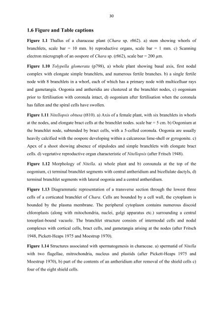
30 <br />
1.6 Figure and Table captions<br />
Figure 1.1 Thallus of a characeae plant (Chara sp. r862). a) stem showing whorls of<br />
branchlets, scale bar = 10 mm. b) reproductive organs, scale bar = 1 mm. c) Scanning<br />
electron micrograph of an oospore of Chara sp. (r862), scale bar = 200 µm.<br />
Figure 1.10 Tolypella glomerata (p798), a) whole plant showing basal axis, first nodal<br />
complex with elongate simple branchlets, and numerous fertile branches. b) a single fertile<br />
node with 8 branchlets in a whorl, each of which has a primary node with multicelluar rays<br />
and gametangia. Oogonia and antheridia are clustered at the branchlet nodes, c) oogonium<br />
prior to fertilisation with coronula intact, d) oogonium after fertilisation when the coronula<br />
has fallen and the spiral cells have swollen.<br />
Figure 1.11 Nitellopsis obtusa (t810). a) Axis of a female plant, with six branchlets in whorls<br />
at the nodes, and elongate bract cells at the branchlet nodes. scale bar = 5 cm. b) Oogonium at<br />
the branchlet node, subtended by bract cells, with a 5-celled coronula. Oogonia are usually<br />
heavily calcified with the oospore developing within a calcareous lime-shell or gyrogonite. c)<br />
Apex of a shoot showing absence of stipulodes and simple branchlets with elongate bract<br />
cells. d) vegetative reproductive organ characteristic of Nitellopsis (after Fritsch 1948).<br />
Figure 1.12 Morphology of Nitella. a) whole plant and b) corounula at the top of the<br />
oogonium, c) terminal branchlet segments with central antheridium and bicellulate dactyls, d)<br />
terminal branchlet segments with lateral oogonia and a central antheridium.<br />
Figure 1.13 Diagrammatic representation of a transverse section through the lowest three<br />
cells of a corticated branchlet of Chara. Cells are bounded by a cell wall, the cytoplasm is<br />
bounded by the plasma membrane. <strong>The</strong> peripheral cytoplasm contains numerous discoid<br />
chloroplasts (along with mitochondria, nuclei, golgi apparatus etc.) surrounding a central<br />
tonoplast-bound vacuole. <strong>The</strong> branchlet structure consists of intermodal cells and nodal<br />
complexes with cortical cells, bract cells, and gametangia arising at the nodes (after Fritsch<br />
1948, Pickett-Heaps 1975 and Moestrup 1970).<br />
Figure 1.14 Structures associated with spermatogenesis in characeae. a) spermatid of Nitella<br />
with two flagellae, mitrochondria, nucleus and plastids (after Pickett-Heaps 1975 and<br />
Moestrup 1970), b) part of the contents of an antheridium after removal of the shield cells c)<br />
four of the eight shield cells.