surgical technique
surgical technique
surgical technique

Create successful ePaper yourself
Turn your PDF publications into a flip-book with our unique Google optimized e-Paper software.
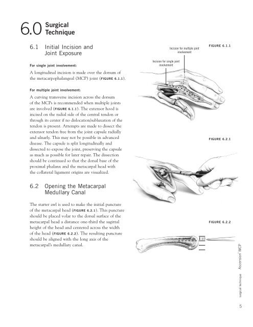
6.0 Surgical<br />
Technique<br />
6.1 Initial Incision and<br />
Joint Exposure<br />
For single joint involvement:<br />
A longitudinal incision is made over the dorsum of<br />
the metacarpophalangeal (MCP) joint (FIGURE 6.1.1).<br />
Incision for single joint<br />
involvement<br />
Incision for multiple joint<br />
involvement<br />
FIGURE 6.1.1<br />
For multiple joint involvement:<br />
A curving transverse incision across the dorsum<br />
of the MCPs is recommended when multiple joints<br />
are involved (FIGURE 6.1.1). The extensor hood is<br />
incised on the radial side of the central tendon or<br />
through its center if no dislocation/subluxation of the<br />
tendon is present. Attempts are made to dissect the<br />
extensor tendon free from the joint capsule radially<br />
and ulnarly. This may not be possible in advanced<br />
disease. The capsule is split longitudinally and<br />
dissected to expose the joint, preserving the capsule<br />
as much as possible for later repair. The dissection<br />
should be continued so that the dorsal base of the<br />
proximal phalanx and the metacarpal head with<br />
the collateral ligament origins are visualized.<br />
FIGURE 6.2.1<br />
6.2 Opening the Metacarpal<br />
Medullary Canal<br />
The starter awl is used to make the initial puncture<br />
of the metacarpal head (FIGURE 6.2.1). This puncture<br />
should be placed volar to the dorsal surface of the<br />
metacarpal head a distance one-third the sagittal<br />
height of the head and centered across the width<br />
of the head (FIGURE 6.2.2). The resulting puncture<br />
should be aligned with the long axis of the<br />
metacarpal’s medullary canal.<br />
1/3<br />
FIGURE 6.2.2<br />
5<strong>surgical</strong> <strong>technique</strong> Ascension® MCP