surgical technique
surgical technique
surgical technique

Create successful ePaper yourself
Turn your PDF publications into a flip-book with our unique Google optimized e-Paper software.
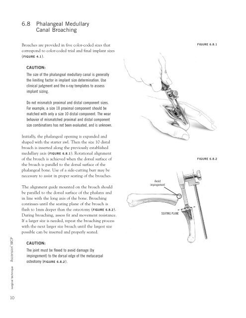
6.8 Phalangeal Medullary<br />
Canal Broaching<br />
Broaches are provided in five color-coded sizes that<br />
correspond to color-coded trial and final implant sizes<br />
(FIGURE 4.1).<br />
FIGURE 6.8.1<br />
CAUTION:<br />
The size of the phalangeal medullary canal is generally<br />
the limiting factor in implant size determination. Use<br />
clinical judgment and the x-ray templates to assess<br />
implant sizing.<br />
Do not mismatch proximal and distal component sizes.<br />
For example, a size 10 proximal component should be<br />
matched with only a size 10 distal component. The wear<br />
behavior of mismatched proximal and distal component<br />
size combinations has not been evaluated, and is unknown.<br />
Initially, the phalangeal opening is expanded and<br />
shaped with the starter awl. Then the size 10 distal<br />
broach is inserted along the previously established<br />
medullary axis (FIGURE 6.8.1). Rotational alignment<br />
of the broach is achieved when the dorsal surface of<br />
the broach is parallel to the dorsal surface of the<br />
phalangeal bone. Use of a side-cutting burr may be<br />
necessary to assist in proper seating of the broaches.<br />
The alignment guide mounted on the broach should<br />
be parallel to the dorsal surface of the phalanx and<br />
in line with the long axis of the bone. Broaching<br />
continues until the seating plane of the broach is<br />
flush to 1mm deeper than the osteotomy (FIGURE 6.8.2).<br />
During broaching, assess fit and movement resistance.<br />
If a larger size is needed, repeat the broaching process<br />
with the next larger size broach until the largest size<br />
possible can be inserted and properly seated.<br />
Avoid<br />
impingement<br />
SEATING PLANE<br />
FIGURE 6.8.2<br />
<strong>surgical</strong> <strong>technique</strong> Ascension ® MCP<br />
CAUTION:<br />
The joint must be flexed to avoid damage (by<br />
impingement) to the dorsal edge of the metacarpal<br />
osteotomy (FIGURE 6.8.2).<br />
10