1061504_Rev. A ENDOBUTTON Tech - EUROCIENCIA
1061504_Rev. A ENDOBUTTON Tech - EUROCIENCIA
1061504_Rev. A ENDOBUTTON Tech - EUROCIENCIA

You also want an ePaper? Increase the reach of your titles
YUMPU automatically turns print PDFs into web optimized ePapers that Google loves.
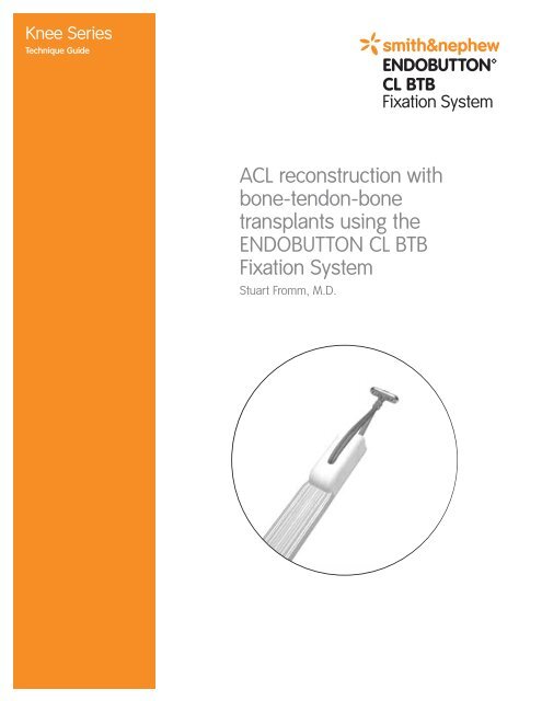
Knee Series<br />
<strong>Tech</strong>nique Guide<br />
ACL reconstruction with<br />
bone-tendon-bone<br />
transplants using the<br />
<strong>ENDOBUTTON</strong> CL BTB<br />
Fixation System<br />
Stuart Fromm, M.D.
<strong>ENDOBUTTON</strong> CL BTB Fixation System Stuart Fromm, M.D.<br />
ACL Reconstruction with Bone-Tendon-Bone Transplants<br />
As described by Stuart Fromm, M.D.<br />
In the late 1980s Smith & Nephew's ACUFEX instrumentation helped<br />
standardize ACL reconstruction.<br />
In 1999 the Smith & Nephew <strong>ENDOBUTTON</strong> CL Fixation Device<br />
for hamstring tendons further simplified ACL reconstruction and<br />
increased initial reconstruction strength.<br />
With the increasing popularity of bone-tendon-bone grafts, Smith &<br />
Nephew developed the <strong>ENDOBUTTON</strong> CL BTB Fixation System.<br />
This new BTB system offers the surgeon many advantages: the bone<br />
blocks are fully appositioned in the femoral tunnel, thus the tibial<br />
and femoral bone blocks cannot protrude; perforation of the posterior<br />
femoral cortex will not compromise fixation; and revisions become<br />
simpler. In addition, by directly obtaining the measurements for the<br />
total graft length, femoral tunnel length, and desired graft insertion<br />
depth, calculation is eliminated and potential errors are minimized.<br />
The technique is easy, reproducible, dependable, and delivers a<br />
strong fixation that avoids complications seen with interference<br />
screws such as screw divergence, posterior blow-out, laceration<br />
of the graft, and screw breakage.<br />
Stuart Fromm, M.D.<br />
Black Hills Orthopedic & Spine Center, P.C.<br />
Rapid City, South Dakota<br />
2
Overview<br />
Graft Harvesting<br />
and Preparation<br />
If necessary perform appropriate<br />
notchplasty using the Smith &<br />
Nephew NOTCHMASTER<br />
Curette or Smith & Nephew<br />
NOTCHBLASTER Burr).<br />
Harvest the bone-patella-bone<br />
tendon graft in the preferred<br />
manner (Figure 1). Recommended<br />
bone plug lengths are 20 mm.<br />
4. Depending on the type of<br />
tibial fixation used, prepare<br />
the tibial bone block in the<br />
preferred manner.<br />
For an exceptionally long graft,<br />
you may need to adjust the<br />
tunnels as described under the<br />
Tibial Tunnel Positioning and<br />
Femoral Tunnel Location sections.<br />
1. Measure the overall length of<br />
the graft (Figure 2; example:<br />
90 mm).<br />
2 Prepare the desired bone plug<br />
diameters in the preferred<br />
manner, e.g., utilizing Smith &<br />
Nephew COMPACTION Pliers.<br />
Measure bone block diameters<br />
using sizing tubes.<br />
Figure 1: Graft harvesting site<br />
3. Drill a 2.0 mm diameter hole<br />
in the femoral bone block<br />
perpendicular to the cortex<br />
(Figure 2). This hole should<br />
be placed at the midpoint of<br />
the bone block or toward<br />
the bone tendon junction.<br />
The continuous loop of the<br />
<strong>ENDOBUTTON</strong> CL BTB Fixation<br />
System will eventually be<br />
passed through this hole.<br />
If you desire to pass the<br />
Continuous Loop (CL) at the<br />
bone-tendon junction, a 2 mm<br />
hole in the femoral bone block<br />
is needed for a #2 guide<br />
suture. The #2 guide suture<br />
is used to keep the femoral<br />
bone block concentric with<br />
the bone tunnels when pulling<br />
the graft through.<br />
Figure 2: All-over graft length and hole<br />
3
<strong>ENDOBUTTON</strong> CL BTB Fixation System Stuart Fromm, M.D.<br />
Tibial Tunnel Positioning<br />
Position the tibial tunnel using<br />
the Smith & Nephew ACUFEX<br />
DIRECTOR Drill Guide as<br />
follows (Figure 3):<br />
The remaining distal stump of the<br />
torn ACL is the major orientation<br />
guide.<br />
Figure 3: Positioning of the tibial aiming device<br />
Using the Elbow Aimer:<br />
place the tip of the aimer in<br />
the posterior fibers of the ACL<br />
footprint. The 2.4 mm guide wire<br />
will protrude through the tibial<br />
plateau several millimeters<br />
anterior (depending on the angle<br />
of the tibial tunnel) to the tip of<br />
the aimer.<br />
Using the Tip Aimer: place the<br />
tip of the aimer exactly at the<br />
point where the 2.4 mm guide<br />
wire will protrude through the<br />
tibial plateau.<br />
Place the 2.4 mm guide wire in<br />
the preferred position (Figure 4).<br />
The angle of the aimer may be<br />
adjusted depending on the graft<br />
length. To achieve a longer tibial<br />
tunnel, increase the angle of the<br />
aimer arm.<br />
Figure 4: Placing of the 2.4 mm guide wire<br />
4
Tibial Tunnel Drilling<br />
Confirm the exact diameter of the<br />
bone blocks and perform notch<br />
assessment prior to drilling.<br />
If necessary, reposition the<br />
2.4 mm guide wire using the<br />
Offset Guide.<br />
Drill the tibial tunnel using a<br />
standard Cannulated Drill Bit<br />
that matches the graft diameter<br />
(Figure 5).<br />
Femoral Tunnel Location<br />
The knee is usually flexed at 90°.<br />
Position the endoscopic femoral<br />
aimer with the appropriate offset<br />
hook (Figure 6: X mm) at the overthe-top<br />
position, in direct contact<br />
with the bony cortex.<br />
Note: When using the<br />
<strong>ENDOBUTTON</strong> CL BTB System<br />
for femoral fixation, perforation<br />
of the posterior femoral cortex<br />
does not compromise fixation.<br />
If a longer femoral tunnel<br />
is desired (e.g., with an<br />
exceptionally long graft),<br />
the knee can be flexed slightly<br />
less than 90° (Figure 7).<br />
Advance the 2.7 mm passing pin<br />
through the femoral offset guide<br />
and drill through the femur until<br />
the passing pin “breaks” through<br />
the lateral femoral cortex.<br />
Feel the 2.7 mm passing pin just<br />
under the skin after it exits the<br />
cortex to determine its position<br />
with respect to the tourniquet.<br />
Figure 5: Drilling the tibial tunnel<br />
X mm<br />
Figure 6: Positioning the endoscopic femoral aimer at the over-the-top position<br />
Figure 7: Femoral tunnel lengths with knee in different flexions<br />
5
<strong>ENDOBUTTON</strong> CL BTB Fixation System Stuart Fromm, M.D.<br />
B. Femoral insertion length (ex. 25 mm)<br />
Figure 8: Femoral socket drilling<br />
C. 10 mm<br />
A. Overall graft length<br />
(ex. 90 mm)<br />
Femoral Tunnel Drilling<br />
The endoscopic cannulated<br />
drill bit will mimic the ACL<br />
graft, allowing for a direct<br />
determination of femoral tunnel<br />
depth. To position the tibial bone<br />
block of the bone-tendon-bone<br />
graft flush with the distal tibial<br />
tunnel:<br />
1. Advance the endoscopic<br />
cannulated drill bit over<br />
the 2.7 mm passing pin.<br />
Measuring directly off the<br />
drill bit where it enters the<br />
tibial tunnel, drill the femoral<br />
socket to a depth matching<br />
the overall length of the<br />
bone-tendon-bone graft<br />
(Figure 8-A). Example: if the<br />
total graft length is 90 mm,<br />
drill the femoral tunnel to a<br />
depth where 90 mm is read<br />
directly off the endoscopic<br />
cannulated drill bit where it<br />
enters the tibial tunnel.<br />
Caution: Ensure the<br />
endoscopic cannulated drill bit<br />
does not breach the lateral<br />
femoral cortex, otherwise<br />
femoral fixation with the<br />
<strong>ENDOBUTTON</strong> CL BTB System<br />
will be compromised.<br />
6<br />
Graft insertion<br />
length (ex. 25 mm)<br />
Figure 9: Marking the graft<br />
Blue line<br />
2. With the endoscopic<br />
cannulated drill bit pushed<br />
against the back wall of the<br />
femoral socket, directly read<br />
the measurement off the drill<br />
bit at the base of the femoral<br />
socket (Figure 8-B; example:<br />
25 mm). Using a surgical<br />
marker, place a<br />
corresponding “blue line” on<br />
the graft (Figure 9). This mark<br />
indicates the depth of the<br />
graft in the femoral tunnel.
3. With the endoscopic cannulated<br />
drill bit still in place, drill the<br />
femoral socket an additional<br />
10 mm minimum (Figure 8-C)<br />
to allow the <strong>ENDOBUTTON</strong><br />
device to be rotated once<br />
passed.<br />
Caution: Ensure the endoscopic<br />
cannulated drill bit does not<br />
breach the lateral femoral cortex,<br />
otherwise femoral fixation with<br />
the <strong>ENDOBUTTON</strong> CL BTB System<br />
will be compromised.<br />
4. With the 2.7 mm passing pin<br />
still in place, use the 4.5 mm<br />
<strong>ENDOBUTTON</strong> Drill Bit to drill<br />
completely through the<br />
femoral cortex (Figure 10).<br />
Determination of the<br />
CL Length<br />
Measure the total length of the<br />
femoral channel length using<br />
the <strong>ENDOBUTTON</strong> Depth Probe<br />
(Figure 11; example: 60 mm).<br />
This will correspond to the<br />
distance from the <strong>ENDOBUTTON</strong><br />
device to the “blue line” on<br />
the graft.<br />
Figure 10: Drilling with the <strong>ENDOBUTTON</strong> Drill through the cortex<br />
Femoral channel<br />
length (ex. 60 mm)<br />
Position the graft on the<br />
<strong>ENDOBUTTON</strong> Holder of the<br />
GRAFTMASTER Graft Preparation<br />
Board such that the “blue line”<br />
aligns at the distance obtained<br />
from the <strong>ENDOBUTTON</strong> Depth<br />
Probe (Figure 12; example: 60 mm<br />
from the <strong>ENDOBUTTON</strong> device).<br />
Figure 11: Measuring the femoral channel<br />
CL length needed (ex. 45 mm)<br />
The Continuous Loop (CL) length<br />
needed is read directly off the<br />
<strong>ENDOBUTTON</strong> Holder at the<br />
desired point of attachment to<br />
the graft.<br />
Figure 12. Determination of CL length on the GRAFTMASTER Board<br />
7
<strong>ENDOBUTTON</strong> CL BTB Fixation System Stuart Fromm, M.D.<br />
2.<br />
3.<br />
Attaching the <strong>ENDOBUTTON</strong><br />
CL BTB Fixation System to<br />
the Graft<br />
The <strong>ENDOBUTTON</strong> CL BTB<br />
System can be divided into<br />
three distinct “areas” (Figure 13):<br />
1. Long CL loop<br />
1.<br />
2. Short CL loop<br />
3. <strong>ENDOBUTTON</strong> Fixation Device<br />
Figure 13: <strong>ENDOBUTTON</strong> CL BTB components<br />
a.<br />
b.<br />
c. d.<br />
Pass the Continuous Loop (CL)<br />
through the graft and attach it<br />
to the <strong>ENDOBUTTON</strong> device<br />
as shown in Figure 14.<br />
1. Feed the long CL loop through<br />
the prepared hole<br />
of the femoral bone block<br />
(respectively through the<br />
desired position at the<br />
tendon-bone junction). The<br />
short CL loop should face<br />
toward the bone block<br />
(Figure 14-a).<br />
2. Feed the long CL loop through<br />
the short CL loop (Figure 14-b).<br />
3. Slip the long CL loop over<br />
the <strong>ENDOBUTTON</strong> device<br />
(Figures 14-c and d).<br />
e. f.<br />
4. Tighten the construct<br />
(Figures 14-e and f).<br />
Figure 14: Attaching the <strong>ENDOBUTTON</strong> CL BTB System<br />
8
Graft Passage<br />
Attach a #5 polyester braided<br />
suture to one outside hole of<br />
the <strong>ENDOBUTTON</strong> device to lead<br />
and pass the <strong>ENDOBUTTON</strong><br />
device. Attach a trailing #2<br />
polyester braided suture to the<br />
opposite outside hole of the<br />
<strong>ENDOBUTTON</strong> device to rotate<br />
the <strong>ENDOBUTTON</strong> device as it<br />
exits the anterolateral femoral<br />
cortex. Both sutures are passed<br />
through the eyelet of a passing<br />
pin (Figure 15).<br />
#2 Suture<br />
#5 Suture<br />
Figure 15: Attachment of leading and trailing<br />
suture to the <strong>ENDOBUTTON</strong> device<br />
Total Channel<br />
Length<br />
Suture<br />
Span<br />
The passing pin is used for<br />
suture passage by piercing the<br />
quadriceps and skin proximally.<br />
The #5 suture is pulled first,<br />
advancing the <strong>ENDOBUTTON</strong>/<br />
graft complex into the femoral<br />
tunnel (Figure 16) until the<br />
<strong>ENDOBUTTON</strong> device has<br />
cleared the femoral cortex.<br />
Insertion<br />
Length<br />
Turning<br />
Radius<br />
The trailing #2 suture is pulled,<br />
rotating the <strong>ENDOBUTTON</strong> device<br />
immediately external to the femur<br />
(Figure 17).<br />
Figure 16: Passage of the graft<br />
Hold the <strong>ENDOBUTTON</strong> device<br />
perpendicular to the femoral<br />
cortex and pull back on the graft,<br />
locking the <strong>ENDOBUTTON</strong> device<br />
on the outer femoral cortex.<br />
Secure fixation should be felt<br />
at that point with the “blue line”<br />
flush at the base of the femoral<br />
tunnel.<br />
Figure 17: Rotating the <strong>ENDOBUTTON</strong> device outside<br />
the cortex<br />
9
<strong>ENDOBUTTON</strong> CL BTB Fixation System Stuart Fromm, M.D.<br />
Tibial Fixation<br />
Tension the graft and fix the<br />
tibial bone plug as desired,<br />
i.e., with a Smith & Nephew<br />
interference screw, tying over<br />
a screw and washer, or both<br />
(Figure 18).<br />
Figure 18: Flex and extend the knee while pulling on the graft prior to tibial graft fixation<br />
10
Additional Instruction<br />
Prior to performing this technique,<br />
consult the Instructions for Use<br />
documentation provided with<br />
individual components — including<br />
indications, contraindications,<br />
warnings, cautions, and instructions.<br />
Courtesy of Smith & Nephew, Inc.,<br />
Endoscopy Division<br />
Caution: U.S. Federal law restricts this device<br />
to sale by or on the order of a physician.<br />
Endoscopy<br />
Smith & Nephew, Inc.<br />
Andover, MA 01810<br />
USA<br />
www.smith-nephew.com<br />
978 749 1000<br />
978 749 1108 Fax<br />
800 343 5717 U.S. Customer Service<br />
Trademarks of Smith & Nephew. Certain marks<br />
registered U.S. Patent & Trademark Office.<br />
©2004 Smith & Nephew, Inc. All Rights Reserved.<br />
Printed in USA<br />
04/04 <strong>1061504</strong> <strong>Rev</strong>. A