In Vitro Studies - ConvaTec
In Vitro Studies - ConvaTec
In Vitro Studies - ConvaTec

You also want an ePaper? Increase the reach of your titles
YUMPU automatically turns print PDFs into web optimized ePapers that Google loves.
Shallow Wound Microbial Model<br />
Table 2 shows the percentage growth of S. aureus and P. aeruginosa in the indented agar surface beneath the silver-containing dressings over<br />
a 48 hour contact period. Additionally, lateral spreading of both P. aeruginosa and S. aureus on the prominent agar beyond the edges of the<br />
indented agar surface was observed in association with Foam dressings A and C but this was not observed with SCHD or Foam B dressing.<br />
Test Dressing Challenge Bacterial Growth Mean % Bacterial Growth<br />
Organism (within agar indentation) indentation)<br />
Silver-Containing Dressings:<br />
Replicates<br />
1 2 3<br />
SCHD S. aureus 0.0 0.0 1.2 0.4<br />
P. aeruginosa 2.7 3.6 20.8 9.0<br />
Foam A S. aureus 30.8 25.3 21.0 25.7<br />
P. aeruginosa 100.0 100.0 100.0 100.0<br />
Foam B S. aureus 17.3 18.4 14.6 16.7<br />
P. aeruginosa 96.1 97.9 99.4 97.8<br />
Foam C S. aureus 73.0 65.9 80.6 73.2<br />
P. aeruginosa 100.0 100.0 100.0 100.0<br />
Positive Control (no dressing) S. aureus 100.0 - - 100.0<br />
Positive Control (no dressing) P. aeruginosa 100.0 - - 100.0<br />
Table 2: Percentage growth of P. aeruginosa and S. aureus in the indented agar area<br />
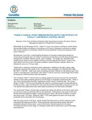
SCHD with S. aureus<br />
(0.0%) growth<br />
SCHD with P. aeruginosa<br />
(3.6%) growth<br />
Foam A with S. aureus<br />
(21.0%) growth<br />
Foam A with P. aeruginosa<br />
(100.0%) growth<br />
Figure 2: Examples of S. aureus and P. aeruginosa growth beneath<br />
SCHD in the indented agar. Figures in brackets indicate percentage<br />
bacterial growth relative to total indented agar surface area for an<br />
individual replicate (left – replicate 1; right – replicate 2).<br />
Figure 3: Examples of S. aureus and P. aeruginosa growth beneath<br />
Foam A dressing in the indented agar. Figures in brackets indicate<br />
percentage bacterial growth relative to total indented agar surface<br />
area for an individual replicate (left – replicate 3; right – replicate 3).<br />
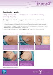
Foam B with S. aureus<br />
(17.3%) growth<br />
Foam B with P. aeruginosa<br />
(97.9%) growth<br />
Foam C with S. aureus<br />
(73.0%) growth<br />
Foam C with P. aeruginosa<br />
(100.0%) growth<br />
Figure 4: Examples of S. aureus and P. aeruginosa growth beneath<br />
Foam B dressing in the indented agar. Figures in brackets indicate<br />
percentage bacterial growth relative to total indented agar surface<br />
area for an individual replicate (left – replicate 1; right – replicate 2).<br />
Conclusion<br />
Figure 5: Examples of S. aureus and P. aeruginosa growth beneath<br />
Foam C in the indented agar. Figures in brackets indicate percentage<br />
bacterial growth relative to total indented agar surface area for an<br />
individual replicate (left – replicate 1; right – replicate 3).<br />
• Using an in vitro tissue conformability model SCHD was observed to conform more closely to the tissue surface than the tested<br />
silver-containing foam dressings following hydration.<br />
• There was no evidence of fluid accumulation at the tissue/dressing interface with SCHD, but this was observed with the tested<br />
silver-containing foam dressings.<br />
• Using an in vitro shallow wound microbial model, it was observed that SCHD killed more bacteria (both P. aeruginosa and S. aureus)<br />
beneath the dressing than any of the tested silver-containing foam dressings.<br />
• Additionally, SCHD was observed to not allow the spread of bacteria beyond the edge of the simulated wound.<br />
Hydrofiber, AQUACEL, Versiva, XC and DuoDERM are registered trademarks of <strong>ConvaTec</strong> <strong>In</strong>c.<br />
© 2010 <strong>ConvaTec</strong> <strong>In</strong>c AP-009703-MM (AM,EM,SP)