In Vitro Studies - ConvaTec
In Vitro Studies - ConvaTec
In Vitro Studies - ConvaTec

You also want an ePaper? Increase the reach of your titles
YUMPU automatically turns print PDFs into web optimized ePapers that Google loves.
The Importance of Dressing Conformability to Antimicrobial<br />
Action of Silver-Containing Wound Dressings: <strong>In</strong> <strong>Vitro</strong> <strong>Studies</strong><br />
Phil Bowler, Samantha Jones, Victoria Towers, Rebecca Booth, David Parsons and Mike Walker<br />
<strong>ConvaTec</strong> Global Development Centre, Deeside, Flintshire UK<br />
<strong>In</strong>troduction<br />
Chronic wounds are polymicrobial, with bacterial colonisation originating from external sources such as surrounding skin, the gut and the<br />
mouth. As a consequence, bacterial burden exists predominantly in superficial wound tissue where biofilm inevitably develops, providing the<br />
colonising bacteria with an environment that may give them a competitive advantage over the host.<br />
<strong>In</strong> order to minimise the opportunity for infection in chronic wounds, it is important that the superficial bioburden is controlled and maintained in<br />
a state that is not problematic to the host. <strong>In</strong> this respect, wound management practices such as cleansing, sharp debridement and the use of<br />
antimicrobial dressings are important. <strong>In</strong> order to maximise antimicrobial potency in superficial wound tissue, selection of a dressing that conforms<br />
well to a wound’s unique topography is likely to be important in order to maximise exposure between bacteria and the antimicrobial agent.<br />
<strong>In</strong> these in vitro studies, models were utilised that enabled both visualisation of the conformability of silver-containing dressings with a simulated<br />
wound tissue, and visualisation of the antimicrobial activity of these dressings in a shallow wound microbial model.<br />
Materials and Methods<br />
The silver-containing wound dressings used are listed in Table 1.<br />
Dressing<br />
Dressing Type<br />
Hydrofiber ® dressing<br />
Foam A dressing<br />
Foam B dressing<br />
Foam C dressing<br />
Table 1: List of silver-containing dressings used:<br />
Results and Discussion<br />
Tissue Conformability Model<br />
Silver-containing Hydrofiber ® dressing (SCHD,<br />
AQUACEL ® Ag) containing ionic silver, was covered with<br />
adhesive Hydrocolloid cover dressing (DuoDERM ®<br />
EXTRA THIN)<br />
Adhesive foam dressing containing silver sulfadiazine<br />
Non-adhesive foam dressing containing silver sulfadiazine<br />
Silicone adhesive dressing foam containing silver sulphate<br />
Tissue Conformability Model<br />
• Porcine muscle tissue was used to simulate an<br />
irregular wound surface.<br />
• A Crono pump (PCA Model - 20 ml) was used to<br />
deliver Alcian blue-dyed physiological saline.<br />
• An Olympus SZ61 Light microscope, with QImaging<br />
camera and Image Pro Plus 5.0 image analysis software<br />
(MediaCybernetics, UK) was used to capture the images.<br />
• SCHD, Foam A and Foam C were tested in this model.<br />
Shallow Wound Microbial Model<br />
Challenge organisms: Pseudomonas aeruginosa (NCIMB 8626) and Staphylococcus aureus (NCIMB 9518).<br />
• A suspension of S. aureus (approximately 4x10 3 cfu/ml) was inoculated into the centre of the indentation within the agar plate.<br />
• Each silver-containing dressing (SCHD applied with an adhesive Hydrofiber cover dressing (Versiva ® XC ® ) and foams A, B and C, (n=3))<br />
was gently applied such that it covered the agar indentation.<br />
• Positive controls (n=1 for each challenge organism) were also utilised to confirm the extent of bacterial growth in the absence of silver-containing dressings.<br />
• All agar plates were then incubated at 35°C (±3°C) for 48 hours prior to removal of dressings.<br />
• Photographs of the agar plates were also taken following dressing removal.<br />
• This procedure was repeated using P. aeruginosa as the challenge organism.<br />
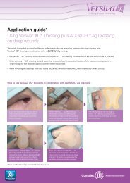
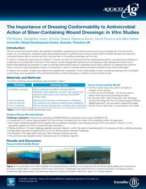
Figure 1: As dyed saline was made available at the dressing/tissue interface it was observed that the SCHD rapidly gelled and conformed to<br />
the tissue surface (left). <strong>In</strong> comparison both Foam A and Foam C dressings were observed not to conform as closely to the tissue surface<br />
(middle and right respectively), and there was evidence of fluid accumulation within the “open” non-contact spaces.<br />
Hydrofiber, AQUACEL, Versiva, XC and DuoDERM are registered trademarks of <strong>ConvaTec</strong> <strong>In</strong>c.<br />
© 2010 <strong>ConvaTec</strong> <strong>In</strong>c
Shallow Wound Microbial Model<br />
Table 2 shows the percentage growth of S. aureus and P. aeruginosa in the indented agar surface beneath the silver-containing dressings over<br />
a 48 hour contact period. Additionally, lateral spreading of both P. aeruginosa and S. aureus on the prominent agar beyond the edges of the<br />
indented agar surface was observed in association with Foam dressings A and C but this was not observed with SCHD or Foam B dressing.<br />
Test Dressing Challenge Bacterial Growth Mean % Bacterial Growth<br />
Organism (within agar indentation) indentation)<br />
Silver-Containing Dressings:<br />
Replicates<br />
1 2 3<br />
SCHD S. aureus 0.0 0.0 1.2 0.4<br />
P. aeruginosa 2.7 3.6 20.8 9.0<br />
Foam A S. aureus 30.8 25.3 21.0 25.7<br />
P. aeruginosa 100.0 100.0 100.0 100.0<br />
Foam B S. aureus 17.3 18.4 14.6 16.7<br />
P. aeruginosa 96.1 97.9 99.4 97.8<br />
Foam C S. aureus 73.0 65.9 80.6 73.2<br />
P. aeruginosa 100.0 100.0 100.0 100.0<br />
Positive Control (no dressing) S. aureus 100.0 - - 100.0<br />
Positive Control (no dressing) P. aeruginosa 100.0 - - 100.0<br />
Table 2: Percentage growth of P. aeruginosa and S. aureus in the indented agar area<br />
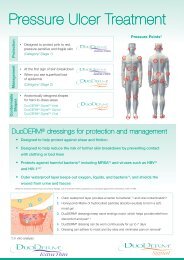
SCHD with S. aureus<br />
(0.0%) growth<br />
SCHD with P. aeruginosa<br />
(3.6%) growth<br />
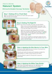
Foam A with S. aureus<br />
(21.0%) growth<br />
Foam A with P. aeruginosa<br />
(100.0%) growth<br />
Figure 2: Examples of S. aureus and P. aeruginosa growth beneath<br />
SCHD in the indented agar. Figures in brackets indicate percentage<br />
bacterial growth relative to total indented agar surface area for an<br />
individual replicate (left – replicate 1; right – replicate 2).<br />
Figure 3: Examples of S. aureus and P. aeruginosa growth beneath<br />
Foam A dressing in the indented agar. Figures in brackets indicate<br />
percentage bacterial growth relative to total indented agar surface<br />
area for an individual replicate (left – replicate 3; right – replicate 3).<br />
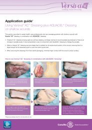
Foam B with S. aureus<br />
(17.3%) growth<br />
Foam B with P. aeruginosa<br />
(97.9%) growth<br />
Foam C with S. aureus<br />
(73.0%) growth<br />
Foam C with P. aeruginosa<br />
(100.0%) growth<br />
Figure 4: Examples of S. aureus and P. aeruginosa growth beneath<br />
Foam B dressing in the indented agar. Figures in brackets indicate<br />
percentage bacterial growth relative to total indented agar surface<br />
area for an individual replicate (left – replicate 1; right – replicate 2).<br />
Conclusion<br />
Figure 5: Examples of S. aureus and P. aeruginosa growth beneath<br />
Foam C in the indented agar. Figures in brackets indicate percentage<br />
bacterial growth relative to total indented agar surface area for an<br />
individual replicate (left – replicate 1; right – replicate 3).<br />
• Using an in vitro tissue conformability model SCHD was observed to conform more closely to the tissue surface than the tested<br />
silver-containing foam dressings following hydration.<br />
• There was no evidence of fluid accumulation at the tissue/dressing interface with SCHD, but this was observed with the tested<br />
silver-containing foam dressings.<br />
• Using an in vitro shallow wound microbial model, it was observed that SCHD killed more bacteria (both P. aeruginosa and S. aureus)<br />
beneath the dressing than any of the tested silver-containing foam dressings.<br />
• Additionally, SCHD was observed to not allow the spread of bacteria beyond the edge of the simulated wound.<br />
Hydrofiber, AQUACEL, Versiva, XC and DuoDERM are registered trademarks of <strong>ConvaTec</strong> <strong>In</strong>c.<br />
© 2010 <strong>ConvaTec</strong> <strong>In</strong>c AP-009703-MM (AM,EM,SP)