AQUACEL® Ag Made Easy - ConvaTec
AQUACEL® Ag Made Easy - ConvaTec
AQUACEL® Ag Made Easy - ConvaTec

You also want an ePaper? Increase the reach of your titles
YUMPU automatically turns print PDFs into web optimized ePapers that Google loves.
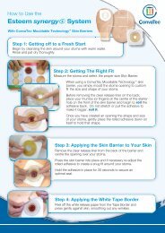
PRODUCTS FOR PRACTICE<br />
the wound the dressing should be layered<br />
to fill the wound and avoid the creation of<br />
dead space.<br />
If the wound bed is relatively dry, it<br />
is recommended that AQUACEL ® <strong>Ag</strong><br />
Dressing is placed in the wound and then<br />
moistened with sterile saline over the<br />
wound area only. The vertical absorption<br />
of the dressing will help to maintain the<br />
moist area over the wound only and<br />
reduce the risk of periwound maceration.<br />
For partial thickness burns, AQUACEL ®<br />
<strong>Ag</strong> Dressing should overlap 5cm<br />
(approximately 2 inches) onto the skin<br />
surrounding the burn. The dressing<br />
should be covered with sterile gauze and<br />
secured with medical tape or a retention<br />
bandage. In this situation, adherence of<br />
AQUACEL ® <strong>Ag</strong> Dressing to the wound<br />
bed is a desired characteristic that helps<br />
to reduce the frequency of dressing<br />
changes 21,24 . Lack of adherence within<br />
one to two days of dressing application<br />
may indicate secondary deepening of the<br />
burn, or development of a complication<br />
such as infection, and indicate<br />
reassessment of the wound 21 .<br />
For deep wounds<br />
Select the appropriate width of<br />
AQUACEL ® <strong>Ag</strong> Ribbon Dressing with<br />
Strengthening Fiber. When using<br />
AQUACEL ® <strong>Ag</strong> Ribbon Dressing with<br />
Strengthening Fiber in deep wounds,<br />
at least 2.5 cm (approximately 1 inch)<br />
should be left outside the wound to aid<br />
retrieval of the dressings. Deep wounds<br />
should be filled with the dressing by no<br />
more than 80% (almost to the top), as the<br />
dressing will swell as it absorbs the fluid.<br />
Apply a cover dressing<br />
The dressing should be covered with<br />
a secondary dressing that keeps the<br />
wound moist. The choice of secondary<br />
dressing will depend on the level of<br />
exudation (eg for a lightly exuding<br />
wound DuoDERM ® Dressing (<strong>ConvaTec</strong>)<br />
or for a lightly to moderately exuding<br />
wound Versiva ® XC ® Gelling Foam<br />
Dressing (<strong>ConvaTec</strong>) may be appropriate).<br />
See individual package inserts for<br />
instructions regarding cover dressing<br />
usage and removal. If covering with<br />
gauze, the dressings should be changed<br />
when wound fluid strikes through the<br />
outer layer.<br />
Removal<br />
While the dressing may have to be<br />
changed every two to three days<br />
initially, it can be left on the wound<br />
for up to seven days when the wound<br />
is almost closed. The dressing should<br />
be changed when it is saturated with<br />
wound fluid or if the cover dressing’s<br />
edges are bunching, rolling or leaking.<br />
The dressing should be removed<br />
when medically indicated (eg when<br />
wound fluid comes out of the dressing,<br />
there is too much bleeding, or there<br />
is increased pain). If residual dressing<br />
is left in the wound, irrigate with a<br />
AQUACEL ® <strong>Ag</strong> case study<br />
This case study features a female patient with recurrent venous leg ulcers<br />
This female patient, aged 77 years, has chronic venous insufficiency following a deep venous thrombosis<br />
years previously. Since 1990, she has had recurrent venous leg ulcers on the lower parts of both legs. The<br />
current ulcers, two on the left leg and one on the right, started in 2007.<br />
The patient has severe arthritis of the left knee that limits her ability to walk less than 200 metres per day.<br />
She is on the waiting list for a knee replacement, but this cannot take place until the venous leg ulcers have<br />
healed. As a result, she was referred to the wound clinic in early 2010.<br />
At the initial visit, the patient said that the wounds had increased in area, but was unclear by how much and<br />
over what period. She mentioned that she was currently experiencing more wound-related pain than usual.<br />
The ulcers were being treated with a paraffin-impregnated cotton viscose primary dressing and an absorbent<br />
cellulose fluff secondary dressing beneath Class I compression stockings.<br />
The wound beds were green/yellow and producing considerable watery exudate. These signs, along with the<br />
increase in pain and wound area, indicated infection.<br />
AQUACEL® <strong>Ag</strong> Dressing was chosen because of its antiseptic properties, high absorption and ease of use. At<br />
each dressing change, the ulcers underwent sharp debridement and cleansing with tap water-soaked gauze.<br />
The AQUACEL® <strong>Ag</strong> Dressing was covered with an absorbent cellulose fluff dressing that was held in place<br />
by an elasticated tubular bandage. The patient’s stockings did not provide sufficient compression, and so<br />
ambulatory compression bandaging with short-stretch bandages was commenced.<br />
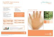
The wound on the right lower leg is used here as an example (see pictures). On presentation, the wound area<br />
was 64cm 2 . After five months of treatment, the condition of the wound bed had improved, there was visible<br />
granulation tissue and the area of the wound was 50cm 2 . By the end of December 2010, the wound area had<br />
reduced to 6.5 cm 2 . At the end of January 2011, the level of exudate production was so low that AQUACEL® <strong>Ag</strong><br />
Dressing was discontinued.<br />
Treatment was continued with an enzyme alginogel. By the beginning of February 2011, the wound area<br />
had reduced further to 1.5 cm 2 . The other two ulcers had healed and, because the oedema of the legs has<br />
resolved, the patient is now wearing therapeutic elastic stockings.<br />
In this case, AQUACEL® <strong>Ag</strong> Dressing antimicrobial and moisture retentive properties helped to promote<br />
healing in the patient’s long-standing venous leg ulcers that were exhibiting signs of infection.<br />
The wound on 25 January, 2010 (64cm 2 )<br />
The wound on 30 June, 2010 (50cm 2 )<br />
The wound on 2 February, 2011 (1.5cm 2 )<br />
4<br />
MADE EASY AQUACEL.indd 6 08/04/2011 09:26