White paper: William G. Stevenson, MD ... - Biosense Webster
White paper: William G. Stevenson, MD ... - Biosense Webster
White paper: William G. Stevenson, MD ... - Biosense Webster

You also want an ePaper? Increase the reach of your titles
YUMPU automatically turns print PDFs into web optimized ePapers that Google loves.
IRRIGATED RF ABLATION: POWER TITRATION AND<br />
FLUID MANAGEMENT FOR OPTIMAL SAFETY AND EFFICACY<br />
INTRODUCTION<br />
Radiofrequency (RF) catheter ablation damages<br />
cardiac tissue by heating. High frequency electrical<br />
current is passed though the myocardial tissue<br />
between the catheter electrode and a dispersive<br />
electrode on the body surface. Although the tip<br />
electrode has a low resistance, the tissue through<br />
which current must flow has a substantially greater<br />
resistance. Current flowing through this resistance<br />
generates heat, like the heating element in a toaster.<br />
This heat is conducted to the surrounding tissue.<br />
Thus there is a very hot area beneath the electrode<br />
due to resistive heating; this heat is then conducted<br />
to the surrounding tissue, where the temperature is<br />
slightly lower. Irreversible damage to myocytes<br />
occurs when tissue temperatures exceed 48° to<br />
50°C. 1,2 The hotter the region of maximum temperature,<br />
the larger the “kill zone” in which the temperature<br />
exceeds this critical limit. If a larger lesion<br />
is desired, one can try to increase current flow into<br />
the tissue to increase heating. This is done by<br />
increasing the driving force for current, which is the<br />
voltage. With most RF generators the parameter<br />
that is adjusted to increase heating is the power,<br />
which is measured in watts (W). Watts = voltage x<br />
current. Thus as power is increased, both current<br />
(milliamperes, or mA) and voltage are increased.<br />
It is important to recognize that the electrode on<br />
the ablation catheter is not heated directly by the<br />
current. The temperature of the electrode increases<br />
only because it is in contact with the tissue that is<br />
being heated. Heat conducts from the tissue to the<br />
electrode. Thus, there is a difference between the<br />
tissue temperature and electrode temperature. For<br />
example, tissue temperature might reach 100°C<br />
when the temperature of the electrode is only<br />
70°C. This difference between electrode temperature<br />
and tissue temperature increases if the electrode is<br />
cooled by irrigation or by the brisk flow of surrounding<br />
blood. In fact, the hottest region in the tissue may<br />
be just below the surface of the tissue, because<br />
the surface is cooled by flowing blood.<br />
There is a limit to which current can be increased<br />
to increase tissue temperature and thereby enlarge<br />
the size of the ablation lesion. When any part of the<br />
<strong>William</strong> G. <strong>Stevenson</strong>, <strong>MD</strong>, FACC<br />
Brigham and Women’s Hospital, Boston, MA<br />
electrode reaches a temperature exceeding 75ºC<br />
and certainly by 100ºC, blood proteins coagulate on<br />
the electrode. 3 Heating these proteins forms a layer<br />
of coagulum or char, which is a high-resistance barrier<br />
to further current flow. Formation of this coagulum<br />
produces an increase in impedance. 4 Typically the<br />
impedance reported by the RF generator reflects<br />
the resistance to current flow through the entire<br />
circuit of catheter, connecting cable, patient, cutaneous<br />
electrode and its connecting cable. In most<br />
patients this will be somewhere between 90 and<br />
120 ohms. A sudden increase in impedance of<br />
more than 20 ohms is almost always an indication<br />
of coagulum formation. If current delivery continues,<br />
impedance rapidly increases, reaching the limit at<br />
which the RF generator will automatically shut off.<br />
(Please refer to the Stockert User Manual for default<br />
setting for impedance.) With most RF generators,<br />
the level at which an automatic shut-off occurs is<br />
much greater than that at which the coagulum<br />
begins to form. Monitoring impedance is often useful<br />
during ablation, particularly during irrigated ablation.<br />
When the catheter is in tissue contact and tissue<br />
heating begins, the measured impedance begins<br />
decreasing. A fall at the beginning of RF application<br />
of approximately 10%, followed by a slow decrease<br />
for another 3 – 5%, is an indication of lesion creation<br />
caused by tissue heating. Small fluctuations in<br />
impedance (increases or decreases of 5 to 10<br />
ohms) are usually caused by catheter movement;<br />
slight increases in impedance may be due to coagulum.<br />
Coagulum typically begins forming around<br />
the collar of the electrode where it meets the<br />
shaft. If only a small amount forms, it may have no<br />
effect on measured impedance. There is concern<br />
that this coagulum could be a risk for embolization,<br />
a risk that has not been proven but is of concern.<br />
With standard RF ablation, power is typically adjusted<br />
to achieve a target temperature that is lower than<br />
that which allows coagulum to form. Temperature<br />
during RF ablation is monitored from temperature<br />
sensors (either a thermocouple or thermister)<br />
embedded in the tip electrode. This provides a<br />
useful but incomplete indication of electrode temperature.<br />
1,5,6 When the electrode is in flowing blood,<br />
a temperature gradient exists across its surface;
the temperature is not uniform, but can be > 15ºC<br />
higher at the region that is in contact with the tissue<br />
compared to the portion that is in the bloodstream.<br />
Electrode temperature is cooler on the side that is<br />
facing the flow of blood than on the side opposite<br />
the direction of flow. Thus a portion of the electrode<br />
can reach temperatures exceeding 75ºC, causing<br />
coagulum formation at a time when the temperature<br />
reported by the sensor(s) is lower. Extensive clinical<br />
experience has shown that with solid 4 or 5 mm<br />
electrodes, limiting the electrode temperature to<br />
60ºC is usually effective in preventing coagulum<br />
formation. With 8 mm electrodes, we frequently<br />
observe coagulum formation at this temperature<br />
during long ablation applications because there is<br />
a greater range of temperature across the larger<br />
electrode.<br />
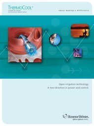
THE THERMOCOOL® IRRIGATED ABLATION<br />
CATHETER<br />
The THERMOCOOL® Catheter allows saline to flow<br />
through a lumen in the catheter, emerging from six<br />
small holes in the tip electrode. The saline is at<br />
room temperature (e.g., 23ºC) and acts to cool the<br />
electrode. Cooling the electrode in this manner<br />
allows more power to be applied, with more current<br />
flowing through the tissue, before the temperature<br />
at the surface of the tissue exceeds 75ºC and coagulum<br />
to begins to form. It has also been suggested<br />
that irrigation washes away blood proteins before<br />
coagulum can form on the electrode. Thus, the<br />
lesions that can be created are larger than those<br />
achieved with similar sized, nonirrigated electrodes.<br />
Appropriate use of the THERMOCOOL® Catheter<br />
requires recognition of the important difference<br />
that irrigation causes in the temperature measured<br />
from the electrode. During mapping, irrigation is<br />
performed with a continuous infusion of 2 ml/min to<br />
maintain patency of the small pores in the electrode.<br />
During ablation, the flow rate is increased to either<br />
17 ml/min or 30 ml/min. With the increase in flow,<br />
electrode temperature falls below 35ºC. As RF<br />
current is applied, the temperature of the electrode<br />
increases, but with active electrode cooling, the<br />
electrode remains substantially cooler than the<br />
underlying tissue. Typically, the temperature<br />
increases from 35ºC to 38 – 45ºC. Substantial<br />
lesions may be created even though measured<br />
electrode temperature remains below 40ºC. During<br />
irrigation at 17 ml/min with up to 30 W or 30 ml/min<br />
with up to 50 W, the electrode temperature generally<br />
remains below 50ºC. Reaching 50ºC can indicate<br />
that the irrigation pump has been shut off or is<br />
disconnected from the catheter. This can also be an<br />
indication of inadequate cooling or coagulum formation<br />
blocking the irrigation ducts; immediate assessment is<br />
warranted. With tissue heating, impedance does<br />
fall, similar to that observed with nonirrigated RF<br />
ablation. Failure of impedance to decrease with<br />
initiation of RF energy suggests that the electrode<br />
catheter is not in good contact.<br />
Because electrode temperature is a poor indicator<br />
of tissue heating, it is recommended that irrigated<br />
RF ablation be performed in a power-control mode,<br />
rather than a temperature-control mode. In the<br />
temperature control that is often used with nonirrigated<br />
ablation, the RF generator is set to a target<br />
temperature and maximum power, and the generator<br />
automatically increases power to reach the target<br />
temperature. In the power-control mode, recommended<br />
for the THERMOCOOL® Catheter, the operator specifies<br />
the power setting and gradually increases or<br />
decreases power as desired. An upper temperature<br />
limit is set for safety, so that the generator will<br />
automatically shut down or reduce power if the<br />
upper temperature limit is exceeded. Operating in<br />
the power-control mode allows the operator to<br />
exert more control over energy delivery. Thus, if<br />
low power (15 to 20 W) produces the desired effect<br />
of reduction of the recorded electrogram or<br />
emergence of double potentials indicative of<br />
conduction block with a temperature that reaches<br />
only 40ºC, the power can be kept low to avoid<br />
excessive energy delivery and reduce the risk of<br />
steam pops. Pops occur when temperature reaches<br />
100ºC either in the tissue or at the electrode<br />
tip-tissue interface. Sudden formation of steam<br />
can result in an explosion, sometimes audible as a<br />
“pop,” 5 that can cause cardiac perforation. 1,5,6 It is<br />
likely that the chance of a steam pops increases<br />
with greater RF energy. If low power is ineffective,<br />
power is increased by the operator. If greater than<br />
30 W is required, the irrigation flow rate is<br />
increased to 30 ml/min.<br />
FLUID ADMINISTRATION DURING CATHETER<br />
ABLATION WITH THE THERMOCOOL® CATHETER<br />
Saline irrigation from the THERMOCOOL® Catheter<br />
goes directly into the patient’s bloodstream.<br />
Awareness and management of this volume load is<br />
important. In susceptible patients, administration of<br />
this volume load can cause acute pulmonary<br />
edema. The amount administered depends on the<br />
flow rate, number of RF lesions, and duration of<br />
each lesion. The flow rate recommended for use<br />
with the THERMOCOOL® Catheter is shown in Table<br />
1, and the number of lesions that would result in<br />
administration of one liter of saline for different
lesion durations and flow rates is shown in Table 2.<br />
It is recommended that the irrigation rate be<br />
increased to the ablation rate for 5 seconds before<br />
ablation and continued for 5 seconds after ablation.<br />
As shown in Table 2, one liter of saline is infused<br />
into the patient with every 29 RF lesions if the<br />
duration of application is 1 minute with a flow rate<br />
of 30 ml/min. If 2-minute RF applications with an<br />
irrigation flow of 30 ml are used, the patient will<br />
receive one liter of saline with every 15 RF applications.<br />
To appreciate the potential impact on the<br />
patient’s volume status, consider that one liter of<br />
normal saline contains 9 grams of sodium chloride,<br />
which includes 3.6 grams of sodium. Patients with<br />
heart failure or hypertension are typically advised to<br />
reduce the sodium in their diet to 2.4 grams daily.<br />
In addition to the volume load, each liter of saline<br />
irrigation contains 1,000 Units of heparin. Thus,<br />
there is the potential for greater anticoagulation<br />
than would be achieved without irrigation.<br />
Monitoring activated coagulation times (ACT) is<br />
prudent during long procedures.<br />
TABLE 1. RECOMMENDED IRRIGATION RATES<br />
RF Power Irrigation Rate<br />
(watts) (ml/min)<br />
Between lesion applications<br />
during mapping<br />
0 2 ml/min<br />
5 sec before to 5 sec after RF < 30 W 17 ml/min<br />
5 sec before to 5 sec after RF 30 - 50 W 30 ml/min<br />
TABLE 2. NUMBER OF RF APPLICATIONS TO<br />
INFUSE 1000 ML OF SALINE FOR DIFFERENT<br />
INFUSION RATES AND RF DURATIONS<br />
Duration Number of RF Total minutes<br />
Flow of each RF lesions/liter of RF for 1<br />
(ml/min) application (sec) of saline* liter of saline<br />
17 60 50 50<br />
17 90 35 53<br />
17 120 27 54<br />
30 60 29 29<br />
30 90 20 30<br />
30 120 15 31<br />
*includes 5 seconds of flow before and after each lesion<br />
CONSEQUENCES OF SALINE ADMINISTRATION<br />
DURING IRRIGATION<br />
Approximately 60% of total body weight in women<br />
and 50% in men is water. The majority -- 55% to<br />
75% -- of total body water is located within cells.<br />
The remainder (25% to 45%) is extracellular.<br />
Approximately a quarter of the extracellular water is<br />
located in the vascular system, with the remainder<br />
in the interstitial space. Total blood volume is<br />
approximately 7% of body weight. The total solute<br />
concentration is tightly regulated to an osmolality of<br />
285 – 295 mosm largely through renal mechanisms<br />
and diffusion of water between the vasculature,<br />
interstitial and intracellular spaces. There is a<br />
continuous flow of saline from the intravascular<br />
space across capillary walls into the interstitial<br />
space from which lymphatics return it back to the<br />
vascular space. Flow of water from the intravascular<br />
space into the interstitial space is determined by the<br />
balance between hydrostatic and osmotic pressure<br />
within the capillaries and that in the interstitium.<br />
Administration of a volume load, such as saline<br />
irrigation, produces an increase in venous pressure,<br />
which increases capillary pressure. The amount of<br />
fluid leaving the vasculature increases. If lymphatic<br />
drainage is able to keep up with the increase in<br />
fluid, no symptoms occur. The increase in saline<br />
load also results in an increase in renal excretion<br />
of saline to restore homeostasis. If, however, the<br />
capacity of the lymphatics is exceeded, fluid accumulates<br />
in the interstitial spaces, causing edema.<br />
In the lungs, this edema increases the work of<br />
breathing and, when severe, can impair the<br />
exchange of oxygen from alveoli to blood. As the<br />
severity increases, fluid accumulates within the<br />
alveoli themselves, which may lead to increased<br />
respiratory rate, agitation, dyspnea, evidence of<br />
sympathetic activation with diaphoresis and tachycardia.<br />
Symptoms are worse in the supine position,<br />
as on the catheterization table. Patients who are<br />
minimally sedated may become agitated. Symptoms<br />
are absent in those who are heavily sedated or<br />
under general anesthesia. Peripheral oxygen saturation,<br />
however, will eventually begin to fall. Elevated<br />
filling pressures may be recorded from a pulmonary<br />
artery catheter (PCWP, pulmonary capillary wedge<br />
pressure) or left atrial transseptal sheath if these<br />
are present. Obvious edema in the extremities or<br />
dependent areas (over the lower back in a supine<br />
patient) is a relatively late manifestation of volume<br />
administration.<br />
Treatment is to ensure adequate peripheral oxygenation<br />
with administration of oxygen and to<br />
initiate diuresis with intravenous administration of<br />
potent diuretics. In patients with poor cardiac function,<br />
administration of an inotropic agent, such as<br />
dopamine, may be required to attempt to improve<br />
renal perfusion and to acutely lower cardiac filling<br />
pressures by increasing cardiac output. In severe
cases, endotracheal intubation and assisted<br />
ventilation may be required to achieve adequate<br />
peripheral oxygenation until diuresis is achieved.<br />
PREVENTION OF COMPLICATIONS FROM VOLUME<br />
ADMINISTRATION<br />
Before the procedure – identify the patient at risk for<br />
volume overload<br />
Most patients will respond to the volume infused<br />
through the irrigation catheter by excreting it through<br />
the kidneys, increasing urine output. However, many<br />
patients who undergo ablation have factors that reduce<br />
their ability to handle this volume load, making them<br />
susceptible to developing pulmonary edema or heart<br />
failure during or after the procedure (Table 3). Patients<br />
with congestive heart failure or renal insufficiency and<br />
the elderly are particularly susceptible.<br />
TABLE 3. FACTORS THAT PREDISPOSE TO<br />
PULMONARY EDEMA<br />
1. History of heart failure<br />
2. Heart disease (even with no prior history of heart failure)<br />
a. Aortic valve disease<br />
b. Mitral valve disease<br />
c. Depressed LV ejection fraction (e.g., < 35%)<br />
d. Cardiomyopathy<br />
e. Previous myocardial infarction<br />
f. Incessant tachycardia<br />
3. Renal insufficiency (e.g., serum creatinine > 1.5 mg/dl)<br />
4. Diabetes mellitus<br />
5. Advanced age<br />
6. Multi-system disease (e.g., autoimmune disease) or<br />
aggressive therapy (chemotherapy or radiotherapy)<br />
Prior to ablation, it is prudent to consider these factors<br />
and to plan to manage the fluid administered, with<br />
administration of diuretics if needed. Patients with<br />
heart failure should ideally have their volume status<br />
optimized so that they are not volume overloaded prior<br />
to starting the procedure. Ablation with the<br />
THERMOCOOL® Catheter should not be attempted in<br />
patients who cannot tolerate administration of additional<br />
saline, particularly if they have renal insufficiency such<br />
that they may not respond well to diuretics. Patients<br />
who have heart failure with dyspnea at rest or minimal<br />
exertion (NYHA class III or IV), cardiogenic shock, acute<br />
myocardial infarction, or severe renal insufficiency<br />
(serum creatinine > 2.5 mg/dl) should usually not be<br />
considered candidates for ablation with this technology.<br />
Even in patients without these factors, it is prudent<br />
to assess blood electrolytes, markers of renal or<br />
liver failure, and left ventricular ejection fraction, as<br />
severe abnormalities indicate impaired ability to excrete<br />
the volume that may be administered during ablation.<br />
©<strong>Biosense</strong> <strong>Webster</strong>, Inc. 2005 0605002.5 Order # C0029<br />
During the procedure<br />
Continuously monitor intake and output. If a long<br />
procedure is anticipated, placement of a urinary<br />
catheter may be considered. If fluid balance becomes<br />
markedly positive (e.g., > 1 – 1.5 liters), administration<br />
of an intravenous diuretic agent such as furosemide<br />
may be reasonable. Be aware that the development<br />
of agitation, increase in respiratory rate, or falling<br />
oxygen saturation may be manifestations of volume<br />
overload, warranting diuresis. Blood electrolytes,<br />
especially potassium, should be monitored in cases<br />
of frequent administration of diuretics.<br />
After the procedure<br />
Review the patient’s fluid balance. In some cases,<br />
symptoms of volume overload may not become<br />
apparent until after the procedure. Consider instituting<br />
diuresis if the intake/output is markedly positive. A<br />
large diuresis can also deplete serum potassium<br />
and magnesium. If administration and diuresis of<br />
large volumes has occurred, consider checking<br />
electrolytes and administering appropriate supplementation<br />
if warranted.<br />
REFERENCES<br />
1. <strong>Stevenson</strong> WG, Cooper J, Sapp J. Optimizing RF output for<br />
cooled RF ablation. J Cardiovasc Electrophysiol. 2004;15:S24-7.<br />
2. Nakagawa H, Wittkampf FH, Yamanashi WS, Pitha JV, Imai S,<br />
Campbell B, Arruda M, Lazzara R, Jackman WM. Inverse<br />
relationship between electrode size and lesion size during<br />
radiofrequency ablation with active electrode cooling.<br />
Circulation. 1998;98:458-65.<br />
3. Matsudaira K, Nakagawa H, Wittkampf Fred HM, Yamanashi W,<br />
Imia S, Pitha J V, Lazzara R, Jackman WM. High incidence of<br />
thrombus formation without impedance rise during radiofrequency<br />
ablation using electrode temperature control. PACE.<br />
May 2003; 26:1227-37.<br />
4. Haines D. Biophysics of ablation: application to technology.<br />
J Cardiovasc Electrophysiol. 2004;15:S2-S11.<br />
5. Scavee C, Jais P, Hsu LF, Sanders P, Hocini M, Weerasooriya R,<br />
Macle L, Raybaud F, Clementy J, Haissaguerre M. Prospective<br />
randomised comparison of irrigated-tip and large-tip catheter<br />
ablation of cavotricuspid isthmus-dependent atrial flutter.<br />
Eur Heart J. 2004;25:963-9.<br />
6. Jais P, Haissaguerre M, Shah DC, Takahashi A, Hocini M,<br />
Lavergne T, Lafitte S, Le Mouroux A, Fischer B, Clementy J.<br />
Successful irrigated-tip catheter ablation of atrial flutter resistant<br />
to conventional radiofrequency ablation. Circulation.<br />
1998;98:835-8.<br />
<strong>Biosense</strong> <strong>Webster</strong>, Inc.<br />
3333 Diamond Canyon Road<br />
Diamond Bar, CA 91765<br />
Tel: 909-839-8500<br />
Tel: 800-729-9010<br />
Fax: 909-468-2905<br />
www.biosensewebster.com