knee joint moments and patellofemoral pain syndrome in runners part i
knee joint moments and patellofemoral pain syndrome in runners part i
knee joint moments and patellofemoral pain syndrome in runners part i
You also want an ePaper? Increase the reach of your titles
YUMPU automatically turns print PDFs into web optimized ePapers that Google loves.
KNEE JOINT MOMENTS AND PATELLOFEMORAL PAIN SYNDROME IN RUNNERS<br />
PART I: A CASE CONTROL STUDY; PART II: A PROSPECTIVE COHORT STUDY<br />
D.J. Stefanyshyn 1 , P. Stergiou 1 , V.M.Y. Lun 2 , W.H. Meeuwisse 2 , B.M. Nigg 1<br />
1 Human Performance Laboratory, University of Calgary, Calgary, Alberta<br />
2 Sport Medic<strong>in</strong>e Centre, University of Calgary, Calgary, Alberta<br />
INTRODUCTION<br />
The <strong>knee</strong> has been shown to be the most common site of runn<strong>in</strong>g <strong>in</strong>juries <strong>and</strong><br />
Patellofemoral Pa<strong>in</strong> Syndrome (PFPS) has been shown to be the most common diagnosis<br />
with<strong>in</strong> the area of the <strong>knee</strong> 1 . There is a lack of underst<strong>and</strong><strong>in</strong>g as to the factors responsible<br />
for the onset of PFPS <strong>in</strong> <strong>runners</strong>. Some researchers have speculated that the mechanical<br />
etiology of PFPS <strong>in</strong> <strong>runners</strong> may be an <strong>in</strong>creased <strong>in</strong>ternal rotation of the tibia caused by an<br />
<strong>in</strong>creased pronation of the foot dur<strong>in</strong>g the stance phase <strong>in</strong> runn<strong>in</strong>g 2,3 . Others have shown<br />
factors such as Q-angle to be strong discrim<strong>in</strong>ators between <strong>runners</strong> with <strong>and</strong> without<br />
PFPS 4 . It may be speculated that <strong>knee</strong> <strong>jo<strong>in</strong>t</strong> load<strong>in</strong>g (i.e., <strong>knee</strong> <strong>jo<strong>in</strong>t</strong> <strong>moments</strong>) may play a<br />
functional role <strong>in</strong> the onset of PFPS <strong>in</strong> <strong>runners</strong>. Also, there exists a lack of prospective<br />
studies which have identified risk factors related to runn<strong>in</strong>g <strong>in</strong>juries 5 . Therefore, the<br />
purposes of these studies were to 1) identify functional variables which may be related to<br />
PFPS <strong>in</strong> <strong>runners</strong> through a Case Control study design <strong>and</strong> 2) to see if these factors are<br />
related to the onset of PFPS <strong>in</strong> a group of <strong>runners</strong> with the use of a Prospective Cohort<br />
Study.<br />
METHODS<br />
Case Control Study<br />
Two groups of <strong>runners</strong> were used as subjects <strong>in</strong> this study. The first group (n=20) <strong>in</strong>cluded<br />
<strong>runners</strong> who had been positively diagnosed with PFPS. The second group (n=20) <strong>in</strong>cluded<br />
<strong>runners</strong> who had never been diagnosed with PFPS. K<strong>in</strong>ematic data were collected us<strong>in</strong>g a<br />
4-camera high speed (200 Hz) 3-dimensional motion analysis system. Reflective markers<br />
(3 per segment) were placed on the rearfoot, lower leg <strong>and</strong> upper leg. K<strong>in</strong>etic data were<br />
collected (1000 Hz) us<strong>in</strong>g a force platform. Three trials were collected per subject. The<br />
runn<strong>in</strong>g speed (4.0 +/- 0.2 m/s) was monitored us<strong>in</strong>g two photo cells at shoulder height.<br />
All trials were collected with the subject runn<strong>in</strong>g <strong>in</strong> the same shoe (Adidas Cushion). A<br />
st<strong>and</strong>ard <strong>in</strong>verse dynamics approach was used to calculate the <strong>knee</strong> <strong>jo<strong>in</strong>t</strong> <strong>moments</strong> <strong>in</strong> 3-<br />
dimensions <strong>and</strong> these were compared between the two groups us<strong>in</strong>g an ANOVA.<br />
Prospective Cohort Study<br />
Basel<strong>in</strong>e biomechanical data were collected us<strong>in</strong>g a similar procedure as mentioned above<br />
on 145 <strong>runners</strong> prior to the beg<strong>in</strong>n<strong>in</strong>g of the summer runn<strong>in</strong>g season. Dur<strong>in</strong>g a 6 month<br />
period, a total of 6 <strong>runners</strong> were diagnosed with PFPS. These 6 <strong>in</strong>jured subjects were<br />
matched to 12 non-<strong>in</strong>jured subjects (2 matched controls per <strong>in</strong>jured subject). The criteria<br />
for match<strong>in</strong>g subjects <strong>in</strong>cluded that the non-<strong>in</strong>jured subjects had similar weekly tra<strong>in</strong><strong>in</strong>g<br />
distances, similar years of runn<strong>in</strong>g experience, similar mass <strong>and</strong> same gender. A<br />
comparison of <strong>knee</strong> <strong>jo<strong>in</strong>t</strong> <strong>moments</strong> <strong>in</strong> 3 dimensions was performed similar to the methods<br />
used <strong>in</strong> Part I. Additionally, three barefoot runn<strong>in</strong>g trials were collected for this <strong>part</strong>.<br />
Results<br />
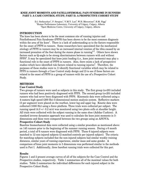
Figures 1 <strong>and</strong> 2 present average curves of all of the subjects for the Case Control <strong>and</strong> the<br />
Prospective studies, respectively. Table 1 summarizes all of the maximal values for both<br />
studies. Table 2 summarizes the <strong>in</strong>dividual differences for each <strong>in</strong>jured subject <strong>in</strong> the<br />
Prospective Cohort Study.
Figure 1: Stance phase <strong>knee</strong> <strong>jo<strong>in</strong>t</strong> <strong>moments</strong> <strong>in</strong> the sagittal, frontal <strong>and</strong> transverse planes compar<strong>in</strong>g asymptomatic<br />
(n=20) to PFPS (n=20) <strong>runners</strong>, runn<strong>in</strong>g heel-toe, with a shoe, at 4.0 m/s.(CASE CONTROL Study Design)<br />
Figure 2: Stance phase <strong>knee</strong> <strong>jo<strong>in</strong>t</strong> <strong>moments</strong> <strong>in</strong> the sagittal, frontal <strong>and</strong> transverse planes compar<strong>in</strong>g asymptomatic<br />
(n=12) to PFPS (n=6) <strong>runners</strong>, runn<strong>in</strong>g heel-toe, with a shoe, at 4.0 m/s.(PROSPECTIVE Study Design)<br />
Knee Moment [Nm]<br />
study condition group n<br />
max extension (SD) p-value max abduction (SD) p-value max ext rotation (SD) p-value<br />
asymp 20 124.1 (47.5) -103.1 (35.6) 27.9 (16.2)<br />
case control shoe<br />
0.69 0.16 0.14<br />
pfps 20 129.1 (29.6) -127.3 (66.1) 35.8 (16.5)<br />
asymp 12 132.1 (31.2) -49.9 (33.6) 12.9 (10.2)<br />
shoe<br />
0.05<br />
0.16<br />
0.12<br />
pfps 6 101.7 (18.8) -74.7 (29.2) 21.5 (10.5)<br />
prospective<br />
asymp 12 128.9 (33.0) -45.5 (31.2) 11.8 (7.8)<br />
barefoot<br />
0.03 *<br />
0.08<br />
0.03 *<br />
pfps 6 94.6 (16.1) -75.2 (27.0) 22.4 (10.2)<br />
Table 1: Comparison of average maximal <strong>knee</strong> <strong>moments</strong> <strong>and</strong> st<strong>and</strong>ard deviations <strong>in</strong> the sagittal, frontal <strong>and</strong> transverse<br />
planes for both study designs. Only shoe runn<strong>in</strong>g was collected for the Case Control Study.<br />
condition variable [unit] sub 1 sub 2 sub 3 sub 4 sub 5 sub 6<br />
∆ (max extension moment) % -25.5 -23.8 -3.9 -18.5 -19.1 -47.5<br />
shoe ∆ (max abduction moment) % 139.9 21.2 72.7 -48.1 73.8 38.6<br />
∆ (max external rot moment) % 176.2 -14.0 96.9 -57.5 81.4 119.2<br />
∆ (max extension moment) % -35.6 -27.5 -2.6 -32.0 -35.9 -25.8<br />
barefoot ∆ (max abduction moment) % 165.1 81.5 89.4 -21.3 54.3 22.4<br />
∆ (max external rot moment) % 239.7 74.5 129.2 -13.7 74.2 34.1<br />
Table 2: Individual subject values for the Prospective Study. Values shown are differences <strong>in</strong> % from the average of the<br />
asymptomatic group. A positive value <strong>in</strong>dicates a higher moment compared to the asymptomatic average.<br />
DISCUSSION<br />
Increased <strong>knee</strong> <strong>jo<strong>in</strong>t</strong> load<strong>in</strong>g <strong>in</strong> the frontal (ab-adduction <strong>moments</strong>) <strong>and</strong> transverse<br />
(external-<strong>in</strong>ternal rotation <strong>moments</strong>) planes may lead to <strong>in</strong>creased stresses with<strong>in</strong> or around<br />
the <strong>patellofemoral</strong> <strong>jo<strong>in</strong>t</strong> <strong>and</strong> thus lead<strong>in</strong>g to <strong>pa<strong>in</strong></strong>. Both maximal abduction <strong>and</strong> maximal<br />
external rotation <strong>knee</strong> <strong>moments</strong> were approximately 20% higher for the <strong>in</strong>jured <strong>runners</strong> <strong>in</strong><br />
the case control study. No statistical differences were seen. Results of the prospective<br />
study showed a similar trend. These differences became statistically significant for the<br />
external rotation moment when look<strong>in</strong>g at barefoot runn<strong>in</strong>g. More importantly, the PFPS<br />
group had a 65% higher maximal <strong>knee</strong> abduction moment <strong>and</strong> a 90% higher maximal<br />
external <strong>knee</strong> rotation moment. For the prospective study, <strong>in</strong>dividual differences revealed<br />
that 5 of the 6 <strong>in</strong>jured subjects had a 30% or greater value for the abduction <strong>and</strong> external<br />
rotation moment for barefoot runn<strong>in</strong>g. This pattern was also evident for 8 of the 20 <strong>in</strong>jured<br />
subjects <strong>in</strong> the Case Control study. There is a strong possibility that <strong>in</strong>creased <strong>knee</strong> <strong>jo<strong>in</strong>t</strong><br />
<strong>moments</strong> are contribut<strong>in</strong>g factors lead<strong>in</strong>g to the onset of PFPS <strong>in</strong> <strong>runners</strong>.<br />
REFERENCES<br />
1. Clement, D.B. et al. Phys & Spts Med 9(5): 47-58, 1981.<br />
2. James, S.L. et al. Amer J Spts Med 6(2): 40-49, 1978.<br />
3. Nigg, B.M. et al. J Biomech 26(8): 909-916, 1993.<br />
4. Messier, S.P. et al. Med Sci Spts Exec 23(9): 1008-1015, 1991.<br />
5. Van Mechelen, W. Sports Med 19(3): 161-165, 1995.


