
You also want an ePaper? Increase the reach of your titles
YUMPU automatically turns print PDFs into web optimized ePapers that Google loves.
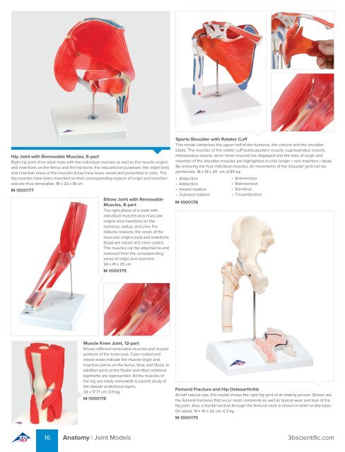
Hip Joint with Removable Muscles, 6-part<br />
Right hip joint of an adult male with the individual muscles as well as the muscle origins<br />
and insertions on the femur and the hip bone. For educational purposes, the origin (red)<br />
and insertion areas of the muscles (blue) have been raised and presented in color. The<br />
hip muscles have been mounted on their corresponding regions of origin and insertion<br />
and are thus removable. 18 x 32 x 18 cm<br />
M-1000177<br />
Elbow Joint with Removable<br />
Muscles, 8-part<br />
The right elbow of a male with<br />
individual muscles plus muscular<br />
origins and insertions on the<br />
humerus, radius, and ulna. For<br />
didactic reasons, the areas of the<br />
muscular origins (red) and insertions<br />
(blue) are raised and color-coded.<br />
The muscles can be attached to and<br />
removed from the corresponding<br />
areas of origin and insertion.<br />
24 x 41 x 25 cm<br />
M-1000179<br />
Sports Shoulder with Rotator Cuff<br />
This model comprises the upper half of the humerus, the clavicle and the shoulder<br />
blade. The muscles of the rotator cuff (subscapularis muscle, supraspinatus muscle,<br />
infraspinatus muscle, teres minor muscle) are displayed and the sites of origin and<br />
insertion of the shoulder muscles are highlighted in color (origin = red; insertion = blue).<br />
By removing the four individual muscles, all movements of the shoulder joint can be<br />
performed. 18 x 18 x 24 cm; 0.85 kg<br />
• Abduction<br />
• Adduction<br />
• Inward rotation<br />
• Outward rotation<br />
M-1000176<br />
• Anteversion<br />
• Retroversion<br />
• Elevation<br />
• Circumduction<br />
Muscle Knee Joint, 12-part<br />
Shows different removable muscles and muscle<br />
portions of the knee area. Color coded and<br />
raised areas indicate the muscle origin and<br />
insertion points on the femur, tibia, and fibula. In<br />
addition parts of the fibular and tibial collateral<br />
ligaments are represented. All the muscles of<br />
the leg are easily removable to permit study of<br />
the deeper anatomical layers.<br />
33 x 17 17 cm; 0.9 kg<br />
M-1000178<br />
Femoral Fracture and Hip Osteoarthritis<br />
At half natural-size, this model shows the right hip joint of an elderly person. Shown are<br />
the femoral fractures that occur most commonly as well as typical wear and tear of the<br />
hip joint. Also, a frontal section through the femoral neck is shown in relief on the base.<br />
On stand. 14 x 10 x 22 cm; 0.3 kg<br />
M-1000175<br />
16 Anatomy | Joint Models 3bscientific.com