
You also want an ePaper? Increase the reach of your titles
YUMPU automatically turns print PDFs into web optimized ePapers that Google loves.
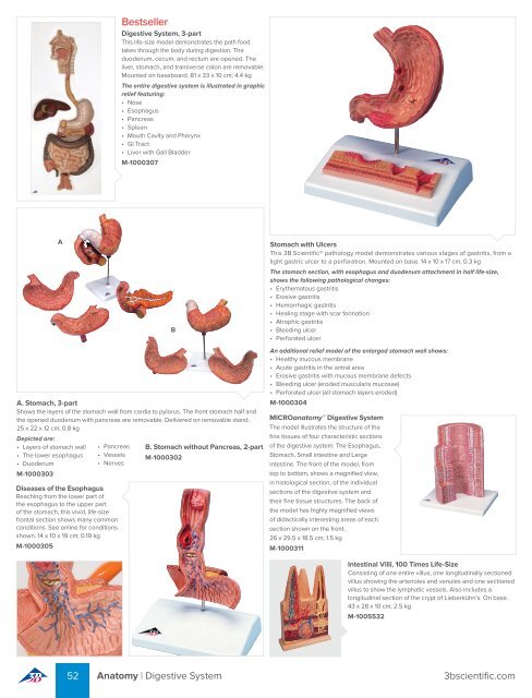
Bestseller<br />
Digestive System, 3-part<br />
This life-size model demonstrates the path food<br />
takes through the body during digestion. The<br />
duodenum, cecum, and rectum are opened. The<br />
liver, stomach, and transverse colon are removable.<br />
Mounted on baseboard. 81 x 33 x 10 cm; 4.4 kg<br />
The entire digestive system is illustrated in graphic<br />
relief featuring:<br />
• Nose<br />
• Esophagus<br />
• Pancreas<br />
• Spleen<br />
• Mouth Cavity and Pharynx<br />
• GI Tract<br />
• Liver with Gall Bladder<br />
M-1000307<br />
A<br />
B<br />
Stomach with Ulcers<br />
This <strong>3B</strong> <strong>Scientific</strong>® pathology model demonstrates various stages of gastritis, from a<br />
light gastric ulcer to a perforation. Mounted on base. 14 x 10 x 17 cm; 0.3 kg<br />
The stomach section, with esophagus and duodenum attachment in half life-size,<br />
shows the following pathological changes:<br />
• Erythematous gastritis<br />
• Erosive gastritis<br />
• Hemorrhagic gastritis<br />
• Healing stage with scar formation<br />
• Atrophic gastritis<br />
• Bleeding ulcer<br />
• Perforated ulcer<br />
A. Stomach, 3-part<br />
Shows the layers of the stomach wall from cardia to pylorus. The front stomach half and<br />
the opened duodenum with pancreas are removable. Delivered on removable stand.<br />
25 x 22 x 12 cm; 0.8 kg<br />
Depicted are:<br />
• Layers of stomach wall<br />
• The lower esophagus<br />
• Duodenum<br />
M-1000303<br />
• Pancreas<br />
• Vessels<br />
• Nerves<br />
Diseases of the Esophagus<br />
Reaching from the lower part of<br />
the esophagus to the upper part<br />
of the stomach, this vivid, life-size<br />
frontal section shows many common<br />
conditions. See online for conditions<br />
shown. 14 x 10 x 19 cm; 0.19 kg<br />
M-1000305<br />
B. Stomach without Pancreas, 2-part<br />
M-1000302<br />
An additional relief model of the enlarged stomach wall shows:<br />
• Healthy mucous membrane<br />
• Acute gastritis in the antral area<br />
• Erosive gastritis with mucous membrane defects<br />
• Bleeding ulcer (eroded muscularis mucosae)<br />
• Perforated ulcer (all stomach layers eroded)<br />
M-1000304<br />
MICROanatomy Digestive System<br />
The model illustrates the structure of the<br />
fine tissues of four characteristic sections<br />
of the digestive system: The Esophagus,<br />
Stomach, Small intestine and Large<br />
intestine. The front of the model, from<br />
top to bottom, shows a magnified view,<br />
in histological section, of the individual<br />
sections of the digestive system and<br />
their fine tissue structures. The back of<br />
the model has highly magnified views<br />
of didactically interesting areas of each<br />
section shown on the front.<br />
26 x 29.5 x 18.5 cm; 1.5 kg<br />
M-1000311<br />
Intestinal Villi, 100 Times Life-Size<br />
Consisting of one entire villus, one longitudinally sectioned<br />
villus showing the arterioles and venules and one sectioned<br />
villus to show the lymphatic vessels. Also includes a<br />
longitudinal section of the crypt of Lieberkühn’s. On base.<br />
43 x 28 x 10 cm; 2.5 kg<br />
M-1005532<br />
52 Anatomy | Digestive System 3bscientific.com