Shape Memory Materials for Biomedical Applications
Shape Memory Materials for Biomedical Applications
Shape Memory Materials for Biomedical Applications

You also want an ePaper? Increase the reach of your titles
YUMPU automatically turns print PDFs into web optimized ePapers that Google loves.
<strong>Shape</strong> <strong>Memory</strong> <strong>Materials</strong><br />
<strong>for</strong> <strong>Biomedical</strong> <strong>Applications</strong><br />
REVIEWS<br />
By Fatiha El Feninat, Gaetan Laroche, Michel Fiset,andDiego Mantovani*<br />
<strong>Shape</strong> memory properties provide a very attractive insight into materials science, opening unexplored<br />
horizons and giving access to unconventional functions in every material class (metals, polymers, and<br />
ceramics). In this regard, the biomedical field, <strong>for</strong>ever in search of materials that display unconventional<br />
properties able to satisfy the severe specifications required by their implantation, is now showing<br />
great interest in shape memory materials, whose mechanical properties make them extremely attractive<br />
<strong>for</strong> many biomedical applications. However, their biocompatibility, particularly <strong>for</strong> long-term and permanent<br />
applications, has not yet been fully established and is there<strong>for</strong>e the object of controversy. On<br />
the other hand, shape memory polymers (SMPs) show promise, although thus far, their biomedical<br />
applications have been limited to the exploration. This paper will first review the most common biomedical<br />
applications of shape memory alloys and SMPs and address their critical biocompatibility<br />
concerns. Finally, some engineering implications of their use as biomaterials will be examined.<br />
1. Introduction<br />
Medical implants have undoubtedly made an indelible<br />
mark on our world during the last century. More than<br />
100 millions humans carry at least one major internal medical<br />
device. The prosthesis industry has topped 50 billion US$ in<br />
annual sales, with approximately 150 universities throughout<br />
the world proposing an undergraduate program in bioengineering<br />
or biomedical engineering. Despite that, however,<br />
most medical devices have been constructed using a significantly<br />
restricted number of conventional metallic, ceramic,<br />
polymeric, and composite biomaterials.<br />
Medicine and improvisation hardly appear to be a likely<br />
pair, yet since ancient times, resourceful doctors have carried<br />
out difficult procedures, often having to work with materials<br />
on hand. [1] Wounds were sutured with plant fibers (animalderived<br />
materials by ancient Greeks, Chinese, and Egyptians),<br />
and prosthetic limbs were fashioned from wood. Metals<br />
were eventually introduced in dentistry, and early this<br />
past century, when stainless steel became available, corrosion-resistant<br />
alloys were used to make a variety of prostheses.<br />
As with their predecessors, today medical practitioners<br />
will often attempt to cure diseases or improve quality<br />
of life by replacing a defective body part with a substitute.<br />
While the designing process leading to the development of<br />
successful artificial organs has been improved over the years,<br />
bioengineers remain limited to fabricating devices with offthe-shelf<br />
materials which were not designed specifically <strong>for</strong><br />
the application. The easy availability of industrial materials,<br />
along with the multiple specific and challenging constraints<br />
to which an artificial organ is submitted when implanted in<br />
the body, are the principal factors which may explain why<br />
today, only a dozen materials are routinely used to construct<br />
internal artificial organs. In this regard, motivated by the<br />
increasing need <strong>for</strong> custom-made materials <strong>for</strong> specific medical<br />
applications, materials scientists, metallurgists, chemists,<br />
mechanical and chemical engineers, as well as researchers in<br />
other disciplines, have progressively begun an interdisciplinary<br />
work in the hopes of creating high-per<strong>for</strong>mance biomate-<br />
±<br />
[*] Prof. D. Mantovani, Dr. F. El Feninat<br />
Research Center, St. François d'Assise Hospital,<br />
Department of Mining, Metallurgy and <strong>Materials</strong><br />
Engineering<br />
Laval University, Pouliot Building, Room 1745-E<br />
Quebec City, G1K 7P4 (Canada)<br />
E-mail: Diego.Mantovani@gmn.ulaval.ca<br />
Dr. M. Fiset<br />
Department of Mining, Metallurgy and <strong>Materials</strong><br />
Engineering, Laval University<br />
Dr. G. Laroche<br />
Department of Surgery, Laval University<br />
ADVANCED ENGINEERING MATERIALS 2002, 4, No. 3 Ó WILEY-VCH Verlag GmbH, D-69469 Weinheim, 2002 1438-1656/02/0303-0091 $ 17.50+.50/0 91
Mantovani et al./<strong>Shape</strong> <strong>Memory</strong> <strong>Materials</strong> <strong>for</strong> <strong>Biomedical</strong> <strong>Applications</strong><br />
REVIEWS<br />
rials, or tailoring those industrial materials with specific<br />
properties into high-potential biomaterial candidates.<br />
Among the last industrial materials elected to the rank of<br />
biomaterials are shape memory alloys (SMA), which have<br />
been proposed <strong>for</strong> use in a wide variety of internal applications,<br />
including orthopaedic, dental, surgical, and (only later)<br />
cardiovascular devices. The unique properties of SMA allow<br />
<strong>for</strong> a variety of applications in implantology. As it was pre-<br />
Fatiha El Feninat received her M.Sc.A degree in chemical engineering from École Polytechnique de<br />
MontrØal. Recently, she obtained a Ph.D. degree in chemistry from the University of MontrØal, <strong>for</strong> her<br />
works on the characterisation of human dentin by atomic <strong>for</strong>ce microscopy. In November 2000, she<br />
joined the Department of Mining, Metallurgy and <strong>Materials</strong> Engineering at Laval University and the<br />
Research Center of the St-François d'Assise Hospital as Postdoctoral Fellow. Her research focus on surface<br />
modifications and characterisation of shape memory alloys in order to be used as long-term safe biomaterials,<br />
and atomic <strong>for</strong>ce microscopy.<br />
GaØtan Laroche received both his B.Sc. (1986) and Ph.D. (1990) from the chemistry department at Laval<br />
University. He joined the Research Center of St-François d'Assise Hospital in 1992 and the Surgery<br />
Department at Laval University where he is professor since 1994. His main research interests are<br />
related to the physicochemical characterisation of biomaterials, molecular transport through biomaterials<br />
and surface engineering of biomaterials to improve their biocompatibility.<br />
M. Fiset obtained a B.Sc. degree in physics and Ph.D. degree in metallurgy from Laval University. He<br />
joined the department of Mining, Metallurgy and <strong>Materials</strong> Engineering at Laval University in 1977 as<br />
professor of materials science. One of his major research interests has been the field of abrasive wear, with<br />
particular reference to alloy white cast irons, laser materials processing, as well as advanced alloys <strong>for</strong><br />
biomedical applications.<br />
Diego Mantovani obtained a B.Sc. degree in Bioengineering from the Politecnico di Milano, Italy, and a<br />
B.Sc. in Biomaterials and Artificial Organs from the UniversitØ de Technologie de Compi›gne, France.<br />
Then, he obtained a Ph.D. from Laval University in 1998 and a D.Sc. from the UniversitØ de Technologie<br />
de Compi›gne in 1999, <strong>for</strong> his studies on materials and biomaterials. He is professor at the Laval<br />
University department of Mining, Metallurgy and <strong>Materials</strong> Engineering and researcher at the<br />
Research Center of the St-François d'Assise Hospital in Biomaterials and Bioengineering since January<br />
2000. With his team, he carries out project-oriented and multidisciplinary research in biomaterials, artificial<br />
organs and bioengineering. Focus is on functional materials <strong>for</strong> blood-contact applications, structure-properties<br />
relationships, micro-mechanics, surface properties modifications, and bioreactors design<br />
<strong>for</strong> reparative medicine.<br />
92 ADVANCED ENGINEERING MATERIALS 2002, 4, No. 3
Mantovani et al./<strong>Shape</strong> <strong>Memory</strong> <strong>Materials</strong> <strong>for</strong> <strong>Biomedical</strong> <strong>Applications</strong><br />
viously published in extensively reviews. [2,3] In general, each<br />
material used to fabricate industrial products designed to be<br />
implanted in the human body <strong>for</strong> small, medium or longterm<br />
periods must be tested to demonstrate its ªbiocompatibilityº.<br />
In vitro studies, followed by in vivo studies, and<br />
finally, clinical studies, are the three successive and general<br />
steps, with increased levels of scrutiny, required by the U.S.<br />
Food and Drug Administration (FDA) to authorise their use<br />
as implant. Consequently, only a few products developed by<br />
industrial R&D may effectively be used in implantology.<br />
Today, the use of endovascular stents, orthopaedic staples<br />
and dental braces are widely acknowledged world-wide,<br />
which is not the case <strong>for</strong> intra-cranial staples, which are not<br />
permitted by the FDA <strong>for</strong> use in the USA.<br />
The objective of this paper is first to review the most common<br />
applications of shape memory materials (SMMs), focusing<br />
on shape memory metallic alloys, which are widely used,<br />
and shape memory polymers (SMPs), <strong>for</strong> which strong R&D<br />
industrial ef<strong>for</strong>ts are ongoing. Secondly, this paper aim is to<br />
show the challenges facing their unique properties, while<br />
addressing some of the critical concerns with regard to the<br />
nature of the biological environment these materials will have<br />
to integrate. We will conclude by outlining some of the realistic<br />
implications such as societies expectations and the quality<br />
of life <strong>for</strong> the patient. The goal here is there<strong>for</strong>e not to categorise<br />
materials and biomaterials as being either ªbadº or<br />
ªgoodº, but rather to stimulate reader implication in a true<br />
interdisciplinary discussion towards an actual advancement<br />
of this ever-growing research field.<br />
2. <strong>Shape</strong> <strong>Memory</strong> Alloys<br />
From a chronological point of view, in the thirties the<br />
pseudoelastic effect was already being observed in Au±Cd<br />
alloy. [4] This was followed in 1938 by the observation of the<br />
shape memory effect in Cu±Zn alloy. [5] It was, however, in<br />
the 1960s, that Buehler et al., [6] at Naval Ordnance Laboratory,<br />
discovered the shape memory effect in nickel±titanium alloys,<br />
commonly known as Nitinol alloys (<strong>for</strong> nickel titanium Naval<br />
Ordnance Laboratory).<br />
From a more scientific point of view, there exists an<br />
exhaustive wide variety of metallic alloys which demonstrate<br />
shape memory and/or superelastic effects and which have<br />
been investigated in the past and are well reported in the literature.<br />
[4,7±18] These works focused on metallic alloys, including,<br />
<strong>for</strong> example, binary systems such as NiTi, CuZn, AuCd,<br />
CuSn, TiPd, NiAl, and InTi, as well as ternary systems such<br />
as NiTiCu, [19] and CuZnAl. [20,21] Moreover, it has been shown<br />
that introducing copper in a binary NiTi alloy increases its<br />
ability to change shape when heated (shape memory properties).<br />
[22±24]<br />
From a metallurgical point of view, nitinol is a nickel±titanium<br />
alloy of near-equiatomic composition, which implies<br />
that nickel represents approximately 50 % of its chemical<br />
composition. This alloy is particularly interesting because of<br />
its significant mechanical properties, and above all, because<br />
of its ability to show high elastic de<strong>for</strong>mation, or ªpseudoelasticityº,<br />
and ªshape memory effectº which are not present<br />
in other conventional metallic alloys. It has to be underlined<br />
that the terms pseudoelasticity and superelasticity are often<br />
used synonymously in the literature, even if the specific<br />
metallurgical mean and the proper use of these terms was<br />
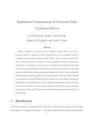
previously discussed by Ostuka and Wayman. [25] Figure 1<br />
shows the stress±temperature relation where the hysteresis<br />
describing the trans<strong>for</strong>mation of the SMA is presented. This<br />
hysteresis is characterised by four temperatures (Ms, Mf, As,<br />
and Af) which indicate the initial and final trans<strong>for</strong>mation<br />
temperatures. Two stable trans<strong>for</strong>mation phases are present<br />
at different temperatures: the martensite phase is stable at<br />
low temperature in contrast the austenite one which is stable<br />
at high temperature. These two trans<strong>for</strong>mations are reversible<br />
without inducing any diffusion between the existing phases.<br />
The most significant properties of the SMA are ruled by the<br />
phase transitions between the austenite (ªhigh temperatureº<br />
phase) and the martensite (ªlow temperatureº phase), and<br />
reciprocally. The phase transitions as a function of temperature<br />
are thus particularly important in order to control the<br />
properties. In a previous study, [2] we carefully described the<br />
five characteristic properties and proposed an integrated<br />
global overview of the various effects which can be observed<br />
on SMAs:<br />
l the one-way shape memory effect, where the change in<br />
shape is regulated by the transition from martensite to<br />
austenite,<br />
l the two-way shape memory effect, with the learning process<br />
by mechanical cycles and the one-way shape memory<br />
effect, where the changes in shape are regulated by the<br />
phase transitions (martensite±austenite followed by austenite±martensite),<br />
l the superelastic effect, where the de<strong>for</strong>mations are regulated<br />
by the phase transitions (austenite±martensite, then<br />
martensite±austenite),<br />
Fig. 1. Austenite trans<strong>for</strong>mation and hysteresis (H) following a temperature change.<br />
As = Austenite start, Af = Austenite finish, Ms = Martensite start, and Mf = Martensite<br />
finish.<br />
REVIEWS<br />
ADVANCED ENGINEERING MATERIALS 2002, 4, No. 3 93
Mantovani et al./<strong>Shape</strong> <strong>Memory</strong> <strong>Materials</strong> <strong>for</strong> <strong>Biomedical</strong> <strong>Applications</strong><br />
REVIEWS<br />
l the superthermic effect, where the material replaces the<br />
accumulated internal constraints during the learning process<br />
by the external constraints,<br />
l the rubber-like effect, observed in the repeated mechanical<br />
cycles of learning.<br />
The interconnections between temperature, <strong>for</strong>ce, and<br />
geometrical shape are complex, which make it difficult to predict<br />
the behaviour of SMA in each specific application. Most<br />
of the current applications use alloys that allow us to retain<br />
two of three parameters, with the third fixed by the choice of<br />
alloy elements and thermomechanical treatment. The NiTi<br />
general properties are primarily related to a temperature or<br />
stress that is induced by the martensitic trans<strong>for</strong>mation. [26]<br />
However, the mechanical properties of NiTi alloy depend<br />
more specifically on the trans<strong>for</strong>mation temperature of transition,<br />
[6] and the damping capacity of alloys which can reach<br />
90 % <strong>for</strong> impact loads. [27] The properties of nickel±titanium<br />
alloys have been extensively investigated and reported by<br />
various authors. [25,28] The high mechanical properties of NiTi<br />
alloy may be helpful in self-expanding and self-compressing,<br />
[29] situations which make NiTi easily malleable when<br />
used in medical devices. This malleability is important, particularly<br />
in the case of alloys used in endovascular therapy.<br />
The instrument handles can be bent with enormous precision<br />
to the proper shape required <strong>for</strong> surgery, and recover their<br />
initial shape after heating. [30]<br />
From an industrial point of view, among the wide variety<br />
of shape memory metallic alloys available only those able to<br />
recover a substantial amount under strain (when the austenitic±martensitic<br />
phase change occurs) have been considered in<br />
the design of industrial products. Thus far, this signifies that<br />
only the equiatomic (or the near equiatomic) NiTi alloys and<br />
a few of the copper-based alloys have been largely commercialised.<br />
Moreover, only NiTi alloys have been introduced in<br />
the medical device industry and are currently overwhelmingly<br />
used as biomaterials through other potential metallic<br />
alloys, because the latter, while presenting similar properties,<br />
are at times expensive (such as gold-based alloys, [31,32] )ordo<br />
not exhibit mechanical properties and thermal stability as<br />
competitively as do NiTi alloys. [33] Finally, in some other<br />
cases, they exhibit a far greater risk of toxicity. [34±36]<br />
The field of NiTi alloys is expanding rapidly: In 1998, TiNi<br />
Alloy Company reported that many devices of various sizes<br />
had been introduced <strong>for</strong> medical and others fields and that<br />
the sales of these devices had reached more than a hundred<br />
million USD per year. [37] We will there<strong>for</strong>e present a review<br />
of the various biomedical applications of NiTi-based SMA<br />
and its biocompatibility when used in the fabricating of biomedical<br />
devices.<br />
Since the discovery of NiTi alloy, and particularly in the<br />
early 1970s, many studies have exploited the potential of NiTi<br />
<strong>for</strong> medical applications. [38±41] However, it was not until the<br />
1990s that adequate medical investigations led to a breakthrough<br />
with the development the first commercial stent. [42,43]<br />
Based on superelastic effect wires, [44] NiTi alloys were used in<br />
orthodontic therapy because of their high flexibility in bending<br />
without kicking. [45±49] Studies emerged on the use of NiTi<br />
wires <strong>for</strong> the correction of malocclusions and impacted<br />
canines. [50] In 1978, Andreasen and Morrow [51] reported on<br />
the advantages of NiTi orthodontic wires over conventional<br />
orthodontic wire. During the early stages of orthodontic therapy,<br />
constant low stress to the dentition over time is required<br />
in order to minimise tissue destruction such as root resorption<br />
during tooth movement. [52,53] Superelastic NiTi wire easily<br />
gains these important <strong>for</strong>ces, which are particularly favourable<br />
<strong>for</strong> large malaligned teeth. Another advantage of NiTi<br />
orthodontic wires is that it is possible to provide rapid orthodontic<br />
treatments, resulting in less patient discom<strong>for</strong>t because<br />
fewer adjustments and wire changes are required. [30,52]<br />
Other interesting medical applications of NiTi alloy have<br />
been reported in orthopaedic, [54] and other bone-related<br />
operations. [55] Nitinol has been to be more effective than<br />
others materials [56] in connecting broken bones. Staples made<br />
of NiTi SMAs have been used to fix small bone fragments. [57]<br />
In cervical anterior fusion, the NiTi staple was used in fifty<br />
patients; with successful results <strong>for</strong> 80 % of the cases<br />
(36 months). [58] Superelastic NiTi catheters facilitate access to<br />
areas in human body which are at times more difficult to<br />
reach using other materials.<br />
Because of its superelastic properties, NiTi alloy is exceptionally<br />
flexible, which enables its use in non-invasive surgery<br />
to reach narrow places. The NiTi wires are also shaped<br />
<strong>for</strong> use in prostheses, tissue anchoring and connection, [59] as<br />
well as stents. [60,61] When inserted into the human body, the<br />
superelastic NiTi-based stents are capable of self-expanding.<br />
This property allows us to use these stents in gastroenterology,<br />
cardiovascular and radiology fields. The U.S. Food and<br />
Drug Administration has accepted the vascular NiTi device<br />
reported by Simon and his research team. [41] This device,<br />
called Simon Nitinol Filter (SNF), is used <strong>for</strong> treating pulmonary<br />
embolism. Other applications in cardiovascular surgery<br />
have been reported. [62±64] In 1990, it was reported in Britain<br />
that 7.5 per 100 000 of the population develops oesophageal<br />
carcinoma [65] and only the palliation of oesophageal carcinoma<br />
was suitable by using self-expanding stents, [66] which<br />
offer the advantage of being easily implantable <strong>for</strong> providing<br />
effective malignant palliation.<br />
The follow-up in most of the studies on NiTi as implant<br />
material in humans may un<strong>for</strong>tunately have been limited in<br />
evaluating its toxicity which may arise after many years of<br />
implantation. When such a long-term application is not<br />
required, the use of this alloy may there<strong>for</strong>e be considered, as<br />
in the case of the preliminary bracket alignment stage of<br />
orthodontic treatment. [52] However, further investigations<br />
must to be envisaged prior to any long-term implantation of<br />
these materials in humans. If it is to be considered fully useful,<br />
the SMA must fulfil the requirements of both short and<br />
long-term biological (biocompatibility) and chemical (degradation,<br />
corrosion, and dissolution) reliabilities when used <strong>for</strong><br />
human concerns.<br />
94 ADVANCED ENGINEERING MATERIALS 2002, 4, No. 3
Mantovani et al./<strong>Shape</strong> <strong>Memory</strong> <strong>Materials</strong> <strong>for</strong> <strong>Biomedical</strong> <strong>Applications</strong><br />
3. <strong>Shape</strong> <strong>Memory</strong> Polymers<br />
Contrary to popular belief, shape memory is not a property<br />
known exclusively to metallic alloys. Nevertheless, it<br />
must to be emphasised that only metallic alloys are capable<br />
of showing shape memory properties because of a crystalline<br />
structure change, i.e., from austenite to martensite, or viceversa.<br />
Other materials exhibit similar properties, and there<strong>for</strong>e<br />
are defined as ªadaptive or smartº materials. Smart or<br />
adaptive are adjectives given to those materials that provide<br />
a specific response in a particular environment. For example,<br />
it means that they are able to assume pre-definite shapes and<br />
dimensions when their environment reaches at a determined<br />
temperature, or that they are capable of displaying a specific<br />
<strong>for</strong>ce at a particular temperature, etc. There<strong>for</strong>e, it may be<br />
understood that those specific materials, namely, polymers<br />
with high, specific shape memory characteristics, have<br />
received early recognition as potential candidates as implant<br />
materials. In fact, the need <strong>for</strong> these new materials is well<br />
appreciated because of the controversy regarding the biocompatibility<br />
of nickel-containing SMAs (as will be discussed<br />
later in this paper), their difficult fabrication and/or their<br />
cost. The primary advantage of polymers over other materials<br />
in biomedical applications is their easier availability and their<br />
wide range of mechanical and physical properties. [67,68]<br />
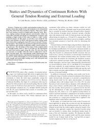
As shown in Figure 2, the polymeric materials show the<br />
properties more closely to those of soft biological tissue, if<br />
compared to those of metals which are more similar to those<br />
of hard biological tissue. The international scientific community<br />
has there<strong>for</strong>e focused its attention on new polymeric<br />
materials offering the specific high advantage of returning to<br />
some previously defined shape under the appropriate thermal<br />
conditions. A variety of publications and patents have<br />
covered the development of polymers exhibiting these<br />
unusual properties. [69±76]<br />
The mechanism through which selected polymers demonstrate<br />
specific shape memory properties is related to their<br />
intrinsic non-crystalline molecular structure. As it is wellknown,<br />
the glass transition temperature (T g ) characterises<br />
polymers, making them unique in contrast to other materials<br />
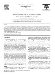
such as metals or ceramics. However, polymers display different<br />
states depending on the temperature range within a<br />
few degrees of T g . In fact, at this temperature, significant<br />
changes in the mechanical and thermodynamical properties<br />
may be observed [77] (Fig. 3):<br />
l above this temperature, the polymers are in their rubbery<br />
state, whereas at this stage, the polymers are elastic and<br />
soft, [78]<br />
l below the transition temperature, the polymeric materials<br />
become brittle and hard. At this point, the rubbery state is<br />
replaced by a glassy behavior,<br />
l across the glass temperature, the elastic modulus of polymers<br />
may exhibit a large, reversible change (Fig. 3).<br />
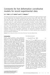
Through the shape memory effect shown in Figure 4, it<br />
may be possible to de<strong>for</strong>m the polymers below T g and return<br />
them to their original shape by heating the polymers to higher<br />
temperature than T g . However, if the materials are de<strong>for</strong>med<br />
above their glass transition temperature under external<br />
<strong>for</strong>ce, the de<strong>for</strong>mation will be fixed and maintained after<br />
removal of the external <strong>for</strong>ce. However, a subsequent heating<br />
of the material above its T g will allow it to recover its original<br />
shape, thereby generating a lower <strong>for</strong>ce. [68]<br />
The shape memory and elastic properties make polymers<br />
highly interesting when used as smart (or adaptive) materials<br />
<strong>for</strong> industrial applications. [78] SMPs are basically characterised<br />
by a low temperature transition which is in the range<br />
of room temperature; [79] this feature makes the SMPs suitable<br />
<strong>for</strong> biomedical devices, as the latter are implanted at body<br />
temperature (37 C). Another advantage supporting the preferential<br />
use of polymers as biomaterials, is the potential to<br />
target specific complementary properties simply by copolymerising<br />
two or more monomers. For example, the copolymerisation<br />
of vinyl chloride (VC) with ethylene, allows us to<br />
combine some properties of polyethylene that is soft and elastic,<br />
and poly(vinyl chloride) (PVC) that is mostly hard. The<br />
copolymerisation of styrene and butadiene produces a styrene±butadiene<br />
copolymer with shape memory properties. In<br />
this case, the different temperature-dependent behaviours of<br />
the copolymerised styrene and butadiene moieties enable the<br />
copolymer to preserve its stiffness (styrene) while the butadiene<br />
segments help maintain its flexible character (butadiene),<br />
thereby leading to the shape memory capabilities.<br />
REVIEWS<br />
Fig. 2. Schematic stress versus strain diagram <strong>for</strong> metals, polymeric materials, and biological<br />
tissues.<br />
Fig. 3. Elasticity versus temperature of amorphous polymers.<br />
ADVANCED ENGINEERING MATERIALS 2002, 4, No. 3 95
Mantovani et al./<strong>Shape</strong> <strong>Memory</strong> <strong>Materials</strong> <strong>for</strong> <strong>Biomedical</strong> <strong>Applications</strong><br />
REVIEWS<br />
Fig. 4. De<strong>for</strong>mation at different temperatures obtained after external loading.<br />
Polymers exhibit shape memory effects which completely<br />
differ from those of metallic alloys. Rubber is a typical SMP<br />
that is capable of expanding many times be<strong>for</strong>e returning to<br />
its original shape as a function of applied stress. [80] Contrary<br />
to the shape memory effects in metallic alloys, the effects in<br />
polymers are controllable not only by heating but also by<br />
exposure to light or through chemical reactions. [81,82] Indeed,<br />
crosslinking agents may be added to polymer <strong>for</strong>mulations;<br />
these crosslinks are selected in reason of their potential to<br />
experience isomerisation under photo-irradiation. The most<br />
simple example is azobenzene which is used as a crosslinking<br />
agent <strong>for</strong> polymer fabrics. By isomerisation through the ultraviolet<br />
irradiation of azobenzene, the trans<strong>for</strong>mation from the<br />
cis to the trans <strong>for</strong>m induces the shape change in the polymers.<br />
[83] As demonstrated by Hirai and his collaborators, [84] it<br />
is possible to introduce shape memorizing properties by<br />
crosslinking the polymer through chemical bonding. In addition<br />
to introducing the shape memory effect, this chemical<br />
crosslinking leads to a three-dimensional network which may<br />
significantly improve the physical properties such as acceptable<br />
elasticity and excellent strength. Of considerable interest<br />
is poly(vinyl alcohol) (PVA) crosslinked with glutaraldehyde.<br />
Without crosslinks, the use of the PVA gel is un<strong>for</strong>tunately<br />
limited in terms of thermal stability, [85] as heating this gel can<br />
in fact disrupt the hydrogen bonds and consequently the stability<br />
of the gel; this stability is also lost when the gel is placed<br />
in boiling water. [86]<br />
During the past 15 years, Nippon Zeon Co. and others [87±91]<br />
have developed a wide variety of SMPs. In the early 1997s,<br />
Liang et al. [92] developed new, easy-to-shape polymers. At<br />
Mitsubishi Heavy Industries in Japan, Hayashi developed<br />
shape memory segmented polyurethane (PU) copolymer<br />
which is characterised by two distinct elements: one, the hard,<br />
<strong>for</strong> the physical crosslinks and the other, the soft to introduce<br />
the shape memory effect. This material is capable of recovering<br />
the entire plastic de<strong>for</strong>mation up to 400 % when heated<br />
above the glass transition temperature; however, this recovery<br />
is much lower than that of SMA (8 % at initial elongation).<br />
Unlike metal alloys, [80] polymers demonstrate a recovery<br />
stress, between 0.98 and 2.94 MPa (10 and 30 kg f/cm 2 ) which<br />
is lower than that of metal alloys between 147 and 294 MPa<br />
(1500 and 3000 kg f/cm 2 ). There<strong>for</strong>e, despite the advantages of<br />
being relatively low-cost and easily processed, their application<br />
is often restricted because of their lack in recovering<br />
stress. [93]<br />
<strong>Shape</strong> memory polynorbornene, with a glass temperature<br />
of 35 C, has been used as an occluder device <strong>for</strong> patent ductus<br />
arteriosus (PDA) occlusion. [94] However, in vitro studies<br />
indicate that this technique requires that the temperature<br />
shape-changeable materials be easy introduced when used in<br />
intravascular surgery. Under this temperature, the occluder<br />
expands completely in the ductus and reduces the leak<br />
caused by the incomplete occlusion [95] at the PDA. However,<br />
because the compatibility of this material has not been tested,<br />
its ability to safely remain in contact with natural and living<br />
tissue cannot be predicted.<br />
Very recently, researchers have developed SMPs that are<br />
both compatible with the body and biodegradable upon interaction<br />
with physiological environment. These SMPs have<br />
been studied by Langer and Lendlein and their respective<br />
team [96] to produce scaffolds <strong>for</strong> engineering new organs and<br />
coronary stents. Such stents could be compressed and fed<br />
96 ADVANCED ENGINEERING MATERIALS 2002, 4, No. 3
Mantovani et al./<strong>Shape</strong> <strong>Memory</strong> <strong>Materials</strong> <strong>for</strong> <strong>Biomedical</strong> <strong>Applications</strong><br />
through a tiny hole in the body into a blocked artery. Then,<br />
the warmth of body would trigger the polymer's expansion<br />
into original shape. Instead of requiring a second surgery <strong>for</strong><br />
removing the SMPs, the polymer would gradually dissolve in<br />
the body over time. Others reported that the development of<br />
biodegradable materials suited <strong>for</strong> polymers will serve biomedical<br />
applications such as stents, catheters, and sutures. [97]<br />
As shown in this paper, polymers change their shape in 45 s<br />
at 65 C. The biodegradability of these materials will thus be<br />
an advantage in reducing the number of invasive surgeries. [93]<br />
Bernnan further explained that devices used <strong>for</strong> short-term<br />
endovascular applications will more readily degrade after<br />
successive tissue healing occurs. There<strong>for</strong>e, follow-up surgery<br />
will be obligatory, which will mean less discom<strong>for</strong>t <strong>for</strong> the<br />
patient. In some cases, biodegradable polymers are the only<br />
solution in applications such as reconstruction and functionality<br />
of blood vessels.<br />
4. Biocompatibility Concerns<br />
As discussed above, the development of metallic and polymeric<br />
adaptive (or smart) materials <strong>for</strong> biomedical applications<br />
is progressing rapidly because of their unique properties.<br />
[43] These materials are to be part of internal medical<br />
devices in intimate contact with tissue and body fluids, there<strong>for</strong>e<br />
particular attention must be given to the interface<br />
between the SMMs and the natural tissue upon implantation.<br />
Being synthetic (man-made), thus <strong>for</strong>eign to the body, these<br />
adaptive materials must first satisfy the basic criteria such as<br />
biofunctionality, biostability, and biocompatibility during<br />
implantation. This last element refers to the ability of the<br />
material to remain non-toxic while maintaining its initial<br />
functionality <strong>for</strong> the duration of implantation.<br />
Several studies have assessed the biocompatibility of the<br />
shape memory metallic alloys; however, thorough, more systematic<br />
studies of their biocompatibility when in contact with<br />
blood flow have only partially addressed the crucial question<br />
regarding their security, particularly in long-term applications,<br />
<strong>for</strong> which the biocompatibility of SMAs remains controversial,<br />
as we will present below. The rigorous investigation<br />
of the biocompatibility of biomaterials is of primary concern,<br />
because it will allow us to predict (albeit without certitude)<br />
their behaviour when implanted in humans. The objective<br />
being to guarantee the best possible quality of life <strong>for</strong> the<br />
patient, the biomaterialist's responsibility is to supply to the<br />
bioengineer with artificial organ materials which will remain<br />
stable <strong>for</strong> the rest of the patient life.<br />
Body fluids, such as blood, constitute an aggressive environment<br />
<strong>for</strong> a metallic implant. [98] Nitinol there<strong>for</strong>e represents<br />
the most widely used element in orthodontic and orthopaedic<br />
implants, and in stents. The clinical use of stents <strong>for</strong><br />
intravascular application has been improved by studying<br />
their surface properties and characteristics. [99,100] Preliminary<br />
studies have concluded that NiTi-based devices <strong>for</strong> use as<br />
peripheral arteries in human have led to interesting<br />
results. [101] Shih and his research group were the first to demonstrate<br />
the potential cytotoxicity of NiTi stent wires on rat<br />
aortic smooth muscle cells. [102] They observed cellular death<br />
following incubation of nitinol in cultured media and cell<br />
growth inhibition, and discussed that these phenomena were<br />
related to the concentration of nickel ions existing in NiTi<br />
stent as well as exposure time in the corrosive media. This<br />
finding is in agreement with several studies reporting that<br />
the release of the Ni ions from NiTi alloys has a significant<br />
effect, the dissolution of Ni may possibly contribute to the inhibition<br />
of cell replication [103] and proper cell function, [104±106]<br />
as these ions are considered both toxic and carcinogenic to<br />
cultured cells. [107±109] Each of these studies was consistent<br />
with the reports by Uo et al. [110] who observed the presence of<br />
severe tissue damage with inflammatory response around the<br />
Ni implants.<br />
When used to make catheters, or parts of catheters, NiTi<br />
alloys have a distinct advantage because of their properties,<br />
and particularly with regard to their easier insertion, which<br />
constitutes a very safe and justifiable choice <strong>for</strong> short-term<br />
(some hours) applications. [111] However, when the application<br />
requires longer periods of residence inside the body, a major<br />
question arises concerning its corrosion resistance, (particularly<br />
on the nickel presence) and its enormous potential to be<br />
cytotoxic, carcinogenic and eventually mutagenic. This potential<br />
must be further investigated and unequivocally stated:<br />
Appropriate procedures and rigorous standards have to be<br />
elaborated. Yet despite the many investigations, [112±114] the<br />
question is more complex than one would imagine. The possibility<br />
that nickel ions may react with the physiological environment<br />
is both realistic and theoretically possible. Each metal<br />
possesses its own intrinsic toxicity to cells, often depending<br />
on the concentration of its presence. Thus, the corrosion resistance<br />
of an alloy and the toxicity of individual metals (and<br />
their respective ions) in an alloy are the two principal factors<br />
that determine its long-term biocompatibility, [115] with results<br />
such as corrosion and other undesirable effects such as toxicity<br />
and carcinogenesis. [116±121] Moreover, this corrosive reaction<br />
may weaken the alloys mechanical properties. [122] Caution<br />
is there<strong>for</strong>e required when addressing the possibility<br />
that nickel is released into the human body and causes a<br />
potential risk when it is used long-term, as the dissolved Ni<br />
ions are capable of stimulating and activating natural tissue<br />
as well as adverse reactions. [123] It is <strong>for</strong> this reason, their<br />
applications have been sometimes limited. [124] Matsumoto et<br />
al. [125] reported that the subcutaneous implantation of nitinol<br />
rods in rabbits <strong>for</strong> 4 weeks led to the elution of Ni, causing a<br />
significant increase of the Ni concentration in the blood.<br />
Moreover, nitinol rods implanted intramedullary in rats<br />
exhibited significant surface corrosion after 60 weeks of<br />
implantation. [59]<br />
These studies identified the problem, which is a definite<br />
lack of evidence to support unequivocally the long-term biocompatibility<br />
of NiTi alloy. However, if the Ni dissolution<br />
REVIEWS<br />
ADVANCED ENGINEERING MATERIALS 2002, 4, No. 3 97
Mantovani et al./<strong>Shape</strong> <strong>Memory</strong> <strong>Materials</strong> <strong>for</strong> <strong>Biomedical</strong> <strong>Applications</strong><br />
REVIEWS<br />
from alloy was significant enough to cause corrosion after<br />
60 weeks, thereby affecting the natural tissue what are the effects<br />
if implanted <strong>for</strong> longer period of time (i.e., the rest of the<br />
patient's life)? Regardless, specific investigations of long-term<br />
implantation must there<strong>for</strong>e be carefully designed and methodically<br />
per<strong>for</strong>med following rigorous procedures be<strong>for</strong>e the<br />
safety of nitinol can be absolutely started and certified.<br />
It there<strong>for</strong>e appears clear that today there are no available<br />
conclusive data on the biocompatibility of NiTi. Nickel is<br />
among those solid metals recognised as being potentially carcinogenic<br />
when used in human and animal models. In 1976,<br />
many investigations of nickel compounds reported that they<br />
may induce cancers in animals models. [126,127] Many type of<br />
cancers have been related to the exposure to nickel. Despite<br />
the advantages such as shape memory and superelasticity,<br />
the nickel released in the body may cause both toxic [128] and<br />
allergic [129] reactions. The implantation of nitinol alloy in rabbit<br />
paravertebral muscles [130] resulted in an inflammatory<br />
response which may have caused cell damage. In contrast to<br />
the above mentioned studies, several studies agree as to the<br />
safety of NiTi alloy. [131,132] Moreover, devices fabricated with<br />
SMAs are still present in the medical market. The controversy<br />
still continues.<br />
The use of this alloy in practical applications depends on<br />
the environment and the level of wear, specific to the application,<br />
as well as other factors. We are far from suggesting that<br />
NiTi alloys be banned from the medical market. For example,<br />
to overcome their potential and acknowledged Ni-leakage<br />
and the relative biocompatibility problems, devices made<br />
with NiTi alloys could be treated with various surface modifications<br />
to enhance their corrosion resistance and/or to prevent<br />
Ni leakage. However, the biocompatibility aspect of NiTi<br />
alloy must be rigorous by investigating so that we may precisely<br />
validate the long-term effects of the implant as well as<br />
eliminate any apprehension on the part of potential users.<br />
Orthopaedic and cardiovascular surgery remain the two<br />
major fields <strong>for</strong> the use of SMAs. However, they do impose a<br />
variety of constraints and environments on the implant which<br />
will require that validation studies seek out different<br />
approaches.<br />
As mentioned earlier in this review, SMPs may also be<br />
used as biomaterials because of their unusual and interesting<br />
properties. However, their short, medium and long-term biocompatibility<br />
have to be previously assessed. In fact, despite<br />
the fact that theoretically, polymers are well-recognised as<br />
high-potential biomaterials, because of their good biocompatibility,<br />
we must consider that large scale production (industrial)<br />
of polymers is very hard to achieve without additives,<br />
and that in many cases, the presence of these additives has<br />
resulted in biocompatibility problems in long-term implantation.<br />
Certain additives such as plasticizers, stabilisers and,<br />
sometimes, pigments are in fact often used in developing<br />
polymeric implants. These additives may show toxic effects<br />
under human constraints, such leaching by fluids, temperature,<br />
strain, stress and so on. The use of polymers as biomaterials<br />
show some difficulties, <strong>for</strong> example, the ultra high<br />
molecular weight of polyethylene used in hip joint replacements<br />
led to the implant's failure after a long period (with the<br />
duration depending on each patient involved). [133,134] This<br />
failure was attributed to the loss of functionality of the<br />
implant and, the generating of wear debris from implant<br />
materials with osteolysis. [135,136] In fact, in vivo evaluation of<br />
polyethylene hip replacement sockets after 15 years of<br />
implantation, revealed a significant surface degradation. [137]<br />
The question there<strong>for</strong>e is this: Are polyethylene debris likely<br />
to be cyto-, muta-, and/or geno-toxic?<br />
In platelet retention experiments, it has been shown that<br />
some polymers from the PU family may be highly thrombogenic.<br />
[138,139] The authors concluded that the response of blood<br />
to PU surfaces depended on the PU surfaces and on the<br />
sequence of PU segments: In fact, the PU segmented copolymers<br />
displayed excellent blood compatibility only when the<br />
PU soft segment was polytetramethylene oxide, which suggests<br />
that a successful application is only possible by selecting<br />
the specific PU polymer <strong>for</strong> a particular application. On the<br />
other hand, following the introduction of polyethylene as a<br />
soft segment, a lack of biocompatibility was observed. [140]<br />
And although, the incorporation of carboxylate ion into PU<br />
reduced the deposition and activation of the adherent platelet,<br />
[141] Okkema and Cooper [142] demonstrated that the carboxylate<br />
ion had no statistically significant effect on platelet<br />
adhesion. Following the implantation of polyurethane foamcovered<br />
implants, some authors observed the presence of toluene<br />
diamine (TDA) in the patient's urine. [143±145] Exposure to<br />
TDA released from the coating was known to cause a cancer<br />
in animals, and <strong>for</strong> this reason this type of implant was taken<br />
off the market in 1991.<br />
In addition to the presence of additives, the issue chemical<br />
stability is of prime importance and must be carefully considered<br />
when designing a SMP which will be suitable <strong>for</strong><br />
implantation. While some polymers are known to be chemically<br />
highly stable upon implantation in humans, (i.e., poly-<br />
(tetrafluoroethylene), PTFE, and poly(ethyleneterephtalate)),<br />
others may be more susceptible to chemical degradation<br />
because of their intrinsic molecular structure. Indeed, several<br />
polymers contain chemical moieties which may be readily<br />
hydrolysed or oxidised within the aggressive, physiological<br />
environment of the human body. In other words, the chemical<br />
structure of an eventually perfect shape memory that<br />
polymer displays all of the appropriate mechanical characteristics<br />
must also meet the criteria <strong>for</strong> chemical stability to prevent<br />
the failure of the SMP-made biomedical device.<br />
Despite some success in biomedical applications, the use<br />
of polymers in acceptable permanent implants has yet to be<br />
reported, particularly in long-term applications. We must<br />
first keep in mind that biocompatibility of biomaterial<br />
depends on many parameters (both intrinsic and extrinsic)<br />
and that it cannot be easily assessed. In addition, as the<br />
expected duration of the implantation is directly related to<br />
the short or long-term material's ability to maintain its stabil-<br />
98 ADVANCED ENGINEERING MATERIALS 2002, 4, No. 3
Mantovani et al./<strong>Shape</strong> <strong>Memory</strong> <strong>Materials</strong> <strong>for</strong> <strong>Biomedical</strong> <strong>Applications</strong><br />
ity, the biocompatibility must be a priority when selecting<br />
biomaterials <strong>for</strong> specific applications. Ideally, biomaterials<br />
used as long-term medical implants must retain their properties<br />
and functionality <strong>for</strong> the remainder of the patient's life.<br />
Finally, we believe there is an urgent need <strong>for</strong> further systematic<br />
investigations on the biocompatibility of SMMs.<br />
5. Surface Engineering Concerns<br />
Although, the presence of nickel guarantees the mechanical<br />
per<strong>for</strong>mance of the NiTi alloy, the latter's biocompatibility<br />
has not been established beyond a reasonable doubt. In fact,<br />
despite numerous clinical applications of NiTi alloy, [146±150] its<br />
long-term biocompatibility has not been fully certified and<br />
has given rise to controversy. In short-term applications,<br />
Ryhänen demonstrated that the NiTi SMA has the same biocompatibility<br />
as stainless steel. [59] In long-term applications, it<br />
was proposed that the NiTi surface has to be treated or coated<br />
in order to inhibit any potential toxic effects. [151±154] The possibility<br />
of enhancing the corrosion resistance makes these materials<br />
attractive <strong>for</strong> biomedical applications as cardiovascular<br />
devices and others. In the present section, we will highlight<br />
some directions which could be successfully adopted to circumvent<br />
the potential toxicity of these Ni-containing alloys.<br />
In fact, a surface treatment would probably make nickel±titanium<br />
SMA more suitable <strong>for</strong> human implantation: the presence<br />
of nickel in the alloy would be masked, thus improving<br />
the corrosion resistance. In fact, surface treatment opens the<br />
door to many possibilities. [150,155,156] Results in laser treatment<br />
are exciting [157] and other surface treatments and coatings<br />
may lead to an improved sensitivity to corrosion. However,<br />
we believe that the changes of shape and dimension associated<br />
with nickel±titanium during the austenite±martensite<br />
transition may cause the film to delaminate. For this reason,<br />
the adhesion properties of any covering will have be extensively<br />
investigated.<br />
Because the long-term outcome is not fully understood,<br />
and/or due to the lack of biocompatibility or of shape memorising<br />
materials, many techniques to solve the problem of<br />
the biocompatibility have been developed to modify the<br />
material's surface. Surface modifications may change the surface<br />
tremendously but an excellent surface biocompatibility<br />
may be preserved. Various modification methods have previously<br />
been proposed to protect the surface of materials<br />
against corrosion and/or to prevent the release of toxic elements<br />
such as Ni ions. Among the available surface engineering<br />
techniques, those including thin film deposition, [155,158±160]<br />
and plasma surface treatment, [161,162] deserve an attention in<br />
the surface modification of biomaterials.<br />
Electropolishing has already been tested as a surface modification<br />
method to improve the corrosion behavior of<br />
NiTi. [163,164] The authors believe that this treatment allows the<br />
development of a layer of TiO 2 on the surface of the alloy<br />
which may act a barrier against further Ni diffusion. TrØpanier<br />
et al. [149] showed that nitinol surface treatments by electropolishing,<br />
nitric acid etching, or heating are helpful in<br />
improving the stent corrosion resistance. Another study demonstrated<br />
that mechanically polishing nitinol increases the Ti<br />
concentration which may in turn favour the development of a<br />
stable oxide Ti layer on the surface. [165]<br />
Another technique which consists in coating the NiTi alloy<br />
with a thin polymer film can be used to provide a protective<br />
barrier which will inhibit the diffusion of released Ni. [166] An<br />
overall protective polymer surface film may ensure outstanding<br />
corrosion resistance and biocompatibility. These findings<br />
are in agreement with other studies which indicate that the<br />
coating of nitinol by a polymeric film does in fact improve<br />
the corrosion resistance. [155,159] Moreover, the surface modification<br />
of stents with polymers would be an excellent means<br />
to achieve long-term local delivery of anti-thrombotic agent.<br />
Basically, a smooth metal surface is required to prevent the<br />
activation of the clotting process by trapped corpuscular<br />
blood components. Coating the NiTi with polymers, such as<br />
PTFE-like polymer [155] using plasma, has been known to<br />
improve the corrosion resistance. On the other hand, the<br />
implantation of nitinol stents coated by polyurethane in rabbit<br />
carotid arteries resulted in an increased inflammatory<br />
response. [167]<br />
Surface treatments may also be used to change the material<br />
surface topography, as shown by Kimura and Sohmura, [168]<br />
who un<strong>for</strong>tunately demonstrated that the coating of NiTi<br />
with bioceramics (TiN and CTiN) failed because of the cracking<br />
of the coating on a major de<strong>for</strong>mation due to the memory<br />
effect. There<strong>for</strong>e, as shown by many authors, the surface<br />
modification may induce the bulk material to alter in many<br />
materials during the sterilisation process. [169±171]<br />
Polymers are also good candidates to provide thin films to<br />
coat the surface of metallic biomaterials to inhibit the leakage<br />
of potentially toxic elements and improve their biocompatibility,<br />
or merely <strong>for</strong> the required sterilisation of the device.<br />
For example, this last process was shown to be beneficial in<br />
preventing the degradation of implant materials. The sterilisation<br />
by gamma-irradiation of polyethylene showed no surface<br />
oxidative degradation after 16 years of implantation. [137]<br />
In fact, the gamma-irradiation of polyethylene induced crosslinking,<br />
which is known to have a significant effect on both<br />
the mechanical as well as the physical properties. Thierry et<br />
al. [172] and others [78] showed that the sterilisation could chemically<br />
modify NiTi surface characteristics, however, the use of<br />
this technique remains uncertain, as the obtained results were<br />
not reproducible. The coating of nitinol devices with polymers<br />
by means of surface coating reactors (i.e., radio-frequency<br />
or microwave plasma systems) may represent a very<br />
promising alternative, although these new modified surfaces<br />
must to be thoroughly characterised and extensively studied.<br />
Despite their interesting properties, biomedical applications<br />
thus far of SMPs have been limited. We do believe, however,<br />
that these materials represent a valid choice in the new<br />
and exciting field of tissue engineering which has become a<br />
REVIEWS<br />
ADVANCED ENGINEERING MATERIALS 2002, 4, No. 3 99
Mantovani et al./<strong>Shape</strong> <strong>Memory</strong> <strong>Materials</strong> <strong>for</strong> <strong>Biomedical</strong> <strong>Applications</strong><br />
REVIEWS<br />
serious alternative to the regeneration (rather than replacement)<br />
of diseased tissues, even organs, that require the use of<br />
innovative scaffolds <strong>for</strong> initial cell attachment and tissue<br />
development. [173±175] These scaffolds must be virtually biocompatible,<br />
at times bioresorbable, and they must create the<br />
three-dimensional network to which the cells will attach and<br />
grow. An extensive summary of polymeric scaffolds was presented<br />
by Agrawal and Ray, [176] in which various scaffolds<br />
made of synthetic biodegradable polymers such as poly(lactic<br />
acid)s (PLA), poly(glycolic acid) (PGA) and their copolymers<br />
(PLGA) [177±179] were investigated. PLA is considered scaffold<br />
material <strong>for</strong> the support of cell growth; however, it was found<br />
that this material was not chemically reactive enough. To<br />
overcome this problem, many authors proposed surface modification<br />
by introducing reactive groups. [174,180,181] Many other<br />
polymeric scaffolds have been developed <strong>for</strong> tissue engineering<br />
applications such as breast reconstruction, [182] as well as<br />
the replacement and regeneration of damaged bone [183] and<br />
cartilage. [184,185] For example, polyanhydrides have been used<br />
as successful scaffolds <strong>for</strong> orthopaedic implants [186,187] and<br />
tyrosine-derived polycarbonates have produced interesting<br />
results when used as scaffolds in tissue engineering. As<br />
shown by Choueka et al., [188] these polymers exhibited an intimate<br />
contact with bone. Hydrogels have been developed as<br />
scaffolding materials <strong>for</strong> use either in biomedical [189,190] or tissue<br />
engineering applications, [191] such as peripheral nerve<br />
repair, because of their appropriate mechanical properties, as<br />
shown by Kuo and Ma. [192]<br />
In general, tissue engineering requires that synthetic materials<br />
display carefully tailored bulk and surface properties,<br />
and are specifically designed to function as scaffolds to<br />
promote tissue growth and organisation by providing a<br />
three-dimensional framework with characteristics that welcome<br />
favourable cell responses. More specifically, we believe<br />
that SMPs can provide new challenges by exhibiting the<br />
appropriate and required matching of their mechanical and<br />
micro-mechanical properties to those of hosting and surrounding<br />
cells and tissue.<br />
6. Conclusions<br />
This review of medical applications of SMMs is perhaps<br />
not exhaustive, however, the objective was to show their<br />
obvious potential in the field of medicine. <strong>Shape</strong> memory<br />
ceramics, in particular which are a new exciting class of materials<br />
recently discovered and now being examined, have been<br />
voluntary emitted from this review, as their potential biomedical<br />
applications remain unexplored. In the coming years,<br />
as biology and material sciences evolve, we will most certainly<br />
witness true revolution in medicine. Challenging new<br />
concepts in conventional vascular surgery have begun in the<br />
field of endovascular surgery, and minimally invasive laparoscopy<br />
surgical interventions are now being combined with<br />
magnetic resonance imaging to push the science beyond the<br />
existing medical frontiers. New horizons must been opened,<br />
and clinicians, scientists and industrialists must quickly and<br />
truly work in close collaboration, as mastering such complex<br />
problems necessarily requires a multidisciplinary approach.<br />
As a result, numerous applications have been considered and<br />
many more are envisaged. This is undoubtedly the perspective<br />
by which the development of SMMs must be regarded<br />
and analysed. Because of their revolutionary properties, these<br />
alloys have been the stimulus <strong>for</strong> the most audacious applications<br />
since the 70's and have broken more than one some scientific<br />
barrier. However, as we deepen our knowledge, our<br />
criticism must become more rigorous. We must learn from<br />
past experience and adopt a more rational, and less emotional,<br />
approach if we are to face and overcome tomorrow's<br />
technological challenges.<br />
Received: June 25, 2001<br />
Final version: October 23, 2001<br />
±<br />
[1] M. Jacoby, Chem. Eng. News 2001, 79(6), 30.<br />
[2] D. Mantovani, J. Min. Met. Mater. Soc. 2000, 52(10), 36.<br />
[3] T. Deurig, A. Pelton, D. Stöckel, Mater. Sci. Eng. A 1999,<br />
273±275, 149.<br />
[4] A. Ölander, Z. Kristallogr. A 1932, 83, 145.<br />
[5] A. B Greninger, V. G. Mooradian, Trans. AIME 1938,<br />
128, 337.<br />
[6] W. J. Buehler, F. E. Wang, Ocean Eng. 1967, 1, 105.<br />
[7] F. T. Worrell, J. Appl. Phys. 1948, 19, 929.<br />
[8] M. J. Duggin, Acta Metall. 1966, 14, 123.<br />
[9] T. Tadaki, Y. Katano, K. Shimizu, Acta Metall. 1978, 26,<br />
883.<br />
[10] V. V. Martynov, K. Enami, L. G. Khandros, S. Nenno,<br />
A. V. Tkachenko, Phys. Met. Metallogr. 1983, 55, 136.<br />
[11] Y. Kudoh, M. Tokonami, S. Miyazaki, K. Otsuka, Acta<br />
Metall. 1985, 33, 2049.<br />
[12] T. Ohba, Y. Emura, S. Miyazaki, K. Otsuka, Mater.<br />
Trans., JIM 1990, 31, 12.<br />
[13] J. Ye, M. Tokonami, K. Otsuka, Metall. Trans. A 1990, 21,<br />
2669.<br />
[14] Y. Noda, S. M. Shapiro, G. Shirane, Y. Yamada, K. Fuchizaki,<br />
L. E. Tanner, Mater. Sci. Forum 1990, 56±58, 299.<br />
[15] H. Sakamato, H. Tsuzuki, K. Shimizu, Mater. Sci. Forum<br />
1990, 56±58, 305.<br />
[16] M. Liu, T. R. Finlayson, T. F. Smith, Mater. Sci. Forum<br />
1990, 56±58, 311.<br />
[17] Y. Shugo, Mater. Sci. Forum 1990, 56±58, 631.<br />
[18] A. Hedayat, J. Rechtien, K. Mukhejee, J. Mater. Sci. Med.<br />
1992, 3, 65.<br />
[19] X. Wen, N. Zhang, X. Li, Z. Cao, Bio-Med. Mater. Eng.<br />
1997, 9,1.<br />
[20] V. D. Asanovic, B. F. Perovic, I. D. Radusinovic, in<br />
<strong>Shape</strong> <strong>Memory</strong> Alloys: Proceedings of the International Symposium,<br />
38th Conference of Metallurgists of CIM (Eds:<br />
F. Trochu, V. Brailovski, A. Galibois), QuØbec, Canada<br />
1999, p. 185.<br />
100 ADVANCED ENGINEERING MATERIALS 2002, 4, No. 3
Mantovani et al./<strong>Shape</strong> <strong>Memory</strong> <strong>Materials</strong> <strong>for</strong> <strong>Biomedical</strong> <strong>Applications</strong><br />
[21] D. Bernard, C. Remise, L'H. Yahia, E. Phan, M. Assad,<br />
F. Boudreault, in <strong>Shape</strong> <strong>Memory</strong> Alloys: Proceedings of the<br />
International Symposium, 38th Conference of Metallurgists<br />
of CIM (Eds: F. Trochu, V. Brailovski, A. Galibois), QuØbec,<br />
Canada 1999, p. 339.<br />
[22] O. Mercier, K. N. Melton, Metall. Trans. A 1979, 10, 357.<br />
[23] R. H. Bricknell, K. N. Melton, O. Mercier, Metall. Trans.<br />
A 1979, 10, 693.<br />
[24] T. Saburi, T. Komatsu, S. Neenno, Y. Watanabe, J. Less-<br />
Common Met. 1986, 118, 217.<br />
[25] K. Otsuka, C. M. Wayman, in <strong>Shape</strong> <strong>Memory</strong> <strong>Materials</strong><br />
(Eds: K. Otsuka, C. M. Wayman), Cambridge University<br />
Press, Cambridge 1998, p. 149.<br />
[26] M. Ahlers, Prog. Mater. Sci. 1986, 30(3), 135.<br />
[27] J. Van Humbeeck, Proc. Int. Symp. (Eds: M. M. Rath,<br />
M. S. Misra), Toronto, Canada 1985, p.5.<br />
[28] T. Saburi, in <strong>Shape</strong> <strong>Memory</strong> <strong>Materials</strong> (Eds: K. Otsuka,<br />
C. M. Wayman), Cambridge 1998, p. 49.<br />
[29] U. Blum, New Eng. J. Med. 1997, 336, 13.<br />
[30] D. E. Hodgson, M. H. Wu, R. J. Biermann, <strong>Shape</strong> <strong>Memory</strong><br />
Alloys, whitepaper by SMA-Inc., San Jose, CA 1999,<br />
http://www.sma-inc.com/SMAPaper.html.<br />
[31] G. B. Brooks, Gold. Bull. 1972, 6, 8.<br />
[32] G. V. Raynor, Gold. Bull. 1976, 9, 50.<br />
[33] C. W. H. Lam, W. H. Zou, C. Y. Chung, J. K. L. Lai,<br />
Proceedings of the Second International Conference on <strong>Shape</strong><br />
<strong>Memory</strong> and Superelastic Technologies, Pacific Grove, CA<br />
1997, p. 101.<br />
[34] M. Webb, S. M. Weinzierl, Br. J. Cancer 1972, 26, 292.<br />
[35] F. W. Sunderman, Fed. Proc. 1978, 37, 40.<br />
[36] J. C. Wataha, C. T. Hanks, R. G. Craig, J. Biomed. Mater.<br />
Res. 1991, 25, 1133.<br />
[37] A. D. Johnson, J. Krämer, State-of-the Art of <strong>Shape</strong> <strong>Memory</strong><br />
Actuators, whitepaper 1998, http://www.sma-mems.com/act98.html).<br />
[38] D. E. Cutright, S. N. Bhaskar, B. Perez, R. M. Johnson,<br />
G. S. J. Cowan, Oral Surg. Oral Med. Oral Phatol. 1973,<br />
35, 578.<br />
[39] T. Iwabuchi, S. Suzuki, K. Ebina, T. Honma, J. Neurosurg.<br />
1975, 42, 733.<br />
[40] L. S. Castleman, S. M. Motzkin, F. P. Alicandri, M. Giani,<br />
L. Pirrotta, P. Puddu, G. Girolomoni, J. Biomed.<br />
Mater. Res. 1976, 10, 695.<br />
[41] M. Simon, R. Kaplow, E. Salzman, D. Freiman, Radiology<br />
1977, 125, 87.<br />
[42] I. Y. U. Khmelevskaya, E. P. Ryklina, T. V. Morozova,<br />
Proceedings 1st International Conference on <strong>Shape</strong> <strong>Memory</strong><br />
and Superelastic Technologies, Pacific Grove, CA, March<br />
1994, p. 495.<br />
[43] W. V. Moorleghem, M. Chandrasekaran, D. Reynaerts,<br />
J. Peirs, H. V. Brussel, J. Biomed. Mater. Res. 1998, 8, 55.<br />
[44] S. Miyazaki, Y. Ohmi, K Otsuka, Y. Suzuki, J. de Phys.<br />
1982, 43 Suppl. 12, C4-225.<br />
[45] Y. Oshura, J. Jpn. Orthod. Soc. 1984, 43, 71.<br />
[46] C. J. Burstone, B. Quin, J. Y. Morton, Am. J. Orthod.<br />
1985, 187, 445.<br />
[47] H. Yoshizawa, E. Shigeura, K. Suzuki, S. Fukuyo,<br />
K. Hashimoto, E. Sirenji, Dental Implant 1985, 10, 12.<br />
[48] T. W. Duerig, Mater. Sci. Forum 1990, 56±58, 679.<br />
[49] R. Sachdeva, S. Fukuyo, R. Sachedeva, S. Fukuyo,<br />
K. Suzuki, Y. Oshida, S. Miyazaki, Mater. Sci. Forum<br />
1990, 56±58, 693.<br />
[50] G. F. Andreasen, T. B. Hilleman, J. Am. Dent. Assoc.<br />
1971, 182, 1373.<br />
[51] G. F. Andreasen, R. E. Morrow, Am. J. Orthod. 1978, 73,<br />
142.<br />
[52] R. C. L. Sachdeva, S. Miyazaki, in Engineering Aspect of<br />
<strong>Shape</strong> <strong>Memory</strong> Alloys (Eds: T. W. Duerig, K. N. Melton,<br />
D. Stöckel, C. M. Wayman), Butterworth-Heinemann,<br />
Toronto 1990, p. 452.<br />
[53] F. J. Gil, E. Fernµndez, J. M. Manero, J. A. Planell, J. Sabriµ,<br />
M. Cortada, L. Giner, Biomed. Mater. Eng. 1996, 6,<br />
153.<br />
[54] Y. Sekiguchi, in Application of shape memory alloys: Medical<br />
applications (Ed: H. Funakubo), Gordon and Breach<br />
Science, New York 1987, p. 226.<br />
[55] J. Ryhänen, M. Kallioinen, W. Serto, P. Perämäki, J. Junila,<br />
P. Sandvik, E. Niemela, J. Tuukkanen, J. Biomed.<br />
Mater. Res. 1999, 47, 472.<br />
[56] H. Ohnishi, Z. Zouki, Artif. Organs 1983, 12, 862.<br />
[57] J. Musialek, P. Filip, J. Nieslanik, Arch. Orthop. Trauma.<br />
Surg. 1998, 117, 341.<br />
[58] O. Rica, in <strong>Shape</strong> <strong>Memory</strong> and Superelastic Technologies<br />
Proceedings of SMST (Eds: A. R. Pelton, D. E. Hodgson,<br />
S. M. Russell, T. W. Duerig), Pacific Grove, CA, March<br />
1997, p. 623.<br />
[59] J. Ryhänen, Doctoral Thesis, Oulu University, Finland<br />
1999.<br />
[60] C. T. Dotter, R. W. Buschmann, M. K. McKinney,<br />
J. Rosch, Radiology 1983, 147, 259.<br />
[61] A. Cragg, G. Lund, J. Rysavy, F. Castaneda, W. Castanela-Zuniga,<br />
K. Amplatz, Radiology 1983, 147, 261.<br />
[62] P. P. de Jaegere, F. D. Eefting, J. J. Popma, P. W. Serruys,<br />
Semin. Interv. Cardiol. 1996, 1, 233.<br />
[63] S. N. Oesterie, R. Whitbourn, P. J. Fitzgeral, A. C.<br />
Yeung, S. H. Stertzer, M. D. Dake, P. G. Yock, R. Virmani,<br />
Am. Heart J. 1998, 136, 578.<br />
[64] H. Schwarzenberg, S. Muller-Hulsbeck, C. C. Gluer,<br />
F. Wesner, M. Heller, Am. J. Roentgenol. 1998, 170, 1181.<br />
[65] J. M. Muller, H. Erasmi, M. Stelzner, Br. J. Surg. 1990,<br />
77, 845.<br />
[66] B. Acunas, I. Rozanes, S. Akpinar, A. Tunaci, M. Tunaci,<br />
G. Acunas, Radiology 1996, 199, 648.<br />
[67] A. V. Tobolsky, in Properties and Structures of Polymers<br />
(Eds: A. V. Tobolsky, H. F. Mark), Wiley, New York<br />
1971, p. 214.<br />
[68] Y. Shirai, S. Hayashi, Development of polymeric shape memory<br />
material, Mitsibushi, Technical Bulletin 1988, 184,1.<br />
REVIEWS<br />
ADVANCED ENGINEERING MATERIALS 2002, 4, No. 3 101
Mantovani et al./<strong>Shape</strong> <strong>Memory</strong> <strong>Materials</strong> <strong>for</strong> <strong>Biomedical</strong> <strong>Applications</strong><br />
REVIEWS<br />
[69] A. Bhattacharyya, H. Tobushi, J. Polym. Eng. Sci. 2000,<br />
40, 2498.<br />
[70] K. Komuro, H. Watanabe, H. Onishi, Japanese Patent<br />
366 486 A(2), 1990.<br />
[71] T. Oosawa, T. Matsumoto, Japan Kokai, Japanese Patent<br />
05 305 666, 1993.<br />
[72] H. Yoshtsugu, T. Yoshtsugu, Japanese Patent 04 100 853,<br />
1992.<br />
[73] S. Hayashi, US Patent 5 135 786, 1992.<br />
[74] S. Takei, in Development and <strong>Applications</strong> of <strong>Shape</strong> <strong>Memory</strong><br />
Polymers (in Japanese) (Ed: M. Irie), CMC, Tokyo 1989,<br />
p. 11.<br />
[75] T. Hirai, H. Maruyama, T. Suzuki, S. Hayasi, J. Appl.<br />
Polym. Sci. 1992, 45, 1849.<br />
[76] P. Frenger, Biomed. Sci. Instrum. 1993, 29, 47.<br />
[77] E. M. James, Physical Properties of Polymers Handbook,<br />
AIP, New York 1996, p. 139.<br />
[78] R. I. Leininger, D. M. Bigg, in Handbook of Biomaterials<br />
Evaluation: Scientific, Technical, and Clinical Testing of<br />
Implant <strong>Materials</strong> (Ed: A. F. Von Recum), Macmillan,<br />
New York 1986, p. 24.<br />
[79] J. D. Chiodo, E. H. Billet, IEEE Int. Symp. Electron. Environ.<br />
1999, 151.<br />
[80] M. Irie, in <strong>Shape</strong> memory materials (Eds: K. Otsuka, C. M.<br />
Wayman), Cambridge University Press, Cambridge<br />
1998, p. 203.<br />
[81] M. Irie, Adv. Polym. Sci. 1990, 94, 27.<br />
[82] Hayashi, in International Progress in Urethane, University<br />
of Detroit (Mercy) Press, Detroit, MI 1993, p. 90.<br />
[83] C. D. Eisenbach, Polymers 1982, 21, 1175.<br />
[84] T. Hirai, H. Maruyama, T. Suzuki, S. Hayasi, J. Appl.<br />
Polym. Sci. 1992, 45, 1849.<br />
[85] T. Hirai, Y. Asada, S. Hayashi, J. Appl. Polym. Sci. 1989,<br />
38, 491.<br />
[86] K. Yamaura, H. Katoh, T. Tanigami, S. Matsuzawa,<br />
J. Appl. Polym. Sci. 1987, 37, 2347.<br />
[87] M. Ishii, K. Wada, S. Minatono, Development of shape<br />
memory polymers, Kuraray Co. Ltd. 1987, 7(12), 28.<br />
[88] H. Watanabe, H. Onishi, Block copolymers with shape<br />
memory, Nippon Zeon Co. Ltd., 27 October 1988.<br />
[89] T. Matsuki, J. Kuwata, <strong>Shape</strong>-memory thermoplastic block<br />
polyester±polyethers, Toray Industries, Inc., 15 September<br />
1990.<br />
[90] S. Hayashi, Properties and applications of polyurethane-series<br />
shape memory polymer, in International Progress<br />
Urethanes, Univ. of Detroit Press, Detroit, MI, 1993, p. 453.<br />
[91] W. J. Maloney, R. L. Smith, F. Castro, D. J. Schurman,<br />
J. Bone Jt. Surg. Am. 1993, 75, 835.<br />
[92] C. Liang, C. A. Rogers, E. Malafeew, J. Intell. Mater. Sys.<br />
Struct. 1997, 8, 380.<br />
[93] S. Ashley, in <strong>Shape</strong> shifters: <strong>Shape</strong> memory polymers find<br />
use in medicine and clothing, Scientific American: Science<br />
and The Citizen, May 2001.<br />
[94] E. Echigo, T. Matsuda, T. Kamiya, E. Tsuda, K. Suda, K.<br />
Huroe, Y. Ono, K. Yazawa, ASAIO Trans. 1990, 36, M195.<br />
[95] W. J. Rashkind, C. É. Mullins, W. E. Hellenbrand, M. A.<br />
Tait, Circulation 1987, 75, 583.<br />
[96] R. S. Langer, A. Lendlein, A. Schmidt, H. Grablowitz,<br />
US Patent 6 160 084, 2000.<br />
[97] M. Bernnan, Chem. Eng. News 2001, 79(6),5.<br />
[98] L. L. Shrier, R. A. Jarman, G. T. Burstein, in Corrosion-<br />
Metal/Environment Reactions (Ed: J. Hill), Butterworth-<br />
Heineman, Ox<strong>for</strong>d 1995,p.3.<br />
[99] J. C. Palmaz, Am. J. Radiol. 1993, 160, 613.<br />
[100] J. C. Palmaz, Tex. Heart Inst. J. 1997, 24, 156.<br />
[101] R. Beyar, R. Shofti, E. Grenedier, M. Henry, O. Globerman,<br />
M. Beyar, Cathet. Cardiol. Diagn. 1994, 32, 162.<br />
[102] C. C. Shih, S. J. Lin, Y. L. Chen, Y. Y. Su, S. T. Lai, G. J.<br />
Wu, C. F. Kwok, K. W. Chung, J. Biomed. Mater. Res.<br />
2000, 52, 395.<br />
[103] G. A. Gristina, Science 1987, 237, 1588.<br />
[104] R. G. Craig, C. T. Hanks, J. Dent. Res. 1990, 69, 1542.<br />
[105] N. Jacobsen, Scand. J. Dent. Res. 1977, 85, 567.<br />
[106] E. J. Evans, I. T. Thomas, Biomaterials 1986, 7, 25.<br />
[107] J. C. Wataha, C. T. Hanks, R. G. Craig, J. Biomed. Mater.<br />
Res. 1991, 25, 1133.<br />
[108] F. W. Sunderman, Fed. Proc. 1978, 37, 40.<br />
[109] M. Webb, S. M. Weinzierl, Br. J. Cancer 1972, 26, 292.<br />
[110] M. Uo, F. Watari, A. Yokoyama, H. Matsumo, Biomaterials<br />
2001, 22, 677.<br />
[111] A. J. Carter, D. Scott, J. R. Laird, L. Bailey, J. A. Kovach,<br />
T. G. Hoopes, K. Pierce, K. Health, K. Hess,<br />
A. Farb, R. Virmani, Cathet. Cardiovasc. Diagn. 1998,<br />
44, 193.<br />
[112] M. Berger-Gorbet, B. Broxup, C. H. Rivard, L'H. Yahia,<br />
J. Biomed. Mater. Res. 1996, 32, 243.<br />
[113] S. Shabaovsky, J. Cunnuck, J. Anderegg, B. Harmon,<br />
R. Sachdeva, in Proceedings of The First International Conference<br />
<strong>Shape</strong> <strong>Memory</strong> and Superelastic Technologies (Eds:<br />
A. R. Pelton, D. E. Hodgson, T. W. Duerig), Pacific<br />
Grove, CA, March 1994.<br />
[114] M. Assad, S. Lombardi, S. Berneche, E. A. Desrosiers,<br />
L'H. Yahia, C. H. Rivard, Ann. Chir. 1994, 48, 731.<br />
[115] J. Black, in Biological Per<strong>for</strong>mance of <strong>Materials</strong>: Fundamentals<br />
of Biocompatibility, 2nd ed., Marcel Decker, New<br />
York 1992, p. 38.<br />
[116] M. Kononen, M. Hormia, J. Kivilahti, J. Hautaniemi,<br />
I. Thesleff, J. Biomed. Mater. Res. 1992, 26, 1325.<br />
[117] W. J. Maloney, R. L. Smith, F. Castro, D. J. Schurman,<br />
J. Bone Jt. Surg. Am. 1993, 75, 835.<br />
[118] A. S. Shanbhag, J. J. Jacobs, J. Black, J. O. Galante, T. T.<br />
Glant, J. Biomed. Mater. Res. 1994, 28, 81.<br />
[119] C. Li, K. H. Wu, in Proceedings of The First International<br />
Conference on <strong>Shape</strong> <strong>Memory</strong> and Superelastic Technologies<br />
(Eds: A. R. Pelton, D. E. Hodgson, T. W. Duerig), Pacific<br />
Grove, CA, March 1994, p. 227.<br />
[120] K. Takamura, K. Hayashi, N. Ishinishi, T. Yamada,<br />
Y. Sugioka, J. Biomed. Mater. Res. 1994, 28, 589.<br />
[121] M. Okuyama, M. L'H. Yahia, D. L. Piron, S. Lombardi,<br />
Biomater. Living Syst. Int. 1995, 3, 145.<br />
102 ADVANCED ENGINEERING MATERIALS 2002, 4, No. 3
Mantovani et al./<strong>Shape</strong> <strong>Memory</strong> <strong>Materials</strong> <strong>for</strong> <strong>Biomedical</strong> <strong>Applications</strong><br />
[122] M. G. Fontana, in Corrosion engineering: Modern theory<br />
and applications, 3rd ed., McGraw-Hill, New York 1986,<br />
p. 445.<br />
[123] H. Oonishi, E. Tsuji, M. Miyaga, J. Hamada, Y. Suzuki,<br />
T. Nabeshima, T. Hamaguchi, N. Okabe, Trans. Soc. Biomater.<br />
1984, 7, 183.<br />
[124] G. Bensmann, F. Baumgart, J. Haasters, Trans. Soc. Biomater.<br />
1982, 4, 90.<br />
[125] K. Matsumoto, N. Tajima, S. Kuwahara, J. Jpn. Orthop.<br />
Assoc. 1993, 67, 267.<br />
[126] F. W. Sunderman Jr., K. S. Kasprzak, T. J. Lau, P. P.<br />
Minghetti, R. M. Maenza, N. Becker, C. Onkelinx, P. J.<br />
Goldblatt, Cancer Res. 1976, 36, 1790.<br />
[127] E. E. Morse, T. Y. Lee, R. F. Reiss, F. W. Sunderman Jr.,<br />
Ann. Clin. Lab. Sci. 1977, 7, 17.<br />
[128] K. Takamura, K. Hayashi, N. Ishinishi, T. Yamada,<br />
Y. Sugioka, J. Biomed. Mater. Res. 1994, 28, 583.<br />
[129] D. F Williams, Toxicity of Implanted Metals. Fundamental<br />
Aspects of Biocompatibility, CRC Press, Boca Raton, FL<br />
1981, p. 45.<br />
[130] P. G. Laing, A. B. Ferguson Jr., E. S. Hodge, J. Biomed.<br />
Mater. Res. 1967, 1, 135.<br />
[131] J. L. M. Putters, D. M. K. S. Kaulesar Sukul, G. R. de<br />
Zeeuw, A. Bijima, P. A. Besselink, Eur. Surg. Res. 1992,<br />
24, 378.<br />
[132] R. Venugopalan, J. Biomed. Mater. Res. 1999, 48, 829.<br />
[133] H. G. Willert, J. Ludwig, M. Semlitsh, J. Bone Jt. Surg.<br />
Am. A 1974, 56, 1368.<br />
[134] T. P. Schmalzried, M. Jasty, W. H. Harris, J. Bone Jt.<br />
Surg. Am. A 1992, 74, 849.<br />
[135] H. G. Willert, H. Bertram, G. H. Buchorn, Clin. Orthop.<br />
Rel. Res. 1990, 258, 95.<br />
[136] A. S. Shanbhag, J. J. Jacobs, T. T. Glant, J. L. Gilbert,<br />
J. Black, J. O. Galante, J. Bone Jt. Surg. Am. B 1994, 76, 60.<br />
[137] H. Oonishi, Y. Kadoya, S. Masuda, J. Biomed. Mater. Res.<br />
(Appl. Biomater.) 2001, 58, 167.<br />
[138] M. D. Lelah, L. K. Lambrecht, B. R. Young, S. L. Cooper,<br />
J. Biomed. Mater. Res. 1983, 17, 1.<br />
[139] A. Takahara, J. I. Tashita, T. Kajiyama, M. Takayanagi,<br />
W. J. Macknight, Polymers 1985, 26, 987.<br />
[140] J. H. Silver, J. W. Marchant, S. L. Cooper, J. Biomed.<br />
Mater. Res. 1993, 27, 1443.<br />
[141] K. Y. Chen, J. F. Kuo, C. Y. Chen, Biomaterials 2000, 21,<br />
161.<br />
[142] A. Z. Okkema, S. L. Cooper, Biomaterials 1991, 12, 668.<br />
[143] S. C. Chang, D. C. Birdsell, C. Y. Gradeen, Clin. Chem.<br />
1991, 37, 2413.<br />
[144] O.Sepai, S. Czech, P. Eckert, D. Henschler, G. Sabbioni,<br />
Toxicol. Lett. 1995, 77, 371.<br />
[145] T. R. Hester, N. F. Ford, P. J. Gale, J. L. Hammett,<br />
R. Raymond, D. Tumbull, V. H. Frankos, M. B. Cohen,<br />
Plast. Reconstr. Surg. 1997, 100, 1291.<br />
[146] K. Dai, Y. Chu, Biomed. Mater. Eng. 1996, 6, 233.<br />
[147] D. J. Wever, A. G. Velhuizen, M. M. Sanders, J. M.<br />
Schakenraad, J. R. Van Horn, Biomaterials 1997, 18, 1115.<br />
[148] M. Assad, L'H. Yahia, C. H. Rivard, N. Lemieux,<br />
J. Biomed. Mater. Res. 1998, 41, 154.<br />
[149] J. Ryhänen, M. Kallioinen, J. Tuukkanen, J. Junita,<br />
E. Niemelä, P. Sandvik, W. Serlo, J. Biomed. Mater. Res.<br />
1998, 41, 481.<br />
[150] C. TrØpanier, T. K. Leung, M. Tabrizian, L'H. Yahia,<br />
J. G. Bienvenu, J. F. Tanguay, D. L. Piron, L. Bilodeau,<br />
J. Biomed. Mater. Res. 1999, 48, 165.<br />
[151] S. Lombardi, Thesis, École Polytechnique, MontrØal,<br />
Canada 1995.<br />
[152] K. Endo, Dent. Mater. J. 1995, 14(2), 185.<br />
[153] K. Endo, Dent. Mater. J. 1995, 14(2), 199.<br />
[154] L'H. Yahia, S. Lombardi, D. L. Piron, J. E. Klemberg-<br />
Sapieha, M. R. Wertheimer, Med. Prog. Technol. 1997,<br />
21(4), 187.<br />
[155] F. Villermaux, M. Tabrizian, L'H. Yahia, G. Czeremuszkin,<br />
D. L. Piron, Biomed. Mater. Eng. 1996, 6, 241.<br />
[156] C. C. Shih, S. J. Lin, K. H. Chung, Y. Y. Su, J. Biomed.<br />
Mater. Res. 2000, 52, 323.<br />
[157] M. Tabrizian, F. Villermaux, L'H. Yahia, M. Meunier,<br />
G. Czermuszkin, M. Wertheimer, D. L. Piron, Biocompatibility<br />
issues <strong>for</strong> NiTi shape memory alloys medical<br />
devices, A one-day seminar on shape memory alloys,<br />
Mississagua, ON, Canada, December 1996.<br />
[158] L. Chandra, M. Allen, R. Butter, N. Rushton, A. H. Lettington,<br />
T. W. Clyne, J. Mater. Sci.: Mater. Med. 1995, 6,581.<br />
[159] F. Villermaux, M. Tabrizian, L'H. Yahia, M. Menier,<br />
D. L. Piron, Appl. Surf. Sci. 1997, 110, 62.<br />
[160] A. J. Hendry, R. M. Pilliar, J. Biomed. Mater. Res. (Appl.<br />
Biomater.) 2001, 58, 156.<br />
[161] S. Lombardi, L'H. Yahia, J. E. Klemberg-Sapieha, D. L.<br />
Piron, A. Selmani, C. H. Rivard, G. Drouin, in Proceedings<br />
of the First International Conference on <strong>Shape</strong> <strong>Memory</strong><br />
and Superelastic Technology (Eds: A. Petlon, D. Hodgson,<br />
T. Duerig), Pacific Grove, CA, March 1994.<br />
[162] E. S. Cartisle, M. R. Mariappan, K. D. Nelson, B. E.<br />
Thomes, R. B. Timmons, A. Constantinescu, R. C. Eberhart,<br />
P. E. Bankey, Tissue Eng. 2000, 6, 45.<br />
[163] D. J. Wever, A. G. Vedhnizen, J. de Vries, H. J.<br />
Busscher, D. R. A. Uges, J. R. Van Horn, Biomaterials<br />
1998, 19, 761.<br />
[164] C. TrØpanier, M. Tabrizian, L'H. Yahia, L. Bilodeau,<br />
D. L. Piron, J. Biomed. Mater. Res. 1998, 43, 423.<br />
[165] S. A. Shabalovskaya, Bio-Med. Mater. Eng. 1996, 6, 267.<br />
[166] L'H. Yahia, S. Lombardi, D. L. Piron, O. Okuyama, in<br />
38th Annual Conference of Metallurgists of CIM on <strong>Shape</strong><br />
<strong>Memory</strong> Alloys: Fundamental, Modeling and Industrial<br />
<strong>Applications</strong> (Eds: F. Trochu, V. Braillovski, A. Galibois),<br />
QuØbec, Canada 1999, p. 383.<br />
[167] E. Rechavia, F. Litvack, M. C. Fishbien, M. Nakamura,<br />
N. Eigler, Cathet. Cardiovasc. Diagn. 1998, 45, 202.<br />
[168] H. Kimura, T. Sohmura, Dent. Mater. Apparatus 1987,<br />
6(1), 73.<br />
[169] S. Shabalovskaya, J. Anderegg, J. Vac. Sci. Technol. A<br />
1995, 13, 2624.<br />
REVIEWS<br />
ADVANCED ENGINEERING MATERIALS 2002, 4, No. 3 103
Mantovani et al./<strong>Shape</strong> <strong>Memory</strong> <strong>Materials</strong> <strong>for</strong> <strong>Biomedical</strong> <strong>Applications</strong><br />
REVIEWS<br />
[170] P. J. Vezeau, G. F. Koorbusch, R. A. Draughn, J. C. Keller,<br />
J. Oral Maxillofac. Surg. 1996, 54, 738.<br />
[171] J. Silvaggio, L. M. M. Hicks, J. Endodont. 1997, 23, 731.<br />
[172] B. Thierry, M. Tabrizian, O. Savadogo, L'H. Yahia,<br />
J. Biomed. Mater. Res. 2000, 49, 88.<br />
[173] J. A. Hubbell, Biotechnology 1995, 13, 565.<br />
[174] B. D. Ratner, J. Mol. Recognit. 1996, 9, 617.<br />
[175] C. W. Patrick Jr., A. G. Mikos, L. V. McIntire, in Frontier<br />
in Tissue Engineering (Eds: C. W. Patrick, A. G. Mikos,<br />
L. V. McIntire), Elsevier Science, New York 1998, p. 700.<br />
[176] C. M. Agrawal, R. B. Ray, J. Biomed. Mater. Res. 2001, 55,<br />
141.<br />
[177] A. G. Mikos, A. J. Thorsen, L. A. Czerwonka, Y. Bao,<br />
R. Langer, D. N. Winslow, J. P. Vacanti, Polymers 1994,<br />
35, 1068.<br />
[178] P. X. Ma, R. Langer, in Polymers in Medicine and Pharmacy<br />
(Eds: A. G. Mikos, K. W. Leong, M. L. Radomsky,<br />
J. A. Tamada, M. J. Yaszemski), <strong>Materials</strong> Research<br />
Society, Pittsburgh, PA 1995, p. 99.<br />
[179] R. Zhang, P. X. Ma, J. Biomed. Mater. Res. 1999, 45, 285.<br />
[180] D. L. Elbert, J. A. Hubbell, Annu. Rev. Mater Sci. 1996,<br />
26, 365.<br />
[181] R. A. Quirk, W. C. Chan, M. C. Davies, S. J. B. Tendler,<br />
K. M. Shakesheff, Biomaterials 2001, 22, 865.<br />
[182] C. W. Patrick Jr., P. B. Chauvin, G. L. Robb, in Frontiers<br />
in Tissue Engineering (Eds: C. W. Patrick, A. G. Mikos,<br />
L. V. McIntire), Elsevier Science, New York 1998, p. 369.<br />
[183] C. M. Agrawal, J. Bert, J. D. Heckman, B. D. Boyan, Biomaterials<br />
1995, 16, 1255.<br />
[184] K. A. Athanasiou, D. Korvick, R. C. Schenck, Tissue<br />
Eng. 1997, 3, 363.<br />
[185] G. Vunjak-Navokovic, I. Martin, B. Obradovic, S. Treppo,<br />
A. J. Grodzinsky, R. Langer, L. E. Freed, J. Orthop.<br />
Res. 1999, 17, 130.<br />
[186] S. E. Ibim, K. E. Uhrich, R. Bronson, S. F. El-Amin,<br />
R. Langer, C. T. Laurencin, Biomaterials 1998, 19, 941.<br />
[187] S. E. Ibim, K. E. Uhrich, M. Attawia, V. R. Shastri, S. F.<br />
El-Amin, R. Bronson, R. Langer, C. T. Laurencin,<br />
J. Biomed. Mater. Res. 1998, 43, 374.<br />
[188] J. Choueka, J. L. Koval, H. Alexander, K. S. James, L. A.<br />
Hooper, J. Kohn, J. Biomed. Mater. Res. 1996, 31, 35.<br />
[189] J. E. Babensee, R. M. Cornelius, J. L. Brash, M. V. Sefton,<br />
Biomaterials 1998, 17, 839.<br />
[190] S. Lu, K. S. Anseth, J. Controlled Release 1999, 57, 291.<br />
[191] T. Hadlock, J. Elisseeff, R. Langer, J. Vacanti, M. Cheney,<br />
Arch. Otolaryngol Head Nick Surg. 1998, 124, 1081.<br />
[192] C. K. Kuo, P. X. Ma, Biomaterials 2001, 22, 511.<br />
______________________<br />
104 ADVANCED ENGINEERING MATERIALS 2002, 4, No. 3