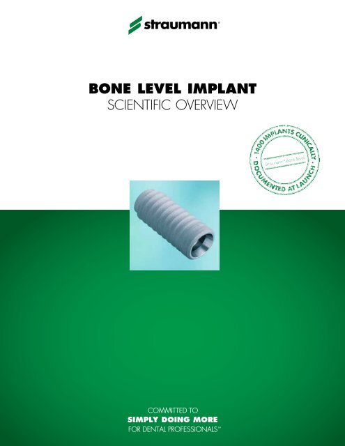
BONE LEVEL IMPLANT SCIENTIFIC OVERVIEW - Straumann
BONE LEVEL IMPLANT SCIENTIFIC OVERVIEW - Straumann
BONE LEVEL IMPLANT SCIENTIFIC OVERVIEW - Straumann

Create successful ePaper yourself
Turn your PDF publications into a flip-book with our unique Google optimized e-Paper software.
<strong>BONE</strong> <strong>LEVEL</strong> <strong>IMPLANT</strong><br />
<strong>SCIENTIFIC</strong> <strong>OVERVIEW</strong>
STUDY <strong>OVERVIEW</strong> ON STRAUMANN ® <strong>BONE</strong> <strong>LEVEL</strong> <strong>IMPLANT</strong><br />
Pre-clinical studies<br />
Topic Objective Study Setup, Principal Investigator<br />
Property tests<br />
Biomechanical stability Ultimate strength and fatigue strength of the <strong>Straumann</strong> ®<br />
Bone Level implant in comparison to competitor implants.<br />
Reliability Screw fit after cyclic mechanical load of the <strong>Straumann</strong> ®<br />
Bone Level implant.<br />
Hydropulser tests, ISO 14801<br />
M. Wieland, H. Hornberger, Switzerland<br />
Hydropulser tests, ISO 14801<br />
M. Wieland, H. Hornberger, Switzerland<br />
Microgap Measurements of gap size. Scanning electron microscopy on polished<br />
micrograph sections<br />
M. de Wild, Switzerland<br />
Bone maintenance<br />
Bone maintenance with submerged and non-submerged<br />
implants placed at different heights.<br />
Delayed restoration, 60 implants<br />
D. Cochran, USA<br />
Animal<br />
Interproximal bone maintenance with adjacently placed<br />
implants.<br />
Immediate abutment placement,<br />
72 implants<br />
D. Tarnow, USA<br />
Clinical studies<br />
Topic Objective Study Setup, Principal Investigator<br />
Basic clinical evidence<br />
Performance and esthetic outcome in single tooth gaps in the<br />
anterior maxilla.<br />
Single center, 20 patients<br />
D. Buser, Switzerland<br />
Esthetics<br />
Submerged vs. non-submerged placement in the anterior<br />
maxilla or mandible.<br />
12 centers in Europe and USA<br />
134 patients, C. Hämmerle, Switzerland<br />
Human<br />
Various treatments Implant success and survival rate in daily dental practice. Over 100 active centers worldwide,<br />
over 1500 implants<br />
Immediacy<br />
Immediate or delayed provisional restoration versus no<br />
provisional restoration in healed alveolar ridges.<br />
Single center, 24 patients<br />
N. Donos, UK<br />
Small diameter implants<br />
(3.3 mm)<br />
Edentulous mandibles restored with a removable prosthesis on<br />
2 small diameter <strong>Straumann</strong> ® Roxolid TM Bone Level implants.<br />
8 international centers, 91 patients<br />
B. Al-Nawas, Germany<br />
Clinical studies are ongoing.<br />
Additional scientific evidence<br />
Topic Objective Link to <strong>Straumann</strong> ® Bone Level Implant<br />
<strong>Straumann</strong> ® SLActive ®<br />
More than 21 published and 12 ongoing scientific studies<br />
substantiate this groundbreaking surface technology.<br />
The <strong>Straumann</strong> ® Bone Level implant is<br />
available with the <strong>Straumann</strong> SLActive<br />
surface.<br />
Thread geometry Field study to demonstrate the performance of the <strong>Straumann</strong> ®<br />
Tapered Effect thread geometry.<br />
The <strong>Straumann</strong> Bone Level implant<br />
features the same thread geometry as the<br />
<strong>Straumann</strong> Tapered Effect implant.
STRAUMANN ® <strong>BONE</strong> <strong>LEVEL</strong> <strong>IMPLANT</strong><br />
Property Tests*<br />
Study<br />
Mechanical testing of ultimate strength<br />
1200<br />
Principal<br />
investigators<br />
Setup<br />
M. Wieland, H. Hornberger, Switzerland<br />
According to fatigue test for endosseous dental<br />
implants (ISO 14801). A total of 19 implants were<br />
measured.<br />
Ultimate strength (N)<br />
1000<br />
800<br />
600<br />
400<br />
200<br />
End-point<br />
Key findings<br />
Static compression tests were performed for<br />
Ø 3.3 mm, Ø 4.1 mm, and Ø 4.8 mm <strong>Straumann</strong> ®<br />
Bone Level implants as well as for different competitor<br />
implants until ultimate implant strength was reached.<br />
<strong>Straumann</strong> ® Bone Level implants reach ultimate<br />
strength of competitor implants with smaller<br />
diameters tested in this study.<br />
0<br />
BL 3.3 Comp. A BL 4.1 Comp. B<br />
4.0<br />
4.3<br />
BL 4.8<br />
Comparison of ultimate strength of the three Bone Level implants<br />
and two competitor implants (n = 3 – 5). Error bars show standard<br />
deviation.<br />
Study<br />
Principal<br />
investigators<br />
Setup<br />
End-point<br />
Mechanical testing of fatigue strength<br />
M. Wieland, H. Hornberger, Switzerland<br />
According to fatigue test for endosseous dental<br />
implants (ISO 14801). A total of 142 implants were<br />
measured.<br />
Load-cycle diagrams were performed for Ø 3.3 mm,<br />
Ø 4.1 mm, and Ø 4.8 mm <strong>Straumann</strong> ® Bone Level<br />
implants and for 13 competitor implants.<br />
Fatigue strength (N)<br />
500<br />
450<br />
400<br />
350<br />
300<br />
250<br />
200<br />
150<br />
100<br />
50<br />
<strong>Straumann</strong> ®<br />
Bone Level implants<br />
BL 3.3<br />
BL 4.1<br />
BL 4.8<br />
Average of measured<br />
competitor implants<br />
0<br />
3.0 3.5 4.0 4.5 5.0 5.5<br />
Shoulder diameter (mm)<br />
Key findings<br />
In this evaluation, average measured fatigue strength<br />
of 3 <strong>Straumann</strong> ® Bone Level implants was higher<br />
than the average failure strength of 13 competitor<br />
implants.<br />
Fatigue strength of <strong>Straumann</strong> ® Bone Level implants (green dots<br />
and regression line) in relation to the fatigue strength of 13 competitor<br />
implants (black dots and regression line), plotted against implant<br />
shoulder diameter.<br />
Study<br />
Microgap measurements on polished micrograph<br />
sections<br />
Principal<br />
investigator<br />
Setup<br />
M. De Wild, Switzerland<br />
Measurement of microgap between implant and<br />
abutment using polished micrograph sections and<br />
scanning electron microscopy.<br />
End-point<br />
Key findings<br />
Microgap measurements were performed.<br />
The mean microgap is below 1 µm.<br />
Representative polished micrograph section<br />
(magnification: left 50×; right 2500×)<br />
* Data on file
The influence on non-matching implant and abutment diameters on radiographic crestal bone in dogs.<br />
RE. Jung, AA. Jones, FL. Higginbottom, TG. Wilson, J. Schoofield, D. Buser, CHF. Hämmerle, DL. Cochran<br />
J Periodontol 2008; 79: 260–270<br />
Introduction<br />
An important factor in crestal bone loss around two-piece implants<br />
appears to be the position of the implant-abutment interface relative to<br />
the bone crest. A second relevant factor seems to be the abutment<br />
diameter relative to the implant diameter. A smaller abutment diameter<br />
may help to reduce bone loss by displacing the implant-abutment<br />
interface, and the inflammatory cell infiltrate, away from the crestal<br />
bone (Figure 1). The aim of this study was therefore to evaluate<br />
radiographic crestal bone changes around <strong>Straumann</strong> Bone Level<br />
implants with smaller abutment diameters placed at different levels<br />
relative to the alveolar crest.<br />
0.4 mm for Ø 4.1 mm implants<br />
Fig. 1: Platform Switching with <strong>Straumann</strong> Bone Level implants.<br />
Materials and Methods<br />
In five dogs, a total of 60 bone level implants with smaller abutment<br />
diameters were placed bilaterally, either submucosally or transmucosally.<br />
In each side of the mandible, the implants were randomly<br />
placed with the implant shoulder either at the level of the alveolar<br />
bone crest or 1.0 mm above or 1.0 mm below (Figure 2). In the<br />
transmucosal group, the healing abutments were different lengths so<br />
that the final occlusal height was the same for each implant. Healing<br />
abutments were placed on the submucosal implants after 4 weeks.<br />
Prostheses (gold crowns) were placed on titanium meso abutments on<br />
all implants 12 weeks after implant placement. Radiographic analysis<br />
was performed at implant placement, crown placement and every<br />
month for up to 6 months.<br />
Submucosal group<br />
Transmucosal group<br />
+1.0 mm -1.0 mm 0.0 mm<br />
+1.0 mm -1.0 mm 0.0 mm<br />
Fig. 2: Submucosal and transmucosal group with bone level implants placed<br />
either at the level of the alveolar bone crest or 1.0 mm above or<br />
1.0 mm below.
Results<br />
Very small changes of the crestal bone level were detected in the<br />
X-rays for both groups (Figures 3 and 4). For implants placed<br />
submucosally, the mean bone loss at 6 months after loading was<br />
+0.17 mm (slight bone gain), -1.32 mm and -0.15 mm for implants<br />
placed above, below or at the crestal bone level, respectively (Figure<br />
5). Corresponding bone loss for transmucosal implants placed above,<br />
below and at the crestal bone level was -0.20 mm, -1.40 mm and<br />
-0.47 mm, respectively (Figure 6). No significant differences in bone<br />
loss or the level of bone-to-implant contact (BIC) were noted between<br />
submucosal or transmucosal implants. Bone loss was greatest at<br />
implants placed below the level of the crestal bone; however, the<br />
distance from the implant shoulder to the first BIC with these implants<br />
was similar to that for implants placed at the level of the crestal bone<br />
(mean 0.19 mm and 0.26 mm for submucosal implants, mean<br />
0.14 mm and 0.44 mm for transmucosal implants).<br />
Fig. 3: Transmucosal implants 6 months after loading.<br />
Conclusions<br />
<strong>Straumann</strong> ® Bone Level implants did not show radiographic<br />
differences in the crestal bone level after 6 months of loading in an<br />
animal model.<br />
There is no significant difference between submucosal and<br />
transmucosal approaches in this canine study.<br />
Fig. 4: Submucosal implants 6 months after<br />
loading. Note the slight bone growth<br />
of the middle implant between crown<br />
placement and 6-month follow-up.<br />
0.17 mm -1.32 mm -0.15 mm<br />
-0.20 mm -1.40 mm -0.47 mm<br />
+1.0 mm -1.0 mm 0.0 mm<br />
Fig. 5: Very little bone loss was detected in the submerged group<br />
6 months after loading.<br />
+1.0 mm -1.0 mm 0.0 mm<br />
Fig. 6: Very little bone loss was detected in the transmucosal group<br />
6 months after loading.<br />
Order the article reprint from <strong>Straumann</strong> USCLR060
Changes in crestal bone and soft tissue at two adjacent <strong>Straumann</strong> Bone Level implants<br />
with different inter-implant distances.<br />
N. Elian, M. Bloom, G. Cardaropoli, B. Ehrlich, M. Dard, D. Tarnow<br />
Introduction<br />
Bone loss around implants has both a lateral as well as a vertical<br />
component, and studies have shown that implants placed too close<br />
together can reduce the height of the inter- implant bone crest. i,ii<br />
Platform-switching may help to reduce the inter-implant bone loss i , so it<br />
was therefore suggested that platform switching in combination with a<br />
bone level implant that shows minimal bone loss may allow the closer<br />
placement of implants without compromising the inter-implant bone<br />
and soft tissue.<br />
1.0<br />
0.0<br />
-1.0<br />
-2.0<br />
3 mm 4 mm<br />
-3.0<br />
Fig. 1: Bone level at buccal, distal, ligual and mesial sides after 8 weeks<br />
Materials and Methods<br />
The mandibular premolars and the first molar from 12 minipigs were<br />
extracted. After 3 months of healing 72 implants were placed using a<br />
template guide. Three bone level implants with SLActive ® surfaces were<br />
placed at bone crest level on one side of the mandible with an interimplant<br />
distance of 3 mm while on the contralateral side the distance<br />
was 4 mm. One stage procedure was used with abutment placement<br />
at time of surgery utilizing a transmucosal abutment healing cap.<br />
Radiographic measurements of the bone levels were done at<br />
placement and after 8 weeks. The following histomorphometric<br />
measurements were performed in a defined region of interest (ROI):<br />
first bone to implant contact (fBIC), length of barrier epithelium (lBE),<br />
length of junctional epithelium (lJE) and length of connective tissue in<br />
contact with the implant surface (lCT).<br />
Results<br />
Of the 72 implants placed, one implant was lost (in the 4 mm group)<br />
after 8 weeks.<br />
Radiographic analysis: A mean bone gain of 0.2 mm ± 0.6 mm<br />
adjacent to the implants was recorded in the 3 mm inter-proximal<br />
distance group while a bone gain of 0.2 mm ± 0.3 mm in the 4 mm<br />
inter-proximal distance group was measured. There were no<br />
significant differences between the groups for the bone level<br />
measurements.<br />
Histological results were referenced in figure 1.<br />
Histomorphometric analysis: The results showed that the mean<br />
overall BIC was not significantly different between the two groups<br />
(86.9 ± 7.3 % and 83.0 ± 10.2 % in the 4 mm and 3 mm<br />
groups, respectively). Similar results were obtained for BIC at the<br />
interproximal sides only. There was also no significant difference in<br />
bone density between the two groups<br />
(63.4 ± 6.1 % and 65.1 ± 5.7 % for the 3 mm and 4 mm groups,<br />
respectively). There were no significant difference in lBE, lJE, and<br />
lCT for the 3 mm and 4 mm groups, respectively.<br />
Bone to implant contact<br />
100 %<br />
75 %<br />
50 %<br />
25 %<br />
0 %<br />
4 mm group 3 mm group<br />
Fig. 2: Mean BIC in the region of interest<br />
Conclusions<br />
Mean bone gain of 0.2 mm was observed radiographically<br />
adjacent to implants in both the 3 mm and 4 mm groups<br />
Bone was maintained in the inter-proximal areas in both groups<br />
Soft tissue remained stable over 8 weeks in both groups<br />
There were no significant differences between 3 mm and 4 mm<br />
inter-implant distances for any of the parameters<br />
Results from this study suggests that <strong>Straumann</strong> Bone Level<br />
implants can be placed as close as 3 mm apart without<br />
compromising the inter-implant bone and soft tissue<br />
i<br />
Tarnow DP, Cho SC, Wallace SS. The effect of inter-implant distance on the height<br />
of the inter-implant bone crest. J Periodontol 2008; 71: 546 – 549.<br />
ii<br />
Kuperschmidt I, Levin L, Schwartz-Arad D. Inter-implant bone height changes in<br />
anterior maxillary immediate and non-immediate adjacent dental implants. J Periodontol<br />
2007; 78: 991 – 996.
Early implant placement with simultaneous guided bone regeneration following single-tooth extraction<br />
in the esthetic zone: 12-month results of a prospective study with 20 consecutive patients.<br />
D. Buser, S. Halbritter, C. Hart, MM. Bornstein, L. Grütter, V. Chappuis, UC. Belser<br />
J Periodontol 2009;80:152-162<br />
Introduction<br />
Early implant placement following extraction of a single tooth is a procedure<br />
used by many clinicians in the maxillary anterior zone, 1 but there is a lack<br />
of documentation on esthetic outcomes. Four studies for single-tooth implant<br />
placement reported mucosal recessions in post-extraction sites. The aim of<br />
this study, therefore, was to prospectively assess esthetic outcomes of early<br />
implant placement in single-tooth gaps in the esthetic zone with <strong>Straumann</strong> ®<br />
Bone Level implants.<br />
Materials and Methods<br />
A total of 20 patients requiring single-tooth replacement in the<br />
anterior maxilla were entered into the study. Exclusion criteria were systemic<br />
diseases that could alter the tissue integration of dental implants, pregnancy,<br />
or smoking > 10 cigarettes per day. After tooth extraction the socket was<br />
allowed to heal for 4 – 8 weeks. Bone level implants were subsequently<br />
placed, sealed with healing caps, with simultaneous contour augmentation<br />
using GBR with deproteinized bovine bone mineral and a collagen<br />
membrane. Reopening was performed 8 – 12 weeks later (Day 0). Within<br />
7 days, provisional crowns were placed, which were gradually enlarged if<br />
necessary to optimize soft tissue contours. Final all-ceramic restorations were<br />
placed after 6 months.<br />
Table 1: Clinical and radiographic parameters at<br />
3, 6, and 12 months<br />
3 mos 6 mos 12 mos<br />
mPLI 0.08 ± 0.24 0.08 ± 0.20 0.36 ±<br />
0.33*<br />
mSBI 0.26 ± 0.29 0.16 ± 0.23 0.21 ± 0.17<br />
PD 3.69 ± 0.62 3.75 ± 0.46 4.43 ±<br />
0.57*<br />
KM 4.06 ± 1.43 4.10 ± 1.41 4.50 ± 1.54<br />
DIB 0.09 ± 0.16 0.14 ± 0.25 0.18 ±<br />
0.20*<br />
*Values are statistically significant compared to values at previous measurements<br />
Mean pink and white esthetic scores were 8.10 and 8.65,<br />
respectively (total score = 16.75), indicating favorable<br />
esthetic outcomes. The maximum for both pink and white<br />
esthetic scores is 10, and the threshold for clinical<br />
acceptability is 6/10 for each index.<br />
The clinical parameters measured were modified plaque index (mPLI),<br />
modified sulcus bleeding index (mSBI), probing depth (PD), width of<br />
keratinized mucosa (KM), distance from mucosal margin to implant shoulder<br />
(DIM), distance from implant shoulder to first bone-to-implant contact (DIB),<br />
and mid-facial height of implant crown and contralateral tooth. Pink esthetic<br />
score and white esthetic scores were evaluated as primary endpoint.<br />
Results<br />
All implants were successfully integrated at 12 months, with no sign of<br />
implant mobility. The peri-implant soft tissues revealed little tendency to bleed<br />
following probing and were clinically healthy. The implants fulfilled<br />
successful tissue integration, and the results were in line with those from<br />
other prospective studies with the same parameters. 3, 4, 5, 6 Mean mPLI,<br />
mSBI, and PD values at 12 months were 0.36, 0.21, and 4.43 mm,<br />
respectively (Table 1). A wide KM band was seen at 3 months, which<br />
remained stable at 6 and 12 months (Table 1).<br />
Mean DIB values at 3, 6, and 12 months were 0.09, 0.14, and 0.18,<br />
respectively (Table 1). The radiographic analysis indicated that 15 of 20<br />
implants showed minimal bone resorption (Figure 1), and only one implant<br />
showed bone loss > 0.5 mm, with minor mucosal recession of 0.5 – 1.0<br />
mm. Mean DIM values at 12 months were -6.68, -6.00, -3.53, and -3.84<br />
for mesial, distal, facial, and oral, respectively.<br />
Conclusions<br />
Minimal crestal bone resorption was demonstrated<br />
Good esthetic and clinical results were seen over 12<br />
months<br />
The risk of mucosal recession was low<br />
Strict success criteria were fulfilled, resulting in 100 %<br />
success and survival rates at 12 months<br />
The 20 implants from this study showed an average bone<br />
loss of 0.18 mm between the day of loading and 12<br />
months of follow-up<br />
20 %<br />
5 %<br />
75 %<br />
Bone loss<br />
0.0–0.25 mm<br />
0.26–0.5 mm<br />
0.51–1.0 mm<br />
Fig. 1: The majority of patients (75 %) showed < 0.25 mm bone loss<br />
Predictable contour augmentation with an anorganic bovine bone mineral<br />
therefore showed a reduced risk of mucosal recession, compared to other<br />
7, 8, 9<br />
studies that have shown mucosal recession of 30 – 40 %.<br />
Order the article reprint from <strong>Straumann</strong> USCLR061
Randomized, controlled clinical study comparing submerged versus transmucosal placement of bone level implants<br />
in the anterior maxilla or mandible by evaluation of the change in bone level between first stage surgery and 12<br />
months post-surgery<br />
CHF Hämmerle, M. Sanz, St. Chen, W. Martin, J. Jackowski, L. Cordaro, CJ Ivanoff, J. Ganeles, R. Jung<br />
Introduction<br />
The surgical procedure and implant design both influence esthetic<br />
outcomes. For example, a submerged technique may be preferred to<br />
establish esthetics and function in anterior sites, and implants where<br />
the metallic shoulder is reduced may help to improve the esthetics of<br />
the restorations. The marginal bone change over time is another important<br />
factor, 1,2 with a historical success criterion being bone loss of<br />
no more than 0.5 mm in the first year and < 0.2 mm annually<br />
thereafter.<br />
This investigation was designed to evaluate the amount of bone level<br />
change with submerged and transmucosal healing, and to assess any<br />
difference in bone level change between the two procedures with<br />
<strong>Straumann</strong> ® Bone Level SLActive ® implants.<br />
Materials and Methods<br />
Implants to replace single teeth in the anterior region (maxilla or mandible)<br />
were placed in a total of 146 patients in 12 centers in seven<br />
countries. A temporary crown was placed between 8 and 14 weeks,<br />
and the final reconstruction was placed after 26 weeks. The primary<br />
parameter was evaluation of change in bone level, measured by<br />
standardized radiographs taken at the surgery (baseline), provisional<br />
placement (approx. 14 weeks), final crown placement (6 months) and<br />
12 months, with annual follow-up intended for up to 5 years. Secondary<br />
parameters included soft tissue recession, implant survival and<br />
success and prosthesis success.<br />
Results<br />
The Intent-to-Treat (ITT) population for the 1-year results included 127<br />
patients (60 and 67 in the transmucosal and submerged groups,<br />
respectively, with a mean age of 45.5 and 47.3 years, respectively).<br />
Bone loss based on 12 month ITT population<br />
Mean bone loss (mm)<br />
0.5<br />
0.0<br />
-0.5<br />
-1.0<br />
-1.5<br />
-2.0<br />
Fig. 1: Mean bone level change from baseline at 6 and 12 months<br />
60<br />
50<br />
40<br />
30<br />
20<br />
10<br />
0<br />
0<br />
Baseline<br />
Implant<br />
placement<br />
- 0.3<br />
6 12<br />
Final<br />
prosthesis<br />
Bone level change in % and mm categories<br />
Baseline to 12 months<br />
0.8 2.4 0.8 2.4<br />
> - 3.0 mm<br />
> - 3.0 to - 2.5 mm<br />
> - 2.5 to - 2.0 mm<br />
> - 2.0 to - 1.5 mm<br />
month<br />
8<br />
> - 1.5 to - 1.0 mm<br />
20.8<br />
> - 1.0 to - 0.5 mm<br />
53.6<br />
- 0.47<br />
10.4<br />
Fig. 2: Percentage of implants showing different categories of bone level change<br />
> - 0.5 to 0.0 mm<br />
> - 0.0 to + 0.5 mm<br />
mean bone loss (mm)<br />
0.8 0 0<br />
> + 0.5 to 1.0 mm<br />
> + 1,0 to 1.5 mm<br />
> + 1.5 to 2.0 mm<br />
Based on the 12 month ITT population data, the mean change in<br />
bone level after 6 months, was -0.30 ± 0.47 mm (-0.32 ± 0.47 mm<br />
and -0.29 ± 0.35 mm for the submerged and transmucosal groups,<br />
respectively), while after 12 months the mean change in bone level<br />
was -0.47 ± 0.64 mm (-0.47 ± 0.64 mm and -0.48 ± 0.65 mm for<br />
the submerged and transmucosal groups, respectively) (Fig. 1). There<br />
was therefore no significant difference in bone level change between<br />
the two groups. Almost two-thirds of implants (64.8 %) showed less<br />
than 0.5 mm bone loss over 12 months (Fig. 2). The implant survival<br />
and success rate was 99.2 %.
Patient satisfaction with the final prosthesis was extremely high; 99 %<br />
of patients reported their level of satisfaction as excellent or good<br />
(Fig. 3).<br />
General patient satisfaction with final prosthesis<br />
ITT population at 1-year visit<br />
good 22 %<br />
fair 1 % poor 0 %<br />
Conclusions<br />
Marginal bone level change was small and not significantly<br />
different between submerged and transmucosal treatment groups<br />
The marginal bone level change from implant placement (baseline)<br />
to 12 months was -0.47 mm (mean). The marginal bone level<br />
change from implant loading (6 months) to 12 months was<br />
-0.17 mm (mean)<br />
Extremely high survival and success rates were observed (99.2 %<br />
for both)<br />
Patient satisfaction with the outcome was extremely high (99 %)<br />
excellent 77 %<br />
Fig. 3: General patient satisfaction with final prosthesis at 12 months<br />
Discussion<br />
Traditional implant success criteria include an acceptable bone loss of<br />
≤ 0.5 mm in the first year and < 0.2 mm annually thereafter. 3<br />
Recently, however, there have been suggestions for these criteria to be<br />
revised, indicating that a more acceptable bone loss for modern<br />
implant systems would be 0.3 mm over 5 years. However, many of<br />
the studies on which this suggestion is based use placement of the<br />
temporary or final prosthesis rather than placement of the implant as<br />
the baseline measurement for bone level change. 4<br />
Studies that use implant placement as the baseline measurement for<br />
bone level change have shown relevant bone loss before loading; 4, 5<br />
therefore, using prosthesis placement as the baseline may give an<br />
inaccurate reflection of the real amount of bone loss. 6, 7 A more<br />
accurate picture can be obtained by measuring bone levels at implant<br />
placement and at regular intervals thereafter (Fig 1). Knowledge of the<br />
amount of bone level change to expect has a huge clinical relevance<br />
in treatment planning to achieve an optimum esthetic outcome; for<br />
example, unexpected bone loss can cause substantial soft tissue<br />
recession, resulting in an esthetic failure.<br />
1<br />
Hermann JS, Buser D, Schenk RK, Schoolfield JD, Cochran DL. Biologic<br />
width around one- and two-piece titanium implants. Clin Oral Implants Res<br />
2001;12:559 – 571.<br />
2<br />
Hermann JS, Schoolfield JD, Schenk RK, Buser D, Cochran DL. Influence of<br />
the size of the microgap on crestal bone changes around titanium implants.<br />
A histometric evaluation of unloaded non-submerged implants in the canine<br />
mandible. J Periodontol 2001;72:1372 – 1383.<br />
3<br />
Albrektsson T, Zarb G, Worthington P, Eriksson AR. The long-term efficacy of<br />
currently used dental implants: a review and proposed criteria of success. Int<br />
J Oral Maxillofac Implants 1986;1:11 – 25.<br />
4<br />
Engquist B, Åstrand P, Dahlgren S, Engquist E, Feldmann H, Gröndahl K.<br />
marginal bone reaction to oral implants: a prospective comparative study<br />
of Astra Tech and Brånemark System implants. Clin Oral Implants Res<br />
2002;13:30 – 37.<br />
5<br />
Åstrand P, Engquist B, Dahlgren S, Gröndahl K, Engquist E, Feldmann H.<br />
Astra Tech and Brånemark system implants: a 5-year prospective study of<br />
marginal bone reactions. Clin Oral Implants Res 2004;15:413 – 420.<br />
6<br />
Cooper L, Felton DA, Kugelberg CF, Ellner S, Chaffee N, Molina AL,<br />
Moriarty JD, Paquette D, Palmqvist U. A multicenter 12-month evaluation<br />
of single-tooth implants restored 3 weeks after 1-stage surgery. Int J Oral<br />
Maxillofac Implants 2001;16:182 – 192.<br />
7<br />
De Bruyn H, Van de Velde T, Collaert B. Immediate functional loading of<br />
TiOblast dental implants in full-arch edentulous mandibles: 1 3-year prospective<br />
study. Clin Oral Implants Res 2008;19:717 – 723.
A non-interventional study to document the implant success and survival rate of <strong>Straumann</strong> ® Bone Level implants<br />
in daily dental practice.<br />
Interim report<br />
P. Bullon, A. Filippi, J. Kälber, M. Roccuzzo, D. Weingart, F. Higginbottom<br />
Introduction<br />
Results from clinical studies with dental implants can be affected by<br />
a number of factors, including the choice of center and the patient<br />
inclusion/exclusion criteria. It may therefore be difficult to extrapolate<br />
the high success and survival rates achieved in formal clinical trials,<br />
with a specifically defined group of patients, to an everyday<br />
clinical practice setting.<br />
The aim of this study therefore was to document the success and<br />
survival rate of <strong>Straumann</strong> ® Bone Level implants in daily practice for<br />
up to 3 years.<br />
Materials and methods<br />
Patients were enrolled at 123 centers in nine countries. There were no<br />
specific inclusion or exclusion criteria for patients enrolled in the study.<br />
In addition, no specific loading protocol was defined, and implants<br />
were allowed to be placed at the discretion of the clinician in<br />
accordance with the product labeling. The study was initiated in<br />
November 2006.<br />
The primary objective of the study is implant survival rate 1 year after<br />
abutment placement. Secondary objectives include implant success<br />
and survival rate over 3 years, change in crestal bone level, loading<br />
protocol, type of restoration, and performance of prosthetic<br />
components.<br />
Interim results<br />
As of July 2008, 843 patients had been treated with 1501 implants.<br />
Dental risk factors (e.g., bone defects, bruxism, periodontitis,<br />
insufficient oral hygiene) were noted in 38.3 % of patients, while<br />
systemic risk factors (e.g., controlled diabetes, metabolic disease,<br />
osteoporosis) were noted in 13.8 %. Most patients received one or<br />
two implants; the mean number of implants per patient was 1.8.<br />
Almost half (47 %) of the implants were placed in the esthetic zone<br />
(Figs. 1 and 2).<br />
No. of implants<br />
No. of implants<br />
140<br />
120<br />
100<br />
80<br />
60<br />
40<br />
20<br />
0<br />
Fig. 1: Implant distribution in the maxilla<br />
140<br />
120<br />
100<br />
80<br />
60<br />
40<br />
20<br />
0<br />
18 17<br />
48 47<br />
16 15 14 13 12 11 21 22 23 24 25 26 27 28<br />
Fig. 2: Implant distribution in the mandible<br />
Tooth position (FDI)<br />
46 45 44 43 42 41 31 32 33 34 35 36 37 38<br />
Tooth position (FDI)<br />
Most implants (56.4 %) were placed using the submerged technique;<br />
34.7 % were placed transmucosally, and 8.5 % were placed semisubmerged.<br />
Augmentations were performed at just over half (55 %)<br />
of the implant sites. Conventional loading was used in the majority<br />
(61.6 %) of cases. Of the 578 final restorations documented, 348<br />
(60.2 %) were single crowns, 165 (28.6 %) were bridges, 44 (7.6<br />
%) were full prostheses, and 11 (1.9 %) were partial prostheses. As of<br />
July 2008, only 12 implants have been reported as failures.<br />
Interim conclusions<br />
The excellent success of the <strong>Straumann</strong> ® Bone Level implant in<br />
daily practice can be demonstrated<br />
The results are comparable to those achieved in controlled<br />
clinical studies<br />
The <strong>Straumann</strong> ® Bone Level implant is successful in a variety<br />
of treatment options
International Headquarters<br />
Institut <strong>Straumann</strong> AG<br />
Peter Merian-Weg 12<br />
CH-4002 Basel, Switzerland<br />
Phone +41 (0)61 965 11 11<br />
Fax +41 (0)61 965 11 01<br />
www.straumann.com<br />
<strong>Straumann</strong> USA<br />
<strong>Straumann</strong> USA, LLC<br />
60 Minuteman Road<br />
Andover, MA 01810<br />
Phone 800/448 8168<br />
978/747 2500<br />
Fax 978/747 2490<br />
www.straumannusa.com<br />
<strong>Straumann</strong> Canada<br />
<strong>Straumann</strong> Canada Limited<br />
3115 Harvester Road, 1st Floor<br />
Burlington, ON L7N 3N8<br />
Phone 800/363 4024<br />
905/319 2900<br />
Fax 905/319 2911<br />
www.straumann.ca<br />
12/09 USLIT 302 <strong>Straumann</strong> ® is a registered trademarks of <strong>Straumann</strong> Holding AG or its affiliates. All rights reserved. © <strong>Straumann</strong> USA, LLC 2009.