Stana 1 - Studia Universitatis Vasile Goldis, Seria Stiintele Vietii
Stana 1 - Studia Universitatis Vasile Goldis, Seria Stiintele Vietii
Stana 1 - Studia Universitatis Vasile Goldis, Seria Stiintele Vietii

Create successful ePaper yourself
Turn your PDF publications into a flip-book with our unique Google optimized e-Paper software.
<strong>Studia</strong> <strong>Universitatis</strong> “<strong>Vasile</strong> Goldiş”, <strong>Seria</strong> Ştiinţele Vieţii<br />
Vol. 22, issue 3, 2012, pp. 351-358<br />
© 2012 <strong>Vasile</strong> <strong>Goldis</strong> University Press (www.studiauniversitatis.ro)<br />
TRACHEOSTOMIZED PACIENT – NURSING AND CASE REPORT<br />
Teodora OLARIU, Iustin OLARIU, Monica SOLOMON<br />
”<strong>Vasile</strong> <strong>Goldis</strong>” West University of Arad, România<br />
ABSTRACT. Tracheostomy is an intervention that is practiced in the case where there is a need of the patient's<br />
ventilation for a period longer than 7 days. Tracheotomy is a surgical intervention by incision was made with the<br />
purpose of trachea penetration air in the shaft asthma, with the later date of trachea on the skin. It was an<br />
intervention Tracheostomy which can be achieved by conventional surgical processes, which require<br />
transporting the patient in the operating room, or by the procedure percutaneous, process which may be carried<br />
out at cog. With proper care patient undergoing tracheostomy has the right objective prevention of infections and<br />
the earliest opportunity you feel the place of incision, sucking up but not be limited, carrying out daily hygiene<br />
cannula, performing ventilation, specific methods of nutrition.<br />
Keywords: tracheostomy, larinx, trachea, neck surgery<br />
Theoretical considerations<br />
A long time tracheostomy was widely criticized<br />
because it is considered that the wound resulting from<br />
tracheal incision is incurable due to existence of cartilage<br />
edges.<br />
Tracheostomy was resumed in 1952 in Denmark,<br />
after the polio epidemic.<br />
In order to describe the intervention we present<br />
anatomically and topographically the larynx and trachea.<br />
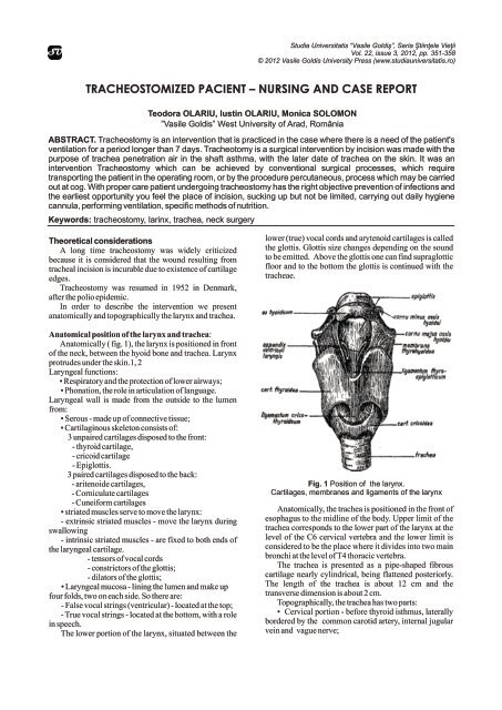
Anatomical position of the larynx and trachea:<br />
Anatomically ( fig. 1), the larynx is positioned in front<br />
of the neck, between the hyoid bone and trachea. Larynx<br />
protrudes under the skin.1, 2<br />
Laryngeal functions:<br />
• Respiratory and the protection of lower airways;<br />
• Phonation, the role in articulation of language.<br />
Laryngeal wall is made from the outside to the lumen<br />
from:<br />
• Serous - made up of connective tissue;<br />
• Cartilaginous skeleton consists of:<br />
3 unpaired cartilages disposed to the front:<br />
- thyroid cartilage,<br />
- cricoid cartilage<br />
- Epiglottis.<br />
3 paired cartilages disposed to the back:<br />
- aritenoide cartilages,<br />
- Corniculate cartilages<br />
- Cuneiform cartilages<br />
• striated muscles serve to move the larynx:<br />
- extrinsic striated muscles - move the larynx during<br />
swallowing<br />
- intrinsic striated muscles - are fixed to both ends of<br />
the laryngeal cartilage.<br />
- tensors of vocal cords<br />
- constrictors of the glottis;<br />
- dilators of the glottis;<br />
• Laryngeal mucosa - lining the lumen and make up<br />
four folds, two on each side. So there are:<br />
- False vocal strings (ventricular) - located at the top;<br />
- True vocal strings - located at the bottom, with a role<br />
in speech.<br />
The lower portion of the larynx, situated between the<br />
lower (true) vocal cords and arytenoid cartilages is called<br />
the glottis. Glottis size changes depending on the sound<br />
to be emitted. Above the glottis one can find supraglottic<br />
floor and to the bottom the glottis is continued with the<br />
tracheae.<br />
Fig. 1 Position of the larynx.<br />
Cartilages, membranes and ligaments of the larynx<br />
Anatomically, the trachea is positioned in the front of<br />
esophagus to the midline of the body. Upper limit of the<br />
trachea corresponds to the lower part of the larynx at the<br />
level of the C6 cervical vertebra and the lower limit is<br />
considered to be the place where it divides into two main<br />
bronchi at the level of T4 thoracic vertebra.<br />
The trachea is presented as a pipe-shaped fibrous<br />
cartilage nearly cylindrical, being flattened posteriorly.<br />
The length of the trachea is about 12 cm and the<br />
transverse dimension is about 2 cm.<br />
Topographically, the trachea has two parts:<br />
• Cervical portion - before thyroid isthmus, laterally<br />
bordered by the common carotid artery, internal jugular<br />
vein and vague nerve;
Olariu T. et al.<br />
• Thoracic portion – behind the sternum and laterally dilated esophagus during swallowing. Musculo-elastic<br />
bordered by the mediastinal side face of the lungs.<br />
fibrous membrane shows a tracheal smooth muscle called<br />
From structural point of view, the trachea is made the transverse muscles, whose contraction or relaxation<br />
from the outside inwards as follows: changes the size of the tracheal orifice. The 16 to 20<br />
• Adventitia - a tunic of conjunctive nature;<br />
cartilaginous rings are linked together by collagen and<br />
• fibro cartilaginous layer - containing 16-20 elastic fibers called the inter-ring filaments.<br />
cartilaginous overlapping, incomplete rings, opening<br />
• The mucosa - made up of cylindrical pseudo<br />
towards the esophagus. Posterior to the esophagus, the stratified ciliated epithelium, lines the lumen of the<br />
end of the rings are joined by a fibro-elastic membrane trachea.<br />
that allows the passage of alimentary bolus through the<br />
352 <strong>Studia</strong> <strong>Universitatis</strong> “<strong>Vasile</strong> Goldiş”, <strong>Seria</strong> Ştiinţele Vieţii<br />
Vol. 22, issue 3, 2012, pp. 351-358<br />
© 2012 <strong>Vasile</strong> <strong>Goldis</strong> University Press (www.studiauniversitatis.ro)
What is tracheostomy?<br />
Tracheotomy is a surgical procedure which consists<br />
of the incision of the trachea, in the cervical portion, in<br />
order to ensure the patient's breathing.<br />
Tracheostomy is a surgical incision of the trachea<br />
which allows the air to penetrate into the bronchial tree,<br />
with subsequent fixation of the trachea to the skin.<br />
The term tracheostomy originates in Greek language :<br />
"tracheia artery" = rough artery, "stoma" = opening or<br />
hole.<br />
Tracheostomy can be achieved by conventional<br />
surgical procedures or by percutaneous procedure.<br />
Percutaneous tracheostomy can be performed at bedside.<br />
Surgical tracheotomy - requires bringing the patient<br />
into the operating room. Ensure all sterile field antiseptic<br />
and aseptic methods. The patient is placed supine with the<br />
cervical region in extension, position achieved by<br />
applying a roll under the shoulders.<br />
It is considered that vertical line marks along the<br />
midline are: thyroid cartilage, cricoid cartilage and<br />
sternal fork. the tracheal rings are palpated . under the<br />
cricoid cartilage. The incision is performed 2 cm below<br />
the cricoid cartilage in the cervical region. A puncture is<br />
made in the trachea below the ring, the guiding rod is<br />
introduced for performing progressive dilation of<br />
tracheal opening in order to introduce the tracheal<br />
cannula. The ventilator is connected to the cannula and<br />
the guiding rod is withdrawn. Anesthesia is maintained<br />
through the cannula.<br />
Since this is a very complex method and requires<br />
deep anesthesia, it’s not used in emergency situations.<br />
When the tracheostomy is performed?<br />
Recommendations for tracheostomy refers to the<br />
cases where mechanical ventilation is required for a<br />
period exceeding seven days, avoiding the stenosis of the<br />
trachea, tracheomalacia stenosis or formation of esotracheal<br />
fistula.3, 4 Incidents and accidents 4, 5,6,7,8:<br />
• Bleeding from vasculo-nervous pack or from<br />
In what situations is indicated tracheostomy:<br />
thyroidian istmus;<br />
1. Airway obstruction by: • Posterior puncture of the trachea and entry into the<br />
• Foreign bodies;<br />
oesophagus.<br />
• Inflammatory diseases,<br />
• Tearing of the a trachea;<br />
•edema due to an infection, burns, trauma, • False entries with the imposibility of the<br />
anaphylactic shock;<br />
introduction of the cannula.i;<br />
• glottis or supraglottic pathology that cause upper • Subcutaneous emphysema.<br />
airway obstruction;<br />
• any other pathology that may cause upper airway Percutaneous tracheostomy:<br />
obstruction .<br />
• could be made at bedside, not requiring the transport<br />
2. If mechanical ventilation is needed for a long time: of the patient to the operating room;<br />
• Severe obstructive lung disease;<br />
• Bleeding is minimal;<br />
• Severe brain disorders;<br />
• Low risk of infection;<br />
• MODS<br />
• Tracheal stenosis rate lower;<br />
• ARDS<br />
• posttraheostomy faint scar.<br />
• Other lung disease requiring mechanical ventilation<br />
for a long period.<br />
Percutaneous tracheostomy kits (PORTEX company)<br />
3. Draining lung secretions, tracheo-bronchial toilet. includes:<br />
4. Head and neck surgery, if necessary, • Scalpel;<br />
5. Uncontrolled sleep apnea • 10 cc syringe<br />
6. Prolonged coma. • 14-G, needle with flexula;<br />
•Cannulas for tracheostomy with special<br />
In what situations tracheostomy is not performed 4: mandrenwithr two fixation bands ;<br />
• Children younger than 8 years;<br />
• Guiding string with input device.<br />
• Patients with major problems of neck anatomy;<br />
• Goitre ;<br />
The technique<br />
• High positioning of brachio-cephalic trunk; • identification, by palpation of the thyroid and<br />
• Tumors.<br />
cricoid cartilage, and the first three rings of the trachea;<br />
• The incision is preferable to be made between the<br />
Difficulties in performing tracheostomy may occur in: first and second or second and third tracheal ring;<br />
• Patients with severe thrombocytopenia,<br />
• The needle is introduced between the tracheal rings,<br />
• Bleeding Time longer than 10 minutes;<br />
until penetration into the trachea, then remove the needle<br />
• Infection at the site of choice.<br />
and let the flexula in place;<br />
Tracheotomy Techniques, 5,9,10:<br />
• The guide is inserted through the flexula , and its<br />
• Surgical techniques:<br />
direction is checked by video laryngoscopy, to clearly<br />
1. Greggs process - with forceps dilators, identify its direction;<br />
2. Ciaglia method - with progressive dilatation. • The flexula is extracted and the guide remains in<br />
• percutaneous tracheostomy<br />
place;<br />
<strong>Studia</strong> <strong>Universitatis</strong> “<strong>Vasile</strong> Goldiş”, <strong>Seria</strong> Ştiinţele Vieţii<br />
Vol. 22, issue 3, 2012, pp. 351-358<br />
© 2012 <strong>Vasile</strong> <strong>Goldis</strong> University Press (www.studiauniversitatis.ro)<br />
Tracheostomized pacient – nursing and case report<br />
353
Olariu T. et al.<br />
• The dilation device is inserted through rotation length of the cannula. The aspiration of the secretions<br />
maneuvers and the orifice is expanded.<br />
can be done with the fibroscope.<br />
• The dilation device is extracted and a special forceps • Daily cleaning of the cannula;<br />
is introduced through the guide. The tracheal and the • Making the ventilation;<br />
existing orifice in the soft tissues are extended with the • Specific nutritional methods.<br />
forceps that opens in horizontally and vertically panes.<br />
• Dilation of the orifice must fit the size of the cannula. Precautions:<br />
When the required size is reached the forceps is • The pressure in the balloon is check by the nurse and<br />
withdrawn and the cannula is introduced through the physician for the cannulae with balloon.<br />
guide. After the cannula is positioned the guide and the • 100% oxygen will be administered for pre<br />
mandren of the cannula are withdrawn.<br />
oxygenation and post oxygenation of patient for three<br />
• The balloon of the cannula is inflated. The ventilator minutes.<br />
circuit is connected to the cannula, and the patient’s • Aspiration of secretions time will be 10 seconds.<br />
ventilation is checked by auscultation. Cannula is fixed Time between successive maneuvers will be 30 seconds,<br />
with two existing bands in the kit.<br />
the patient need to be calm and breathe normally.<br />
• The orotracheal intubation probe is withdrawn only • Use saline if secretions are very thick, viscous, and<br />
when the tracheostomy cannula is positioned correctly. coughing is ineffective (max. 1ml/instillation).<br />
• The balloon will be deflated periodically to prevent<br />
Accidents and incidents<br />
accumulation of secretions from the top of it;<br />
• Accidental decannulation<br />
• Pressure in the balloon should be checked with a<br />
• The obstruction of the cannula<br />
manometer at 2-4 hours intervals (VN 15-20 mm Hg<br />
• Local or respiratory infections<br />
column).<br />
• Hipoxia if the procedure is taking too long. • Humidification and heating of the respiratory gases<br />
• Air penetration into the mediastin, pneumothorax, will be done through a nebulizer or aerosol device for<br />
false entry, subcutaneous emphysema due to the patient breathing spontaneously or through the<br />
positioning of the canula into the para-tracheal space.<br />
humidification device of the ventilator for the patients<br />
• bleeding from vascular-nervous package or thyroid mechanically ventilated.<br />
isthmus;<br />
• Perforation of the trachea and entering back into the Case Report<br />
esophagus, trachea<br />
Patient B.I., 51 years old living in Arad comes to the<br />
• tearing of the posterior wall of the trachea and County Hospital Arad ENT emergency room on<br />
trachea-esophageal fistula;<br />
12.02.2012 presenting disphagia laryngeal discomfort,<br />
• Creation of false paths unable introduction cannula; marked dysphonia, pronounced dyspnea on effort.<br />
• Stenosis of the trachea.<br />
The patient reported that the first signs of disease<br />
• Secondary bleeding due to infections or vascular appeared three months ago. The onset was with pain<br />
erosions.<br />
when swallowing, first for solid foods and then liquid<br />
• Aparition of tracheal granulomma which can lead to foods. Then the patient says that hoarseness ensued and<br />
the impossibility of closing the tracheostomy.<br />
later fatigue, initially with great efforts then with efforts<br />
increasingly smaller.<br />
Nursing of the patient with tracheostomy<br />
Patient is to be hospitalized for emergency respiratory<br />
Objectives:<br />
failure , and the ENT doctor's diagnosis is infiltrating<br />
• Preventing infection at the incision site - by cleaning tumor of the hemilaringe and the patient is being<br />
the stoma and changing the dressing every time is prepared for surgery: tracheostomy.<br />
required. The inner cannulae will be changed daily. From the discussion with Mr. BI we find that he is<br />
Reusable cannulas will be washed and cleaned, the living together with his family ,he is an engineer at a<br />
maneuvers not exceeding 15 minutes;<br />
private company, and he is a smoker for over 20 years,<br />
• Prevention of lesions of the skin incision area;<br />
drink two coffees a day and he drinks a lot of soft drinks.<br />
• Vacuuming secretions will be performed:<br />
The patient doesn’t acknowledge being allergic to any<br />
- When breathing becomes noisy known medication and agrees with the surgery.<br />
- When mucus appears at the end of the tracheal<br />
cannula,<br />
Preoperative Medication: Diazepam and Phenobarbital.<br />
- When there signs of respiratory failure are Half an hour before surgery: Mialgin and Atropine.<br />
present (dropping of SpO2; tachypneea, changes in pulse Dosage and mode of administration have been indicated<br />
and blood pressure, cyanosis, restlessnessor agitation. by the anesthetist.<br />
- The suction of the secretions will be done Vital functions at admission:<br />
gently and not exceeding the length of the cannula Pulse = 80 beats / min<br />
carrying not to harm the tracheal wall.); BP = 130/70 mm Hg<br />
- It can be practiced superficial aspirations in the Respiratory frequency = 15 breaths / min<br />
hole cannula, or deep aspirations, that will exceed the Temperature = 36.8 C<br />
354 <strong>Studia</strong> <strong>Universitatis</strong> “<strong>Vasile</strong> Goldiş”, <strong>Seria</strong> Ştiinţele Vieţii<br />
Vol. 22, issue 3, 2012, pp. 351-358<br />
© 2012 <strong>Vasile</strong> <strong>Goldis</strong> University Press (www.studiauniversitatis.ro)
Tracheostomized pacient – nursing and case report<br />
G = 62 kg<br />
laboratory findings<br />
Biopsy exam on 14/02/2012.<br />
fashion.<br />
On 21.02.2012 the result is: Fragment of a laryngeal • Tertiary - seeks recovery – the role of the assistance<br />
tumor. Spinocellular cell carcinoma.<br />
is to provide individual care until gaining personal<br />
The patient care plan is drawn up under the 14 basic independence.<br />
needs of the concept of Virginia Henderson.<br />
The difficulty is in the addiction causes and<br />
Dependency manifestations of the patent derive from represents an important obstacle to achieve satisfying the<br />
lack of strength, will and knowledge of the difficulty of basic needs.<br />
his own situation.<br />
Sources of difficulty include:<br />
The levels of care for the patient with tracheostomy • physical, psychological, social, spiritual factors ;<br />
are :<br />
• Factors linked to insufficient knowledge of the<br />
• Primary - maintaining and promoting health.<br />
disease.<br />
• Secondary - seeks curative interventions and it’s Plan for tracheostomized patient care is presented in<br />
purpose is to find the ensuing problems in a timely Table 1.<br />
<strong>Studia</strong> <strong>Universitatis</strong> “<strong>Vasile</strong> Goldiş”, <strong>Seria</strong> Ştiinţele Vieţii<br />
Vol. 22, issue 3, 2012, pp. 351-358<br />
© 2012 <strong>Vasile</strong> <strong>Goldis</strong> University Press (www.studiauniversitatis.ro)<br />
355
Olariu T. et al.<br />
Tracheostomized patient care plan<br />
Table 1<br />
356 <strong>Studia</strong> <strong>Universitatis</strong> “<strong>Vasile</strong> Goldiş”, <strong>Seria</strong> Ştiinţele Vieţii<br />
Vol. 22, issue 3, 2012, pp. 351-358<br />
© 2012 <strong>Vasile</strong> <strong>Goldis</strong> University Press (www.studiauniversitatis.ro)
Tracheostomized pacient – nursing and case report<br />
<strong>Studia</strong> <strong>Universitatis</strong> “<strong>Vasile</strong> Goldiş”, <strong>Seria</strong> Ştiinţele Vieţii<br />
Vol. 22, issue 3, 2012, pp. 351-358<br />
© 2012 <strong>Vasile</strong> <strong>Goldis</strong> University Press (www.studiauniversitatis.ro)<br />
357
Olariu T. et al.<br />
CONCLUSIONS<br />
tube during percutaneous tracheostomy: another<br />
1.Tracheostomy is a mutilating surgery that<br />
use for videolaryngoscopy. British Journal of<br />
immobilizes the patient for a certain period, and limits Anaesthesia 2008; 101(1):129.<br />
body functions and changes its normal behaviour.<br />
Udwadia FE. Mechanical ventilation in the critically ill.<br />
2.The care plan for patient with tracheostomy is In: Udwadia FE, Principles of Critical Care, Ed a 2a,<br />
designed considering the 14 basic needs described by<br />
University Press, Oxford 2005, p.309-38.<br />
Virginia Henderson.<br />
3.A fundamental need is a vital necessity, essential to<br />
human beings to ensure their well being, the physical and<br />
mental status..3<br />
4.For reaching the minimum physiological and<br />
psychological balance, the patient must be able to meet its<br />
needs at a basic level.<br />
5.Addiction is an inability of a person to behave or<br />
perform alone, without help, actions that would enable to<br />
meet his basic needs.<br />
REFERENCES<br />
Mircea Ifrim, Gheorghe Niculescu, Cris Precup - Atlas de<br />
anatomie topografica, Regiuni viscerale; “<strong>Vasile</strong><br />
<strong>Goldis</strong>” University Press, 2008, p. 48, 76.<br />
Viorica – Doina Sandu, Cistina Pasca, Erika Kis –<br />
Anatomia si igiena omului, Ed. Presa Universitara<br />
Clujeana, 2000, p. 01-306.<br />
Lucretia Titirca- GHID DE NURSING - Editura Viaţa<br />
Medicală Românească.<br />
Teodora Olariu – Anestezie si urgente in terapie<br />
intensiva, Ed. UVVG Arad, 2008.<br />
Ioana Basarab Micle -Traheostomia percutanata in<br />
terapia intensiva sub control laringoscopic,<br />
Timisoara, 2009, p. 316.<br />
Constanta Stoica, Florentina Anghel - Ingrijirea<br />
pacientului traheostomizat, sectia ATI, SCU<br />
Bucuresti<br />
Cooper RM, Pacey JA, Bishop MJ, McCluskey ZA.<br />
Early clinical experience with a new<br />
videolaryngoscope (GlideScope) in 728 patients.<br />
Canadian Journal of Anesthesia 2005; 191-198.<br />
Smyrnios NA, Connolly A, Wilson MM. Effects of<br />
multifaceted, multidisciplinary, hospital-wide<br />
quality improvement program on weaninig from<br />
mecanical ventilation. Cri Care Med 2002;<br />
30:1224-33.<br />
Coplin WM, Pierson DJ, Cooley KD. Implications of<br />
extubation delay in brain injured patients meeting<br />
standard weaning criteria. Am J Respir Crit Care<br />
Med 2000; 161:1530–6.<br />
Brook AD, Ahrens TS, Schaiff R. Effect of a<br />
nursingimplemented sedation protocol on the<br />
duration of mechanical ventilation. Crit Care Med<br />
1999; 27:2609–15.<br />
Pulido JD, Usman F, Cury JD, Bajwa AA, Koch K, Laos<br />
L. Modification of percutaneous tracheostomy by<br />
direct visualisation of endotracheal tube positioning<br />
with Glidescope prior to performing procedure.<br />
American College of Chest Physicians.<br />
Leonard, R.C.; Lewis, R.H.; Singh, B.; et al.: Late<br />
outcome from percutaneous tracheostomy using the<br />
Portex kit. Chest 1999; 115(4) 1070-5.<br />
Gillies M, Smith J, Langrish C. Positioning the tracheal<br />
358 <strong>Studia</strong> <strong>Universitatis</strong> “<strong>Vasile</strong> Goldiş”, <strong>Seria</strong> Ştiinţele Vieţii<br />
Vol. 22, issue 3, 2012, pp. 351-358<br />
© 2012 <strong>Vasile</strong> <strong>Goldis</strong> University Press (www.studiauniversitatis.ro)