histo-anatomical aspects of vegetative organs of thymus dacicus ...
histo-anatomical aspects of vegetative organs of thymus dacicus ...
histo-anatomical aspects of vegetative organs of thymus dacicus ...

You also want an ePaper? Increase the reach of your titles
YUMPU automatically turns print PDFs into web optimized ePapers that Google loves.
Studia Universitatis<br />
HISTO-ANATOMICAL ASPECTS OF VEGETATIVE ORGANS OF<br />
THYMUS DACICUS BORB. AND THYMUS GLABBRESCENS WILLD.<br />
Irina Berciu * , Constantin Toma<br />
Department <strong>of</strong> Biology, „Al. I. Cuza” University , Iasi<br />
* Correspondence: Berciu Irina, “Alexandru Ioan Cuza” University, Iasi, Department <strong>of</strong> Biology, Carol I Bd., no.<br />
20A, 700506, Iasi, Romania, tel: +400749036689, E-mail: irinaberciu@yahoo.com<br />
Received: march 2008; Published: may 2008<br />
ABSTRACT. The authors analyze the structure <strong>of</strong> <strong>vegetative</strong> <strong>organs</strong> <strong>of</strong> two Thymus species from Romania<br />
flora, evidencing the constant and particular <strong>histo</strong>-<strong>anatomical</strong> features <strong>of</strong> this species. Peculiar attention has<br />
been given to the structure, distribution and morphology <strong>of</strong> the glandular hairs, which are always multicellular<br />
having a basal cell, a unicellular stalk and 1, 2 or 8 cells gland.<br />
Keywords: anatomy, <strong>thymus</strong>, glandular hair<br />
INTRODUCTION<br />
Thymus (Lamiaceae) is a genus <strong>of</strong> around 350<br />
species in Europe, Northern Africa, Asia, Canary<br />
Islands. In Romania flora are 16 species and 12<br />
hybrids. (Oprea A., 2005). Different Thymus species<br />
are used around the world as medicinal, ornamental<br />
and spicy plants and are a source for essential oils.<br />
Thymus <strong>dacicus</strong> Borb. is a perennial plant with<br />
initial vigorous recumbent stems, then ascendents, very<br />
branched. The leaves are elliptic or prolonged, green<br />
in color, both faces are covered with hairs, nervures<br />
little proeminent. The inflorescence is capitate. The<br />
calyx is 3-4 mm long, the corolla is lilac-red, 6-7 mm<br />
long (Guşuleac M., 1961).<br />
Thymus glabrescens Willd. is a perennial plant with<br />
the aerial steam strong, highly branched. The floral<br />
branches are serially disposed, being covered with little<br />
hairs only on the sides. The leaves are ovoid to semiround<br />
or elliptical (Ciocârlan V., 2000).<br />
This paper is a first stage <strong>of</strong> research regarding the<br />
structure <strong>of</strong> glandular trichomes and the essential oils<br />
extracted from the two species, for the purpose <strong>of</strong><br />
eventually linking the cyto-<strong>histo</strong>logical information<br />
with the biochemical data.<br />
RESULTS AND DISCUSSION<br />
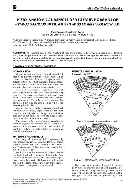
The root. (Fig.1-2.)<br />
Fig. 1. T. <strong>dacicus</strong>- Cross-section <strong>of</strong> root<br />
MATERIALS AND METHODS<br />
The biological material studied is represented by<br />
two Thymus species: T. Dacicus Borb., a species<br />
collected in June 2007 from Cheile Bicazului (Neamt)<br />
and T. Glabrescens Willd., species collected in May<br />
2007 from Barnova (Iasi).<br />
For <strong>histo</strong>-<strong>anatomical</strong> research the vegetal material<br />
was firstly fixed and preserved in ethylic alcohol 70%.<br />
We have made cross sections at the <strong>vegetative</strong> <strong>organs</strong><br />
(root, stem – the superior third part, the medium third<br />
part and the inferior third part; foliar limb), sections<br />
lately colored by iod-green and carmine-red.<br />
Studia Universitatis “Vasile Goldiș”, Seria Ştiințele Vieţii (Life Sciences Series), vol. 18, 2008<br />
© 2008 Vasile Goldis University Press<br />
http://www.studiauniversitatis.ro<br />
Fig. 2. T. glabrescens -Cross-section <strong>of</strong> root<br />
There have not been observed major differences at<br />
the two species analyses; both <strong>of</strong> them present a root<br />
with an early secondary structure generated by the<br />
activity <strong>of</strong> both lateral meristems: the cambium and the<br />
phellogen. The phellogens are generated by a few<br />
21
Studia Universitatis<br />
pr<strong>of</strong>ound cortical layers and form some thin periderms,<br />
the ritidome. First periderms are partial detached and<br />
parts <strong>of</strong> primary cortical parenchyma adhere by de<br />
latest periderm. The cork cells present a radial<br />
disposition and the phellogen cells are tangential<br />
oblongs, with the thin walls.<br />
The central cylinder present a thin secondary,<br />
external phloem ring (consisting in sieved tubes,<br />
companion cells and parenchyma cells) and a few (2-<br />
3) thicker secondary xylem rings, both resulted from<br />
the activity <strong>of</strong> the cambium. Annual rings <strong>of</strong> secondary<br />
xylem are streaky unequal as thickness, every one <strong>of</strong><br />
them having xylem vessels <strong>of</strong> large diameter and less<br />
lately xylem vessels <strong>of</strong> smaller diameter, separeted by<br />
libriform. The xylem parenchima is terminal and is<br />
form by the end <strong>of</strong> each year <strong>of</strong> vegetation.<br />
First two annual rings presents a few vessel with a<br />
smoll diameter and in the center <strong>of</strong> root are a few cells<br />
<strong>of</strong> parenchim moderate thickened. Looking with<br />
attention, in center is distinguishing 3 bundles <strong>of</strong> xylem<br />
from primary structure.<br />
The stem (Fig. 3-8)<br />
In cross section <strong>of</strong> the superior level <strong>of</strong> the stem,<br />
both analyzed species present round ribs. At T. <strong>dacicus</strong><br />
the epidermis presents izodiametric cells, with<br />
thickened internal and external walls, the external wall<br />
being covered by a very thick cuticle. Rarely secretory<br />
trichomes and tector trichomes are present, especially<br />
in the ribs. The tector trichomes are mostly localized<br />
on the ribs; their lengthiness is variable being<br />
formatted on ones or more cells. At T. glabrescens the<br />
epidermis presents isomorphs and izodiametric cells,<br />
with thickened external walls and covered by a very<br />
thick cuticle. From place to place, are present the<br />
stomata, many tector trichomes and secretory<br />
trichomes, mostly with unicellular gland. The tector<br />
trichomes have different lengthiness, being unicellular,<br />
bicellulars or multicellulars.<br />
On both analyzed species, the cortex is<br />
collenchymatised in the ribs and parenchymaticcellulosed<br />
in the rest.<br />
The cortex presents a Casparyan type endodermis,<br />
with large cells, weakly tangentially elongated cells, at<br />
both investigated species.<br />
At T. <strong>dacicus</strong> the central cylinder is thick with<br />
primary structure, but the conducting vessels are<br />
represented by a tenuous external phloem ring<br />
(consisting in sieved tubes and companion cells) and a<br />
thicker xylem ring. Between this two rings there is a<br />
thicker procambium ring (consisting in 3-4 layers). The<br />
pith is thick, parenchymatic-cellulosed, <strong>of</strong> meatus type;<br />
the cells from perimedular aria are moderated<br />
colenchimatouses.<br />
At T. glabrescens the central cylinder present a<br />
particular secondary structure, being formed from a<br />
very tenuous external phloem ring (consisting in sieved<br />
tubes, companion cells and a few parenchyma cells)<br />
and a thicker xylem ring formed mostly by libriform<br />
fibers with external wall very thick and partial<br />
lignified. In the inferior part <strong>of</strong> the xylem ring on<br />
perceive a few xylem vessels that can be solitary or<br />
grouped in cross. The pith is thick, parenchymaticcellulosed,<br />
<strong>of</strong> meatus type; most <strong>of</strong> the cells from<br />
central part being disorganizated or in process <strong>of</strong><br />
disorganization, resulting a big aerifer cavity, with<br />
fitful configuration.<br />
22<br />
Fig. 3. T. <strong>dacicus</strong>-Cross-section <strong>of</strong> stem (superior<br />
level), x400<br />
Fig. 4. T. glabrescens-Cross-section <strong>of</strong> stem (superior<br />
level), x400<br />
In cross section <strong>of</strong> the median level <strong>of</strong> the stem, at<br />
T. <strong>dacicus</strong>, in comparison with superior level, the<br />
cuticle is thicker (especially in ribs); the cortical<br />
parenchyma presents big aerifer cavity and the central<br />
cylinder present a secondary structure being<br />
Studia Universitatis “Vasile Goldiş”, Seria Ştiințele Vieţii (Life Sciences Series), vol. 18, 2008<br />
© 2008 Vasile Goldis University Press<br />
http://www.studiauniversitatis.ro
Studia Universitatis<br />
represented by a tenuous external phloem ring<br />
(consisting in sieved tubes, companion cells and a few<br />
parenchyma cells) and a thicker xylem ring (with<br />
libriform and a few vassels).<br />
At T. glabrescens in median level <strong>of</strong> the stem the<br />
structure is similar with the superior level, with the<br />
followings different: the cuticle is thicker, the tector<br />
hairs are more numerous, the xylem ring presents more<br />
vessels, dispersed in libriform fundamental mass.<br />
In cross section <strong>of</strong> the inferior level <strong>of</strong> the stem, at<br />
T. <strong>dacicus</strong>, the configuration <strong>of</strong> cross section remain<br />
the same with the preceding level, but the ribs are a<br />
little salient. The xylem ring is thicker and the central<br />
aerifer cavity is tighter.<br />
At T. glabrescens the structure remain the same<br />
with the preceding level, with the mention that the<br />
configuration <strong>of</strong> cross section is round and the cuticle<br />
is very thick.<br />
Fig. 5. T. <strong>dacicus</strong>-Cross-section <strong>of</strong> stem (middle level),<br />
x400<br />
Fig. 6. T. glabrescens-Cross-section <strong>of</strong> stem (middle<br />
level), x400<br />
Fig. 7. T. <strong>dacicus</strong>-Cross-section <strong>of</strong> stem (inferior level),<br />
x400<br />
The foliar blade (fig. 9).<br />
At both analyzed species, in front side view, the<br />
epidermis consists <strong>of</strong> irregularly-shaped cells, with<br />
weak waved walls. Both epidermis present stomata <strong>of</strong><br />
diacytic type, so, the limb are amfistomatic. Here and<br />
there a lot <strong>of</strong> secretory and non-secretory (tector)<br />
Fig. 8. T. glabrescens-Cross-section <strong>of</strong> stem (inferior<br />
level), x400<br />
trichomes are present. The borders <strong>of</strong> foliar blade have<br />
practically all cells transformed in aculeiform hairs,<br />
unicellular, with external walls very thick; a few hairs<br />
are multicellular having the terminal cell with blind<br />
apex.<br />
Studia Universitatis “Vasile Goldiș”, Seria Ştiințele Vieţii (Life Sciences Series), vol. 18, 2008<br />
© 2008 Vasile Goldis University Press<br />
http://www.studiauniversitatis.ro<br />
23
Studia Universitatis<br />
Fig. 9 A T. <strong>dacicus</strong>- the lower epidermis <strong>of</strong> the limb<br />
Fig. 9 B T. glabrescens-- the lower epidermis <strong>of</strong> the limb<br />
Fig. 10. Secretory trichomes belonging to T. <strong>dacicus</strong>,<br />
x400<br />
Fig. 11. Secretory trichomes belonging to T.<br />
glabrescens, x400<br />
24<br />
Studia Universitatis “Vasile Goldiş”, Seria Ştiințele Vieţii (Life Sciences Series), vol. 18, 2008<br />
© 2008 Vasile Goldis University Press<br />
http://www.studiauniversitatis.ro
Studia Universitatis<br />
In cross section, at both analyzed species, the<br />
mesophyll is formed by palisade tissue at the upper<br />
side and lacunary tissue at the lower one, so, the blade<br />
has a bifacial-heter<strong>of</strong>acial (dorsiventral) structure.<br />
At T. <strong>dacicus</strong>, the palisade tissue is bistratified,<br />
dense, with the hypodermic layer cells higher and with<br />
winding lateral walls. The lacunary tissue present 5-6<br />
layers by rounded cells or fitful cells, with small aerifer<br />
lacunae between cells. To the border <strong>of</strong> foliar blade<br />
whole the mesophyll is on palisadyc type. The<br />
conducted vessels form more bundles, the biggest one<br />
presents at the periphery <strong>of</strong> the phloem cordons <strong>of</strong><br />
sclerenchymatous fibers, with very thick walls but<br />
moderated lignified.<br />
At T. glabrescens, the palisade tissue is bistratified,<br />
with hypodermic layer cells higher. The lacunary tissue<br />
compass about 4 layers cells, isodiametric or tangent<br />
elongated, with small aerifer lacunas between them.<br />
REFERENCES<br />
Bailey Ciocârlan V., Flora ilustrată a României.<br />
Pteridophyta et Spermatophyta, Ed. Ceres,<br />
Bucureşti, pp. 670-675, 2000;<br />
Guşuleac M., Thymus, In Flora Republicii Populare<br />
Române, VIII, Ed. Acad. RPR, Bucureşti, pp.<br />
301-334, 1961;<br />
Metcalfe C.R., Chalk L., Anatomy <strong>of</strong> Dicotyledons,<br />
Clarendon Press, Oxford, 2, pp. 1041-1053,<br />
1950;<br />
Toma C., Berciu I., Morphological peculiaries <strong>of</strong><br />
germination and structure <strong>of</strong> seedling in<br />
Thymus vulgaris L.; Romanian Biological<br />
Sciences, V, 1-2, pp. 136-137, 2007.<br />
Toma C., Rugină R., Anatomia plantelor medicinale.<br />
Atlas. Ed.Acad. Rom., Bucureşti, pp. 169-172,<br />
1998.<br />
CONCLUSIONS<br />
At both analyzed species, the root presents a<br />
secondary structure, resulting for din activity on both<br />
lateral meristems: the cambium and the fellogen.<br />
On both analyzed species there are two types <strong>of</strong><br />
trichomes: tector trichomes, more <strong>of</strong>ten multicelular,<br />
and secretors trichomes, always multicelular,<br />
consisting in a basal cell, a unicellular pedicel and a<br />
uni- or multicellular gland.<br />
The endoderm <strong>of</strong> Casparyan types became visible<br />
in the median third <strong>of</strong> the T. <strong>dacicus</strong> stem.<br />
At T. <strong>dacicus</strong> to the border <strong>of</strong> foliar blade whole the<br />
mesophyll is on palisadyc type.<br />
On both analyzed species, the stomata are diacitic<br />
type and there are presents on both sides <strong>of</strong> the foliar<br />
blade.<br />
Studia Universitatis “Vasile Goldiș”, Seria Ştiințele Vieţii (Life Sciences Series), vol. 18, 2008<br />
© 2008 Vasile Goldis University Press<br />
http://www.studiauniversitatis.ro<br />
25
Studia Universitatis<br />
26<br />
Studia Universitatis “Vasile Goldiş”, Seria Ştiințele Vieţii (Life Sciences Series), vol. 18, 2008<br />
© 2008 Vasile Goldis University Press<br />
http://www.studiauniversitatis.ro