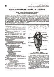
histo-anatomical aspects of vegetative organs of thymus dacicus ...
histo-anatomical aspects of vegetative organs of thymus dacicus ...
histo-anatomical aspects of vegetative organs of thymus dacicus ...

Create successful ePaper yourself
Turn your PDF publications into a flip-book with our unique Google optimized e-Paper software.
Studia Universitatis<br />
pr<strong>of</strong>ound cortical layers and form some thin periderms,<br />
the ritidome. First periderms are partial detached and<br />
parts <strong>of</strong> primary cortical parenchyma adhere by de<br />
latest periderm. The cork cells present a radial<br />
disposition and the phellogen cells are tangential<br />
oblongs, with the thin walls.<br />
The central cylinder present a thin secondary,<br />
external phloem ring (consisting in sieved tubes,<br />
companion cells and parenchyma cells) and a few (2-<br />
3) thicker secondary xylem rings, both resulted from<br />
the activity <strong>of</strong> the cambium. Annual rings <strong>of</strong> secondary<br />
xylem are streaky unequal as thickness, every one <strong>of</strong><br />
them having xylem vessels <strong>of</strong> large diameter and less<br />
lately xylem vessels <strong>of</strong> smaller diameter, separeted by<br />
libriform. The xylem parenchima is terminal and is<br />
form by the end <strong>of</strong> each year <strong>of</strong> vegetation.<br />
First two annual rings presents a few vessel with a<br />
smoll diameter and in the center <strong>of</strong> root are a few cells<br />
<strong>of</strong> parenchim moderate thickened. Looking with<br />
attention, in center is distinguishing 3 bundles <strong>of</strong> xylem<br />
from primary structure.<br />
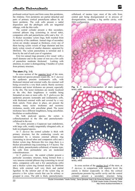
The stem (Fig. 3-8)<br />
In cross section <strong>of</strong> the superior level <strong>of</strong> the stem,<br />
both analyzed species present round ribs. At T. <strong>dacicus</strong><br />
the epidermis presents izodiametric cells, with<br />
thickened internal and external walls, the external wall<br />
being covered by a very thick cuticle. Rarely secretory<br />
trichomes and tector trichomes are present, especially<br />
in the ribs. The tector trichomes are mostly localized<br />
on the ribs; their lengthiness is variable being<br />
formatted on ones or more cells. At T. glabrescens the<br />
epidermis presents isomorphs and izodiametric cells,<br />
with thickened external walls and covered by a very<br />
thick cuticle. From place to place, are present the<br />
stomata, many tector trichomes and secretory<br />
trichomes, mostly with unicellular gland. The tector<br />
trichomes have different lengthiness, being unicellular,<br />
bicellulars or multicellulars.<br />
On both analyzed species, the cortex is<br />
collenchymatised in the ribs and parenchymaticcellulosed<br />
in the rest.<br />
The cortex presents a Casparyan type endodermis,<br />
with large cells, weakly tangentially elongated cells, at<br />
both investigated species.<br />
At T. <strong>dacicus</strong> the central cylinder is thick with<br />
primary structure, but the conducting vessels are<br />
represented by a tenuous external phloem ring<br />
(consisting in sieved tubes and companion cells) and a<br />
thicker xylem ring. Between this two rings there is a<br />
thicker procambium ring (consisting in 3-4 layers). The<br />
pith is thick, parenchymatic-cellulosed, <strong>of</strong> meatus type;<br />
the cells from perimedular aria are moderated<br />
colenchimatouses.<br />
At T. glabrescens the central cylinder present a<br />
particular secondary structure, being formed from a<br />
very tenuous external phloem ring (consisting in sieved<br />
tubes, companion cells and a few parenchyma cells)<br />
and a thicker xylem ring formed mostly by libriform<br />
fibers with external wall very thick and partial<br />
lignified. In the inferior part <strong>of</strong> the xylem ring on<br />
perceive a few xylem vessels that can be solitary or<br />
grouped in cross. The pith is thick, parenchymaticcellulosed,<br />
<strong>of</strong> meatus type; most <strong>of</strong> the cells from<br />
central part being disorganizated or in process <strong>of</strong><br />
disorganization, resulting a big aerifer cavity, with<br />
fitful configuration.<br />
22<br />
Fig. 3. T. <strong>dacicus</strong>-Cross-section <strong>of</strong> stem (superior<br />
level), x400<br />
Fig. 4. T. glabrescens-Cross-section <strong>of</strong> stem (superior<br />
level), x400<br />
In cross section <strong>of</strong> the median level <strong>of</strong> the stem, at<br />
T. <strong>dacicus</strong>, in comparison with superior level, the<br />
cuticle is thicker (especially in ribs); the cortical<br />
parenchyma presents big aerifer cavity and the central<br />
cylinder present a secondary structure being<br />
Studia Universitatis “Vasile Goldiş”, Seria Ştiințele Vieţii (Life Sciences Series), vol. 18, 2008<br />
© 2008 Vasile Goldis University Press<br />
http://www.studiauniversitatis.ro