histo-anatomical aspects of vegetative organs of thymus dacicus ...
histo-anatomical aspects of vegetative organs of thymus dacicus ...
histo-anatomical aspects of vegetative organs of thymus dacicus ...

Create successful ePaper yourself
Turn your PDF publications into a flip-book with our unique Google optimized e-Paper software.
Studia Universitatis<br />
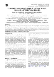
In cross section, at both analyzed species, the<br />
mesophyll is formed by palisade tissue at the upper<br />
side and lacunary tissue at the lower one, so, the blade<br />
has a bifacial-heter<strong>of</strong>acial (dorsiventral) structure.<br />
At T. <strong>dacicus</strong>, the palisade tissue is bistratified,<br />
dense, with the hypodermic layer cells higher and with<br />
winding lateral walls. The lacunary tissue present 5-6<br />
layers by rounded cells or fitful cells, with small aerifer<br />
lacunae between cells. To the border <strong>of</strong> foliar blade<br />
whole the mesophyll is on palisadyc type. The<br />
conducted vessels form more bundles, the biggest one<br />
presents at the periphery <strong>of</strong> the phloem cordons <strong>of</strong><br />
sclerenchymatous fibers, with very thick walls but<br />
moderated lignified.<br />
At T. glabrescens, the palisade tissue is bistratified,<br />
with hypodermic layer cells higher. The lacunary tissue<br />
compass about 4 layers cells, isodiametric or tangent<br />
elongated, with small aerifer lacunas between them.<br />
REFERENCES<br />
Bailey Ciocârlan V., Flora ilustrată a României.<br />
Pteridophyta et Spermatophyta, Ed. Ceres,<br />
Bucureşti, pp. 670-675, 2000;<br />
Guşuleac M., Thymus, In Flora Republicii Populare<br />
Române, VIII, Ed. Acad. RPR, Bucureşti, pp.<br />
301-334, 1961;<br />
Metcalfe C.R., Chalk L., Anatomy <strong>of</strong> Dicotyledons,<br />
Clarendon Press, Oxford, 2, pp. 1041-1053,<br />
1950;<br />
Toma C., Berciu I., Morphological peculiaries <strong>of</strong><br />
germination and structure <strong>of</strong> seedling in<br />
Thymus vulgaris L.; Romanian Biological<br />
Sciences, V, 1-2, pp. 136-137, 2007.<br />
Toma C., Rugină R., Anatomia plantelor medicinale.<br />
Atlas. Ed.Acad. Rom., Bucureşti, pp. 169-172,<br />
1998.<br />
CONCLUSIONS<br />
At both analyzed species, the root presents a<br />
secondary structure, resulting for din activity on both<br />
lateral meristems: the cambium and the fellogen.<br />
On both analyzed species there are two types <strong>of</strong><br />
trichomes: tector trichomes, more <strong>of</strong>ten multicelular,<br />
and secretors trichomes, always multicelular,<br />
consisting in a basal cell, a unicellular pedicel and a<br />
uni- or multicellular gland.<br />
The endoderm <strong>of</strong> Casparyan types became visible<br />
in the median third <strong>of</strong> the T. <strong>dacicus</strong> stem.<br />
At T. <strong>dacicus</strong> to the border <strong>of</strong> foliar blade whole the<br />
mesophyll is on palisadyc type.<br />
On both analyzed species, the stomata are diacitic<br />
type and there are presents on both sides <strong>of</strong> the foliar<br />
blade.<br />
Studia Universitatis “Vasile Goldiș”, Seria Ştiințele Vieţii (Life Sciences Series), vol. 18, 2008<br />
© 2008 Vasile Goldis University Press<br />
http://www.studiauniversitatis.ro<br />
25