flow cytrometric analysis of red blood cells in polycythemia vera
flow cytrometric analysis of red blood cells in polycythemia vera
flow cytrometric analysis of red blood cells in polycythemia vera

Create successful ePaper yourself
Turn your PDF publications into a flip-book with our unique Google optimized e-Paper software.
Studia Universitatis “Vasile Goldiş”, Seria Şti<strong>in</strong>ţele Vieţii<br />
Vol. 21, issue 1, 2011, pp. 29-35<br />
© 2011 Vasile Goldis University Press (www.studiauniversitatis.ro)<br />
FLOW CYTROMETRIC ANALYSIS OF RED BLOOD CELLS IN<br />
POLYCYTHEMIA VERA<br />
Ana-Maria GHEORGHE 1 , Alexandr<strong>in</strong>a RUGINA 1 , Andreea Delia MOICEAN 2 ,<br />
Aurel ARDELEAN 3 , Daniela BRATOSIN 1,3 *<br />
1 National Institute <strong>of</strong> Biological Science Research and Development (INCDSB), Bucharest, Romania<br />
2 Cl<strong>in</strong>ical Institute Fundeni, Center <strong>of</strong> Hematology & Bone Marrow Transplantation ’’St. Berceanu’’, Bucharest<br />
3 ″Vasile Goldis″ Western University <strong>of</strong> Arad, Faculty <strong>of</strong> Natural Sciences, Arad, Romania<br />
ABSTRACT. Polycythemia <strong>vera</strong> (PV) is characterized by an absolute <strong>in</strong>crease <strong>in</strong> the <strong>red</strong> <strong>blood</strong> cell mass, but<br />
the mechanisms are not completely understood. In this study, we identified by <strong>flow</strong> cytometric <strong>analysis</strong><br />
morphological forms that deviate from the classical discoid shape, who had a more viability determ<strong>in</strong>ed by<br />
Calce<strong>in</strong>-AM method and a normal phosphatidylser<strong>in</strong>e exposure level. Measurement <strong>of</strong> glycoconjugate<br />
sialylation us<strong>in</strong>g lect<strong>in</strong>es demonstrates a low degree <strong>of</strong> sialilation <strong>of</strong> membrane glycoconjugates <strong>of</strong><br />
Polycythemia <strong>vera</strong> RBCs but which is close to normal after treatment and an <strong>in</strong>creased percentage <strong>of</strong> <strong>cells</strong><br />
with active caspase-8 and -3 compa<strong>red</strong> to normal RBCs, show<strong>in</strong>g that the organism tries to restore<br />
the apoptotic mechanism for ma<strong>in</strong>ta<strong>in</strong><strong>in</strong>g the normal hematocrit. Our observations may contribute for<br />
understand<strong>in</strong>g the survival <strong>of</strong> RBCs <strong>in</strong> Polycythemia <strong>vera</strong> and may also participate <strong>in</strong> elucidation <strong>of</strong> the<br />
mechanism <strong>in</strong> pathogenesis <strong>of</strong> this disease.<br />
Keywords: <strong>polycythemia</strong> <strong>vera</strong>; PV; rbcs, <strong>red</strong> <strong>blood</strong> <strong>cells</strong>; erythrocyte viability; phosphatidylser<strong>in</strong>e exposure;<br />
caspase-3; caspase-8; Annex<strong>in</strong>-V; Calce<strong>in</strong>-AM; <strong>flow</strong> cytometry<br />
INTRODUCTION<br />
Polycythemia is literally translated as "many <strong>cells</strong><br />
<strong>in</strong> the <strong>blood</strong>". Only erythrocytosis (an alternative term<br />
for these disorders) produces <strong>polycythemia</strong> s<strong>in</strong>ce<br />
leukocytes and platelets are present <strong>in</strong> <strong>blood</strong> <strong>in</strong> far<br />
smaller proportions. This disease may be due to<br />
<strong>in</strong>creased proliferation or decreased apoptosis <strong>of</strong><br />
erythroid progenitors or to delayed erythroid<br />
differentiation with an <strong>in</strong>creased number <strong>of</strong> progenitor<br />
cell divisions (Prchal J.T., 2001). Polycythemia <strong>vera</strong><br />
(PV) is a malignant disorder <strong>of</strong> hematopoietic stem<br />
<strong>cells</strong> which is characterized by clonal<br />
myeloproliferation with <strong>in</strong>creased production <strong>of</strong><br />
morphologically normal mature <strong>red</strong> <strong>blood</strong> <strong>cells</strong>, white<br />
<strong>cells</strong> and platelets (Berl<strong>in</strong>, N.I., 1975; Adamson et<br />
al.,1976; Spivak, J.L. 2002; Bai et al.,2004). First<br />
described <strong>in</strong> 1892 by Vaquez (Vaquez H.,1892)<br />
Polycythemia <strong>vera</strong> is a disease with an <strong>in</strong>cidence <strong>of</strong> at<br />
least 2 per 100 000 and is a tril<strong>in</strong>eage hematopoietic<br />
cell hyperplasia (Berglund, S. & Zetterval O.,1992;<br />
Mcnally, et al.,1997). Prolonged <strong>red</strong> cell survival,<br />
another theoretical cause <strong>of</strong> <strong>polycythemia</strong>, has not yet<br />
been demonstrated (Berl<strong>in</strong> et al., 1951; Fernandez-<br />
Luna et al., 1998). Recent <strong>in</strong>vestigations have focused<br />
on a number <strong>of</strong> molecules <strong>in</strong>volved <strong>in</strong> signal<br />
transduction pathways mediated by erythropoiet<strong>in</strong><br />
(Epo) and other growth factors, but human erythroid<br />
malignancies (PV and erythroleukemia) are associated<br />
with erythropoiet<strong>in</strong>-<strong>in</strong>dependent growth and<br />
differentiation (Carneskog et al.,1998; Ugo et<br />
al.,2004). Despite recent advances <strong>in</strong> the<br />
characterization <strong>of</strong> the malignant PV clone, the<br />
molecular mechanism and the abnormalities associated<br />
with the development <strong>of</strong> this disorder rema<strong>in</strong> unknown<br />
(Spivak, J.L., 2002). Regard<strong>in</strong>g these controversed<br />
results, we conside<strong>red</strong> <strong>of</strong> <strong>in</strong>terest to analyse and<br />
characterise <strong>red</strong> <strong>blood</strong> <strong>cells</strong> <strong>in</strong> Polycythaemia <strong>vera</strong><br />
regarded as cancerous <strong>cells</strong> by apply<strong>in</strong>g methods for<br />
characterisation <strong>of</strong> apoptotic <strong>cells</strong>. Cell viability and<br />
death were analyzed by <strong>flow</strong> cytometry, a method<br />
ideally adapted for the study <strong>of</strong> cell death and for rapid<br />
and <strong>in</strong>dividual <strong>analysis</strong> <strong>of</strong> a large number <strong>of</strong> <strong>cells</strong>.<br />
MATERIALS AND METHODS<br />
Chemicals<br />
Fluoresce<strong>in</strong> conjugated Annex<strong>in</strong>-V (Annex<strong>in</strong>-V-<br />
FITC), HEPES b<strong>in</strong>d<strong>in</strong>g buffer (HEPES buffer pH 7.4<br />
conta<strong>in</strong><strong>in</strong>g 2.5mM calcium chloride) were obta<strong>in</strong>ed<br />
from Pharm<strong>in</strong>gen (San Diego, CA, USA), Calce<strong>in</strong>-AM<br />
from Sigma Aldrich (St. Louis, M0, USA) and<br />
CaspGLOW TM Fluoresce<strong>in</strong> active caspase-8 and<br />
caspase-3 sta<strong>in</strong><strong>in</strong>g kits from BioVision Research<br />
Products (Mounta<strong>in</strong> View, CA, USA).<br />
Fluoresce<strong>in</strong>ylisothiocyanate lect<strong>in</strong>s (FITC-lect<strong>in</strong>s):<br />
MAA (Maackia amurensis agglut<strong>in</strong><strong>in</strong>), SNA<br />
(Sambucus nigra agglut<strong>in</strong><strong>in</strong>) and RCA 120 (Ric<strong>in</strong>us<br />
communis agglut<strong>in</strong><strong>in</strong>) were from EY Laboratories (San<br />
Mateo, CA, USA).<br />
The <strong>flow</strong> cytometer was a Becton-Dick<strong>in</strong>son<br />
FACScan apparatus (San Jose, CA, USA) with<br />
CellQuestPro s<strong>of</strong>tware for acquisition and <strong>analysis</strong>.<br />
Isolation <strong>of</strong> erythrocytes<br />
Human <strong>blood</strong> was taken up on hepar<strong>in</strong> from the<br />
patients with PV before and after treatment. The<br />
diagnosis <strong>of</strong> the disease was established accord<strong>in</strong>g to<br />
commonly accepted cl<strong>in</strong>ical and laboratory criteria<br />
(Berk et al., 1986). The <strong>blood</strong> samples were washed<br />
*Correspondence: Daniela Bratos<strong>in</strong>, National Institute for Biological Science Research and Development, Spl. Independentei, Nº 296,<br />
060031 Bucharest, Romania, Tel/Fax: 40.21.2200881, E-mail: bratos<strong>in</strong>d@yahoo.com<br />
Article received: November 2010; published: February 2011
Gheorghe A., Rug<strong>in</strong>a Alx., Moicean A., Ardelean A., Bratos<strong>in</strong> D.<br />
thrice by centrifugation (5 m<strong>in</strong>, 1000 x g at 4°C) with<br />
phosphate buffe<strong>red</strong> sal<strong>in</strong>e (PBS):Na 2 HPO 4 1.8 mM,<br />
KH 2 PO 4 140 mM, NaCl 2.7 mM, KCl, pH 7.4. After<br />
centrifugation, plasma, platelets and leukocytes were<br />
removed by aspiration and the <strong>red</strong> <strong>blood</strong> <strong>cells</strong> were<br />
resuspended (10 7 <strong>cells</strong> per ml) for further experiments<br />
<strong>in</strong> isotonic phosphate-buffe<strong>red</strong> sal<strong>in</strong>e (PBS) solution<br />
pH 7.4.<br />
Flow cytometric <strong>analysis</strong><br />
Flow cytometric analyses were performed on a<br />
FACScan cytometer us<strong>in</strong>g CellQuestPro s<strong>of</strong>tware for<br />
acquisition and <strong>analysis</strong>. Cells <strong>in</strong> suspension <strong>in</strong> isotonic<br />
PBS buffer pH 7.4 were gated for the light scatter<br />
channels on l<strong>in</strong>ear ga<strong>in</strong>s, and the fluorescence channels<br />
were set on a logarithmic scale with a m<strong>in</strong>imum <strong>of</strong><br />
10,000 <strong>cells</strong> analyzed <strong>in</strong> each condition.<br />
Morphological changes assessment <strong>of</strong> <strong>red</strong><br />
<strong>blood</strong> <strong>cells</strong> <strong>in</strong> Polycythemia <strong>vera</strong> by light<br />
scatte<strong>red</strong> measurements<br />
Analysis <strong>of</strong> the scatte<strong>red</strong> light by <strong>flow</strong> cytometry <strong>in</strong><br />
the mode FSC/SSC provides <strong>in</strong>formations about cell<br />
size and structure. The <strong>in</strong>tensity <strong>of</strong> light scatte<strong>red</strong> <strong>in</strong> a<br />
forward direction (FSC) correlates with cell size. The<br />
<strong>in</strong>tensity <strong>of</strong> scatte<strong>red</strong> light measu<strong>red</strong> at a right angle to<br />
the laser beam (side scatter/SSC), on the other hand,<br />
correlates with granularity, refractiveness and presence<br />
<strong>of</strong> <strong>in</strong>tracellular structures that can reflect the lightwere<br />
associated with cell shr<strong>in</strong>kage. RBCs <strong>in</strong> suspension <strong>in</strong><br />
isotonic PBS buffer, pH 7.4 were gated under forward<br />
and side scatter parameters (FSC versus SSC).<br />
Flow cytometric measurement <strong>of</strong> the b<strong>in</strong>d<strong>in</strong>g<br />
<strong>of</strong> fluoresce<strong>in</strong> isothiocyanate (FITC)-labeled<br />
lect<strong>in</strong>s to RBCs<br />
Accord<strong>in</strong>g to the protocol described by Bratos<strong>in</strong><br />
(Bratos<strong>in</strong> et al., 1995), a solution (50μl) <strong>of</strong> FITClabeled<br />
lect<strong>in</strong>s <strong>in</strong> phosphate buffe<strong>red</strong> sal<strong>in</strong>e (PBS)-<br />
phenylmethysulfonyl fluoride (PMSF) buffer, pH 7.4<br />
(10 mM Na 2 HPO 4 , 1.8 mM KH 2 PO 4 , 140 mM NaCl,<br />
2.7 mM KCl, 0.2 mM PMSF), was added to 50 μl <strong>of</strong> a<br />
<strong>red</strong> <strong>blood</strong> cell suspension <strong>in</strong> the same buffer<br />
(correspond<strong>in</strong>g to 2x10 6 <strong>red</strong> <strong>cells</strong>). After 1h <strong>in</strong>cubation<br />
at 4°C <strong>in</strong> the dark, 10,000 <strong>cells</strong> were analyzed directly.<br />
The b<strong>in</strong>d<strong>in</strong>g for each lect<strong>in</strong> was first studied at<br />
concentrations rang<strong>in</strong>g from 0 to 50 mM <strong>in</strong> order to<br />
determ<strong>in</strong>e the optimal lect<strong>in</strong> concentration. This<br />
experimental protocol was applied to the follow<strong>in</strong>g<br />
FITC-labeled lect<strong>in</strong>s: Sambucus nigra agglut<strong>in</strong><strong>in</strong><br />
(SNA) and Maackia amurensis agglut<strong>in</strong><strong>in</strong> (MAA),<br />
specific for sialic acids and Ric<strong>in</strong>us communis<br />
agglut<strong>in</strong><strong>in</strong> (RCA 120 ) specific for β-galactosyl term<strong>in</strong>al<br />
residues. Experiments were performed at least three<br />
times with three replicates each time.<br />
Flow cytometric measurement <strong>of</strong> cell viability<br />
us<strong>in</strong>g Calce<strong>in</strong>-AM<br />
Cell viability assessment was studied accord<strong>in</strong>g to<br />
the procedure <strong>of</strong> Bratos<strong>in</strong> et al. (Bratos<strong>in</strong> et al., 2005)<br />
30<br />
based on the use <strong>of</strong> acetoxymethyl ester calce<strong>in</strong><br />
(Calce<strong>in</strong>-AM), a fluoresce<strong>in</strong> derivative and nonfluorescent<br />
vital dye that passively crosses the<br />
membrane <strong>of</strong> viable <strong>cells</strong> and is converted by cytosolic<br />
esterases <strong>in</strong>to calce<strong>in</strong> that produces <strong>in</strong>tense green (530<br />
nm) signal, and is reta<strong>in</strong>ed by <strong>cells</strong> with <strong>in</strong>tact plasma<br />
membranes. From dy<strong>in</strong>g or damaged <strong>cells</strong> with<br />
compromised membrane <strong>in</strong>tegrity unhydrolysed<br />
substrat and their fluorescent products are rapidly<br />
extruded from <strong>cells</strong>.<br />
The membrane-permeable dye Calce<strong>in</strong>-AM was<br />
prepa<strong>red</strong> as a stock solution <strong>of</strong> 10 mM <strong>in</strong><br />
dimethylsulfoxide sto<strong>red</strong> at -20°C and as a work<strong>in</strong>g<br />
solution <strong>of</strong> 100 µm <strong>in</strong> PBS buffer pH 7.4. RBCs (4<br />
x10 5 <strong>in</strong> 200 µl PBS buffer, pH 7.4) were <strong>in</strong>cubated with<br />
10 µl Calce<strong>in</strong>-AM work<strong>in</strong>g solution (f<strong>in</strong>al<br />
concentration <strong>in</strong> Calce<strong>in</strong>-AM: 5 µm) for 45 m<strong>in</strong> at<br />
37°C <strong>in</strong> the dark and then diluted <strong>in</strong> 0.5 ml <strong>of</strong> PBS<br />
buffer for immediate <strong>flow</strong> cytometric <strong>analysis</strong> <strong>of</strong><br />
Calce<strong>in</strong> fluorescence retention <strong>in</strong> <strong>cells</strong>. Experiments<br />
were performed at least three times with three<br />
replicates each time.<br />
Flow cytometric <strong>analysis</strong> <strong>of</strong><br />
phosphatidylser<strong>in</strong>e exposure<br />
To 10 µl <strong>of</strong> the RBCs suspension (10 7 <strong>cells</strong> per ml)<br />
<strong>in</strong> PBS buffer, pH 7.4, were added 90 µl <strong>of</strong> b<strong>in</strong>d<strong>in</strong>g<br />
HEPES buffer, pH 7.4 and 5µl (0.05 µg) <strong>of</strong> FITC-<br />
Annex<strong>in</strong> V solution. After <strong>in</strong>cubation for 15 m<strong>in</strong> at<br />
room temperature <strong>in</strong> the dark, 400 µl <strong>of</strong> HEPES (N-(2-<br />
hydroxymethyl)piperaz<strong>in</strong>e-N’-(2-ethane)sulfonic acid)<br />
buffer, pH 7.4 were added and the suspension was<br />
analysed <strong>in</strong> the <strong>flow</strong> cytometer and gated for<br />
biparametric histograms FL1 (FITC fluorescence)<br />
versus FL2 (RBC aut<strong>of</strong>luorescence). Ten thousand<br />
fluorescent particles <strong>of</strong> each gated population were<br />
analyzed. Data were collected on a Becton Dick<strong>in</strong>son<br />
FACScan cytometer and analyzed us<strong>in</strong>g CellQuestPro<br />
s<strong>of</strong>tware. The light scatter channels were set on l<strong>in</strong>ear<br />
ga<strong>in</strong>s and the fluorescence channels on a logarithmic<br />
scale. Experiments were performed at least three times<br />
with three replicates each time.<br />
CaspGLOW TM fluoresce<strong>in</strong> active caspase-3 or -<br />
8 <strong>flow</strong> cytometric analyses<br />
10 6 RBCs were first <strong>in</strong>cubated for 1h with 1 µl <strong>of</strong><br />
FITC-IETD-fmk for caspase-8 activity or FITC-<br />
DEVD-fmk for caspase-3 activity at 37°C under 5%<br />
CO 2 atmosphere. After 3 treatments with 0.5 ml <strong>of</strong><br />
wash<strong>in</strong>g buffer, the <strong>cells</strong> were gated for parametric<br />
histograms FL1 (FITC fluorescence <strong>of</strong> CaspGLOW TM ).<br />
Experiments were carried out <strong>in</strong> triplicate.<br />
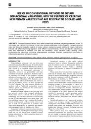
Scann<strong>in</strong>g electron microscopy (SEM) <strong>analysis</strong><br />
Erythrocytes were fixed for 4 h with a 1.25%<br />
glutaraldehyde solution <strong>in</strong> 0.1 M sodium cacodylate<br />
buffer pH 7.2 and post-fixed for 4 h <strong>in</strong> 1% osmium<br />
tetraoxide <strong>in</strong> the same buffer. The suspensions were<br />
then filte<strong>red</strong> onto 0.2 μ Anodisc filters and dehydrated<br />
<strong>in</strong> an ethanol series. After dry<strong>in</strong>g with carbon dioxide<br />
Studia Universitatis “Vasile Goldiş”, Seria Şti<strong>in</strong>ţele Vieţii<br />
Vol. 21, issue 1, 2011, pp. 29-35<br />
© 2011 Vasile Goldis University Press (www.studiauniversitatis.ro)
Flow <strong>cytrometric</strong> <strong>analysis</strong> <strong>of</strong> <strong>red</strong><br />
<strong>blood</strong> <strong>cells</strong> <strong>in</strong> Polycythemia <strong>vera</strong><br />
by the critical po<strong>in</strong>t method and sputter-coat<strong>in</strong>g with<br />
gold, samples were exam<strong>in</strong>ed on a 35 CF JEOL SEM.<br />
RESULTS AND DISCUSSIONS<br />
Light scatter<strong>in</strong>g properties <strong>of</strong> <strong>red</strong> <strong>blood</strong> <strong>cells</strong><br />
<strong>in</strong> Polycythemia <strong>vera</strong><br />
As shown <strong>in</strong> Figures 1 and 2, <strong>flow</strong> cytometric<br />
analyses announce significant morphological changes<br />
<strong>of</strong> <strong>red</strong> <strong>blood</strong> <strong>cells</strong> <strong>in</strong> Polycythemia <strong>vera</strong>. In fact, as<br />
demonstrated <strong>in</strong> Figure 1 and 2, the XGeo Mean values<br />
(cell side scatter) vary from 260 (Patient PV) compa<strong>red</strong><br />
to 299.63 (M), the statistical value for normal RBCs<br />
be<strong>in</strong>g 310 ± 25. In the same way, the YGeo Mean<br />
values (cell density scatter) vary from 196.87 (Patient<br />
PV) to 256.35 (M-To), the statistical value <strong>of</strong> normal<br />
RBCs be<strong>in</strong>g 298± 30. These morphological changes<br />
dim<strong>in</strong>ishes after treatment as shown <strong>in</strong> Figures 2, PVt,<br />
where the YGeo Mean <strong>in</strong>crease to 222.58, value closer<br />
to YGeo Mean <strong>of</strong> normal erythrocytes (M).<br />
Fig. 1. Comparative dot-plot <strong>analysis</strong> FSC/SSC <strong>of</strong> morphological changes <strong>of</strong> <strong>red</strong> <strong>blood</strong> <strong>cells</strong> <strong>in</strong> Polycythemia <strong>vera</strong>. (M):<br />
control erythrocytes; (PV): <strong>red</strong> <strong>blood</strong> <strong>cells</strong> <strong>in</strong> Polycythemia <strong>vera</strong> before and after treatment (PVt); Abscissae: forward<br />
scatter (cell size); ord<strong>in</strong>ates: side scatter (cell density, granularity and refractiveness). Number <strong>of</strong> counted <strong>cells</strong>: 10,000.<br />
Results presented are from one representative experiment <strong>of</strong> three performed<br />
Fig. 2. Comparative histogram <strong>of</strong> X- and YGeoMean values <strong>of</strong> normal (M) and Polycythemia <strong>vera</strong> erythrocytes before<br />
(PV) and after treatment (PVt). Values refer to dot-plot analyses <strong>of</strong> Figure 1<br />
Scann<strong>in</strong>g electron microscopy <strong>analysis</strong><br />
Scann<strong>in</strong>g electron microscopy entirely confirmed<br />
these data and led to the discovery <strong>of</strong> a dysmorphic<br />
RBCs, especially stomatocytes, presented <strong>in</strong> Figure 3a<br />
to 3f, which coexist with discocytes.<br />
Cell viability <strong>of</strong> erythrocytes<br />
We have applied the <strong>flow</strong> cytometric assay we<br />
previously developed for the measurement <strong>of</strong><br />
erythrocyte viability (Bratos<strong>in</strong> et al., 2005). As<br />
described <strong>in</strong> "Materials and methods", the assay is<br />
based on the use <strong>of</strong> Calce<strong>in</strong>-AM, a non-fluorescent<br />
vital dye that passively crosses the cell membrane <strong>of</strong><br />
Studia Universitatis “Vasile Goldiş”, Seria Şti<strong>in</strong>ţele Vieţii<br />
Vol. 21, issue 1, 2011, pp. 29-35<br />
© 2011 Vasile Goldis University Press (www.studiauniversitatis.ro)<br />
viable <strong>cells</strong> and is converted by cytosolic esterases <strong>in</strong>to<br />
green fluorescent calce<strong>in</strong> which is reta<strong>in</strong>ed only by<br />
<strong>cells</strong> with <strong>in</strong>tact membranes. The results we obta<strong>in</strong>ed,<br />
illustrated by the Figure 4, reveal <strong>in</strong> RBCs <strong>of</strong><br />
Polycythemia <strong>vera</strong> patient (PV), an Calce<strong>in</strong> means <strong>of</strong><br />
fluorescence <strong>in</strong>tensity <strong>of</strong> 160, almost double compa<strong>red</strong><br />
to MFI <strong>of</strong> normal RBCs (M). After treatment, MFI<br />
slightly decreases to 156 (PVt).<br />
This <strong>in</strong>creased esterase activity <strong>in</strong> erythrocytes<br />
frompatients with Polycythemia <strong>vera</strong> could be a<br />
possible cause <strong>of</strong> a longer life-time, which would lead<br />
to an <strong>in</strong>crease <strong>in</strong> circulat<strong>in</strong>g hematocrit.<br />
31
Gheorghe A., Rug<strong>in</strong>a Alx., Moicean A., Ardelean A., Bratos<strong>in</strong> D.<br />
Fig. 3 Comparative scann<strong>in</strong>g electron microscopy<br />
(SEM) <strong>of</strong> normal (M) and Polycythemia <strong>vera</strong> <strong>red</strong> <strong>blood</strong><br />
<strong>cells</strong> (PV). Details <strong>of</strong> some dysmorphic Polycythemia<br />
<strong>vera</strong> erythrocytes (a to f)<br />
Measurement <strong>of</strong> glycoconjugate sialylation<br />
Classically, the sialic acid residues which are<br />
situated <strong>in</strong> term<strong>in</strong>al position <strong>of</strong> the glycan moieties <strong>of</strong><br />
membrane glycoconjugates are conside<strong>red</strong> as<br />
antirecognition signals for phagocytic <strong>cells</strong>. Their<br />
removal by sialidases, by demask<strong>in</strong>g the penultimate β-<br />
galactosyl residues <strong>of</strong> glycans <strong>in</strong>duces the capture <strong>of</strong><br />
<strong>cells</strong> mediated by a specific lect<strong>in</strong> present <strong>in</strong> the<br />
macrophage membrane. As demonstrated by Figure 5,<br />
<strong>analysis</strong> by cyt<strong>of</strong>luorimetry <strong>of</strong> the b<strong>in</strong>d<strong>in</strong>g <strong>of</strong> FITClabeled<br />
lect<strong>in</strong>s specific for sialic acid and β-galactosyl<br />
residues shows that the means <strong>of</strong> fluorescence<br />
<strong>in</strong>tensity (MFI) for Maackia amurensis agglut<strong>in</strong><strong>in</strong><br />
MAA specify for α-1,3-l<strong>in</strong>ked sialic acids is<br />
much smaller (23.05) compa<strong>red</strong> to MFI for normal<br />
RBCs (47.88). After treatment (PVt), the MFI <strong>in</strong>crease<br />
to 36.77 value.<br />
This desialilation is also evidenced by Wheat germ<br />
agglut<strong>in</strong><strong>in</strong> (WGA) that b<strong>in</strong>ds to N-acetylglucosam<strong>in</strong>e,<br />
but also can <strong>in</strong>teract with some glycoprote<strong>in</strong>s via sialic<br />
acid residues. Sambucus nigra agglut<strong>in</strong><strong>in</strong> (SNA),<br />
specific for α-1,6-l<strong>in</strong>ked sialic acids also show a<br />
discrete desialilation.<br />
Fig. 4. Comparative <strong>flow</strong> cytometric histogram <strong>analysis</strong> <strong>of</strong> erythrocytes viability determ<strong>in</strong>ed by cell esterase activity<br />
measurement us<strong>in</strong>g Calce<strong>in</strong>-AM. Human normal <strong>red</strong> <strong>blood</strong> <strong>cells</strong> (M), <strong>red</strong> <strong>blood</strong> <strong>cells</strong> <strong>in</strong> Polycythaemia <strong>vera</strong> before (PV)<br />
and after treatment (PVt). Numbers represent fluorescence mean values (MFI). Abscissae: log scale green fluorescence<br />
<strong>in</strong>tensity <strong>of</strong> Calce<strong>in</strong> (FL1). Ord<strong>in</strong>ates: relative cell number. Number <strong>of</strong> counted <strong>cells</strong>: 10,000. Results presented are from<br />
one representative experiment <strong>of</strong> three performed.<br />
In the same way, as demonstrated by Figure 5,<br />
b<strong>in</strong>d<strong>in</strong>g <strong>of</strong> Ric<strong>in</strong>us comMunis agglut<strong>in</strong><strong>in</strong> (RCA 120 ),<br />
specific for β-galactosyl residues, has not been<br />
fixed more on Polycythemia <strong>vera</strong> <strong>red</strong> <strong>blood</strong> <strong>cells</strong><br />
(MFI=61.7) compa<strong>red</strong> to normal erythrocytes (76.73)<br />
or after treatment, when b<strong>in</strong>d<strong>in</strong>g is also lower (46.31),<br />
phenomenon that rema<strong>in</strong>s an enigma.<br />
Study <strong>of</strong> death by annex<strong>in</strong>-V-FITC labell<strong>in</strong>g<br />
Study <strong>of</strong> death by Annex<strong>in</strong>-V-FITC label<strong>in</strong>g<br />
phosphatidylser<strong>in</strong>e exposure on the outer leaflet <strong>of</strong><br />
plasma membrane is regarded as one <strong>of</strong> the signals<br />
allow<strong>in</strong>g macrophages to <strong>in</strong>gest erythrocytes. In<br />
Polycythaemia <strong>vera</strong> we did not observe any difference<br />
<strong>of</strong> phosphatidylser<strong>in</strong>e externalization between normal<br />
and Polycythemia <strong>vera</strong> RBCs. The value (1.48%) is <strong>in</strong><br />
32<br />
the limit <strong>of</strong> normal RBCs statistical value: 1.5 ± 1%<br />
(data not shown)<br />
Red <strong>blood</strong> <strong>cells</strong> <strong>in</strong> Polycythemia <strong>vera</strong> express<br />
active caspases-8 and 3<br />
We then assessed whether erythrocytes <strong>in</strong><br />
Polycythemia <strong>vera</strong> express active caspases. In order to<br />
precise the nature <strong>of</strong> activated caspases, we used the<br />
CaspGLOW TM specific cell permeable fluorogenic<br />
substrates FITC-IETD-fmk and FITC-DEVD-fmk,<br />
which are labeled <strong>in</strong>hibitors <strong>of</strong> caspases-8 and -3,<br />
respectively.<br />
As shown <strong>in</strong> Figure 6, a significant fluorescence<br />
was detected by <strong>flow</strong> cytometry. Thus, by compar<strong>in</strong>g<br />
% <strong>of</strong> <strong>cells</strong> with active caspases, can be observed a<br />
lower level <strong>of</strong> caspase-8 active <strong>in</strong> the RBCS <strong>of</strong><br />
Studia Universitatis “Vasile Goldiş”, Seria Şti<strong>in</strong>ţele Vieţii<br />
Vol. 21, issue 1, 2011, pp. 29-35<br />
© 2011 Vasile Goldis University Press (www.studiauniversitatis.ro)
Flow <strong>cytrometric</strong> <strong>analysis</strong> <strong>of</strong> <strong>red</strong><br />
<strong>blood</strong> <strong>cells</strong> <strong>in</strong> Polycythemia <strong>vera</strong><br />
Polycythemia <strong>vera</strong> (PV) before (0.5%) and after<br />
treatment (0.58). Inexplicably, the % <strong>of</strong> <strong>red</strong><br />
<strong>cells</strong> with active caspase-3 is higher <strong>in</strong> both RBCs <strong>of</strong><br />
Polycythemia <strong>vera</strong>, before (PV) and after treatment<br />
(PVt), 9.88% and 9.14% respectively, compa<strong>red</strong> with<br />
normal RBCs, 2.84% (M), demonstrat<strong>in</strong>g that the<br />
organism tries to restore the apoptotic mechanism for<br />
ma<strong>in</strong>ta<strong>in</strong><strong>in</strong>g the normal hematocrit.<br />
Fig. 5. Overlay (s<strong>in</strong>gle parameter) <strong>of</strong> <strong>flow</strong> cytometric <strong>analysis</strong> <strong>of</strong> the b<strong>in</strong>d<strong>in</strong>g <strong>of</strong> FITC-lect<strong>in</strong>s specific for term<strong>in</strong>al<br />
monosaccharides: sialic acid and N-acetylglucosam<strong>in</strong>e (Wheat germ agglut<strong>in</strong><strong>in</strong>: WGA), α-1,3-l<strong>in</strong>ked sialic acids (Maackia<br />
amurensis agglut<strong>in</strong><strong>in</strong>: MAA), α-1,6-l<strong>in</strong>ked sialic acids (Sambucus nigra agglut<strong>in</strong><strong>in</strong>: SNA) and β-galactosyl residues<br />
(Ric<strong>in</strong>us communis agglut<strong>in</strong><strong>in</strong>: RCA 120). MFI: means <strong>of</strong> fluorescence <strong>in</strong>tensity. Number <strong>of</strong> counted <strong>cells</strong>: 10,000.<br />
Numbers represent fluorescence mean values (MFI). Data shown are from a representative experiment <strong>of</strong> three<br />
performed giv<strong>in</strong>g similar results.<br />
Studia Universitatis “Vasile Goldiş”, Seria Şti<strong>in</strong>ţele Vieţii<br />
Vol. 21, issue 1, 2011, pp. 29-35<br />
© 2011 Vasile Goldis University Press (www.studiauniversitatis.ro)<br />
33
Gheorghe A., Rug<strong>in</strong>a Alx., Moicean A., Ardelean A., Bratos<strong>in</strong> D.<br />
Fig. 6. Flow cytometric <strong>analysis</strong> <strong>of</strong> caspase-8 and 3 activities <strong>in</strong> RBCs <strong>of</strong> Polycythemia <strong>vera</strong> before (PV) and after<br />
treatment (PVt) compa<strong>red</strong> to normal erythrocytes (M) us<strong>in</strong>g CaspGLOW TM . M1: region <strong>of</strong> RBCs present<strong>in</strong>g actives<br />
caspases. Abscissae: log scale green fluorescence <strong>in</strong>tensity <strong>of</strong> FITC-IETC-fmk for caspase-8 and <strong>of</strong> FITC-DEVD-fmk for<br />
caspase-3. Ord<strong>in</strong>ates: relative cell number. Number <strong>of</strong> counted <strong>cells</strong>: 10,000. Data shown are from a representative<br />
experiment <strong>of</strong> three performed giv<strong>in</strong>g similar results.<br />
Erythropoiesis is a process <strong>of</strong> <strong>red</strong> <strong>blood</strong> <strong>cells</strong><br />
production from hematopoietic stem <strong>cells</strong> result<strong>in</strong>g<br />
from balanced proliferation, apoptosis, and<br />
differentiation that are tightly regulated by <strong>in</strong>tr<strong>in</strong>sic and<br />
extr<strong>in</strong>sic signals <strong>in</strong> a differentiation stage-specific<br />
manner. The imbalance or aberrant activation <strong>of</strong> these<br />
signals has pathological consequences (Adamson et al.,<br />
1976; Kralovics et al., 2006; Nussenzveig et al., 2007).<br />
In a previous article (Bulai et al., 2003), authors<br />
showed that the sialic acids <strong>of</strong> human erythrocyte<br />
membranes have a large diversity <strong>in</strong>dependent <strong>of</strong> <strong>blood</strong><br />
groups. Indeed O-acylated-N-acetylneuram<strong>in</strong>ic acids<br />
(acetylated, lactylated), O-methylated and O-sulphated<br />
were present at a significant level but N-<br />
glycolylneuram<strong>in</strong>ic acid and its O-alkylated or O-<br />
acylated derivatives were not detected.<br />
In addition, the diversity <strong>of</strong> sialic acids <strong>in</strong> the<br />
erythroleukemia RBCs membranes was extremely<br />
<strong>red</strong>uced as compa<strong>red</strong> to normal <strong>cells</strong>, s<strong>in</strong>ce only four<br />
entities <strong>in</strong>stead <strong>of</strong> 10 have been characterised by their<br />
specific fragmentation mass spectra: Neu5Ac (91.5%),<br />
Neu5-Ac1,7L (7.5%), Neu4,5Ac2 and Neu4,5Ac29Lt,<br />
the two latter represent<strong>in</strong>g 0.5% each <strong>of</strong> the total sialic<br />
acids. This diffe<strong>red</strong> from membranes <strong>of</strong> normal <strong>cells</strong><br />
<strong>in</strong> which were also present Neu5,7Ac2, Neu5,9Ac2,<br />
Neu5Ac9Lt, Neu5Ac8S and Neu (the de-N-acetylated<br />
form <strong>of</strong> Neu5Ac) and <strong>in</strong> most cases traces <strong>of</strong> Kdn. The<br />
absence <strong>of</strong> these compounds which are present <strong>in</strong> the<br />
34<br />
membranes <strong>of</strong> normal <strong>cells</strong> was not due to a lack <strong>of</strong><br />
sensitivity <strong>of</strong> the method which is able to quantify 50<br />
times lower than those <strong>of</strong> the m<strong>in</strong>or compounds present<br />
<strong>in</strong> Polycythemia <strong>vera</strong> RBCs and to detect quantities<br />
1000 times lower. From these analyses two essential<br />
conclusions could be drawn. On the one hand, Neu5Gc<br />
was absent from Polycythemia <strong>vera</strong> RBCs and,<br />
therefore, the concept that Neu5Gc and its derivatives<br />
could be markers <strong>of</strong> malignant transformation <strong>in</strong><br />
humans was not susta<strong>in</strong>ed. (Bratos<strong>in</strong> et al., 2007).<br />
CONCLUSIONS<br />
Regard<strong>in</strong>g these controversed results, we<br />
conside<strong>red</strong> <strong>of</strong> <strong>in</strong>terest to analyse and characterise by<br />
<strong>flow</strong> cytometry the RBCs <strong>in</strong> Polycythemia <strong>vera</strong><br />
regarded as cancerous <strong>cells</strong> by apply<strong>in</strong>g the methods<br />
for characterisation <strong>of</strong> apoptotic <strong>cells</strong>, and <strong>in</strong> this study,<br />
we identified that analyses <strong>of</strong> <strong>red</strong> <strong>blood</strong> <strong>cells</strong> <strong>in</strong><br />
Polycythemia <strong>vera</strong> by light scatte<strong>red</strong> measurements and<br />
scann<strong>in</strong>g microscopy identified morphological forms<br />
that deviate from classical discoid shape, show<strong>in</strong>g cupshaped<br />
or stomatocytes.<br />
Measurement <strong>of</strong> glycoconjugate sialylation us<strong>in</strong>g<br />
lect<strong>in</strong>es demonstrates a low degree <strong>of</strong> membrane<br />
glycoconjugates sialilation <strong>of</strong> Polycythemia <strong>vera</strong> RBCs<br />
but which is close to normal after treatment. The RBCs<br />
viability determ<strong>in</strong>ed by Calce<strong>in</strong>-AM methods<br />
showed a high viability <strong>of</strong> Polycythemia <strong>vera</strong> RBCs<br />
Studia Universitatis “Vasile Goldiş”, Seria Şti<strong>in</strong>ţele Vieţii<br />
Vol. 21, issue 1, 2011, pp. 29-35<br />
© 2011 Vasile Goldis University Press (www.studiauniversitatis.ro)
Flow <strong>cytrometric</strong> <strong>analysis</strong> <strong>of</strong> <strong>red</strong><br />
<strong>blood</strong> <strong>cells</strong> <strong>in</strong> Polycythemia <strong>vera</strong><br />
compa<strong>red</strong> to normal erythrocytes and after<br />
treatment the erythrocyte viability have a light<br />
<strong>red</strong>uction. Phosphatidylser<strong>in</strong>e exposure level <strong>of</strong> <strong>red</strong><br />
<strong>blood</strong> <strong>cells</strong> <strong>in</strong> Polycythemia <strong>vera</strong> was<br />
not different from normal erythrocytes, although it is a<br />
marker <strong>of</strong> apoptosis, and <strong>in</strong>creased percentage <strong>of</strong> <strong>cells</strong><br />
with active caspase-8 and -3 before and after treatment<br />
compa<strong>red</strong> to normal RBCs demonstrates that the<br />
organism tries to restore the apoptotic mechanism for<br />
ma<strong>in</strong>ta<strong>in</strong><strong>in</strong>g the normal hematocrit.<br />
Our observations may contribute for understand<strong>in</strong>g<br />
the survival <strong>of</strong> <strong>red</strong> <strong>blood</strong> <strong>cells</strong> <strong>in</strong> Polycythemia <strong>vera</strong><br />
and participate to elucidation <strong>of</strong> this mechanism <strong>in</strong><br />
pathogenesis <strong>of</strong> this disease.<br />
ACKNOWLEDGMENTS<br />
Authors are grateful with many thanks to Dr.<br />
Francis Goudaliez, Director <strong>of</strong> MacoPharma C°,<br />
Tourco<strong>in</strong>g, France, for provid<strong>in</strong>g reagents and<br />
FACScan <strong>flow</strong> cytometer. We express our gratitude to<br />
Victoria Andrei for skilful assistance.<br />
REFERENCES<br />
Adamson J.W., Fialkow P.J., Murphy S., Prchal.F.,<br />
Ste<strong>in</strong>mann L. Polycythemia <strong>vera</strong>: stem- cell and<br />
probable clonal orig<strong>in</strong> <strong>of</strong> the disease, N Engl J<br />
Med ., 295, 913–916, 1976.<br />
Bai J., Shao Z.H., Liu H., Shi J., He G.S., Cao Y.R.,<br />
Cui Z.Z., Wu Y.H., Sun C.L., Endogenous<br />
erythroid colony assay <strong>in</strong> patients with<br />
<strong>polycythemia</strong> <strong>vera</strong> and its cl<strong>in</strong>ical significance,<br />
Ch<strong>in</strong>. Med. J. (Engl), 117, 668-672, 2004.<br />
Berglund S., Zetterval O. Incidence <strong>of</strong> <strong>polycythemia</strong><br />
<strong>vera</strong> <strong>in</strong> a def<strong>in</strong>ed population, Eur. J. Haematol.,<br />
48, 20-26, 1992.<br />
Berk P.D., Goldberg J.D., Donovan P.B. Fruchtman<br />
S.M., Berl<strong>in</strong> N.I., Wasserman L.R., Therapeutic<br />
recommendations <strong>in</strong> <strong>polycythemia</strong> <strong>vera</strong> based<br />
on Polycythemia Vera Study Group protocols,<br />
Sem<strong>in</strong>. Hematol., 23 132-143, 1986.<br />
Berl<strong>in</strong> N.I., Lawrence J.H., Lee H.C., Age and sex<br />
distributions <strong>of</strong> hematological malignancies <strong>in</strong><br />
the U.K, Science 114, 385-387, 1951.<br />
Berl<strong>in</strong> N.I., Diagnosis and classification <strong>of</strong> the<br />
<strong>polycythemia</strong>s, Sem<strong>in</strong>. Hematol.12, 339-351,<br />
1975.<br />
Bratos<strong>in</strong> D., Mazurier J., Debray H., Lecocq M., Boilly<br />
B., Alonso C., Moisei M., Motaş C., Montreuil J.,<br />
Flow cyt<strong>of</strong>luorimetric <strong>analysis</strong> <strong>of</strong> young and<br />
senescent human erythrocytes probed with lect<strong>in</strong>s.<br />
Evidence that sialic acids control their life span,<br />
Glycoconjugate Journal, 12, 258-267, 1995<br />
Bratos<strong>in</strong> D, Estaquier J, Petit F, Arnoult D, Quatannens<br />
B, Tissier JP, Slomianny C, Sartiaux C, Alonso<br />
C, Huart JJ, Montreuil J, Ameisen JC,<br />
Programmed cell death <strong>in</strong> mature erythrocytes:<br />
a model for <strong>in</strong>vestigat<strong>in</strong>g death effector<br />
pathways operat<strong>in</strong>g <strong>in</strong> the absence <strong>of</strong><br />
mitochondria. Cell Death Differ, 8, 1143-1156,<br />
2001.<br />
Studia Universitatis “Vasile Goldiş”, Seria Şti<strong>in</strong>ţele Vieţii<br />
Vol. 21, issue 1, 2011, pp. 29-35<br />
© 2011 Vasile Goldis University Press (www.studiauniversitatis.ro)<br />
Bratos<strong>in</strong> D., Mitr<strong>of</strong>an L., Palii C., Estaquier J.,<br />
Montreuil J., A novel fluorescence assay for<br />
determ<strong>in</strong>ation <strong>of</strong> human erythrocyte viability<br />
us<strong>in</strong>g Calce<strong>in</strong>-AM and <strong>flow</strong> cytometry,<br />
Cytometry A, 66A, 78-84, 2005.<br />
Bratos<strong>in</strong> D., Palii C., Moicean A. D., Zanetta J-P,<br />
Montreuil J., Reduced diversity <strong>of</strong> the human<br />
erythrocyte membrane sialic acids <strong>in</strong><br />
<strong>polycythemia</strong> <strong>vera</strong> and absence <strong>of</strong> N-<br />
glycolylneuram<strong>in</strong>ic acid, Biochimie, 88, 11,<br />
2006.<br />
Bulai T., Bratos<strong>in</strong> D., Pons A., Montreuil J., Zanetta<br />
J.P., Diversity <strong>of</strong> the human erythrocyte<br />
membrane sialic acids <strong>in</strong> relation with <strong>blood</strong><br />
groups, FEBS Lett., 534, 185-189, 2003.<br />
Carneskog J., Kutti J., Wadenvik H., Lundberg P.A.,<br />
L<strong>in</strong>dstedt G., Plasma erythropoiet<strong>in</strong> by highdetectability<br />
immunoradiometric assay <strong>in</strong><br />
untreated and treated patients with<br />
polycythaemia <strong>vera</strong> and essential<br />
thrombocythaemia, Eur.J. Haematol., 60, 278-<br />
282, 1998.<br />
Darzynkiewicz Z., Juan G., Li X., Gorczyca W.,<br />
Murakami T., Traganos F., Cytometry <strong>in</strong> cell<br />
necrobiology: <strong>analysis</strong> <strong>of</strong> apoptosis and<br />
accidental cell death (necrosis). Cytometry, 27,<br />
1-20, 1997.<br />
Fernandez-Luna J. L., Silva M., Richard C., Sanz C.,<br />
Benito A, Pathogenesis <strong>of</strong> <strong>polycythemia</strong> <strong>vera</strong>,<br />
Haematologica 83,150-158, 1998.<br />
Kralovics R., Teo S.S., Li S., Theocharides A., Buser<br />
A.S., Tichelli A., Skoda R.C., Acquisition <strong>of</strong><br />
the V617F mutation <strong>of</strong> JAK2 is a late genetic<br />
event <strong>in</strong> a subset <strong>of</strong> patients with<br />
myeloproliferative disorders, Blood , 108,1377–<br />
1380, 2006.<br />
Mcnally R.J., Rowland D., Roman E., Cartwright R.A.,<br />
Age and sex distributions <strong>of</strong> hematological<br />
malignancies <strong>in</strong> the U.K Hematol. Oncol., 15,<br />
173-189, 1997.<br />
Nussenzveig R.H., Swierczek S.I., Jel<strong>in</strong>ek J., Gaikwad<br />
A., Liu E., Verstovsek S., Prchal J.F., Prchal<br />
J.T., Polycythemia <strong>vera</strong> is not <strong>in</strong>itiated by<br />
JAK2V617F mutation, Exp Hematol., 35, 32–<br />
38, 2007.<br />
Spivak J.L., Polycythemia <strong>vera</strong>: myths, mechanisms,<br />
and management, Blood, 100, 4272 – 4290,<br />
2002.<br />
Vaquez H., Sur une forme spéciale de cyanose<br />
s’accompagnant d’hyperglobulie excessive et<br />
persistante, C.R. Soc. Biol. (Paris) 44, 384-388,<br />
1892.<br />
Ugo V., Marzac C., Teyssandier I., Larbret F., Lécluse<br />
Y., Debili N., Va<strong>in</strong>chenker W., Casadevall N.,<br />
Multiple signal<strong>in</strong>g pathways are <strong>in</strong>volved <strong>in</strong><br />
erythropoiet<strong>in</strong>- <strong>in</strong>dependent differentiation <strong>of</strong><br />
erythroid progenitors <strong>in</strong> <strong>polycythemia</strong> <strong>vera</strong>,<br />
Exp. Hematol., 32, 179-187, 2004.<br />
35