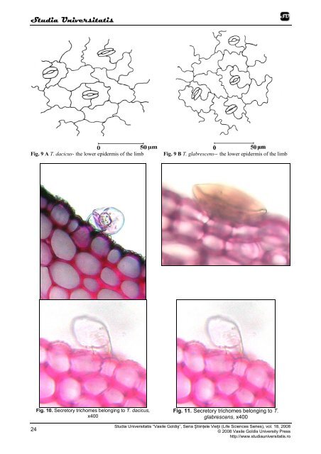
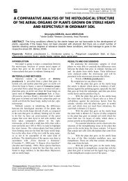
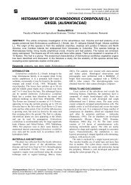
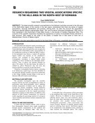
Studia Universitatis Fig. 9 A T. <strong>dacicus</strong>- the lower epidermis <strong>of</strong> the limb Fig. 9 B T. glabrescens-- the lower epidermis <strong>of</strong> the limb Fig. 10. Secretory trichomes belonging to T. <strong>dacicus</strong>, x400 Fig. 11. Secretory trichomes belonging to T. glabrescens, x400 24 Studia Universitatis “Vasile Goldiş”, Seria Ştiințele Vieţii (Life Sciences Series), vol. 18, 2008 © 2008 Vasile Goldis University Press http://www.studiauniversitatis.ro
Studia Universitatis In cross section, at both analyzed species, the mesophyll is formed by palisade tissue at the upper side and lacunary tissue at the lower one, so, the blade has a bifacial-heter<strong>of</strong>acial (dorsiventral) structure. At T. <strong>dacicus</strong>, the palisade tissue is bistratified, dense, with the hypodermic layer cells higher and with winding lateral walls. The lacunary tissue present 5-6 layers by rounded cells or fitful cells, with small aerifer lacunae between cells. To the border <strong>of</strong> foliar blade whole the mesophyll is on palisadyc type. The conducted vessels form more bundles, the biggest one presents at the periphery <strong>of</strong> the phloem cordons <strong>of</strong> sclerenchymatous fibers, with very thick walls but moderated lignified. At T. glabrescens, the palisade tissue is bistratified, with hypodermic layer cells higher. The lacunary tissue compass about 4 layers cells, isodiametric or tangent elongated, with small aerifer lacunas between them. REFERENCES Bailey Ciocârlan V., Flora ilustrată a României. Pteridophyta et Spermatophyta, Ed. Ceres, Bucureşti, pp. 670-675, 2000; Guşuleac M., Thymus, In Flora Republicii Populare Române, VIII, Ed. Acad. RPR, Bucureşti, pp. 301-334, 1961; Metcalfe C.R., Chalk L., Anatomy <strong>of</strong> Dicotyledons, Clarendon Press, Oxford, 2, pp. 1041-1053, 1950; Toma C., Berciu I., Morphological peculiaries <strong>of</strong> germination and structure <strong>of</strong> seedling in Thymus vulgaris L.; Romanian Biological Sciences, V, 1-2, pp. 136-137, 2007. Toma C., Rugină R., Anatomia plantelor medicinale. Atlas. Ed.Acad. Rom., Bucureşti, pp. 169-172, 1998. CONCLUSIONS At both analyzed species, the root presents a secondary structure, resulting for din activity on both lateral meristems: the cambium and the fellogen. On both analyzed species there are two types <strong>of</strong> trichomes: tector trichomes, more <strong>of</strong>ten multicelular, and secretors trichomes, always multicelular, consisting in a basal cell, a unicellular pedicel and a uni- or multicellular gland. The endoderm <strong>of</strong> Casparyan types became visible in the median third <strong>of</strong> the T. <strong>dacicus</strong> stem. At T. <strong>dacicus</strong> to the border <strong>of</strong> foliar blade whole the mesophyll is on palisadyc type. On both analyzed species, the stomata are diacitic type and there are presents on both sides <strong>of</strong> the foliar blade. Studia Universitatis “Vasile Goldiș”, Seria Ştiințele Vieţii (Life Sciences Series), vol. 18, 2008 © 2008 Vasile Goldis University Press http://www.studiauniversitatis.ro 25