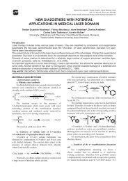
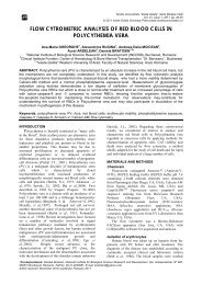
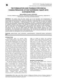
Read full article - Studia Universitatis Vasile Goldis, Seria Stiintele ...
Read full article - Studia Universitatis Vasile Goldis, Seria Stiintele ...
Read full article - Studia Universitatis Vasile Goldis, Seria Stiintele ...

Create successful ePaper yourself
Turn your PDF publications into a flip-book with our unique Google optimized e-Paper software.
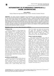
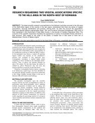
A comparative analysis of the histological structure of the aerial organs<br />
of plants grown on sterile heaps and respectively in ordinary soil<br />
normal soil, although the sclerenchyma, the<br />
collenchyma, and the liber tissue seem less developed.<br />
2. A stem of Centaurea cyanus L.<br />
The Centaurea genus is well represented on the<br />
sterile heaps, especially on the one from Sasar, not so<br />
much in the number of species, but mainly in the<br />
number of plants and the area on which they grow.<br />
This is the reason why I have decided to have<br />
microscopic samples of the stem, and to compare them<br />
with plants that grow in normal soil. Thus, one can<br />
observe that the plants from this species as well as<br />
other species that grow on the sterile, show a higher<br />
degree of weathering and ageing than the plants that<br />
grow in normal soil. Thus, at the level of the stem we<br />
can observe in the cross-section that:<br />
- With the plant that grew on the sterile heap<br />
the epiderm is badly exfoliated compared to the plant<br />
from the normal soil, which shows a higher degree of<br />
weathering, a thing which can be seen with the naked<br />
eye through macroscopic analysis. This exfoliation<br />
may be due to the anorganic dust which causes the<br />
dehydration of the cells leading to the detaching of<br />
some of them and thus creating flaws.<br />
- With the plant that grew on the sterile heap<br />
the outer bark is also more exfoliated than with the one<br />
that grew in normal soil (which also has 3-4 layers of<br />
cells), thus continuing the process started in the<br />
epiderm.<br />
- With the plant that grew on the sterile heap<br />
the inner bark shows no flaws, but shows a slight<br />
tendency towards growing thinner (2-3 layers)<br />
compared to the plant that grew in normal soil (3-4<br />
layers) most probably as a manifestation of the lack of<br />
the nourishing substances that the sterile soil has not<br />
been able to offer.<br />
- With the plant that grew on the sterile heap<br />
the medullary rays are narrower compared to the plant<br />
that grew in normal farming soil with which the rays<br />
are normal up to the bark.<br />
- With the plant that grew on the sterile heap<br />
the ligule (which contains more metaxylem) and the<br />
liber are narrower than normal, having denser fascicles,<br />
although it shows no other visible alterations, but the<br />
majority of tissues show the same direction determined<br />
by the scarcity of nourishing substances, which the<br />
sterile heap is incapable of ensuring if there is no<br />
adequate phyto-soil placating. In the witness plant, the<br />
ligule has metaxylem at the inside and protoxylem at<br />
the outside, the liber representing one third of the ligule<br />
thickness.<br />
- With the plant that grew on the sterile heap as<br />
a reaction of defence against pollutant factors, and the<br />
poor nourishing conditions, the strenghthening tissue<br />
has a tendency of growth (being almost uninterrupted<br />
except for some very thin medullary rays) taking up<br />
some of the space of the medullary rays,too, a<br />
manifestation which can be observed in many other<br />
plants that grow in a polluted environment. With the<br />
witness plant, the strenghthening tissue is continual,set<br />
among the perixyl.<br />
- With the plant that grew on the sterile heap<br />
the medulla is normal, but has smaller cells than the<br />
witness plant.<br />
3. Aerial stem of Polygonum cuspidatum Sieb. et<br />
Zucc. (Reynoutria japonica Houtt.).<br />
The aerial stem of Polygonum cuspidatum Sieb. et<br />
Zucc. (Reynoutria japonica Houtt.) is at least similar at<br />
the plant from the sterile soil to the plant grown in<br />
normal soil. Thus:<br />
- With the plant that grew on the sterile heap,<br />
(Fig. 11), the cuticle is somehow thicker than with the<br />
plant grown in normal soil (fig. 12), against the<br />
background of the perrenial self-defence against an<br />
aggressive environment.<br />
- With the plant that grew on the sterile heap<br />
the bark has several chloroplasts, 7-8 layers of cells<br />
similar to the one from the witness plant, showing no<br />
clear decelable microscopic alterations,except for the<br />
fact that it is thinner.<br />
- With the plant that grew on the sterile heap<br />
the strenghthening tissue is better developed and more<br />
continual, rarely interrupted by medullary rays, against<br />
the same tendency of defence against the environment<br />
factors, whereas with the witness plant it is<br />
discontinual due to the perixyls (Fig.10).<br />
- With the plant that grew on the sterile heap,<br />
the liber is more clearly fragmented than with the plant<br />
from the normal soil, a character which we have<br />
generally encountered at the plants from sterile soils,<br />
probably because of a sudden dehydration due to the<br />
loss of water at the level of the sterile substratum on<br />
the hill side.<br />
- With the plant that grew on the sterile heap,<br />
the ligule contains less metaxylem, that is the ducts<br />
have not been able to grow to this stage, probably<br />
because of the lack of nutrients, besides the ample<br />
dehydration, whereas with the witness plant the ligulw<br />
has less than 20 % very compact metaxylem.<br />
- With the plant that grew on the sterile heap<br />
the medullary rays are unaltered compared to normal<br />
(Fig.9), being clear, up to the strenghthening tissue.<br />
With the plant that grew on the sterile heap the<br />
medulla has somewhat smaller cells than the normal<br />
plant, but no major differences are evident, except that<br />
the cells are larger in the centre, with fewer<br />
amyloplasts, the cells being smaller at the exterior and<br />
having more amyloplasts.<br />
<strong>Studia</strong> <strong>Universitatis</strong> “<strong>Vasile</strong> Goldiş”, <strong>Seria</strong> Ştiințele Vieţii<br />
Vol. 19, issue 1, 2009, pp. 171-176<br />
© 2009 <strong>Vasile</strong> <strong>Goldis</strong> University Press (www.studiauniversitatis.ro)<br />
173