- Page 1 and 2: THE MOLECULAR ARCHITECTURE OF MAMES
- Page 3 and 4: "Bugs are not going to inherit the
- Page 5 and 6: DEDICATION This Ph.D thesis is dedi
- Page 7 and 8: ACKNOWLEDGMENTS I owe my deepest gr
- Page 9 and 10: 2.1.1 Mamestra configurata ........
- Page 11 and 12: 4.3.3.3.1 Alkaline phosphatase ....
- Page 13 and 14: 5.3.3.4 Localization...............
- Page 15 and 16: 8.2.1 Preparation of dsRNA.........
- Page 17 and 18: LIST OF FIGURES Figure 1.1 The alim
- Page 19 and 20: Figure 5.12 Two dimensional gel ele
- Page 21 and 22: LIST OF ABBREVIATIONS Ac...........
- Page 23 and 24: Ld ................................
- Page 25 and 26: wt.................................
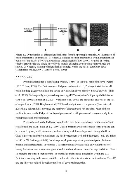
- Page 27: 1.2 The Peritrophic Matrix The pres
- Page 31 and 32: 1.2.3.1 Mechanical protection and s
- Page 33 and 34: 1.2.3.3 Compartmentalization of dig
- Page 35 and 36: the anterior midgut and disintegrat
- Page 37 and 38: of larvae reared on the leaves of r
- Page 39 and 40: 2. GENERAL MATERIAL AND METHODS Thi
- Page 41 and 42: Blots were prehybridized, hybridize
- Page 43 and 44: samples were mixed with loading buf
- Page 45 and 46: over the m/z range 50 to 1900. LC-M
- Page 47 and 48: acetylglucosamine (UDP-GlcNAc) as t
- Page 49 and 50: For SEM analyses, PMs were dissecte
- Page 51 and 52: Chapter 2) was conducted using tota
- Page 53 and 54: significant difference was detected
- Page 55 and 56: The PMs from all three stages studi
- Page 57 and 58: amino acids (Figure 3.4) including
- Page 59 and 60: 1 McCHS-B MATKPKTPGFTGLGDDSEDESEYTP
- Page 61 and 62: 36 1 70 AiCHI MKAILATLAVLAVVTTAIEAD
- Page 63 and 64: 38 1 70 AiNAG MWLQKYSLCAVYITLLSVICV
- Page 65 and 66: 3.3.4 Expression Analysis of the Ge
- Page 67 and 68: Figure 3.10 Expression of Mamestra
- Page 69 and 70: H. virescens (Ryerse et al., 1992)
- Page 71 and 72: 3.4.3 Tissue Specific Expression of
- Page 73 and 74: (Chamankhah et al., 2003), McSerpin
- Page 75 and 76: 4. SURVEY AND PRELIMINARY CHARACTER
- Page 77 and 78: Table 4.1 RT-PCR primers used in ex
- Page 79 and 80:
Table 4.2 Proteins identified from
- Page 81 and 82:
Figure 4.2 Two dimensional gel elec
- Page 83 and 84:
Figure 4.3 Expression of Mamestra c
- Page 85 and 86:
Trypsins Chymotrypsins Elastase 57
- Page 87 and 88:
(Figure 4.5 continued) 351 420 McAP
- Page 89 and 90:
(Figure 4.5 continued) 1051 1102 Mc
- Page 91 and 92:
4.3.3.2.3 Insect intestinal lipases
- Page 93 and 94:
4.3.3.2.5 α-Amylase Mascot analysi
- Page 95 and 96:
4.3.4.3 REPAT Mascot analysis ident
- Page 97 and 98:
Appendix A). All proteins were pred
- Page 99 and 100:
eported to be restricted to midgut
- Page 101 and 102:
and Ellar, 2007; Angelucci et al.,
- Page 103 and 104:
the M. configurata PM. McALP1 is pr
- Page 105 and 106:
larvae; whereas HMG176 is expressed
- Page 107 and 108:
4.4.4 Proteins without Orthologs Th
- Page 109 and 110:
5.2 Material and Methods 5.2.1 Rapi
- Page 111 and 112:
Table 5.2 (continued) Primer 1 Sequ
- Page 113 and 114:
uffer (without β-mercaptoethanol)
- Page 115 and 116:
5.3.2 Insect Intestinal Mucins (McI
- Page 117 and 118:
proline (9.3%), histidine (11.1%),
- Page 119 and 120:
molting larvae (Figure 5.4B). A low
- Page 121 and 122:
Figure 5.6 One dimensional (1D) and
- Page 123 and 124:
(Figure 5.7 continued) 421 429 BmCD
- Page 125 and 126:
meliloti (RhiNODB) and Colletotrich
- Page 127 and 128:
5.3.3.5 Demonstration of chitin dea
- Page 129 and 130:
similar colour changes, indicating
- Page 131 and 132:
5.3.4.3 Gene expression Expression
- Page 133 and 134:
transfer to a gelatin impregnated p
- Page 135 and 136:
produced and stored in secretory ve
- Page 137 and 138:
The results of CDA activity assays
- Page 139 and 140:
catalytically active. Of the 10 lip
- Page 141 and 142:
6. SURVEY, CHARACTERIZATION AND EVO
- Page 143 and 144:
PAD, conserved aromatic amino acids
- Page 145 and 146:
6.2.5 Antisera Production, Protein
- Page 147 and 148:
McPPAD1 MIAKFLTTVL LLNVVLTAEI PQKNA
- Page 149 and 150:
with the 951 bp cDNA. The McPPAD2 p
- Page 151 and 152:
The anti-rMcPPAD1 antiserum reacted
- Page 153 and 154:
Four types of PPADs were identified
- Page 155 and 156:
Figure 6.7 (legend is on the next p
- Page 157 and 158:
Figure 6.8 Model showing the propos
- Page 159 and 160:
Figure 6.9 Modeling of Mamestra con
- Page 161 and 162:
6.4.1.2 Expression Genes encoding P
- Page 163 and 164:
Table 6.4 (continued) Species Prote
- Page 165 and 166:
however, this is not present in McP
- Page 167 and 168:
that bonds could form between C2 an
- Page 169 and 170:
to form a bond with C3 in the eight
- Page 171 and 172:
Mamestra configurata nucleopolyhedr
- Page 173 and 174:
solution. In parallel, PMs without
- Page 175 and 176:
Figure 7.1 Analysis of strongly ass
- Page 177 and 178:
Figure 7.3 Analysis of Mamestra con
- Page 179 and 180:
kDa representing the degradation pr
- Page 181 and 182:
8. TARGETING GENES INVOLVED IN PERI
- Page 183 and 184:
Table 8.1 RT-PCR primers used for a
- Page 185 and 186:
50 µl 0.1% Triton-100 in nuclease
- Page 187 and 188:
Figure 8.1 RT-PCR analysis to detec
- Page 189 and 190:
clear as the same dosages and dsRNA
- Page 191 and 192:
RNAi to be initiated by dsRNA feedi
- Page 193 and 194:
2003), which could inhibit the acti
- Page 195 and 196:
in vitro (Cho et al., 2000), which
- Page 197 and 198:
glycan, two GlcNAc residues are lin
- Page 199 and 200:
The current study is also the first
- Page 201 and 202:
TcCHS2 are specialized for synthesi
- Page 203 and 204:
Blair, D.E., Hekmat, O., Schuttelko
- Page 205 and 206:
Chapman, R.F., 1985. Structure of t
- Page 207 and 208:
Dong, D-J., He, H-J., Chai, L-Q., W
- Page 209 and 210:
Filho, B.P.D., Lemos, F.J.A., Secun
- Page 211 and 212:
Grossi de Sa, M.F., Chrispeels, M.J
- Page 213 and 214:
Huang, X., Madan, A., 1999. CAP3: A
- Page 215 and 216:
Kim, M.G., Shin, S.W., Bae, K.S., K
- Page 217 and 218:
Lepore, L.S., Roelvink, P.R., Grana
- Page 219 and 220:
novel, very large fluorescent lipoc
- Page 221 and 222:
Palli, S.R., Locke, M., 1987. Hemol
- Page 223 and 224:
Ponnuvel, K.M., Nakazawa, H., Furuk
- Page 225 and 226:
Schorderet, S., Pearson, R.D., Vuoc
- Page 227 and 228:
Strobl, S., Maskos, K., Betz, M., W
- Page 229 and 230:
Tomoyasu, Y., Miller, S.C., Tomita,
- Page 231 and 232:
Wang, S., Jayaram, A.S., Hemphala,
- Page 233 and 234:
Zhou, H-X., Tan, X-M., Li, C-Y., Wa
- Page 235 and 236:
Appendix A. (continued) Protein Acc
- Page 237 and 238:
Appendix A. (continued) Protein Acc
- Page 239 and 240:
Appendix A. (continued) Protein Acc
- Page 241 and 242:
Appendix A. (continued) Protein Acc
- Page 243 and 244:
218 Appendix B. Amino acid composit
- Page 245 and 246:
220 Appendix D. Data from serine pr
- Page 247 and 248:
Appendix F. Domain organization and
- Page 249 and 250:
Appendix F. (continued) Spodoptera
- Page 251 and 252:
Appendix F. (continued) The colour
- Page 253 and 254:
Appendix G. (continued) 27. McPM1 C
- Page 255 and 256:
Appendix G. (continued) 81. HaIIM4
- Page 257 and 258:
Appendix G. (continued) 135. SeCBP6
- Page 259 and 260:
234 Appendix I. Tandem repeats with