Rapid detection of Dirofilaria immitis in mosquito vectors and dogs ...
Rapid detection of Dirofilaria immitis in mosquito vectors and dogs ...
Rapid detection of Dirofilaria immitis in mosquito vectors and dogs ...

You also want an ePaper? Increase the reach of your titles
YUMPU automatically turns print PDFs into web optimized ePapers that Google loves.
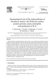
Veter<strong>in</strong>ary Parasitology 168 (2010) 255–260<br />
Contents lists available at ScienceDirect<br />
Veter<strong>in</strong>ary Parasitology<br />
journal homepage: www.elsevier.com/locate/vetpar<br />
<strong>Rapid</strong> <strong>detection</strong> <strong>of</strong> Dir<strong>of</strong>ilaria <strong>immitis</strong> <strong>in</strong> <strong>mosquito</strong> <strong>vectors</strong> <strong>and</strong> <strong>dogs</strong><br />
us<strong>in</strong>g a real-time fluorescence resonance energy transfer PCR <strong>and</strong><br />
melt<strong>in</strong>g curve analysis<br />
Tongjit Thanchomnang a , Pewpan M. Intapan a , Viraphong Lulitanond b ,<br />
Somboon Sangmaneedet c , Sudchit Chungpivat d , Piyanan Taweethavonsawat d ,<br />
Wej Choochote e , Wanchai Maleewong a, *<br />
a Department <strong>of</strong> Parasitology, Faculty <strong>of</strong> Medic<strong>in</strong>e <strong>and</strong> Diagnostic Center for Emerg<strong>in</strong>g Infectious Diseases, Khon Kaen University, Khon Kaen 40002, Thail<strong>and</strong><br />
b Department <strong>of</strong> Microbiology, Faculty <strong>of</strong> Medic<strong>in</strong>e <strong>and</strong> Diagnostic Center for Emerg<strong>in</strong>g Infectious Diseases, Khon Kaen University, Khon Kaen 40002, Thail<strong>and</strong><br />
c Department <strong>of</strong> Pathology, Faculty <strong>of</strong> Veter<strong>in</strong>ary Science, Khon Kaen University, Khon Kaen 40002, Thail<strong>and</strong><br />
d Department <strong>of</strong> Pathology, Faculty <strong>of</strong> Veter<strong>in</strong>ary Science, Chulalongkorn University, Bangkok 10330, Thail<strong>and</strong><br />
e Department <strong>of</strong> Parasitology, Faculty <strong>of</strong> Medic<strong>in</strong>e, Chiang Mai University, Chiang Mai 50200, Thail<strong>and</strong><br />
ARTICLE<br />
INFO<br />
ABSTRACT<br />
Article history:<br />
Received 13 October 2009<br />
Received <strong>in</strong> revised form 27 November 2009<br />
Accepted 7 December 2009<br />
Keywords:<br />
Dir<strong>of</strong>ilaria <strong>immitis</strong><br />
Detection<br />
Dogs<br />
Mosquito<br />
Parasite<br />
Real-time PCR<br />
A real-time fluorescence resonance energy transfer (FRET) PCR supplemented with<br />
melt<strong>in</strong>g curve analysis for the rapid molecular <strong>detection</strong> <strong>of</strong> Dir<strong>of</strong>ilaria <strong>immitis</strong> <strong>in</strong> <strong>mosquito</strong><br />
<strong>vectors</strong> <strong>and</strong> dog blood samples was developed. This real-time FRET PCR was based on the<br />
fluorescence melt<strong>in</strong>g curve analysis <strong>of</strong> a hybrid between an amplicon generated from the<br />
D. <strong>immitis</strong> ribosomal RNA gene sequence <strong>and</strong> specific fluorophore-labeled probes. The<br />
sensitivity, specificity, accuracy, <strong>and</strong> positive <strong>and</strong> negative predictive values <strong>of</strong> this<br />
method were all 100%. Besides be<strong>in</strong>g highly sensitive <strong>and</strong> specific, this PCR is fast <strong>and</strong><br />
<strong>of</strong>fers a high throughput. Therefore it is a suitable <strong>and</strong> powerful tool for the diagnosis <strong>and</strong><br />
for epidemiological surveys <strong>of</strong> can<strong>in</strong>e dir<strong>of</strong>ilariasis as well as for molecular xenomonitor<strong>in</strong>g<br />
<strong>of</strong> D. <strong>immitis</strong> <strong>in</strong> <strong>mosquito</strong> <strong>vectors</strong>.<br />
ß 2009 Elsevier B.V. All rights reserved.<br />
1. Introduction<br />
The heartworm Dir<strong>of</strong>ilaria <strong>immitis</strong> is a <strong>mosquito</strong>-borne<br />
pathogenic nematode <strong>of</strong> domestic <strong>dogs</strong> <strong>and</strong> some canids<br />
(McCall et al., 2008). The worm has also been found <strong>in</strong><br />
seals, horses, beavers, bears, nutrias <strong>and</strong> muskrats (Mar et<br />
al., 2002). Its life cycle consists <strong>of</strong> two phases, the first <strong>in</strong><br />
<strong>mosquito</strong>es <strong>and</strong> the second <strong>in</strong> the def<strong>in</strong>itive hosts. The<br />
sexually mature worms reside <strong>in</strong> the right side <strong>of</strong> the<br />
hearts <strong>of</strong> the def<strong>in</strong>itive hosts <strong>and</strong> release micr<strong>of</strong>ilariae <strong>in</strong>to<br />
the blood circulation. After susceptible <strong>mosquito</strong>es <strong>in</strong>gest<br />
* Correspond<strong>in</strong>g author at: Department <strong>of</strong> Parasitology, Faculty <strong>of</strong><br />
Medic<strong>in</strong>e, Khon Kaen University, Khon Kaen 40002, Thail<strong>and</strong>.<br />
Tel.: +66 43 348387; fax: +66 43 202475.<br />
E-mail address: wanch_ma@kku.ac.th (W. Maleewong).<br />
the larvae from a micr<strong>of</strong>ilaremic host, the worms develop<br />
to the <strong>in</strong>fective, third stage (L3) with<strong>in</strong> 10–14 days. When<br />
the <strong>in</strong>fective L3 are transmitted to the def<strong>in</strong>itive host by an<br />
<strong>in</strong>fected <strong>mosquito</strong> dur<strong>in</strong>g a blood meal, the parasites<br />
develop <strong>in</strong> the subcutaneous tissue, abdomen or thorax<br />
<strong>and</strong> migrate to the pulmonary arteries by day 90–120<br />
(McCall et al., 2008). People liv<strong>in</strong>g <strong>in</strong> heartworm endemic<br />
areas are at risk <strong>of</strong> be<strong>in</strong>g <strong>in</strong>fected by <strong>mosquito</strong>es (Orihel<br />
<strong>and</strong> Eberhard, 1998). Several reports have revealed<br />
occasional <strong>in</strong>fections <strong>of</strong> humans (Abadie et al., 1965;<br />
Moorhouse, 1978; Goldste<strong>in</strong> <strong>and</strong> Smith, 1985; Rodríguez<br />
et al., 2002) which is why this helm<strong>in</strong>thiasis could be<br />
regarded as a parasitic zoonosis.<br />
An important goal <strong>of</strong> the elim<strong>in</strong>ation programme for<br />
filariasis is the <strong>in</strong>terruption <strong>of</strong> the transmission <strong>of</strong> the<br />
<strong>in</strong>fection. Therefore, the availability <strong>of</strong> effective methods to<br />
monitor the presence or absence <strong>of</strong> filarial larvae <strong>in</strong> the<br />
0304-4017/$ – see front matter ß 2009 Elsevier B.V. All rights reserved.<br />
doi:10.1016/j.vetpar.2009.12.002
256<br />
T. Thanchomnang et al. / Veter<strong>in</strong>ary Parasitology 168 (2010) 255–260<br />
<strong>mosquito</strong> vector <strong>and</strong>/or micr<strong>of</strong>ilaria <strong>in</strong> the def<strong>in</strong>itive hosts<br />
is important. Microscopical methods for the <strong>detection</strong> <strong>of</strong><br />
filarial larvae <strong>in</strong> <strong>mosquito</strong> <strong>vectors</strong> <strong>and</strong>/or <strong>of</strong> micr<strong>of</strong>ilaria <strong>in</strong><br />
the blood circulation are labour-<strong>in</strong>tensive, have a low<br />
sensitivity <strong>and</strong> require highly experienced personnel.<br />
Serological tests based on enzyme-liked immunosorbent<br />
assays <strong>and</strong> immunochromatography techniques are considered<br />
highly specific, but false negative results can occur<br />
<strong>in</strong> very light <strong>in</strong>fections (McCall et al., 2008).<br />
Conventional polymerase cha<strong>in</strong> reaction (c-PCR) assays<br />
us<strong>in</strong>g different primers have been developed to detect<br />
filarial DNA <strong>in</strong> def<strong>in</strong>itive hosts <strong>and</strong> <strong>mosquito</strong> <strong>vectors</strong> (Favia<br />
et al., 1996; Mar et al., 2002; Cancr<strong>in</strong>i et al., 2003; Casiraghi<br />
et al., 2006; Lee et al., 2007). The methods are sensitive <strong>and</strong><br />
accurate <strong>in</strong> discrim<strong>in</strong>at<strong>in</strong>g the micr<strong>of</strong>ilaria <strong>of</strong> the different<br />
filarial species that <strong>in</strong>fect <strong>dogs</strong>. However, all <strong>of</strong> these<br />
procedures dem<strong>and</strong> analysis by agarose gel electrophoresis<br />
which is slow, has a limited throughput <strong>and</strong> has a<br />
tendency to carry-over contam<strong>in</strong>ation as well as illusive<br />
results. Therefore this study was conducted to develop a<br />
real-time fluorescence resonance energy transfer (FRET)<br />
PCR comb<strong>in</strong>ed with a melt<strong>in</strong>g curve analysis which does<br />
not need agarose gel electrophoresis for the <strong>detection</strong> <strong>of</strong> D.<br />
<strong>immitis</strong> DNA <strong>in</strong> <strong>mosquito</strong> <strong>vectors</strong> <strong>and</strong> <strong>in</strong>fected dog blood<br />
samples.<br />
2. Materials <strong>and</strong> methods<br />
2.1. Mosquitoes <strong>and</strong> experimental <strong>in</strong>fection<br />
Aedes aegypti, Culex pipiens qu<strong>in</strong>quefasciatus, Mansonia<br />
uniformis, <strong>and</strong> Armigeres spp. <strong>mosquito</strong>es were collected <strong>in</strong><br />
urban areas <strong>of</strong> Khon Kaen Prov<strong>in</strong>ce, Northeastern Thail<strong>and</strong>.<br />
Additionally Aedes togoi was collected from Koh Nom Sao,<br />
Chanthaburi Prov<strong>in</strong>ce, Eastern Thail<strong>and</strong>. All <strong>mosquito</strong><br />
larvae were taken from their breed<strong>in</strong>g places <strong>and</strong> reared<br />
<strong>in</strong> an <strong>in</strong>sectarium. The Ae. aegypti, Ae. togoi <strong>and</strong> Cx.<br />
qu<strong>in</strong>quefasciatus <strong>mosquito</strong>es were artificially <strong>in</strong>fected with<br />
D. <strong>immitis</strong> (stra<strong>in</strong> from Khon Kaen Prov<strong>in</strong>ce), nocturnally<br />
subperiodic B. malayi (stra<strong>in</strong> from Narathiwat Prov<strong>in</strong>ce,<br />
Southern Thail<strong>and</strong>) (Maleewong et al., 1987) <strong>and</strong> nocturnally<br />
periodic W. bancr<strong>of</strong>ti (stra<strong>in</strong> from Burmese immigrant,<br />
Tak Prov<strong>in</strong>ce Northwestern Thail<strong>and</strong>) (Lulitanond et<br />
al., 2004), respectively.<br />
An 8-year-old mixed-breed female dog naturally<br />
<strong>in</strong>fected with D. <strong>immitis</strong> was used as micr<strong>of</strong>ilariae (mf)<br />
donor (54 mf/20 ml blood). Female 3–5-day-old <strong>mosquito</strong>es<br />
(fasted for 12 h) were allowed to feed on the D.<br />
<strong>immitis</strong>-<strong>in</strong>fected blood by membrane feed<strong>in</strong>g. Briefly, a<br />
culture flask conta<strong>in</strong><strong>in</strong>g warm water was covered with a<br />
Parafilm membrane <strong>and</strong> filled with <strong>in</strong>fected blood. The<br />
membrane feeder was placed over a netted <strong>mosquito</strong> cup<br />
<strong>and</strong> the <strong>mosquito</strong>es were permitted to feed on the blood<br />
for 1 h. The fed <strong>mosquito</strong>es were reared at 27 8C. To ensure<br />
<strong>in</strong>fectivity, each <strong>mosquito</strong> was dissected <strong>in</strong> normal sal<strong>in</strong>e<br />
solution 14 days post feed<strong>in</strong>g, <strong>and</strong> the number <strong>of</strong> larval<br />
worms found <strong>in</strong> each was counted under a stereomicroscope<br />
<strong>and</strong> recorded. Only <strong>in</strong>fected <strong>mosquito</strong>es were used<br />
for the experiments. After dissection, all D. <strong>immitis</strong> larvae,<br />
<strong>and</strong> the <strong>in</strong>sect’s body were mixed <strong>and</strong> put <strong>in</strong>to a 1.5-ml<br />
microcentrifuge tube, labeled, <strong>and</strong> kept at 20 * C for DNA<br />
extraction. The range, mean SD <strong>and</strong> median parasite<br />
number per <strong>in</strong>fected <strong>mosquito</strong> were 1–10, 5.40 3.23 <strong>and</strong><br />
5.50 larval worms, respectively.<br />
W. bancr<strong>of</strong>ti <strong>in</strong>fected Cx. qu<strong>in</strong>quefasciatus, B. malayi<br />
<strong>in</strong>fected Ae. togoi as well as non-<strong>in</strong>fected Ae. aegypti, non<strong>in</strong>fected<br />
Cx. qu<strong>in</strong>quefasciatus, non-<strong>in</strong>fected Ma. uniformis<br />
<strong>and</strong> non-<strong>in</strong>fected Armigeres spp. were used for DNA<br />
extraction. The eluted DNA was used as the source for<br />
specificity evaluation.<br />
2.2. Source <strong>of</strong> blood specimens <strong>and</strong> control DNAs for<br />
sensitivity <strong>and</strong> specificity<br />
A total <strong>of</strong> 71 EDTA dog blood samples were obta<strong>in</strong>ed<br />
from the Animal Hospital, Faculty <strong>of</strong> Veter<strong>in</strong>ary Science,<br />
Chulalongkorn University <strong>and</strong> the Animal Hospital, Faculty<br />
<strong>of</strong> Veter<strong>in</strong>ary Science, Khon Kaen University. The specimens<br />
were microscopically exam<strong>in</strong>ed <strong>and</strong> the presence <strong>of</strong><br />
parasite objects was confirmed by sta<strong>in</strong><strong>in</strong>g a thick blood<br />
film (20 ml blood/slide) with Giemsa (World Health<br />
Organization, 1991) <strong>and</strong> by special sta<strong>in</strong><strong>in</strong>g for acid<br />
phosphatase activity (Yen <strong>and</strong> Mak, 1978). Out <strong>of</strong> these<br />
71 samples, 30 were positive for D. <strong>immitis</strong> (mean<br />
micr<strong>of</strong>ilarial density SD = 29.73 3.85; median = 17.5;<br />
range = 1–158), 6 for Brugia pahangi, 2 for Trypanosoma<br />
evansi, 1 for Ehrlichia canis, 2 for Hepatozoon canis, 1 for E.<br />
canis <strong>and</strong> H. canis, 2 for E. canis <strong>and</strong> Babesia spp., <strong>and</strong> 1 for<br />
Babesia spp. Twenty-six negative blood samples from healthy<br />
<strong>dogs</strong> <strong>and</strong> one negative blood sample <strong>of</strong> a healthy cat were<br />
obta<strong>in</strong>ed as controls. Adult D. <strong>immitis</strong> were recovered from<br />
heart <strong>of</strong> <strong>in</strong>fected dog. All specimens were bl<strong>in</strong>dly labeled <strong>and</strong><br />
further processed by DNA extraction.<br />
One DNA specimen extracted from Plasmodium falciparum<br />
<strong>in</strong>fected human red blood cells was received from<br />
the frozen bank, Department <strong>of</strong> Parasitology, Faculty <strong>of</strong><br />
Medic<strong>in</strong>e, Khon Kaen University.<br />
The study protocol was approved by the Animal Ethics<br />
Committee <strong>of</strong> Khon Kaen University, based on the Ethics <strong>of</strong><br />
Animal Experimentation <strong>of</strong> the National Research Council<br />
<strong>of</strong> Thail<strong>and</strong> (Reference No. 0514.1.12.2/50).<br />
2.3. Preparation <strong>of</strong> specimens for real-time FRET PCR<br />
Each <strong>mosquito</strong> specimen, was put <strong>in</strong> a 1.5-ml microcentrifuge<br />
tube, homogenized with disposable polypropylene<br />
pestles (Bellco Glass, V<strong>in</strong>el<strong>and</strong>, NJ), <strong>and</strong> extracted<br />
us<strong>in</strong>g the Nucleosp<strong>in</strong> Tissue kit (Macherey-Nagel, Duren,<br />
Germany). The DNAs were eluted <strong>in</strong> 100 ml <strong>of</strong> 5 mM Tris–<br />
HCl, pH 8.5 <strong>and</strong> stored at 20 8C until analysis. 5 ml <strong>of</strong> each<br />
DNA was used per reaction. Regard<strong>in</strong>g blood samples,<br />
50 ml were used per sample for DNA extraction <strong>and</strong><br />
purification as above.<br />
2.4. Real-time FRET PCR assay<br />
The LightCycler PCR <strong>detection</strong> <strong>and</strong> analysis systems<br />
(LightCycler 2.0, Roche Applied Science, Mannhe<strong>in</strong>, Germany)<br />
were used for amplification <strong>and</strong> quantification. The<br />
reaction was performed <strong>in</strong> glass capillaries. Specific<br />
primers, i.e. DI-F (5 0 ATG ATG ATT GCT CAA TTA AGT<br />
AGA C 3 0 ) <strong>and</strong> DI-R (5 0 GAT AAT CTG ATC GAT ATT GAC CCT
T. Thanchomnang et al. / Veter<strong>in</strong>ary Parasitology 168 (2010) 255–260 257<br />
3 0 ) (Proligo, S<strong>in</strong>gapore), were designed to b<strong>in</strong>d to the D.<br />
<strong>immitis</strong> ribosomal RNA gene sequence (GenBank accession<br />
number AF217800). This target sequence was previously<br />
used to design highly sensitive <strong>and</strong> specific primers for the<br />
<strong>detection</strong> <strong>of</strong> D. <strong>immitis</strong> DNA by c-PCR (Mar et al., 2002).<br />
For amplification <strong>detection</strong>, the LightCycler FastStart<br />
DNA Master HybProbe Kit (Roche Applied Science) was<br />
used as recommended by the manufacturer. Briefly, a pair<br />
<strong>of</strong> adjacent oligoprobes was hybridized with the D. <strong>immitis</strong><br />
ribosomal RNA gene. One probe had been labeled at the 5 0<br />
end with the LightCycler Red 640 fluorophore (5 0 Red 640-<br />
GCT CGT GGA TCG ATG AAG AAC GCA GCT-Phosphate 3 0 )<br />
(DILC640 probe) <strong>and</strong> the other had been labeled at the 3 0<br />
end with 530 fluoresce<strong>in</strong> (5 0 ATT TTT CAA TAA CTC TAA<br />
GCG GGG GAT CAC C- Flou 530 3 0 ) (DIFL530 probe) (Tib<br />
Molbiol, Berl<strong>in</strong>, Germany). Probe <strong>and</strong> primers were<br />
designed by the LC probe design s<strong>of</strong>tware (Roche Applied<br />
Science). A schematic diagram <strong>of</strong> the hybridization<br />
analysis <strong>of</strong> the primers <strong>and</strong> probes is shown <strong>in</strong> Fig. 1.<br />
When the probes hybridized to the same DNA str<strong>and</strong><br />
<strong>in</strong>ternal to the PCR primers, the probes came <strong>in</strong> close<br />
proximity <strong>and</strong> produced a FRET (Intapan et al., 2009).<br />
The PCR mixture conta<strong>in</strong>ed LightCycler Faststart DNA<br />
Master HybProbe (Roche Applied Science), 2 mM MgCl 2 ,<br />
0.5 mM DI-F primer, 0.5 mM DI-R primer, 0.4 mM DILC640<br />
probe <strong>and</strong> 0.2 mM DIFL530 probe. The total reaction<br />
volume was 20 ml. The samples were run through 45<br />
cycles <strong>of</strong> repeated denaturation (10 s at 95 8C), anneal<strong>in</strong>g<br />
(15 s at 45 8C), <strong>and</strong> extension (15 s at 72 * C). The<br />
temperature transition rate was 20 8C/s. After amplification,<br />
a melt<strong>in</strong>g curve was produced by heat<strong>in</strong>g the product<br />
at 20 8C/s to 95 8C, cool<strong>in</strong>g it to 40 8C, keep<strong>in</strong>g it at 40 8C for<br />
30 s <strong>and</strong> then slowly heat<strong>in</strong>g it at 0.1 8C/s to 75 8C. The<br />
fluorescence <strong>in</strong>tensity change was measured throughout<br />
the slow heat<strong>in</strong>g phase.<br />
In order to determ<strong>in</strong>e the specificity <strong>of</strong> the oligonucleotide<br />
hybridization based on the FRET technique, DNA<br />
extracted from samples other than D. <strong>immitis</strong>-<strong>in</strong>fected<br />
<strong>mosquito</strong>es <strong>and</strong> <strong>in</strong>fected dog samples were analyzed<br />
separately. Each run conta<strong>in</strong>ed at least one negative<br />
control consist<strong>in</strong>g <strong>of</strong> 5 ml distilled water.<br />
For improved visualization <strong>of</strong> the melt<strong>in</strong>g temperatures<br />
(T m ), melt<strong>in</strong>g peaks were derived as previously described<br />
(Thanchomnang et al., 2008). Melt<strong>in</strong>g curves were used to<br />
determ<strong>in</strong>e the specific PCR products, which were confirmed<br />
by conventional gel electrophoresis. The cycle number (Cn)<br />
<strong>in</strong>dicat<strong>in</strong>g the target sequence copy number was presented<br />
as the number <strong>of</strong> PCR cycles needed for the fluorescence<br />
signal <strong>of</strong> the amplicons to exceed the detected threshold<br />
value. The correlation between the worm loads <strong>and</strong> the Cn<br />
was analyzed by Pearson Correlation Test.<br />
2.5. Dir<strong>of</strong>ilaria <strong>immitis</strong>-positive control plasmids<br />
A positive control plasmid was constructed by clon<strong>in</strong>g a<br />
PCR product <strong>of</strong> the D. <strong>immitis</strong> ribosomal DNA gene (Mar et<br />
al., 2002) <strong>in</strong>to the pCR4-TOPO vector (Invitrogen, Carlsbad,<br />
CA), accord<strong>in</strong>g to the manufacturer’s <strong>in</strong>structions. The PCR<br />
products were obta<strong>in</strong>ed by c-PCR us<strong>in</strong>g the primers DI-F<br />
<strong>and</strong> DI-R. The plasmid was propagated <strong>in</strong> Escherichia coli<br />
<strong>and</strong> purified by us<strong>in</strong>g the Wizard 1 plus SV M<strong>in</strong>ipreps kit<br />
(Promega Coporation, Madison, WI). The nucleotide<br />
sequence <strong>of</strong> the <strong>in</strong>serted gene was sequenced <strong>in</strong> both<br />
directions <strong>and</strong> revealed to be identical to the D. <strong>immitis</strong><br />
ribosomal RNA gene (GenBank accession number<br />
AF217800). The size <strong>of</strong> the plasmid was 4250 bp (<strong>in</strong>clud<strong>in</strong>g<br />
the 294-bp ribosomal RNA gene sequence).<br />
3. Results<br />
3.1. St<strong>and</strong>ardization <strong>of</strong> the real-time PCR<br />
The sensitivity <strong>of</strong> real-time FRET PCR was determ<strong>in</strong>ed<br />
us<strong>in</strong>g 5 ml <strong>of</strong> serial dilutions (4–4 10 8 copies) <strong>of</strong> D.<br />
<strong>immitis</strong>-positive control plasmid <strong>in</strong> water. The <strong>detection</strong><br />
limit <strong>of</strong> the ribosomal RNA gene target DNA sequence was<br />
4 10 4 copies <strong>of</strong> positive control plasmid (Fig. 2) or as little<br />
as 5 10 3 ng D. <strong>immitis</strong> genomic DNA (figure not shown)<br />
when consider<strong>in</strong>g 35 cycles as the cut-<strong>of</strong>f <strong>detection</strong> limit.<br />
No fluorescence signal was obta<strong>in</strong>ed when purified DNA<br />
from W. bancr<strong>of</strong>ti-<strong>in</strong>fected Cx. qu<strong>in</strong>quefasciatus, B. malayi<strong>in</strong>fected<br />
Ae. togoi, non-<strong>in</strong>fected <strong>mosquito</strong>es (Ae. aegypti, Cx.<br />
qu<strong>in</strong>quefasciatus, Ma. uniformis, <strong>and</strong> Armigeres spp.), non<strong>in</strong>fected<br />
dog <strong>and</strong> cat blood samples, dog blood samples with<br />
<strong>in</strong>fections other than D. <strong>immitis</strong>, orP. falciparum-<strong>in</strong>fected<br />
human red blood cells were tested. The capability <strong>of</strong><br />
<strong>detection</strong> was determ<strong>in</strong>ed with each pool <strong>of</strong> 1, 5, 10, 15<br />
<strong>and</strong> 20 un<strong>in</strong>fected Ae. aegypti adult <strong>mosquito</strong>es <strong>in</strong>oculated<br />
with one <strong>in</strong>fected <strong>mosquito</strong> that nourished one D. <strong>immitis</strong> L3.<br />
After DNA extraction <strong>and</strong> exam<strong>in</strong>ation <strong>of</strong> the real-time FRET<br />
PCR assay, this method could detect D. <strong>immitis</strong> DNA from as<br />
little as one D. <strong>immitis</strong> larva <strong>in</strong>fected <strong>in</strong> one <strong>mosquito</strong> vector<br />
spiked <strong>in</strong> 20 un<strong>in</strong>fected Ae. aegypti (Fig. 3).<br />
3.2. Real-time FRET PCR for the <strong>detection</strong> <strong>of</strong> D. <strong>immitis</strong> <strong>in</strong><br />
<strong>in</strong>fected <strong>mosquito</strong>es <strong>and</strong> <strong>dogs</strong><br />
Fig. 1. Schematic illustration <strong>of</strong> the PCR primers (DI-F <strong>and</strong> DI-R primers),<br />
anchor, <strong>and</strong> <strong>detection</strong> probes hybridized with the D. <strong>immitis</strong> ribosomal<br />
RNA gene (AF217800). The probe DIFL530 was labeled with 530<br />
fluoresce<strong>in</strong> at the 3 0 end <strong>and</strong> served as anchor probe for the sensor<br />
DILC640 probe. The sensor probe was labeled with LightCycler Red 640<br />
fluorophore (LC red 640) at the 5 0 end. S<strong>in</strong>gle circle, 530 fluoresce<strong>in</strong>;<br />
double circle, LC red 640. The left <strong>and</strong> right l<strong>in</strong>es <strong>in</strong>dicate the <strong>in</strong>ternal<br />
transcribed spacer region 1 (ITS1) <strong>and</strong> <strong>in</strong>ternal transcribed spacer region 2<br />
(ITS2), respectively whereas the middle dashed l<strong>in</strong>e <strong>in</strong>dicates the 5.8S<br />
ribosomal RNA gene.<br />
The method comb<strong>in</strong>ed with a melt<strong>in</strong>g curve analysis <strong>of</strong><br />
the amplicon products was applied to detect D. <strong>immitis</strong><br />
DNA <strong>in</strong> <strong>in</strong>fected <strong>mosquito</strong>es <strong>and</strong> <strong>dogs</strong>. A total <strong>of</strong> 30 D.<br />
<strong>immitis</strong>-<strong>in</strong>fected <strong>and</strong> 30 non-<strong>in</strong>fected Ae. aegypti as well as<br />
30 D. <strong>immitis</strong>-<strong>in</strong>fected <strong>and</strong> 26 non-<strong>in</strong>fected dog samples<br />
<strong>and</strong> 15 dog samples <strong>in</strong>fected with other biological agents<br />
were separately analyzed. The melt<strong>in</strong>g curve analyses are<br />
shown <strong>in</strong> Fig. 4. When us<strong>in</strong>g D. <strong>immitis</strong>-specific primers<br />
<strong>and</strong> probes, the mean SD, range <strong>and</strong> the median <strong>of</strong> the T m
258<br />
T. Thanchomnang et al. / Veter<strong>in</strong>ary Parasitology 168 (2010) 255–260<br />
Fig. 2. Amplification plot <strong>of</strong> fluorescence (y-axis) vs cycle numbers (x-axis) show<strong>in</strong>g the analytical sensitivity <strong>of</strong> the real-time FRET PCR for detect<strong>in</strong>g D.<br />
<strong>immitis</strong> plasmid DNA. (A) D. <strong>immitis</strong> 4 10 8 copies/reaction; (B) D. <strong>immitis</strong> plasmid 4 10 7 copies/reaction; (C) D. <strong>immitis</strong> plasmid 4 10 6 copies/reaction;<br />
(D) D. <strong>immitis</strong> plasmid 4 10 5 copies/reaction; (E) D. <strong>immitis</strong> plasmid 4 10 4 copies/reaction; (F) D. <strong>immitis</strong> plasmid 4 10 3 copies/reaction; (G) D. <strong>immitis</strong><br />
plasmid 4 10 2 ,4 10, 4 copies/reaction, <strong>and</strong> distilled water, respectively.<br />
Fig. 3. Amplification plot <strong>of</strong> fluorescence (y-axis) vs cycle numbers (x-axis) show<strong>in</strong>g the analytical sensitivity <strong>of</strong> the real-time FRET PCR for detect<strong>in</strong>g one D.<br />
<strong>immitis</strong> larva harbored <strong>in</strong> one <strong>in</strong>fected <strong>mosquito</strong> <strong>in</strong>oculated <strong>in</strong> pools <strong>of</strong> 1, 5, 10, 15 <strong>and</strong> 20 non-<strong>in</strong>fected <strong>mosquito</strong>es (left figure) <strong>and</strong> the representative<br />
melt<strong>in</strong>g curve analysis <strong>of</strong> two fluorophore-labeled probes hybridized to the amplification products <strong>of</strong> the D. <strong>immitis</strong> ribosomal RNA gene. The melt<strong>in</strong>g<br />
temperature (T m ) <strong>of</strong> the double-str<strong>and</strong>ed fragment is visualized by plott<strong>in</strong>g the negative derivative <strong>of</strong> the change <strong>in</strong> fluorescence divided by the change <strong>in</strong><br />
temperature <strong>in</strong> relation to the temperature [ (d/dT) Fluorescence (640/530)]. The turn<strong>in</strong>g po<strong>in</strong>t <strong>of</strong> this converted melt<strong>in</strong>g curve results <strong>in</strong> a peak <strong>and</strong><br />
permits identification <strong>of</strong> the fragment specific T m (right figure). (A) D. <strong>immitis</strong> positive control plasmid (4 10 7 copies/reaction); (B)–(F) are the results <strong>of</strong><br />
DNA extracted from one D. <strong>immitis</strong> larva harbored <strong>in</strong> one <strong>in</strong>fected <strong>mosquito</strong> <strong>in</strong>oculated <strong>in</strong> 1, 5, 10, 15 <strong>and</strong> 20 <strong>of</strong> non-<strong>in</strong>fected <strong>mosquito</strong>es, respectively; (G)<br />
negative control conta<strong>in</strong><strong>in</strong>g no DNA.
T. Thanchomnang et al. / Veter<strong>in</strong>ary Parasitology 168 (2010) 255–260 259<br />
Fig. 4. Representative melt<strong>in</strong>g curve analyses <strong>of</strong> two fluorophore-labeled probes hybridized to the amplification products <strong>of</strong> D. <strong>immitis</strong> ribosomal RNA gene.<br />
The left figure shows the melt<strong>in</strong>g peaks <strong>of</strong> D. <strong>immitis</strong> <strong>in</strong>fected <strong>mosquito</strong>es (A), D. <strong>immitis</strong> <strong>in</strong>fected dog blood (B), a positive control plasmid (C), D. <strong>immitis</strong><br />
genomic DNA (D), non-<strong>in</strong>fected Ae. aegypti, non-<strong>in</strong>fected Cx. qu<strong>in</strong>quefasciatus, W. bancr<strong>of</strong>ti-<strong>in</strong>fected <strong>and</strong> B. malayi-<strong>in</strong>fected <strong>mosquito</strong>es DNA <strong>and</strong> the negative<br />
control conta<strong>in</strong><strong>in</strong>g no DNA (E). The right figure shows the melt<strong>in</strong>g peaks <strong>of</strong> a positive control plasmid (F) <strong>and</strong> P. falciparum-<strong>in</strong>fected human red blood cells,<br />
non-<strong>in</strong>fected cat <strong>and</strong> dog blood samples, B. pahangi-, Babesia spp.-, H. canis-, E. canis-, <strong>and</strong> T. evansi-<strong>in</strong>fected dog blood DNA, respectively, as well as non<strong>in</strong>fected<br />
Armigeres spp. <strong>and</strong> non-<strong>in</strong>fected Ma. uniformis (G) DNA.<br />
Fig. 5. Ethidium bromide sta<strong>in</strong><strong>in</strong>g patterns <strong>of</strong> the PCR products on a 1% agarose gel. The arrows <strong>in</strong>dicate the 294 bp D. <strong>immitis</strong> specific b<strong>and</strong>s. Negative control<br />
conta<strong>in</strong><strong>in</strong>g no DNA (lanes 1 <strong>and</strong> 11); the PCR products obta<strong>in</strong>ed from the positive control plasmid (lanes 2 <strong>and</strong> 12); D. <strong>immitis</strong> genomic DNA (lane 3); D.<br />
<strong>immitis</strong>-<strong>in</strong>fected <strong>mosquito</strong>es (lane 4); D. <strong>immitis</strong>-<strong>in</strong>fected dog sample (lane 5); non-<strong>in</strong>fected Ae. aegypti (lane 6); non-<strong>in</strong>fected Cx. qu<strong>in</strong>quefasciatus (lane 7);<br />
non-<strong>in</strong>fected dog blood (lane 8); W. bancr<strong>of</strong>ti-<strong>in</strong>fected (lane 9) <strong>and</strong> B. malayi-<strong>in</strong>fected <strong>mosquito</strong>es (lane 10); P. falciparum-<strong>in</strong>fected human red blood cells<br />
(lane 13); B. pahangi <strong>in</strong>fected dog blood (lane 14); non-<strong>in</strong>fected cat blood (lane 15); Babesia spp.-<strong>in</strong>fected (lane 16), H. canis-<strong>in</strong>fected (lane 17), E. canis<strong>in</strong>fected<br />
(lane 18) <strong>and</strong> T. evansi -<strong>in</strong>fected dog blood samples (lane 19); non-<strong>in</strong>fected Armigeres spp. (lane 20) <strong>and</strong> non-<strong>in</strong>fected Ma. uniformis (lane 21); lane<br />
M, DNA size markers (1 kb plus DNA ladder from Invitrogen).<br />
values <strong>of</strong> the D. <strong>immitis</strong>-<strong>in</strong>fected <strong>mosquito</strong>es were<br />
58.40 0.11, 58.30–58.58 <strong>and</strong> 58.35, respectively <strong>and</strong> <strong>of</strong><br />
the D. <strong>immitis</strong>-<strong>in</strong>fected <strong>dogs</strong> were 58.52 0.13, 58.47–58.79<br />
<strong>and</strong> 58.53, respectively. A total <strong>of</strong> 30 D. <strong>immitis</strong>-<strong>in</strong>fected Ae.<br />
aegypti (Cn range 24.90–31.0; mean SD = 27.90 2.34;<br />
median = 27.39) <strong>and</strong> 30 D. <strong>immitis</strong>-<strong>in</strong>fected dog samples<br />
(Cn range 23.37–34.40; mean SD = 26.60 3.45; median<br />
= 27.30) were positive by real-time FRET PCR <strong>and</strong> melt<strong>in</strong>g<br />
curve analysis, whereas all <strong>of</strong> the specificity control DNA was<br />
negative. The sensitivity, specificity, accuracy <strong>and</strong> positive<br />
<strong>and</strong> negative predictive values were all 100%.<br />
For validation <strong>of</strong> this method, the DNAs from the D.<br />
<strong>immitis</strong>-<strong>in</strong>fected <strong>mosquito</strong>es, the D. <strong>immitis</strong>-<strong>in</strong>fected <strong>dogs</strong>,<br />
the D. <strong>immitis</strong> genomic DNA, <strong>and</strong> positive control plasmids<br />
were amplified by real-time FRET PCR (Fig. 5, lanes 2–5), to<br />
verify the presence <strong>of</strong> the prom<strong>in</strong>ent 294-bp product,<br />
whereas genomic DNA from other specificity control DNAs<br />
were not amplified. However, when the genomic DNAs from<br />
non-<strong>in</strong>fected dog samples (Fig. 5, lane 8), W. bancr<strong>of</strong>ti<strong>in</strong>fected<br />
Cx. qu<strong>in</strong>quefasciatus (Fig. 5, lanes 9), dog blood<br />
<strong>in</strong>fected with B. pahangi (Fig. 5, lane 14), Babesia spp. (Fig. 5,<br />
lane 16), H. canis (Fig. 5,lane17),E. canis (Fig. 5, lane 18), T.<br />
evansi (Fig. 5, lane 19) <strong>and</strong> non-<strong>in</strong>fected cat blood (Fig. 5, lane<br />
15) were amplified the fa<strong>in</strong>t b<strong>and</strong>s but no specific<br />
fluorescence signal was detected dur<strong>in</strong>g melt<strong>in</strong>g peak<br />
analysis. Significant correlations between the Cn <strong>and</strong> the<br />
micr<strong>of</strong>ilarial load <strong>in</strong> D. <strong>immitis</strong>-<strong>in</strong>fected dog bloods<br />
(R = 0.553; P < 0.05) <strong>and</strong> between the Cn <strong>and</strong> the larval<br />
load <strong>in</strong> <strong>in</strong>fected <strong>mosquito</strong>es (R = 0.591; P < 0.05) were<br />
revealed. These results show that real-time FRET PCR can<br />
present <strong>in</strong>formationonlarvaldensities<strong>in</strong>can<strong>in</strong>e blood<strong>and</strong><strong>in</strong><br />
<strong>vectors</strong>.<br />
4. Discussion<br />
With regards to animal health, the can<strong>in</strong>e <strong>and</strong> fel<strong>in</strong>e<br />
heartworm disease caused by adult D. <strong>immitis</strong> worms is<br />
severe <strong>and</strong> potentially fatal. Laboratory diagnosis <strong>of</strong> the<br />
disease can be done us<strong>in</strong>g blood tests that detect<br />
circulat<strong>in</strong>g micr<strong>of</strong>ilariae or adult antigen, however other
260<br />
T. Thanchomnang et al. / Veter<strong>in</strong>ary Parasitology 168 (2010) 255–260<br />
diagnostic tests are needed to <strong>in</strong>vestigate the severity <strong>of</strong><br />
the disease <strong>and</strong> treatment options (McCall et al., 2008).<br />
Molecular methods to detect filarial DNA are sensitive <strong>and</strong><br />
accurate tools for the differential diagnosis <strong>and</strong> are helpful<br />
<strong>in</strong> cases <strong>of</strong> morphological abnormalities <strong>of</strong> the micr<strong>of</strong>ilariae.<br />
Moreover, molecular procedures are useful for the<br />
survey <strong>of</strong> filarial <strong>in</strong>fections <strong>in</strong> <strong>vectors</strong> (Lee et al., 2007) <strong>and</strong><br />
have potential for mapp<strong>in</strong>g endemic areas <strong>of</strong> diseases.<br />
These methods can be used for surveillance based control<br />
<strong>of</strong> arthropods <strong>and</strong> for the management <strong>of</strong> such vectorborne<br />
diseases.<br />
Recently, quantitative real-time PCR was reported for<br />
<strong>detection</strong> <strong>of</strong> D. repens <strong>in</strong> Austrian <strong>dogs</strong> (Duscher et al.,<br />
2009). The real-time FRET PCR, a variation <strong>of</strong> the real-time<br />
PCR, is be<strong>in</strong>g used effectively for the rapid molecular<br />
<strong>detection</strong> <strong>of</strong> W. bancr<strong>of</strong>ti <strong>and</strong>/or B. malayi DNAs <strong>in</strong><br />
<strong>mosquito</strong> <strong>vectors</strong> (Lulitanond et al., 2004; Thanchomnang<br />
et al., 2008; Intapan et al., 2009). Here, we reported for the<br />
first time a real-time PCR based on two pairs <strong>of</strong> specific<br />
primers <strong>and</strong> <strong>in</strong>dividually labelled hybridization probes<br />
merged with melt<strong>in</strong>g curve analysis for the <strong>detection</strong> <strong>of</strong> D.<br />
<strong>immitis</strong> <strong>in</strong>fection <strong>in</strong> <strong>mosquito</strong> <strong>vectors</strong> <strong>and</strong> <strong>dogs</strong>. As little as<br />
one s<strong>in</strong>gle D. <strong>immitis</strong> L3 <strong>in</strong>fection <strong>of</strong> one <strong>mosquito</strong> mixed <strong>in</strong><br />
20 un<strong>in</strong>fected Ae. aegypti <strong>and</strong> one micr<strong>of</strong>ilaria <strong>in</strong> 50 ml <strong>of</strong><br />
dog blood sample could be detected. These results present<br />
a high sensitivity <strong>and</strong> suggest that this method can be<br />
applied for the field <strong>detection</strong> <strong>of</strong> D. <strong>immitis</strong> <strong>in</strong> <strong>dogs</strong> as well<br />
as for molecular xenomonitor<strong>in</strong>g <strong>of</strong> <strong>mosquito</strong> <strong>vectors</strong>, even<br />
<strong>in</strong> regions with a low <strong>in</strong>tensity level <strong>of</strong> <strong>in</strong>fection. Nevertheless,<br />
this method gave negative results with 3 dog blood<br />
samples that antigen positive-negative micr<strong>of</strong>ilaria for D.<br />
<strong>immitis</strong> (data not shown). The method also had 100%<br />
specificity. No fluorescence was documented when a large<br />
range <strong>of</strong> control DNA from other parasites <strong>and</strong> from<br />
<strong>in</strong>termediate <strong>and</strong> def<strong>in</strong>itive hosts was <strong>in</strong>vestigated. However,<br />
we need further <strong>in</strong>vestigation to test aga<strong>in</strong>st D. repens<br />
DNA for make sure the specificity <strong>of</strong> procedure.<br />
The real-time FRET PCR provides an alternative to the<br />
available classic methods <strong>and</strong> more modern molecular or<br />
immunologic procedures for D. <strong>immitis</strong> <strong>detection</strong> <strong>in</strong> <strong>vectors</strong><br />
<strong>and</strong> <strong>dogs</strong>. Although <strong>detection</strong> <strong>of</strong> D. <strong>immitis</strong> DNA is an<br />
<strong>in</strong>direct method <strong>of</strong> filarial <strong>detection</strong>, it does supply data on<br />
the D. <strong>immitis</strong> epidemiology. The entire procedure is rapid<br />
<strong>and</strong> provides a high throughput s<strong>in</strong>ce there is no need for<br />
present<strong>in</strong>g the amplicons with agarose gel electrophoresis.<br />
A large number <strong>of</strong> samples can be exam<strong>in</strong>ed at the same<br />
time <strong>and</strong> only very small sample volumes are needed for<br />
analysis. The method also abolishes the requirement for<br />
the microscopic exam<strong>in</strong>ations by experienced personnel.<br />
In conclusion, a rapid, specific, <strong>and</strong> sensitive real-time<br />
FRET PCR for the <strong>detection</strong> <strong>of</strong> heartworm <strong>in</strong>fection <strong>in</strong><br />
<strong>mosquito</strong> <strong>vectors</strong> <strong>and</strong> <strong>in</strong> animal blood is presented here.<br />
This method is a suitable <strong>and</strong> powerful tool not only for the<br />
diagnosis <strong>and</strong> for epidemiological surveys <strong>of</strong> can<strong>in</strong>e<br />
dir<strong>of</strong>ilariasis but also for molecular xenomonitor<strong>in</strong>g <strong>of</strong> D.<br />
<strong>immitis</strong> <strong>in</strong> <strong>mosquito</strong> <strong>vectors</strong>. As survey results <strong>of</strong> heartworm<br />
disease are not promptly available <strong>in</strong> Asian<br />
countries (McCall et al., 2008), the method presented <strong>in</strong><br />
this study could potentially support surveys on prevalence<br />
<strong>and</strong> distribution <strong>of</strong> D. <strong>immitis</strong> <strong>in</strong> this area.<br />
Acknowledgments<br />
This study was supported by grants from the Office <strong>of</strong><br />
the Higher Education Commission, the Thail<strong>and</strong> Research<br />
Fund, <strong>and</strong> the Khon Kaen University (the Research <strong>and</strong><br />
Technology Transfer Affairs <strong>and</strong> the Faculty <strong>of</strong> Medic<strong>in</strong>e<br />
research grant, Comm. 51201), Thail<strong>and</strong>. Tongjit Thanchomnang<br />
was supported by CHE Ph.D. Scholarship. The<br />
authors wish to thank Dr. Mark Roselieb for his assistance<br />
<strong>in</strong> prepar<strong>in</strong>g the manuscript <strong>and</strong> laboratory staffs <strong>of</strong> the<br />
Animal Hospital, Faculty <strong>of</strong> Veter<strong>in</strong>ary Science, Chulalongkorn<br />
University <strong>and</strong> the Animal Hospital, Faculty <strong>of</strong><br />
Veter<strong>in</strong>ary Science, Khon Kaen University for provid<strong>in</strong>g<br />
dog blood samples.<br />
References<br />
Abadie, S.H., Swartzwelder, J.C., Holman, R.L., 1965. A human case <strong>of</strong><br />
Dir<strong>of</strong>ilaria <strong>immitis</strong> <strong>in</strong>fection. Am. J. Trop. Med. Hyg. 14, 117–118.<br />
Cancr<strong>in</strong>i, G., Frangipane di Regalbono, A., Ricci, I., Tessar<strong>in</strong>, C., Gabrielli, S.,<br />
Pietrobelli, M., 2003. Aedes albopictus is a natural vector <strong>of</strong> Dir<strong>of</strong>ilaria<br />
<strong>immitis</strong> <strong>in</strong> Italy. Vet. Parasitol. 118, 195–202.<br />
Casiraghi, M., Bazzocchi, C., Mortar<strong>in</strong>o, M., Ott<strong>in</strong>a, E., Genchi, C., 2006. A<br />
simple molecular method for discrim<strong>in</strong>at<strong>in</strong>g common filarial nematodes<br />
<strong>of</strong> <strong>dogs</strong> (Canis familiaris). Vet. Parasitol. 141, 368–372.<br />
Duscher, G., Feiler, A., Wille-Piazzai, W., Bakonyi, T., Leschnik, M., Miterpáková,<br />
M., Kolodziejek, J., Nowotny, N., Joachim, A., 2009. Detection<br />
<strong>of</strong> Dir<strong>of</strong>ilaria <strong>in</strong> Austrian <strong>dogs</strong>. Berl. Munch. Tierarztl. Wochenschr.<br />
122, 199–203.<br />
Favia, G., Lanfrancotti, A., Della Torre, A., Cancr<strong>in</strong>i, G., Coluzzi, M., 1996.<br />
Polymerase cha<strong>in</strong> reaction-identification <strong>of</strong> Dir<strong>of</strong>ilaria repens <strong>and</strong><br />
Dir<strong>of</strong>ilaria <strong>immitis</strong>. Parasitology 113, 567–571.<br />
Goldste<strong>in</strong>, J.D., Smith, D.R., 1985. Dir<strong>of</strong>ilaria <strong>immitis</strong> <strong>in</strong> a portacaval shunt.<br />
Hum. Pathol. 16, 1172–1173.<br />
Intapan, P.M., Thanchomnang, T., Lulitanond, V., Maleewong, W., 2009.<br />
<strong>Rapid</strong> <strong>detection</strong> <strong>of</strong> Wuchereria bancr<strong>of</strong>ti <strong>and</strong> Brugia malayi <strong>in</strong> <strong>mosquito</strong><br />
<strong>vectors</strong> (Diptera: Culicidae) us<strong>in</strong>g a real-time fluorescence resonance<br />
energy transfer multiplex PCR <strong>and</strong> melt<strong>in</strong>g curve analysis. J. Med.<br />
Entomol. 46, 158–164.<br />
Lee, S.E., Kim, H.C., Chong, S.T., Kle<strong>in</strong>, T.A., Lee, W.J., 2007. Molecular<br />
survey <strong>of</strong> Dir<strong>of</strong>ilaria <strong>immitis</strong> <strong>and</strong> Dir<strong>of</strong>ilaria repens by direct PCR for<br />
wild caught <strong>mosquito</strong>es <strong>in</strong> the Republic <strong>of</strong> Korea. Vet. Parasitol. 148,<br />
149–155.<br />
Lulitanond, V., Intapan, P.M., Pipitgool, V., Choochote, W., Maleewong, W.,<br />
2004. <strong>Rapid</strong> <strong>detection</strong> <strong>of</strong> Wuchereria bancr<strong>of</strong>ti <strong>in</strong> <strong>mosquito</strong>es by Light-<br />
Cycler polymerase cha<strong>in</strong> reaction <strong>and</strong> melt<strong>in</strong>g curve analysis. Parasitol.<br />
Res. 94, 337–341.<br />
Maleewong, W., Choochote, W., Sukhavat, K., Khamboonruang, C., Arunyanart,<br />
C., 1987. Scann<strong>in</strong>g electron microscopic study <strong>of</strong> third-stage<br />
larva <strong>of</strong> Wuchereria bancr<strong>of</strong>ti <strong>and</strong> Brugia malayi <strong>in</strong> Thail<strong>and</strong>. Southeast<br />
Asian J. Trop. Med. Public Health 18, 261–264.<br />
Mar, P.H., Yang, I.C., Chang, G.N., Fei, A.C., 2002. Specific polymerase cha<strong>in</strong><br />
reaction for differential diagnosis <strong>of</strong> Dir<strong>of</strong>ilaria <strong>immitis</strong> <strong>and</strong> Dipetalonema<br />
reconditum us<strong>in</strong>g primers derived from <strong>in</strong>ternal transcribed<br />
spacer region 2 (ITS2). Vet. Parasitol. 106, 243–252.<br />
McCall, J.W., Genchi, C., Kramer, L.H., Guerrero, J., Venco, L., 2008. Heartworm<br />
disease <strong>in</strong> animals <strong>and</strong> humans. Adv. Parasitol. 66, 193–285.<br />
Moorhouse, D.E., 1978. Dir<strong>of</strong>ilaria <strong>immitis</strong>: a cause <strong>of</strong> human <strong>in</strong>tra-ocular<br />
<strong>in</strong>fection. Infection 6, 192–193.<br />
Orihel, T.C., Eberhard, M.L., 1998. Zoonotic filariasis. Cl<strong>in</strong>. Microbiol. Rev.<br />
11, 366–381.<br />
Rodríguez, B., Arroyo, R., Caro, L., Orihel, T.C., 2002. Human dir<strong>of</strong>ilariasis <strong>in</strong><br />
Costa Rica. A report <strong>of</strong> three new cases <strong>of</strong> Dir<strong>of</strong>ilaria <strong>immitis</strong> <strong>in</strong>fection.<br />
Parasite 9, 193–195.<br />
Thanchomnang, T., Intapan, P.M., Lulitanond, V., Choochote, W., Manjai,<br />
A., Prasongdee, T.K., Maleewong, W., 2008. <strong>Rapid</strong> <strong>detection</strong> <strong>of</strong> Brugia<br />
malayi <strong>in</strong> <strong>mosquito</strong> <strong>vectors</strong> us<strong>in</strong>g a real-time fluorescence resonance<br />
energy transfer PCR <strong>and</strong> melt<strong>in</strong>g curve analysis. Am. J. Trop. Med. Hyg.<br />
78, 509–513.<br />
World Health Organization, 1991. Basic Laboratory Methods <strong>in</strong> Medical<br />
Parasitology. World Health Organization, Geneva, 114 p.<br />
Yen, P.K., Mak, J.W., 1978. Histochemical differentiation <strong>of</strong> Brugia,<br />
Wuchereria, Dir<strong>of</strong>ilaria <strong>and</strong> Bre<strong>in</strong>lia micr<strong>of</strong>ilariae. Ann. Trop. Med.<br />
Parasitol. 72, 157–162.