iomedical engineering by mandy thoo w rning sign An optical-fibre sensor that can detect tumours in their early stages is on its way. Researchers at <strong>Swinburne</strong> are developing a leadingedge sensor that will help detect and diagnose cancers early, potentially saving many more lives. The new technology is the vision <strong>of</strong> PhD researcher Emma Carland. Inspired by her experience helping sick children in intensive care at The Royal Children’s Hospital in Melbourne, Emma decided to use her biomedical engineering skills to give people a better chance against illnesses. “I maintained and tested life-support medical equipment such as drug pumps and respirators, and saw how the kids rely on these tools in their day-to-day struggle for life,” Carland says. “This was a powerful motivation for me to embark on this research.” Building on existing technology Her work is based on an optical-fibre touch sensor as fine as a human hair built by her supervisors, Dr Paul Stoddart and Dr Scott Wade, last year to prevent injuring delicate ear tissues during cochlear implant insertion. The sensor is built into an optical fibre – a technology that has revolutionised communications – that sends light between its two ends. Due to its tiny size and fast transmission <strong>of</strong> signals, optical fibres are <strong>of</strong>ten used in medicine, including endoscopies and ‘keyhole’ surgeries. “In our touch sensor, light either passes through or is reflected by two sets <strong>of</strong> parallel ‘lines’, or gratings, in the fibre,” says Dr Stoddart, who is an associate pr<strong>of</strong>essor in biomedical engineering and also involved in <strong>Swinburne</strong>’s bionic-eye project. “When the sensor is untouched, the light that reflects from the first grating matches the second one, resulting in a ‘low’ signal. “When you apply pressure to the sensor, the light reflected by the first grating will shift, and now that it no longer matches the second grating, the detector picks this up and emits a ‘high’ signal. The difference between these two signals will tell you how much pressure the sensor experiences.” Sensing cancerous tissue Now, the researchers propose to use the device for early detection <strong>of</strong> tumours by vibrating the sensor against a particular tissue: as the sensor nudges and withdraws from the area, the detected signals will alternate between being either high or low. “A tumour is stiffer than cells from a healthy area,” says Emma. “So, the difference between the sensor’s signals tells you how stiff the tissue is – a diseased tissue, being firmer, will push back at the sensor with more force, resulting in a larger difference.” Dr Stoddart continues, “Once we test the tissues at different vibrating frequencies, we can find out that at this Advances in hearing technology Dr Paul Stoddart is the head <strong>of</strong> Applied Optics at <strong>Swinburne</strong>, which is associated with two <strong>of</strong> the university’s leading research centres, the Centre for Atom Optics and Ultrafast Spectroscopy and the Industrial Research Institute <strong>of</strong> <strong>Swinburne</strong>. He began working on the optical fibre touch sensor with Cochlear Ltd in 2007. The cochlear implant, or bionic ear, helps people with pr<strong>of</strong>ound deafness, and is implanted in 250,000 people worldwide. In the procedure, electrodes are surgically inserted into the ear to stimulate auditory nerves and provide hearing. While it has a high rate <strong>of</strong> success, the procedure still risks damaging delicate membranes in the ear, reducing any remaining hearing. Now, with its long, flexible and hair-like structure, the touch sensor can curl around the spirals <strong>of</strong> the snail-shaped cochlear. “Previously, you’d only find out if the ear membranes were damaged after the surgery,” says Paul Carter from Cochlear Ltd. “But the sensor has proven to be very sensitive and surgeons can use it during the surgery and find out, in real time, when the electrodes bump into the thin cochlea walls.” Dr Stoddart is now working with Cochlear Ltd on a project that attempts to use light, instead <strong>of</strong> electrical currents, to stimulate the cochlear. particular frequency, for a healthy tissue, the signal should be at this range. Larger signal differences mean the tissue is firmer and indicate that they’re more cancerous. “This allows us to make an accurate assessment <strong>of</strong> the tumour’s stage – and the best way to treat it. This is something many tumour tests can’t provide, as they only tell you whether the tissue is diseased or not. We can then build a database with the information and embed it into s<strong>of</strong>tware,” he says. The long, thin and flexible structure <strong>of</strong> the fibre sensor will also allow it to be inserted into endoscopes that explore small tissue regions, such as ear, nose, throat cavities and the colon. “Endoscopies usually take tissue samples and send them to the laboratory for analysis, which could take a while,” Dr Stoddart says. “With the sensor, we can judge the area to see how the tissues respond, which gives us quicker results. “This means we can obtain very precise measurements <strong>of</strong> small tissue regions, which allows for the early identification <strong>of</strong> any abnormal tissues.” Positively affecting outcomes Cancer remains a leading cause <strong>of</strong> death worldwide, with half <strong>of</strong> the nation’s men – and one-third <strong>of</strong> women – likely to experience the illness by the age <strong>of</strong> 85. Finding tumours at early stages – before they spread through the body – makes them easier to be removed or treated, the researchers say. It increases a person’s chances <strong>of</strong> survival, and is what we hope the sensor can achieve. “Emma’s placement allowed her to see the needs and constraints <strong>of</strong> medical tools – she understands that you can’t just build something without considering the people who will use it,” Dr Stoddart says. “Connecting research and practical application is important to get the right outcomes.” And with her passion in biomedical engineering, Emma envisions being in the same field in future years, providing society with the right tools to battle diseases. l case study Breathe easy Biomedical engineering undergraduate Sovit Baral is working on an optical sensor to monitor oxygen in blood. Q: Can you describe your current project? A: We’re developing a noninvasive oximeter to measure the amount <strong>of</strong> oxygen in a person’s veins. Compared with arterial pulse oximeters, these aren’t common in the market – current sensors are invasive and are only used in intensive care. Also, arterial-pulse oximeters only tell you how much oxygen is delivered from the heart, and not whether the circulation to vital organs is adequate. With the device, instead <strong>of</strong> checking a patient’s blood every two hours, we can tell straight away if they are getting enough oxygen. Q: How does it work? A: The device combines a laser and sensor – when you aim the laser at the neck, some <strong>of</strong> the light will be reflected. The sensor picks up the reflected light, which indicates how much oxygen is present in the tissues. Q: Has your industry placement helped you for your studies and future career? A: Definitely – it gave me a chance to address real-life biomedical engineering problems, and the experience has reaffirmed my passion and devotion towards my pr<strong>of</strong>ession, helping me to plan my career path. 8 | swinburne | venture | issue three 2012
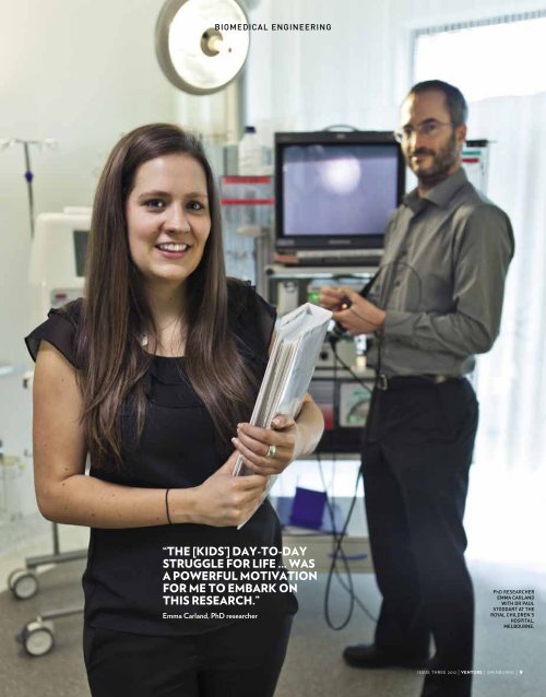
iomedical engineering “the [kids’] day-to-day struggle for life ... was a powerful motivation for me to embark on this research.” Emma Carland, PhD researcher PhD researcher emma carland with Dr paul stoddart at the royal children’s hospital, melbourne. issue three 2012 | venture | swinburne | 9