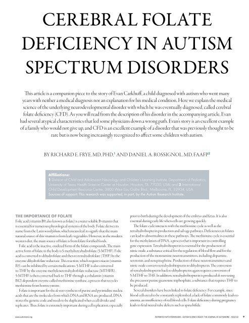
cerebral folate deficiency in autism spectrum disorders - Rossignol ...
cerebral folate deficiency in autism spectrum disorders - Rossignol ...
cerebral folate deficiency in autism spectrum disorders - Rossignol ...

Create successful ePaper yourself
Turn your PDF publications into a flip-book with our unique Google optimized e-Paper software.
Cerebral Folate<br />
Deficiency <strong>in</strong> Autism<br />
Spectrum Disorders<br />
This article is a companion piece to the story of Evan Carkhuff, a child diagnosed with <strong>autism</strong> who went many<br />
years with neither a medical diagnosis nor an explanation for his medical condition. Here we expla<strong>in</strong> the medical<br />
science of the underly<strong>in</strong>g neurodevelopmental disorder with which he was eventually diagnosed, called <strong>cerebral</strong><br />
<strong>folate</strong> <strong>deficiency</strong> (CFD). As you will read from the description of his disorder <strong>in</strong> the accompany<strong>in</strong>g article, Evan<br />
had several atypical characteristics that led some physicians down a wrong path. Evan’s story is an excellent example<br />
of a family who would not give up, and CFD is an excellent example of a disorder that was previously thought to be<br />
rare but is now be<strong>in</strong>g <strong>in</strong>creas<strong>in</strong>gly recognized to affect some children with <strong>autism</strong>.<br />
By Richard E. Frye, MD, PhD, 1 and Daniel A. <strong>Rossignol</strong>, MD, FAAFP 2<br />
Affiliations:<br />
1 Division of Child and Adolescent Neurology and Children’s Learn<strong>in</strong>g Institute, Department of Pediatrics,<br />
University of Texas Health Science Center at Houston, Houston, TX, 77030, USA; and 2 International<br />
Child Development Resource Center, 3800 West Eau Gallie Blvd., Melbourne, FL, 32934, USA.<br />
Sources of support: This research was supported, <strong>in</strong> part, by the Autism Research Institute.<br />
The importance of <strong>folate</strong><br />
Folic acid (vitam<strong>in</strong> B9, also known as <strong>folate</strong>) is a water-soluble B vitam<strong>in</strong> that<br />
is essential for numerous physiological systems of the body. Folate derives its<br />
name from the Lat<strong>in</strong> word folium, which means leaf, to signify that the ma<strong>in</strong><br />
natural source of this vitam<strong>in</strong> is from leafy vegetables. However, <strong>in</strong> the modern<br />
western diet, the ma<strong>in</strong> source of <strong>folate</strong> is from <strong>folate</strong>-fortified foods.<br />
Folic acid is the <strong>in</strong>active, oxidized form of the <strong>folate</strong> compounds. The ma<strong>in</strong><br />
active form of <strong>folate</strong> <strong>in</strong> the body is 5-methyltetrahydro<strong>folate</strong> (5-MTHF). Folic<br />
acid is converted to dihydro<strong>folate</strong> and then to tetrahydro<strong>folate</strong> (THF) by the<br />
enzyme dihydro<strong>folate</strong> reductase. This reaction, which requires niac<strong>in</strong> (vitam<strong>in</strong><br />
B3), can be <strong>in</strong>hibited by certa<strong>in</strong> medications. 5-MTHF is also converted<br />
to THF by the enzyme methylenetetrahydro<strong>folate</strong> reductase (MTHFR).<br />
5-MTHF is then converted back to THF through a cobalam<strong>in</strong> (vitam<strong>in</strong><br />
B12) dependent enzyme called methion<strong>in</strong>e synthase, a process that recycles<br />
methion<strong>in</strong>e from homocyste<strong>in</strong>e.<br />
Folate is important for the de novo synthesis of pur<strong>in</strong>e and pyrimid<strong>in</strong>e nucleic<br />
acids that are the molecules from which DNA and RNA are produced. DNA<br />
stores the genetic code and needs to be duplicated when a cell divides and<br />
replicates. Thus, <strong>folate</strong> is extremely important dur<strong>in</strong>g cell replication, especially<br />
prior to birth dur<strong>in</strong>g the development of the embryo and fetus. It is also<br />
essential dur<strong>in</strong>g early life when cells are grow<strong>in</strong>g quickly.<br />
The <strong>folate</strong> cycle <strong>in</strong>teracts with the methion<strong>in</strong>e cycle as well as the<br />
tetrahydrobiopter<strong>in</strong> production and salvage pathways. Deficiencies <strong>in</strong> <strong>folate</strong>s<br />
can lead to abnormalities <strong>in</strong> these pathways. The methion<strong>in</strong>e cycle is essential<br />
for the methylation of DNA, a process that is important <strong>in</strong> controll<strong>in</strong>g<br />
gene expression. Tetrahydrobiopter<strong>in</strong> is essential for the production of<br />
nitric oxide, a substance critical for the regulation of blood flow and for the<br />
production of the monoam<strong>in</strong>e neurotransmitters, <strong>in</strong>clud<strong>in</strong>g dopam<strong>in</strong>e,<br />
seroton<strong>in</strong>, and norep<strong>in</strong>ephr<strong>in</strong>e. Production of these neurotransmitters and<br />
nitric oxide converts tetrahydrobiopter<strong>in</strong> to dihydropter<strong>in</strong>. The conversion<br />
of tetrahydrobiopter<strong>in</strong> back to dihydropter<strong>in</strong> aga<strong>in</strong> requires conversion of<br />
5-MTHF to THF. In addition, tetrahydrobiopter<strong>in</strong> is produced de novo us<strong>in</strong>g<br />
the precursor pur<strong>in</strong>e guanos<strong>in</strong>e triphosphate, a substance that requires THF to<br />
be produced.<br />
Several <strong>disorders</strong> have been l<strong>in</strong>ked to <strong>folate</strong> <strong>deficiency</strong>. For example, s<strong>in</strong>ce<br />
blood cells need to be constantly replenished, a lack of <strong>folate</strong> commonly leads to<br />
anemia, an <strong>in</strong>sufficiency of red blood cells. Folate <strong>deficiency</strong> dur<strong>in</strong>g pregnancy<br />
leads to fetal neural tube defects such as sp<strong>in</strong>a bifida.<br />
www.<strong>autism</strong>one.org REPRINTED WITH PERMISSION • AUTISM SCIENCE DIGEST: THE JOURNAL OF AUTISMONE • ISSUE 02 9
Richard E. Frye, MD, PhD,<br />
received his MD/PhD from<br />
Georgetown University. He<br />
completed his pediatric residency<br />
at University of Miami, and a child<br />
neurology residency and fellowship<br />
<strong>in</strong> behavioral neurology and<br />
learn<strong>in</strong>g disabilities at Children’s<br />
Hospital Boston. Dr. Frye is board<br />
certified <strong>in</strong> general pediatrics<br />
and <strong>in</strong> neurology with special<br />
competency <strong>in</strong> child neurology.<br />
Currently at the University of Texas<br />
Health Science Center at Houston,<br />
Dr. Frye studies bra<strong>in</strong> structure and<br />
function <strong>in</strong> neurodevelopmental<br />
<strong>disorders</strong>, mitochondrial dysfunction<br />
and metabolic <strong>disorders</strong> <strong>in</strong> <strong>autism</strong>,<br />
and novel <strong>autism</strong> treatments. He<br />
has published numerous papers<br />
and book chapters on children<br />
with <strong>autism</strong>.<br />
10 AUTISM SCIENCE DIGEST: THE JOURNAL OF AUTISMONE • ISSUE 02 • REPRINTED WITH PERMISSION www.<strong>autism</strong>one.org
Cerebral <strong>folate</strong> <strong>deficiency</strong>: A recently<br />
described neurodevelopmental disorder<br />
One decade ago, Ramaekers and colleagues 1 described a new<br />
neurodevelopmental disorder called <strong>cerebral</strong> <strong>folate</strong> <strong>deficiency</strong> (CFD). They<br />
described five patients with normal neurodevelopment until four to six months<br />
of life. Dur<strong>in</strong>g the second half of the first year of life, these patients demonstrated<br />
developmental regression and progressively developed neurological symptoms,<br />
<strong>in</strong>clud<strong>in</strong>g irritability, psychomotor retardation, ataxia, dysk<strong>in</strong>esias, pyramidal<br />
signs, visual loss, and seizures. Patients also demonstrated acquired microcephaly.<br />
5-MTHF was found to be normal <strong>in</strong> the serum and red blood cells but was low<br />
<strong>in</strong> the cerebrosp<strong>in</strong>al fluid. This new disorder was named CFD to describe the<br />
lack of <strong>folate</strong> specifically <strong>in</strong> the central nervous system (CNS).<br />
Cerebral <strong>folate</strong> transporters<br />
To understand CFD, it is necessary to understand that the CNS is a protected<br />
area of the body. The blood-bra<strong>in</strong> barrier highly regulates the entry of<br />
substances <strong>in</strong>to the CNS. For the active form of <strong>folate</strong> (5-MTHF) to enter<br />
the CNS, it must be transported across the blood-bra<strong>in</strong> barrier by one of two<br />
specialized carriers. The primary carrier uses a specialized <strong>folate</strong> receptor known<br />
as <strong>folate</strong> receptor 1 (FR1). Through this system, 5-MTHF b<strong>in</strong>ds to FR1, which<br />
is located on the apical side (blood vessel side) of epithelial cells of the choroid<br />
plexus. FR1 then transports 5-MTHF to the basolateral side of the epithelial<br />
cells. On the basolateral side of the cell, 5-MTHF is released <strong>in</strong>to the CNS. This<br />
transport process requires energy <strong>in</strong> the form of an adenos<strong>in</strong>e-5’-triphosphate<br />
(ATP) dependent mechanism. FR1 is then recycled back to the apical side of<br />
the cell to pick up more 5-MTHF.<br />
A secondary carrier of <strong>folate</strong> through the blood-bra<strong>in</strong> barrier is the reduced<br />
<strong>folate</strong> carrier (RFC). The RFC has a lower aff<strong>in</strong>ity for folic acid and 5-MTHF<br />
than the FR1 system but has a higher aff<strong>in</strong>ity for 5-formyltetrahydro<strong>folate</strong>,<br />
also known as fol<strong>in</strong>ic acid or leucovor<strong>in</strong>. The RFC is also responsible for<br />
transport<strong>in</strong>g 5-MTHF <strong>in</strong>to neurons once it has entered the CNS.<br />
If blood concentrations of <strong>folate</strong> are high enough, <strong>folate</strong> may also diffuse<br />
across the blood-bra<strong>in</strong> barrier without a carrier.<br />
Causes of <strong>cerebral</strong> <strong>folate</strong> <strong>deficiency</strong><br />
Ramaekers’ group 1 exam<strong>in</strong>ed the gene that encodes FR1 to <strong>in</strong>vestigate<br />
whether or not genetic mutations accounted for dysfunction <strong>in</strong> the transport<br />
of 5-MTHF <strong>in</strong>to the CNS but could not identify any such mutations. In 2004,<br />
Ramaekers and Blau 2 expanded their case series to 20 patients, none of whom<br />
were found to have a mutation <strong>in</strong> the FR1 gene. However, these researchers did<br />
f<strong>in</strong>d non-functional FR1 receptors <strong>in</strong> the patients’ cerebrosp<strong>in</strong>al fluid, lead<strong>in</strong>g to<br />
the hypothesis that some type of molecule, potentially an autoantibody, might<br />
be irreversibly b<strong>in</strong>d<strong>in</strong>g to the FR1 prote<strong>in</strong>, caus<strong>in</strong>g it to become dysfunctional<br />
for b<strong>in</strong>d<strong>in</strong>g <strong>folate</strong>. In 2005, Ramaekers and colleagues 3 identified high-aff<strong>in</strong>ity<br />
Ramaekers’ group exam<strong>in</strong>ed<br />
the gene that encodes FR1<br />
to <strong>in</strong>vestigate whether or not<br />
genetic mutations accounted<br />
for dysfunction <strong>in</strong> the transport<br />
of 5-MTHF <strong>in</strong>to the CNS but<br />
could not identify any such<br />
mutations.<br />
block<strong>in</strong>g autoantibodies aga<strong>in</strong>st FR1 <strong>in</strong> the serum of 25 of 28 children with<br />
CFD. These autoantibodies were not found <strong>in</strong> age-matched control subjects.<br />
More recently, Molloy and colleagues 4 described an additional block<strong>in</strong>g FR1<br />
autoantibody (termed a “b<strong>in</strong>d<strong>in</strong>g” antibody), but this autoantibody has yet to be<br />
associated with any pathological disease. Interest<strong>in</strong>gly, although the majority of<br />
cases of <strong>in</strong>dividuals with these autoantibodies have not been reported to have<br />
any obvious <strong>in</strong>flammatory conditions, FR1 autoantibodies have been associated<br />
with juvenile rheumatoid arthritis. 5<br />
In 2006, CFD was l<strong>in</strong>ked to mitochondrial disease <strong>in</strong> a case report of a child<br />
with an <strong>in</strong>complete form of Kearns-Sayre syndrome. 6 Further case reports and<br />
case series later expanded the association between CFD and mitochondrial<br />
<strong>disorders</strong> to <strong>in</strong>clude complex I <strong>deficiency</strong>, 7 Alpers’ disease 8 and complex IV<br />
hyperfunction, 9 as well as a wide variety of mitochondrial <strong>disorders</strong> <strong>in</strong> both<br />
children and adults. 10 In most of these cases, the autoantibodies to FR1 were<br />
not found, suggest<strong>in</strong>g that it was the lack of ATP availability secondary to<br />
mitochondrial dysfunction that resulted <strong>in</strong> the impaired transportation of<br />
5-MTHF <strong>in</strong>to the CNS.<br />
Cerebral <strong>folate</strong> <strong>deficiency</strong> and<br />
<strong>autism</strong> <strong>spectrum</strong> <strong>disorders</strong><br />
Seven of the 20 children portrayed <strong>in</strong> the second case series describ<strong>in</strong>g CFD<br />
were reported to have an <strong>autism</strong> <strong>spectrum</strong> disorder (ASD), 2 while five of the<br />
28 patients first described to have the FR1 autoantibody were found to have<br />
low-function<strong>in</strong>g <strong>autism</strong> with neurological features. 3 Further case reports 9,11<br />
and case series 12,13,14 have expanded the description of CFD <strong>in</strong> children<br />
with idiopathic <strong>autism</strong>. Overall, these reports suggest that early-onset lowfunction<strong>in</strong>g<br />
<strong>autism</strong> with neurological deficits is characteristic of children with<br />
both <strong>autism</strong> and CFD. Interest<strong>in</strong>gly, Rett syndrome, a disorder considered to<br />
be a part of the diagnostic group of <strong>autism</strong> <strong>spectrum</strong> <strong>disorders</strong>, has also been<br />
reported to have reduced 5-MTHF levels <strong>in</strong> the cerebrosp<strong>in</strong>al fluid. 15,16<br />
It should be noted that only some children with <strong>autism</strong> who have CFD<br />
have been reported to possess FR1 autoantibodies. 3,13 Because these reports<br />
of children with idiopathic <strong>autism</strong> and Rett syndrome <strong>in</strong>clude children with<br />
and without the FR1 autoantibody, this suggests that factors other than<br />
the FR1 autoantibody might be important for the development of CFD <strong>in</strong><br />
these children. Although not specifically <strong>in</strong>vestigated, it is possible that many<br />
children with CFD and idiopathic <strong>autism</strong> or Rett syndrome who do not have<br />
the FR1 autoantibody may have mitochondrial disease. Indeed, as previously<br />
noted, mitochondrial disease appears to be associated with CFD, 6-10 and there<br />
appears to be an <strong>in</strong>creased prevalence of mitochondrial disease <strong>in</strong> children with<br />
idiopathic <strong>autism</strong> as compared to the general population. 17,18 At least one case<br />
series has l<strong>in</strong>ked children with mitochondrial disease and regressive-type <strong>autism</strong><br />
to CFD. 9 Interest<strong>in</strong>gly, Rett syndrome has also been l<strong>in</strong>ked to mitochondrial<br />
abnormalities <strong>in</strong> both an animal model 19 and a case report. 20 To a lesser<br />
extent, children with idiopathic <strong>autism</strong> might also manifest dysfunction of<br />
the mitochondria without necessarily fulfill<strong>in</strong>g the criteria for strictly def<strong>in</strong>ed<br />
mitochondrial disease. 18 Thus, it is possible that mitochondrial dysfunction<br />
could contribute to the development of CFD <strong>in</strong> children with idiopathic <strong>autism</strong>.<br />
www.<strong>autism</strong>one.org REPRINTED WITH PERMISSION • AUTISM SCIENCE DIGEST: THE JOURNAL OF AUTISMONE • ISSUE 02 11
Daniel A. <strong>Rossignol</strong>, MD, FAAFP,<br />
received his doctorate of medic<strong>in</strong>e at the<br />
Medical College of Virg<strong>in</strong>ia and completed his<br />
residency <strong>in</strong> family medic<strong>in</strong>e at the University<br />
of Virg<strong>in</strong>ia. He is currently a physician at the<br />
International Child Development Resource<br />
Center (ICDRC) <strong>in</strong> Melbourne, Florida. Com<strong>in</strong>g<br />
from an academic background, Dr. <strong>Rossignol</strong><br />
searched the medical literature look<strong>in</strong>g for<br />
a solution after both of his children were<br />
diagnosed with <strong>autism</strong>, and he has made it his<br />
mission to research and publish <strong>in</strong> <strong>autism</strong>. In the<br />
last 5 years, he has published 16 articles and 3<br />
book chapters concern<strong>in</strong>g <strong>autism</strong>.<br />
12<br />
AUTISM SCIENCE DIGEST: THE JOURNAL OF AUTISMONE • ISSUE 02 • REPRINTED WITH PERMISSION<br />
www.<strong>autism</strong>one.org
Diagnos<strong>in</strong>g <strong>cerebral</strong> <strong>folate</strong> <strong>deficiency</strong><br />
Table 1 outl<strong>in</strong>es the signs, symptoms and conditions associated with CFD. It is<br />
important to consider CFD <strong>in</strong> children with Rett syndrome or mitochondrial<br />
disease with or without autistic features. A comb<strong>in</strong>ation of the neurological<br />
symptoms outl<strong>in</strong>ed <strong>in</strong> Table 1 that are not expla<strong>in</strong>ed by a specific neurological<br />
condition should also prompt consideration of CFD. Given the accompany<strong>in</strong>g<br />
case description, it is clear that CFD can present with atypical features. Thus, it<br />
is important to keep a high <strong>in</strong>dex of suspicion for this disorder <strong>in</strong> children with<br />
unexpla<strong>in</strong>ed neurodevelopmental symptoms.<br />
Table 1<br />
When to Suspect Cerebral Folate Deficiency<br />
• Low-function<strong>in</strong>g <strong>autism</strong><br />
• Mitochondrial disease or dysfunction<br />
• Rett syndrome<br />
• Epilepsy or seizures<br />
• Abnormal electroencephalogram: subcl<strong>in</strong>ical<br />
electrical discharges or slow<strong>in</strong>g<br />
• Ataxia<br />
• Microcephaly<br />
• Dysk<strong>in</strong>esia: choreoathetosis, ballismus<br />
• Pyramidal tract abnormalities<br />
• Irritability<br />
• Insomnia<br />
• Delayed myel<strong>in</strong>ation<br />
• Frontotemporal atrophy<br />
Table 2 outl<strong>in</strong>es the diagnostic workup for CFD. As shown <strong>in</strong> the table, it is<br />
important to beg<strong>in</strong> by rul<strong>in</strong>g out systemic deficiencies <strong>in</strong> <strong>folate</strong> or cobalam<strong>in</strong><br />
that might cause symptoms similar to CFD (Step 1). Next, it is essential to<br />
test for FR1 autoantibodies (Step 2). If the FR1 b<strong>in</strong>d<strong>in</strong>g autoantibodies are<br />
discovered, it is important to <strong>in</strong>vestigate the function of other organs that<br />
use the FR1 receptor for <strong>folate</strong> uptake to ensure that the antibodies are not<br />
the result of a more general autoimmune process (Step 3). It should be noted<br />
that, because the reported relationship between FR1 autoantibodies and<br />
cerebrosp<strong>in</strong>al fluid levels of 5-MTHF is nonl<strong>in</strong>ear, some <strong>in</strong>dividuals with FR1<br />
autoantibodies have normal levels of cerebrosp<strong>in</strong>al fluid 5-MTHF. 14 If FR1<br />
autoantibodies are negative, it is possible that an underly<strong>in</strong>g mitochondrial<br />
disorder might be result<strong>in</strong>g <strong>in</strong> secondary CFD. Thus, if CFD is still suspected<br />
despite a negative FR1 b<strong>in</strong>d<strong>in</strong>g autoantibody, a screen<strong>in</strong>g for mitochondrial<br />
<strong>disorders</strong> us<strong>in</strong>g established guidel<strong>in</strong>es 18 is recommended (Step 4). If the FR1<br />
autoantibody is detected or a mitochondrial disorder is diagnosed, a lumbar<br />
puncture is required to confirm the diagnosis of CFD (Step 5).<br />
A thorough workup should also measure levels of tetrahydrobiopter<strong>in</strong><br />
because <strong>folate</strong> is essential <strong>in</strong> the production of this cofactor. As noted<br />
previously, deficits <strong>in</strong> tetrahydrobiopter<strong>in</strong> can lead to reduced production of<br />
the monoam<strong>in</strong>e neurotransmitters. Interest<strong>in</strong>gly, abnormalities <strong>in</strong> monoam<strong>in</strong>e<br />
neurotransmitter metabolites have been reported <strong>in</strong> CFD 2,21 and may<br />
improve with fol<strong>in</strong>ic acid treatment. Neurotransmitter metabolites <strong>in</strong> the<br />
cerebrosp<strong>in</strong>al fluid should therefore also be measured dur<strong>in</strong>g the lumbar<br />
puncture. F<strong>in</strong>ally, because <strong>in</strong>flammatory conditions have been associated with<br />
CFD, 5,8 it is important to measure cerebrosp<strong>in</strong>al fluid neopter<strong>in</strong>, a measure of<br />
<strong>in</strong>flammation, and an IgG <strong>in</strong>dex, a measure of <strong>in</strong>trathecal antibody production.<br />
Unfortunately, a lumbar puncture is an <strong>in</strong>vasive procedure that requires a<br />
specialist with significant experience to perform. For example, at the University<br />
of Texas Health Science Center, an experienced neuroradiologist performs<br />
non-emergent lumbar punctures under general anesthesia with fluoroscopy<br />
guidance. In many cases, parents will not elect for their child to undergo such<br />
an <strong>in</strong>vasive procedure, and, <strong>in</strong> other cases, experienced personnel may not<br />
be readily available. Under these circumstances, empirical treatment with<br />
fol<strong>in</strong>ic acid or 5-MTHF can be a prudent option (see Treatment of <strong>cerebral</strong> <strong>folate</strong><br />
<strong>deficiency</strong>). If empirical treatment is pursued, the patient should be closely<br />
monitored for behavioral and/or cognitive changes and side effects.<br />
Table 2<br />
Diagnostic Workup for Cerebral<br />
Folate Deficiency<br />
1 Rule out systemic <strong>folate</strong> and cobalam<strong>in</strong> <strong>deficiency</strong><br />
a Serum folic acid level<br />
b Serum cobalam<strong>in</strong> level<br />
2 Test for FR1 <strong>folate</strong> receptor autoantibodies<br />
3 If FR1 autoantibodies are positive:<br />
a Test for dysfunction <strong>in</strong> other organs<br />
i Thyroid function tests<br />
ii Renal function tests<br />
b Test for <strong>in</strong>flammatory disease<br />
i Erythrocyte sedimentation rate<br />
ii C-reactive prote<strong>in</strong><br />
iii Ant<strong>in</strong>uclear antibody<br />
4 If FR1 autoantibodies are negative or there are<br />
symptoms of mitochondrial <strong>disorders</strong>: 17<br />
a Test for mitochondrial markers 18<br />
i Fast<strong>in</strong>g serum lactate, pyruvate, quantitative<br />
am<strong>in</strong>o acids, ammonia, metabolic panel, liver<br />
function tests, creat<strong>in</strong>e k<strong>in</strong>ase, acylcarnit<strong>in</strong>e<br />
panel, carnit<strong>in</strong>e panel, and CoQ10 level<br />
ii Fast<strong>in</strong>g ur<strong>in</strong>e organic acids<br />
5 If FR1 autoantibodies are positive or<br />
mitochondrial markers are positive:<br />
a Perform lumbar puncture to confirm <strong>cerebral</strong><br />
<strong>folate</strong> <strong>deficiency</strong><br />
b Test cerebrosp<strong>in</strong>al fluid for<br />
i 5-MTHF<br />
ii Tetrahydrobiopter<strong>in</strong><br />
iii Neurotransmitters<br />
iv Neopter<strong>in</strong><br />
v IgG <strong>in</strong>dex<br />
The first treatment used for CFD was fol<strong>in</strong>ic acid. This therapy, which has<br />
an excellent safety profile, has been shown to normalize cerebrosp<strong>in</strong>al fluid<br />
levels of 5-MTHF <strong>in</strong> children with <strong>autism</strong> and CFD.<br />
www.<strong>autism</strong>one.org REPRINTED WITH PERMISSION • AUTISM SCIENCE DIGEST: THE JOURNAL OF AUTISMONE • ISSUE 02<br />
13
14<br />
Treatment of <strong>cerebral</strong> <strong>folate</strong> <strong>deficiency</strong><br />
Treatments for CFD are outl<strong>in</strong>ed <strong>in</strong> Table 3. The first treatment used for CFD<br />
was fol<strong>in</strong>ic acid. This therapy, which has an excellent safety profile, has been<br />
shown to normalize cerebrosp<strong>in</strong>al fluid levels of 5-MTHF <strong>in</strong> children with<br />
<strong>autism</strong> and CFD. 13 Reports have suggested that treatment with fol<strong>in</strong>ic acid<br />
has led to full control of epilepsy and resolution of bra<strong>in</strong>stem, thalamus, basal<br />
ganglia, and white matter demyel<strong>in</strong>ation <strong>in</strong> a child with complex I <strong>deficiency</strong>, 7<br />
resolution of neurotransmitter abnormalities, 2 and improvements <strong>in</strong> seizures,<br />
attention, motor skills, neurological abnormalities, verbalizations, perseverative<br />
behavior, restricted <strong>in</strong>terests, and social <strong>in</strong>teraction <strong>in</strong> some children with<br />
<strong>autism</strong>. 3,11,12,13<br />
Typical doses of fol<strong>in</strong>ic acid range from 0.5-1 mg/kg/day <strong>in</strong> two divided<br />
doses with a maximum of 50 mg/day. However, some case reports have used<br />
doses as high as 4 mg/kg/day. Therefore, some children may need higher levels<br />
of fol<strong>in</strong>ic acid. As described above, fol<strong>in</strong>ic acid enters the CNS through an<br />
alternative <strong>folate</strong> carrier known as the reduced <strong>folate</strong> carrier. Once it enters<br />
the CNS, fol<strong>in</strong>ic acid can particulate <strong>in</strong> the reactions that use THF. In these<br />
processes, fol<strong>in</strong>ic acid is converted to 5-MTHF, a step that requires cobalam<strong>in</strong><br />
to be recycled to THF. Thus, it is essential that adequate levels of cobalam<strong>in</strong><br />
be available when treat<strong>in</strong>g with fol<strong>in</strong>ic acid. As folic acid (the <strong>in</strong>active, oxidized<br />
form of <strong>folate</strong>) can compete for the b<strong>in</strong>d<strong>in</strong>g site on FR1, it is probably wise to<br />
discont<strong>in</strong>ue the use of folic acid-conta<strong>in</strong><strong>in</strong>g supplements.<br />
Interest<strong>in</strong>gly, the human <strong>folate</strong> receptor cross-reacts with <strong>folate</strong> receptors<br />
conta<strong>in</strong>ed <strong>in</strong> human, bov<strong>in</strong>e (cow), and goat milk. In 2008, Ramaekers and<br />
colleagues 14 demonstrated that a cow’s milk-free diet significantly reduced<br />
the level of FR1 autoantibodies and that re-exposure to milk significantly<br />
<strong>in</strong>creased FR1 autoantibodies. Furthermore, some of the children with<br />
<strong>autism</strong> were found to have marked or partial improvements <strong>in</strong> attention,<br />
communication, and stereotyped movements when placed on a milk-free<br />
diet. This provides compell<strong>in</strong>g evidence that supports parental reports of<br />
improvements with a case<strong>in</strong>-free diet <strong>in</strong> some children with <strong>autism</strong> and<br />
supports previous studies suggest<strong>in</strong>g gastro<strong>in</strong>test<strong>in</strong>al tract immune activation<br />
<strong>in</strong> children with <strong>autism</strong>.<br />
Table 3<br />
Treatments for Cerebral Folate Deficiency<br />
• Discont<strong>in</strong>ue drugs that can <strong>in</strong>terfere with <strong>folate</strong><br />
metabolism<br />
• Start fol<strong>in</strong>ic acid at dose of 0.5 mg/kg/day <strong>in</strong> two<br />
divided doses and <strong>in</strong>crease to 1-4 mg/kg/day <strong>in</strong> two<br />
divided doses (max 50 mg/day)<br />
• Consider cobalam<strong>in</strong> supplementation (vitam<strong>in</strong> B12)<br />
• Stop folic acid supplementation<br />
• Start a cow’s milk-free diet<br />
• Monitor for changes <strong>in</strong> cognition and behavior<br />
• Monitor adverse effects<br />
Potential association of the <strong>cerebral</strong> <strong>folate</strong><br />
antibody with birth defects<br />
Several studies provide <strong>in</strong>terest<strong>in</strong>g and compell<strong>in</strong>g evidence for a relationship<br />
between <strong>folate</strong> receptor autoantibodies and neural tube defects (NTDs). In<br />
2004, for example, Rothenberg and colleagues 22 demonstrated that women<br />
from the United States with a current or previous baby with NTDs were<br />
more likely to have autoantibodies to the human placental <strong>folate</strong> receptor. In a<br />
larger study, Cabrera and colleagues 23 found that mid-gestation levels of both<br />
IgM and IgG autoantibodies to the human <strong>folate</strong> receptor collected from<br />
AUTISM SCIENCE DIGEST: THE JOURNAL OF AUTISMONE • ISSUE 02 • REPRINTED WITH PERMISSION<br />
US women were associated with pregnancies complicated by NTDs. More<br />
recently, a study of Norwegian women by Bovies and colleagues 24 suggested<br />
that mid-gestation autoantibodies were specifically related to NTDs but not<br />
to oral facial clefts. Although another rather large study from Ireland (us<strong>in</strong>g<br />
previously frozen specimens not necessarily collected dur<strong>in</strong>g pregnancy) did<br />
not f<strong>in</strong>d any difference between mothers who had a previous pregnancy with<br />
NTDs as compared to those without an affected pregnancy, 25 the prevalence<br />
of autoantibodies to FR1 was very high <strong>in</strong> this population (approach<strong>in</strong>g 35%),<br />
and the f<strong>in</strong>d<strong>in</strong>gs need duplication <strong>in</strong> populations where the prevalence is lower.<br />
Because these studies reflect important methodological differences (<strong>in</strong>clud<strong>in</strong>g<br />
whether or not autoantibodies were measured dur<strong>in</strong>g pregnancy) as well as<br />
differences <strong>in</strong> national policies regard<strong>in</strong>g dietary <strong>folate</strong> supplementation,<br />
further research is needed to def<strong>in</strong>e whether or not a relationship between <strong>folate</strong><br />
receptor autoantibodies and NTDs truly exists.<br />
Unanswered questions: What Evan can<br />
teach us<br />
It is important to understand that because CFD has only been reported <strong>in</strong> case<br />
reports and case series, there may be a much wider variation <strong>in</strong> the symptoms<br />
associated with CFD. For example, children who do not have neurological<br />
symptoms or seizures will rarely undergo a lumbar puncture to look for CFD.<br />
This is especially true <strong>in</strong> <strong>autism</strong>, where there are diverse op<strong>in</strong>ions regard<strong>in</strong>g the<br />
disorder’s medical basis. It is possible that many more children with ASD than<br />
are currently recognized may suffer from CFD, a treatable condition.<br />
As the accompany<strong>in</strong>g article illustrates, Evan is a good example of the<br />
underrecognition of CFD. Whereas one of the symptoms of classic CFD<br />
is microcephaly, a study has reported that <strong>in</strong> children with ASD and CFD,<br />
microcephaly was not present. 12 Evan, <strong>in</strong> fact, had macrocephaly because of<br />
congenital ventriculomegaly (a condition <strong>in</strong> which the fluid-filled structures <strong>in</strong> the<br />
bra<strong>in</strong> are too large). Could this have been a manifestation of his mother hav<strong>in</strong>g the<br />
FR1 autoantibody? Although the ventriculomegaly that Evan possessed is not a<br />
neural tube defect (and Evan does not have a neural tube defect), it can be related<br />
to NTDs. Evan’s growth IGF-1 <strong>deficiency</strong> raises additional questions, <strong>in</strong>clud<strong>in</strong>g<br />
how it fits <strong>in</strong>to CFD. The fact is, we do not yet know. Evan has taught us that we<br />
need to understand our limitations as we cont<strong>in</strong>ue to search for answers and put<br />
together the pieces of a complicated puzzle.<br />
It is possible that many more<br />
children with ASD than are currently<br />
recognized may suffer from CFD,<br />
a treatable condition.<br />
www.<strong>autism</strong>one.org
References<br />
1. Ramaekers VT, Husler M, Opladen T, Heimann G, Blau N. Psychomotor retardation, spastic<br />
paraplegia, cerebellar ataxia and dysk<strong>in</strong>esia associated with low 5-methyltetrahydro<strong>folate</strong> <strong>in</strong><br />
cerebrosp<strong>in</strong>al fluid: a novel neurometabolic condition respond<strong>in</strong>g to fol<strong>in</strong>ic acid substitution.<br />
Neuropediatrics. 2002 Dec;33(6):301-8.<br />
2. Ramaekers VT, Blau N. Cerebral <strong>folate</strong> <strong>deficiency</strong>. Dev Med Child Neurol. 2004<br />
Dec;46(12):843-51.<br />
3. Ramaekers VT, Rothenberg SP, Sequeira JM, Opladen T, Blau N, Quadros EV, Selhub J.<br />
Autoantibodies to <strong>folate</strong> receptors <strong>in</strong> the <strong>cerebral</strong> <strong>folate</strong> <strong>deficiency</strong> syndrome. N Engl J Med.<br />
2005 May 12;352(19):1985-91.<br />
4. Molloy AM, Quadros EV, Sequeira JM, Troendle JF, Scott JM, Kirke PN, Mills JL. Lack of<br />
association between <strong>folate</strong>-receptor autoantibodies and neural-tube defects. N Engl J Med.<br />
2009 Jul 9;361(2):152-60.<br />
5. Koenig MK, Perez M, Rothenberg S, Butler IJ. Juvenile onset central nervous system <strong>folate</strong><br />
<strong>deficiency</strong> and rheumatoid arthritis. J Child Neurol. 2008 Jan;23(1):106-7. Epub 2007 Dec 3.<br />
6. P<strong>in</strong>eda M, Ormazabal A, Lopez-Gallardo E, Nascimento A, Solano A, Herrero MD,<br />
Vilaseca MA, Briones P, Ibanez L, Montoya J, Artuch R. Cerebral <strong>folate</strong> <strong>deficiency</strong> and<br />
leukoencephalopathy caused by a mitochondrial DNA deletion. Ann Neurol. 2006<br />
Feb;59(2):394-8.<br />
7. Ramaekers VT, Weis J, Sequeira JM, Quadros EV, Blau N. Mitochondrial complex I<br />
encephalomyopathy and <strong>cerebral</strong> 5-methyltetrahydro<strong>folate</strong> <strong>deficiency</strong>. Neuropediatrics. 2007<br />
Aug;38(4):184-7.<br />
8. Hasselmann O, Blau N, Ramaekers VT, Quadros EV, Sequeira JM, Weissert M. Cerebral<br />
<strong>folate</strong> <strong>deficiency</strong> and CNS <strong>in</strong>flammatory markers <strong>in</strong> Alpers disease. Mol Genet Metab. 2010<br />
Jan;99(1):58-61.<br />
9. Frye RE. Complex IV hyperfunction <strong>in</strong> <strong>autism</strong> <strong>spectrum</strong> disorder: a new mitochondrial<br />
syndrome. J Ped Neurol, <strong>in</strong> press.<br />
10. Garcia-Cazorla A, Quadros EV, Nascimento A, Garcia-Silva MT, Briones P, Montoya<br />
J, Ormazabal A, Artuch R, Sequeira JM, Blau N, Arenas J, P<strong>in</strong>eda M, Ramaekers VT.<br />
Mitochondrial diseases associated with <strong>cerebral</strong> <strong>folate</strong> <strong>deficiency</strong>. Neurology. 2008 Apr<br />
15;70(16):1360-2.<br />
11. Moretti P, Sahoo T, Hyland K, Bottiglieri T, Peters S, del Gaudio D, Roa B, Curry S, Zhu H,<br />
F<strong>in</strong>nell RH, Neul JL, Ramaekers VT, Blau N, Bac<strong>in</strong>o CA, Miller G, Scaglia F. Cerebral <strong>folate</strong><br />
<strong>deficiency</strong> with developmental delay, <strong>autism</strong>, and response to fol<strong>in</strong>ic acid. Neurology. 2005<br />
Mar 22;64(6):1088-90.<br />
12. Moretti P, Peters SU, Del Gaudio D, Sahoo T, Hyland K, Bottiglieri T, Hopk<strong>in</strong> RJ, Peach<br />
E, M<strong>in</strong> SH, Goldman D, Roa B, Bac<strong>in</strong>o CA, Scaglia F. Autistic symptoms, developmental<br />
regression, mental retardation, epilepsy, and dysk<strong>in</strong>esias <strong>in</strong> CNS <strong>folate</strong> <strong>deficiency</strong>. J Autism Dev<br />
Disord. 2008 Jul;38(6):1170-7. Epub 2007 Nov 20.<br />
13. Ramaekers VT, Blau N, Sequeira JM, Nassogne MC, Quadros EV. Folate receptor<br />
autoimmunity and <strong>cerebral</strong> <strong>folate</strong> <strong>deficiency</strong> <strong>in</strong> low-function<strong>in</strong>g <strong>autism</strong> with neurological deficits.<br />
Neuropediatrics. 2007 Dec;38(6):276-81.<br />
14. Ramaekers VT, Sequeira JM, Blau N, Quadros EV. A milk-free diet downregulates <strong>folate</strong><br />
receptor autoimmunity <strong>in</strong> <strong>cerebral</strong> <strong>folate</strong> <strong>deficiency</strong> syndrome. Dev Med Child Neurol. 2008<br />
May;50(5):346-52. Epub 2008 Mar 19.<br />
15. Ramaekers VT, Hansen SI, Holm J, Opladen T, Senderek J, Husler M, Heimann G, Fowler<br />
B, Maiwald R, Blau N. Reduced <strong>folate</strong> transport to the CNS <strong>in</strong> female Rett patients. Neurology.<br />
2003 Aug 26;61(4):506-15<br />
16. Ramaekers VT, Sequeira JM, Artuch R, Blau N, Temudo T, Ormazabal A, P<strong>in</strong>eda M,<br />
Aracil A, Roelens F, Laccone F, Quadros EV. Folate receptor autoantibodies and sp<strong>in</strong>al fluid<br />
5-methyltetrahydro<strong>folate</strong> <strong>deficiency</strong> <strong>in</strong> Rett syndrome. Neuropediatrics. 2007 Aug;38(4):<br />
179-83.<br />
17. Frye RE, <strong>Rossignol</strong> DA. Mitochondrial dysfunction can connect the diverse medical<br />
symptoms associated with <strong>autism</strong> <strong>spectrum</strong> <strong>disorders</strong>. Pediatr Res. 2011 May;69(5 Pt 2):41R-7R.<br />
18. <strong>Rossignol</strong> DA, Frye RE. Mitochondrial dysfunction <strong>in</strong> <strong>autism</strong> <strong>spectrum</strong> <strong>disorders</strong>:a systematic<br />
review and meta-analysis. Mol Psychiatry. 2011 Jan 25. [Epub ahead of pr<strong>in</strong>t]<br />
19. Kriaucionis S, Paterson A, Curtis J, Guy J, Macleod N, Bird A. Gene expression analysis<br />
exposes mitochondrial abnormalities <strong>in</strong> a mouse model of Rett syndrome. Mol Cell Biol. 2006<br />
Jul;26(13):5033-42.<br />
20. Condie J, Goldste<strong>in</strong> J, Wa<strong>in</strong>wright MS. Acquired microcephaly, regression of milestones,<br />
mitochondrial dysfunction, and episodic rigidity <strong>in</strong> a 46,XY male with a de novo MECP2 gene<br />
mutation. J Child Neurol. 2010 May;25(5):633-6. Epub 2010 Feb 8.<br />
21. Hansen FJ, Blau N. Cerebral <strong>folate</strong> <strong>deficiency</strong>: life-chang<strong>in</strong>g supplementation with fol<strong>in</strong>ic<br />
acid. Mol Genet Metab. 2005 Apr;84(4):371-3. Epub 2005 Jan 22.<br />
22. Rothenberg SP, da Costa MP, Sequeira JM, Cracco J, Roberts JL, Weedon J, Quadros EV.<br />
Autoantibodies aga<strong>in</strong>st <strong>folate</strong> receptors <strong>in</strong> women with a pregnancy complicated by a neuraltube<br />
defect. N Engl J Med. 2004 Jan 8;350(2):134-42.<br />
23. Cabrera RM, Shaw GM, Ballard JL, Carmichael SL, Yang W, Lammer EJ, F<strong>in</strong>nell RH.<br />
Autoantibodies to <strong>folate</strong> receptor dur<strong>in</strong>g pregnancy and neural tube defect risk. J Reprod<br />
Immunol. 2008 Oct;79(1):85-92. Epub 2008 Sep 18.<br />
24. Boyles AL, Ballard JL, Gorman EB, McConnaughey DR, Cabrera RM, Wilcox AJ, Lie RT,<br />
F<strong>in</strong>nell RH. Association between <strong>in</strong>hibited b<strong>in</strong>d<strong>in</strong>g of folic acid to <strong>folate</strong> receptor alpha <strong>in</strong><br />
maternal serum and <strong>folate</strong>-related birth defects <strong>in</strong> Norway. Hum Reprod. 2011 May 15. [Epub<br />
ahead of pr<strong>in</strong>t]<br />
25. Molloy AM, Quadros EV, Sequeira JM, Troendle JF, Scott JM, Kirke PN, Mills JL. Lack of<br />
association between <strong>folate</strong>-receptor autoantibodies and neural-tube defects. N Engl J Med.<br />
2009 Jul 9;361(2):152-60.<br />
www.<strong>autism</strong>one.org REPRINTED WITH PERMISSION • AUTISM SCIENCE DIGEST: THE JOURNAL OF AUTISMONE • ISSUE 02 15