Autism Studies and Related Medical Conditions, January 2009 - TACA
Autism Studies and Related Medical Conditions, January 2009 - TACA
Autism Studies and Related Medical Conditions, January 2009 - TACA

You also want an ePaper? Increase the reach of your titles
YUMPU automatically turns print PDFs into web optimized ePapers that Google loves.
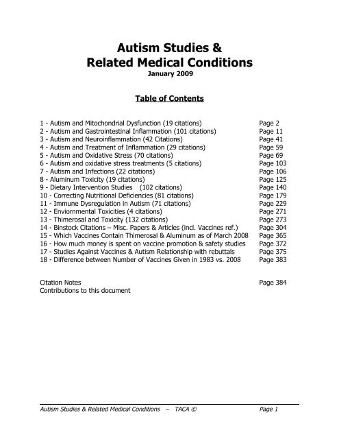
<strong>Autism</strong> <strong>Studies</strong> &<br />
<strong>Related</strong> <strong>Medical</strong> <strong>Conditions</strong><br />
<strong>January</strong> <strong>2009</strong><br />
Table of Contents<br />
1 - <strong>Autism</strong> <strong>and</strong> Mitochondrial Dysfunction (19 citations) Page 2<br />
2 - <strong>Autism</strong> <strong>and</strong> Gastrointestinal Inflammation (101 citations) Page 11<br />
3 - <strong>Autism</strong> <strong>and</strong> Neuroinflammation (42 Citations) Page 41<br />
4 - <strong>Autism</strong> <strong>and</strong> Treatment of Inflammation (29 citations) Page 59<br />
5 - <strong>Autism</strong> <strong>and</strong> Oxidative Stress (70 citations) Page 69<br />
6 - <strong>Autism</strong> <strong>and</strong> oxidative stress treatments (5 citations) Page 103<br />
7 - <strong>Autism</strong> <strong>and</strong> Infections (22 citations) Page 106<br />
8 - Aluminum Toxicity (19 citations) Page 125<br />
9 - Dietary Intervention <strong>Studies</strong> (102 citations) Page 140<br />
10 - Correcting Nutritional Deficiencies (81 citations) Page 179<br />
11 - Immune Dysregulation in <strong>Autism</strong> (71 citations) Page 229<br />
12 - Enviornmental Toxicities (4 citations) Page 271<br />
13 - Thimerosal <strong>and</strong> Toxicity (132 citations) Page 273<br />
14 - Binstock Citations – Misc. Papers & Articles (incl. Vaccines ref.) Page 304<br />
15 - Which Vaccines Contain Thimerosal & Aluminum as of March 2008 Page 365<br />
16 - How much money is spent on vaccine promotion & safety studies Page 372<br />
17 - <strong>Studies</strong> Against Vaccines & <strong>Autism</strong> Relationship with rebuttals Page 375<br />
18 - Difference between Number of Vaccines Given in 1983 vs. 2008 Page 383<br />
Citation Notes Page 384<br />
Contributions to this document<br />
<strong>Autism</strong> <strong>Studies</strong> & <strong>Related</strong> <strong>Medical</strong> <strong>Conditions</strong> – <strong>TACA</strong> © Page 1
1- <strong>Autism</strong> <strong>and</strong> Mitochondrial Dysfunction (19 citations):<br />
Blasi, F., E. Bacchelli, et al. (2006). "SLC25A12 <strong>and</strong> CMYA3 gene variants are not<br />
associated with autism in the IMGSAC multiplex family sample." Eur J Hum Genet<br />
14(1): 123-6.<br />
<strong>Autism</strong> is a severe neurodevelopmental disorder with a complex genetic<br />
predisposition. Linkage findings from several genome scans suggest the presence<br />
of an autism susceptibility locus on chromosome 2q24-q33, making this region<br />
the focus of c<strong>and</strong>idate gene <strong>and</strong> association studies. Recently, significant<br />
association with autism has been reported for single-nucleotide polymorphisms<br />
(SNPs) in the SLC25A12 <strong>and</strong> CMYA3 genes on chromosome 2q. We attempted to<br />
replicate these findings in the collection of families from the International<br />
Molecular Genetic Study of <strong>Autism</strong> Consortium (IMGSAC), using the transmission<br />
disequilibrium test <strong>and</strong> case-control comparison. Our study failed to reveal any<br />
significant association for the SNPs tested at either locus, suggesting that these<br />
variants are unlikely to play a major role in genetic susceptibility to autism in our<br />
sample.<br />
Clark-Taylor, T. <strong>and</strong> B. E. Clark-Taylor (2004). "Is autism a disorder of fatty acid<br />
metabolism? Possible dysfunction of mitochondrial beta-oxidation by long chain acyl-<br />
CoA dehydrogenase." Med Hypotheses 62(6): 970-5.<br />
Long chain acyl-CoA dehydrogenase (LCAD) has recently been shown to be the<br />
mitochondrial enzyme responsible for the beta-oxidation of branched chain <strong>and</strong><br />
unsaturated fatty acids [Biochim. Biophys. Acta 1393 (1998) 35; Biochim.<br />
Biophys. Acta 1485 (2000) 121]. Whilst disorders of short, medium <strong>and</strong> very long<br />
chain acyl dehydrogenases are known, there is no known disorder of LCAD<br />
deficiency in humans. Experimental LCAD deficiency in mice shows an acylcarnitine<br />
profile with prominent elevations of unsaturated fatty acid metabolites<br />
C14:1 <strong>and</strong> C14:2 [Hum. Mol. Genet. 10 (2001) 2069]. A child with autism whose<br />
acyl-carnitine profile also shows these abnormalities is presented, <strong>and</strong> it is<br />
hypothesized that the child may have LCAD deficiency. Additional metabolic<br />
abnormalities seen in this patient include alterations of TCA energy production,<br />
ammonia detoxification, reduced synthesis of omega-3 DHA, <strong>and</strong> abnormal<br />
cholesterol metabolism. These metabolic changes are also seen as secondary<br />
abnormalities in dysfunction of fatty acid beta-oxidation, <strong>and</strong> have also been<br />
reported in autism. It is hypothesized that LCAD deficiency may be a cause of<br />
autism. Similarities between metabolic disturbances in autism, <strong>and</strong> those of<br />
disorders of fatty acid beta-oxidation are discussed.<br />
Filipek, P. A., J. Juranek, et al. (2004). "Relative carnitine deficiency in autism." J<br />
<strong>Autism</strong> Dev Disord 34(6): 615-23.<br />
A r<strong>and</strong>om retrospective chart review was conducted to document serum carnitine<br />
levels on 100 children with autism. Concurrently drawn serum pyruvate, lactate,<br />
ammonia, <strong>and</strong> alanine levels were also available in many of these children.<br />
<strong>Autism</strong> <strong>Studies</strong> & <strong>Related</strong> <strong>Medical</strong> <strong>Conditions</strong> – <strong>TACA</strong> © Page 2
Values of free <strong>and</strong> total carnitine (p < 0.001), <strong>and</strong> pyruvate (p = 0.006) were<br />
significantly reduced while ammonia <strong>and</strong> alanine levels were considerably<br />
elevated (p < 0.001) in our autistic subjects. The relative carnitine deficiency in<br />
these patients, accompanied by slight elevations in lactate <strong>and</strong> significant<br />
elevations in alanine <strong>and</strong> ammonia levels, is suggestive of mild mitochondrial<br />
dysfunction. It is hypothesized that a mitochondrial defect may be the origin of<br />
the carnitine deficiency in these autistic children.<br />
Filipek, P. A., J. Juranek, et al. (2003). "Mitochondrial dysfunction in autistic patients<br />
with 15q inverted duplication." Ann Neurol 53(6): 801-4.<br />
Two autistic children with a chromosome 15q11-q13 inverted duplication are<br />
presented. Both had uneventful perinatal courses, normal electroencephalogram<br />
<strong>and</strong> magnetic resonance imaging scans, moderate motor delay, lethargy, severe<br />
hypotonia, <strong>and</strong> modest lactic acidosis. Both had muscle mitochondrial enzyme<br />
assays that showed a pronounced mitochondrial hyperproliferation <strong>and</strong> a partial<br />
respiratory chain block most parsimoniously placed at the level of complex III,<br />
suggesting c<strong>and</strong>idate gene loci for autism within the critical region may affect<br />
pathways influencing mitochondrial function.<br />
Fillano, J. J., M. J. Goldenthal, et al. (2002). "Mitochondrial dysfunction in patients with<br />
hypotonia, epilepsy, autism, <strong>and</strong> developmental delay: HEADD syndrome." J Child<br />
Neurol 17(6): 435-9.<br />
A group of 12 children clinically presenting with hypotonia, intractable epilepsy,<br />
autism, <strong>and</strong> developmental delay, who did not fall into previously described<br />
categories of mitochondrial encephalomyopathy, were evaluated for<br />
mitochondrial respiratory enzyme activity levels, mitochondrial DNA, <strong>and</strong><br />
mitochondrial structural abnormalities. Reduced levels in specific respiratory<br />
activities were found solely in enzymes with subunits encoded by mitochondrial<br />
DNA in seven of eight biopsied skeletal muscle specimens evaluated. Five cases<br />
exhibited increased levels of large-scale mitochondrial DNA deletions, whereas<br />
pathogenic point mutations previously described in association with<br />
mitochondrial encephalomyopathies were not found. Mitochondrial structural<br />
abnormalities were present in three of four patients examined. Our findings<br />
suggest that mitochondrial dysfunction, including extensive abnormalities in<br />
specific enzyme activities, mitochondrial structure, <strong>and</strong> mitochondrial DNA<br />
integrity, may be present in children with a clinical constellation including<br />
hypotonia, epileptic seizures, autism, <strong>and</strong> developmental delay. The acronym<br />
HEADD is presented here to facilitate pursuit of mitochondrial defects in patients<br />
with this clinical constellation after other causes have been excluded.<br />
Gargus, J. J. <strong>and</strong> F. Imtiaz (2008). "Mitochondrial energy-deficient endophenotype in<br />
autism." American Journal of Biochemistry <strong>and</strong> Biotechnology 4(2): 198-207.<br />
While evidence points to a multigenic etiology of most autism, the<br />
pathophysiology of the disorder has yet to be defined <strong>and</strong> the underlying genes<br />
<strong>Autism</strong> <strong>Studies</strong> & <strong>Related</strong> <strong>Medical</strong> <strong>Conditions</strong> – <strong>TACA</strong> © Page 3
<strong>and</strong> biochemical pathways they subserve remain unknown. <strong>Autism</strong> is considered<br />
to be influenced by a combination of various genetic, environmental <strong>and</strong><br />
immunological factors; more recently, evidence has suggested that increased<br />
vulnerability to oxidative stress may be involved in the etiology of this<br />
multifactorial disorder. Furthermore, recent studies have pointed to a subset of<br />
autism associated with the biochemical endophenotype of mitochondrial energy<br />
deficiency, identified as a subtle impairment in fat <strong>and</strong> carbohydrate oxidation.<br />
This phenotype is similar, but more subtle than those seen in classic<br />
mitochondrial defects. In some cases the beginnings of the genetic<br />
underpinnings of these mitochondrial defects are emerging, such as mild<br />
mitochondrial dysfunction <strong>and</strong> secondary carnitine deficiency observed in the<br />
subset of autistic patients with an inverted duplication of chromosome 15q11-<br />
q13. In addition, rare cases of familial autism associated with sudden infant<br />
death syndrome (SIDS) or associated with abnormalities in cellular calcium<br />
homeostasis, such as malignant hyperthermia or cardiac arrhythmia, are<br />
beginning to emerge. Such special cases suggest that the pathophysiology of<br />
autism may comprise pathways that are directly or indirectly involved in<br />
mitochondrial energy production <strong>and</strong> to further probe this connection three new<br />
avenues seem worthy of exploration: 1) metabolomic clinical studies provoking<br />
controlled aerobic exercise stress to exp<strong>and</strong> the biochemical phenotype, 2) highthroughput<br />
expression arrays to directly survey activity of the genes underlying<br />
these biochemical pathways <strong>and</strong> 3) model systems, either based upon neuronal<br />
stem cells or model genetic organisms, to discover novel genetic <strong>and</strong><br />
environmental inputs into these pathways.<br />
Graf, W. D., J. Marin-Garcia, et al. (2000). "<strong>Autism</strong> associated with the mitochondrial<br />
DNA G8363A transfer RNA(Lys) mutation." J Child Neurol 15(6): 357-61.<br />
We report a family with a heterogeneous group of neurologic disorders<br />
associated with the mitochondrial DNA G8363A transfer ribonucleic acid<br />
(RNA)Lys mutation. The phenotype of one child in the family was consistent with<br />
autism. During his second year of life, he lost previously acquired language skills<br />
<strong>and</strong> developed marked hyperactivity with toe-walking, abnormal reciprocal social<br />
interaction, stereotyped mannerisms, restricted interests, self-injurious behavior,<br />
<strong>and</strong> seizures. Brain magnetic resonance imaging (MRI) <strong>and</strong> repeated serum<br />
lactate studies were normal. His older sister developed signs of Leigh syndrome<br />
with progressive ataxia, myoclonus, seizures, <strong>and</strong> cognitive regression. Her<br />
laboratory studies revealed increased MRI T2-weighted signal in the putamen<br />
<strong>and</strong> posterior medulla, elevated lactate in serum <strong>and</strong> cerebrospinal fluid, <strong>and</strong><br />
absence of cytochrome c oxidase staining in muscle histochemistry. Molecular<br />
analysis in her revealed the G8363A mutation of the mitochondrial transfer<br />
RNA(Lys) gene in blood (82% mutant mitochondrial DNA) <strong>and</strong> muscle (86%).<br />
The proportions of mutant mitochondrial DNA from her brother with autism were<br />
lower (blood 60%, muscle 61%). It is likely that the origin of his autism<br />
phenotype is the pathogenic G8363A mitochondrial DNA mutation. This<br />
<strong>Autism</strong> <strong>Studies</strong> & <strong>Related</strong> <strong>Medical</strong> <strong>Conditions</strong> – <strong>TACA</strong> © Page 4
observation suggests that certain mitochondrial point mutations could be the<br />
basis for autism in some individuals.<br />
Lerman-Sagie, T., E. Leshinsky-Silver, et al. (2004). "Should autistic children be<br />
evaluated for mitochondrial disorders?" J Child Neurol 19(5): 379-81.<br />
<strong>Autism</strong> is etiologically heterogeneous; medical conditions are implicated in only a<br />
minority of cases, whereas metabolic disorders are even less common. Recently,<br />
there have been articles describing the association of autism with mitochondrial<br />
abnormalities. We critically review the current literature <strong>and</strong> conclude that<br />
mitochondrial disorders are probably a rare <strong>and</strong> insignificant cause of pure<br />
autism; however, evidence is accumulating that both autosomal recessive <strong>and</strong><br />
maternally inherited mitochondrial disorders can present with autistic features.<br />
Most patients will present with multisystem abnormalities associated with autistic<br />
behavior. Finding biochemical or structural mitochondrial abnormalities in an<br />
autistic child does not necessarily imply a primary mitochondrial disorder but can<br />
also be secondary to technical inaccuracies or another genetic disorder. Clinicians<br />
should be careful in diagnosing a mitochondrial disorder in an autistic child<br />
because it has important implications for accurate genetic counseling, prognosis,<br />
<strong>and</strong> therapy.<br />
Lombard, J. (1998). "<strong>Autism</strong>: a mitochondrial disorder?" Med Hypotheses 50(6): 497-<br />
500.<br />
<strong>Autism</strong> is a developmental disorder characterized by disturbance in language,<br />
perception <strong>and</strong> socialization. A variety of biochemical, anatomical <strong>and</strong><br />
neuroradiographical studies imply a disturbance of brain energy metabolism in<br />
autistic patients. The underlying etiology of a disturbed bioenergetic metabolism<br />
in autism is unknown. A likely etiological possibility may involve mitochondrial<br />
dysfunction with concomitant defects in neuronal oxidative phosphorylation<br />
within the central nervous system. This hypothesis is supported by a frequent<br />
association of lactic acidosis <strong>and</strong> carnitine deficiency in autistic patients.<br />
Mitochondria are vulnerable to a wide array of endogenous <strong>and</strong> exogenous<br />
factors which appear to be linked by excessive nitric oxide production. Strategies<br />
to augment mitochondrial function, either by decreasing production of<br />
endogenous toxic metabolites, reducing nitric oxide production, or stimulating<br />
mitochondrial enzyme activity may be beneficial in the treatment of autism.<br />
Oliveira, G., A. Ataide, et al. (2007). "Epidemiology of autism spectrum disorder in<br />
Portugal: prevalence, clinical characterization, <strong>and</strong> medical conditions." Dev Med Child<br />
Neurol 49(10): 726-33.<br />
The objective of this study was to estimate the prevalence of autistic spectrum<br />
disorder (ASD) <strong>and</strong> identify its clinical characterization, <strong>and</strong> medical conditions in<br />
a paediatric population in Portugal. A school survey was conducted in elementary<br />
schools, targeting 332,808 school-aged children in the mainl<strong>and</strong> <strong>and</strong> 10,910 in<br />
the Azores isl<strong>and</strong>s. Referred children were directly assessed using the Diagnostic<br />
<strong>Autism</strong> <strong>Studies</strong> & <strong>Related</strong> <strong>Medical</strong> <strong>Conditions</strong> – <strong>TACA</strong> © Page 5
<strong>and</strong> Statistical Manual of Mental Disorders (4th edn), the <strong>Autism</strong> Diagnostic<br />
Interview-Revised, <strong>and</strong> the Childhood <strong>Autism</strong> Rating Scale. Clinical history <strong>and</strong> a<br />
laboratory investigation was performed. In parallel, a systematic multi-source<br />
search of children known to have autism was carried out in a restricted region.<br />
The global prevalence of ASD per 10,000 was 9.2 in mainl<strong>and</strong>, <strong>and</strong> 15.6 in the<br />
Azores, with intriguing regional differences. A diversity of associated medical<br />
conditions was documented in 20%, with an unexpectedly high rate of<br />
mitochondrial respiratory chain disorders.<br />
Oliveira, G., L. Diogo, et al. (2005). "Mitochondrial dysfunction in autism spectrum<br />
disorders: a population-based study." Dev Med Child Neurol 47(3): 185-9.<br />
A minority of cases of autism has been associated with several different organic<br />
conditions, including bioenergetic metabolism deficiency. In a population-based<br />
study, we screened associated medical conditions in a group of 120 children with<br />
autism (current age range 11y 5mo to 14y 4mo, mean age 12y 11mo [SD<br />
9.6mo], male:female ratio 2.9:1). Children were diagnosed using Diagnostic <strong>and</strong><br />
Statistical Manual of Mental Disorders criteria, the <strong>Autism</strong> Diagnostic Interview--<br />
Revised, <strong>and</strong> the Childhood <strong>Autism</strong> Rating Scale; 76% were diagnosed with<br />
typical autism <strong>and</strong> 24% with atypical autism. Cognitive functional level was<br />
assessed with the Griffiths scale <strong>and</strong> the Wechsler Intelligence Scale for Children<br />
<strong>and</strong> was in the normal range in 17%. Epilepsy was present in 19 patients.<br />
Plasma lactate levels were measured in 69 patients, <strong>and</strong> in 14 we found<br />
hyperlactacidemia. Five of 11 patients studied were classified with definite<br />
mitochondrial respiratory chain disorder, suggesting that this might be one of the<br />
most common disorders associated with autism (5 of 69; 7.2%) <strong>and</strong> warranting<br />
further investigation.<br />
Poling, J. S., R. E. Frye, et al. (2006). "Developmental regression <strong>and</strong> mitochondrial<br />
dysfunction in a child with autism." J Child Neurol 21(2): 170-2.<br />
Autistic spectrum disorders can be associated with mitochondrial dysfunction. We<br />
present a singleton case of developmental regression <strong>and</strong> oxidative<br />
phosphorylation disorder in a 19-month-old girl. Subtle abnormalities in the<br />
serum creatine kinase level, aspartate aminotransferase, <strong>and</strong> serum bicarbonate<br />
led us to perform a muscle biopsy, which showed type I myofiber atrophy,<br />
increased lipid content, <strong>and</strong> reduced cytochrome c oxidase activity. There were<br />
marked reductions in enzymatic activities for complex I <strong>and</strong> III. Complex IV<br />
(cytochrome c oxidase) activity was near the 5% confidence level. To determine<br />
the frequency of routine laboratory abnormalities in similar patients, we<br />
performed a retrospective study including 159 patients with autism (Diagnostic<br />
<strong>and</strong> Statistical Manual of Mental Disorders-IV <strong>and</strong> Childhood <strong>Autism</strong> Rating Scale)<br />
not previously diagnosed with metabolic disorders <strong>and</strong> 94 age-matched controls<br />
with other neurologic disorders. Aspartate aminotransferase was elevated in 38%<br />
of patients with autism compared with 15% of controls (P
with autism. These data suggest that further metabolic evaluation is indicated in<br />
autistic patients <strong>and</strong> that defects of oxidative phosphorylation might be<br />
prevalent.<br />
Pons, R., A. L. Andreu, et al. (2004). "Mitochondrial DNA abnormalities <strong>and</strong> autistic<br />
spectrum disorders." J Pediatr 144(1): 81-5.<br />
OBJECTIVES: To further characterize mtDNA defects associated with autistic<br />
features, especially the A3243G mtDNA mutation <strong>and</strong> mtDNA depletion.Study<br />
design Five patients with autistic spectrum disorders <strong>and</strong> family histories of<br />
mitochondrial DNA diseases were studied. We performed mtDNA analysis in all<br />
patients <strong>and</strong> magnetic resonance spectroscopy in three. RESULTS: Three<br />
patients manifested isolated autistic spectrum features <strong>and</strong> two had additional<br />
neurologic symptoms. Two patients harbored the A3243G mutation. In two<br />
others, the A3243G mutation was not found in accessible tissues but was present<br />
in tissues from their mothers. The fifth patient had 72% mtDNA depletion in<br />
skeletal muscle. CONCLUSIONS: Autistic spectrum disorders with or without<br />
additional neurologic features can be early presentations of the A3243G mtDNA<br />
mutation <strong>and</strong> can be a prominent clinical manifestation of mtDNA depletion.<br />
Mitochondrial dysfunction should be considered in patients who have autistic<br />
features <strong>and</strong> associated neurologic findings or who have evidence of maternal<br />
inheritance.<br />
Ramoz, N., J. G. Reichert, et al. (2004). "Linkage <strong>and</strong> association of the mitochondrial<br />
aspartate/glutamate carrier SLC25A12 gene with autism." Am J Psychiatry 161(4): 662-<br />
9.<br />
OBJECTIVE: <strong>Autism</strong>/autistic disorder (MIM number 209850) is a complex, largely<br />
genetic psychiatric disorder. The authors recently mapped a susceptibility locus<br />
for autism to chromosome region 2q24-q33 (MIM number 606053). In the<br />
present study, genes across the 2q24-q33 interval were analyzed to identify an<br />
autism susceptibility gene in this region. METHOD: Mutation screening of<br />
positional c<strong>and</strong>idate genes was performed in two stages. The first stage involved<br />
identifying, in unrelated subjects showing linkage to 2q24-q33, genetic variants<br />
in exons <strong>and</strong> flanking sequence within c<strong>and</strong>idate genes <strong>and</strong> comparing the<br />
frequency of the variants between autistic <strong>and</strong> unrelated nonautistic subjects.<br />
Two single nucleotide polymorphisms (SNPs) that showed evidence for divergent<br />
distribution between autistic <strong>and</strong> nonautistic subjects were identified, both within<br />
SLC25A12, a gene encoding the mitochondrial aspartate/glutamate carrier<br />
(AGC1). In the second stage, the two SNPs in SLC25A12 were further genotyped<br />
in 411 autistic families, <strong>and</strong> linkage <strong>and</strong> association tests were carried out in the<br />
197 informative families. RESULTS: Linkage <strong>and</strong> association were observed<br />
between autistic disorder <strong>and</strong> the two SNPs, rs2056202 <strong>and</strong> rs2292813, found in<br />
SLC25A12. Using either a single affected subject per family or all affected<br />
subjects, evidence for excess transmission was found by the Transmission<br />
Disequilibrium Test for rs2056202, rs2292813, <strong>and</strong> a two-locus G*G haplotype.<br />
<strong>Autism</strong> <strong>Studies</strong> & <strong>Related</strong> <strong>Medical</strong> <strong>Conditions</strong> – <strong>TACA</strong> © Page 7
Similar results were observed using TRANSMIT for the analyses. Evidence for<br />
linkage was supported by linkage analysis with the two SNPs, with a maximal<br />
multipoint nonparametric linkage score of 1.57 <strong>and</strong> a maximal multipoint<br />
heterogeneity lod score of 2.11. Genotype relative risk could be estimated to be<br />
between 2.4 <strong>and</strong> 4.8 for persons homozygous at these loci. CONCLUSIONS: A<br />
strong association of autism with SNPs within the SLC25A12 gene was<br />
demonstrated. Further studies are needed to confirm this association <strong>and</strong> to<br />
decipher any potential etiological role of AGC1 in autism.<br />
Rossignol, D. A. <strong>and</strong> J. J. Bradstreet (2008). "Evidence of mitochondrial dysfunction in<br />
autism <strong>and</strong> implications for treatment." American Journal of Biochemistry <strong>and</strong><br />
Biotechnology 4(2): 208-217.<br />
Classical mitochondrial diseases occur in a subset of individuals with autism <strong>and</strong><br />
are usually caused by genetic anomalies or mitochondrial respiratory pathway<br />
deficits. However, in many cases of autism, there is evidence of mitochondrial<br />
dysfunction (MtD) without the classic features associated with mitochondrial<br />
disease. MtD appears to be more common in autism <strong>and</strong> presents with less<br />
severe signs <strong>and</strong> symptoms. It is not associated with discernable mitochondrial<br />
pathology in muscle biopsy specimens despite objective evidence of lowered<br />
mitochondrial functioning. Exposure to environ-mental toxins is the likely etiology<br />
for MtD in autism. This dysfunction then contributes to a number of diagnostic<br />
symptoms <strong>and</strong> comorbidities observed in autism including: cognitive impairment,<br />
language deficits, abnormal energy metabolism, chronic gastrointestinal<br />
problems, abnormalities in fatty acid oxidation, <strong>and</strong> increased oxidative stress.<br />
MtD <strong>and</strong> oxidative stress may also explain the high male to female ratio found in<br />
autism due to increased male vulnerability to these dysfunctions. Biomarkers for<br />
mitochondrial dysfunction have been identified, but seem widely under-utilized<br />
despite available therapeutic interventions. Nutritional supplementation to<br />
decrease oxidative stress along with factors to improve reduced glutathione, as<br />
well as hyperbaric oxygen therapy (HBOT) represent supported <strong>and</strong> rationale<br />
approaches. The underlying pathophysiology <strong>and</strong> autistic symptoms of affected<br />
individuals would be expected to either improve or cease worsening once<br />
effective treatment for MtD is implemented.<br />
Segurado, R., J. Conroy, et al. (2005). "Confirmation of association between autism <strong>and</strong><br />
the mitochondrial aspartate/glutamate carrier SLC25A12 gene on chromosome 2q31."<br />
Am J Psychiatry 162(11): 2182-4.<br />
OBJECTIVE: <strong>Autism</strong> is a neurodevelopmental disorder with childhood onset <strong>and</strong> a<br />
known major genetic component. A recent study identified a highly significant<br />
association between autism <strong>and</strong> a two-single-nucleotide-polymorphism haplotype<br />
in the SLC25A12 gene, with a homozygote genotype relative risk between 2.4<br />
<strong>and</strong> 4.8. The authors' goal was to investigate this association with autism in Irish<br />
affected child-parent trios because replication in an independent sample is<br />
essential in the validation of such potentially important findings. METHOD:<br />
<strong>Autism</strong> <strong>Studies</strong> & <strong>Related</strong> <strong>Medical</strong> <strong>Conditions</strong> – <strong>TACA</strong> © Page 8
Markers rs2056202 <strong>and</strong> rs2292813 were genotyped in a total of 158 trios (442<br />
individuals). The Transmission Disequilibrium Test was used to examine these<br />
markers for association with autism. RESULTS: In agreement with the recent<br />
study, the authors found significant association between autism <strong>and</strong> the C alleles<br />
of both rs2056202 <strong>and</strong> rs2292813 as well as the two-marker haplotype.<br />
CONCLUSIONS: These findings provide replication of the association between<br />
autism <strong>and</strong> SLC25A12.<br />
Silverman, J. M., J. D. Buxbaum, et al. (2007). "<strong>Autism</strong>-related routines <strong>and</strong> rituals<br />
associated with a mitochondrial aspartate/glutamate carrier SLC25A12 polymorphism."<br />
Am J Med Genet B Neuropsychiatr Genet.<br />
Evidence for a genetic association between autism <strong>and</strong> two single nucleotide<br />
polymorphisms (SNPs), rs2056202 <strong>and</strong> rs2292813, in the mitochondrial<br />
aspartate/glutamate carrier (SLC25A12) gene led us to ask whether any of the<br />
four previously identified familial traits in autism spectrum disorders (ASD) varied<br />
by these SNPs. In 355 ASD cases from 170 sibships we examined levels of the<br />
four traits in these SNPs using ANCOVA models. The primary models selected<br />
unrelated affected cases <strong>and</strong> used age <strong>and</strong> sex as covariates. An ancillary set of<br />
models used all affected siblings <strong>and</strong> included "sibship" as a r<strong>and</strong>om effects<br />
independent variable. We found significantly lower levels of routines <strong>and</strong> rituals<br />
associated with the presence of the less frequent A allele in rs2056206. No other<br />
significant differences were observed. The rs2056202 polymorphism may be<br />
associated with levels of routines <strong>and</strong> rituals in autism <strong>and</strong> related disorders. (c)<br />
2007 Wiley-Liss, Inc.<br />
Trushina, E. <strong>and</strong> C. T. McMurray (2007). "Oxidative stress <strong>and</strong> mitochondrial<br />
dysfunction in neurodegenerative diseases." Neuroscience 145(4): 1233-48.<br />
In recent years, it has become increasingly clear that mitochondrial dysfunction<br />
<strong>and</strong> oxidative damage are major contributors to neuronal loss. Free radicals,<br />
typically generated from mitochondrial respiration, cause oxidative damage of<br />
nucleic acids, lipids, carbohydrates <strong>and</strong> proteins. Despite enormous amount of<br />
effort, however, the mechanism by which oxidative damage causes neuronal<br />
death is not well understood. Emerging data from a number of<br />
neurodegenerative diseases suggest that there may be common features of<br />
toxicity that are related to oxidative damage. In this review, while focusing on<br />
Huntington's disease (HD), we discuss similarities among HD, Friedreich ataxia<br />
<strong>and</strong> xeroderma pigmentosum, which provide insight into shared mechanisms of<br />
neuronal death.<br />
Tsao, C. Y. <strong>and</strong> J. R. Mendell (2007). "Autistic disorder in 2 children with mitochondrial<br />
disorders." J Child Neurol 22(9): 1121-3.<br />
Autistic disorder is a heterogeneous disorder. The majority of the cases are<br />
idiopathic, <strong>and</strong> only a small number of the autistic children have associated<br />
secondary diagnosis. This article reports 2 children with mitochondrial disorders<br />
<strong>Autism</strong> <strong>Studies</strong> & <strong>Related</strong> <strong>Medical</strong> <strong>Conditions</strong> – <strong>TACA</strong> © Page 9
associated with autistic disorder fulfilling the diagnostic criteria of the American<br />
Psychiatric Association Manual of Psychiatric Diseases, 4th edition, <strong>and</strong> briefly<br />
reviews the literature on autistic disorder associated with mitochondrial<br />
disorders.<br />
<strong>Autism</strong> <strong>Studies</strong> & <strong>Related</strong> <strong>Medical</strong> <strong>Conditions</strong> – <strong>TACA</strong> © Page 10
2- <strong>Autism</strong> <strong>and</strong> Gastrointestinal Inflammation (101 citations)<br />
Afzal, M. A. <strong>and</strong> P. D. Minor (2002). "Vaccines, Crohn's disease <strong>and</strong> autism." Mol<br />
Psychiatry 7 Suppl 2: S49-50.<br />
Afzal, M. A., P. D. Minor, et al. (2001). "Measles virus persistence in specimens of<br />
inflammatory bowel disease <strong>and</strong> autism cases." Dig Dis Sci 46(3): 658-60.<br />
Afzal, N., S. Murch, et al. (2003). "Constipation with acquired megarectum in children<br />
with autism." Pediatrics 112(4): 939-42.<br />
OBJECTIVE: Recent evidence suggests that autistic children may have significant<br />
gastrointestinal symptoms. Although constipation occurs in 2% to 5% of healthy<br />
children, its clinical diagnosis is often difficult in children with behavioral<br />
disorders. We thus aimed to assess the prevalence of fecal loading in autistic<br />
children with gastrointestinal symptoms <strong>and</strong> to identify possible predictors of<br />
constipation. METHODS: We studied abdominal radiographs of 103 autistic<br />
children (87 boys) who were referred for gastroenterological assessment, in<br />
comparison with 29 control radiographs from children who were referred to the<br />
emergency department, most with abdominal pain. Radiographs were scored<br />
independently, in blinded manner, by 4 pediatric gastroenterologists <strong>and</strong> a<br />
radiologist. The severity of constipation was determined using a validated index.<br />
Details of stool habit, abdominal pain, dietary history, <strong>and</strong> laxative use were<br />
obtained from case notes. RESULTS: The incidence of constipation in the control<br />
subjects with abdominal pain was higher than reported for normal children.<br />
Despite this, moderate or severe constipation was more frequent in the autistic<br />
group than in the control subjects (36% vs 10%). Analysis of rectosigmoid<br />
loading showed more striking differences (54.4% of autistic children had<br />
moderate/severe loading or acquired megarectum compared with 24.1% of<br />
control subjects). Multivariate regression analysis showed consumption of milk to<br />
be the strongest predictor of constipation in the autistic group, whereas stool<br />
frequency, gluten consumption, soiling, <strong>and</strong> abdominal pain were not predictive<br />
of constipation. CONCLUSIONS: Constipation is a frequent finding in children<br />
with gastrointestinal symptoms <strong>and</strong> autism, particularly in the rectosigmoid<br />
colon, often with acquired megarectum. The absence of any correlation between<br />
the clinical history <strong>and</strong> the degree of fecal impaction in autistic children confirms<br />
the importance of an abdominal radiograph in the assessment of their degree of<br />
constipation.<br />
Ashwood, P., A. Anthony, et al. (2003). "Intestinal lymphocyte populations in children<br />
with regressive autism: evidence for extensive mucosal immunopathology." J Clin<br />
Immunol 23(6): 504-17.<br />
Inflammatory intestinal pathology has been reported in children with regressive<br />
autism (affected children). Detailed analysis of intestinal biopsies in these<br />
children indicates a novel lymphocytic enterocolitis with autoimmune features;<br />
<strong>Autism</strong> <strong>Studies</strong> & <strong>Related</strong> <strong>Medical</strong> <strong>Conditions</strong> – <strong>TACA</strong> © Page 11
however, links with cognitive function remain unclear. To characterize further,<br />
the nature <strong>and</strong> extent of this disease we examined the mucosal infiltrate using<br />
flow cytometry. Duodenal, ileal, <strong>and</strong> colonic biopsies were obtained from 52<br />
affected children, 25 histologically normal, <strong>and</strong> 54 histologically inflamed,<br />
developmentally normal controls. Epithelial <strong>and</strong> lamina propria lymphocyte<br />
populations were isolated <strong>and</strong> examined by multicolor flow cytometry. Adjacent<br />
biopsies were assessed by semiquantitative histopathology. At all sites, CD3(+)<br />
<strong>and</strong> CD3(+)CD8(+) IEL as well as CD3(+) LPL were significantly increased in<br />
affected children compared with developmentally normal noninflamed control<br />
groups (p
than in nondisease controls (p
Nested case-control study. SETTING: UK General Practice Research Database.<br />
SUBJECTS: Children born after 1 <strong>January</strong> 1988 <strong>and</strong> registered with the General<br />
Practice Research Database within 6 months of birth. OUTCOME MEASURES:<br />
Chronic inflammation of the gastrointestinal tract, coeliac disease, food<br />
intolerance, <strong>and</strong> recurrent gastrointestinal symptoms recorded by the general<br />
practitioner. RESULTS: 9 of 96 (9%) children with a diagnosis of autism (cases)<br />
<strong>and</strong> 41 of 449 (9%) children without autism (matched controls) had a history of<br />
gastrointestinal disorders before the index date (the date of first recorded<br />
diagnosis of autism in the cases <strong>and</strong> the same date for controls). The estimated<br />
odds ratio for a history of gastrointestinal disorders among children with autism<br />
compared with children without autism was 1.0 (95% confidence interval 0.5 to<br />
2.2). CONCLUSIONS: No evidence was found that children with autism were<br />
more likely than children without autism to have had defined gastrointestinal<br />
disorders at any time before their diagnosis of autism.<br />
Black, D., H. Prempeh, et al. (1998). "<strong>Autism</strong>, inflammatory bowel disease, <strong>and</strong> MMR<br />
vaccine." Lancet 351(9106): 905-6; author reply 908-9.<br />
Bolte, E. R. (1998). "<strong>Autism</strong> <strong>and</strong> Clostridium tetani." Med Hypotheses 51(2): 133-44.<br />
<strong>Autism</strong> is a severe developmental disability believed to have multiple etiologies.<br />
This paper outlines the possibility of a subacute, chronic tetanus infection of the<br />
intestinal tract as the underlying cause for symptoms of autism observed in some<br />
individuals. A significant percentage of individuals with autism have a history of<br />
extensive antibiotic use. Oral antibiotics significantly disrupt protective intestinal<br />
microbiota, creating a favorable environment for colonization by opportunistic<br />
pathogens. Clostridium tetani is an ubiquitous anaerobic bacillus that produces a<br />
potent neurotoxin. Intestinal colonization by C. tetani, <strong>and</strong> subsequent<br />
neurotoxin release, have been demonstrated in laboratory animals which were<br />
fed vegetative cells. The vagus nerve is capable of transporting tetanus<br />
neurotoxin (TeNT) <strong>and</strong> provides a route of ascent from the intestinal tract to the<br />
CNS. This route bypasses TeNT's normal preferential binding sites in the spinal<br />
cord, <strong>and</strong> therefore the symptoms of a typical tetanus infection are not evident.<br />
Once in the brain, TeNT disrupts the release of neurotransmitters by the<br />
proteolytic cleavage of synaptobrevin, a synaptic vesicle membrane protein. This<br />
inhibition of neurotransmitter release would explain a wide variety of behavioral<br />
deficits apparent in autism. Lab animals injected in the brain with TeNT have<br />
exhibited many of these behaviors. Some children with autism have also shown a<br />
significant reduction in stereotyped behaviors when treated with antimicrobials<br />
effective against intestinal clostridia. When viewed as sequelae to a subacute,<br />
chronic tetanus infection, many of the puzzling abnormalities of autism have a<br />
logical basis. A review of atypical tetanus cases, <strong>and</strong> strategies to test the validity<br />
of this paper's hypothesis, are included.<br />
<strong>Autism</strong> <strong>Studies</strong> & <strong>Related</strong> <strong>Medical</strong> <strong>Conditions</strong> – <strong>TACA</strong> © Page 14
Buie, T. M. (2005). "Gastroesophageal reflux in children with autism: how do children<br />
present <strong>and</strong> can one test these children?" J Pediatr Gastroenterol Nutr 41(4): 505.<br />
Background: Gastroesophageal Reflux (GER) is primarily diagnosed by symptom<br />
description. Children with autism have core difficulty communicating <strong>and</strong> atypical<br />
social relatedness. For this reason, identification of GER in autistic children may<br />
be difficult. The prevalence of GER in autism remains unknown, but several<br />
reports identify esophagitis as a finding in autistic children undergoing<br />
endoscopy. Aims: To evaluate autistic children with GI complaints <strong>and</strong><br />
aggression or self-injurious behavior in order to determine if these behaviors<br />
may be symptoms of GER. Methods: Six consecutive autistic children (ages 8–19<br />
years) undergoing endoscopy <strong>and</strong> scheduled for BRAVO (wireless) pH probe<br />
were evaluated for histology <strong>and</strong> pH meter results. Findings: GER was identified<br />
in 5 of 5 patients tested by BRAVO pH testing. Esophagitis was seen in 3 of 6<br />
patients biopsied. See tables below. Conclusions: 1. Gastroesophageal reflux can<br />
be tested in children with autism using wireless BRAVO pH probe technology. 2.<br />
Aggresive or self-injurious behavior may be a manifestation of pain from GER<br />
<strong>and</strong> should prompt consideration of further investigation. 3. Further study of<br />
non-classic GI symptoms needs to be considered in children with autism.<br />
Cade, R., M. Privette, et al. (2000). "<strong>Autism</strong> <strong>and</strong> schizophrenia: intestinal disorders."<br />
Nutritional Neuroscience 3: 57-72.<br />
DeFelice, M. L., E. D. Ruchelli, et al. (2003). "Intestinal cytokines in children with<br />
pervasive developmental disorders." Am J Gastroenterol 98(8): 1777-82.<br />
OBJECTIVES: A relationship between autism <strong>and</strong> gastrointestinal (GI) immune<br />
dysregulation has been postulated based on incidence of GI complaints as well<br />
as macroscopically observed lymphonodular hyperplasia <strong>and</strong> microscopically<br />
determined enterocolitis in pediatric patients with autism. To evaluate GI<br />
immunity, we quantitatively assessed levels of proinflammatory cytokines,<br />
interleukin (IL)-6, IL-8, <strong>and</strong> IL-1beta, produced by intestinal biopsies of children<br />
with pervasive developmental disorders. METHODS: Fifteen patients, six with<br />
pervasive developmental disorders <strong>and</strong> nine age-matched controls, presenting<br />
for diagnostic colonoscopy were enrolled. Endoscopic biopsies were organ<br />
cultured, supernatants were harvested, <strong>and</strong> IL-6, IL-8, <strong>and</strong> IL-1beta levels were<br />
quantified by ELISA. Tissue histology was evaluated by blinded pathologists.<br />
RESULTS: Concentrations of IL-6 from intestinal organ culture supernatants of<br />
patients with pervasive developmental disorders (median 318.5 pg/ml,<br />
interquartile range 282.0-393.0 pg/ml) when compared with controls (median<br />
436.9 pg/ml, interquartile range 312.6-602.5 pg/ml) were not significantly<br />
different (p = 0.0987). Concentrations of IL-8 (median 84,000 pg/ml,<br />
interquartile range 16,000-143,000 pg/ml) when compared with controls (median<br />
177,000 pg/ml, interquartile range 114,000-244,000 pg/ml) were not significantly<br />
different (p = 0.0707). Concentrations of IL-1beta (median 0.0 pg/ml,<br />
interquartile range 0.0-94.7 pg/ml) when compared with controls (median 0.0<br />
<strong>Autism</strong> <strong>Studies</strong> & <strong>Related</strong> <strong>Medical</strong> <strong>Conditions</strong> – <strong>TACA</strong> © Page 15
pg/ml, interquartile range 0.0-60.2 pg/ml) were not significantly different (p =<br />
0.8826). Tissue histology was nonpathological for all patients. CONCLUSIONS:<br />
We have demonstrated no significant difference in production of IL-6, IL-8, <strong>and</strong><br />
IL-1beta between patients with pervasive developmental disorders <strong>and</strong> agematched<br />
controls. In general, intestinal levels of IL-6 <strong>and</strong> IL-8 were lower in<br />
patients with pervasive developmental disorders than in age-matched controls.<br />
These data fail to support an association between autism <strong>and</strong> GI inflammation.<br />
D'Eufemia, P., M. Celli, et al. (1996). "Abnormal intestinal permeability in children with<br />
autism." Acta Paediatr 85(9): 1076-9.<br />
We determined the occurrence of gut mucosal damage using the intestinal<br />
permeability test in 21 autistic children who had no clinical <strong>and</strong> laboratory<br />
findings consistent with known intestinal disorders. An altered intestinal<br />
permeability was found in 9 of the 21 (43%) autistic patients, but in none of the<br />
40 controls. Compared to the controls, these nine patients showed a similar<br />
mean mannitol recovery, but a significantly higher mean lactulose recovery<br />
(1.64% +/- 1.43 vs 0.38% +/- 0.14; P < 0.001). We speculate that an altered<br />
intestinal permeability could represent a possible mechanism for the increased<br />
passage through the gut mucosa of peptides derived from foods with subsequent<br />
behavioural abnormalities.<br />
Erickson, C. A., K. A. Stigler, et al. (2005). "Gastrointestinal factors in autistic disorder:<br />
a critical review." J <strong>Autism</strong> Dev Disord 35(6): 713-27.<br />
Interest in the gastrointestinal (GI) factors of autistic disorder (autism) has<br />
developed from descriptions of symptoms such as constipation <strong>and</strong> diarrhea in<br />
autistic children <strong>and</strong> advanced towards more detailed studies of GI<br />
histopathology <strong>and</strong> treatment modalities. This review attempts to critically <strong>and</strong><br />
comprehensively analyze the literature as it applies to all aspects of GI factors in<br />
autism, including discussion of symptoms, pathology, nutrition, <strong>and</strong> treatment.<br />
While much literature is available on this topic, a dearth of rigorous study was<br />
found to validate GI factors specific to children with autism.<br />
Finegold, S. M., D. Molitoris, et al. (2002). "Gastrointestinal microflora studies in lateonset<br />
autism." Clin Infect Dis 35(Suppl 1): S6-S16.<br />
Some cases of late-onset (regressive) autism may involve abnormal flora<br />
because oral vancomycin, which is poorly absorbed, may lead to significant<br />
improvement in these children. Fecal flora of children with regressive autism was<br />
compared with that of control children, <strong>and</strong> clostridial counts were higher. The<br />
number of clostridial species found in the stools of children with autism was<br />
greater than in the stools of control children. Children with autism had 9 species<br />
of Clostridium not found in controls, whereas controls yielded only 3 species not<br />
found in children with autism. In all, there were 25 different clostridial species<br />
found. In gastric <strong>and</strong> duodenal specimens, the most striking finding was total<br />
absence of non-spore-forming anaerobes <strong>and</strong> microaerophilic bacteria from<br />
<strong>Autism</strong> <strong>Studies</strong> & <strong>Related</strong> <strong>Medical</strong> <strong>Conditions</strong> – <strong>TACA</strong> © Page 16
control children <strong>and</strong> significant numbers of such bacteria from children with<br />
autism. These studies demonstrate significant alterations in the upper <strong>and</strong> lower<br />
intestinal flora of children with late-onset autism <strong>and</strong> may provide insights into<br />
the nature of this disorder.<br />
Fombonne, E. (1998). "Inflammatory bowel disease <strong>and</strong> autism." Lancet 351(9107):<br />
955.<br />
Fombonne, E. <strong>and</strong> E. H. Cook (2003). "MMR <strong>and</strong> autistic enterocolitis: consistent<br />
epidemiological failure to find an association." Mol Psychiatry 8(2): 133-4.<br />
Furlano, R. I., A. Anthony, et al. (2001). "Colonic CD8 <strong>and</strong> gamma delta T-cell<br />
infiltration with epithelial damage in children with autism." J Pediatr 138(3): 366-72.<br />
OBJECTIVES: We have reported colitis with ileal lymphoid nodular hyperplasia<br />
(LNH) in children with regressive autism. The aims of this study were to<br />
characterize this lesion <strong>and</strong> determine whether LNH is specific for autism.<br />
METHODS: Ileo-colonoscopy was performed in 21 consecutively evaluated<br />
children with autistic spectrum disorders <strong>and</strong> bowel symptoms. Blinded<br />
comparison was made with 8 children with histologically normal ileum <strong>and</strong> colon,<br />
10 developmentally normal children with ileal LNH, 15 with Crohn's disease, <strong>and</strong><br />
14 with ulcerative colitis. Immunohistochemistry was performed for cell lineage<br />
<strong>and</strong> functional markers, <strong>and</strong> histochemistry was performed for<br />
glycosaminoglycans <strong>and</strong> basement membrane thickness. RESULTS: Histology<br />
demonstrated lymphocytic colitis in the autistic children, less severe than<br />
classical inflammatory bowel disease. However, basement membrane thickness<br />
<strong>and</strong> mucosal gamma delta cell density were significantly increased above those<br />
of all other groups including patients with inflammatory bowel disease. CD8(+)<br />
density <strong>and</strong> intraepithelial lymphocyte numbers were higher than those in the<br />
Crohn's disease, LNH, <strong>and</strong> normal control groups; <strong>and</strong> CD3 <strong>and</strong> plasma cell<br />
density <strong>and</strong> crypt proliferation were higher than those in normal <strong>and</strong> LNH control<br />
groups. Epithelial, but not lamina propria, glycosaminoglycans were disrupted.<br />
However, the epithelium was HLA-DR(-), suggesting a predominantly T(H)2<br />
response. INTERPRETATION: Immunohistochemistry confirms a distinct<br />
lymphocytic colitis in autistic spectrum disorders in which the epithelium appears<br />
particularly affected. This is consistent with increasing evidence for gut epithelial<br />
dysfunction in autism.<br />
González, L., K. López, et al. (2006). "Endoscopic <strong>and</strong> histological characteristics of the<br />
digestive mucosa in autistic children with gastrointestinal symptoms." Archivos<br />
Venezolanos De Puericultura Y Pediatria 69: 19-25.<br />
Goodwin, M. S., M. A. Cowen, et al. (1971). "Malabsorption <strong>and</strong> cerebral dysfunction: a<br />
multivariate <strong>and</strong> comparative study of autistic children." J <strong>Autism</strong> Child Schizophr 1(1):<br />
48-62.<br />
<strong>Autism</strong> <strong>Studies</strong> & <strong>Related</strong> <strong>Medical</strong> <strong>Conditions</strong> – <strong>TACA</strong> © Page 17
Horvath, K., J. C. Papadimitriou, et al. (1999). "Gastrointestinal abnormalities in children<br />
with autistic disorder." J Pediatr 135(5): 559-63.<br />
OBJECTIVES: Our aim was to evaluate the structure <strong>and</strong> function of the upper<br />
gastrointestinal tract in a group of patients with autism who had gastrointestinal<br />
symptoms. STUDY DESIGN: Thirty-six children (age: 5.7 +/- 2 years, mean +/-<br />
SD) with autistic disorder underwent upper gastrointestinal endoscopy with<br />
biopsies, intestinal <strong>and</strong> pancreatic enzyme analyses, <strong>and</strong> bacterial <strong>and</strong> fungal<br />
cultures. The most frequent gastrointestinal complaints were chronic diarrhea,<br />
gaseousness, <strong>and</strong> abdominal discomfort <strong>and</strong> distension. RESULTS: Histologic<br />
examination in these 36 children revealed grade I or II reflux esophagitis in 25<br />
(69.4%), chronic gastritis in 15, <strong>and</strong> chronic duodenitis in 24. The number of<br />
Paneth's cells in the duodenal crypts was significantly elevated in autistic children<br />
compared with non-autistic control subjects. Low intestinal carbohydrate<br />
digestive enzyme activity was reported in 21 children (58.3%), although there<br />
was no abnormality found in pancreatic function. Seventy-five percent of the<br />
autistic children (27/36) had an increased pancreatico-biliary fluid output after<br />
intravenous secretin administration. Nineteen of the 21 patients with diarrhea<br />
had significantly higher fluid output than those without diarrhea. CONCLUSIONS:<br />
Unrecognized gastrointestinal disorders, especially reflux esophagitis <strong>and</strong><br />
disaccharide malabsorption, may contribute to the behavioral problems of the<br />
non-verbal autistic patients. The observed increase in pancreatico-biliary<br />
secretion after secretin infusion suggests an upregulation of secretin receptors in<br />
the pancreas <strong>and</strong> liver. Further studies are required to determine the possible<br />
association between the brain <strong>and</strong> gastrointestinal dysfunctions in children with<br />
autistic disorder.<br />
Horvath, K. <strong>and</strong> J. A. Perman (2002). "<strong>Autism</strong> <strong>and</strong> gastrointestinal symptoms." Curr<br />
Gastroenterol Rep 4(3): 251-8.<br />
<strong>Autism</strong> is a collection of behavioral symptoms characterized by dysfunction in<br />
social interaction <strong>and</strong> communication in affected children. It is typically<br />
associated with restrictive, repetitive, <strong>and</strong> stereotypic behavior <strong>and</strong> manifests<br />
within the first 3 years of life. The cause of this disorder is not known. Over the<br />
past decade, a significant upswing in research has occurred to examine the<br />
biologic basis of autism. Recent clinical studies have revealed a high prevalence<br />
of gastrointestinal symptoms, inflammation, <strong>and</strong> dysfunction in children with<br />
autism. Mild to moderate degrees of inflammation were found in both the upper<br />
<strong>and</strong> lower intestinal tract. In addition, decreased sulfation capacity of the liver,<br />
pathologic intestinal permeability, increased secretory response to intravenous<br />
secretin injection, <strong>and</strong> decreased digestive enzyme activities were reported in<br />
many children with autism. Treatment of digestive problems appears to have<br />
positive effects on autistic behavior. These new observations represent only a<br />
piece of the unsolved autism "puzzle" <strong>and</strong> should stimulate more research into<br />
the brain-gut connection.<br />
<strong>Autism</strong> <strong>Studies</strong> & <strong>Related</strong> <strong>Medical</strong> <strong>Conditions</strong> – <strong>TACA</strong> © Page 18
Horvath, K. <strong>and</strong> J. A. Perman (2002). "Autistic disorder <strong>and</strong> gastrointestinal disease."<br />
Curr Opin Pediatr 14(5): 583-7.<br />
Autistic disorder is a pervasive developmental disorder manifested in the first 3<br />
years of life by dysfunction in social interaction <strong>and</strong> communication. Many efforts<br />
have been made to explore the biologic basis of this disorder, but the etiology<br />
remains unknown. Recent publications describing upper gastrointestinal<br />
abnormalities <strong>and</strong> ileocolitis have focused attention on gastrointestinal function<br />
<strong>and</strong> morphology in these children. High prevalence of histologic abnormalities in<br />
the esophagus, stomach, small intestine <strong>and</strong> colon, <strong>and</strong> dysfunction of liver<br />
conjugation capacity <strong>and</strong> intestinal permeability were reported. Three surveys<br />
conducted in the United States described high prevalence of gastrointestinal<br />
symptoms in children with autistic disorder. Treatment of the digestive problems<br />
may have positive effects on their behavior.<br />
Horvath, K., G. Stefanatos, et al. (1998). "Improved social <strong>and</strong> language skills after<br />
secretin administration in patients with autistic spectrum disorders." J Assoc Acad Minor<br />
Phys 9(1): 9-15.<br />
We report three children with autistic spectrum disorders who underwent upper<br />
gastrointestinal endoscopy <strong>and</strong> intravenous administration of secretin to<br />
stimulate pancreaticobiliary secretion. All three had an increased<br />
pancreaticobiliary secretory response when compared with nonautistic patients<br />
(7.5 to 10 mL/min versus 1 to 2 mL/min). Within 5 weeks of the secretin<br />
infusion, a significant amelioration of the children's gastrointestinal symptoms<br />
was observed, as was a dramatic improvement in their behavior, manifested by<br />
improved eye contact, alertness, <strong>and</strong> expansion of expressive language. These<br />
clinical observations suggest an association between gastrointestinal <strong>and</strong> brain<br />
function in patients with autistic behavior.<br />
Hunter, L. C., A. O'Hare, et al. (2003). "Opioid peptides <strong>and</strong> dipeptidyl peptidase in<br />
autism." Dev Med Child Neurol 45(2): 121-8.<br />
It has been hypothesized that autism results from an 'opioid peptide excess'. The<br />
aims of this study were to (1) confirm the presence of opioid peptides in the<br />
urine of children with autism <strong>and</strong> (2) determine whether dipeptidyl peptidase IV<br />
(DPPIV/CD26) is defective in children with autism. Opioid peptides were not<br />
detected in either the urine of children with autism (10 children; nine males, one<br />
female; age range 2 years 6 months to 10 years 1 month) or their siblings (10<br />
children; seven males, three females; age range 2 years 3 months to 12 years 7<br />
months) using liquid chromatography-ultraviolet-mass spectrometric analysis<br />
(LC-UV-MS). Plasma from 11 normally developing adults (25 years 5 months to<br />
55 years 5 months) was also tested. The amount <strong>and</strong> activity of DPPIV in the<br />
plasma were quantified by an ELISA <strong>and</strong> DPPIV enzyme assay respectively;<br />
DPPIV was not found to be defective. The percentage of mononuclear cells<br />
expressing DPPIV (as CD26) was determined by flow cytometry. Children with<br />
<strong>Autism</strong> <strong>Studies</strong> & <strong>Related</strong> <strong>Medical</strong> <strong>Conditions</strong> – <strong>TACA</strong> © Page 19
autism had a significantly lower percentage of cells expressing CD3 <strong>and</strong> CD26,<br />
suggesting that they had lower T-cell numbers than their siblings. In conclusion,<br />
this study failed to replicate the findings of others <strong>and</strong> questions the validity of<br />
the opioid peptide excess theory for the cause of autism.<br />
Jass, J. R. (2005). "The intestinal lesion of autistic spectrum disorder." Eur J<br />
Gastroenterol Hepatol 17(8): 821-2.<br />
This editorial briefly reviews the significance of lymphoid nodular hyperplasia in<br />
the intestinal tract of children with autistic spectrum disorder. The distinction<br />
between physiological <strong>and</strong> pathological lymphoid hyperplasia of the intestinal<br />
tract is of importance in the context of a possible causative link with autism. A<br />
primary intestinal lesion may occur as part of the broad spectrum of<br />
immunological disorders to which autistic children are prone. This could result in<br />
increased intestinal permeability to peptides of dietary origin which may then<br />
lead to disruption of neuroregulatory mechanisms required for normal brain<br />
development. Alternatively, there could be a primary defect in the translocation<br />
<strong>and</strong> processing of factors derived from the intestinal lumen. These possibilities<br />
deserve further investigation <strong>and</strong> should not be lost in the fog of the controversy<br />
regarding the role of measles/mumps/rubella vaccination in the aetiology of<br />
autistic spectrum disorder.<br />
Jyonouchi, H., L. Geng, et al. (2005). "Evaluation of an association between<br />
gastrointestinal symptoms <strong>and</strong> cytokine production against common dietary proteins in<br />
children with autism spectrum disorders." J Pediatr 146(5): 605-10.<br />
OBJECTIVE: To evaluate an association between cytokine production with<br />
common dietary proteins as a marker of non-allergic food hypersensitivity (NFH)<br />
<strong>and</strong> gastrointestinal (GI) symptoms in young children with autism spectrum<br />
disorders (ASD). STUDY DESIGN: Peripheral blood mononuclear cells (PBMCs)<br />
were obtained from 109 ASD children with or without GI symptoms (GI [+] ASD,<br />
N = 75 <strong>and</strong> GI (-) ASD, N = 34], from children with NFH (N = 15), <strong>and</strong> control<br />
subjects (N = 19). Diarrhea <strong>and</strong> constipation were the major GI symptoms. We<br />
measured production of type 1 T-helper cells (Th1), type 2 T-helper cells (Th2),<br />
<strong>and</strong> regulatory cytokines by PBMCs stimulated with whole cow's milk protein<br />
(CMP), its major components (casein, beta-lactoglobulin, <strong>and</strong> alphalactoalbumin),<br />
gliadin, <strong>and</strong> soy. RESULTS: PBMCs obtained from GI (+) ASD<br />
children produced more tumor necrosis factor-alpha (TNF-alpha)/interleukin-12<br />
(IL-12) than those obtained from control subjects with CMP, beta-lactoglobulin,<br />
<strong>and</strong> alpha-lactoalbumin, irrespective of objective GI symptoms. They also<br />
produced more TNF-alpha with gliadin, which was more frequently observed in<br />
the group with loose stools. PBMCs obtained from GI (-) ASD children produced<br />
more TNF-alpha/IL-12 with CMP than those from control subjects, but not with<br />
beta-lactoglobulin, alpha-lactoalbumin, or gliadin. Cytokine production with<br />
casein <strong>and</strong> soy were unremarkable. CONCLUSION: A high prevalence of elevated<br />
TNF-alpha/IL-12 production by GI (+) ASD PBMCs with CMP <strong>and</strong> its major<br />
<strong>Autism</strong> <strong>Studies</strong> & <strong>Related</strong> <strong>Medical</strong> <strong>Conditions</strong> – <strong>TACA</strong> © Page 20
components indicates a role of NFH in GI symptoms observed in children with<br />
ASD.<br />
Jyonouchi, H., L. Geng, et al. (2005). "Dysregulated innate immune responses in young<br />
children with autism spectrum disorders: their relationship to gastrointestinal symptoms<br />
<strong>and</strong> dietary intervention." Neuropsychobiology 51(2): 77-85.<br />
OBJECTIVE: Our previous study indicated an association between cellular<br />
immune reactivity to common dietary proteins (DPs) <strong>and</strong> excessive<br />
proinflammatory cytokine production with endotoxin (lipopolysaccharide, LPS), a<br />
major stimulant of innate immunity in the gut mucosa, in a subset of autism<br />
spectrum disorder (ASD) children. However, it is unclear whether such abnormal<br />
LPS responses are intrinsic in these ASD children or the results of chronic<br />
gastrointestinal (GI) inflammation secondary to immune reactivity to DPs. This<br />
study further explored possible dysregulated production of proinflammatory <strong>and</strong><br />
counter-regulatory cytokines with LPS in ASD children <strong>and</strong> its relationship to GI<br />
symptoms <strong>and</strong> the effects of dietary intervention measures. METHODS: This<br />
study includes ASD children (median age 4.8 years) on the unrestricted (n =<br />
100) or elimination (n = 77) diet appropriate with their immune reactivity.<br />
Controls include children with non-allergic food hypersensitivity (NFH; median<br />
age 2.9 years) on the unrestricted (n = 14) or elimination (n = 16) diet, <strong>and</strong><br />
typically developing children (median age 4.5 years, n = 13). The innate immune<br />
responses were assessed by measuring production of proinflammatory (TNFalpha,<br />
IL-1beta, IL-6, <strong>and</strong> IL-12) <strong>and</strong> counter-regulatory (IL-1ra, IL-10, <strong>and</strong><br />
sTNFRII) cytokines by peripheral blood mononuclear cells (PBMCs) with LPS. The<br />
results were also compared to T-cell responses with common DPs <strong>and</strong> control T-<br />
cell mitogens assessed by measuring T-cell cytokine production. RESULTS: ASD<br />
<strong>and</strong> NFH PBMCs produced higher levels of TNF-alpha with LPS than controls<br />
regardless of dietary interventions. However, only in PBMCs from ASD children<br />
with positive gastrointestinal (GI(+)) symptoms, did we find a positive<br />
association between TNF-alpha levels produced with LPS <strong>and</strong> those with cow's<br />
milk protein (CMP) <strong>and</strong> its major components regardless of dietary interventions.<br />
In the unrestricted diet group, GI(+) ASD PBMCs produced higher IL-12 than<br />
controls <strong>and</strong> less IL-10 than GI(-) ASD PBMCs with LPS. GI(+) ASD but not GI(-)<br />
ASD or NFH PBMCs produced less counter-regulatory cytokines with LPS in the<br />
unrestricted diet group than in the elimination diet group. There was no<br />
significant difference among the study groups with regard to cytokine production<br />
in responses to T-cell mitogens <strong>and</strong> other recall antigens. Conclusion: Our results<br />
revealed that there are findings limited to GI(+) ASD PBMCs in both the<br />
unrestricted <strong>and</strong> elimination diet groups. Thus our findings indicate intrinsic<br />
defects of innate immune responses in GI(+) ASD children but not in NFH or GI(-<br />
) ASD children, suggesting a possible link between GI <strong>and</strong> behavioral symptoms<br />
mediated by innate immune abnormalities.<br />
<strong>Autism</strong> <strong>Studies</strong> & <strong>Related</strong> <strong>Medical</strong> <strong>Conditions</strong> – <strong>TACA</strong> © Page 21
Jyonouchi, H., S. Sun, et al. (2002). "Innate immunity associated with inflammatory<br />
responses <strong>and</strong> cytokine production against common dietary proteins in patients with<br />
autism spectrum disorder." Neuropsychobiology 46(2): 76-84.<br />
OBJECTIVES: Children with autism spectrum disorder (ASD) frequently reveal<br />
various gastrointestinal (GI) symptoms that may resolve with an elimination diet<br />
along with apparent improvement of some of the behavioral symptoms. Evidence<br />
suggests that ASD may be accompanied by aberrant (inflammatory) innate<br />
immune responses. This may predispose ASD children to sensitization to<br />
common dietary proteins (DP), leading to GI inflammation <strong>and</strong> aggravation of<br />
some behavioral symptoms. METHODS: We measured IFN-gamma, IL-5, <strong>and</strong><br />
TNF-alpha production against representative DPs [gliadin, cow's milk protein<br />
(CMP), <strong>and</strong> soy] by peripheral blood mononuclear cells (PBMCs) from ASD <strong>and</strong><br />
control children [those with DP intolerance (DPI), ASD siblings, <strong>and</strong> healthy<br />
unrelated children]. We evaluated the results in association with proinflammatory<br />
<strong>and</strong> counter-regulatory cytokine production with endotoxin (LPS), a microbial<br />
product of intestinal flora <strong>and</strong> a surrogate stimulant for innate immune<br />
responses. RESULTS: ASD PBMCs produced elevated IFN-gamma <strong>and</strong> TNF-alpha,<br />
but not IL-5 with common DPs at high frequency as observed in DPI PBMCs. ASD<br />
PBMCs revealed increased proinflammatory cytokine responses with LPS at high<br />
frequency with positive correlation between proinflammatory cytokine production<br />
with LPS <strong>and</strong> IFN-gamma <strong>and</strong> TNF-alpha production against DPs. Such<br />
correlation was less evident in DPI PBMCs. CONCLUSION: Immune reactivity to<br />
DPs may be associated with apparent DPI <strong>and</strong> GI inflammation in ASD children<br />
that may be partly associated with aberrant innate immune response against<br />
endotoxin, a product of the gut bacteria.<br />
Kawashima, H., T. Mori, et al. (2000). "Detection <strong>and</strong> sequencing of measles virus from<br />
peripheral mononuclear cells from patients with inflammatory bowel disease <strong>and</strong><br />
autism." Dig Dis Sci 45(4): 723-9.<br />
It has been reported that measles virus may be present in the intestine of<br />
patients with Crohn's disease. Additionally, a new syndrome has been reported in<br />
children with autism who exhibited developmental regression <strong>and</strong> gastrointestinal<br />
symptoms (autistic enterocolitis), in some cases soon after MMR vaccine. It is not<br />
known whether the virus, if confirmed to be present in these patients, derives<br />
from either wild strains or vaccine strains. In order to characterize the strains<br />
that may be present, we have carried out the detection of measles genomic RNA<br />
in peripheral mononuclear cells (PBMC) in eight patients with Crohn's disease,<br />
three patients with ulcerative colitis, <strong>and</strong> nine children with autistic enterocolitis.<br />
As controls, we examined healthy children <strong>and</strong> patients with SSPE, SLE, HIV-1 (a<br />
total of eight cases). RNA was purified from PBMC by Ficoll-paque, followed by<br />
reverse transcription using AMV; cDNAs were subjected to nested PCR for<br />
detection of specific regions of the hemagglutinin (H) <strong>and</strong> fusion (F) gene<br />
regions. Positive samples were sequenced directly, in nucleotides 8393-8676 (H<br />
region) or 5325-5465 (from noncoding F to coding F region). One of eight<br />
<strong>Autism</strong> <strong>Studies</strong> & <strong>Related</strong> <strong>Medical</strong> <strong>Conditions</strong> – <strong>TACA</strong> © Page 22
patients with Crohn disease, one of three patients with ulcerative colitis, <strong>and</strong><br />
three of nine children with autism, were positive. Controls were all negative. The<br />
sequences obtained from the patients with Crohn's disease shared the<br />
characteristics with wild-strain virus. The sequences obtained from the patients<br />
with ulcerative colitis <strong>and</strong> children with autism were consistent with being<br />
vaccine strains. The results were concordant with the exposure history of the<br />
patients. Persistence of measles virus was confirmed in PBMC in some patients<br />
with chronic intestinal inflammation.<br />
Knivsberg, A. M., K. L. Reichelt, et al. (2002). "A r<strong>and</strong>omised, controlled study of dietary<br />
intervention in autistic syndromes." Nutr Neurosci 5(4): 251-61.<br />
Impaired social interaction, communication <strong>and</strong> imaginative skills characterize<br />
autistic syndromes. In these syndromes urinary peptide abnormalities, derived<br />
from gluten, gliadin, <strong>and</strong> casein, are reported. They reflect processes with opioid<br />
effect. The aim of this single blind study was to evaluate effect of gluten <strong>and</strong><br />
casein-free diet for children with autistic syndromes <strong>and</strong> urinary peptide<br />
abnormalities. A r<strong>and</strong>omly selected diet <strong>and</strong> control group with 10 children in<br />
each group participated. Observations <strong>and</strong> tests were done before <strong>and</strong> after a<br />
period of 1 year. The development for the group of children on diet was<br />
significantly better than for the controls.<br />
Knivsberg, A. M., K. L. Reichelt, et al. (2001). "Reports on dietary intervention in autistic<br />
disorders." Nutr Neurosci 4(1): 25-37.<br />
<strong>Autism</strong> is a developmental disorder for which no cure currently exists. Gluten<br />
<strong>and</strong>/or casein free diet has been implemented to reduce autistic behaviour, in<br />
addition to special education, since early in the eighties. Over the last twelve<br />
years various studies on this dietary intervention have been published in addition<br />
to anecdotal, parental reports. The scientific studies include both groups of<br />
participants as well as single cases, <strong>and</strong> beneficial results are reported in all, but<br />
one study. While some studies are based on urinary peptide abnormalities,<br />
others are not. The reported results are, however, more or less identical;<br />
reduction of autistic behaviour, increased social <strong>and</strong> communicative skills, <strong>and</strong><br />
reappearance of autistic traits after the diet has been broken.<br />
Knivsberg, A. M., K. L. Reichelt, et al. (1995). "Autistic symptoms <strong>and</strong> diet: a follow-up<br />
study." Sc<strong>and</strong> J Ed Research 39: 223-236.<br />
Kuddo, T. <strong>and</strong> K. B. Nelson (2003). "How common are gastrointestinal disorders in<br />
children with autism?" Curr Opin Pediatr 15(3): 339-43.<br />
We could identify no report that describes the prevalence of gastrointestinal<br />
disorders in a representative group of children with a diagnosis of autism<br />
compared with appropriate controls. Thus, we found no evidence upon which to<br />
base a confident conclusion as to whether gastrointestinal symptoms are more<br />
common in children with than without autism. However, the frequency of<br />
<strong>Autism</strong> <strong>Studies</strong> & <strong>Related</strong> <strong>Medical</strong> <strong>Conditions</strong> – <strong>TACA</strong> © Page 23
gastrointestinal symptoms observed in population-based samples of autistic<br />
children indicate that gastrointestinal problems are not nearly as common in<br />
children with autism as reports from pediatric gastroenterology clinics suggest.<br />
Kushak, R. I., H. S. Winter, et al. (2005). "Gastrointestinal symptoms <strong>and</strong> intestinal<br />
disaccharidase activities in children with autism." J Pediatr Gastroenterol Nutr 41(4):<br />
508.<br />
Autistic children frequently suffer from diarrhea, abdominal pain, food intolerance<br />
<strong>and</strong> other gastrointestinal problems (GIP) that may contribute to their behavioral<br />
symptoms. Aim: To determine disaccharidase activities in autistic (AI) <strong>and</strong><br />
nonautistic individuals (NAI) with different GIP. Methods: Specific activities for<br />
lactase, sucrase, maltase, <strong>and</strong> palatinase were studied in duodenal biopsies from<br />
308 AI <strong>and</strong> 206 NAI selected for endoscopy based on a suspicion of GIP.<br />
Disaccharidase activities were analyzed for all patients based upon clinical report<br />
or diagnosis of diarrhea, abdominal pain, food sensitivity, failure to thrive (FTT),<br />
constipation, GER, or a combination of symptoms. Within each diagnostic<br />
category, activities for AI <strong>and</strong> NAI were determined. Cut off values for lactase,<br />
sucrase, maltase, <strong>and</strong> palatinase deficiency were correspondingly 15, 25, 100,<br />
<strong>and</strong> 5 U/g protein. Disaccharidase activities in intestinal biopsies were<br />
determined by Dahlqvist method; protein level was measured by Bradford<br />
method. Results: The frequency of GIP among AI <strong>and</strong> NAI was: diarrhea, 38 vs<br />
18 %; abdominal pain, 36 vs 59 %; food sensitivity, 14 vs 11%; constipation, 4<br />
vs. 0.5%; GER, 3 vs. 11%; FTT, 2 vs. 6%; diarrhea with abdominal pain, 6 vs<br />
5%; diarrhea with food sensitivity, 6 vs 3%; <strong>and</strong> abdominal pain with food<br />
sensitivity, 4 vs 3%. AI with diarrhea (n = 206) demonstrated significantly lower<br />
maltase (P , 0.05) activity than NAI with diarrhea. Frequency of lactase<br />
deficiency in AI with FTT (n = 5) was significantly higher (80% vs 25%; P , 0.05)<br />
than in NAI with FTT <strong>and</strong> frequency of palatinase deficiency in AI with diarrhea<br />
was significantly higher than in NAI (28% vs 11%; P , 0.05) with the same GIP.<br />
AI <strong>and</strong> NAI with other GIP had similar frequency of disaccharidase deficiencies.<br />
Conclusion: Clinical indications for endoscopy based on GIP differ in AI <strong>and</strong> NAI.<br />
The clinical relevance of maltase deficiency in behavioral issues of AI with<br />
diarrhea needs to be determined. For most AI with GIP, the frequency of<br />
disaccharidase deficiency does not appear to differ from NAI.<br />
Levy, S. E., M. C. Souders, et al. (2007). "Relationship of dietary intake to<br />
gastrointestinal symptoms in children with autistic spectrum disorders." Biol Psychiatry<br />
61(4): 492-7.<br />
BACKGROUND: Gastrointestinal (GI) symptoms <strong>and</strong> abnormalities in stool<br />
consistency are frequently reported by parents of children with autism spectrum<br />
disorders (ASD). The purpose of this study was to 1) describe dietary intake of a<br />
cohort of children with ASD compared with normative data <strong>and</strong> 2) determine<br />
whether GI symptoms <strong>and</strong> stool consistency are related to dietary intake.<br />
METHODS: Data from diet diaries of children (3-8 years) with ASD (n = 62) were<br />
<strong>Autism</strong> <strong>Studies</strong> & <strong>Related</strong> <strong>Medical</strong> <strong>Conditions</strong> – <strong>TACA</strong> © Page 24
analyzed by a registered pediatric dietician to compare to RDA st<strong>and</strong>ards for total<br />
calories, protein, carbohydrate, <strong>and</strong> fat. Dietary intake was correlated with<br />
descriptors of stool consistency using cumulative logistic regression methods.<br />
RESULTS: Intake of calories, carbohydrates, <strong>and</strong> fat were in the average range;<br />
protein intake was increased (211% of RDA). Reported frequency of GI<br />
abnormalities, including abnormal stool consistency (e.g., bulky or loose), was<br />
increased (54%). No statistically significant relationships between stool<br />
consistency <strong>and</strong> dietary intake were observed. CONCLUSIONS: In this sample,<br />
there was a high rate of reported gastrointestinal symptoms, despite lack of<br />
medical causes. Intake was adequate for calories <strong>and</strong> carbohydrates <strong>and</strong><br />
increased for protein. The children did not exhibit excessive carbohydrate intake.<br />
There was no association of nutrient intake to changes in stool consistency.<br />
Liu, Z., N. Li, et al. (2005). "Tight junctions, leaky intestines, <strong>and</strong> pediatric diseases."<br />
Acta Paediatr 94(4): 386-93.<br />
BACKGROUND: Tight junctions (TJs) represent the major barrier within the<br />
paracellular pathway between intestinal epithelial cells. Disruption of TJs leads to<br />
intestinal hyperpermeability (the so-called "leaky gut") <strong>and</strong> is implicated in the<br />
pathogenesis of several acute <strong>and</strong> chronic pediatric disease entities that are likely<br />
to have their origin during infancy. AIM: This review provides an overview of<br />
evidence for the role of TJ breakdown in diseases such as systemic inflammatory<br />
response syndrome (SIRS), inflammatory bowel disease, type 1 diabetes,<br />
allergies, asthma, <strong>and</strong> autism. CONCLUSION: A better basic underst<strong>and</strong>ing of<br />
this structure might lead to prevention or treatment of these diseases using<br />
nutritional or other means.<br />
Macdonald, T. T. <strong>and</strong> P. Domizio (2007). "Autistic enterocolitis: is it a histopathological<br />
entity?" Histopathology 51(4): 552-3.<br />
MacFabe, D. F., D. P. Cain, et al. (2007). "Neurobiological effects of intraventricular<br />
propionic acid in rats: possible role of short chain fatty acids on the pathogenesis <strong>and</strong><br />
characteristics of autism spectrum disorders." Behav Brain Res 176(1): 149-69.<br />
Clinical observations suggest that certain gut <strong>and</strong> dietary factors may transiently<br />
worsen symptoms in autism spectrum disorders (ASD), epilepsy <strong>and</strong> some<br />
inheritable metabolic disorders. Propionic acid (PPA) is a short chain fatty acid<br />
<strong>and</strong> an important intermediate of cellular metabolism. PPA is also a by-product of<br />
a subpopulation of human gut enterobacteria <strong>and</strong> is a common food<br />
preservative. We examined the behavioural, electrophysiological,<br />
neuropathological, <strong>and</strong> biochemical effects of treatment with PPA <strong>and</strong> related<br />
compounds in adult rats. Intraventricular infusions of PPA produced reversible<br />
repetitive dystonic behaviours, hyperactivity, turning behaviour, retropulsion,<br />
caudate spiking, <strong>and</strong> the progressive development of limbic kindled seizures,<br />
suggesting that this compound has central effects. Biochemical analyses of brain<br />
homogenates from PPA treated rats showed an increase in oxidative stress<br />
<strong>Autism</strong> <strong>Studies</strong> & <strong>Related</strong> <strong>Medical</strong> <strong>Conditions</strong> – <strong>TACA</strong> © Page 25
markers (e.g., lipid peroxidation <strong>and</strong> protein carbonylation) <strong>and</strong> glutathione S-<br />
transferase activity coupled with a decrease in glutathione <strong>and</strong> glutathione<br />
peroxidase activity. Neurohistological examinations of hippocampus <strong>and</strong> adjacent<br />
white matter (external capsule) of PPA treated rats revealed increased reactive<br />
astrogliosis (GFAP immunoreactivity) <strong>and</strong> activated microglia (CD68<br />
immunoreactivity) suggestive of a neuroinflammatory process. This was coupled<br />
with a lack of cytotoxicity (cell counts, cleaved caspase 3' immunoreactivity), <strong>and</strong><br />
an increase in phosphorylated CREB immunoreactivity. We propose that some<br />
types of autism may be partial forms of genetically inherited or acquired<br />
disorders involving altered PPA metabolism. Thus, intraventricular administration<br />
of PPA in rats may provide a means to model some aspects of human ASD in<br />
rats.<br />
McGinnis, W. R. (2001). "Mercury <strong>and</strong> autistic gut disease." Environ Health Perspect<br />
109(7): A303-4.<br />
Molloy, C. A. <strong>and</strong> P. Manning-Courtney (2003). "Prevalence of chronic gastrointestinal<br />
symptoms in children with autism <strong>and</strong> autistic spectrum disorders." <strong>Autism</strong> 7(2): 165-<br />
71.<br />
The purpose of this study was to estimate the prevalence of chronic<br />
gastrointestinal symptoms in a general population of children with autism or<br />
autistic spectrum disorder (ASD). The study site was a clinic specializing in ASD<br />
in a large pediatric medical center serving a 10 county area in the midwestern<br />
USA. In a sample of 137 children, age 24-96 months, classified as having autism<br />
or ASD by the <strong>Autism</strong> Diagnostic Observation Schedule-Generic, 24 percent had<br />
a history of at least one chronic gastrointestinal symptom. The most common<br />
symptom was diarrhea, which occurred in 17 percent. There was no association<br />
between chronic gastrointestinal symptoms <strong>and</strong> a history of developmental<br />
regression. The potential phenotypic association between autism <strong>and</strong><br />
gastrointestinal symptoms is discussed.<br />
O'Brien, S. J., I. G. Jones, et al. (1998). "<strong>Autism</strong>, inflammatory bowel disease, <strong>and</strong> MMR<br />
vaccine." Lancet 351(9106): 906-7; author reply 908-9.<br />
Parracho, H. M., M. O. Bingham, et al. (2005). "Differences between the gut microflora<br />
of children with autistic spectrum disorders <strong>and</strong> that of healthy children." J Med<br />
Microbiol 54(Pt 10): 987-91.<br />
Children with autistic spectrum disorders (ASDs) tend to suffer from severe<br />
gastrointestinal problems. Such symptoms may be due to a disruption of the<br />
indigenous gut flora promoting the overgrowth of potentially pathogenic microorganisms.<br />
The faecal flora of patients with ASDs was studied <strong>and</strong> compared<br />
with those of two control groups (healthy siblings <strong>and</strong> unrelated healthy<br />
children). Faecal bacterial populations were assessed through the use of a<br />
culture-independent technique, fluorescence in situ hybridization, using<br />
<strong>Autism</strong> <strong>Studies</strong> & <strong>Related</strong> <strong>Medical</strong> <strong>Conditions</strong> – <strong>TACA</strong> © Page 26
oligonucleotide probes targeting predominant components of the gut flora. The<br />
faecal flora of ASD patients contained a higher incidence of the Clostridium<br />
histolyticum group (Clostridium clusters I <strong>and</strong> II) of bacteria than that of healthy<br />
children. However, the non-autistic sibling group had an intermediate level of the<br />
C. histolyticum group, which was not significantly different from either of the<br />
other subject groups. Members of the C. histolyticum group are recognized toxinproducers<br />
<strong>and</strong> may contribute towards gut dysfunction, with their metabolic<br />
products also exerting systemic effects. Strategies to reduce clostridial population<br />
levels harboured by ASD patients or to improve their gut microflora profile<br />
through dietary modulation may help to alleviate gut disorders common in such<br />
patients.<br />
Quigley, E. M. <strong>and</strong> D. Hurley (2000). "<strong>Autism</strong> <strong>and</strong> the gastrointestinal tract." Am J<br />
Gastroenterol 95(9): 2154-6.<br />
Reichelt, K. L. (1991). "[Gluten-free diet in infantile autism]." Tidsskr Nor Laegeforen<br />
111(10): 1286-7.<br />
Reichelt, K. L., K. Hole, et al. (1981). "Biologically active peptide-containing fractions in<br />
schizophrenia <strong>and</strong> childhood autism." Adv Biochem Psychopharmacol 28: 627-43.<br />
It is well documented that peptides have a major role in the effective functioning<br />
of higher animals at all levels from enzyme stabilization to homeostatic<br />
mechanisms governing essential functions such as eating, sexual behavior, <strong>and</strong><br />
temperature regulation. The effects of exogenously administered peptides on<br />
neurotransmitter release, uptake, metabolism <strong>and</strong> behavioral consequences are<br />
also well established. We have attempted to extend these findings by postulating<br />
peptidergic neurons as transducers of multisignal inputs, <strong>and</strong> that development<br />
of pathological states may be due to genetically-determined reduced levels of<br />
activity of key peptidases, leading to excretion of regulatory peptides into the<br />
circulation. We have been able to demonstrate that, in schizophrenia <strong>and</strong> autism<br />
(in well defined clinical cases), the patterns of peptides <strong>and</strong> associated proteins<br />
from urinary samples differ considerably from each other <strong>and</strong> from normal<br />
controls. In addition to this, further purification of the material obtained has led<br />
to the discovery of a number of factors capable of modulating the function of<br />
major neurotransmitters. Some of these are in the final stages of characterization<br />
as peptides, while the remainder are also probably peptides, as purification has<br />
been followed by both biological testing <strong>and</strong> chemical analysis for peptidic<br />
material. We have outlined a number of parameters which we consider relevant<br />
in any attempt to put psychiatric disorders on a biological foundation. Any new<br />
advances in the neurochemical underst<strong>and</strong>ing of such disorders must take into<br />
consideration the observations of several different disciplines including genetics<br />
<strong>and</strong> psychology. However, at this stage of research it is far too early to speculate<br />
on the relevance of the various biological activities to the etiology <strong>and</strong><br />
symptomatology of schizophrenia <strong>and</strong> childhood autism.<br />
<strong>Autism</strong> <strong>Studies</strong> & <strong>Related</strong> <strong>Medical</strong> <strong>Conditions</strong> – <strong>TACA</strong> © Page 27
Reichelt, K. L. <strong>and</strong> A. M. Knivsberg (2003). "Can the pathophysiology of autism be<br />
explained by the nature of the discovered urine peptides?" Nutr Neurosci 6(1): 19-28.<br />
Opioid peptides derived from food proteins (exorphins) have been found in urine<br />
of autistic patients. Based on the work of several groups, we try to show that<br />
exorphins <strong>and</strong> serotonin uptake stimulating factors may explain many of the<br />
signs <strong>and</strong> symptoms seen in autistic disorders. The individual symptoms ought to<br />
be explainable by the properties <strong>and</strong> behavioural effects of the found peptides.<br />
The data presented form the basis of an autism model, where we suggest that<br />
exorphins <strong>and</strong> serotonin uptake modulators are key mediators for the<br />
development of autism. This may be due to a genetically based peptidase<br />
deficiency in at least two or more peptidases <strong>and</strong>, or of peptidase regulating<br />
proteins made manifest by a dietary overload of exorphin precursors such as by<br />
increased gut uptake.<br />
S<strong>and</strong>ler, R. H., S. M. Finegold, et al. (2000). "Short-term benefit from oral vancomycin<br />
treatment of regressive-onset autism." J Child Neurol 15(7): 429-35.<br />
In most cases symptoms of autism begin in early infancy. However, a subset of<br />
children appears to develop normally until a clear deterioration is observed. Many<br />
parents of children with "regressive"-onset autism have noted antecedent<br />
antibiotic exposure followed by chronic diarrhea. We speculated that, in a<br />
subgroup of children, disruption of indigenous gut flora might promote<br />
colonization by one or more neurotoxin-producing bacteria, contributing, at least<br />
in part, to their autistic symptomatology. To help test this hypothesis, 11 children<br />
with regressive-onset autism were recruited for an intervention trial using a<br />
minimally absorbed oral antibiotic. Entry criteria included antecedent broadspectrum<br />
antimicrobial exposure followed by chronic persistent diarrhea,<br />
deterioration of previously acquired skills, <strong>and</strong> then autistic features. Short-term<br />
improvement was noted using multiple pre- <strong>and</strong> post-therapy evaluations. These<br />
included coded, paired videotapes scored by a clinical psychologist blinded to<br />
treatment status; these noted improvement in 8 of 10 children studied.<br />
Unfortunately, these gains had largely waned at follow-up. Although the protocol<br />
used is not suggested as useful therapy, these results indicate that a possible gut<br />
flora-brain connection warrants further investigation, as it might lead to greater<br />
pathophysiologic insight <strong>and</strong> meaningful prevention or treatment in a subset of<br />
children with autism.<br />
Schneider, C. K., R. D. Melmed, et al. (2006). "Oral human immunoglobulin for children<br />
with autism <strong>and</strong> gastrointestinal dysfunction: a prospective, open-label study." J <strong>Autism</strong><br />
Dev Disord 36(8): 1053-64.<br />
Immunoglobulin secretion onto mucosal surfaces is a major component of the<br />
mucosal immune system. We hypothesized that chronic gastrointestinal (GI)<br />
disturbances associated with autistic disorder (AD) may be due to an underlying<br />
deficiency in mucosal immunity, <strong>and</strong> that orally administered immunoglobulin<br />
<strong>Autism</strong> <strong>Studies</strong> & <strong>Related</strong> <strong>Medical</strong> <strong>Conditions</strong> – <strong>TACA</strong> © Page 28
would be effective in alleviating chronic GI dysfunction in these individuals. In<br />
this pilot study, twelve male subjects diagnosed with AD were evaluated using a<br />
GI severity index (GSI) while receiving daily dosing with encapsulated human<br />
immunoglobulin. Following eight weeks of treatment, 50% of the subjects met<br />
prespecified criteria for response in GI signs <strong>and</strong> symptoms <strong>and</strong> showed<br />
significant behavioral improvement as assessed by the <strong>Autism</strong> Behavior Checklist<br />
<strong>and</strong> parent <strong>and</strong> physician rated Clinical Global Impression of Improvement.<br />
Senior, K. (2002). "Possible autoimmune enteropathy found in autistic children." Lancet<br />
359(9318): 1674.<br />
Smeeth, L., A. Hall, et al. (2002). "<strong>Autism</strong>, bowel inflammation, <strong>and</strong> measles." Lancet<br />
359(9323): 2112-3.<br />
Song, Y., C. Liu, et al. (2004). "Real-time PCR quantitation of clostridia in feces of<br />
autistic children." Appl Environ Microbiol 70(11): 6459-65.<br />
Based on the hypothesis that intestinal clostridia play a role in late-onset autism,<br />
we have been characterizing clostridia from stools of autistic <strong>and</strong> control<br />
children. We applied the TaqMan real-time PCR procedure to detect <strong>and</strong><br />
quantitate three Clostridium clusters <strong>and</strong> one Clostridium species, C. bolteae, in<br />
stool specimens. Group- <strong>and</strong> species-specific primers targeting the 16S rRNA<br />
genes were designed, <strong>and</strong> specificity of the primers was confirmed with DNA<br />
from related bacterial strains. In this procedure, a linear relationship exists<br />
between the threshold cycle (CT) fluorescence value <strong>and</strong> the number of bacterial<br />
cells (CFU). The assay showed high sensitivity: as few as 2 cells of members of<br />
cluster I, 6 cells of cluster XI, 4 cells of cluster XIVab, <strong>and</strong> 0.6 cell of C. bolteae<br />
could be detected per PCR. Analysis of the real-time PCR data indicated that the<br />
cell count differences between autistic <strong>and</strong> control children for C. bolteae <strong>and</strong> the<br />
following Clostridium groups were statistically significant: mean counts of C.<br />
bolteae <strong>and</strong> clusters I <strong>and</strong> XI in autistic children were 46-fold (P = 0.01), 9.0-fold<br />
(P = 0.014), <strong>and</strong> 3.5-fold (P = 0.004) greater than those in control children,<br />
respectively, but not for cluster XIVab (2.6 x 10(8) CFU/g in autistic children <strong>and</strong><br />
4.8 x 10(8) CFU/g in controls; respectively). More subjects need to be studied.<br />
The assay is a rapid <strong>and</strong> reliable method, <strong>and</strong> it should have great potential for<br />
quantitation of other bacteria in the intestinal tract.<br />
Taylor, B., E. Miller, et al. (2002). "Measles, mumps, <strong>and</strong> rubella vaccination <strong>and</strong> bowel<br />
problems or developmental regression in children with autism: population study." BMJ<br />
324(7334): 393-6.<br />
Objectives: To investigate whether measles, mumps, <strong>and</strong> rubella (MMR)<br />
vaccination is associated with bowel problems <strong>and</strong> developmental regression in<br />
children with autism, looking for evidence of a "new variant" form of autism.<br />
Design: Population study with case note review linked to independently recorded<br />
vaccine data. Setting: Five health districts in north east London. Participants: 278<br />
<strong>Autism</strong> <strong>Studies</strong> & <strong>Related</strong> <strong>Medical</strong> <strong>Conditions</strong> – <strong>TACA</strong> © Page 29
children with core autism <strong>and</strong> 195 with atypical autism, mainly identified from<br />
computerised disability registers <strong>and</strong> born between 1979 <strong>and</strong> 1998. Main<br />
outcome measures: Recorded bowel problems lasting at least three months, age<br />
of reported regression of the child's development where it was a feature, <strong>and</strong><br />
relation of these to MMR vaccination. Results: The proportion of children with<br />
developmental regression (25% overall) or bowel symptoms (17%) did not<br />
change significantly (P value for trend 0.50 <strong>and</strong> 0.47, respectively) during the 20<br />
years from 1979, a period which included the introduction of MMR vaccination in<br />
October 1988. No significant difference was found in rates of bowel problems or<br />
regression in children who received the MMR vaccine before their parents<br />
became concerned about their development (where MMR might have caused or<br />
triggered the autism with regression or bowel problem), compared with those<br />
who received it only after such concern <strong>and</strong> those who had not received the<br />
MMR vaccine. A possible association between non-specific bowel problems <strong>and</strong><br />
regression in children with autism was seen but this was unrelated to MMR<br />
vaccination. Conclusions: These findings provide no support for an MMR<br />
associated "new variant" form of autism with developmental regression <strong>and</strong><br />
bowel problems, <strong>and</strong> further evidence against involvement of MMR vaccine in the<br />
initiation of autism.<br />
Thrower, D. (2002). "<strong>Autism</strong>, bowel inflammation, <strong>and</strong> measles." Lancet 359(9323):<br />
2113.<br />
Torrente, F., A. Anthony, et al. (2004). "Focal-enhanced gastritis in regressive autism<br />
with features distinct from Crohn's <strong>and</strong> Helicobacter pylori gastritis." Am J Gastroenterol<br />
99(4): 598-605.<br />
BACKGROUND: Immunohistochemistry allowed recent recognition of a distinct<br />
focal gastritis in Crohn's disease. Following reports of lymphocytic colitis <strong>and</strong><br />
small bowel enteropathy in children with regressive autism, we aimed to see<br />
whether similar changes were seen in the stomach. We thus studied gastric<br />
antral biopsies in 25 affected children, in comparison to 10 with Crohn's disease,<br />
10 with Helicobacter pylori infection, <strong>and</strong> 10 histologically normal controls. All<br />
autistic, Crohn's, <strong>and</strong> normal patients were H. pylori negative. METHODS: Snapfrozen<br />
antral biopsies were stained for CD3, CD4, CD8, gammadelta T cells, HLA-<br />
DR, IgG, heparan sulphate proteoglycan, IgM, IgA, <strong>and</strong> C1q. Cell proliferation<br />
was assessed with Ki67. RESULTS: Distinct patterns of gastritis were seen in the<br />
disease states: diffuse, predominantly CD4+ infiltration in H. pylori, <strong>and</strong> focalenhanced<br />
gastritis in Crohn's disease <strong>and</strong> autism, the latter distinguished by<br />
striking dominance of CD8+ cells, together with increased intraepithelial<br />
lymphocytes in surface, foveolar <strong>and</strong> gl<strong>and</strong>ular epithelium. Proliferation of<br />
foveolar epithelium was similarly increased in autism, Crohn's disease <strong>and</strong> H.<br />
pylori compared to controls. A striking finding, seen only in 20/25 autistic<br />
children, was colocalized deposition of IgG <strong>and</strong> C1q on the subepithelial<br />
basement membrane <strong>and</strong> the surface epithelium. CONCLUSIONS: These findings<br />
<strong>Autism</strong> <strong>Studies</strong> & <strong>Related</strong> <strong>Medical</strong> <strong>Conditions</strong> – <strong>TACA</strong> © Page 30
demonstrate a focal CD8-dominated gastritis in autistic children, with novel<br />
features. The lesion is distinct from the recently recognized focal gastritis of<br />
Crohn's disease, which is not CD8-dominated. As in the small intestine, there is<br />
epithelial deposition of IgG.<br />
Torrente, F., P. Ashwood, et al. (2002). "Small intestinal enteropathy with epithelial IgG<br />
<strong>and</strong> complement deposition in children with regressive autism." Mol Psychiatry 7(4):<br />
375-82, 334.<br />
We have reported lymphocytic colitis in children with regressive autism, with<br />
epithelial damage prominent. We now compare duodenal biopsies in 25 children<br />
with regressive autism to 11 with coeliac disease, five with cerebral palsy <strong>and</strong><br />
mental retardation <strong>and</strong> 18 histologically normal controls. Immunohistochemistry<br />
was performed for lymphocyte <strong>and</strong> epithelial lineage <strong>and</strong> functional markers. We<br />
determined the density of intraepithelial <strong>and</strong> lamina propria lymphocyte<br />
populations, <strong>and</strong> studied mucosal immunoglobulin <strong>and</strong> complement C1q<br />
localisation. St<strong>and</strong>ard histopathology showed increased enterocyte <strong>and</strong> Paneth<br />
cell numbers in the autistic children. Immunohistochemistry demonstrated<br />
increased lymphocyte infiltration in both epithelium <strong>and</strong> lamina propria with<br />
upregulated crypt cell proliferation, compared to normal <strong>and</strong> cerebral palsy<br />
controls. Intraepithelial lymphocytes <strong>and</strong> lamina propria plasma cells were lower<br />
than in coeliac disease, but lamina propria T cell populations were higher <strong>and</strong><br />
crypt proliferation similar. Most strikingly, IgG deposition was seen on the<br />
basolateral epithelial surface in 23/25 autistic children, co-localising with<br />
complement C1q. This was not seen in the other conditions. These findings<br />
demonstrate a novel form of enteropathy in autistic children, in which increases<br />
in mucosal lymphocyte density <strong>and</strong> crypt cell proliferation occur with epithelial<br />
IgG deposition. The features are suggestive of an autoimmune lesion.<br />
Uhlmann, V., C. M. Martin, et al. (2002). "Potential viral pathogenic mechanism for new<br />
variant inflammatory bowel disease." Mol Pathol 55(2): 84-90.<br />
AIMS: A new form of inflammatory bowel disease (ileocolonic lymphonodular<br />
hyperplasia) has been described in a cohort of children with developmental<br />
disorder. This study investigates the presence of persistent measles virus in the<br />
intestinal tissue of these patients (new variant inflammatory bowel disease) <strong>and</strong><br />
a series of controls by molecular analysis. METHODS: Formalin fixed, paraffin<br />
wax embedded <strong>and</strong> fresh frozen biopsies from the terminal ileum were examined<br />
from affected children <strong>and</strong> histological normal controls. The measles virus Fusion<br />
(F) <strong>and</strong> Haemagglutinin (H) genes were detected by TaqMan reverse<br />
transcription polymerase chain reaction (RT-PCR) <strong>and</strong> the Nucleocapsid (N) gene<br />
by RT in situ PCR. Localisation of the mRNA signal was performed using a<br />
specific follicular dendritic cell antibody. RESULTS: Seventy five of 91 patients<br />
with a histologically confirmed diagnosis of ileal lymphonodular hyperplasia <strong>and</strong><br />
enterocolitis were positive for measles virus in their intestinal tissue compared<br />
with five of 70 control patients. Measles virus was identified within the follicular<br />
<strong>Autism</strong> <strong>Studies</strong> & <strong>Related</strong> <strong>Medical</strong> <strong>Conditions</strong> – <strong>TACA</strong> © Page 31
dendritic cells <strong>and</strong> some lymphocytes in foci of reactive follicular hyperplasia. The<br />
copy number of measles virus ranged from one to 300,00 copies/ng total RNA.<br />
CONCLUSIONS: The data confirm an association between the presence of<br />
measles virus <strong>and</strong> gut pathology in children with developmental disorder.<br />
Valicenti-McDermott, M., K. McVicar, et al. (2006). "Frequency of gastrointestinal<br />
symptoms in children with autistic spectrum disorders <strong>and</strong> association with family<br />
history of autoimmune disease." J Dev Behav Pediatr 27(2 Suppl): S128-36.<br />
This is a cross-sectional study that compares lifetime prevalence of<br />
gastrointestinal (GI) symptoms in children with autistic spectrum disorders<br />
(ASDs) <strong>and</strong> children with typical development <strong>and</strong> with other developmental<br />
disabilities (DDs) <strong>and</strong> examines the association of GI symptoms with a family<br />
history of autoimmune disease. A structured interview was performed in 50<br />
children with ASD <strong>and</strong> 2 control groups matched for age, sex, <strong>and</strong> ethnicity-50<br />
with typical development <strong>and</strong> 50 with other DDs. Seventy-four percent were boys<br />
with a mean age of 7.6 years (SD, +/-3.6). A history of GI symptoms was elicited<br />
in 70% of children with ASD compared with 28% of children with typical<br />
development (p
Chlamydia pneumoniae <strong>and</strong> Streptococcus group A." J Neuroimmunol 129(1-2): 168-<br />
77.<br />
We measured autoantibodies against nine different neuron-specific antigens <strong>and</strong><br />
three cross-reactive peptides in the sera of autistic subjects <strong>and</strong> healthy controls<br />
by means of enzyme-linked immunosorbent assay (ELISA) testing. The antigens<br />
were myelin basic protein (MBP), myelin-associated glycoprotein (MAG),<br />
ganglioside (GM1), sulfatide (SULF), chondroitin sulfate (CONSO4), myelin<br />
oligodendrocyte glycoprotein (MOG), alpha,beta-crystallin (alpha,beta-CRYS),<br />
neurofilament proteins (NAFP), tubulin <strong>and</strong> three cross-reactive peptides,<br />
Chlamydia pneumoniae (CPP), streptococcal M protein (STM6P) <strong>and</strong> milk<br />
butyrophilin (BTN). Autistic children showed the highest levels of IgG, IgM <strong>and</strong><br />
IgA antibodies against all neurologic antigens as well as the three cross-reactive<br />
peptides. These antibodies are specific because immune absorption<br />
demonstrated that only neuron-specific antigens or their cross-reactive epitopes<br />
could significantly reduce antibody levels. These antibodies may have been<br />
synthesized as a result of an alteration in the blood-brain barrier. This barrier<br />
promotes access of preexisting T-cells <strong>and</strong> central nervous system antigens to<br />
immunocompetent cells, which may start a vicious cycle. These results suggest a<br />
mechanism by which bacterial infections <strong>and</strong> milk antigens may modulate<br />
autoimmune responses in autism.<br />
Vojdani, A., T. O'Bryan, et al. (2004). "Immune response to dietary proteins, gliadin <strong>and</strong><br />
cerebellar peptides in children with autism." Nutr Neurosci 7(3): 151-61.<br />
The mechanisms behind autoimmune reaction to nervous system antigens in<br />
autism are not understood. We assessed the reactivity of sera from 50 autism<br />
patients <strong>and</strong> 50 healthy controls to specific peptides from gliadin <strong>and</strong> the<br />
cerebellum. A significant percentage of autism patients showed elevations in<br />
antibodies against gliadin <strong>and</strong> cerebellar peptides simultaneously. For examining<br />
cross-reaction between dietary proteins <strong>and</strong> cerebellar antigens, antibodies were<br />
prepared in rabbits, <strong>and</strong> binding of rabbit anti-gliadin, anti-cerebellar peptides,<br />
anti-MBP, anti-milk, anti-egg, anti-soy <strong>and</strong> anti-corn to either gliadin- or<br />
cerebellar-antigen-coated wells was measured. In comparison to anti-gliadin<br />
peptide binding to gliadin peptide at 100%, the reaction of anti-cerebellar<br />
peptide to gliadin peptide was 22%, whereas the binding of anti-myelin basic<br />
protein (MBP), anti-milk, anti-egg <strong>and</strong> anti-soy to gliadin was less than 10%.<br />
Further examination of rabbit anti-gliadin (EQVPLVQQ) <strong>and</strong> anti-cerebellar<br />
(EDVPLLED) 8 amino acid (AA) peptides with human serum albumin (HSA) <strong>and</strong><br />
an unrelated peptide showed no binding, but the reaction of these antibodies<br />
with both the cerebellar <strong>and</strong> gliadin peptides was greater than 60%. This crossreaction<br />
was further confirmed by DOT-immunoblot <strong>and</strong> inhibition studies. We<br />
conclude that a subgroup of patients with autism produce antibodies against<br />
Purkinje cells <strong>and</strong> gliadin peptides, which may be responsible for some of the<br />
neurological symptoms in autism.<br />
<strong>Autism</strong> <strong>Studies</strong> & <strong>Related</strong> <strong>Medical</strong> <strong>Conditions</strong> – <strong>TACA</strong> © Page 33
Vojdani, A., J. B. Pangborn, et al. (2003). "Infections, toxic chemicals <strong>and</strong> dietary<br />
peptides binding to lymphocyte receptors <strong>and</strong> tissue enzymes are major instigators of<br />
autoimmunity in autism." Int J Immunopathol Pharmacol 16(3): 189-99.<br />
Similar to many complex autoimmune diseases, genetic <strong>and</strong> environmental<br />
factors including diet, infection <strong>and</strong> xenobiotics play a critical role in the<br />
development of autism. In this study, we postulated that infectious agent<br />
antigens such as streptokinase, dietary peptides (gliadin <strong>and</strong> casein) <strong>and</strong> ethyl<br />
mercury (xenobiotic) bind to different lymphocyte receptors <strong>and</strong> tissue enzyme<br />
(DPP IV or CD26). We assessed this hypothesis first by measuring IgG, IgM <strong>and</strong><br />
IgA antibodies against CD26, CD69, streptokinase (SK), gliadin <strong>and</strong> casein<br />
peptides <strong>and</strong> against ethyl mercury bound to human serum albumin in patients<br />
with autism. A significant percentage of children with autism developed anti-SK,<br />
anti-gliadin <strong>and</strong> casein peptides <strong>and</strong> anti-ethyl mercury antibodies, concomitant<br />
with the appearance of anti-CD26 <strong>and</strong> anti-CD69 autoantibodies. These<br />
antibodies are synthesized as a result of SK, gliadin, casein <strong>and</strong> ethyl mercury<br />
binding to CD26 <strong>and</strong> CD69, indicating that they are specific. Immune absorption<br />
demonstrated that only specific antigens, like CD26, were capable of significantly<br />
reducing serum anti-CD26 levels. However, for direct demonstration of SK,<br />
gliadin, casein <strong>and</strong> ethyl mercury to CD26 or CD69, microtiter wells were coated<br />
with CD26 or CD69 alone or in combination with SK, gliadin, casein or ethyl<br />
mercury <strong>and</strong> then reacted with enzyme labeled rabbit anti-CD26 or anti-CD69.<br />
Adding these molecules to CD26 or CD69 resulted in 28-86% inhibition of CD26<br />
or CD69 binding to anti-CD26 or anti-CD69 antibodies. The highest % binding of<br />
these antigens or peptides to CD26 or CD69 was attributed to SK <strong>and</strong> the lowest<br />
to casein peptides. We, therefore, propose that bacterial antigens (SK), dietary<br />
peptides (gliadin, casein) <strong>and</strong> Thimerosal (ethyl mercury) in individuals with predisposing<br />
HLA molecules, bind to CD26 or CD69 <strong>and</strong> induce antibodies against<br />
these molecules. In conclusion, this study is apparently the first to demonstrate<br />
that dietary peptides, bacterial toxins <strong>and</strong> xenobiotics bind to lymphocyte<br />
receptors <strong>and</strong>/or tissue enzymes, resulting in autoimmune reaction in children<br />
with autism.<br />
Wakefield, A., C. Stott, et al. (2006). "Gastrointestinal comorbidity, autistic regression<br />
<strong>and</strong> Measles-containing vaccines: positive re-challenge <strong>and</strong> biological gradient." <strong>Medical</strong><br />
Veritas 3: 796-802.<br />
Background: A temporal association between exposure to measles-containing<br />
vaccine (MCV) <strong>and</strong> autistic-like developmental regression in a sub-set of children<br />
with enterocolitis has been reported. Measles virus (MV) was detected in ileal<br />
biopsies from these children at higher prevalence than in developmentally normal<br />
pediatric controls. This study tested the hypothesis of a dose-response effect of<br />
MCV exposure on intestinal pathology, as evidence of a causal association.<br />
Methodology/Principle Findings: Children with normal early development <strong>and</strong><br />
autistic-like developmental regression were divided into two groups: re-exposed<br />
children (n=23), who had received more than one dose of a measles-containing<br />
<strong>Autism</strong> <strong>Studies</strong> & <strong>Related</strong> <strong>Medical</strong> <strong>Conditions</strong> – <strong>TACA</strong> © Page 34
vaccine (MCV), <strong>and</strong> once-exposed children (n=23), who had received only one<br />
dose of MCV. The groups were matched for sex, age, <strong>and</strong> time-elapsed from first<br />
exposure to endoscopy. Com-parisons included: secondary (2o) gastrointestinal<br />
(GI) <strong>and</strong> related physical symptoms <strong>and</strong> observer-blinded scores of endoscopic<br />
<strong>and</strong> histological disease. Re-exposed children scored significantly higher than<br />
once-exposed for 2o physical symptoms including incontinence, presence of<br />
severe ileal lymphoid hyperplasia, number of biopsies with epithelial damage <strong>and</strong><br />
number of children with acute inflammation. Markers of acute inflammation<br />
included number of children affected <strong>and</strong> proportion of biopsies affected.<br />
Conclusion/Significance: The data identify a re-challenge effect on symptoms <strong>and</strong><br />
a biological gradient effect on intestinal pathology, which links MCV exposure to<br />
autistic-like developmental regression <strong>and</strong> enterocolitis.<br />
Wakefield, A. J. (2002). "Enterocolitis, autism <strong>and</strong> measles virus." Mol Psychiatry 7<br />
Suppl 2: S44-6.<br />
Wakefield, A. J. (2002). "The gut-brain axis in childhood developmental disorders." J<br />
Pediatr Gastroenterol Nutr 34 Suppl 1: S14-7.<br />
Wakefield, A. J., A. Anthony, et al. (2000). "Enterocolitis in children with developmental<br />
disorders." Am J Gastroenterol 95(9): 2285-95.<br />
OBJECTIVE: Intestinal pathology, i.e., ileocolonic lymphoid nodular hyperplasia<br />
(LNH) <strong>and</strong> mucosal inflammation, has been described in children with<br />
developmental disorders. This study describes some of the endoscopic <strong>and</strong><br />
pathological characteristics in a group of children with developmental disorders<br />
(affected children) that are associated with behavioral regression <strong>and</strong> bowel<br />
symptoms, <strong>and</strong> compares them with pediatric controls. METHODS:<br />
Ileocolonoscopy <strong>and</strong> biopsy were performed on 60 affected children (median age<br />
6 yr, range 3-16; 53 male). Developmental diagnoses were autism (50 patients),<br />
Asperger's syndrome (five), disintegrative disorder (two), attention deficit<br />
hyperactivity disorder (ADHD) (one), schizophrenia (one), <strong>and</strong> dyslexia (one).<br />
Severity of ileal LNH was graded (0-3) in both affected children <strong>and</strong> 37<br />
developmentally normal controls (median age 11 yr, range 2-13 yr) who were<br />
investigated for possible inflammatory bowel disease (IBD). Tissue sections were<br />
reviewed by three pathologists <strong>and</strong> scored on a st<strong>and</strong>ard proforma. Data were<br />
compared with ileocolonic biopsies from 22 histologically normal children<br />
(controls) <strong>and</strong> 20 children with ulcerative colitis (UC), scored in an identical<br />
manner. Gut pathogens were sought routinely. RESULTS: Ileal LNH was present<br />
in 54 of 58 (93%) affected children <strong>and</strong> in five of 35 (14.3%) controls (p <<br />
0.001). Colonic LNH was present in 18 of 60 (30%) affected children <strong>and</strong> in two<br />
of 37 (5.4%) controls (p < 0.01). Histologically, reactive follicular hyperplasia<br />
was present in 46 of 52 (88.5%) ileal biopsies from affected children <strong>and</strong> in four<br />
of 14 (29%) with UC, but not in non-IBD controls (p < 0.01). Active ileitis was<br />
present in four of 51 (8%) affected children but not in controls. Chronic colitis<br />
<strong>Autism</strong> <strong>Studies</strong> & <strong>Related</strong> <strong>Medical</strong> <strong>Conditions</strong> – <strong>TACA</strong> © Page 35
was identified in 53 of 60 (88%) affected children compared with one of 22<br />
(4.5%) controls <strong>and</strong> in 20 of 20 (100%) with UC. Scores of frequency <strong>and</strong><br />
severity of inflammation were significantly greater in both affected children <strong>and</strong><br />
those with UC, compared with controls (p < 0.001). CONCLUSIONS: A new<br />
variant of inflammatory bowel disease is present in this group of children with<br />
developmental disorders.<br />
Wakefield, A. J., P. Ashwood, et al. (2005). "The significance of ileo-colonic lymphoid<br />
nodular hyperplasia in children with autistic spectrum disorder." Eur J Gastroenterol<br />
Hepatol 17(8): 827-36.<br />
BACKGROUND: Intestinal mucosal pathology, characterized by ileo-colonic<br />
lymphoid nodular hyperplasia (LNH) <strong>and</strong> mild acute <strong>and</strong> chronic inflammation of<br />
the colorectum, small bowel <strong>and</strong> stomach, has been reported in children with<br />
autistic spectrum disorder (ASD). AIM: To assess ileo-colonic LNH in ASD <strong>and</strong><br />
control children <strong>and</strong> to test the hypothesis that there is an association between<br />
ileo-colonic LNH <strong>and</strong> ASD in children. PATIENTS AND METHODS: One hundred<br />
<strong>and</strong> forty-eight consecutive children with ASD (median age 6 years; range 2-16;<br />
127 male) with gastrointestinal symptoms were investigated by ileo-colonoscopy.<br />
Macroscopic <strong>and</strong> histological features were scored <strong>and</strong> compared with 30<br />
developmentally normal (non-inflammatory bowel disease, non-coeliac disease)<br />
controls (median age 7 years; range 1-11; 25 male) showing mild non-specific<br />
colitis in 16 cases (13 male) <strong>and</strong> normal colonic histology in 14 cases (12 male).<br />
Seventy-four ASD children <strong>and</strong> 23 controls also underwent upper gastrointestinal<br />
endoscopy. The influence on ileal LNH of dietary restriction, age at colonoscopy,<br />
<strong>and</strong> co-existent LNH elsewhere in the intestine, was examined. RESULTS: The<br />
prevalence of LNH was significantly greater in ASD children compared with<br />
controls in the ileum (129/144 (90%) vs. 8/27 (30%), P < 0.0001) <strong>and</strong> colon<br />
(88/148 (59%) vs. 7/30 (23%), P = 0.0003), whether or not controls had coexistent<br />
colonic inflammation. The severity of ileal LNH was significantly greater<br />
in ASD children compared with controls, with moderate to severe ileal LNH<br />
present in 98 of 144 (68%) ASD children versus 4 of 27 (15%) controls (P <<br />
0.0001). Severe ileal LNH was associated with co-existent colonic LNH in ASD<br />
children (P = 0.01). The presence <strong>and</strong> severity of ileal LNH was not influenced by<br />
either diet or age at colonoscopy (P = 0.2). Isolated ileal LNH without evidence<br />
of pathology elsewhere in the intestine was a rare event, occurring in less than<br />
3% of children overall. On histopathological examination, hyperplastic lymphoid<br />
follicles are significantly more prevalent in the ileum of ASD children (84/138;<br />
61%) compared with controls (2/23; 9%, P = 0.0001). CONCLUSION: Ileocolonic<br />
LNH is a characteristic pathological finding in children with ASD <strong>and</strong><br />
gastrointestinal symptoms, <strong>and</strong> is associated with mucosal inflammation.<br />
Differences in age at colonoscopy <strong>and</strong> diet do not account for these changes.<br />
The data support the hypothesis that LNH is a significant pathological finding in<br />
ASD children.<br />
<strong>Autism</strong> <strong>Studies</strong> & <strong>Related</strong> <strong>Medical</strong> <strong>Conditions</strong> – <strong>TACA</strong> © Page 36
Wakefield, A. J., S. H. Murch, et al. (1998). "Ileal-lymphoid-nodular hyperplasia, nonspecific<br />
colitis, <strong>and</strong> pervasive developmental disorder in children." Lancet 351(9103):<br />
637-41.<br />
BACKGROUND: We investigated a consecutive series of children with chronic<br />
enterocolitis <strong>and</strong> regressive developmental disorder. METHODS: 12 children<br />
(mean age 6 years [range 3-10], 11 boys) were referred to a paediatric<br />
gastroenterology unit with a history of normal development followed by loss of<br />
acquired skills, including language, together with diarrhoea <strong>and</strong> abdominal pain.<br />
Children underwent gastroenterological, neurological, <strong>and</strong> developmental<br />
assessment <strong>and</strong> review of developmental records. Ileocolonoscopy <strong>and</strong> biopsy<br />
sampling, magnetic-resonance imaging (MRI), electroencephalography (EEG),<br />
<strong>and</strong> lumbar puncture were done under sedation. Barium follow-through<br />
radiography was done where possible. Biochemical, haematological, <strong>and</strong><br />
immunological profiles were examined. FINDINGS: Onset of behavioural<br />
symptoms was associated, by the parents, with measles, mumps, <strong>and</strong> rubella<br />
vaccination in eight of the 12 children, with measles infection in one child, <strong>and</strong><br />
otitis media in another. All 12 children had intestinal abnormalities, ranging from<br />
lymphoid nodular hyperplasia to aphthoid ulceration. Histology showed patchy<br />
chronic inflammation in the colon in 11 children <strong>and</strong> reactive ileal lymphoid<br />
hyperplasia in seven, but no granulomas. Behavioural disorders included autism<br />
(nine), disintegrative psychosis (one), <strong>and</strong> possible postviral or vaccinal<br />
encephalitis (two). There were no focal neurological abnormalities <strong>and</strong> MRI <strong>and</strong><br />
EEG tests were normal. Abnormal laboratory results were significantly raised<br />
urinary methylmalonic acid compared with age-matched controls (p=0.003), low<br />
haemoglobin in four children, <strong>and</strong> a low serum IgA in four children.<br />
INTERPRETATION: We identified associated gastrointestinal disease <strong>and</strong><br />
developmental regression in a group of previously normal children, which was<br />
generally associated in time with possible environmental triggers.<br />
Wakefield, A. J., J. M. Puleston, et al. (2002). "Review article: the concept of enterocolonic<br />
encephalopathy, autism <strong>and</strong> opioid receptor lig<strong>and</strong>s." Aliment Pharmacol Ther<br />
16(4): 663-74.<br />
There is growing awareness that primary gastrointestinal pathology may play an<br />
important role in the inception <strong>and</strong> clinical expression of some childhood<br />
developmental disorders, including autism. In addition to frequent<br />
gastrointestinal symptoms, children with autism often manifest complex<br />
biochemical <strong>and</strong> immunological abnormalities. The gut-brain axis is central to<br />
certain encephalopathies of extra-cranial origin, hepatic encephalopathy being<br />
the best characterized. Commonalities in the clinical characteristics of hepatic<br />
encephalopathy <strong>and</strong> a form of autism associated with developmental regression<br />
in an apparently previously normal child, accompanied by immune-mediated<br />
gastrointestinal pathology, have led to the proposal that there may be analogous<br />
mechanisms of toxic encephalopathy in patients with liver failure <strong>and</strong> some<br />
children with autism. Aberrations in opioid biochemistry are common to these<br />
<strong>Autism</strong> <strong>Studies</strong> & <strong>Related</strong> <strong>Medical</strong> <strong>Conditions</strong> – <strong>TACA</strong> © Page 37
two conditions, <strong>and</strong> there is evidence that opioid peptides may mediate certain<br />
aspects of the respective syndromes. The generation of plausible <strong>and</strong> testable<br />
hypotheses in this area may help to identify new treatment options in<br />
encephalopathies of extra-cranial origin. Therapeutic targets for this autistic<br />
phenotype may include: modification of diet <strong>and</strong> entero-colonic microbial milieu<br />
in order to reduce toxin substrates, improve nutritional status <strong>and</strong> modify<br />
mucosal immunity; anti-inflammatory/immunomodulatory therapy; <strong>and</strong> specific<br />
treatment of dysmotility, focusing, for example, on the pharmacology of local<br />
opioid activity in the gut.<br />
White, J. F. (2003). "Intestinal pathophysiology in autism." Exp Biol Med (Maywood)<br />
228(6): 639-49.<br />
<strong>Autism</strong> is a life-long developmental disorder affecting as many as 1 in 500<br />
children. The causes for this profound disorder are largely unknown. Recent<br />
research has uncovered pathology in the gastrointestinal tract of autistic<br />
children. The pathology, reported to extend from the esophagus to the colon, is<br />
described here along with other studies pointing to a connection between diet<br />
<strong>and</strong> the severity of symptoms expressed in autism. The evidence that there is<br />
impaired intestinal permeability in autism is reviewed, <strong>and</strong> various theories are<br />
discussed by which a leaky gut could develop. Lastly, some possible ways in<br />
which impaired gastrointestinal function might influence brain function are<br />
discussed.<br />
Krigsman A, Boris M, Goldblatt A. Frequency of histologic enterocolitis <strong>and</strong><br />
lymphonodular hyperplasia in autistic children presenting for ileocolonoscopy. IMFAR.<br />
May 7th, 2004.<br />
Kuddo T, Nelson KB. How common are gastrointestinal disorders in children with<br />
autism. Curr Opin Pediatr 2003: 15(3); 339-343.<br />
Levy S, Souders MC, Wray J, Jawad AF, Gallagher PR, Coplan J, Belchic JK, Gerdes M,<br />
Mitchell R, Mulberg AE. Children with autistic spectrum disorders. I: Comparison of<br />
placebo <strong>and</strong> single dose of human synthetic secretin. Arch. Dis. Child. 2003;88;731-736.<br />
Melmed RD, Schneider CK, Fabes RA. Metabolic markers <strong>and</strong> gastrointestinal symptoms<br />
in children with autism <strong>and</strong> related disorders. J Pediatr Gastroenterol Nutr<br />
2000:31(suppl 2)S31-32.<br />
Torrente F, Machado N, Perez-Machado M, Furlano R, Thomson M, Davies S, Wakefield<br />
AJ, Walker-Smith JA, Murch SH. Enteropathy with T cell infiltration <strong>and</strong> epithelial IgG<br />
deposition in autism. Mol Psychiatry. 2002;7:375-382.<br />
<strong>Autism</strong> <strong>Studies</strong> & <strong>Related</strong> <strong>Medical</strong> <strong>Conditions</strong> – <strong>TACA</strong> © Page 38
Walker SJ, Hepner K, Segal J, Krigsman A. Persistent ileal measles virus in a large<br />
cohort of regressive autistic children with ileocolitis <strong>and</strong> lymphonodular hyperplasia:<br />
revisitation of an earlier study. IMFAR. June 1, 2006.<br />
Belluzzi A, Brignola C, Campieri M, Pera A, Boschi S, Miglioli M. Effect of an entericcoated<br />
fish-oil preparation on relapses in Crohn's disease. N Engl J Med. 1996 Jun<br />
13;334(24):1557-60.<br />
Biller JA, Katz AJ, Flores AF, Buie TM, Gorbach SL. Treatment of recurrent Clostridium<br />
difficile colitis with Lactobacillus GG. J Pediatr Gastroenterol Nutr. 1995 Aug;21(2):224-<br />
6.<br />
Bousvaros A, Sylvester F, Kugathasan S, Szigethy E, Fiocchi C, Colletti R, Otley A, Amre<br />
D, Ferry G, Czinn SJ, Splawski JB, Oliva-Hemker M, Hyams JS, Faubion WA, Kirschner<br />
BS, Dubinsky MC; <strong>and</strong> the Members of the Challenges in Pediatric IBD Study Groups.<br />
Challenges in pediatric inflammatory bowel disease. Inflamm Bowel Dis. 2006<br />
Sep;12(9):885-913.<br />
Buts JP, De Keyser N, De Raedemaeker L. Saccharomyces boulardii enhances rat<br />
intestinal enzyme expression by endoluminal release of polyamines. Pediatr Res. 1994<br />
Oct;36(4):522-7.<br />
Buts JP, De Keyser N. Effects of Saccharomyces boulardii on intestinal mucosa. Dig Dis<br />
Sci. 2006 Aug;51(8):1485-92.<br />
Buts JP, De Keyser N, Mar<strong>and</strong>i S, Hermans D, Sokal EM, Chae YH, Lambotte L,<br />
Chanteux H, Tulkens PM. Saccharomyces boulardii upgrades cellular adaptation after<br />
proximal enterectomy in rats. Gut. 1999 Jul;45(1):89-96.<br />
Gottschall, Elaine G. Breaking the Vicious Cycle: Intestinal Health Through Diet.<br />
Ontario: Kirkton Press. 1994.<br />
Haskey N, Dahl WJ. Synbiotic therapy: a promising new adjunctive therapy for<br />
ulcerative colitis. Nutr Rev. 2006 Mar;64(3):132-8.<br />
Hassall E. Decisions in diagnosing <strong>and</strong> managing chronic gastroesophageal reflux<br />
disease in children. J Pediatr. 2005 Mar;146(3 Suppl):S3-12.<br />
Homan M, Baldassano RN, Mamula P. Managing complicated Crohn's disease in children<br />
<strong>and</strong> adolescents. Nat Clin Pract Gastroenterol Hepatol. 2005 Dec;2(12):572-9.<br />
Itoh T, Fujimoto Y, Kawai Y, Toba T, Saito T. Inhibition of food-borne pathogenic<br />
bacteria by bacteriocins from Lactobacillus gasseri. Lett Appl Microbiol. 1995<br />
Sep;21(3):137-41.<br />
<strong>Autism</strong> <strong>Studies</strong> & <strong>Related</strong> <strong>Medical</strong> <strong>Conditions</strong> – <strong>TACA</strong> © Page 39
Kaila M, Isolauri E, Soppi E, Virtanen E, Laine S, Arvilommi H. Enhancement of the<br />
circulating antibody secreting cell response in human diarrhea by a human Lactobacillus<br />
strain. Pediatr Res. 1992 Aug;32(2):141-4.<br />
Lin MY, Savaiano D, Harl<strong>and</strong>er S. Influence of nonfermented dairy products containing<br />
bacterial starter cultures on lactose maldigestion in humans. J Dairy Sci. 1991<br />
Jan;74(1):87-95.<br />
Macdonald A. Omega-3 fatty acids as adjunctive therapy in Crohns disease.<br />
Gastroenterol Nurs. 2006 Jul-Aug;29(4):295-301.<br />
Majamaa H, Isolauri E, Saxelin M, Vesikari T. Lactic acid bacteria in the treatment of<br />
acute rotavirus gastroenteritis. J Pediatr Gastroenterol Nutr. 1995 Apr;20(3):333-8.<br />
North American Society for Pediatric Gastroenterology, Hepatology <strong>and</strong> Nutrition.<br />
Evaluation <strong>and</strong> treatment of constipation in children: summary of updated<br />
recommendations of the North American Society for Pediatric Gastroenterology,<br />
Hepatology <strong>and</strong> Nutrition. J Pediatr Gastroenterol Nutr. 2006 Sep;43(3):405-7.<br />
Romano C, Cucchiara S, Barabino A, Annese V, Sferlazzas C. Usefulness of omega-3<br />
fatty acid supplementation in addition to mesalazine in maintaining remission in<br />
pediatric Crohn's disease: a double-blind, r<strong>and</strong>omized, placebo-controlled study. World J<br />
Gastroenterol. 2005 Dec 7;11(45):7118-21.<br />
Saavedra JM, Bauman NA, Oung I, Perman JA, Yolken RH. Feeding of Bifidobacterium<br />
bifidum <strong>and</strong> Streptococcus thermophilus to infants in hospital for prevention of<br />
diarrhoea <strong>and</strong> shedding of rotavirus. Lancet. 1994 Oct 15;344(8929):1046-9.<br />
Shermak MA, Saavedra JM, Jackson TL, Huang SS, Bayless TM, Perman JA. Effect of<br />
yogurt on symptoms <strong>and</strong> kinetics of hydrogen production in lactose-malabsorbing<br />
children. Am J Clin Nutr. 1995 Nov;62(5):1003-6.<br />
Sougioultzis S, Simeonidis S, Bhaskar KR, Chen X, Anton PM, Keates S, Pothoulakis C,<br />
Kelly CP. Saccharomyces boulardii produces a soluble anti-inflammatory factor that<br />
inhibits NF-kappaB-mediated IL-8 gene expression. Biochem Biophys Res Commun.<br />
2006 Apr 28;343(1):69-76.<br />
Sutas Y, Hurme M, Isolauri E. Down-regulation of anti-CD3 antibody-induced IL-4<br />
production by bovine caseins hydrolysed with Lactobacillus GG-derived enzymes. Sc<strong>and</strong><br />
J Immunol. 1996 Jun;43(6):687-9.<br />
Working Group of the Japanese Society for Pediatric Gastroenterology, Hepatology <strong>and</strong><br />
Nutrition; Konno M, Kobayashi A, Tomomasa T, Kaneko H, Toyoda S, Nakazato Y, Nezu<br />
<strong>Autism</strong> <strong>Studies</strong> & <strong>Related</strong> <strong>Medical</strong> <strong>Conditions</strong> – <strong>TACA</strong> © Page 40
R, Maisawa S, Miki K. Guidelines for the treatment of Crohn's disease in children.<br />
Pediatr Int. 2006 Jun;48(3):349-52.<br />
<strong>Autism</strong> <strong>Studies</strong> & <strong>Related</strong> <strong>Medical</strong> <strong>Conditions</strong> – <strong>TACA</strong> © Page 41
3 - <strong>Autism</strong> <strong>and</strong> Neuroinflammation (42 citations):<br />
Ahlsen, G., L. Rosengren, et al. (1993). "Glial fibrillary acidic protein in the cerebrospinal<br />
fluid of children with autism <strong>and</strong> other neuropsychiatric disorders." Biol Psychiatry<br />
33(10): 734-43.<br />
The cerebrospinal fluid (CSF) of 47 children <strong>and</strong> adolescents with autism was<br />
analyzed for the contents of two astroglial proteins, the glial fibrillary acidic<br />
protein (GFA) <strong>and</strong> S 100. The results were contrasted with those obtained in<br />
similarly aged cases with other neuropsychiatric disorders (n = 25) <strong>and</strong> in normal<br />
children (n = 10). S-100 did not discriminate the groups from each other.<br />
However, GFA in autism <strong>and</strong> autistic-like conditions was at a level almost three<br />
times that in the normal group. The results could implicate gliosis <strong>and</strong> unspecific<br />
brain damage in autism. An alternative model would be increased synapse<br />
turnover regardless of underlying cause.<br />
Bradstreet, J. J., S. Smith, et al. (2007). "Spironolactone might be a desirable<br />
immunologic <strong>and</strong> hormonal intervention in autism spectrum disorders." Med Hypotheses<br />
68(5): 979-87.<br />
Multiple studies now demonstrate that autism is medically characterized, in part,<br />
by immune system dysregulation, including evidence of neuroglial activation <strong>and</strong><br />
gastrointestinal inflammation. This neuroglial process has further been<br />
characterized as neuroinflammation. In addition, a subset of autistic children<br />
exhibit higher than average levels of <strong>and</strong>rogens. Spironolactone is an aldosterone<br />
antagonist <strong>and</strong> potassium-sparing diuretic with a desirable safety profile. It<br />
possesses potent anti-inflammatory <strong>and</strong> immune modifying properties that might<br />
make it an excellent medical intervention for autism spectrum disorders.<br />
Furthermore, spironolactone demonstrates substantial anti-<strong>and</strong>rogen properties<br />
that might further enhance its appeal in autism, particularly in a definable subset<br />
of hyper<strong>and</strong>rogenic autistic children. One case report is briefly reviewed<br />
demonstrating objective clinical improvements in an autistic child after<br />
spironolactone administration. Additional research in controlled trials is now<br />
needed to further define the risks <strong>and</strong> benefits of spironolactone use in children<br />
with autism.<br />
Braunschweig, D., P. Ashwood, et al. (2007). "<strong>Autism</strong>: Maternally derived antibodies<br />
specific for fetal brain proteins." Neurotoxicology.<br />
<strong>Autism</strong> is a profound disorder of neurodevelopment with poorly understood<br />
biological origins. A potential role for maternal autoantibodies in the etiology of<br />
some cases of autism has been proposed in previous studies. To investigate this<br />
hypothesis, maternal plasma antibodies against human fetal <strong>and</strong> adult brain<br />
proteins were analyzed by western blot in 61 mothers of children with autistic<br />
disorder <strong>and</strong> 102 controls matched for maternal age <strong>and</strong> birth year (62 mothers<br />
of typically developing children (TD) <strong>and</strong> 40 mothers of children with non-ASD<br />
developmental delays (DD)). We observed reactivity to two protein b<strong>and</strong>s at<br />
<strong>Autism</strong> <strong>Studies</strong> & <strong>Related</strong> <strong>Medical</strong> <strong>Conditions</strong> – <strong>TACA</strong> © Page 42
approximately 73 <strong>and</strong> 37kDa in plasma from 7 of 61 (11.5%) mothers of children<br />
with autism (AU) against fetal but not adult brain, which was not noted in either<br />
control group (TD; 0/62 p=0.0061 <strong>and</strong> DD; 0/40 p=0.0401). Further, the<br />
presence of reactivity to these two b<strong>and</strong>s was associated with parent report of<br />
behavioral regression in AU children when compared to the TD (p=0.0019) <strong>and</strong><br />
DD (0.0089) groups. Individual reactivity to the 37kDa b<strong>and</strong> was observed<br />
significantly more often in the AU population compared with TD (p=0.0086) <strong>and</strong><br />
DD (p=0.002) mothers, yielding a 5.69-fold odds ratio (95% confidence interval<br />
2.09-15.51) associated with this b<strong>and</strong>. The presence of these antibodies in the<br />
plasma of some mothers of children with autism, as well as the differential<br />
findings between mothers of children with early onset <strong>and</strong> regressive autism may<br />
suggest an association between the transfer of IgG autoantibodies during early<br />
neurodevelopment <strong>and</strong> the risk of developing of autism in some children.<br />
Chez, M. G., T. Dowling, et al. (2007). "Elevation of tumor necrosis factor-alpha in<br />
cerebrospinal fluid of autistic children." Pediatr Neurol 36(6): 361-5.<br />
Recent reports implicating elevated cytokines in the central nervous system in a<br />
small number of patients studied with autism have reported clinical regression.<br />
These studies have not focused on tumor necrosis factor-alpha as a possible<br />
marker for inflammatory damage. A series of 10 children with autism had clinical<br />
evaluation of their serum <strong>and</strong> spinal fluid for inflammatory changes <strong>and</strong> possible<br />
metabolic disease as part of their neurological evaluation. Elevation of<br />
cerebrospinal fluid levels of tumor necrosis factor-alpha was significantly higher<br />
(mean = 104.10 pg/mL) than concurrent serum levels (mean = 2.78 pg/mL) in<br />
all of the patients studied. The ratio of the cerebrospinal fluid levels to serum<br />
levels averaged 53.7:1. This ratio is significantly higher than the elevations<br />
reported for other pathological states for which cerebrospinal fluid <strong>and</strong> serum<br />
tumor necrosis factor-alpha levels have been simultaneously measured. This<br />
observation may offer a unique insight into central nervous system inflammatory<br />
mechanisms that may contribute to the onset of autism <strong>and</strong> may serve as a<br />
potential clinical marker. More controlled study of this potentially important<br />
observation may prove valuable.<br />
Connolly, A. M., M. Chez, et al. (2006). "Brain-derived neurotrophic factor <strong>and</strong><br />
autoantibodies to neural antigens in sera of children with autistic spectrum disorders,<br />
L<strong>and</strong>au-Kleffner syndrome, <strong>and</strong> epilepsy." Biol Psychiatry 59(4): 354-63.<br />
BACKGROUND: Brain derived neurotrophic factor (BDNF) elevation in newborn<br />
sera predicts intellectual/social developmental abnormalities. Other<br />
autoantibodies (AAs) to endothelial cells (ECs) <strong>and</strong> myelin basic protein (MBP)<br />
are also elevated in some children. We tested relationships between BDNF, BDNF<br />
AAs, <strong>and</strong> other AAs in children with these disorders. METHODS: BDNF levels <strong>and</strong><br />
IgG/IgM autoantibodies to BDNF, ECs, MBP, <strong>and</strong> histones were measured in<br />
children with autism, childhood disintegrative disorder (CDD), pervasive<br />
developmental delay-not otherwise specified (PDD-nos), acquired epilepsy,<br />
<strong>Autism</strong> <strong>Studies</strong> & <strong>Related</strong> <strong>Medical</strong> <strong>Conditions</strong> – <strong>TACA</strong> © Page 43
L<strong>and</strong>au-Kleffner syndrome (LKS); healthy children (HC), <strong>and</strong> children with nonneurological<br />
illnesses (NNI). RESULTS: Mean BDNF levels were elevated in<br />
children with autism <strong>and</strong> CDD, (p < or = 0.0002) compared to HC or NNI. Mean<br />
IgG <strong>and</strong> IgM BDNF AAs were elevated in children with autism, CDD <strong>and</strong> epilepsy<br />
(p < or = 0.0005) compared to HC but not to NNI. Mean IgM AA EC titers<br />
detected by immunocytochemistry were higher in autism, PDD-NOS, epilepsy,<br />
<strong>and</strong> LKS (p < or = 0.005) compared to HC <strong>and</strong> NNI. While mean ELISA IgG EC<br />
AAs were higher in autism <strong>and</strong> PPD-NOS (p < 0.005) compared to HC but not<br />
NNI, ELISA IgM EC AAs were higher in children with autism, CDD, PDD-NOS, <strong>and</strong><br />
epilepsy compared to both HC <strong>and</strong> NNI (p < 0.0005). Mean anti-MBP IgG <strong>and</strong><br />
IgM titers were higher in all study groups (p < 0.005) except for LKS compared<br />
to both HC <strong>and</strong> NNI. CONCLUSION: Children with developmental disorders <strong>and</strong><br />
epilepsy have higher AAs to several neural antigens compared to controls. The<br />
presence of both BDNF AAs <strong>and</strong> elevated BDNF levels in some children with<br />
autism <strong>and</strong> CDD suggests a previously unrecognized interaction between the<br />
immune system <strong>and</strong> BDNF.<br />
Connolly, A. M., M. G. Chez, et al. (1999). "Serum autoantibodies to brain in L<strong>and</strong>au-<br />
Kleffner variant, autism, <strong>and</strong> other neurologic disorders." J Pediatr 134(5): 607-13.<br />
OBJECTIVE: Etiologically unexplained disorders of language <strong>and</strong> social<br />
development have often been reported to improve in patients treated with<br />
immune-modulating regimens. Here we determined the frequency of<br />
autoantibodies to brain among such children. DESIGN: We collected sera from a<br />
cohort of children with (1) pure L<strong>and</strong>au-Kleffner syndrome (n = 2), (2) L<strong>and</strong>au-<br />
Kleffner syndrome variant (LKSV, n = 11), <strong>and</strong> (3) autistic spectrum disorder<br />
(ASD, n = 11). None had received immune-modulating treatment before the<br />
serum sample was obtained. Control sera (n = 71) were from 29 healthy<br />
children, 22 with non-neurologic illnesses (NNIs), <strong>and</strong> 20 children with other<br />
neurologic disorders (ONDs). We identified brain autoantibodies by<br />
immunostaining of human temporal cortex <strong>and</strong> antinuclear autoantibodies using<br />
commercially available kits. RESULTS: IgG anti-brain autoantibodies were<br />
present in 45% of sera from children with LKSV, 27% with ASD, <strong>and</strong> 10% with<br />
ONDs compared with 2% from healthy children <strong>and</strong> control children with NNIs.<br />
IgM autoantibodies were present in 36% of sera from children with ASD, 9%<br />
with LKSV, <strong>and</strong> 15% with ONDs compared with 0% of control sera. Labeling<br />
studies identified one antigenic target to be endothelial cells. Antinuclear<br />
antibodies with titers >/=1:80 were more common in children with ASD <strong>and</strong><br />
control children with ONDs. CONCLUSION: Children with LKSV <strong>and</strong> ASD have a<br />
greater frequency of serum antibodies to brain endothelial cells <strong>and</strong> to nuclei<br />
than children with NNIs or healthy children. The presence of these antibodies<br />
raises the possibility that autoimmunity plays a role in the pathogenesis of<br />
language <strong>and</strong> social developmental abnormalities in a subset of children with<br />
these disorders.<br />
<strong>Autism</strong> <strong>Studies</strong> & <strong>Related</strong> <strong>Medical</strong> <strong>Conditions</strong> – <strong>TACA</strong> © Page 44
Cook, E. H., Jr., B. D. Perry, et al. (1993). "Receptor inhibition by immunoglobulins:<br />
specific inhibition by autistic children, their relatives, <strong>and</strong> control subjects." J <strong>Autism</strong><br />
Dev Disord 23(1): 67-78.<br />
Forty-two parents of children with autistic disorder, 15 children with autistic<br />
disorder, 17 siblings of children with autistic disorder, <strong>and</strong> 12 unrelated normal<br />
adult controls were studied to determine if immunoglobulins isolated from their<br />
plasma would inhibit binding of the 5HT1A agonist, [3H]-8-hydroxy-N,N-dipropyl-<br />
2-aminotetralin (DPAT) to 5HT1A receptors in human hippocampal membranes.<br />
There were no significant differences among the means of percentage inhibition<br />
of DPAT binding of parents, children with autistic disorder, siblings, or unrelated<br />
controls. In addition, there were no differences in the proportion of subjects with<br />
> 15% DPAT inhibition among autistic children, their parents, their siblings, or<br />
unrelated controls. Immunoglobulin inhibition was not specific for the 5HT1A<br />
receptor binding site, since immunoglobulins inhibited binding to 5HT2, D1, D2,<br />
<strong>and</strong> alpha 2-adrenergic binding sites. The immunoglobulins isolated from normal<br />
controls inhibited [3H]-rauwolscine binding at alpha 2-adrenergic sites less than<br />
immunoglobulins of children with autistic disorder <strong>and</strong> their parents <strong>and</strong> siblings.<br />
This study did not support the hypothesis that autoantibodies to 5HT1A or 5HT2<br />
receptors are characteristic of autistic disorder.<br />
Cooper, E. L. (2003). "Neuroimmunology of autism: a multifaceted hypothesis." Int J<br />
Immunopathol Pharmacol 16(3): 289-92.<br />
Cortesi, M., E. Alfei, et al. (2007). "Linking autism, regression <strong>and</strong> L<strong>and</strong>au-Kleffner<br />
syndrome: integrative role of nerve growth factor." Med Hypotheses 68(5): 1178-9.<br />
Dalton, P., R. Deacon, et al. (2003). "Maternal neuronal antibodies associated with<br />
autism <strong>and</strong> a language disorder." Ann Neurol 53(4): 533-7.<br />
Neurodevelopmental disorders could be caused by maternal antibodies or other<br />
serum factors. We detected serum antibodies binding to rodent Purkinje cells <strong>and</strong><br />
other neurons in a mother of three children: the first normal, the second with<br />
autism, <strong>and</strong> the third with a severe specific language disorder. We injected the<br />
serum (0.5-1.0 ml/day) into pregnant mice during gestation <strong>and</strong> found altered<br />
exploration <strong>and</strong> motor coordination <strong>and</strong> changes in cerebellar magnetic<br />
resonance spectroscopy in the mouse offspring, comparing with offspring of mice<br />
injected with sera from mothers of healthy children. This evidence supports a<br />
role for maternal antibodies in some forms of neurodevelopmental disorder.<br />
Kern, J. K. (2003). "Purkinje cell vulnerability <strong>and</strong> autism: a possible etiological<br />
connection." Brain Dev 25(6): 377-82.<br />
<strong>Autism</strong> is a neurological disorder of unknown etiology. The onset of the abnormal<br />
growth <strong>and</strong> development within the brain is also not known. Current thought by<br />
experts in autism is that the time of onset is prenatal, occurring prior to 30<br />
weeks gestation. However, autism comprises a heterogeneous population in that<br />
<strong>Autism</strong> <strong>Studies</strong> & <strong>Related</strong> <strong>Medical</strong> <strong>Conditions</strong> – <strong>TACA</strong> © Page 45
parents report either that their child was abnormal from birth, or that their child<br />
was developmentally normal until sometime after birth, at which time the child<br />
began to regress or deteriorate. Anecdotal reports suggest that some children<br />
with autism have significant illness or clinical events prior to the development of<br />
autistic symptoms. Conceivably, these children may become autistic from<br />
neuronal cell death or brain damage sometime after birth as result of insult. To<br />
support this theory is that marked Purkinje cell loss, the most consistent finding<br />
in the autistic disorder, can result from insult. Evidence suggests that the<br />
Purkinje cell is selectively vulnerable. This article discusses a theory that the<br />
selective vulnerability of the Purkinje cell may play a role in the etiology of<br />
autism, <strong>and</strong> suggests that a future direction in autism research may be to<br />
investigate the possibility of neuronal cell loss from insult as a cause of autism.<br />
Results of a small pilot survey are also discussed.<br />
Kirkman, N. J., J. E. Libbey, et al. (2008). "How Relevant are GFAP Autoantibodies in<br />
<strong>Autism</strong> <strong>and</strong> Tourette Syndrome?" J <strong>Autism</strong> Dev Disord 38(2): 333-41.<br />
Controversy exists over the role of autoantibodies to central nervous system<br />
antigens in autism <strong>and</strong> Tourette Syndrome. We investigated plasma<br />
autoantibody titers to glial fibrillary acidic protein (GFAP) in children with classic<br />
onset (33) <strong>and</strong> regressive onset (26) autism, controls (25, healthy age- <strong>and</strong><br />
gender-matched) <strong>and</strong> individuals with Tourette Syndrome (24) by enzyme-linked<br />
immunosorbent assays. We found a significant difference in autoantibody titers<br />
to GFAP, not accounted for by age, between the Tourette (significantly lower)<br />
<strong>and</strong> regressive autism groups. However, no differences were found between:<br />
classic/regressive; classic/controls; classic/Tourette; regressive/controls; or<br />
controls/Tourette. Autoantibody responses against GFAP are unlikely to play a<br />
pathogenic role in autism or Tourette Syndrome.<br />
Kozlovskaia, G. V., T. P. Kliushnik, et al. (2000). "[Nerve growth factor auto-antibodies<br />
in children with various forms of mental dysontogenesis <strong>and</strong> in schizophrenia high risk<br />
group]." Zh Nevrol Psikhiatr Im S S Korsakova 100(3): 50-2.<br />
The level of autoantibodies (AAb) to nerve growth factor was evaluated in blood<br />
serum of 163 children with different forms of mental dysontogenesis of<br />
endogenic, residual-organic, psychogenic <strong>and</strong> deprivative origin. Significant<br />
elevation of the level of AAb was found in all forms of psychic dysontogenesis.<br />
The most significant elevation of the level of AAb (p < 0.01), as compared with<br />
the controls (45 children), was characteristic for endogenic forms of<br />
dysontogenesis (schizophrenia, early children's autism, schizotypic diathesis).<br />
The level of AAb was also found as an indicator of the acuteness of the<br />
pathologic state. Besides, its elevation was observed 1-2 weeks prior to the<br />
onset of the clinical exacerbation. Elevation of AAb level was also found in<br />
psychic dysontogenesis of residual-organic nature (children with perinatal<br />
encephalopathy), but it was not so significant as compared with the controls (p<br />
< 0.05%). The analysis in the age dynamics of children from this group<br />
<strong>Autism</strong> <strong>Studies</strong> & <strong>Related</strong> <strong>Medical</strong> <strong>Conditions</strong> – <strong>TACA</strong> © Page 46
evealed, that AAb level may serve as some prognostic index of the severity of<br />
psychic dysontogenesis. The level of AAb differs some states in schizotypic<br />
diathesis <strong>and</strong> deprivative dysontogenesis, which are clinically quite similar. The<br />
method for the estimation of serum AAb level may be proposed as screening in<br />
prophylactic medical examination of children from the first year of life under<br />
conditions of pediatric outpatient service for identification of risk-groups by<br />
psychic dysontogenesis to perform early special psychoprophylaxis.<br />
Libbey, J. E., H. H. Coon, et al. (2008). "Are There Enhanced MBP Autoantibodies in<br />
<strong>Autism</strong>?" J <strong>Autism</strong> Dev Disord 38(2): 324-32.<br />
Autoantibodies to central nervous system antigens, such as myelin basic protein<br />
(MBP), may play a role in autism. We measured autoantibody titers to MBP in<br />
children with autism, both classic onset <strong>and</strong> regressive onset forms, controls<br />
(healthy age- <strong>and</strong> gender-matched) <strong>and</strong> individuals with Tourette syndrome via<br />
enzyme-linked immunosorbent assays. We found a significant difference in<br />
autoantibody titers to MBP, not accounted for by age or medication, between<br />
Tourette <strong>and</strong> classic autism (both significantly lower) when compared to<br />
regressive autism, but not when compared to controls. Autoantibody responses<br />
against MBP are unlikely to play a pathogenic role in autism.<br />
MacFabe, D. F., D. P. Cain, et al. (2007). "Neurobiological effects of intraventricular<br />
propionic acid in rats: possible role of short chain fatty acids on the pathogenesis <strong>and</strong><br />
characteristics of autism spectrum disorders." Behav Brain Res 176(1): 149-69.<br />
Clinical observations suggest that certain gut <strong>and</strong> dietary factors may transiently<br />
worsen symptoms in autism spectrum disorders (ASD), epilepsy <strong>and</strong> some<br />
inheritable metabolic disorders. Propionic acid (PPA) is a short chain fatty acid<br />
<strong>and</strong> an important intermediate of cellular metabolism. PPA is also a by-product of<br />
a subpopulation of human gut enterobacteria <strong>and</strong> is a common food<br />
preservative. We examined the behavioural, electrophysiological,<br />
neuropathological, <strong>and</strong> biochemical effects of treatment with PPA <strong>and</strong> related<br />
compounds in adult rats. Intraventricular infusions of PPA produced reversible<br />
repetitive dystonic behaviours, hyperactivity, turning behaviour, retropulsion,<br />
caudate spiking, <strong>and</strong> the progressive development of limbic kindled seizures,<br />
suggesting that this compound has central effects. Biochemical analyses of brain<br />
homogenates from PPA treated rats showed an increase in oxidative stress<br />
markers (e.g., lipid peroxidation <strong>and</strong> protein carbonylation) <strong>and</strong> glutathione S-<br />
transferase activity coupled with a decrease in glutathione <strong>and</strong> glutathione<br />
peroxidase activity. Neurohistological examinations of hippocampus <strong>and</strong> adjacent<br />
white matter (external capsule) of PPA treated rats revealed increased reactive<br />
astrogliosis (GFAP immunoreactivity) <strong>and</strong> activated microglia (CD68<br />
immunoreactivity) suggestive of a neuroinflammatory process. This was coupled<br />
with a lack of cytotoxicity (cell counts, cleaved caspase 3' immunoreactivity), <strong>and</strong><br />
an increase in phosphorylated CREB immunoreactivity. We propose that some<br />
types of autism may be partial forms of genetically inherited or acquired<br />
<strong>Autism</strong> <strong>Studies</strong> & <strong>Related</strong> <strong>Medical</strong> <strong>Conditions</strong> – <strong>TACA</strong> © Page 47
disorders involving altered PPA metabolism. Thus, intraventricular administration<br />
of PPA in rats may provide a means to model some aspects of human ASD in<br />
rats.<br />
MacFabe, D. F., K. Rodríguez-Capote, et al. (2008). "A novel rodent model of autism:<br />
intraventricular infusions of propionic acid increase locomotor activity <strong>and</strong> induce<br />
neuroinflammation <strong>and</strong> oxidative stress in discrete regions of adult rat brain " American<br />
Journal of Biochemistry <strong>and</strong> Biotechnology 4(2): 146-166.<br />
Innate neuroinflammatory changes, increased oxidative stress <strong>and</strong> disorders of<br />
glutathione metabolism may be involved in the pathophysiology of autism<br />
spectrum disorders (ASD). Propionic acid (PPA) is a dietary <strong>and</strong> gut bacterial<br />
short chain fatty acid which can produce brain <strong>and</strong> behavioral changes<br />
reminiscent of ASD following intraventricular infusion in rats. Adult Long-Evans<br />
rats were given intraventricular infusions of either PPA (500ug uL-1, 4µl<br />
animal-1) or phosphate buffered saline (PBS) vehicle, twice daily for 7 days.<br />
Immediately following the second daily infusion, the locomotor activity of each<br />
rat was assessed in an automated open field (Versamax) for 30 min. PPA-treated<br />
rats showed significant increases in locomotor activity compared to PBS vehicle<br />
controls. Following the last treatment day, specific brain regions were assessed<br />
for neuroinflammatory or oxidative stress markers. Immunohistochemical<br />
analyses revealed reactive astrogliosis (GFAP), activated microglia (CD68, Iba1)<br />
without apoptotic cell loss (Caspase 3 <strong>and</strong> NeuN) in hippocampus <strong>and</strong> white<br />
matter (external capsule) of PPA treated rats. Biomarkers of protein <strong>and</strong> lipid<br />
peroxidation, total glutathione (GSH) as well as the activity of the antioxidant<br />
enzymes superoxide dismutase (SOD), catalase, glutathione peroxidase (GPx),<br />
glutathione reductase (GR) <strong>and</strong> glutathione S-transferase (GST) were examined<br />
in brain homogenates. Some brain regions of PPA treated animals (neocortex,<br />
hippocampus, thalamus, striatum) showed increased lipid <strong>and</strong> protein oxidation<br />
accompanied by decreased total GSH in neocortex. Catalase activity was<br />
decreased in most brain regions of PPA treated animals suggestive of reduced<br />
antioxidant enzymatic activity. GPx <strong>and</strong> GR activity was relatively unaffected by<br />
PPA treatment while GST was increased perhaps indicating involvement of GSH<br />
in the removal of PPA or related catabolites. Impairments in GSH <strong>and</strong> catalase<br />
levels may render CNS cells more susceptible to oxidative stress from a variety of<br />
toxic insults. Overall, these findings are consistent with those found in ASD<br />
patients <strong>and</strong> further support intraventricular PPA administration as an animal<br />
model of ASD.<br />
Martinez Bermejo, A., I. Pascual Castroviejo, et al. (1989). "[Acquired aphasia syndrome<br />
with epilepsy (L<strong>and</strong>au-Kleffner syndrome) secondary to cerebral arteritis. 4 cases]."<br />
Neurologia 4(8): 296-9.<br />
We report four cases of acquired aphasia with epilepsy syndrome or L<strong>and</strong>au-<br />
Kleffner syndrome in three boys <strong>and</strong> one girl who were previously in good<br />
health. Along with the clinical picture, we describe cerebral arteriographic<br />
<strong>Autism</strong> <strong>Studies</strong> & <strong>Related</strong> <strong>Medical</strong> <strong>Conditions</strong> – <strong>TACA</strong> © Page 48
findings, which consisted of obstructive arteriopathy of small <strong>and</strong> medium size<br />
vessels in delimited areas. A specific cause of the arteriopathy was not found in<br />
any case. We discuss the treatment (corticosteroids <strong>and</strong> calcium antagonists)<br />
<strong>and</strong> the course of the patients. In front of the possibility of being isolated cases,<br />
we discuss the interest of performing cerebral angiographic studies in patients<br />
with L<strong>and</strong>au-Kleffner syndrome in initial stage in order to confirm the consistence<br />
of these findings <strong>and</strong> the adequate treatment.<br />
Mazur-Kolecka, B., I. L. Cohen, et al. (2007). "Altered development of neuronal<br />
progenitor cells after stimulation with autistic blood sera." Brain Res 1168: 11-20.<br />
Changes of brain structure <strong>and</strong> functions in people with autism may result from<br />
altered neuronal development, however, no adequate cellular or animal models<br />
are available to study neurogenesis in autism. Neuronal development can be<br />
modeled in culture of neuronal progenitor cells (NPCs) stimulated with serum to<br />
differentiate into neurons. Because sera from people with autism <strong>and</strong> agematched<br />
controls contain different levels of numerous biologically active factors,<br />
we hypothesized that development of human NPCs induced to differentiate into<br />
neurons with sera from children with autism reflects the altered early neuronal<br />
development that leads to autism. The control <strong>and</strong> autistic sera were collected<br />
from siblings aged below 6 years that lived in the same environment. The effect<br />
of sera on differentiation of NPC neurospheres into neuronal colonies was tested<br />
in 72-h-long cultures by morphometry, immunocytochemistry <strong>and</strong><br />
immunoblotting. We found that sera from children with autism significantly<br />
reduced NPCs' proliferation, but stimulated cell migration, development of small<br />
neurons with processes, length of processes <strong>and</strong> synaptogenesis. These results<br />
suggest that development of network of processes <strong>and</strong> synaptogenesis--the<br />
specific events in the brain during postnatal ontogenesis--are altered in autism.<br />
Further studies in this cell culture model may explain some of the cellular<br />
alterations described in autistic patients.<br />
Ni, L., G. Acevedo, et al. (2007). "Toll-like receptor lig<strong>and</strong>s <strong>and</strong> CD154 stimulate<br />
microglia to produce a factor(s) that promotes excess cholinergic differentiation in the<br />
developing rat basal forebrain: implications for neurodevelopmental disorders." Pediatr<br />
Res 61(1): 15-20.<br />
Maternal inflammation plays a role in the etiology of certain neurodevelopmental<br />
disorders including autism <strong>and</strong> schizophrenia. Because maternal inflammation<br />
can lead to activation of fetal microglia, we have examined effects of inflamed<br />
microglia on cultured neural progenitors from rat embryonic septal region <strong>and</strong><br />
basal forebrain. These cells give rise to cholinergic neurons projecting to cortex<br />
<strong>and</strong> hippocampus. Microglia stimulated with lipopolysaccharide (LPS),<br />
peptidoglycan, Poly I:C <strong>and</strong> CD154 produce conditioned media (CM) that<br />
promotes excessive numbers of cholinergic neurons <strong>and</strong> levels of choline<br />
acetyltransferase (ChAT) activity 6-8 times that of untreated cultures. Expression<br />
of the neural-specific transcription factor MATH1 increases substantially within 1<br />
<strong>Autism</strong> <strong>Studies</strong> & <strong>Related</strong> <strong>Medical</strong> <strong>Conditions</strong> – <strong>TACA</strong> © Page 49
h of plating in LPS-CM. Untreated cultures do not attain equivalent levels until 6<br />
h. By contrast, expression of glial-related transcription factors in LPS-CM-treated<br />
cultures never attains the elevated levels of untreated cultures. LPS-CM-treated<br />
clones derived from individual progenitors labeled with a LacZ-expressing<br />
retrovirus showed >2.5-fold increase in the percentage of cholinergic cells<br />
compared with untreated clones. Thus, CM from activated microglia prompts<br />
excess cholinergic differentiation from undifferentiated progenitors suggesting<br />
that microglial inflammation during critical stages can lead to aberrant brain<br />
development.<br />
Pardo, C. A., D. L. Vargas, et al. (2005). "Immunity, neuroglia <strong>and</strong> neuroinflammation in<br />
autism." Int Rev Psychiatry 17(6): 485-95.<br />
<strong>Autism</strong> is a complex neurodevelopmental disorder of early onset that is highly<br />
variable in its clinical presentation. Although the causes of autism in most<br />
patients remain unknown, several lines of research support the view that both<br />
genetic <strong>and</strong> environmental factors influence the development of abnormal<br />
cortical circuitry that underlies autistic cognitive processes <strong>and</strong> behaviors. The<br />
role of the immune system in the development of autism is controversial. Several<br />
studies showing peripheral immune abnormalities support immune hypotheses,<br />
however until recently there have been no immune findings in the CNS. We<br />
recently demonstrated the presence of neuroglial <strong>and</strong> innate neuroimmune<br />
system activation in brain tissue <strong>and</strong> cerebrospinal fluid of patients with autism,<br />
findings that support the view that neuroimmune abnormalities occur in the brain<br />
of autistic patients <strong>and</strong> may contribute to the diversity of the autistic phenotypes.<br />
The role of neuroglial activation <strong>and</strong> neuroinflammation are still uncertain but<br />
could be critical in maintaining, if not also in initiating, some of the CNS<br />
abnormalities present in autism. A better underst<strong>and</strong>ing of the role of<br />
neuroinflammation in the pathogenesis of autism may have important clinical <strong>and</strong><br />
therapeutic implications.<br />
Pascual-Castroviejo, I., V. Lopez Martin, et al. (1992). "Is cerebral arteritis the cause of<br />
the L<strong>and</strong>au-Kleffner syndrome? Four cases in childhood with angiographic study." Can J<br />
Neurol Sci 19(1): 46-52.<br />
Four children with L<strong>and</strong>au-Kleffner syndrome were studied over a six year<br />
period. They presented with acquired aphasia, epilepsy, <strong>and</strong> focal or generalized<br />
EEG discharges which were exacerbated during sleep. In addition, cerebral<br />
angiography demonstrated isolated arteritis of some branches of the carotid<br />
arteries in all cases. Computed tomographic <strong>and</strong> magnetic resonance images<br />
were normal. Nicardipine in a dose of 1 to 2 mg/kg/day, added to conventional<br />
anticonvulsant drugs provided effective supplementary control of seizures, of<br />
paroxysmal EEG discharges, <strong>and</strong> of language <strong>and</strong> behavioural disturbances, even<br />
several years after the onset of the disorder <strong>and</strong> in patients whose response to<br />
other medications, including steroids, had been poor. Interruption of nicardipine<br />
administration was followed by relapse of the language disorder. Repeat<br />
<strong>Autism</strong> <strong>Studies</strong> & <strong>Related</strong> <strong>Medical</strong> <strong>Conditions</strong> – <strong>TACA</strong> © Page 50
angiography was performed in all four patients <strong>and</strong> showed recanalization of<br />
obstructed vessels in two cases. Focal cerebral vasculitis may be the<br />
pathogenesis of the L<strong>and</strong>au-Kleffner syndrome <strong>and</strong> calcium channel blockers<br />
such as nicardipine may be effective <strong>and</strong> specific therapy.<br />
Pascual-Castroviejo, I. <strong>and</strong> S. I. Pascual Pascual (2000). "[Cerebral arteritis <strong>and</strong> psychic<br />
involution in children. A report of one case with a good response to treatment]." Rev<br />
Neurol 31(4): 311-3.<br />
OBJECTIVE: To show the importance of the cerebral arteritis as etiology of the<br />
language <strong>and</strong> the intellectual involution in children. CLINICAL CASE: A boy<br />
started to show psychic <strong>and</strong> language involution since 18 months of life to arrive<br />
to an autistic behavior. After showing normal results in all the studies performed<br />
in order to investigate the possible etiologies, cerebral arteriography was<br />
performed. Cerebral arteritis affecting especially the right opercular artery was<br />
disclosed. Oral nicardipine administration was follow-up of a complete<br />
recuperation. CONCLUSIONS: Cerebral arteritis is very seldom managed as the<br />
cause of intellectual <strong>and</strong>/or language involution in children as it also occurs with<br />
the syndrome of acquired aphasia. However, this pathology has a good response<br />
not only to corticoids but also to calcium channel antagonists as it occurred in<br />
our patient.<br />
Purcell, A. E., O. H. Jeon, et al. (2001). "Postmortem brain abnormalities of the<br />
glutamate neurotransmitter system in autism." Neurology 57(9): 1618-28.<br />
BACKGROUND: <strong>Studies</strong> examining the brains of individuals with autism have<br />
identified anatomic <strong>and</strong> pathologic changes in regions such as the cerebellum<br />
<strong>and</strong> hippocampus. Little, if anything, is known, however, about the molecules<br />
that are involved in the pathogenesis of this disorder. OBJECTIVE: To identify<br />
genes with abnormal expression levels in the cerebella of subjects with autism.<br />
METHOD: Brain samples from a total of 10 individuals with autism <strong>and</strong> 23<br />
matched controls were collected, mainly from the cerebellum. Two cDNA<br />
microarray technologies were used to identify genes that were significantly upor<br />
downregulated in autism. The abnormal mRNA or protein levels of several<br />
genes identified by microarray analysis were investigated using PCR with reverse<br />
transcription <strong>and</strong> Western blotting. alpha-Amino-3-hydroxy-5-methyl-4-<br />
isoxazoleproprionic acid (AMPA)- <strong>and</strong> NMDA-type glutamate receptor densities<br />
were examined with receptor autoradiography in the cerebellum, caudateputamen,<br />
<strong>and</strong> prefrontal cortex. RESULTS: The mRNA levels of several genes<br />
were significantly increased in autism, including excitatory amino acid transporter<br />
1 <strong>and</strong> glutamate receptor AMPA 1, two members of the glutamate system.<br />
Abnormalities in the protein or mRNA levels of several additional molecules in the<br />
glutamate system were identified on further analysis, including glutamate<br />
receptor binding proteins. AMPA-type glutamate receptor density was decreased<br />
in the cerebellum of individuals with autism (p < 0.05). CONCLUSIONS: Subjects<br />
with autism may have specific abnormalities in the AMPA-type glutamate<br />
<strong>Autism</strong> <strong>Studies</strong> & <strong>Related</strong> <strong>Medical</strong> <strong>Conditions</strong> – <strong>TACA</strong> © Page 51
eceptors <strong>and</strong> glutamate transporters in the cerebellum. These abnormalities<br />
may be directly involved in the pathogenesis of the disorder.<br />
Silva, S. C., C. Correia, et al. (2004). "Autoantibody repertoires to brain tissue in autism<br />
nuclear families." J Neuroimmunol 152(1-2): 176-82.<br />
The hypothesis of an immune dysfunction in autism spectrum disorders has<br />
previously been put forward without, however, compelling evidence of a direct<br />
relation to its etiology or pathogenesis. To further underst<strong>and</strong> if autoimmunity<br />
could play a significant role in autism, we analyzed autoantibody repertoires to<br />
brain tissue extract in the plasma of 171 autism children, their parents, <strong>and</strong> 54<br />
controls, by quantitative immunoblotting. Multiparametric analysis revealed<br />
significant differences between patients <strong>and</strong> controls, <strong>and</strong> showed that one single<br />
reactivity in Section 32 of the blot had the most power to discriminate between<br />
these samples. Family correlation coefficients <strong>and</strong> heritability estimates did not<br />
provide any evidence that this reactivity was genetically determined. While the<br />
molecular weight of the target protein suggested that it might be an isoform of<br />
Myelin Basic Protein (MBP), inhibition assays with human MBP argued against<br />
this hypothesis. The study evidences the widespread occurrence of<br />
autoreactivities to brain tissue in autism patients, which may represent the<br />
immune system's neuroprotective response to a previous brain injury occurred<br />
during neurodevelopment. The molecular identification of the target protein in<br />
Section 32 will contribute to the underst<strong>and</strong>ing of the role of immune responses<br />
against brain antigens in autistic patients.<br />
Singer, H. S., C. M. Morris, et al. (2007). "Antibodies against fetal brain in sera of<br />
mothers with autistic children." J Neuroimmunol.<br />
Serum antibodies in 100 mothers of children with autistic disorder (MCAD) were<br />
compared to 100 age-matched mothers with unaffected children (MUC) using as<br />
antigenic substrates human <strong>and</strong> rodent fetal <strong>and</strong> adult brain tissues, GFAP, <strong>and</strong><br />
MBP. MCAD had significantly more individuals with Western immunoblot b<strong>and</strong>s at<br />
36 kDa in human fetal <strong>and</strong> rodent embryonic brain tissue. The density of b<strong>and</strong>s<br />
was greater in fetal brain at 61 kDa. MCAD plus developmental regression had<br />
greater reactivity against human fetal brain at 36 <strong>and</strong> 39 kDa. Data support a<br />
possible complex association between genetic/metabolic/environmental factors<br />
<strong>and</strong> the placental transfer of maternal antibodies in autism.<br />
Singer, H. S., C. M. Morris, et al. (2006). "Antibrain antibodies in children with autism<br />
<strong>and</strong> their unaffected siblings." J Neuroimmunol 178(1-2): 149-55.<br />
Serum autoantibodies to human brain, identified by ELISA <strong>and</strong> Western<br />
immunoblotting, were evaluated in 29 children with autism spectrum disorder<br />
(22 with autistic disorder), 9 non-autistic siblings <strong>and</strong> 13 controls. More autistic<br />
subjects than controls had b<strong>and</strong>s at 100 kDa in caudate, putamen <strong>and</strong> prefrontal<br />
cortex (p
matched non-autistic siblings had denser b<strong>and</strong>s (peak height <strong>and</strong>/or area under<br />
the curve) at 73 kDa in the cerebellum <strong>and</strong> cingulate gyrus than did controls<br />
(p
first to report an association between virus serology <strong>and</strong> brain autoantibody in<br />
autism; it supports the hypothesis that a virus-induced autoimmune response<br />
may play a causal role in autism.<br />
Singh, V. K. <strong>and</strong> W. H. Rivas (2004). "Prevalence of serum antibodies to caudate<br />
nucleus in autistic children." Neurosci Lett 355(1-2): 53-6.<br />
<strong>Autism</strong> may involve autoimmunity to brain. We studied regional distribution of<br />
antibodies to rat caudate nucleus, cerebral cortex, cerebellum, brain stem <strong>and</strong><br />
hippocampus. The study included 30 normal <strong>and</strong> 68 autistic children. Antibodies<br />
were assayed by immunoblotting. Autistic children, but not normal children, had<br />
antibodies to caudate nucleus (49% positive sera), cerebral cortex (18% positive<br />
sera) <strong>and</strong> cerebellum (9% positive sera). Brain stem <strong>and</strong> hippocampus were<br />
negative. Antibodies to caudate nucleus were directed towards three proteins<br />
having 160, 115 <strong>and</strong> 49 kD molecular weights. Since a significant number of<br />
autistic children had antibodies to caudate nucleus, we propose that an<br />
autoimmune reaction to this brain region may cause neurological impairments in<br />
autistic children. Thus, the caudate nucleus might be involved in the<br />
neurobiology of autism.<br />
Singh, V. K., R. Warren, et al. (1997). "Circulating autoantibodies to neuronal <strong>and</strong> glial<br />
filament proteins in autism." Pediatr Neurol 17(1): 88-90.<br />
Autoimmunity may be a pathogenic factor in autism, a behavioral disorder of<br />
early childhood onset. Circulating autoantibodies are produced in organ-specific<br />
autoimmunity; therefore, we investigated them in the plasma of autistic subjects,<br />
mentally retarded (MR) subjects, <strong>and</strong> healthy controls. Autoantibodies (IgG<br />
isotype) to neuron-axon filament protein (anti-NAFP) <strong>and</strong> glial fibrillary acidic<br />
protein (anti-GFAP) were analyzed by the Western immunoblotting technique.<br />
We found a significant increase in incidence of anti-NAFP (P = .004) <strong>and</strong> anti-<br />
GFAP (P = .002) in autistic subjects, but not in MR subjects. Clinically, these<br />
autoantibodies may be related to autoimmune pathology in autism.<br />
Singh, V. K., R. P. Warren, et al. (1993). "Antibodies to myelin basic protein in children<br />
with autistic behavior." Brain Behav Immun 7(1): 97-103.<br />
Based on a possible pathological relationship of autoimmunity to autism,<br />
antibodies reactive with myelin basic protein (anti-MBP) were investigated in the<br />
sera of autistic children. Using a screening serum dilution of 1:400 in the proteinimmunoblotting<br />
technique, approximately 58% (19 of 33) sera of autistic<br />
children (< or = 10 years of age) were found to be positive for anti-MBP. This<br />
result in autistics was significantly (p < or = .0001) different from the controls (8<br />
of 88 or only 9% positive), which included age-matched children with normal<br />
health, idiopathic mental retardation (MR) <strong>and</strong> Down syndrome (DS), <strong>and</strong> normal<br />
adults of 20 to 40 years of age. Since autism is a syndrome of unknown etiology,<br />
it is possible that anti-MBP antibodies are associated with the development of<br />
autistic behavior.<br />
<strong>Autism</strong> <strong>Studies</strong> & <strong>Related</strong> <strong>Medical</strong> <strong>Conditions</strong> – <strong>TACA</strong> © Page 54
Todd, R. D. <strong>and</strong> R. D. Ciaranello (1985). "Demonstration of inter- <strong>and</strong> intraspecies<br />
differences in serotonin binding sites by antibodies from an autistic child." Proc Natl<br />
Acad Sci U S A 82(2): 612-6.<br />
Serotonin (5-HT) binding sites from bovine <strong>and</strong> rat cerebral cortex membranes<br />
share pharmacological properties that allow both to be subclassified by the same<br />
criteria. We show here that [3H]5-HT binding sites from human cortex also<br />
possess pharmacological properties that follow the same subclassification<br />
scheme as for bovine <strong>and</strong> rat cortex. In addition, we show that solubilized 5-HT1<br />
<strong>and</strong> 5-HT3 sites from all three species have an s20,w value of 3.4. Despite these<br />
similar pharmacological <strong>and</strong> physical characteristics, we can demonstrate<br />
antigenic differences between receptor types <strong>and</strong> species. Human 5-HT1A sites<br />
can be distinguished from human 5-HT1B, 5-HT2, <strong>and</strong> 5-HT3 sites <strong>and</strong> from<br />
equivalent sites in rat <strong>and</strong> bovine cortex. The anti-human 5-HT1A antibodies<br />
were discovered in the blood of an autistic child <strong>and</strong> may have clinical or<br />
etiologic significance for this disorder.<br />
Todd, R. D., J. M. Hickok, et al. (1988). "Antibrain antibodies in infantile autism." Biol<br />
Psychiatry 23(6): 644-7.<br />
Vargas, D. L., C. Nascimbene, et al. (2005). "Neuroglial activation <strong>and</strong><br />
neuroinflammation in the brain of patients with autism." Ann Neurol 57(1): 67-81.<br />
<strong>Autism</strong> is a neurodevelopmental disorder characterized by impaired<br />
communication <strong>and</strong> social interaction <strong>and</strong> may be accompanied by mental<br />
retardation <strong>and</strong> epilepsy. Its cause remains unknown, despite evidence that<br />
genetic, environmental, <strong>and</strong> immunological factors may play a role in its<br />
pathogenesis. To investigate whether immune-mediated mechanisms are<br />
involved in the pathogenesis of autism, we used immunocytochemistry, cytokine<br />
protein arrays, <strong>and</strong> enzyme-linked immunosorbent assays to study brain tissues<br />
<strong>and</strong> cerebrospinal fluid (CSF) from autistic patients <strong>and</strong> determined the<br />
magnitude of neuroglial <strong>and</strong> inflammatory reactions <strong>and</strong> their cytokine<br />
expression profiles. Brain tissues from cerebellum, midfrontal, <strong>and</strong> cingulate<br />
gyrus obtained at autopsy from 11 patients with autism were used for<br />
morphological studies. Fresh-frozen tissues available from seven patients <strong>and</strong><br />
CSF from six living autistic patients were used for cytokine protein profiling. We<br />
demonstrate an active neuroinflammatory process in the cerebral cortex, white<br />
matter, <strong>and</strong> notably in cerebellum of autistic patients. Immunocytochemical<br />
studies showed marked activation of microglia <strong>and</strong> astroglia, <strong>and</strong> cytokine<br />
profiling indicated that macrophage chemoattractant protein (MCP)-1 <strong>and</strong> tumor<br />
growth factor-beta1, derived from neuroglia, were the most prevalent cytokines<br />
in brain tissues. CSF showed a unique proinflammatory profile of cytokines,<br />
including a marked increase in MCP-1. Our findings indicate that innate<br />
neuroimmune reactions play a pathogenic role in an undefined proportion of<br />
<strong>Autism</strong> <strong>Studies</strong> & <strong>Related</strong> <strong>Medical</strong> <strong>Conditions</strong> – <strong>TACA</strong> © Page 55
autistic patients, suggesting that future therapies might involve modifying<br />
neuroglial responses in the brain.<br />
Vojdani, A., A. W. Campbell, et al. (2002). "Antibodies to neuron-specific antigens in<br />
children with autism: possible cross-reaction with encephalitogenic proteins from milk,<br />
Chlamydia pneumoniae <strong>and</strong> Streptococcus group A." J Neuroimmunol 129(1-2): 168-<br />
77.<br />
We measured autoantibodies against nine different neuron-specific antigens <strong>and</strong><br />
three cross-reactive peptides in the sera of autistic subjects <strong>and</strong> healthy controls<br />
by means of enzyme-linked immunosorbent assay (ELISA) testing. The antigens<br />
were myelin basic protein (MBP), myelin-associated glycoprotein (MAG),<br />
ganglioside (GM1), sulfatide (SULF), chondroitin sulfate (CONSO4), myelin<br />
oligodendrocyte glycoprotein (MOG), alpha,beta-crystallin (alpha,beta-CRYS),<br />
neurofilament proteins (NAFP), tubulin <strong>and</strong> three cross-reactive peptides,<br />
Chlamydia pneumoniae (CPP), streptococcal M protein (STM6P) <strong>and</strong> milk<br />
butyrophilin (BTN). Autistic children showed the highest levels of IgG, IgM <strong>and</strong><br />
IgA antibodies against all neurologic antigens as well as the three cross-reactive<br />
peptides. These antibodies are specific because immune absorption<br />
demonstrated that only neuron-specific antigens or their cross-reactive epitopes<br />
could significantly reduce antibody levels. These antibodies may have been<br />
synthesized as a result of an alteration in the blood-brain barrier. This barrier<br />
promotes access of preexisting T-cells <strong>and</strong> central nervous system antigens to<br />
immunocompetent cells, which may start a vicious cycle. These results suggest a<br />
mechanism by which bacterial infections <strong>and</strong> milk antigens may modulate<br />
autoimmune responses in autism.<br />
Vojdani, A., T. O'Bryan, et al. (2004). "Immune response to dietary proteins, gliadin <strong>and</strong><br />
cerebellar peptides in children with autism." Nutr Neurosci 7(3): 151-61.<br />
The mechanisms behind autoimmune reaction to nervous system antigens in<br />
autism are not understood. We assessed the reactivity of sera from 50 autism<br />
patients <strong>and</strong> 50 healthy controls to specific peptides from gliadin <strong>and</strong> the<br />
cerebellum. A significant percentage of autism patients showed elevations in<br />
antibodies against gliadin <strong>and</strong> cerebellar peptides simultaneously. For examining<br />
cross-reaction between dietary proteins <strong>and</strong> cerebellar antigens, antibodies were<br />
prepared in rabbits, <strong>and</strong> binding of rabbit anti-gliadin, anti-cerebellar peptides,<br />
anti-MBP, anti-milk, anti-egg, anti-soy <strong>and</strong> anti-corn to either gliadin- or<br />
cerebellar-antigen-coated wells was measured. In comparison to anti-gliadin<br />
peptide binding to gliadin peptide at 100%, the reaction of anti-cerebellar<br />
peptide to gliadin peptide was 22%, whereas the binding of anti-myelin basic<br />
protein (MBP), anti-milk, anti-egg <strong>and</strong> anti-soy to gliadin was less than 10%.<br />
Further examination of rabbit anti-gliadin (EQVPLVQQ) <strong>and</strong> anti-cerebellar<br />
(EDVPLLED) 8 amino acid (AA) peptides with human serum albumin (HSA) <strong>and</strong><br />
an unrelated peptide showed no binding, but the reaction of these antibodies<br />
with both the cerebellar <strong>and</strong> gliadin peptides was greater than 60%. This cross-<br />
<strong>Autism</strong> <strong>Studies</strong> & <strong>Related</strong> <strong>Medical</strong> <strong>Conditions</strong> – <strong>TACA</strong> © Page 56
eaction was further confirmed by DOT-immunoblot <strong>and</strong> inhibition studies. We<br />
conclude that a subgroup of patients with autism produce antibodies against<br />
Purkinje cells <strong>and</strong> gliadin peptides, which may be responsible for some of the<br />
neurological symptoms in autism.<br />
Warren, R. P., P. Cole, et al. (1990). "Detection of maternal antibodies in infantile<br />
autism." J Am Acad Child Adolesc Psychiatry 29(6): 873-7.<br />
Maternal antibodies reactive with antigenic proteins expressed on the cell surface<br />
of paternal lymphocytes can be detected in couples with histories of more than<br />
one miscarriage or stillbirth. It is possible, but not proven, that these antibodies<br />
also react with tissues of the fetus <strong>and</strong> result in fetal death. Since many mothers<br />
of autistic children have a history of pregnancy disorder, antibodies were studied<br />
in 11 mothers of autistic children who were 6 years of age or younger. Six of the<br />
mothers had antibodies that reacted with lymphocytes of the autistic child. Five<br />
of these six mothers had a history of pregnancy disorder. Since antigens<br />
expressed on lymphocytes are found on cells of the central nervous system <strong>and</strong>,<br />
perhaps, other tissues of the developing embryo, it is suggested that aberrant<br />
maternal immunity may be associated with the development of some cases of<br />
infantile autism.<br />
Weizman, A., R. Weizman, et al. (1982). "Abnormal immune response to brain tissue<br />
antigen in the syndrome of autism." Am J Psychiatry 139(11): 1462-5.<br />
Cell-mediated immune response to human myelin basic protein was studied by<br />
the macrophage migration inhibition factor test in 17 autistic patients <strong>and</strong> a<br />
control group of 11 patients suffering from other mental diseases included in the<br />
differential diagnosis of the syndrome of autism. Of the 17 autistic patients, 13<br />
demonstrated inhibition of macrophage migration, whereas none of the<br />
nonautistic patients showed such a response. The results indicate the existence<br />
of a cell-mediated immune response to brain tissue in the syndrome of autism.<br />
Zakharenko, O. M., T. P. Kliushnik, et al. (1999). "[Nerve growth factor auto-antibodies<br />
in the sera of mothers of schizophrenic children <strong>and</strong> children from high risk group]." Zh<br />
Nevrol Psikhiatr Im S S Korsakova 99(3): 44-6.<br />
A level of autoantibodies (aAB) to nerve growth factor (NGF) was measured in<br />
blood serum of children from 4 groups: 1) schizophrenic patients; 2) children<br />
from the families, in which one of the parents suffered with schizophrenia (high<br />
risk groups of schizophrenia); 3) children with residual-organic damages of CNS;<br />
4) control group. This index was also determined in their mothers. Significant<br />
elevation of a titer of aAB to NGF was observed in blood of children from groups<br />
1 <strong>and</strong> 2 as well as in their mothers, as compared with 3 <strong>and</strong> 4 groups. Among<br />
the mothers of the children from 1 <strong>and</strong> 2 groups there were met women with<br />
different endogenous mental disorders, with the disorders of personality as well<br />
as mentally healthy women. An increase of a level of aAB to NGF was found in all<br />
the women from groups 1 <strong>and</strong> 2, independently of their mental status including<br />
<strong>Autism</strong> <strong>Studies</strong> & <strong>Related</strong> <strong>Medical</strong> <strong>Conditions</strong> – <strong>TACA</strong> © Page 57
mentally healthy women. Such results allow to consider elevated level of aAB to<br />
NGF as a risk factor of mental pathology.<br />
Zerrate, M. C., M. Pletnikov, et al. (2007). "Neuroinflammation <strong>and</strong> behavioral<br />
abnormalities after neonatal terbutaline treatment in rats: implications for autism." J<br />
Pharmacol Exp Ther 322(1): 16-22.<br />
<strong>Autism</strong> is a neurodevelopmental disorder presenting before 3 years of age with<br />
deficits in communication <strong>and</strong> social skills <strong>and</strong> repetitive behaviors. In addition to<br />
genetic influences, recent studies suggest that prenatal drug or chemical<br />
exposures are risk factors for autism. Terbutaline, a beta2-adrenoceptor agonist<br />
used to arrest preterm labor, has been associated with increased concordance<br />
for autism in dizygotic twins. We studied the effects of terbutaline on microglial<br />
activation in different brain regions <strong>and</strong> behavioral outcomes in developing rats.<br />
Newborn rats were given terbutaline (10 mg/kg) daily on postnatal days (PN) 2<br />
to 5 or PN 11 to 14 <strong>and</strong> examined 24 h after the last dose <strong>and</strong> at PN 30.<br />
Immunohistochemical studies showed that administration of terbutaline on PN 2<br />
to 5 produced a robust increase in microglial activation on PN 30 in the cerebral<br />
cortex, as well as in cerebellar <strong>and</strong> cerebrocortical white matter. None of these<br />
effects occurred in animals given terbutaline on PN 11 to 14. In behavioral tests,<br />
animals treated with terbutaline on PN 2 to 5 showed consistent patterns of<br />
hyper-reactivity to novelty <strong>and</strong> aversive stimuli when assessed in a novel open<br />
field, as well as in the acoustic startle response test. Our findings indicate that<br />
beta2-adrenoceptor overstimulation during an early critical period results in<br />
microglial activation associated with innate neuroinflammatory pathways <strong>and</strong><br />
behavioral abnormalities, similar to those described in autism. This study<br />
provides a useful animal model for underst<strong>and</strong>ing the neuropathological<br />
processes underlying autism spectrum disorders.<br />
Zimmerman, A. W., S. L. Connors, et al. (2007). "Maternal antibrain antibodies in<br />
autism." Brain Behav Immun 21(3): 351-7.<br />
<strong>Autism</strong> is a neurodevelopmental disorder of prenatal onset that is behaviorally<br />
defined. There is increasing evidence for systemic <strong>and</strong> neuroimmune<br />
mechanisms in children with autism. Although genetic factors are important,<br />
atypical prenatal maternal immune responses may also be linked to the<br />
pathogenesis of autism. We tested serum reactivity in 11 mothers <strong>and</strong> their<br />
autistic children, maternal controls, <strong>and</strong> several groups of control children, to<br />
prenatal, postnatal, <strong>and</strong> adult rat brain proteins, by immunoblotting. Similar<br />
patterns of reactivity to prenatal (gestational day 18), but not postnatal (day 8)<br />
or adult rat brain proteins were identified in autistic children, their mothers, <strong>and</strong><br />
children with other neurodevelopmental disorders, <strong>and</strong> differed from mothers of<br />
normal children, normal siblings of children with autism <strong>and</strong> normal child<br />
controls. Specific patterns of antibody reactivity were present in sera from the<br />
autism mothers, from 2 to 18 years after the birth of their affected children <strong>and</strong><br />
were unrelated to birth order. Immunoblotting using specific antigens for myelin<br />
<strong>Autism</strong> <strong>Studies</strong> & <strong>Related</strong> <strong>Medical</strong> <strong>Conditions</strong> – <strong>TACA</strong> © Page 58
asic protein (MBP) <strong>and</strong> glial acidic fibrillary protein (GFAP) suggests that these<br />
proteins were not targets of the maternal antibodies. The identification of<br />
specific serum antibodies in mothers of children with autism that recognize<br />
prenatally expressed brain antigens suggests that these autoantibodies could<br />
cross the placenta <strong>and</strong> alter fetal brain development.<br />
Zimmerman, A. W., H. Jyonouchi, et al. (2005). "Cerebrospinal fluid <strong>and</strong> serum markers<br />
of inflammation in autism." Pediatr Neurol 33(3): 195-201.<br />
Systemic immune abnormalities have no known relevance to brain dysfunction in<br />
autism. In order to find evidence for neuroinflammation, we compared levels of<br />
sensitive indicators of immune activation: quinolinic acid, neopterin, <strong>and</strong><br />
biopterin, as well as multiple cytokines <strong>and</strong> cytokine receptors, in cerebrospinal<br />
fluid <strong>and</strong> serum from children with autism, to control subjects with other<br />
neurologic disorders. In cerebrospinal fluid from 12 children with autism,<br />
quinolinic acid (P = 0.037) <strong>and</strong> neopterin (P = 0.003) were decreased, <strong>and</strong><br />
biopterin (P = 0.040) was elevated, compared with control subjects. In sera from<br />
35 persons with autism, among cytokines, only tumor necrosis factor receptor II<br />
was elevated compared with controls (P < 0.02). Decreased quinolinic acid <strong>and</strong><br />
neopterin in cerebrospinal fluid are paradoxical <strong>and</strong> suggest dysmaturation of<br />
metabolic pathways <strong>and</strong> absence of concurrent infection, respectively, in autism.<br />
Alternatively, they may be produced by microglia but remain localized <strong>and</strong> not<br />
expressed in cerebrospinal fluid.<br />
<strong>Autism</strong> <strong>Studies</strong> & <strong>Related</strong> <strong>Medical</strong> <strong>Conditions</strong> – <strong>TACA</strong> © Page 59
4 - <strong>Autism</strong> <strong>and</strong> Treatment of Inflammation (29 citations)<br />
Boris, M., A. Goldblatt, et al. (2005). "Improvement in children with autism treated with<br />
intravenous gamma globulin." J Nutritional Environmental Medicine 15(4): 169-176.<br />
Boris, M., C. C. Kaiser, et al. (2007). "Effect of pioglitazone treatment on behavioral<br />
symptoms in autistic children." J Neuroinflammation 4: 3.<br />
INTRODUCTION: <strong>Autism</strong> is complex neuro-developmental disorder which has a<br />
symptomatic diagnosis in patients characterized by disorders in<br />
language/communication, behavior, <strong>and</strong> social interactions. The exact causes for<br />
autism are largely unknown, but is has been speculated that immune <strong>and</strong><br />
inflammatory responses, particularly those of Th2 type, may be involved.<br />
Thiazolidinediones (TZDs) are agonists of the peroxisome proliferator activated<br />
receptor gamma (PPARgamma), a nuclear hormone receptor which modulates<br />
insulin sensitivity, <strong>and</strong> have been shown to induce apoptosis in activated T-<br />
lymphocytes <strong>and</strong> exert anti-inflammatory effects in glial cells. The TZD<br />
pioglitazone (Actos) is an FDA-approved PPARgamma agonist used to treat type<br />
2 diabetes, with a good safety profile, currently being tested in clinical trials of<br />
other neurological diseases including AD <strong>and</strong> MS. We therefore tested the safety<br />
<strong>and</strong> therapeutic potential of oral pioglitazone in a small cohort of children with<br />
diagnosed autism. CASE DESCRIPTION: The rationale <strong>and</strong> risks of taking<br />
pioglitazone were explained to the parents, consent was obtained, <strong>and</strong> treatment<br />
was initiated at either 30 or 60 mg per day p.o. A total of 25 children (average<br />
age 7.9 +/- 0.7 year old) were enrolled. Safety was assessed by measurements<br />
of metabolic profiles <strong>and</strong> blood pressure; effects on behavioral symptoms were<br />
assessed by the Aberrant Behavior Checklist (ABC), which measures<br />
hyperactivity, inappropriate speech, irritability, lethargy, <strong>and</strong> stereotypy, done at<br />
baseline <strong>and</strong> after 3-4 months of treatment. DISCUSSION AND EVALUATION: In<br />
a small cohort of autistic children, daily treatment with 30 or 60 mg p.o.<br />
pioglitazone for 3-4 months induced apparent clinical improvement without<br />
adverse events. There were no adverse effects noted <strong>and</strong> behavioral<br />
measurements revealed a significant decrease in 4 out of 5 subcategories<br />
(irritability, lethargy, stereotypy, <strong>and</strong> hyperactivity). Improved behaviors were<br />
inversely correlated with patient age, indicating stronger effects on the younger<br />
patients. CONCLUSION: Pioglitazone should be considered for further testing of<br />
therapeutic potential in autistic patients.<br />
Bradstreet, J. J., S. Smith, et al. (2007). "Spironolactone might be a desirable<br />
immunologic <strong>and</strong> hormonal intervention in autism spectrum disorders." Med Hypotheses<br />
68(5): 979-87.<br />
Multiple studies now demonstrate that autism is medically characterized, in part,<br />
by immune system dysregulation, including evidence of neuroglial activation <strong>and</strong><br />
gastrointestinal inflammation. This neuroglial process has further been<br />
characterized as neuroinflammation. In addition, a subset of autistic children<br />
<strong>Autism</strong> <strong>Studies</strong> & <strong>Related</strong> <strong>Medical</strong> <strong>Conditions</strong> – <strong>TACA</strong> © Page 60
exhibit higher than average levels of <strong>and</strong>rogens. Spironolactone is an aldosterone<br />
antagonist <strong>and</strong> potassium-sparing diuretic with a desirable safety profile. It<br />
possesses potent anti-inflammatory <strong>and</strong> immune modifying properties that might<br />
make it an excellent medical intervention for autism spectrum disorders.<br />
Furthermore, spironolactone demonstrates substantial anti-<strong>and</strong>rogen properties<br />
that might further enhance its appeal in autism, particularly in a definable subset<br />
of hyper<strong>and</strong>rogenic autistic children. One case report is briefly reviewed<br />
demonstrating objective clinical improvements in an autistic child after<br />
spironolactone administration. Additional research in controlled trials is now<br />
needed to further define the risks <strong>and</strong> benefits of spironolactone use in children<br />
with autism.<br />
Brudnak, M. A., B. Riml<strong>and</strong>, et al. (2002). "Enzyme-based therapy for autism spectrum<br />
disorders -- is it worth another look?" Med Hypotheses 58(5): 422-8.<br />
<strong>Autism</strong> is a developmental disease usually manifesting within the first three years<br />
of life. To date, no causative agent has been found. Similarly, treatment options<br />
have been limited. Of the treatment options available, a number of them have<br />
been nutritionally based in an attempt to address one or more of the theories<br />
regarding the etiology of the disease. An example would be enzyme therapy for<br />
the digestion of purported offending neuroactive peptides collectively known as<br />
exorphins. This paper discusses the exorphin theory of autism <strong>and</strong> subsequent<br />
treatment with dietary enzyme therapy. Novel data are presented in support of<br />
the theory that enzymes play a critical role in autism. Forty-six patients between<br />
the ages of 5 <strong>and</strong> 31 were selected for inclusion in the study based on a<br />
diagnosis placing them in the category of the autism spectrum disorders (ASD).<br />
The diets were supplemented with a novel dietary enzyme formulation,<br />
ENZYMAID, for a period of 12 weeks. Progress was tracked according to the<br />
Symptom Outcome Survey (SOS) (1) form method of symptom charting <strong>and</strong><br />
presented in a table for further analysis. The novel enzyme formula, ENZYMAID,<br />
beneficially <strong>and</strong> safely affected all 13 of the parameters measured.<br />
Improvements ranged from 50-90%, depending on the parameter measured.<br />
Enzyme therapy to treat ASD may indeed a viable option in treatment protocols.<br />
These results indicate that further controlled studies are warranted.<br />
Chez, M. G., T. Dowling, et al. (2007). "Elevation of tumor necrosis factor-alpha in<br />
cerebrospinal fluid of autistic children." Pediatr Neurol 36(6): 361-5.<br />
Recent reports implicating elevated cytokines in the central nervous system in a<br />
small number of patients studied with autism have reported clinical regression.<br />
These studies have not focused on tumor necrosis factor-alpha as a possible<br />
marker for inflammatory damage. A series of 10 children with autism had clinical<br />
evaluation of their serum <strong>and</strong> spinal fluid for inflammatory changes <strong>and</strong> possible<br />
metabolic disease as part of their neurological evaluation. Elevation of<br />
cerebrospinal fluid levels of tumor necrosis factor-alpha was significantly higher<br />
(mean = 104.10 pg/mL) than concurrent serum levels (mean = 2.78 pg/mL) in<br />
<strong>Autism</strong> <strong>Studies</strong> & <strong>Related</strong> <strong>Medical</strong> <strong>Conditions</strong> – <strong>TACA</strong> © Page 61
all of the patients studied. The ratio of the cerebrospinal fluid levels to serum<br />
levels averaged 53.7:1. This ratio is significantly higher than the elevations<br />
reported for other pathological states for which cerebrospinal fluid <strong>and</strong> serum<br />
tumor necrosis factor-alpha levels have been simultaneously measured. This<br />
observation may offer a unique insight into central nervous system inflammatory<br />
mechanisms that may contribute to the onset of autism <strong>and</strong> may serve as a<br />
potential clinical marker. More controlled study of this potentially important<br />
observation may prove valuable.<br />
Elder, J. H., M. Shankar, et al. (2006). "The gluten-free, casein-free diet in autism:<br />
results of a preliminary double blind clinical trial." J <strong>Autism</strong> Dev Disord 36(3): 413-20.<br />
This study tested the efficacy of a gluten-free <strong>and</strong> casein-free (GFCF) diet in<br />
treating autism using a r<strong>and</strong>omized, double blind repeated measures crossover<br />
design. The sample included 15 children aged 2-16 years with autism spectrum<br />
disorder. Data on autistic symptoms <strong>and</strong> urinary peptide levels were collected in<br />
the subjects' homes over the 12 weeks that they were on the diet. Group data<br />
indicated no statistically significant findings even though several parents reported<br />
improvement in their children. Although preliminary, this study demonstrates<br />
how a controlled clinical trial of the GFCF diet can be conducted, <strong>and</strong> suggests<br />
directions for future research.<br />
Fayad, M. N., R. Choueiri, et al. (1997). "L<strong>and</strong>au-Kleffner syndrome: consistent<br />
response to repeated intravenous gamma-globulin doses: a case report." Epilepsia<br />
38(4): 489-94.<br />
PURPOSE: Although several treatments have been tried for L<strong>and</strong>au-Kleffner<br />
syndrome (LKS) too many patients are refractory to known therapies. We report<br />
an 8-year-old girl who failed other therapies but who had a consistent response<br />
after treatment with intravenous (i.v.) gamma-globulin. METHODS: We<br />
monitored the girl from the age of 6 years, when she presented with a 6-month<br />
history of loss of language with normal hearing, normal brain magnetic<br />
resonance imaging (MRI), increased cerebrospinal fluid (CSF) IgG index, <strong>and</strong> an<br />
EEG showing almost continuous, predominantly left-sided spike- <strong>and</strong> slow-wave<br />
complexes. She had no clinical seizures <strong>and</strong> did not respond to consecutive trials<br />
of valproate (VPA), clonazepam (CZP), prednisone, <strong>and</strong> carbamazepine (CBZ).<br />
She received three courses of intravenous (i.v.) gamma-globulin; after each<br />
course, clinical <strong>and</strong> electrographic improvement lasted a few months. After each<br />
of the initial two courses, clinical improvement lasted 3-4 months but was<br />
followed by recurrence of the spikes on the EEG <strong>and</strong> by speech deterioration.<br />
RESULTS: However, her last remission has been continuous for the past 16<br />
months. Her CSF IgG index became normal after the first i.v. gamma-globulin<br />
infusion. CONCLUSIONS: Based on our experience with this patient <strong>and</strong> on other<br />
investigators' experience, we believe that further research into immunologic<br />
mechanisms <strong>and</strong> therapies of this syndrome are warranted.<br />
<strong>Autism</strong> <strong>Studies</strong> & <strong>Related</strong> <strong>Medical</strong> <strong>Conditions</strong> – <strong>TACA</strong> © Page 62
Feasby, T., B. Banwell, et al. (2007). "Guidelines on the use of intravenous immune<br />
globulin for neurologic conditions." Transfus Med Rev 21(2 Suppl 1): S57-107.<br />
Canada's per capita use of intravenous immune globulin (IVIG) grew by<br />
approximately 115% between 1998 <strong>and</strong> 2006, making Canada one of the world's<br />
highest per capita users of IVIG. It is believed that most of this growth is<br />
attributable to off-label usage. To help ensure IVIG use is in keeping with an<br />
evidence-based approach to the practice of medicine, the National Advisory<br />
Committee on Blood <strong>and</strong> Blood Products (NAC) <strong>and</strong> Canadian Blood Services<br />
convened a panel of national experts to develop an evidence-based practice<br />
guideline on the use of IVIG for neurologic conditions. The m<strong>and</strong>ate of the<br />
expert panel was to review evidence regarding use of IVIG for 22 neurologic<br />
conditions <strong>and</strong> formulate recommendations on IVIG use for each. A panel of 6<br />
clinical experts, one expert in practice guideline development <strong>and</strong> 4<br />
representatives from the NAC met to review the evidence <strong>and</strong> reach consensus<br />
on the recommendations for the use of IVIG. The primary sources used by the<br />
panel were 2 recent evidence-based reviews. Recommendations were based on<br />
interpretation of the available evidence <strong>and</strong>, where evidence was lacking,<br />
consensus of expert clinical opinion. A draft of the practice guideline was<br />
circulated to neurologists in Canada for feedback. The results of this process<br />
were reviewed by the expert panel, <strong>and</strong> modifications to the draft guideline were<br />
made where appropriate. This practice guideline will provide the NAC with a<br />
basis for making recommendations to provincial <strong>and</strong> territorial health ministries<br />
regarding IVIG use management. Recommendations for use of IVIG were made<br />
for 14 conditions, including acute disseminated encephalomyelitis, chronic<br />
inflammatory demyelinating polyneuropathy, dermatomyositis, diabetic<br />
neuropathy, Guillain-Barre syndrome, Lambert-Eaton myasthenic syndrome,<br />
multifocal motor neuropathy, multiple sclerosis, myasthenia gravis, opsoclonusmyoclonus,<br />
pediatric autoimmune neuropsychiatric disorders associated with<br />
streptococcal infections, polymyositis, Rasmussen's encephalitis, <strong>and</strong> stiff person<br />
syndrome; IVIG was not recommended for 8 conditions including<br />
adrenoleukodystrophy, amyotropic lateral sclerosis, autism, critical illness<br />
polyneuropathy, inclusion body, myositis, intractable childhood epilepsy,<br />
paraproteinemic neuropathy (IgM variant), <strong>and</strong> POEMS syndrome. Development<br />
<strong>and</strong> dissemination of evidence-based clinical practice guidelines may help to<br />
facilitate appropriate use of IVIG.<br />
Golubchik, P., M. Lewis, et al. (2007). "Neurosteroids in child <strong>and</strong> adolescent<br />
psychopathology." Eur Neuropsychopharmacol 17(3): 157-64.<br />
Neurosteroids play a significant role in neurodevelopment <strong>and</strong> are involved in a<br />
wide variety of psychopathological processes. There is accumulating evidence on<br />
their role in adult psychopathology, including Alzheimer disease, schizophrenia,<br />
mood disorder, anxiety disorders <strong>and</strong> post-traumatic stress disorder. Little is<br />
known, however, about the possible role of neurosteroids in child <strong>and</strong> adolescent<br />
psychopathology although there is increasing evidence for their critical role from<br />
<strong>Autism</strong> <strong>Studies</strong> & <strong>Related</strong> <strong>Medical</strong> <strong>Conditions</strong> – <strong>TACA</strong> © Page 63
the early stages of brain development until adolescence. In this review we focus<br />
on the involvement of neurosteroids in neurodevelopment <strong>and</strong> mental disorders<br />
in children <strong>and</strong> adolescents. Adequate physiological levels protect the developing<br />
neural system from insult <strong>and</strong> contribute to the regulation of brain organization<br />
<strong>and</strong> function. Neurosteroids may be involved in the pathophysiology <strong>and</strong><br />
pharmacotherapy of a variety of disorders in children <strong>and</strong> adolescents, including<br />
schizophrenia, depression, eating disorders, aggressive behavior <strong>and</strong> attention<br />
deficit. The complex interaction between neurosteroids, neurodevelopment, lifeevents,<br />
genetics <strong>and</strong> mental disorders in children <strong>and</strong> adolescents merits further<br />
investigation.<br />
Gupta, S. (1999). "Treatment of children with autism with intravenous<br />
immunoglobulin." J Child Neurol 14(3): 203-5.<br />
Gupta, S. (2000). "Immunological treatments for autism." J <strong>Autism</strong> Dev Disord 30(5):<br />
475-9.<br />
Several investigators, including ourselves, have reported significant changes in<br />
various immune responses in children with autism. These changes demonstrate<br />
dysregulation of the immune system (deficiency in some components of the<br />
immune system <strong>and</strong> excesses in others). In addition, certain genes in the major<br />
histocompatibility complex (that regulates immune responses) appear to be<br />
involved in autism. Based upon immunological abnormalities, various treatment<br />
modalities have been applied to children with autism. In this brief review, these<br />
immunological changes <strong>and</strong> various biological therapies are analyzed <strong>and</strong><br />
summarized.<br />
Gupta, S., S. Aggarwal, et al. (1996). "Dysregulated immune system in children with<br />
autism: beneficial effects of intravenous immune globulin on autistic characteristics." J<br />
<strong>Autism</strong> Dev Disord 26(4): 439-52.<br />
Gupta, S., B. Riml<strong>and</strong>, et al. (1996). "Pentoxifylline: brief review <strong>and</strong> rationale for its<br />
possible use in the treatment of autism." J Child Neurol 11(6): 501-4.<br />
Johnson, S. M. <strong>and</strong> E. Holl<strong>and</strong>er (2003). "Evidence that eicosapentaenoic acid is<br />
effective in treating autism." J Clin Psychiatry 64(7): 848-9.<br />
Knivsberg, A. M., K. L. Reichelt, et al. (2002). "A r<strong>and</strong>omised, controlled study of dietary<br />
intervention in autistic syndromes." Nutr Neurosci 5(4): 251-61.<br />
Impaired social interaction, communication <strong>and</strong> imaginative skills characterize<br />
autistic syndromes. In these syndromes urinary peptide abnormalities, derived<br />
from gluten, gliadin, <strong>and</strong> casein, are reported. They reflect processes with opioid<br />
effect. The aim of this single blind study was to evaluate effect of gluten <strong>and</strong><br />
casein-free diet for children with autistic syndromes <strong>and</strong> urinary peptide<br />
abnormalities. A r<strong>and</strong>omly selected diet <strong>and</strong> control group with 10 children in<br />
<strong>Autism</strong> <strong>Studies</strong> & <strong>Related</strong> <strong>Medical</strong> <strong>Conditions</strong> – <strong>TACA</strong> © Page 64
each group participated. Observations <strong>and</strong> tests were done before <strong>and</strong> after a<br />
period of 1 year. The development for the group of children on diet was<br />
significantly better than for the controls.<br />
Knivsberg, A. M., K. L. Reichelt, et al. (1995). "Autistic symptoms <strong>and</strong> diet: a follow-up<br />
study." Sc<strong>and</strong> J Ed Research 39: 223-236.<br />
Millward, C., M. Ferriter, et al. (2004). "Gluten- <strong>and</strong> casein-free diets for autistic<br />
spectrum disorder." Cochrane Database Syst Rev(2): CD003498.<br />
BACKGROUND: It has been suggested that peptides from gluten <strong>and</strong> casein may<br />
have a role in the origins of autism <strong>and</strong> that the physiology <strong>and</strong> psychology of<br />
autism might be explained by excessive opioid activity linked to these peptides.<br />
Research has reported abnormal levels of peptides in the urine <strong>and</strong> cerebrospinal<br />
fluid of persons with autism. If this is the case, diets free of gluten <strong>and</strong> /or casein<br />
should reduce the symptoms associated with autism. OBJECTIVES: To determine<br />
the efficacy of gluten- <strong>and</strong>/or casein- free diets as an intervention to improve<br />
behaviour, cognitive <strong>and</strong> social functioning in individuals with autism. SEARCH<br />
STRATEGY: Electronic searching of abstracts from the Cochrane Library (Issue 3,<br />
2003), PsycINFO (1971- May 2003), EMBASE (1974- May 2003), CINAHL (1982-<br />
May 2003), MEDLINE (1986- May 2003), ERIC (1965-2003), LILACS (to 2003)<br />
<strong>and</strong> the specialist register of the Cochrane Complementary Medicine Field<br />
(<strong>January</strong> 2004). Review bibliographies were also examined to identify potential<br />
trials. SELECTION CRITERIA: All r<strong>and</strong>omised controlled trials involving<br />
programmes which eliminated gluten, casein or both gluten <strong>and</strong> casein from the<br />
diets of individuals diagnosed with autistic spectrum disorder. DATA<br />
COLLECTION AND ANALYSIS: Abstracts of studies identified in searches of<br />
electronic databases were read <strong>and</strong> assessed to determine whether they might<br />
meet the inclusion criteria. The authors independently selected the relevant<br />
studies from the reports identified in this way. As only one trial fitted the<br />
inclusion criteria, no meta-analysis is currently possible <strong>and</strong> data are presented in<br />
narrative form. MAIN RESULTS: The one trial included reported results on four<br />
outcomes. Unsurprisingly in such a small-scale study, the results for three of<br />
these outcomes (cognitive skills, linguistic ability <strong>and</strong> motor ability) had wide<br />
confidence intervals that spanned the line of nil effect. However, the fourth<br />
outcome, reduction in autistic traits, reported a significant beneficial treatment<br />
effect for the combined gluten- <strong>and</strong> casein- free diet. REVIEWERS'<br />
CONCLUSIONS: This is an important area of investigation <strong>and</strong> large scale, good<br />
quality r<strong>and</strong>omised controlled trials are needed.<br />
Plioplys, A. V. (1998). "Intravenous immunoglobulin treatment of children with autism."<br />
J Child Neurol 13(2): 79-82.<br />
Since autism has been associated with immunologic abnormalities suggesting an<br />
autoimmune cause of autistic symptoms in a subset of patients, this study was<br />
undertaken to investigate whether intravenous immunoglobulin (i.v.Ig) would<br />
<strong>Autism</strong> <strong>Studies</strong> & <strong>Related</strong> <strong>Medical</strong> <strong>Conditions</strong> – <strong>TACA</strong> © Page 65
improve autistic symptoms. Ten autistic children with immunologic abnormalities,<br />
demonstrated on blood tests, were enrolled in this study. Their ages ranged from<br />
4 to 17 years, with two girls <strong>and</strong> eight boys. Eight children (1 female <strong>and</strong> 7 male)<br />
historically had undergone autistic regression. Intravenous immunoglobulin, 200<br />
to 400 mg/kg, was administered every 6 weeks for an intended treatment<br />
program of four infusions. In five children, there was no detectable change in<br />
behavior during the treatment program. In four children, there was a mild<br />
improvement noted in attention span <strong>and</strong> hyperactivity. In none of these children<br />
did the parents feel that the improvement was sufficient to warrant further<br />
continuation of the infusions beyond the termination of the program. Only in one<br />
child was there a very significant improvement, with almost total amelioration of<br />
autistic symptoms over the time period of the four infusions. Once the treatment<br />
program was completed, this child gradually deteriorated over a 5-month time<br />
period <strong>and</strong> fully reverted to his previous autistic state. In this treatment program,<br />
five children had no response to intravenous immunoglobulin. In the four<br />
children who showed mild improvements, those improvements may simply have<br />
been due to nonspecific effects of physician intervention <strong>and</strong> parental<br />
expectation (ie, placebo effect). However, in one child there was a very<br />
significant amelioration of autistic symptoms. There were no distinguishing<br />
historic or laboratory features in this child who improved. Given a positive<br />
response rate of only 10% in this study, along with the high economic costs of<br />
the immunologic evaluations <strong>and</strong> the intravenous immunoglobulin treatments,<br />
the use of intravenous immunoglobulin to treat autistic children should be<br />
undertaken only with great caution, <strong>and</strong> only under formal research protocols.<br />
Plioplys, A. V. (2000). "Intravenous immunoglobulin treatment in autism." J <strong>Autism</strong> Dev<br />
Disord 30(1): 73-4.<br />
Robinson, P., D. Anderson, et al. (2007). "Evidence-based guidelines on the use of<br />
intravenous immune globulin for hematologic <strong>and</strong> neurologic conditions." Transfus Med<br />
Rev 21(2 Suppl 1): S3-8.<br />
In Canada, intravenous immune globulin (IVIG) use has increased by 115% over<br />
the past 7 to 8 years. Given this increased usage, Canadian Blood Services <strong>and</strong><br />
the National Advisory Committee on Blood <strong>and</strong> Blood Products for Canada<br />
identified the need to develop <strong>and</strong> disseminate evidence-based guidelines to<br />
facilitate appropriate IVIG use. As a result, guidelines for IVIG use in hematologic<br />
<strong>and</strong> neurologic conditions have been developed <strong>and</strong> are published in this<br />
supplement of Transfusion Medicine Reviews. This commentary provides a brief<br />
description of the process used to develop these guidelines <strong>and</strong> includes a<br />
summary of the recommendations for IVIG use in the various conditions<br />
evaluated.<br />
<strong>Autism</strong> <strong>Studies</strong> & <strong>Related</strong> <strong>Medical</strong> <strong>Conditions</strong> – <strong>TACA</strong> © Page 66
Rossignol, D. A., L. W. Rossignol, et al. (2007). "The effects of hyperbaric oxygen<br />
therapy on oxidative stress, inflammation, <strong>and</strong> symptoms in children with autism: an<br />
open-label pilot study." BMC Pediatr 7(1): 36.<br />
ABSTRACT: BACKGROUND: Recently, hyperbaric oxygen therapy (HBOT) has<br />
increased in popularity as a treatment for autism. Numerous studies document<br />
oxidative stress <strong>and</strong> inflammation in individuals with autism; both of these<br />
conditions have demonstrated improvement with HBOT, along with enhancement<br />
of neurological function <strong>and</strong> cognitive performance. In this study, children with<br />
autism were treated with HBOT at atmospheric pressures <strong>and</strong> oxygen<br />
concentrations in current use for this condition. Changes in markers of oxidative<br />
stress <strong>and</strong> inflammation were measured. The children were evaluated to<br />
determine clinical effects <strong>and</strong> safety. METHODS: Eighteen children with autism,<br />
ages 3-16 years, underwent 40 hyperbaric sessions of 45 minutes duration each<br />
at either 1.5 atmospheres (atm) <strong>and</strong> 100% oxygen, or at 1.3 atm <strong>and</strong> 24%<br />
oxygen. Measurements of C-reactive protein (CRP) <strong>and</strong> markers of oxidative<br />
stress, including plasma oxidized glutathione (GSSG), were assessed by fasting<br />
blood draws collected before <strong>and</strong> after the 40 treatments. Changes in clinical<br />
symptoms, as rated by parents, were also assessed. The children were closely<br />
monitored for potential adverse effects. RESULTS: At the endpoint of 40<br />
hyperbaric sessions, neither group demonstrated statistically significant changes<br />
in mean plasma GSSG levels, indicating intracellular oxidative stress appears<br />
unaffected by either regimen. A trend towards improvement in mean CRP was<br />
present in both groups; the largest improvements were observed in children with<br />
initially higher elevations in CRP. When all 18 children were pooled, a significant<br />
improvement in CRP was found (p = 0.021). Pre- <strong>and</strong> post-parental observations<br />
indicated statistically significant improvements in both groups, including<br />
motivation, speech, <strong>and</strong> cognitive awareness (p < 0.05). No major adverse<br />
events were observed. CONCLUSIONS: In this prospective pilot study of children<br />
with autism, HBOT at a maximum pressure of 1.5 atm with up to 100% oxygen<br />
was safe <strong>and</strong> well tolerated. HBOT did not appreciably worsen oxidative stress<br />
<strong>and</strong> significantly decreased inflammation as measured by CRP levels. Parental<br />
observations support anecdotal accounts of improvement in several domains of<br />
autism. However, since this was an open-label study, definitive statements<br />
regarding the efficacy of HBOT for the treatment of individuals with autism must<br />
await results from double-blind, controlled trials. Trial Registration:<br />
clinicaltrials.gov NCT00324909.<br />
Schneider, C. K., R. D. Melmed, et al. (2006). "Oral human immunoglobulin for children<br />
with autism <strong>and</strong> gastrointestinal dysfunction: a prospective, open-label study." J <strong>Autism</strong><br />
Dev Disord 36(8): 1053-64.<br />
Immunoglobulin secretion onto mucosal surfaces is a major component of the<br />
mucosal immune system. We hypothesized that chronic gastrointestinal (GI)<br />
disturbances associated with autistic disorder (AD) may be due to an underlying<br />
deficiency in mucosal immunity, <strong>and</strong> that orally administered immunoglobulin<br />
<strong>Autism</strong> <strong>Studies</strong> & <strong>Related</strong> <strong>Medical</strong> <strong>Conditions</strong> – <strong>TACA</strong> © Page 67
would be effective in alleviating chronic GI dysfunction in these individuals. In<br />
this pilot study, twelve male subjects diagnosed with AD were evaluated using a<br />
GI severity index (GSI) while receiving daily dosing with encapsulated human<br />
immunoglobulin. Following eight weeks of treatment, 50% of the subjects met<br />
prespecified criteria for response in GI signs <strong>and</strong> symptoms <strong>and</strong> showed<br />
significant behavioral improvement as assessed by the <strong>Autism</strong> Behavior Checklist<br />
<strong>and</strong> parent <strong>and</strong> physician rated Clinical Global Impression of Improvement.<br />
Shenoy, S., S. Arnold, et al. (2000). "Response to steroid therapy in autism secondary<br />
to autoimmune lymphoproliferative syndrome." J Pediatr 136(5): 682-7.<br />
We report a child who developed autoimmune lymphoproliferative syndrome<br />
(ALPS) secondary to a heterozygous dominant negative mutation in the death<br />
domain of the Fas receptor. Previously developmentally normal, he had<br />
symptoms of autism with rapid regression in developmental milestones<br />
coincident with the onset of lymphoproliferation <strong>and</strong> autoimmune hemolytic<br />
anemia. Low-dose steroid therapy induced early <strong>and</strong> complete remission in the<br />
ALPS phenotype. There was subjective improvement, followed by objective<br />
improvement in speech <strong>and</strong> developmental milestones. We propose that autism<br />
may be part of the autoimmune disease spectrum of ALPS in this child, <strong>and</strong> this<br />
case represents a novel manifestation <strong>and</strong> target organ involvement in this<br />
disease.<br />
Singh, V. K., H. H. Fudenberg, et al. (1988). "Immunodiagnosis <strong>and</strong> immunotherapy in<br />
autistic children." Ann N Y Acad Sci 540: 602-4.<br />
Stefanatos, G. A., W. Grover, et al. (1995). "Case study: corticosteroid treatment of<br />
language regression in pervasive developmental disorder." J Am Acad Child Adolesc<br />
Psychiatry 34(8): 1107-11.<br />
The authors describe a child whose language <strong>and</strong> behavior regressed at 22<br />
months <strong>and</strong> in whom pervasive developmental disorder was later diagnosed. At 6<br />
years, he displayed a profound receptive-expressive aphasia accompanied by<br />
behavioral disturbances characterized by hyperactivity, impaired social<br />
interactions, tantrums, gestural stereotypies, <strong>and</strong> echolalia. A single-photon<br />
emission computed tomography scan <strong>and</strong> steady-state auditory evoked<br />
potentials suggested bitemporal <strong>and</strong> left frontal pathophysiology. The overall<br />
profile resembled L<strong>and</strong>au-Kleffner syndrome, but no electroencephalographic<br />
disturbance was evident. Corticosteroid treatment resulted in amelioration of<br />
language abilities <strong>and</strong> behavior. These findings suggest that the factors<br />
underlying language regression in pervasive developmental disorder can, in<br />
special circumstances, be amenable to pharmacological treatment.<br />
Stubbs, E. G., S. S. Budden, et al. (1980). "Transfer factor immunotherapy of an autistic<br />
child with congenital cytomegalovirus." J <strong>Autism</strong> Dev Disord 10(4): 451-8.<br />
<strong>Autism</strong> <strong>Studies</strong> & <strong>Related</strong> <strong>Medical</strong> <strong>Conditions</strong> – <strong>TACA</strong> © Page 68
Tsuru, T., M. Mori, et al. (2000). "Effects of high-dose intravenous corticosteroid<br />
therapy in L<strong>and</strong>au-Kleffner syndrome." Pediatr Neurol 22(2): 145-7.<br />
Two children with L<strong>and</strong>au-Kleffner syndrome were successfully treated with<br />
antiepileptic drugs <strong>and</strong> a high-dose intravenous corticosteroid. A combination of<br />
valproate <strong>and</strong> a benzodiazepine (clonazepam or diazepam) ameliorated epileptic<br />
seizures <strong>and</strong> electroencephalographic spikes <strong>and</strong> waves, but speech disturbances<br />
persisted. Both patients were treated with an intravenous infusion of high-dose<br />
methylprednisolone sodium succinate (20 mg/kg daily) for 3 consecutive days.<br />
This infusion was repeated three times with a 4-day interval between treatments,<br />
which resulted in a rapid improvement in speech ability. After intravenous<br />
therapy, prednisolone was given orally (2 mg/kg daily for 1 month, then<br />
gradually withdrawn), which maintained the clinical improvement in speech.<br />
Wakefield, A. J., J. M. Puleston, et al. (2002). "Review article: the concept of enterocolonic<br />
encephalopathy, autism <strong>and</strong> opioid receptor lig<strong>and</strong>s." Aliment Pharmacol Ther<br />
16(4): 663-74.<br />
There is growing awareness that primary gastrointestinal pathology may play an<br />
important role in the inception <strong>and</strong> clinical expression of some childhood<br />
developmental disorders, including autism. In addition to frequent<br />
gastrointestinal symptoms, children with autism often manifest complex<br />
biochemical <strong>and</strong> immunological abnormalities. The gut-brain axis is central to<br />
certain encephalopathies of extra-cranial origin, hepatic encephalopathy being<br />
the best characterized. Commonalities in the clinical characteristics of hepatic<br />
encephalopathy <strong>and</strong> a form of autism associated with developmental regression<br />
in an apparently previously normal child, accompanied by immune-mediated<br />
gastrointestinal pathology, have led to the proposal that there may be analogous<br />
mechanisms of toxic encephalopathy in patients with liver failure <strong>and</strong> some<br />
children with autism. Aberrations in opioid biochemistry are common to these<br />
two conditions, <strong>and</strong> there is evidence that opioid peptides may mediate certain<br />
aspects of the respective syndromes. The generation of plausible <strong>and</strong> testable<br />
hypotheses in this area may help to identify new treatment options in<br />
encephalopathies of extra-cranial origin. Therapeutic targets for this autistic<br />
phenotype may include: modification of diet <strong>and</strong> entero-colonic microbial milieu<br />
in order to reduce toxin substrates, improve nutritional status <strong>and</strong> modify<br />
mucosal immunity; anti-inflammatory/immunomodulatory therapy; <strong>and</strong> specific<br />
treatment of dysmotility, focusing, for example, on the pharmacology of local<br />
opioid activity in the gut.<br />
Zimmerman, A. W. (2000). "Commentary: immunological treatments for autism: in<br />
search of reasons for promising approaches." J <strong>Autism</strong> Dev Disord 30(5): 481-4.<br />
<strong>Autism</strong> <strong>Studies</strong> & <strong>Related</strong> <strong>Medical</strong> <strong>Conditions</strong> – <strong>TACA</strong> © Page 69
5 - <strong>Autism</strong> <strong>and</strong> Oxidative Stress (70 citations):<br />
Abbott, L. C. <strong>and</strong> S. S. Nahm (2004). "Neuronal nitric oxide synthase expression in<br />
cerebellar mutant mice." Cerebellum 3(3): 141-51.<br />
Nitric oxide (NO) is a diffusible, multifunctional signaling molecule found in many<br />
areas of the brain. NO signaling is involved in a wide array of neurophysiological<br />
functions including synaptogenesis, modulation of neurotransmitter release,<br />
synaptic plasticity, central nervous system blood flow <strong>and</strong> cell death. NO<br />
synthase (NOS) activity regulates the production of NO <strong>and</strong> the cerebellum<br />
expresses high levels of nitric oxide synthase (NOS) in granule, stellate <strong>and</strong><br />
basket cells. Cerebellar mutant mice provide excellent opportunities to study<br />
changes of NO/NOS concentrations <strong>and</strong> activities to gain a greater underst<strong>and</strong>ing<br />
of the roles of NO <strong>and</strong> NOS in cerebellar function. Here, we have reviewed the<br />
current underst<strong>and</strong>ing of the functional roles of NO <strong>and</strong> NOS in the cerebellum<br />
<strong>and</strong> present NO/NOS activities that have been described in various cerebellar<br />
mutant mice <strong>and</strong> NOS knockout mice. NO appears to exert neuroprotective<br />
effects at low to moderate concentrations, whereas NO becomes neurotoxic as<br />
the concentration increases. Excessive NO production can cause oxidative stress<br />
to neurons, ultimately impairing neuronal function <strong>and</strong> result in neuronal cell<br />
death. Based on their genetics <strong>and</strong> cerebellar histopathology, some of cerebellar<br />
mutant mice display similarities with human neurological conditions <strong>and</strong> may<br />
prove to be valuable models to study several human neurological disorders, such<br />
as autism <strong>and</strong> schizophrenia.<br />
Amminger, G. P., G. E. Berger, et al. (2007). "Omega-3 fatty acids supplementation in<br />
children with autism: a double-blind r<strong>and</strong>omized, placebo-controlled pilot study." Biol<br />
Psychiatry 61(4): 551-3.<br />
BACKGROUND: There is increasing evidence that fatty acid deficiencies or<br />
imbalances may contribute to childhood neurodevelopmental disorders.<br />
METHODS: We conducted a r<strong>and</strong>omized, double-blind, placebo-controlled 6-<br />
week pilot trial investigating the effects of 1.5 g/d of omega-3 fatty acids (.84<br />
g/d eicosapentaenoic acid, .7 g/d docosahexaenoic acid) supplementation in 13<br />
children (aged 5 to 17 years) with autistic disorders accompanied by severe<br />
tantrums, aggression, or self-injurious behavior. The outcome measure was the<br />
Aberrant Behavior Checklist (ABC) at 6 weeks. RESULTS: We observed an<br />
advantage of omega-3 fatty acids compared with placebo for hyperactivity <strong>and</strong><br />
stereotypy, each with a large effect size. Repeated-measures ANOVA indicated a<br />
trend toward superiority of omega-3 fatty acids over placebo for hyperactivity.<br />
No clinically relevant adverse effects were elicited in either group.<br />
CONCLUSIONS: The results of this study provide preliminary evidence that<br />
omega-3 fatty acids may be an effective treatment for children with autism.<br />
<strong>Autism</strong> <strong>Studies</strong> & <strong>Related</strong> <strong>Medical</strong> <strong>Conditions</strong> – <strong>TACA</strong> © Page 70
Anderson, M. P., B. S. Hooker, et al. (2008). "Bridging from cells to cognition in autism<br />
pathophysiology: biological pathways to defective brain function <strong>and</strong> plasticity "<br />
American Journal of Biochemistry <strong>and</strong> Biotechnology 4(2): 167-176.<br />
We review evidence to support a model where the disease process underlying<br />
autism may begin when an in utero or early postnatal environmental, infectious,<br />
seizure, or autoimmune insult triggers an immune response that increases<br />
reactive oxygen species (ROS) production in the brain that leads to DNA damage<br />
(nuclear <strong>and</strong> mitochondrial) <strong>and</strong> metabolic enzyme blockade <strong>and</strong> that these<br />
inflammatory <strong>and</strong> oxidative stressors persist beyond early development (with<br />
potential further exacerbations), producing ongoing functional consequences. In<br />
organs with a high metabolic dem<strong>and</strong> such as the central nervous system, the<br />
continued use of mitochondria with damaged DNA <strong>and</strong> impaired metabolic<br />
enzyme function may generate additional ROS which will cause persistent<br />
activation of the innate immune system leading to more ROS production. Such a<br />
mechanism would self-sustain <strong>and</strong> possibly progressively worsen. The<br />
mitochondrial dysfunction <strong>and</strong> altered redox signal transduction pathways found<br />
in autism would conspire to activate both astroglia <strong>and</strong> microglia. These<br />
activated cells can then initiate a broad-spectrum proinflammatory gene<br />
response. Beyond the direct effects of ROS on neuronal function, receptors on<br />
neurons that bind the inflammatory mediators may serve to inhibit neuronal<br />
signaling to protect them from excitotoxic damage during various pathologic<br />
insults (e.g., infection). In autism, over-zealous neuroinflammatory responses<br />
could not only influence neural developmental processes, but may more<br />
significantly impair neural signaling involved in cognition in an ongoing fashion.<br />
This model makes specific predictions in patients <strong>and</strong> experimental animal<br />
models <strong>and</strong> suggests a number of targets sites of intervention. Our model of<br />
potentially reversible pathophysiological mechanisms in autism motivates our<br />
hope that effective therapies may soon appear on the horizon.<br />
Bell, J. G., E. E. MacKinlay, et al. (2004). "Essential fatty acids <strong>and</strong> phospholipase A2 in<br />
autistic spectrum disorders." Prostagl<strong>and</strong>ins Leukot Essent Fatty Acids 71(4): 201-4.<br />
A health questionnaire based on parental observations of clinical signs of fatty<br />
acid deficiency (FAD) showed that patients with autism <strong>and</strong> Asperger's syndrome<br />
(ASP) had significantly higher FAD scores (6.34+/-4.37 <strong>and</strong> 7.64+/-6.20,<br />
respectively) compared to controls (1.78+/-1.68). Patients with regressive autism<br />
had significantly higher percentages of 18:0,18:2n-6 <strong>and</strong> total saturates in their<br />
RBC membranes compared to controls, while 24:0, 22:5n-6, 24:1 <strong>and</strong> the 20:4n-<br />
6/20:5n-3 ratio were significantly higher in both regressive autism <strong>and</strong> ASP<br />
groups compared to controls. By comparison, the 18:1n-9 <strong>and</strong> 20:4n-6 values<br />
were significantly lower in patients with regressive autism compared to controls<br />
while 22:5n-3, total n-3 <strong>and</strong> total dimethyl acetals were significantly lower in<br />
both regressive autism <strong>and</strong> ASP groups compared to controls. Storage of RBC at<br />
-20 degrees C for 6 weeks resulted in significant reductions in highly unsaturated<br />
fatty acid levels in polar lipids of patients with regressive autism, compared to<br />
<strong>Autism</strong> <strong>Studies</strong> & <strong>Related</strong> <strong>Medical</strong> <strong>Conditions</strong> – <strong>TACA</strong> © Page 71
patients with classical autism or ASP, or controls. Patients diagnosed with both<br />
autism <strong>and</strong> ASP showed significantly increased levels of EPA ( approximately<br />
200%) <strong>and</strong> DHA ( approximately 40%), <strong>and</strong> significantly reduced levels of ARA (<br />
approximately 20%), 20:3n-6 <strong>and</strong> ARA/EPA ratio in their RBC polar lipids, when<br />
supplemented with EPA-rich fish oils, compared to controls <strong>and</strong> nonsupplemented<br />
patients with autism. Patients with both regressive autism <strong>and</strong><br />
classical autism/Asperger's syndrome had significantly higher concentrations of<br />
RBC type IV phospholipase A2 compared to controls. However, patients with<br />
autism/ASP, who had taken EPA supplements, had significantly reduced PLA2<br />
concentrations compared to unsupplemented patients with classical autism or<br />
ASP.<br />
Bell, J. G., J. R. Sargent, et al. (2000). "Red blood cell fatty acid compositions in a<br />
patient with autistic spectrum disorder: a characteristic abnormality in<br />
neurodevelopmental disorders?" Prostagl<strong>and</strong>ins Leukot Essent Fatty Acids 63(1-2): 21-<br />
5.<br />
The fatty acid compositions of red blood cell (RBC) phospholipids from a patient<br />
with autistic spectrum disorder (ASD) had reduced percentages of highly<br />
unsaturated fatty acids (HUFA) compared to control samples. The percentage of<br />
HUFA in the RBC from the autistic patient was dramatically reduced (up to 70%)<br />
when the sample was stored for 6 weeks at -20 degrees C. However, only minor<br />
HUFA reductions were recorded in control samples stored similarly, or when the<br />
autistic sample was stored at -80 degrees C. A similar instability in RBC HUFA<br />
compositions upon storage at -20 degrees C has been recorded in schizophrenic<br />
patients. In a number of other neurodevelopmental conditions, including<br />
attention deficit hyperactivity disorder (ADHD) <strong>and</strong> dyslexia, reduced<br />
concentrations of RBC HUFA have been recorded. The extent <strong>and</strong> nature of these<br />
aberrations require further assessment to determine a possible common<br />
biochemical origin of neurodevelopmental disorders in general. To facilitate this,<br />
a large scale assessment of RBC fatty acid compositions in patients with ASD,<br />
<strong>and</strong> related disorders, should be performed as a matter of urgency.<br />
Supplementing cells in culture with the tryptophan metabolite indole acrylic acid<br />
(IAA) affected the levels of cellular HUFA <strong>and</strong> prostagl<strong>and</strong>in production. Indole<br />
acroyl glycine (IAG), a metabolite of IAA excreted in urine, is found in high<br />
concentrations in patients with neurodevelopmental disorders including ASD,<br />
ADHD, dyslexia, Asperger's syndrome <strong>and</strong> obsessive compulsive disorder.<br />
Bello, S. C. (2007). "<strong>Autism</strong> <strong>and</strong> environmental influences: review <strong>and</strong> commentary."<br />
Rev Environ Health 22(2): 139-56.<br />
Progress has been slow in identifying pre- <strong>and</strong> post-natal environmental<br />
exposures that might trigger the features that characterize autism. During the<br />
past thirty years, research in the field of autism has been conducted in a setting<br />
in which diagnostic criteria for this condition have changed <strong>and</strong> broadened, <strong>and</strong><br />
differences of opinion regarding diagnostic issues <strong>and</strong> diagnostic terminology<br />
<strong>Autism</strong> <strong>Studies</strong> & <strong>Related</strong> <strong>Medical</strong> <strong>Conditions</strong> – <strong>TACA</strong> © Page 72
continue. The documented prevalence of all forms of autism has increased<br />
steadily during this time, suggesting one or more environmental contributors.<br />
Not established, however, is whether an increasing incidence of autism is<br />
responsible for increasing prevalence. The increase in documented prevalence<br />
could result from exp<strong>and</strong>ing <strong>and</strong> changing case definitions <strong>and</strong> increased<br />
reporting due to increased awareness on the part of professionals who work with<br />
children <strong>and</strong> by the public. This review provides a background for the evolving<br />
story of autism <strong>and</strong> describes the research on the relation between autism <strong>and</strong><br />
the environment, with a particular focus on some of the more recently proposed<br />
environmental triggers. Critical analysis of this body of scientific research in a<br />
historical framework helps to explain the often controversial nature of the<br />
proposed relations between autism <strong>and</strong> environmental factors, as well as to<br />
rationalize some of the pitfalls in research design <strong>and</strong> in the often questionable<br />
interpretation of data so obtained.<br />
Beversdorf, D. Q., S. E. Manning, et al. (2005). "Timing of prenatal stressors <strong>and</strong><br />
autism." J <strong>Autism</strong> Dev Disord 35(4): 471-8.<br />
Recent evidence supports a role for genetics in autism, but other findings are<br />
difficult to reconcile with a purely genetic cause. Pathological changes in the<br />
cerebellum in autism are thought to correspond to an event before 30-32 weeks<br />
gestation. Our purpose was to determine whether there is an increased incidence<br />
of stressors in autism before this time period. Surveys regarding incidence <strong>and</strong><br />
timing of prenatal stressors were distributed to specialized schools <strong>and</strong> clinics for<br />
autism <strong>and</strong> Down syndrome, <strong>and</strong> to mothers of children without<br />
neurodevelopmental diagnoses in walk-in clinics. Incidence of stressors during<br />
each 4-week block of pregnancy was recorded. Incidence of stressors in the<br />
blocks prior to <strong>and</strong> including the predicted time period (21-32 weeks gestation)<br />
in each group of surveys was compared to the other prenatal blocks. A higher<br />
incidence of prenatal stressors was found in autism at 21-32 weeks gestation,<br />
with a peak at 25-28 weeks. This does support the possibility of prenatal<br />
stressors as a potential contributor to autism, with the timing of stressors<br />
consistent with the embryological age suggested by neuroanatomical findings<br />
seen in the cerebellum in autism. Future prospective studies would be needed to<br />
confirm this finding.<br />
Blaylock, R. (2003). "Interactions of cytokines, excitotoxins, <strong>and</strong> reactive nitrogen <strong>and</strong><br />
oxygen species in autism spectrum disorders." J Amer Nutr Assoc 6: 21-35.<br />
Boadi, W. Y., L. Thaire, et al. (1991). "Effects of dietary factors on antioxidant enzymes<br />
in rats exposed to hyperbaric oxygen." Vet Hum Toxicol 33(2): 105-9.<br />
To delineate the effect of dietary supplementation with vitamin E (Vit E) alone or<br />
in combination with riboflavin (Rib) or selenium (Se) or both, on biological<br />
oxidative damage in rat brain <strong>and</strong> lungs we exposed rats to hyperbaric oxygen<br />
(HBO) <strong>and</strong> measured the activities of glutathione reductase (GSSG-R),<br />
<strong>Autism</strong> <strong>Studies</strong> & <strong>Related</strong> <strong>Medical</strong> <strong>Conditions</strong> – <strong>TACA</strong> © Page 73
glutathione peroxidase (GSH-Px), superoxide dismutase (SOD) <strong>and</strong> glucose-6-<br />
phosphate dehydrogenase (G-6-PD) prior to or 48 h after exposure. Rats fed the<br />
dietary supplements, <strong>and</strong> a control group maintained on an unsupplemented<br />
diet, for 30 d, were each divided into 2 subgroups, of which 1 was exposed to<br />
4.5 absolute atmospheres (ATA) of 100% oxygen for 30 min, hereafter referred<br />
to as "exposed". The remaining subgroups were left unexposed. Pre-exposure<br />
GSSG-R activity in brain was elevated in all experimentally fed groups (ranging<br />
from 23 to 84%) compared with the unexposed control, whereas GSH-Px, G-6-<br />
PD <strong>and</strong> SOD activities were unchanged. The lungs showed significant increases<br />
in pre-exposure GSSG-R, ranging from 15 to 28%, <strong>and</strong> GSH-Px, ranging from 13<br />
to 23%, activities in all the groups fed the supplemental nutrients, except those<br />
on Vit E alone. Increases in G-6-PD activity were observed only in those fed<br />
supplements of Rib. In most cases exposure to oxygen caused an increase in<br />
GSSG-R, GSH-Px <strong>and</strong> G-6-PD activities. However the increases were higher in the<br />
supplemented groups.(ABSTRACT TRUNCATED AT 250 WORDS)<br />
Boso, M., E. Emanuele, et al. (2006). "Alterations of circulating endogenous secretory<br />
RAGE <strong>and</strong> S100A9 levels indicating dysfunction of the AGE-RAGE axis in autism."<br />
Neurosci Lett 410(3): 169-73.<br />
An excess accumulation of advanced glycation end products (AGEs) has been<br />
reported in autism brains. Through their interaction with their putative receptor<br />
RAGE, AGEs can promote neuroinflammation, oxidative stress <strong>and</strong> neuronal<br />
degeneration. To shed more light on the possible alterations of the AGEs-RAGE<br />
axis in autism, hereto we measured plasma levels of endogenous secretory RAGE<br />
(esRAGE) <strong>and</strong> its proinflammatory lig<strong>and</strong> S100A9 in 18 young adults with autistic<br />
spectrum disorder (ASD) <strong>and</strong> 18 age- <strong>and</strong> gender-matched healthy comparison<br />
subjects. The Childhood <strong>Autism</strong> Rating Scale (CARS) was used to assess the<br />
severity of autistic symptoms. Significantly reduced levels of esRAGE (P =<br />
0.0023) <strong>and</strong> elevated concentrations of S100A9 (P = 0.0012) were found in ASD<br />
patients as compared to controls. In autistic patients, there was a statistically<br />
significant positive correlation between CARS scores <strong>and</strong> S100A9 levels (r = 0.49,<br />
P = 0.035), but no significant correlation was seen between esRAGE <strong>and</strong> S100A9<br />
values (r = -0.23, P = 0.34). Our results of a significantly reduced peripheral<br />
level of esRAGE coupled with elevated S100A9 point to a subtle but definite<br />
dysfunction of the AGEs/RAGE axis in autism that could play a role in the<br />
pathophysiology of this disorder.<br />
Bransfield, R. C., J. S. Wulfman, et al. (2007). "The association between tick-borne<br />
infections, Lyme borreliosis <strong>and</strong> autism spectrum disorders." Med Hypotheses.<br />
Chronic infectious diseases, including tick-borne infections such as Borrelia<br />
burgdorferi may have direct effects, promote other infections <strong>and</strong> create a<br />
weakened, sensitized <strong>and</strong> immunologically vulnerable state during fetal<br />
development <strong>and</strong> infancy leading to increased vulnerability for developing autism<br />
spectrum disorders. A dysfunctional synergism with other predisposing <strong>and</strong><br />
<strong>Autism</strong> <strong>Studies</strong> & <strong>Related</strong> <strong>Medical</strong> <strong>Conditions</strong> – <strong>TACA</strong> © Page 74
contributing factors may contribute to autism spectrum disorders by provoking<br />
innate <strong>and</strong> adaptive immune reactions to cause <strong>and</strong> perpetuate effects in<br />
susceptible individuals that result in inflammation, molecular mimicry, kynurenine<br />
pathway changes, increased quinolinic acid <strong>and</strong> decreased serotonin, oxidative<br />
stress, mitochondrial dysfunction <strong>and</strong> excitotoxicity that impair the development<br />
of the amygdala <strong>and</strong> other neural structures <strong>and</strong> neural networks resulting in a<br />
partial Kluver-Bucy Syndrome <strong>and</strong> other deficits resulting in autism spectrum<br />
disorders <strong>and</strong>/or exacerbating autism spectrum disorders from other causes<br />
throughout life. Support for this hypothesis includes multiple cases of mothers<br />
with Lyme disease <strong>and</strong> children with autism spectrum disorders; fetal<br />
neurological abnormalities associated with tick-borne diseases; similarities<br />
between tick-borne diseases <strong>and</strong> autism spectrum disorder regarding symptoms,<br />
pathophysiology, immune reactivity, temporal lobe pathology, <strong>and</strong> brain imaging<br />
data; positive reactivity in several studies with autistic spectrum disorder patients<br />
for Borrelia burgdorferi (22%, 26% <strong>and</strong> 20-30%) <strong>and</strong> 58% for mycoplasma;<br />
similar geographic distribution <strong>and</strong> improvement in autistic symptoms from<br />
antibiotic treatment. It is imperative to research these <strong>and</strong> all possible causes of<br />
autism spectrum disorders in order to prevent every preventable case <strong>and</strong> treat<br />
every treatable case until this disease has been eliminated from humanity.<br />
Chauhan, A. <strong>and</strong> V. Chauhan (2006). "Oxidative stress in autism." Pathophysiology<br />
13(3): 171-81.<br />
<strong>Autism</strong> is a severe developmental disorder with poorly understood etiology.<br />
Oxidative stress in autism has been studied at the membrane level <strong>and</strong> also by<br />
measuring products of lipid peroxidation, detoxifying agents (such as<br />
glutathione), <strong>and</strong> antioxidants involved in the defense system against reactive<br />
oxygen species (ROS). Lipid peroxidation markers are elevated in autism,<br />
indicating that oxidative stress is increased in this disease. Levels of major<br />
antioxidant serum proteins, namely transferrin (iron-binding protein) <strong>and</strong><br />
ceruloplasmin (copper-binding protein), are decreased in children with autism.<br />
There is a positive correlation between reduced levels of these proteins <strong>and</strong> loss<br />
of previously acquired language skills in children with autism. The alterations in<br />
ceruloplasmin <strong>and</strong> transferrin levels may lead to abnormal iron <strong>and</strong> copper<br />
metabolism in autism. The membrane phospholipids, the prime target of ROS,<br />
are also altered in autism. The levels of phosphatidylethanolamine (PE) are<br />
decreased, <strong>and</strong> phosphatidylserine (PS) levels are increased in the erythrocyte<br />
membrane of children with autism as compared to their unaffected siblings.<br />
Several studies have suggested alterations in the activities of antioxidant<br />
enzymes such as superoxide dismutase, glutathione peroxidase, <strong>and</strong> catalase in<br />
autism. Additionally, altered glutathione levels <strong>and</strong> homocysteine/methionine<br />
metabolism, increased inflammation, excitotoxicity, as well as mitochondrial <strong>and</strong><br />
immune dysfunction have been suggested in autism. Furthermore, environmental<br />
<strong>and</strong> genetic factors may increase vulnerability to oxidative stress in autism.<br />
Taken together, these studies suggest increased oxidative stress in autism that<br />
<strong>Autism</strong> <strong>Studies</strong> & <strong>Related</strong> <strong>Medical</strong> <strong>Conditions</strong> – <strong>TACA</strong> © Page 75
may contribute to the development of this disease. A mechanism linking<br />
oxidative stress with membrane lipid abnormalities, inflammation, aberrant<br />
immune response, impaired energy metabolism <strong>and</strong> excitotoxicity, leading to<br />
clinical symptoms <strong>and</strong> pathogenesis of autism is proposed.<br />
Chauhan, A., V. Chauhan, et al. (2004). "Oxidative stress in autism: increased lipid<br />
peroxidation <strong>and</strong> reduced serum levels of ceruloplasmin <strong>and</strong> transferrin--the antioxidant<br />
proteins." Life Sci 75(21): 2539-49.<br />
<strong>Autism</strong> is a neurological disorder of childhood with poorly understood etiology<br />
<strong>and</strong> pathology. We compared lipid peroxidation status in the plasma of children<br />
with autism, <strong>and</strong> their developmentally normal non-autistic siblings by<br />
quantifying the levels of malonyldialdehyde, an end product of fatty acid<br />
oxidation. Lipid peroxidation was found to be elevated in autism indicating that<br />
oxidative stress is increased in this disease. Levels of major antioxidant proteins<br />
namely, transferrin (iron-binding protein) <strong>and</strong> ceruloplasmin (copper-binding<br />
protein) in the serum, were significantly reduced in autistic children as compared<br />
to their developmentally normal non-autistic siblings. A striking correlation was<br />
observed between reduced levels of these proteins <strong>and</strong> loss of previously<br />
acquired language skills in children with autism. These results indicate altered<br />
regulation of transferrin <strong>and</strong> ceruloplasmin in autistic children who lose acquired<br />
language skills. It is suggested that such changes may lead to abnormal iron <strong>and</strong><br />
copper metabolism in autism, <strong>and</strong> that increased oxidative stress may have<br />
pathological role in autism.<br />
Chauhan, A., A. Sheikh, et al. (2008). "Increased copper-mediated oxidation of<br />
membrane phosphatidylethanolamine in autism." American Journal of Biochemistry <strong>and</strong><br />
Biotechnology 4(2): 95-100.<br />
We have previously reported that levels of phosphatidylethanolamine (PE) in the<br />
erythrocyte membrane <strong>and</strong> of ceruloplasmin, a copper-transport antioxidant<br />
protein, in the serum are lower in children with autism than in control subjects.<br />
In the present study, we report that (a) copper oxidizes <strong>and</strong> reduces the levels of<br />
membrane PE <strong>and</strong> (b) copper-mediated oxidation of PE is higher in lymphoblasts<br />
from autistic subjects than from control subjects. The effect of copper was<br />
examined on the oxidation of liposomes composed of brain lipids from mice <strong>and</strong><br />
also on the lymphoblasts from autism <strong>and</strong> control subjects. Among the various<br />
metal cations (copper, iron, calcium, cadmium <strong>and</strong> zinc), only copper was found<br />
to oxidize <strong>and</strong> decrease the levels of PE. The metal cations did not affect the<br />
levels of other phospholipids. The action of copper on PE oxidation was timedependent<br />
<strong>and</strong> concentration-dependent. No difference was observed between<br />
copper-mediated oxidation of diacyl-PE <strong>and</strong> alkenyl-PE (plasmalogen), suggesting<br />
that plasmalogenic <strong>and</strong> non-plasmalogenic PE are equally oxidized by copper.<br />
Together, these studies suggest that ceruloplasmin <strong>and</strong> copper may contribute to<br />
oxidative stress <strong>and</strong> to reduced levels of membrane PE in autism.<br />
<strong>Autism</strong> <strong>Studies</strong> & <strong>Related</strong> <strong>Medical</strong> <strong>Conditions</strong> – <strong>TACA</strong> © Page 76
Chauhan, V., A. Chauhan, et al. (2004). "Alteration in amino-glycerophospholipids levels<br />
in the plasma of children with autism: a potential biochemical diagnostic marker." Life<br />
Sci 74(13): 1635-43.<br />
Currently, there is no biochemical test to assist in the behavioral diagnosis of<br />
autism. We observed that levels of phosphatidylethanolamine (PE) were<br />
decreased while phosphatidylserine (PS) were increased in the erythrocyte<br />
membranes of children with autism as compared to their non-autistic<br />
developmentally normal siblings. A new method using Trinitrobenezene sulfonic<br />
acid (TNBS) for the quantification of PE <strong>and</strong> PS (amino-glycerophospholipids, i.e.,<br />
AGP) in the plasma of children was developed <strong>and</strong> st<strong>and</strong>ardized. Wavelength<br />
scans of TNBS-PE <strong>and</strong> TNBS-PS complexes gave two peaks at 320 nm <strong>and</strong> 410<br />
nm. When varying concentrations of PS <strong>and</strong> PE were used, a linear regression<br />
line was observed at 410 nm with TNBS. Using this assay, the levels of AGP were<br />
found to be significantly increased in the plasma of children with autism as<br />
compared to their non-autistic normal siblings. It is proposed that plasma AGP<br />
levels may function as a potential diagnostic marker for autism.<br />
Chez, M. G., C. P. Buchanan, et al. (2002). "Double-blind, placebo-controlled study of L-<br />
carnosine supplementation in children with autistic spectrum disorders." J Child Neurol<br />
17(11): 833-7.<br />
L-Carnosine, a dipeptide, can enhance frontal lobe function or be<br />
neuroprotective. It can also correlate with gamma-aminobutyric acid (GABA)-<br />
homocarnosine interaction, with possible anticonvulsive effects. We investigated<br />
31 children with autistic spectrum disorders in an 8-week, double-blinded study<br />
to determine if 800 mg L-carnosine daily would result in observable changes<br />
versus placebo. Outcome measures were the Childhood <strong>Autism</strong> Rating Scale, the<br />
Gilliam <strong>Autism</strong> Rating Scale, the Expressive <strong>and</strong> Receptive One-Word Picture<br />
Vocabulary tests, <strong>and</strong> Clinical Global Impressions of Change. Children on placebo<br />
did not show statistically significant changes. After 8 weeks on L-carnosine,<br />
children showed statistically significant improvements on the Gilliam <strong>Autism</strong><br />
Rating Scale (total score <strong>and</strong> the Behavior, Socialization, <strong>and</strong> Communication<br />
subscales) <strong>and</strong> the Receptive One-Word Picture Vocabulary test (all P < .05).<br />
Improved trends were noted on other outcome measures. Although the<br />
mechanism of action of L-carnosine is not well understood, it may enhance<br />
neurologic function, perhaps in the enterorhinal or temporal cortex.<br />
Corbett, B. A., S. Mendoza, et al. (2006). "Cortisol circadian rhythms <strong>and</strong> response to<br />
stress in children with autism." Psychoneuroendocrinology 31(1): 59-68.<br />
BACKGROUND: <strong>Autism</strong> is a severe neurodevelopmental disorder characterized by<br />
impairment in communication, social interaction, repetitive behaviors <strong>and</strong><br />
difficulty adapting to novel experiences. The Hypothalamic-Pituitary-<br />
Adrenocortical (HPA) system responds consistently to perceived novel or<br />
unfamiliar situations <strong>and</strong> can serve as an important biomarker of the response to<br />
a variety of different stimuli. Previous research has suggested that children with<br />
<strong>Autism</strong> <strong>Studies</strong> & <strong>Related</strong> <strong>Medical</strong> <strong>Conditions</strong> – <strong>TACA</strong> © Page 77
autism may exhibit dysfunction of the HPA system, but it is not clear whether<br />
altered neuroendrocrine regulation or altered responsiveness underlies the<br />
differences between children with <strong>and</strong> without autism. In order to provide<br />
preliminary data concerning HPA regulation <strong>and</strong> responsiveness, we compared<br />
circadian rhythms <strong>and</strong> response to a non-social, environmental stressor in<br />
children with <strong>and</strong> without autism. METHODS: Circadian rhythms of cortisol were<br />
estimated in children with (N=12) <strong>and</strong> without (N=10) autism via analysis of<br />
salivary samples collected in the morning, afternoon <strong>and</strong> evening on 2<br />
consecutive days. HPA responsiveness was assessed by examining the time<br />
course of changes in salivary cortisol in response to a mock MRI. RESULTS: Both<br />
groups showed expected circadian variation with higher cortisol concentration in<br />
morning than in the evening samples. The children with autism, but not typical<br />
children, showed a more variable circadian rhythm as well as statistically<br />
significant elevations in cortisol following exposure to a novel, nonsocial stimulus.<br />
CONCLUSIONS: The results suggest that children with autism process <strong>and</strong><br />
respond idiosyncratically to novel <strong>and</strong> threatening events resulting in an<br />
exaggerated cortisol response.<br />
Danfors, T., A. L. von Knorring, et al. (2005). "Tetrahydrobiopterin in the treatment of<br />
children with autistic disorder: a double-blind placebo-controlled crossover study." J Clin<br />
Psychopharmacol 25(5): 485-9.<br />
Twelve children, all boys, aged 4 to 7 years, with a diagnosis of autistic disorder<br />
<strong>and</strong> low concentrations of spinal 6R-l-erythro-5,6,7,8-tetrahydrobiopterin<br />
(tetrahydrobiopterin) were selected to participate in a double-blind, r<strong>and</strong>omized,<br />
placebo-controlled, crossover study. The children received a daily dose of 3 mg<br />
tetrahydrobiopterin per kilogram during 6 months alternating with placebo.<br />
Treatment-induced effects were assessed with the Childhood <strong>Autism</strong> Rating Scale<br />
every third month. The results showed small nonsignificant changes in the total<br />
scores of Childhood <strong>Autism</strong> Rating Scale after 3- <strong>and</strong> 6-month treatment. Post<br />
hoc analysis looking at the 3 core symptoms of autism, that is, social interaction,<br />
communication, <strong>and</strong> stereotyped behaviors, revealed a significant improvement<br />
of the social interaction score after 6 months of active treatment. In addition, a<br />
high positive correlation was found between response of the social interaction<br />
score <strong>and</strong> IQ. The results indicate a possible effect of tetrahydrobiopterin<br />
treatment.<br />
Deth, R., C. Muratore, et al. (2008). "How environmental <strong>and</strong> genetic factors combine<br />
to cause autism: A redox/methylation hypothesis." Neurotoxicology 29(1): 190-201.<br />
Recently higher rates of autism diagnosis suggest involvement of environmental<br />
factors in causing this developmental disorder, in concert with genetic risk<br />
factors. Autistic children exhibit evidence of oxidative stress <strong>and</strong> impaired<br />
methylation, which may reflect effects of toxic exposure on sulfur metabolism.<br />
We review the metabolic relationship between oxidative stress <strong>and</strong> methylation,<br />
with particular emphasis on adaptive responses that limit activity of cobalamin<br />
<strong>Autism</strong> <strong>Studies</strong> & <strong>Related</strong> <strong>Medical</strong> <strong>Conditions</strong> – <strong>TACA</strong> © Page 78
<strong>and</strong> folate-dependent methionine synthase. Methionine synthase activity is<br />
required for dopamine-stimulated phospholipid methylation, a unique membranedelimited<br />
signaling process mediated by the D4 dopamine receptor that<br />
promotes neuronal synchronization <strong>and</strong> attention, <strong>and</strong> synchrony is impaired in<br />
autism. Genetic polymorphisms adversely affecting sulfur metabolism,<br />
methylation, detoxification, dopamine signaling <strong>and</strong> the formation of neuronal<br />
networks occur more frequently in autistic subjects. On the basis of these<br />
observations, a "redox/methylation hypothesis of autism" is described, in which<br />
oxidative stress, initiated by environment factors in genetically vulnerable<br />
individuals, leads to impaired methylation <strong>and</strong> neurological deficits secondary to<br />
reductions in the capacity for synchronizing neural networks.<br />
Dolske, M. C., J. Spollen, et al. (1993). "A preliminary trial of ascorbic acid as<br />
supplemental therapy for autism." Prog Neuropsychopharmacol Biol Psychiatry 17(5):<br />
765-74.<br />
1. This study presents the results of a 30-week double-blind, placebo-controlled<br />
trial exploring the effectiveness of ascorbic acid (8g/70kg/day) as a supplemental<br />
pharmacological treatment for autistic children in residential treatment. 2.<br />
Residential school children (N = 18) were r<strong>and</strong>omly assigned to either ascorbateascorbate-placebo<br />
treatment order group or ascorbate-placebo-ascorbate<br />
treatment order group. Each treatment phase lasted 10 weeks <strong>and</strong> behaviors<br />
were rated weekly using the Ritvo-Freeman scale. 3. Significant group by phase<br />
interactions were found for total scores <strong>and</strong> also sensory motor scores indicating<br />
a reduction in symptom severity associated with the ascorbic acid treatment. 4.<br />
These results were consistent with a hypothesized dopaminergic mechanism of<br />
action of ascorbic acid.<br />
Evans, T. A., S. L. Siedlak, et al. (2008). "The autistic phenotype exhibits a remarkably<br />
localized modification of brain protein by products of free radical-induced lipid oxidation<br />
" American Journal of Biochemistry <strong>and</strong> Biotechnology 4(2): 61-72.<br />
Oxidative damage has been documented in the peripheral tissues of autism<br />
patients. In this study, we sought evidence of oxidative injury in autistic brain.<br />
Carboxyethyl pyrrole (CEP) <strong>and</strong> iso[4]levugl<strong>and</strong>in (iso[4]LG)E2-protein adducts,<br />
that are uniquely generated through peroxidation of docosahexaenoate <strong>and</strong><br />
arachidonate-containing lipids respectively, <strong>and</strong> heme oxygenase-1 were<br />
detected immunocytochemically in cortical brain tissues <strong>and</strong> by ELISA in blood<br />
plasma. Significant immunoreactivity toward all three of these markers of<br />
oxidative damage in the white matter <strong>and</strong> often extending well into the grey<br />
matter of axons was found in every case of autism examined. This striking<br />
threadlike pattern appears to be a hallmark of the autistic brain as it was not<br />
seen in any control brain, young or aged, used as controls for the oxidative<br />
assays. Western blot <strong>and</strong> immunoprecipitation analysis confirmed neurofilament<br />
heavy chain to be a major target of CEP-modification. In contrast, in plasma<br />
from 27 autism spectrum disorder patients <strong>and</strong> 11 age-matched healthy controls<br />
<strong>Autism</strong> <strong>Studies</strong> & <strong>Related</strong> <strong>Medical</strong> <strong>Conditions</strong> – <strong>TACA</strong> © Page 79
we found similar levels of plasma CEP (124.5 ± 57.9 versus 110.4 ± 30.3<br />
pmol/mL), iso[4]LGE2 protein adducts (16.7 ± 5.8 versus 13.4 ± 3.4 nmol/mL),<br />
anti-CEP (1.2 ± 0.7 versus 1.2 ± 0.3) <strong>and</strong> anti-iso[4]LGE2 autoantibody titre (1.3<br />
± 1.6 versus 1.0 ± 0.9), <strong>and</strong> no differences between the ratio of NO2Tyr/Tyr<br />
(7.81 E-06 ± 3.29 E-06 versus 7.87 E-06 ± 1.62 E-06). These findings provide<br />
the first direct evidence of increased oxidative stress in the autistic brain. It<br />
seems likely that oxidative injury of proteins in the brain would be associated<br />
with neurological abnormalities <strong>and</strong> provide a cellular basis at the root of autism<br />
spectrum disorders.<br />
Gargus, J. J. <strong>and</strong> F. Imtiaz (2008). "Mitochondrial energy-deficient endophenotype in<br />
autism." American Journal of Biochemistry <strong>and</strong> Biotechnology 4(2): 198-207.<br />
While evidence points to a multigenic etiology of most autism, the<br />
pathophysiology of the disorder has yet to be defined <strong>and</strong> the underlying genes<br />
<strong>and</strong> biochemical pathways they subserve remain unknown. <strong>Autism</strong> is considered<br />
to be influenced by a combination of various genetic, environmental <strong>and</strong><br />
immunological factors; more recently, evidence has suggested that increased<br />
vulnerability to oxidative stress may be involved in the etiology of this<br />
multifactorial disorder. Furthermore, recent studies have pointed to a subset of<br />
autism associated with the biochemical endophenotype of mitochondrial energy<br />
deficiency, identified as a subtle impairment in fat <strong>and</strong> carbohydrate oxidation.<br />
This phenotype is similar, but more subtle than those seen in classic<br />
mitochondrial defects. In some cases the beginnings of the genetic<br />
underpinnings of these mitochondrial defects are emerging, such as mild<br />
mitochondrial dysfunction <strong>and</strong> secondary carnitine deficiency observed in the<br />
subset of autistic patients with an inverted duplication of chromosome 15q11-<br />
q13. In addition, rare cases of familial autism associated with sudden infant<br />
death syndrome (SIDS) or associated with abnormalities in cellular calcium<br />
homeostasis, such as malignant hyperthermia or cardiac arrhythmia, are<br />
beginning to emerge. Such special cases suggest that the pathophysiology of<br />
autism may comprise pathways that are directly or indirectly involved in<br />
mitochondrial energy production <strong>and</strong> to further probe this connection three new<br />
avenues seem worthy of exploration: 1) metabolomic clinical studies provoking<br />
controlled aerobic exercise stress to exp<strong>and</strong> the biochemical phenotype, 2) highthroughput<br />
expression arrays to directly survey activity of the genes underlying<br />
these biochemical pathways <strong>and</strong> 3) model systems, either based upon neuronal<br />
stem cells or model genetic organisms, to discover novel genetic <strong>and</strong><br />
environmental inputs into these pathways.<br />
Jackson, M. J. <strong>and</strong> P. J. Garrod (1978). "Plasma zinc, copper, <strong>and</strong> amino acid levels in<br />
the blood of autistic children." J <strong>Autism</strong> Child Schizophr 8(2): 203-8.<br />
Plasma zinc, copper, <strong>and</strong> amino acid levels have been measured in a group of<br />
autistic children. All three variables were found to be normal. These findings are<br />
in disagreement with the previously reported results of some other workers but if<br />
<strong>Autism</strong> <strong>Studies</strong> & <strong>Related</strong> <strong>Medical</strong> <strong>Conditions</strong> – <strong>TACA</strong> © Page 80
confirmed would indicate that autism cannot simply be attributed to a disorder of<br />
zinc metabolism.<br />
James, S. J., P. Cutler, et al. (2004). "Metabolic biomarkers of increased oxidative stress<br />
<strong>and</strong> impaired methylation capacity in children with autism." Am J Clin Nutr 80(6): 1611-<br />
7.<br />
BACKGROUND: <strong>Autism</strong> is a complex neurodevelopmental disorder that usually<br />
presents in early childhood <strong>and</strong> that is thought to be influenced by genetic <strong>and</strong><br />
environmental factors. Although abnormal metabolism of methionine <strong>and</strong><br />
homocysteine has been associated with other neurologic diseases, these<br />
pathways have not been evaluated in persons with autism. OBJECTIVE: The<br />
purpose of this study was to evaluate plasma concentrations of metabolites in<br />
the methionine transmethylation <strong>and</strong> transsulfuration pathways in children<br />
diagnosed with autism. DESIGN: Plasma concentrations of methionine, S-<br />
adenosylmethionine (SAM), S-adenosylhomocysteine (SAH), adenosine,<br />
homocysteine, cystathionine, cysteine, <strong>and</strong> oxidized <strong>and</strong> reduced glutathione<br />
were measured in 20 children with autism <strong>and</strong> in 33 control children. On the<br />
basis of the abnormal metabolic profile, a targeted nutritional intervention trial<br />
with folinic acid, betaine, <strong>and</strong> methylcobalamin was initiated in a subset of the<br />
autistic children. RESULTS: Relative to the control children, the children with<br />
autism had significantly lower baseline plasma concentrations of methionine,<br />
SAM, homocysteine, cystathionine, cysteine, <strong>and</strong> total glutathione <strong>and</strong><br />
significantly higher concentrations of SAH, adenosine, <strong>and</strong> oxidized glutathione.<br />
This metabolic profile is consistent with impaired capacity for methylation<br />
(significantly lower ratio of SAM to SAH) <strong>and</strong> increased oxidative stress<br />
(significantly lower redox ratio of reduced glutathione to oxidized glutathione) in<br />
children with autism. The intervention trial was effective in normalizing the<br />
metabolic imbalance in the autistic children. CONCLUSIONS: An increased<br />
vulnerability to oxidative stress <strong>and</strong> a decreased capacity for methylation may<br />
contribute to the development <strong>and</strong> clinical manifestation of autism.<br />
James, S. J., S. Melnyk, et al. (2006). "Metabolic endophenotype <strong>and</strong> related genotypes<br />
are associated with oxidative stress in children with autism." Am J Med Genet B<br />
Neuropsychiatr Genet 141(8): 947-56.<br />
<strong>Autism</strong> is a behaviorally defined neurodevelopmental disorder usually diagnosed<br />
in early childhood that is characterized by impairment in reciprocal<br />
communication <strong>and</strong> speech, repetitive behaviors, <strong>and</strong> social withdrawal. Although<br />
both genetic <strong>and</strong> environmental factors are thought to be involved, none have<br />
been reproducibly identified. The metabolic phenotype of an individual reflects<br />
the influence of endogenous <strong>and</strong> exogenous factors on genotype. As such, it<br />
provides a window through which the interactive impact of genes <strong>and</strong><br />
environment may be viewed <strong>and</strong> relevant susceptibility factors identified.<br />
Although abnormal methionine metabolism has been associated with other<br />
neurologic disorders, these pathways <strong>and</strong> related polymorphisms have not been<br />
<strong>Autism</strong> <strong>Studies</strong> & <strong>Related</strong> <strong>Medical</strong> <strong>Conditions</strong> – <strong>TACA</strong> © Page 81
evaluated in autistic children. Plasma levels of metabolites in methionine<br />
transmethylation <strong>and</strong> transsulfuration pathways were measured in 80 autistic<br />
<strong>and</strong> 73 control children. In addition, common polymorphic variants known to<br />
modulate these metabolic pathways were evaluated in 360 autistic children <strong>and</strong><br />
205 controls. The metabolic results indicated that plasma methionine <strong>and</strong> the<br />
ratio of S-adenosylmethionine (SAM) to S-adenosylhomocysteine (SAH), an<br />
indicator of methylation capacity, were significantly decreased in the autistic<br />
children relative to age-matched controls. In addition, plasma levels of cysteine,<br />
glutathione, <strong>and</strong> the ratio of reduced to oxidized glutathione, an indication of<br />
antioxidant capacity <strong>and</strong> redox homeostasis, were significantly decreased.<br />
Differences in allele frequency <strong>and</strong>/or significant gene-gene interactions were<br />
found for relevant genes encoding the reduced folate carrier (RFC 80G > A),<br />
transcobalamin II (TCN2 776G > C), catechol-O-methyltransferase (COMT 472G<br />
> A), methylenetetrahydrofolate reductase (MTHFR 677C > T <strong>and</strong> 1298A > C),<br />
<strong>and</strong> glutathione-S-transferase (GST M1). We propose that an increased<br />
vulnerability to oxidative stress (endogenous or environmental) may contribute<br />
to the development <strong>and</strong> clinical manifestations of autism.<br />
Johannesson, T., J. Kristinsson, et al. (2003). "[Neurodegenerative diseases,<br />
antioxidative enzymes <strong>and</strong> copper. A review of experimental research.]." Laeknabladid<br />
89(9): 659-671.<br />
Introduction: In almost all degenerative diseases of the brain aggregation of<br />
proteins inside neurons or extracellulary, is a common pathological phenomenon<br />
regardless of etiology. It is assumed that the biochemical pathways leading to<br />
aggregation are more harmful than the aggregations themselves <strong>and</strong> most likely<br />
imply production of free oxygen radicals. This oxidative stress is in the body met<br />
by free radical scavengers in the form of specific chemical substances <strong>and</strong><br />
antioxidative enzymes. It has therefore been postulated that defective free<br />
radical defense is a common pathway in most neurodegenerative diseases in<br />
humans as well as in other mammals. Material <strong>and</strong> methods: The concentration<br />
of copper <strong>and</strong> the activity of two antioxidative copper containing enzymes,<br />
ceruloplasmin <strong>and</strong> superoxide dismutase (SOD 1), was analyzed in the blood. A<br />
series of case control studies were performed in Alzheimer s disease (AD),<br />
Parkinson s disease (PD) <strong>and</strong> amyotrophic lateral sclerosis (ALS) as well as in<br />
Down s syndrome <strong>and</strong> autism. Furthermore, a study in sheep was conducted in<br />
different areas with different risks of infection of scrapie. In that study, in<br />
addition, the activity of the selenium-containing enzyme, glutathione peroxidase,<br />
was determined as well as the concentration of manganese in blood. Results:<br />
The oxidative activity of ceruloplasmin <strong>and</strong> SOD1 was shown to be significantly<br />
lowered in Alzheimer s disease without any signs of copper deficiency. In<br />
Parkinson s disease, the oxidative activity of ceruloplasmin was also on the whole<br />
shown to be signifcantly lowered, <strong>and</strong> furthermore, it decreased significantly as<br />
well as the SOD1 activity with duration of the disease. In ALS, the means of all of<br />
the determinations were shown to be the same, but the equality of variances<br />
<strong>Autism</strong> <strong>Studies</strong> & <strong>Related</strong> <strong>Medical</strong> <strong>Conditions</strong> – <strong>TACA</strong> © Page 82
differed significantly in the patients compared to their controls. In Down s<br />
syndrome past the age of 40, when Alzheimer s type changes appear in the<br />
brain, the SOD1 activity <strong>and</strong> the ceruloplasmin specific oxidative activity (activity<br />
in relation to concentration) was significantly lowered compared with the<br />
younger patients. In autism, a non-degenerative affection of the central nervous<br />
system, there was no difference between patients <strong>and</strong> their controls. In the<br />
sheep, the results indicated a relationship between decreased glutathione<br />
peroxidase activity, <strong>and</strong> possibly also SOD1 activity, <strong>and</strong> increased susceptibility<br />
to scrapie infection. No connection was found between ceruloplasmin oxidative<br />
activity <strong>and</strong> susceptibility to scrapie infection. Susceptibility to scrapie infection<br />
was apparantly not conntected with low levels of copper or high levels of<br />
manganese in blood of the animals. Discussion: The results indicate that the<br />
oxidative defenses in four neurodegenerative diseases with different clinical<br />
features are defective as the activity of two copper containing antioxidative<br />
enzymes, ceruloplasmin <strong>and</strong> SOD1, was found defective in all of them. In a<br />
developmental syndrome (autism), where neither active degenerative changes<br />
nor aggregations are found, no such changes in enzyme activity were detected.<br />
The results thus support the idea that deranged oxidative defense is a common<br />
denominator in the pathogenesis of these diseases. As far as sheep is concerned,<br />
the results also indicate, that there is a defect in oxidative defense connected<br />
with increased susceptibility to scrapie infection in the form of lowered<br />
glutathione peroxidase activity.<br />
Johnson, S. (2001). "Micronutrient accumulation <strong>and</strong> depletion in schizophrenia,<br />
epilepsy, autism <strong>and</strong> Parkinson's disease?" Med Hypotheses 56(5): 641-5.<br />
Zinc has several crucial functions in brain development <strong>and</strong> maintenance: it binds<br />
to p53, preventing it from binding to supercoiled DNA <strong>and</strong> ensuring that p53<br />
cause the expression of several paramount genes, such as the one that encodes<br />
for the type I receptors to pituitary adenine cylase-activator peptide (PACAP),<br />
which directs embryonic development of the brain cortex, adrenal gl<strong>and</strong>s, etc.; it<br />
is required for the production of CuZnSOD <strong>and</strong> Zn-thionein, which are essential<br />
to prevent oxidative damage; it is required for many proteins, some of them with<br />
Zn fingers, many of them essential enzymes for growth <strong>and</strong> homeostasis. For<br />
example, the synthesis of serotonin involves Zn enzymes <strong>and</strong> since serotonin is<br />
necessary for melatonin synthesis, a Zn deficiency may result in low levels of<br />
both hormones. Unfortunately, Zn levels tend to be low when there is excess Cu<br />
<strong>and</strong> Cd. Moreover, high estrogen levels tend to cause increased absorption of Cu<br />
<strong>and</strong> Cd, <strong>and</strong> smoking <strong>and</strong> eating food contaminated with Cd result in high levels<br />
of the latter. Furthermore, ethanol ingestion increases the elimination of Zn <strong>and</strong><br />
Mg (which acts as a cofactor for CuZnSOD).Increased Cu levels may also be<br />
found in people with Wilson's disease, which is a rather rare disease. However,<br />
the heterozygote form (only one faulty copy of the chromosome) is not so rare.<br />
Therefore, the developing fetus of a pregnant women who is low in Zn <strong>and</strong> high<br />
in Cu may experience major difficulties in the early development of the brain,<br />
<strong>Autism</strong> <strong>Studies</strong> & <strong>Related</strong> <strong>Medical</strong> <strong>Conditions</strong> – <strong>TACA</strong> © Page 83
which may later manifest themselves as schizophrenia, autism or epilepsy.<br />
Similarly, a person who gradually accumulates Cu, will tend to experience a<br />
gradual depletion of Zn, with a corresponding increase in oxidative damage,<br />
eventually leading to Parkinson's disease. Also discussed are the crucial roles of<br />
histidine, histamine, vitamin D, essential fatty acids, vitamin E, peroxynitrate,<br />
etc. in the possible oxidative damage involved in these mental diseases.<br />
Jory, J. <strong>and</strong> W. R. McGinnis (2008). "Red-cell trace minerals in children with autism."<br />
American Journal of Biochemistry <strong>and</strong> Biotechnology 4(2): 101-104.<br />
Abnormalities in mineral-dependent antioxidant enzymes in children with autism<br />
raise interest in the determination of trace mineral status in this population. A<br />
cross sectional investigation of red cell mineral levels was carried out among 20<br />
children with autism <strong>and</strong> 15 controls. Children with autism demonstrated<br />
significantly lower red cell selenium (p
end products (AGE's) in autism brains. These data suggest that homozygosity for<br />
A419 GLO1 resulting in Glu111 is a predisposing factor in the etiology of autism.<br />
Kern, J. K. <strong>and</strong> A. M. Jones (2006). "Evidence of toxicity, oxidative stress, <strong>and</strong> neuronal<br />
insult in autism." J Toxicol Environ Health B Crit Rev 9(6): 485-99.<br />
According to the <strong>Autism</strong> Society of America, autism is now considered to be an<br />
epidemic. The increase in the rate of autism revealed by epidemiological studies<br />
<strong>and</strong> government reports implicates the importance of external or environmental<br />
factors that may be changing. This article discusses the evidence for the case<br />
that some children with autism may become autistic from neuronal cell death or<br />
brain damage sometime after birth as result of insult; <strong>and</strong> addresses the<br />
hypotheses that toxicity <strong>and</strong> oxidative stress may be a cause of neuronal insult in<br />
autism. The article first describes the Purkinje cell loss found in autism, Purkinje<br />
cell physiology <strong>and</strong> vulnerability, <strong>and</strong> the evidence for postnatal cell loss. Second,<br />
the article describes the increased brain volume in autism <strong>and</strong> how it may be<br />
related to the Purkinje cell loss. Third, the evidence for toxicity <strong>and</strong> oxidative<br />
stress is covered <strong>and</strong> the possible involvement of glutathione is discussed.<br />
Finally, the article discusses what may be happening over the course of<br />
development <strong>and</strong> the multiple factors that may interplay <strong>and</strong> make these children<br />
more vulnerable to toxicity, oxidative stress, <strong>and</strong> neuronal insult.<br />
Kinney, D. K., A. M. Miller, et al. (2008). "<strong>Autism</strong> prevalence following prenatal<br />
exposure to hurricanes <strong>and</strong> tropical storms in Louisiana." J <strong>Autism</strong> Dev Disord 38(3):<br />
481-8.<br />
Hurricanes <strong>and</strong> tropical storms served as natural experiments for investigating<br />
whether autism is associated with exposure to stressful events during sensitive<br />
periods of gestation. Weather service data identified severe storms in Louisiana<br />
from 1980 to 1995 <strong>and</strong> parishes hit by storm centers during this period. <strong>Autism</strong><br />
prevalences in different cohorts were calculated using anonymous data on birth<br />
dates <strong>and</strong> parishes of children diagnosed with autism in the state mental health<br />
system, together with corresponding census data on all live births in Louisiana.<br />
Prevalence increased in dose-response fashion with severity of prenatal storm<br />
exposure, especially for cohorts exposed near the middle or end of gestation (p<br />
< 0.001). Results complement other evidence that factors disrupting<br />
development during sensitive gestational periods may contribute to autism.<br />
Kita, T., G. C. Wagner, et al. (2003). "Current research on methamphetamine-induced<br />
neurotoxicity: animal models of monoamine disruption." J Pharmacol Sci 92(3): 178-95.<br />
Methamphetamine (METH)-induced neurotoxicity is characterized by a longlasting<br />
depletion of striatal dopamine (DA) <strong>and</strong> serotonin as well as damage to<br />
striatal dopaminergic <strong>and</strong> serotonergic nerve terminals. Several hypotheses<br />
regarding the mechanism underlying METH-induced neurotoxicity have been<br />
proposed. In particular, it is thought that endogenous DA in the striatum may<br />
play an important role in mediating METH-induced neuronal damage. This<br />
<strong>Autism</strong> <strong>Studies</strong> & <strong>Related</strong> <strong>Medical</strong> <strong>Conditions</strong> – <strong>TACA</strong> © Page 85
hypothesis is based on the observation of free radical formation <strong>and</strong> oxidative<br />
stress produced by auto-oxidation of DA consequent to its displacement from<br />
synaptic vesicles to cytoplasm. In addition, METH-induced neurotoxicity may be<br />
linked to the glutamate <strong>and</strong> nitric oxide systems within the striatum. Moreover,<br />
using knockout mice lacking the DA transporter, the vesicular monoamine<br />
transporter 2, c-fos, or nitric oxide synthetase, it was determined that these<br />
factors may be connected in some way to METH-induced neurotoxicity. Finally a<br />
role for apoptosis in METH-induced neurotoxicity has also been established<br />
including evidence of protection of bcl-2, expression of p53 protein, <strong>and</strong> terminal<br />
deoxynucleotidyl transferase-mediated dUTP nick-end labeling (TUNEL), activity<br />
of caspase-3. The neuronal damage induced by METH may reflect neurological<br />
disorders such as autism <strong>and</strong> Parkinson's disease.<br />
Lombard, J. (1998). "<strong>Autism</strong>: a mitochondrial disorder?" Med Hypotheses 50(6): 497-<br />
500.<br />
<strong>Autism</strong> is a developmental disorder characterized by disturbance in language,<br />
perception <strong>and</strong> socialization. A variety of biochemical, anatomical <strong>and</strong><br />
neuroradiographical studies imply a disturbance of brain energy metabolism in<br />
autistic patients. The underlying etiology of a disturbed bioenergetic metabolism<br />
in autism is unknown. A likely etiological possibility may involve mitochondrial<br />
dysfunction with concomitant defects in neuronal oxidative phosphorylation<br />
within the central nervous system. This hypothesis is supported by a frequent<br />
association of lactic acidosis <strong>and</strong> carnitine deficiency in autistic patients.<br />
Mitochondria are vulnerable to a wide array of endogenous <strong>and</strong> exogenous<br />
factors which appear to be linked by excessive nitric oxide production. Strategies<br />
to augment mitochondrial function, either by decreasing production of<br />
endogenous toxic metabolites, reducing nitric oxide production, or stimulating<br />
mitochondrial enzyme activity may be beneficial in the treatment of autism.<br />
López-Hurtado, E. <strong>and</strong> J. J. Prieto (2008). "A microscopic study of language-related<br />
cortex in autism." American Journal of Biochemistry <strong>and</strong> Biotechnology 4(2): 130-145.<br />
Impaired language function is a principle criterion for the diagnosis of autism.<br />
The present study of brain from age-matched autistic <strong>and</strong> control subjects<br />
compared brain regions associated with the production <strong>and</strong> processing of<br />
speech. Wernicke's area (Brodmann 22, speech recognition), Broca's area<br />
(Brodmann 44, speech production) <strong>and</strong>the gyrus angularis (Brodmann 39,<br />
reading) from autistic subjects (7-44 years of age) <strong>and</strong> control subjects (8-56<br />
years of age) were examined microscopically. Striking differences in the density<br />
of glial cells, the density of neurons <strong>and</strong>the number of lipofuscin-containing<br />
neurons were observed in the autistic group compared with the control group.<br />
The mean density of glial cells was greater in the autistic cohort than controls in<br />
area 22 (p
cells in area 22 (p
short chain fatty acid which can produce brain <strong>and</strong> behavioral changes<br />
reminiscent of ASD following intraventricular infusion in rats. Adult Long-Evans<br />
rats were given intraventricular infusions of either PPA (500ug uL-1, 4µl<br />
animal-1) or phosphate buffered saline (PBS) vehicle, twice daily for 7 days.<br />
Immediately following the second daily infusion, the locomotor activity of each<br />
rat was assessed in an automated open field (Versamax) for 30 min. PPA-treated<br />
rats showed significant increases in locomotor activity compared to PBS vehicle<br />
controls. Following the last treatment day, specific brain regions were assessed<br />
for neuroinflammatory or oxidative stress markers. Immunohistochemical<br />
analyses revealed reactive astrogliosis (GFAP), activated microglia (CD68, Iba1)<br />
without apoptotic cell loss (Caspase 3 <strong>and</strong> NeuN) in hippocampus <strong>and</strong> white<br />
matter (external capsule) of PPA treated rats. Biomarkers of protein <strong>and</strong> lipid<br />
peroxidation, total glutathione (GSH) as well as the activity of the antioxidant<br />
enzymes superoxide dismutase (SOD), catalase, glutathione peroxidase (GPx),<br />
glutathione reductase (GR) <strong>and</strong> glutathione S-transferase (GST) were examined<br />
in brain homogenates. Some brain regions of PPA treated animals (neocortex,<br />
hippocampus, thalamus, striatum) showed increased lipid <strong>and</strong> protein oxidation<br />
accompanied by decreased total GSH in neocortex. Catalase activity was<br />
decreased in most brain regions of PPA treated animals suggestive of reduced<br />
antioxidant enzymatic activity. GPx <strong>and</strong> GR activity was relatively unaffected by<br />
PPA treatment while GST was increased perhaps indicating involvement of GSH<br />
in the removal of PPA or related catabolites. Impairments in GSH <strong>and</strong> catalase<br />
levels may render CNS cells more susceptible to oxidative stress from a variety of<br />
toxic insults. Overall, these findings are consistent with those found in ASD<br />
patients <strong>and</strong> further support intraventricular PPA administration as an animal<br />
model of ASD.<br />
McGinnis, W. R. (2004). "Oxidative stress in autism." Altern Ther Health Med 10(6): 22-<br />
36; quiz 37, 92.<br />
STATEMENT OF PURPOSE: Indirect markers are consistent with greater oxidative<br />
stress in autism. They include greater free-radical production, impaired<br />
energetics <strong>and</strong> cholinergics, <strong>and</strong> higher excitotoxic markers. Brain <strong>and</strong> gut, both<br />
abnormal in autism, are particularly sensitive to oxidative injury. Higher red-cell<br />
lipid peroxides <strong>and</strong> urinary isoprostanes in autism signify greater oxidative<br />
damage to biomolecules. A preliminary study found accelerated lipofuscin<br />
deposition--consistent with oxidative injury to autistic brain in cortical areas<br />
serving language <strong>and</strong> communication. Double-blind, placebo-controlled trials of<br />
potent antioxidants--vitamin C or carnosine--significantly improved autistic<br />
behavior. Benefits from these <strong>and</strong> other nutritional interventions may be due to<br />
reduction of oxidative stress. Underst<strong>and</strong>ing the role of oxidative stress may help<br />
illuminate the pathophysiology of autism, its environmental <strong>and</strong> genetic<br />
influences, new treatments, <strong>and</strong> prevention. OBJECTIVES: Upon completion of<br />
this article, participants should be able to: 1. Be aware of laboratory <strong>and</strong> clinical<br />
evidence of greater oxidative stress in autism. 2. Underst<strong>and</strong> how gut, brain,<br />
<strong>Autism</strong> <strong>Studies</strong> & <strong>Related</strong> <strong>Medical</strong> <strong>Conditions</strong> – <strong>TACA</strong> © Page 88
nutritional, <strong>and</strong> toxic status in autism are consistent with greater oxidative stress.<br />
3. Describe how anti-oxidant nutrients are used in the contemporary treatment<br />
of autism.<br />
McGinnis, W. R. (2005). "Oxidative stress in autism." Altern Ther Health Med 11(1): 19.<br />
McGinnis, W. R. (2007). "Could oxidative stress from psychosocial stress affect<br />
neurodevelopment in autism?" J <strong>Autism</strong> Dev Disord 37(5): 993-4.<br />
Miller, D. M. <strong>and</strong> J. S. Woods (1993). "Urinary porphyrins as biological indicators of<br />
oxidative stress in the kidney. Interaction of mercury <strong>and</strong> cephaloridine." Biochem<br />
Pharmacol 46(12): 2235-41.<br />
Reduced porphyrins (hexahydroporphyrins, porphyrinogens) are readily oxidized<br />
in vitro by free radicals which are known to mediate oxidative stress in tissue<br />
cells. To determine if increased urinary porphyrin concentrations may reflect<br />
oxidative stress to the kidney in vivo, we measured the urinary porphyrin content<br />
of rats treated with mercury as methyl mercury hydroxide (MMH) or<br />
cephaloridine, both nephrotoxic, oxidative stress-inducing agents. Rats exposed<br />
to MMH at 5 ppm in the drinking water for 4 weeks showed a 4-fold increase in<br />
24-hr total urinary porphyrin content <strong>and</strong> a 1.3-fold increase in urinary<br />
malondialdehyde (MDA), an established measure of oxidative stress in vivo.<br />
Treatment with cephaloridine alone (10-500 mg/kg, i.p.) produced a dose-related<br />
increase in urinary MDA <strong>and</strong> total porphyrin levels up to 1.6 <strong>and</strong> 7 times control<br />
values, respectively. Injection of MMH-treated rats with cephaloridine (500<br />
mg/kg) caused a synergistic (20-fold) increase in urinary porphyrin levels, but an<br />
additive (1.9-fold) increase in the MDA concentration. <strong>Studies</strong> in vitro<br />
demonstrated that cephaloridine stimulated the iron-catalyzed H2O2-dependent<br />
oxidation of porphyrinogens to porphyrins in the absence of either microsomes<br />
or mitochondria. Additionally, porphyrinogens were oxidized to porphyrins in an<br />
iron-dependent microsomal lipid peroxidation system. Moreover, porphyrinogens<br />
served as an effective antioxidant (EC50 approximately 1-2 microM) to lipid<br />
peroxidation. These results demonstrate that MMH <strong>and</strong> cephaloridine<br />
synergistically, as well as individually, promote increased oxidation of reduced<br />
porphyrins in the kidney <strong>and</strong> that this action may be mechanistically linked to<br />
oxidative stress elicited by these chemicals. Increased urinary porphyrin levels<br />
may, therefore, represent a sensitive indicator of oxidative stress in the kidney in<br />
vivo.<br />
Ming, X., M. A. Cheh, et al. (2008). "Evidence of oxidative stress in autism derived from<br />
animal models." American Journal of Biochemistry <strong>and</strong> Biotechnology 4(2): 218-225.<br />
<strong>Autism</strong> is a pervasive neurodevelopmental disorder that leads to deficits in social<br />
interaction, communication <strong>and</strong> restricted, repetitive motor movements. <strong>Autism</strong><br />
is a highly heritable disorder, however, there is mounting evidence to suggest<br />
that toxicant-induced oxidative stress may play a role. The focus of this article<br />
<strong>Autism</strong> <strong>Studies</strong> & <strong>Related</strong> <strong>Medical</strong> <strong>Conditions</strong> – <strong>TACA</strong> © Page 89
will be to review our animal model of autism <strong>and</strong> discuss our evidence that<br />
oxidative stress may be a common underlying mechanism of<br />
neurodevelopmental damage. We have shown that mice exposed to either<br />
methylmercury (MeHg) or valproic acid (VPA) in early postnatal life display<br />
aberrant social, cognitive <strong>and</strong> motor behavior. Interestingly, early exposure to<br />
both compounds has been clinically implicated in the development of autism. We<br />
recently found that Trolox, a water-soluble vitamin E derivative, is capable of<br />
attenuating a number of neurobehavioral alterations observed in mice postnatally<br />
exposed to MeHg. In addition, a number of other investigators have shown that<br />
oxidative stress plays a role in neural injury following MeHg exposure both in<br />
vitro <strong>and</strong> in vivo. New data presented here will show that VPA-induced<br />
neurobehavioral deficits are attenuated by vitamin E as well <strong>and</strong> that the level of<br />
glial fibrillary acidic protein (GFAP), a marker of astrocytic neural injury, is altered<br />
following VPA exposure. Collectively, these data indicate that vitamin E <strong>and</strong> its<br />
derivative are capable of protecting against neurobehavioral deficits induced by<br />
both MeHg <strong>and</strong> VPA. This antioxidant protection suggests that oxidative stress<br />
may be a common mechanism of injury leading to aberrant behavior in both our<br />
animal model as well as in the human disease state.<br />
Ming, X., T. P. Stein, et al. (2005). "Increased excretion of a lipid peroxidation<br />
biomarker in autism." Prostagl<strong>and</strong>ins Leukot Essent Fatty Acids 73(5): 379-84.<br />
It is thought that autism could result from an interaction between genetic <strong>and</strong><br />
environmental factors with oxidative stress as a potential mechanism linking the<br />
two. One genetic factor may be altered oxidative-reductive capacity. This study<br />
tested the hypothesis that children with autism have increased oxidative stress.<br />
We evaluated children with autism for the presence of two oxidative stress<br />
biomarkers. Urinary excretion of 8-hydroxy-2-deoxyguanosine (8-OHdG) <strong>and</strong> 8-<br />
isoprostane-F2alpha (8-iso-PGF2alpha) were determined in 33 children with<br />
autism <strong>and</strong> 29 healthy controls. 8-iso-PGF2alpha levels were significantly higher<br />
in children with autism. The isoprostane levels in autistic subjects were variable<br />
with a bimodal distribution. The majority of autistic subjects showed a moderate<br />
increase in isoprostane levels while a smaller group of autistic children showed<br />
dramatic increases in their isoprostane levels. There was a trend of an increase<br />
in 8-OHdG levels in children with autism but it did not reach statistical<br />
significance. There was no significant correlation between the levels of the<br />
biomarkers <strong>and</strong> vitamin intake, dietary supplements, medicine, medical<br />
disorders, or history of regression. These results suggest that the lipid<br />
peroxidation biomarker is increased in this cohort of autistic children, especially<br />
in the subgroup of autistic children.<br />
Mutter, J., J. Naumann, et al. (2005). "Mercury <strong>and</strong> autism: accelerating evidence?"<br />
Neuro Endocrinol Lett 26(5): 439-46.<br />
The causes of autism <strong>and</strong> neurodevelopmental disorders are unknown. Genetic<br />
<strong>and</strong> environmental risk factors seem to be involved. Because of an observed<br />
<strong>Autism</strong> <strong>Studies</strong> & <strong>Related</strong> <strong>Medical</strong> <strong>Conditions</strong> – <strong>TACA</strong> © Page 90
increase in autism in the last decades, which parallels cumulative mercury<br />
exposure, it was proposed that autism may be in part caused by mercury. We<br />
review the evidence for this proposal. Several epidemiological studies failed to<br />
find a correlation between mercury exposure through thimerosal, a preservative<br />
used in vaccines, <strong>and</strong> the risk of autism. Recently, it was found that autistic<br />
children had a higher mercury exposure during pregnancy due to maternal<br />
dental amalgam <strong>and</strong> thimerosal-containing immunoglobulin shots. It was<br />
hypothesized that children with autism have a decreased detoxification capacity<br />
due to genetic polymorphism. In vitro, mercury <strong>and</strong> thimerosal in levels found<br />
several days after vaccination inhibit methionine synthetase (MS) by 50%.<br />
Normal function of MS is crucial in biochemical steps necessary for brain<br />
development, attention <strong>and</strong> production of glutathione, an important antioxidative<br />
<strong>and</strong> detoxifying agent. Repetitive doses of thimerosal leads to neurobehavioral<br />
deteriorations in autoimmune susceptible mice, increased oxidative stress <strong>and</strong><br />
decreased intracellular levels of glutathione in vitro. Subsequently, autistic<br />
children have significantly decreased level of reduced glutathione. Promising<br />
treatments of autism involve detoxification of mercury, <strong>and</strong> supplementation of<br />
deficient metabolites.<br />
Nataf, R., C. Skorupka, et al. (2006). "Porphyrinuria in childhood autistic disorder:<br />
implications for environmental toxicity." Toxicol Appl Pharmacol 214(2): 99-108.<br />
To address a possible environmental contribution to autism, we carried out a<br />
retrospective study on urinary porphyrin levels, a biomarker of environmental<br />
toxicity, in 269 children with neurodevelopmental <strong>and</strong> related disorders referred<br />
to a Paris clinic (2002-2004), including 106 with autistic disorder. Urinary<br />
porphyrin levels determined by high-performance liquid chromatography were<br />
compared between diagnostic groups including internal <strong>and</strong> external control<br />
groups. Coproporphyrin levels were elevated in children with autistic disorder<br />
relative to control groups. Elevation was maintained on normalization for age or<br />
to a control heme pathway metabolite (uroporphyrin) in the same samples. The<br />
elevation was significant (P < 0.001). Porphyrin levels were unchanged in<br />
Asperger's disorder, distinguishing it from autistic disorder. The atypical molecule<br />
precoproporphyrin, a specific indicator of heavy metal toxicity, was also elevated<br />
in autistic disorder (P < 0.001) but not significantly in Asperger's. A subgroup<br />
with autistic disorder was treated with oral dimercaptosuccinic acid (DMSA) with<br />
a view to heavy metal removal. Following DMSA there was a significant (P =<br />
0.002) drop in urinary porphyrin excretion. These data implicate environmental<br />
toxicity in childhood autistic disorder.<br />
Ng, F., M. Berk, et al. (2008). "Oxidative stress in psychiatric disorders: evidence base<br />
<strong>and</strong> therapeutic implications." Int J Neuropsychopharmacol: 1-26.<br />
Oxidative stress has been implicated in the pathogenesis of diverse disease<br />
states, <strong>and</strong> may be a common pathogenic mechanism underlying many major<br />
psychiatric disorders, as the brain has comparatively greater vulnerability to<br />
<strong>Autism</strong> <strong>Studies</strong> & <strong>Related</strong> <strong>Medical</strong> <strong>Conditions</strong> – <strong>TACA</strong> © Page 91
oxidative damage. This review aims to examine the current evidence for the role<br />
of oxidative stress in psychiatric disorders, <strong>and</strong> its academic <strong>and</strong> clinical<br />
implications. A literature search was conducted using the Medline, Pubmed,<br />
PsycINFO, CINAHL PLUS, BIOSIS Previews, <strong>and</strong> Cochrane databases, with a<br />
time-frame extending to September 2007. The broadest data for oxidative stress<br />
mechanisms have been derived from studies conducted in schizophrenia, where<br />
evidence is available from different areas of oxidative research, including<br />
oxidative marker assays, psychopharmacology studies, <strong>and</strong> clinical trials of<br />
antioxidants. For bipolar disorder <strong>and</strong> depression, a solid foundation for oxidative<br />
stress hypotheses has been provided by biochemical, genetic, pharmacological,<br />
preclinical therapeutic studies <strong>and</strong> one clinical trial. Oxidative pathophysiology in<br />
anxiety disorders is strongly supported by animal models, <strong>and</strong> also by human<br />
biochemical data. Pilot studies have suggested efficacy of N-acetylcysteine in<br />
cocaine dependence, while early evidence is accumulating for oxidative<br />
mechanisms in autism <strong>and</strong> attention deficit hyperactivity disorder. In conclusion,<br />
multi-dimensional data support the role of oxidative stress in diverse psychiatric<br />
disorders. These data not only suggest that oxidative mechanisms may form<br />
unifying common pathogenic pathways in psychiatric disorders, but also<br />
introduce new targets for the development of therapeutic interventions.<br />
Padhye, U. (2003). "Excess dietary iron is the root cause for increase in childhood<br />
autism <strong>and</strong> allergies." Med Hypotheses 61(2): 220-2.<br />
<strong>Autism</strong> is a profoundly <strong>and</strong> poorly understood developmental disorder that<br />
impairs a person's social <strong>and</strong> communication abilities. I propose a hypothesis that<br />
the excessive dietary iron consumed by today's infants is the root cause of<br />
increased cases of <strong>Autism</strong>, allergies <strong>and</strong> other childhood diseases. Iron is a<br />
powerful immune system modulator. Excess iron causes hyperactive immune<br />
system. This hyperactive immune system attacks undigested food peptides. The<br />
chemicals released during these intense allergic reactions can damage<br />
surrounding tissue. Neurodegeneration is caused by combination of, oxidative<br />
stress induced by free iron radicals <strong>and</strong> intense immune reactions. Iron chelators<br />
have shown beneficial results in <strong>Autism</strong> <strong>and</strong> allergies.<br />
Pardo, C. A. <strong>and</strong> C. G. Eberhart (2007). "The neurobiology of autism." Brain Pathol<br />
17(4): 434-47.<br />
Improving clinical tests are allowing us to more precisely classify autism<br />
spectrum disorders <strong>and</strong> diagnose them at earlier ages. This raises the possibility<br />
of earlier <strong>and</strong> potentially more effective therapeutic interventions. To fully<br />
capitalize on this opportunity, however, will require better underst<strong>and</strong>ing of the<br />
neurobiological changes underlying this devastating group of developmental<br />
disorders. It is becoming clear that the normal trajectory of neurodevelopment is<br />
altered in autism, with aberrations in brain growth, neuronal patterning <strong>and</strong><br />
cortical connectivity. Changes to the structure <strong>and</strong> function of synapses <strong>and</strong><br />
dendrites have also been strongly implicated in the pathology of autism by<br />
<strong>Autism</strong> <strong>Studies</strong> & <strong>Related</strong> <strong>Medical</strong> <strong>Conditions</strong> – <strong>TACA</strong> © Page 92
morphological, genetic <strong>and</strong> animal modeling studies. Finally, environmental<br />
factors are likely to interact with the underlying genetic profile, <strong>and</strong> foster the<br />
clinical heterogeneity seen in autism spectrum disorders. In this review we<br />
attempt to link the molecular pathways altered in autism to the<br />
neurodevelopmental <strong>and</strong> clinical changes that characterize the disease. We focus<br />
on signaling molecules such as neurotrophin, Reelin, PTEN <strong>and</strong> hepatocyte<br />
growth factor, neurotransmitters such as serotonin <strong>and</strong> glutamate, <strong>and</strong> synaptic<br />
proteins such as neurexin, SHANK <strong>and</strong> neuroligin. We also discuss evidence<br />
implicating oxidative stress, neuroglial activation <strong>and</strong> neuroimmunity in autism.<br />
Pasca, S. P., B. Nemes, et al. (2006). "High levels of homocysteine <strong>and</strong> low serum<br />
paraoxonase 1 arylesterase activity in children with autism." Life Sci 78(19): 2244-8.<br />
<strong>Autism</strong> is a behaviorally defined disorder of unknown etiology that is thought to<br />
be influenced by genetic <strong>and</strong> environmental factors. High levels of homocysteine<br />
<strong>and</strong> oxidative stress are generally associated with neuropsychiatric disorders. The<br />
purpose of this study was to compare the level of homocysteine <strong>and</strong> other<br />
biomarkers in children with autism to corresponding values in age-matched<br />
healthy children. We measured total homocysteine (tHcy), vitamin B(12),<br />
paraoxonase <strong>and</strong> arylesterase activities of human paraoxonase 1 (PON1) in<br />
plasma <strong>and</strong> glutathione peroxidase (GPx) activity in erythrocytes from 21<br />
children: 12 with autism (age: 8.29 +/- 2.76 years) <strong>and</strong> 9 controls (age: 8.33 +/-<br />
1.82 years). We found statistically significant differences in tHcy levels <strong>and</strong> in<br />
arylesterase activity of PON1 in children with autism compared to the control<br />
group: 9.83 +/- 2.75 vs. 7.51 +/- 0.93 micromol/L (P < or =0.01) <strong>and</strong> 72.57 +/-<br />
11.73 vs. 81.83 +/- 7.39 kU/L (P < or =0.005). In the autistic group there was a<br />
strong negative correlation between tHcy <strong>and</strong> GPx activity <strong>and</strong> the vitamin B(12)<br />
level was low or suboptimal. In conclusion, our study shows that in children with<br />
autism there are higher levels of tHcy, which is negatively correlated with GPx<br />
activity, low PON1 arylesterase activity <strong>and</strong> suboptimal levels of vitamin B(12).<br />
Poling, J. S., R. E. Frye, et al. (2006). "Developmental regression <strong>and</strong> mitochondrial<br />
dysfunction in a child with autism." J Child Neurol 21(2): 170-2.<br />
Autistic spectrum disorders can be associated with mitochondrial dysfunction. We<br />
present a singleton case of developmental regression <strong>and</strong> oxidative<br />
phosphorylation disorder in a 19-month-old girl. Subtle abnormalities in the<br />
serum creatine kinase level, aspartate aminotransferase, <strong>and</strong> serum bicarbonate<br />
led us to perform a muscle biopsy, which showed type I myofiber atrophy,<br />
increased lipid content, <strong>and</strong> reduced cytochrome c oxidase activity. There were<br />
marked reductions in enzymatic activities for complex I <strong>and</strong> III. Complex IV<br />
(cytochrome c oxidase) activity was near the 5% confidence level. To determine<br />
the frequency of routine laboratory abnormalities in similar patients, we<br />
performed a retrospective study including 159 patients with autism (Diagnostic<br />
<strong>and</strong> Statistical Manual of Mental Disorders-IV <strong>and</strong> Childhood <strong>Autism</strong> Rating Scale)<br />
not previously diagnosed with metabolic disorders <strong>and</strong> 94 age-matched controls<br />
<strong>Autism</strong> <strong>Studies</strong> & <strong>Related</strong> <strong>Medical</strong> <strong>Conditions</strong> – <strong>TACA</strong> © Page 93
with other neurologic disorders. Aspartate aminotransferase was elevated in 38%<br />
of patients with autism compared with 15% of controls (P
Rossignol, D. A. (2007). "Hyperbaric oxygen therapy might improve certain<br />
pathophysiological findings in autism." Med Hypotheses 68(6): 1208-27.<br />
<strong>Autism</strong> is a neurodevelopmental disorder currently affecting as many as 1 out of<br />
166 children in the United States. Numerous studies of autistic individuals have<br />
revealed evidence of cerebral hypoperfusion, neuroinflammation <strong>and</strong><br />
gastrointestinal inflammation, immune dysregulation, oxidative stress, relative<br />
mitochondrial dysfunction, neurotransmitter abnormalities, impaired<br />
detoxification of toxins, dysbiosis, <strong>and</strong> impaired production of porphyrins. Many<br />
of these findings have been correlated with core autistic symptoms. For example,<br />
cerebral hypoperfusion in autistic children has been correlated with repetitive,<br />
self-stimulatory <strong>and</strong> stereotypical behaviors, <strong>and</strong> impairments in communication,<br />
sensory perception, <strong>and</strong> social interaction. Hyperbaric oxygen therapy (HBOT)<br />
might be able to improve each of these problems in autistic individuals.<br />
Specifically, HBOT has been used with clinical success in several cerebral<br />
hypoperfusion conditions <strong>and</strong> can compensate for decreased blood flow by<br />
increasing the oxygen content of plasma <strong>and</strong> body tissues. HBOT has been<br />
reported to possess strong anti-inflammatory properties <strong>and</strong> has been shown to<br />
improve immune function. There is evidence that oxidative stress can be reduced<br />
with HBOT through the upregulation of antioxidant enzymes. HBOT can also<br />
increase the function <strong>and</strong> production of mitochondria <strong>and</strong> improve<br />
neurotransmitter abnormalities. In addition, HBOT upregulates enzymes that can<br />
help with detoxification problems specifically found in autistic children. Dysbiosis<br />
is common in autistic children <strong>and</strong> HBOT can improve this. Impaired production<br />
of porphyrins in autistic children might affect the production of heme, <strong>and</strong> HBOT<br />
might help overcome the effects of this problem. Finally, HBOT has been shown<br />
to mobilize stem cells from the bone marrow to the systemic circulation. Recent<br />
studies in humans have shown that stem cells can enter the brain <strong>and</strong> form new<br />
neurons, astrocytes, <strong>and</strong> microglia. It is expected that amelioration of these<br />
underlying pathophysiological problems through the use of HBOT will lead to<br />
improvements in autistic symptoms. Several studies on the use of HBOT in<br />
autistic children are currently underway <strong>and</strong> early results are promising.<br />
Rossignol, D. A. <strong>and</strong> J. J. Bradstreet (2008). "Evidence of mitochondrial dysfunction in<br />
autism <strong>and</strong> implications for treatment." American Journal of Biochemistry <strong>and</strong><br />
Biotechnology 4(2): 208-217.<br />
Classical mitochondrial diseases occur in a subset of individuals with autism <strong>and</strong><br />
are usually caused by genetic anomalies or mitochondrial respiratory pathway<br />
deficits. However, in many cases of autism, there is evidence of mitochondrial<br />
dysfunction (MtD) without the classic features associated with mitochondrial<br />
disease. MtD appears to be more common in autism <strong>and</strong> presents with less<br />
severe signs <strong>and</strong> symptoms. It is not associated with discernable mitochondrial<br />
pathology in muscle biopsy specimens despite objective evidence of lowered<br />
mitochondrial functioning. Exposure to environ-mental toxins is the likely etiology<br />
for MtD in autism. This dysfunction then contributes to a number of diagnostic<br />
<strong>Autism</strong> <strong>Studies</strong> & <strong>Related</strong> <strong>Medical</strong> <strong>Conditions</strong> – <strong>TACA</strong> © Page 95
symptoms <strong>and</strong> comorbidities observed in autism including: cognitive impairment,<br />
language deficits, abnormal energy metabolism, chronic gastrointestinal<br />
problems, abnormalities in fatty acid oxidation, <strong>and</strong> increased oxidative stress.<br />
MtD <strong>and</strong> oxidative stress may also explain the high male to female ratio found in<br />
autism due to increased male vulnerability to these dysfunctions. Biomarkers for<br />
mitochondrial dysfunction have been identified, but seem widely under-utilized<br />
despite available therapeutic interventions. Nutritional supplementation to<br />
decrease oxidative stress along with factors to improve reduced glutathione, as<br />
well as hyperbaric oxygen therapy (HBOT) represent supported <strong>and</strong> rationale<br />
approaches. The underlying pathophysiology <strong>and</strong> autistic symptoms of affected<br />
individuals would be expected to either improve or cease worsening once<br />
effective treatment for MtD is implemented.<br />
Rossignol, D. A. <strong>and</strong> L. W. Rossignol (2006). "Hyperbaric oxygen therapy may improve<br />
symptoms in autistic children." Med Hypotheses 67(2): 216-28.<br />
<strong>Autism</strong> is a neurodevelopmental disorder that currently affects as many as 1 out<br />
of 166 children in the United States. Recent research has discovered that some<br />
autistic individuals have decreased cerebral perfusion, evidence of<br />
neuroinflammation, <strong>and</strong> increased markers of oxidative stress. Multiple<br />
independent single photon emission computed tomography (SPECT) <strong>and</strong> positron<br />
emission tomography (PET) research studies have revealed hypoperfusion to<br />
several areas of the autistic brain, most notably the temporal regions <strong>and</strong> areas<br />
specifically related to language comprehension <strong>and</strong> auditory processing. Several<br />
studies show that diminished blood flow to these areas correlates with many of<br />
the clinical features associated with autism including repetitive, self-stimulatory<br />
<strong>and</strong> stereotypical behaviors, <strong>and</strong> impairments in communication, sensory<br />
perception, <strong>and</strong> social interaction. Hyperbaric oxygen therapy (HBOT) has been<br />
used with clinical success in several cerebral hypoperfusion syndromes including<br />
cerebral palsy, fetal alcohol syndrome, closed head injury, <strong>and</strong> stroke. HBOT can<br />
compensate for decreased blood flow by increasing the oxygen content of<br />
plasma <strong>and</strong> body tissues <strong>and</strong> can even normalize oxygen levels in ischemic<br />
tissue. In addition, animal studies have shown that HBOT has potent antiinflammatory<br />
effects <strong>and</strong> reduces oxidative stress. Furthermore, recent evidence<br />
demonstrates that HBOT mobilizes stem cells from human bone marrow, which<br />
may aid recovery in neurodegenerative diseases. Based upon these findings, it is<br />
hypothesized that HBOT will improve symptoms in autistic individuals. A<br />
retrospective case series is presented that supports this hypothesis.<br />
Rossignol, D. A., L. W. Rossignol, et al. (2007). "The effects of hyperbaric oxygen<br />
therapy on oxidative stress, inflammation, <strong>and</strong> symptoms in children with autism: an<br />
open-label pilot study." BMC Pediatr 7(1): 36.<br />
ABSTRACT: BACKGROUND: Recently, hyperbaric oxygen therapy (HBOT) has<br />
increased in popularity as a treatment for autism. Numerous studies document<br />
oxidative stress <strong>and</strong> inflammation in individuals with autism; both of these<br />
<strong>Autism</strong> <strong>Studies</strong> & <strong>Related</strong> <strong>Medical</strong> <strong>Conditions</strong> – <strong>TACA</strong> © Page 96
conditions have demonstrated improvement with HBOT, along with enhancement<br />
of neurological function <strong>and</strong> cognitive performance. In this study, children with<br />
autism were treated with HBOT at atmospheric pressures <strong>and</strong> oxygen<br />
concentrations in current use for this condition. Changes in markers of oxidative<br />
stress <strong>and</strong> inflammation were measured. The children were evaluated to<br />
determine clinical effects <strong>and</strong> safety. METHODS: Eighteen children with autism,<br />
ages 3-16 years, underwent 40 hyperbaric sessions of 45 minutes duration each<br />
at either 1.5 atmospheres (atm) <strong>and</strong> 100% oxygen, or at 1.3 atm <strong>and</strong> 24%<br />
oxygen. Measurements of C-reactive protein (CRP) <strong>and</strong> markers of oxidative<br />
stress, including plasma oxidized glutathione (GSSG), were assessed by fasting<br />
blood draws collected before <strong>and</strong> after the 40 treatments. Changes in clinical<br />
symptoms, as rated by parents, were also assessed. The children were closely<br />
monitored for potential adverse effects. RESULTS: At the endpoint of 40<br />
hyperbaric sessions, neither group demonstrated statistically significant changes<br />
in mean plasma GSSG levels, indicating intracellular oxidative stress appears<br />
unaffected by either regimen. A trend towards improvement in mean CRP was<br />
present in both groups; the largest improvements were observed in children with<br />
initially higher elevations in CRP. When all 18 children were pooled, a significant<br />
improvement in CRP was found (p = 0.021). Pre- <strong>and</strong> post-parental observations<br />
indicated statistically significant improvements in both groups, including<br />
motivation, speech, <strong>and</strong> cognitive awareness (p < 0.05). No major adverse<br />
events were observed. CONCLUSIONS: In this prospective pilot study of children<br />
with autism, HBOT at a maximum pressure of 1.5 atm with up to 100% oxygen<br />
was safe <strong>and</strong> well tolerated. HBOT did not appreciably worsen oxidative stress<br />
<strong>and</strong> significantly decreased inflammation as measured by CRP levels. Parental<br />
observations support anecdotal accounts of improvement in several domains of<br />
autism. However, since this was an open-label study, definitive statements<br />
regarding the efficacy of HBOT for the treatment of individuals with autism must<br />
await results from double-blind, controlled trials. Trial Registration:<br />
clinicaltrials.gov NCT00324909.<br />
Sajdel-Sulkowska, E. M., B. Lipinski, et al. (2008). "Oxidative stress in autism: elevated<br />
cerebellar 3-nitrotyrosine levels." American Journal of Biochemistry <strong>and</strong> Biotechnology<br />
4(2): 73-84.<br />
It has been suggested that oxidative stress <strong>and</strong>/or mercury compounds play an<br />
important role in the pathophysiology of autism. This study compared for the<br />
first time the cerebellar levels of the oxidative stress marker 3-nitrotyrosine (3-<br />
NT), mercury (Hg) <strong>and</strong> the antioxidant selenium (Se) levels between control <strong>and</strong><br />
autistic subjects. Tissue homogenates were prepared in the presence of protease<br />
inhibitors from the frozen cerebellar tissue of control (n=10; mean age, 15.5<br />
years; mean PMI, 15.5 hours) <strong>and</strong> autistic (n=9; mean age 12.1 years; mean<br />
PMI, 19.3 hours) subjects. The concentration of cerebellar 3-NT, determined by<br />
ELISA, in controls ranged from 13.69 to 49.04 pmol g¯1 of tissue; the<br />
concentration of 3-NT in autistic cases ranged from 3.91 to 333.03 pmol g¯1 of<br />
<strong>Autism</strong> <strong>Studies</strong> & <strong>Related</strong> <strong>Medical</strong> <strong>Conditions</strong> – <strong>TACA</strong> © Page 97
tissue. Mean cerebellar 3-NT was elevated in autism by 68.9% <strong>and</strong> the increase<br />
was statistically significant (p=0.045). Cerebellar Hg, measured by atomic<br />
absorption spectrometry ranged from 0.9 to 35 pmol g¯1 tissue in controls<br />
(n=10) <strong>and</strong> from 3.2 to 80.7 pmol g¯1 tissue in autistic cases (n=9); the 68.2%<br />
increase in cerebellar Hg was not statistically significant. However, there was a<br />
positive correlation between cerebellar 3-NT <strong>and</strong> Hg levels (r=0.7961,<br />
p=0.0001). A small decrease in cerebellar Se levels in autism, measured by<br />
atomic absorption spectroscopy, was not statistically significant but was<br />
accompanied by a 42.9% reduction in the molar ratio of Se to Hg in the autistic<br />
cerebellum. While preliminary, the results of the present study add elevated<br />
oxidative stress markers in brain to the growing body of data reflecting greater<br />
oxidative stress in autism.<br />
Sierra, C., M. A. Vilaseca, et al. (2001). "Oxidative stress in Rett syndrome." Brain Dev<br />
23 Suppl 1: S236-9.<br />
The investigation of parameters that might influence the neurological evolution<br />
of Rett syndrome might also yield new information about its pathogenic<br />
mechanisms. Oxidative stress caused by oxygen free radicals is involved in the<br />
neuropathology of several neurodegenerative disorders, as well as in stroke <strong>and</strong><br />
seizures. To evaluate the free radical metabolism in Rett syndrome, we<br />
measured red blood cell antioxidant enzyme activities (superoxide dismutase,<br />
glutathione peroxidase, glutathione reductase <strong>and</strong> catalase) <strong>and</strong> plasma<br />
malondialdehyde, as lipid peroxidation marker in a group of patients with Rett<br />
syndrome. No significant differences were observed in erythrocyte glutathione<br />
peroxidase, glutathione reductase <strong>and</strong> catalase activities, between the Rett<br />
syndrome patients <strong>and</strong> the control group. Erythrocyte superoxide dismutase<br />
activities were significantly decreased in Rett syndrome patients (P
not been investigated yet, several recent studies proposed that nitric oxide (NO)<br />
<strong>and</strong> other parameters related to oxidative stress may have a pathophysiological<br />
role in autism. METHODS: We assessed the changes in superoxide dismutase<br />
(SOD), glutathione peroxidase (GSH-Px) activities <strong>and</strong> thiobarbituric acid-reactive<br />
substances (TBARS) levels in plasma as well as NO levels in red blood cells (RBC)<br />
in patients with autism (n=27) compared to age- <strong>and</strong> sex-matched normal<br />
controls (n=30). RESULTS: In the autistic group, increased RBC NO levels<br />
(p
chromatography linked t<strong>and</strong>em mass spectrometric (LC/MS/MS) method was<br />
used to determine the levels of SAA metabolites in peripheral blood mononuclear<br />
cells obtained from 11 healthy controls <strong>and</strong> 31 autistic children. Improved<br />
detection sensitivity <strong>and</strong> selectivity of the LC/MS/MS method allowed accurate<br />
quantification using small samples. Results show that leukocytes from autistic<br />
children contained significantly lower concentrations of S-adenosylmethionine (-<br />
35%; p = 0.01), <strong>and</strong> elevated levels of intracellular homocysteine content<br />
(+80%; p=0.003). Additionally, the levels of intracellular total cysteine <strong>and</strong><br />
glutathione (GSH) were reduced by 39% (p=0.004) <strong>and</strong> 25% (p=0.01),<br />
respectively. These autism-associated changes were leukocyte specific in that no<br />
significant alterations in SAA metabolite concentrations were detected in the<br />
plasma samples. Our results provide novel evidence for altered metabolism in<br />
immune cells; furthermore, this data suggest the involvement of inflammation in<br />
autism. Dietary differences between controls <strong>and</strong> patients, however, remain a<br />
potential confounder.<br />
Sweeten, T. L., D. J. Posey, et al. (2003). "High blood monocyte counts <strong>and</strong> neopterin<br />
levels in children with autistic disorder." Am J Psychiatry 160(9): 1691-3.<br />
OBJECTIVE: Leukocyte counts <strong>and</strong> plasma neopterin levels were determined in<br />
autistic children <strong>and</strong> matched healthy comparison subjects. METHOD: Blood from<br />
31 autistic children <strong>and</strong> 28 age- <strong>and</strong> gender-matched healthy comparison<br />
subjects was analyzed for numbers of neutrophils, eosinophils, basophils,<br />
lymphocytes, monocytes, <strong>and</strong> total leukocytes <strong>and</strong> for plasma neopterin levels.<br />
RESULTS: The monocyte count <strong>and</strong> neopterin level were significantly higher in<br />
the autistic children than in the comparison subjects. CONCLUSIONS: These<br />
results suggest that the immune system may be activated in some children with<br />
autism.<br />
Sweeten, T. L., D. J. Posey, et al. (2004). "High nitric oxide production in autistic<br />
disorder: a possible role for interferon-gamma." Biol Psychiatry 55(4): 434-7.<br />
BACKGROUND: Neuroimmune regulation abnormalities have been implicated in<br />
the pathophysiology of autistic disorder. Nitric oxide (NO) is involved in immune<br />
reactivity <strong>and</strong> is known to affect brain neurodevelopmental processes. Recent<br />
evidence indicates that NO, <strong>and</strong> cytokines involved in NO production, may be<br />
high in children with autism. The purpose of this study was to verify that plasma<br />
NO is high in children with autism <strong>and</strong> determine whether this elevation is<br />
related to plasma levels of cytokines involved in NO production. METHODS: The<br />
metabolites of NO, nitrite, <strong>and</strong> nitrate (NOx), along with the cytokines interferongamma<br />
(IFN-gamma), tumor necrosis factor-alpha, <strong>and</strong> interleukin-1beta, were<br />
measured in plasma of 29 children with autism (mean age +/- SD = 6.1 +/- 2.8<br />
years) <strong>and</strong> 27 age- <strong>and</strong> gender-matched healthy comparison subjects using<br />
commercially available assay kits. RESULTS: Plasma levels of NOx were<br />
significantly higher in the autistic subjects (p =.006); plasma levels of the<br />
cytokines did not differ between groups. NOx <strong>and</strong> IFN-gamma levels were<br />
<strong>Autism</strong> <strong>Studies</strong> & <strong>Related</strong> <strong>Medical</strong> <strong>Conditions</strong> – <strong>TACA</strong> © Page 100
positively correlated in the autistic subjects (r =.51; p =.005). CONCLUSIONS:<br />
These results confirm that plasma NO is high in some children with autism <strong>and</strong><br />
suggest that this elevation may be related to IFN-gamma activity.<br />
Torsdottir, G., S. Hreidarsson, et al. (2005). "Ceruloplasmin, superoxide dismutase <strong>and</strong><br />
copper in autistic patients." Basic Clin Pharmacol Toxicol 96(2): 146-8.<br />
Trushina, E. <strong>and</strong> C. T. McMurray (2007). "Oxidative stress <strong>and</strong> mitochondrial<br />
dysfunction in neurodegenerative diseases." Neuroscience 145(4): 1233-48.<br />
In recent years, it has become increasingly clear that mitochondrial dysfunction<br />
<strong>and</strong> oxidative damage are major contributors to neuronal loss. Free radicals,<br />
typically generated from mitochondrial respiration, cause oxidative damage of<br />
nucleic acids, lipids, carbohydrates <strong>and</strong> proteins. Despite enormous amount of<br />
effort, however, the mechanism by which oxidative damage causes neuronal<br />
death is not well understood. Emerging data from a number of<br />
neurodegenerative diseases suggest that there may be common features of<br />
toxicity that are related to oxidative damage. In this review, while focusing on<br />
Huntington's disease (HD), we discuss similarities among HD, Friedreich ataxia<br />
<strong>and</strong> xeroderma pigmentosum, which provide insight into shared mechanisms of<br />
neuronal death.<br />
Williams, T. A., A. E. Mars, et al. (2007). "Risk of autistic disorder in affected offspring<br />
of mothers with a glutathione S-transferase P1 haplotype." Arch Pediatr Adolesc Med<br />
161(4): 356-61.<br />
OBJECTIVE: To test whether polymorphisms of the glutathione S-transferase P1<br />
gene (GSTP1) act in the mother during pregnancy to contribute to the phenotype<br />
of autistic disorder (AD) in her fetus. DESIGN: Transmission disequilibrium<br />
testing (TDT) in case mothers <strong>and</strong> maternal gr<strong>and</strong>parents. SETTING: Autistic<br />
disorder may result from multiple genes <strong>and</strong> environmental factors acting during<br />
pregnancy <strong>and</strong> afterward. Teratogenic alleles act in mothers during pregnancy to<br />
contribute to neurodevelopmental disorders in their offspring; however, only a<br />
h<strong>and</strong>ful have been identified. GSTP1 is a c<strong>and</strong>idate susceptibility gene for AD<br />
because of its tissue distribution <strong>and</strong> its role in oxidative stress, xenobiotic<br />
metabolism, <strong>and</strong> JNK regulation. PARTICIPANTS: We genotyped GSTP1*G313A<br />
<strong>and</strong> GSTP1*C341T polymorphisms in 137 members of 49 families with AD. All<br />
prob<strong>and</strong>s received a clinical diagnosis of AD by <strong>Autism</strong> Diagnostic Interview-<br />
Revised <strong>and</strong> <strong>Autism</strong> Diagnostic Observation Schedule-Generic testing. MAIN<br />
OUTCOME MEASURES: Association of haplotypes with AD was tested by the<br />
TDT-Phase program, using the expectation-maximization (EM) algorithm for<br />
uncertain haplotypes <strong>and</strong> for incomplete parental genotypes, with st<strong>and</strong>ard<br />
measures of statistical significance. RESULTS: The GSTP1*A haplotype was<br />
overtransmitted to case mothers (P = .01 [P = .03 using permutation testing];<br />
odds ratio, 2.67 [95% confidence interval, 1.39-5.13]). Results of the combined<br />
haplotype <strong>and</strong> genotype analyses suggest that the GSTP1-313 genotype alone<br />
<strong>Autism</strong> <strong>Studies</strong> & <strong>Related</strong> <strong>Medical</strong> <strong>Conditions</strong> – <strong>TACA</strong> © Page 101
determined the observed haplotype effect. CONCLUSIONS: Overtransmission of<br />
the GSTP1*A haplotype to case mothers suggests that action in the mother<br />
during pregnancy likely increases the likelihood of AD in her fetus. If this is<br />
confirmed <strong>and</strong> is a result of a gene-environment interaction occurring during<br />
pregnancy, these findings could lead to the design of strategies for prevention or<br />
treatment.<br />
Yao, Y., W. J. Walsh, et al. (2006). "Altered vascular phenotype in autism: correlation<br />
with oxidative stress." Arch Neurol 63(8): 1161-4.<br />
BACKGROUND: <strong>Autism</strong> is a neurologic disorder characterized by impaired<br />
communication <strong>and</strong> social interaction. Results of previous studies showed<br />
biochemical evidence for abnormal platelet reactivity <strong>and</strong> altered blood flow in<br />
children with autism. OBJECTIVE: To evaluate the vascular phenotype in children<br />
with autism. DESIGN AND MAIN OUTCOME MEASURES: Urinary levels of<br />
isoprostane F(2alpha)-VI, a marker of lipid peroxidation; 2,3-dinor-thromboxane<br />
B(2), which reflects platelet activation; <strong>and</strong> 6-keto-prostagl<strong>and</strong>in F(1alpha), a<br />
marker of endothelium activation, were measured by means of gas<br />
chromatography-mass spectrometry in subjects with autism <strong>and</strong> healthy control<br />
subjects. SETTING AND SUBJECTS: Children with a clinical diagnosis of autism<br />
attending the Pfeiffer Treatment Center. RESULTS: Compared with controls,<br />
children with autism had significantly higher urinary levels of isoprostane<br />
F(2alpha)-VI, 2,3-dinor-thromboxane B(2), <strong>and</strong> 6-keto-prostagl<strong>and</strong>in F(1alpha).<br />
Lipid peroxidation levels directly correlated with both vascular biomarker ratios.<br />
CONCLUSION: Besides enhanced oxidative stress, platelet <strong>and</strong> vascular<br />
endothelium activation also could contribute to the development <strong>and</strong> clinical<br />
manifestations of autism.<br />
Yorbik, O., C. Akay, et al. (2004). "Zinc status in autistic children." J Trace Elem Exp<br />
Med 17(2): 101-107.<br />
Yorbik, O., A. Sayal, et al. (2002). "Investigation of antioxidant enzymes in children with<br />
autistic disorder." Prostagl<strong>and</strong>ins Leukot Essent Fatty Acids 67(5): 341-3.<br />
Impaired antioxidant mechanisms are unable to inactivate free radicals that may<br />
induce a number of pathophysiological processes <strong>and</strong> result in cell injury. Thus,<br />
any abnormality in antioxidant defence systems could affect neurodevelopmental<br />
processes <strong>and</strong> could have an important role in the etiology of autistic disorder.<br />
The plasma levels of glutathione peroxidase (GSH-Px) <strong>and</strong> superoxide dismutase<br />
(SOD), <strong>and</strong> erythrocyte levels of GSH-Px were investigated in 45 autistic children<br />
<strong>and</strong> compared with 41 normal controls. Levels of erythrocyte SOD, erythrocyte<br />
<strong>and</strong> plasma GSH-Px were assayed spectrophotometrically. Activities of<br />
erythrocyte SOD, erythrocyte <strong>and</strong> plasma GSH-Px in autistic children were<br />
significantly lower than normals. These results indicate that autistic children have<br />
low levels of activity of blood antioxidant enzyme systems; if similar<br />
<strong>Autism</strong> <strong>Studies</strong> & <strong>Related</strong> <strong>Medical</strong> <strong>Conditions</strong> – <strong>TACA</strong> © Page 102
abnormalities are present in brain, free radical accumulation could damage brain<br />
tissue.<br />
Zoroglu, S. S., F. Armutcu, et al. (2004). "Increased oxidative stress <strong>and</strong> altered<br />
activities of erythrocyte free radical scavenging enzymes in autism." Eur Arch Psychiatry<br />
Clin Neurosci 254(3): 143-7.<br />
There is great evidence in recent years that oxygen free radicals play an<br />
important role in the pathophysiology of many neuropsychiatric disorders. The<br />
present study was performed to assess the changes in red blood cells<br />
thiobarbituric acid-reactive substances (TBARS) levels, <strong>and</strong> superoxide dismutase<br />
(SOD), catalase (CAT), adenosine deaminase (ADA) <strong>and</strong> xanthine oxidase (XO)<br />
activities in patients with autism (n = 27) compared to age- <strong>and</strong> sex-matched<br />
normal controls (n = 26). In the autistic group, increased TBARS levels (p <<br />
0.001) <strong>and</strong> XO (p < 0.001) <strong>and</strong> SOD (p < 0.001) activity, decreased CAT (p <<br />
0.001) activity <strong>and</strong> unchanged ADA activity were detected. It is proposed that<br />
antioxidant status may be changed in autism <strong>and</strong> this new situation may induce<br />
lipid peroxidation. These findings indicated a possible role of increased oxidative<br />
stress <strong>and</strong> altered enzymatic antioxidants, both of which may be relevant to the<br />
pathophysiology of autism.<br />
Zoroglu, S. S., M. Yurekli, et al. (2003). "Pathophysiological role of nitric oxide <strong>and</strong><br />
adrenomedullin in autism." Cell Biochem Funct 21(1): 55-60.<br />
Several studies indicate that nitric oxide (NO) is involved in the<br />
aetiopathogenesis of many neuropsychiatric disorders such as schizophrenia,<br />
bipolar disorder, depression, Alzheimer's disease, Hungtington disease <strong>and</strong><br />
stroke. Although it has not been investigated yet, several recent studies<br />
proposed that NO may have a pathophysiological role in autism. Adrenomedullin<br />
(AM), a recently discovered 52-amino acid peptide hormone, induces<br />
vasorelaxation by activating adenylate cyclase <strong>and</strong> also by stimulating NO<br />
release. AM immune reactivity is present in the brain consistent with a role as a<br />
neurotransmitter. It has been stated that NO <strong>and</strong> AM do function in the<br />
regulation of many neurodevelopmental processes. We hypothesized that NO<br />
<strong>and</strong> AM activities have been affected in autistic patients <strong>and</strong> aimed to examine<br />
these molecules. Twenty-six autistic patients <strong>and</strong> 22 healthy control subjects<br />
were included in this study. AM <strong>and</strong> total nitrite (a metabolite of NO) levels have<br />
been measured in plasma. The mean values of plasma total nitrite <strong>and</strong> AM levels<br />
in the autistic group were significantly higher than control values, respectively (p<br />
< 0.001, p = 0.028). There is no correlation between total nitrite <strong>and</strong> AM levels<br />
(r = 0.11, p = 0.31). Certainly, this subject needs much further research<br />
investigating autistic patients in earlier periods of life <strong>and</strong> with subtypes of the<br />
disorder.<br />
<strong>Autism</strong> <strong>Studies</strong> & <strong>Related</strong> <strong>Medical</strong> <strong>Conditions</strong> – <strong>TACA</strong> © Page 103
6- <strong>Autism</strong> <strong>and</strong> oxidative stress treatments (5 citations):<br />
Amminger, G. P., G. E. Berger, et al. (2007). "Omega-3 fatty acids supplementation in<br />
children with autism: a double-blind r<strong>and</strong>omized, placebo-controlled pilot study." Biol<br />
Psychiatry 61(4): 551-3.<br />
BACKGROUND: There is increasing evidence that fatty acid deficiencies or<br />
imbalances may contribute to childhood neurodevelopmental disorders.<br />
METHODS: We conducted a r<strong>and</strong>omized, double-blind, placebo-controlled 6-<br />
week pilot trial investigating the effects of 1.5 g/d of omega-3 fatty acids (.84<br />
g/d eicosapentaenoic acid, .7 g/d docosahexaenoic acid) supplementation in 13<br />
children (aged 5 to 17 years) with autistic disorders accompanied by severe<br />
tantrums, aggression, or self-injurious behavior. The outcome measure was the<br />
Aberrant Behavior Checklist (ABC) at 6 weeks. RESULTS: We observed an<br />
advantage of omega-3 fatty acids compared with placebo for hyperactivity <strong>and</strong><br />
stereotypy, each with a large effect size. Repeated-measures ANOVA indicated a<br />
trend toward superiority of omega-3 fatty acids over placebo for hyperactivity.<br />
No clinically relevant adverse effects were elicited in either group.<br />
CONCLUSIONS: The results of this study provide preliminary evidence that<br />
omega-3 fatty acids may be an effective treatment for children with autism.<br />
Chez, M. G., C. P. Buchanan, et al. (2002). "Double-blind, placebo-controlled study of L-<br />
carnosine supplementation in children with autistic spectrum disorders." J Child Neurol<br />
17(11): 833-7.<br />
L-Carnosine, a dipeptide, can enhance frontal lobe function or be<br />
neuroprotective. It can also correlate with gamma-aminobutyric acid (GABA)-<br />
homocarnosine interaction, with possible anticonvulsive effects. We investigated<br />
31 children with autistic spectrum disorders in an 8-week, double-blinded study<br />
to determine if 800 mg L-carnosine daily would result in observable changes<br />
versus placebo. Outcome measures were the Childhood <strong>Autism</strong> Rating Scale, the<br />
Gilliam <strong>Autism</strong> Rating Scale, the Expressive <strong>and</strong> Receptive One-Word Picture<br />
Vocabulary tests, <strong>and</strong> Clinical Global Impressions of Change. Children on placebo<br />
did not show statistically significant changes. After 8 weeks on L-carnosine,<br />
children showed statistically significant improvements on the Gilliam <strong>Autism</strong><br />
Rating Scale (total score <strong>and</strong> the Behavior, Socialization, <strong>and</strong> Communication<br />
subscales) <strong>and</strong> the Receptive One-Word Picture Vocabulary test (all P < .05).<br />
Improved trends were noted on other outcome measures. Although the<br />
mechanism of action of L-carnosine is not well understood, it may enhance<br />
neurologic function, perhaps in the enterorhinal or temporal cortex.<br />
Danfors, T., A. L. von Knorring, et al. (2005). "Tetrahydrobiopterin in the treatment of<br />
children with autistic disorder: a double-blind placebo-controlled crossover study." J Clin<br />
Psychopharmacol 25(5): 485-9.<br />
Twelve children, all boys, aged 4 to 7 years, with a diagnosis of autistic disorder<br />
<strong>and</strong> low concentrations of spinal 6R-l-erythro-5,6,7,8-tetrahydrobiopterin<br />
<strong>Autism</strong> <strong>Studies</strong> & <strong>Related</strong> <strong>Medical</strong> <strong>Conditions</strong> – <strong>TACA</strong> © Page 104
(tetrahydrobiopterin) were selected to participate in a double-blind, r<strong>and</strong>omized,<br />
placebo-controlled, crossover study. The children received a daily dose of 3 mg<br />
tetrahydrobiopterin per kilogram during 6 months alternating with placebo.<br />
Treatment-induced effects were assessed with the Childhood <strong>Autism</strong> Rating Scale<br />
every third month. The results showed small nonsignificant changes in the total<br />
scores of Childhood <strong>Autism</strong> Rating Scale after 3- <strong>and</strong> 6-month treatment. Post<br />
hoc analysis looking at the 3 core symptoms of autism, that is, social interaction,<br />
communication, <strong>and</strong> stereotyped behaviors, revealed a significant improvement<br />
of the social interaction score after 6 months of active treatment. In addition, a<br />
high positive correlation was found between response of the social interaction<br />
score <strong>and</strong> IQ. The results indicate a possible effect of tetrahydrobiopterin<br />
treatment.<br />
Dolske, M. C., J. Spollen, et al. (1993). "A preliminary trial of ascorbic acid as<br />
supplemental therapy for autism." Prog Neuropsychopharmacol Biol Psychiatry 17(5):<br />
765-74.<br />
1. This study presents the results of a 30-week double-blind, placebo-controlled<br />
trial exploring the effectiveness of ascorbic acid (8g/70kg/day) as a supplemental<br />
pharmacological treatment for autistic children in residential treatment. 2.<br />
Residential school children (N = 18) were r<strong>and</strong>omly assigned to either ascorbateascorbate-placebo<br />
treatment order group or ascorbate-placebo-ascorbate<br />
treatment order group. Each treatment phase lasted 10 weeks <strong>and</strong> behaviors<br />
were rated weekly using the Ritvo-Freeman scale. 3. Significant group by phase<br />
interactions were found for total scores <strong>and</strong> also sensory motor scores indicating<br />
a reduction in symptom severity associated with the ascorbic acid treatment. 4.<br />
These results were consistent with a hypothesized dopaminergic mechanism of<br />
action of ascorbic acid.<br />
James, S. J., P. Cutler, et al. (2004). "Metabolic biomarkers of increased oxidative stress<br />
<strong>and</strong> impaired methylation capacity in children with autism." Am J Clin Nutr 80(6): 1611-<br />
7.<br />
BACKGROUND: <strong>Autism</strong> is a complex neurodevelopmental disorder that usually<br />
presents in early childhood <strong>and</strong> that is thought to be influenced by genetic <strong>and</strong><br />
environmental factors. Although abnormal metabolism of methionine <strong>and</strong><br />
homocysteine has been associated with other neurologic diseases, these<br />
pathways have not been evaluated in persons with autism. OBJECTIVE: The<br />
purpose of this study was to evaluate plasma concentrations of metabolites in<br />
the methionine transmethylation <strong>and</strong> transsulfuration pathways in children<br />
diagnosed with autism. DESIGN: Plasma concentrations of methionine, S-<br />
adenosylmethionine (SAM), S-adenosylhomocysteine (SAH), adenosine,<br />
homocysteine, cystathionine, cysteine, <strong>and</strong> oxidized <strong>and</strong> reduced glutathione<br />
were measured in 20 children with autism <strong>and</strong> in 33 control children. On the<br />
basis of the abnormal metabolic profile, a targeted nutritional intervention trial<br />
with folinic acid, betaine, <strong>and</strong> methylcobalamin was initiated in a subset of the<br />
<strong>Autism</strong> <strong>Studies</strong> & <strong>Related</strong> <strong>Medical</strong> <strong>Conditions</strong> – <strong>TACA</strong> © Page 105
autistic children. RESULTS: Relative to the control children, the children with<br />
autism had significantly lower baseline plasma concentrations of methionine,<br />
SAM, homocysteine, cystathionine, cysteine, <strong>and</strong> total glutathione <strong>and</strong><br />
significantly higher concentrations of SAH, adenosine, <strong>and</strong> oxidized glutathione.<br />
This metabolic profile is consistent with impaired capacity for methylation<br />
(significantly lower ratio of SAM to SAH) <strong>and</strong> increased oxidative stress<br />
(significantly lower redox ratio of reduced glutathione to oxidized glutathione) in<br />
children with autism. The intervention trial was effective in normalizing the<br />
metabolic imbalance in the autistic children. CONCLUSIONS: An increased<br />
vulnerability to oxidative stress <strong>and</strong> a decreased capacity for methylation may<br />
contribute to the development <strong>and</strong> clinical manifestation of autism.<br />
<strong>Autism</strong> <strong>Studies</strong> & <strong>Related</strong> <strong>Medical</strong> <strong>Conditions</strong> – <strong>TACA</strong> © Page 106
7 - <strong>Autism</strong> & Infection – 22 citations<br />
1: J Neurosci Res. 2007 Apr;85(5):1143-8.<br />
Evidence for Mycoplasma ssp., Chlamydia pneunomiae, <strong>and</strong> human herpes virus-6<br />
coinfections in the blood of patients with autistic spectrum disorders.<br />
Nicolson GL, Gan R, Nicolson NL, Haier J.<br />
The Institute for Molecular Medicine, Huntington Beach, California 92647, USA.<br />
gnicolson@immed.org<br />
We examined the blood of 48 patients from central <strong>and</strong> southern California<br />
diagnosed with autistic spectrum disorders (ASD) by using forensic polymerase<br />
chain reaction <strong>and</strong> found that a large subset (28/48 or 58.3%) of patients showed<br />
evidence of Mycoplasma spp. infections compared with two of 45 (4.7%) age-matched<br />
control subjects (odds ratio = 13.8, P < 0.001). Because ASD patients have a high<br />
prevalence of one or more Mycoplasma spp. <strong>and</strong> sometimes show evidence of<br />
infections with Chlamydia pneumoniae, we examined ASD patients for other<br />
infections. Also, the presence of one or more systemic infections may predispose<br />
ASD patients to other infections, so we examined the prevalence of C. pneumoniae<br />
(4/48 or 8.3% positive, odds ratio = 5.6, P < 0.01) <strong>and</strong> human herpes virus-6<br />
(HHV-6, 14/48 or 29.2%, odds ratio = 4.5, P < 0.01) coinfections in ASD patients.<br />
We found that Mycoplasma-positive <strong>and</strong> -negative ASD patients had similar<br />
percentages of C. pneumoniae <strong>and</strong> HHV-6 infections, suggesting that such<br />
infections occur independently in ASD patients. Control subjects also had low<br />
rates of C. pneumoniae (1/48 or 2.1%) <strong>and</strong> HHV-6 (4/48 or 8.3%) infections, <strong>and</strong><br />
there were no coinfections in control subjects. The results indicate that a large<br />
subset of ASD patients shows evidence of bacterial <strong>and</strong>/or viral infections (odds<br />
ratio = 16.5, P < 0.001). The significance of these infections in ASD is<br />
discussed in terms of appropriate treatment. (c) 2007 Wiley-Liss, Inc.<br />
PMID: 17265454 [PubMed - indexed for MEDLINE]<br />
2: Appl Environ Microbiol. 2004 Nov;70(11):6459-65.<br />
Real-time PCR quantitation of clostridia in feces of autistic children.<br />
Song Y, Liu C, Finegold SM.<br />
Research Service, VA <strong>Medical</strong> Center West Los Angeles, Los Angeles, California<br />
<strong>Autism</strong> <strong>Studies</strong> & <strong>Related</strong> <strong>Medical</strong> <strong>Conditions</strong> – <strong>TACA</strong> © Page 107
90073, USA. yulis1@yahoo.com<br />
Based on the hypothesis that intestinal clostridia play a role in late-onset<br />
autism, we have been characterizing clostridia from stools of autistic <strong>and</strong><br />
control children. We applied the TaqMan real-time PCR procedure to detect <strong>and</strong><br />
quantitate three Clostridium clusters <strong>and</strong> one Clostridium species, C. bolteae, in<br />
stool specimens. Group- <strong>and</strong> species-specific primers targeting the 16S rRNA genes<br />
were designed, <strong>and</strong> specificity of the primers was confirmed with DNA from related<br />
bacterial strains. In this procedure, a linear relationship exists between the<br />
threshold cycle (CT) fluorescence value <strong>and</strong> the number of bacterial cells (CFU).<br />
The assay showed high sensitivity: as few as 2 cells of members of cluster I, 6<br />
cells of cluster XI, 4 cells of cluster XIVab, <strong>and</strong> 0.6 cell of C. bolteae could<br />
be detected per PCR. Analysis of the real-time PCR data indicated that the cell<br />
count differences between autistic <strong>and</strong> control children for C. bolteae <strong>and</strong> the<br />
following Clostridium groups were statistically significant: mean counts of C.<br />
bolteae <strong>and</strong> clusters I <strong>and</strong> XI in autistic children were 46-fold (P = 0.01),<br />
9.0-fold (P = 0.014), <strong>and</strong> 3.5-fold (P = 0.004) greater than those in control<br />
children, respectively, but not for cluster XIVab (2.6 x 10(8) CFU/g in autistic<br />
children <strong>and</strong> 4.8 x 10(8) CFU/g in controls; respectively). More subjects need to<br />
be studied. The assay is a rapid <strong>and</strong> reliable method, <strong>and</strong> it should have great<br />
potential for quantitation of other bacteria in the intestinal tract.<br />
Publication Types:<br />
Evaluation <strong>Studies</strong><br />
Research Support, U.S. Gov't, Non-P.H.S.<br />
PMID: 15528506 [PubMed - indexed for MEDLINE]<br />
3: J Med Microbiol. 2005 Oct;54(Pt 10):987-91.<br />
Differences between the gut microflora of children with autistic spectrum<br />
disorders <strong>and</strong> that of healthy children.<br />
Parracho HM, Bingham MO, Gibson GR, McCartney AL.<br />
Food Microbial Sciences Unit, School of Food Biosciences, The University of<br />
Reading, Whiteknights, PO Box 226, Reading RG6 6AP, UK.<br />
Children with autistic spectrum disorders (ASDs) tend to suffer from severe<br />
gastrointestinal problems. Such symptoms may be due to a disruption of the<br />
indigenous gut flora promoting the overgrowth of potentially pathogenic<br />
<strong>Autism</strong> <strong>Studies</strong> & <strong>Related</strong> <strong>Medical</strong> <strong>Conditions</strong> – <strong>TACA</strong> © Page 108
micro-organisms. The faecal flora of patients with ASDs was studied <strong>and</strong> compared<br />
with those of two control groups (healthy siblings <strong>and</strong> unrelated healthy<br />
children). Faecal bacterial populations were assessed through the use of a<br />
culture-independent technique, fluorescence in situ hybridization, using<br />
oligonucleotide probes targeting predominant components of the gut flora. The<br />
faecal flora of ASD patients contained a higher incidence of the Clostridium<br />
histolyticum group (Clostridium clusters I <strong>and</strong> II) of bacteria than that of<br />
healthy children. However, the non-autistic sibling group had an intermediate<br />
level of the C. histolyticum group, which was not significantly different from<br />
either of the other subject groups. Members of the C. histolyticum group are<br />
recognized toxin-producers <strong>and</strong> may contribute towards gut dysfunction, with their<br />
metabolic products also exerting systemic effects. Strategies to reduce<br />
clostridial population levels harboured by ASD patients or to improve their gut<br />
microflora profile through dietary modulation may help to alleviate gut disorders<br />
common in such patients.<br />
Publication Types:<br />
Comparative Study<br />
Research Support, Non-U.S. Gov't<br />
PMID: 16157555 [PubMed - indexed for MEDLINE]<br />
4: Arch Neurol. 1981 Mar;38(3):191-4.<br />
Acquired reversible autistic syndrome in acute encephalopathic illness in<br />
children.<br />
DeLong GR, Bean SC, Brown FR 3rd.<br />
In seeking the neurologic substrate of the autistic syndrome of childhood,<br />
previous studies have implicated the medial temporal lobe or the ring of<br />
mesolimbic cortex located in the mesial frontal <strong>and</strong> temporal lobes. During an<br />
acute encephalopathic illness, a clinical picture developed in three children<br />
that was consistent with infantile autism. This development was reversible. It<br />
was differentiated from acquired epileptic aphasia, <strong>and</strong> the language disorder was<br />
differentiated aphasia. One child has rises in serum herpes simplex titers, <strong>and</strong> a<br />
computerized tomographic (CT) scan revealed an extensive lesion of the temporal<br />
lobes, predominantly on the left. The other two, with similar clinical syndromes,<br />
had normal CT scans, <strong>and</strong> no etiologic agent was defined. These cases are examples<br />
of an acquired <strong>and</strong> reversible autistic syndrome in childhood, emphasizing the<br />
clinical similarities to bilateral medial temporal lobe disease as described in<br />
man, including the Klüver-Bucy syndrome seen in postencephalitic as well as<br />
<strong>Autism</strong> <strong>Studies</strong> & <strong>Related</strong> <strong>Medical</strong> <strong>Conditions</strong> – <strong>TACA</strong> © Page 109
postsurgical states.<br />
Publication Types:<br />
Case Reports<br />
PMID: 6162440 [PubMed - indexed for MEDLINE]<br />
5: J Neurovirol. 2005 Feb;11(1):1-10.<br />
Autistic disorder <strong>and</strong> viral infections.<br />
Libbey JE, Sweeten TL, McMahon WM, Fujinami RS.<br />
Department of Neurology, University of Utah, Salt Lake City, Utah 84132-2305,<br />
USA.<br />
Autistic disorder (autism) is a behaviorally defined developmental disorder with<br />
a wide range of behaviors. Although the etiology of autism is unknown, data<br />
suggest that autism results from multiple etiologies with both genetic <strong>and</strong><br />
environmental contributions, which may explain the spectrum of behaviors seen in<br />
this disorder. One proposed etiology for autism is viral infection very early in<br />
development. The mechanism, by which viral infection may lead to autism, be it<br />
through direct infection of the central nervous system (CNS), through infection<br />
elsewhere in the body acting as a trigger for disease in the CNS, through<br />
alteration of the immune response of the mother or offspring, or through a<br />
combination of these, is not yet known. Animal models in which early viral<br />
infection results in behavioral changes later in life include the influenza virus<br />
model in pregnant mice <strong>and</strong> the Borna disease virus model in newborn Lewis rats.<br />
Many studies over the years have presented evidence both for <strong>and</strong> against the<br />
association of autism with various viral infections. The best association to date<br />
has been made between congenital rubella <strong>and</strong> autism; however, members of the<br />
herpes virus family may also have a role in autism. Recently, controversy has<br />
arisen as to the involvement of measles virus <strong>and</strong>/or the measles, mumps, rubella<br />
(MMR) vaccine in the development of autism. Biological assays lend support to the<br />
association between measles virus or MMR <strong>and</strong> autism whereas epidemiologic studies<br />
show no association between MMR <strong>and</strong> autism. Further research is needed to clarify<br />
both the mechanisms whereby viral infection early in development may lead to<br />
autism <strong>and</strong> the possible involvement of the MMR vaccine in the development of<br />
autism.<br />
Publication Types:<br />
Research Support, Non-U.S. Gov't<br />
<strong>Autism</strong> <strong>Studies</strong> & <strong>Related</strong> <strong>Medical</strong> <strong>Conditions</strong> – <strong>TACA</strong> © Page 110
Research Support, U.S. Gov't, P.H.S.<br />
Review<br />
PMID: 15804954 [PubMed - indexed for MEDLINE]<br />
6: Eur Child Adolesc Psychiatry. 2002 Jun;11(3):142-6.<br />
Autistic symptoms following herpes encephalitis.<br />
Ghaziuddin M, Al-Khouri I, Ghaziuddin N.<br />
Division of Child <strong>and</strong> Adolescent Psychiatry, University of Michigan <strong>Medical</strong><br />
Center, Ann Arbor 48109-0390, USA. mghaziud@umich.edu<br />
<strong>Autism</strong> is a childhood onset neurodevelopmental disorder characterized by<br />
reciprocal social deficits, communication impairment, <strong>and</strong> rigid ritualistic<br />
interests, with the onset almost always before three years of age. Although the<br />
etiology of the disorder is strongly influenced by genes, environmental factors<br />
are also important. In this context, several reports have described its<br />
association with known medical conditions, including infections affecting the<br />
central nervous system. In this report, we describe an 11-year-old Asian<br />
youngster who developed the symptoms of autism following an episode of herpes<br />
encephalitis. In contrast to previous similar reports, imaging studies suggested<br />
a predominant involvement of the frontal lobes. At follow-up after three years,<br />
he continued to show the core deficits of autism. This case further supports the<br />
role of environmental factors, such as infections, in the etiology of autism, <strong>and</strong><br />
suggests that in a minority of cases, autistic symptoms can develop in later<br />
childhood.<br />
Publication Types:<br />
Case Reports<br />
Research Support, Non-U.S. Gov't<br />
PMID: 12369775 [PubMed - indexed for MEDLINE]<br />
7: J <strong>Autism</strong> Dev Disord. 1992 Mar;22(1):107-13.<br />
Brief report: autism <strong>and</strong> herpes simplex encephalitis.<br />
Ghaziuddin M, Tsai LY, Eilers L, Ghaziuddin N.<br />
<strong>Autism</strong> <strong>Studies</strong> & <strong>Related</strong> <strong>Medical</strong> <strong>Conditions</strong> – <strong>TACA</strong> © Page 111
Department of Child <strong>and</strong> Adolescent Psychiatry, University of Michigan Hospital,<br />
Ann Arbor 48109-0390.<br />
Publication Types:<br />
Case Reports<br />
PMID: 1592760 [PubMed - indexed for MEDLINE]<br />
8: Dev Med Child Neurol. 1991 Oct;33(10):920-4.<br />
Autistic syndrome with onset at age 31 years: herpes encephalitis as a possible<br />
model for childhood autism.<br />
Gillberg IC.<br />
Department of Pediatrics <strong>and</strong> Child Psychiatry, University of Göteborg, Sweden.<br />
The author describes a previously healthy man who contracted herpes encephalitis<br />
at the age of 31 years, <strong>and</strong> over the following months developed all the symptoms<br />
considered diagnostic of autism. This case report casts doubt on the notion of<br />
autism as an exclusively developmental disorder. It is suggested that temporal<br />
lobe damage may cause autism in some cases.<br />
Publication Types:<br />
Case Reports<br />
Research Support, Non-U.S. Gov't<br />
PMID: 1743418 [PubMed - indexed for MEDLINE]<br />
9: Can J Psychiatry. 1991 Nov;36(9):686-92.<br />
<strong>Autism</strong>: its primary psychological <strong>and</strong> neurological deficit.<br />
Fotheringham JB.<br />
Department of Psychiatry, Queen's University, Kingston, Ontario.<br />
<strong>Autism</strong> is a perplexing condition because of its unique presenting signs <strong>and</strong> high<br />
degree of variability. Evidence is presented that the basic underlying<br />
information processing disorder is a dysfunction of the appreciation of the<br />
emotional significance of incoming stimuli <strong>and</strong> attaching motivational value to<br />
the stimuli. It is proposed that this dysfunction occurs when the amygdaloid<br />
nucleus <strong>and</strong>/or its connections are disrupted, resulting in the variability of the<br />
<strong>Autism</strong> <strong>Studies</strong> & <strong>Related</strong> <strong>Medical</strong> <strong>Conditions</strong> – <strong>TACA</strong> © Page 112
presentation of this syndrome among individuals. Herpes simplex encephalitis<br />
sometimes results in signs of autism. The virus has a predilection to attack<br />
specific areas of the brain, which provides information on the probable<br />
underlying neurological dysfunction in autism.<br />
Publication Types:<br />
Review<br />
PMID: 1773407 [PubMed - indexed for MEDLINE]<br />
10: J <strong>Autism</strong> Dev Disord. 1986 Sep;16(3):369-75.<br />
Onset at age 14 of a typical autistic syndrome. A case report of a girl with<br />
herpes simplex encephalitis.<br />
Gillberg C.<br />
Publication Types:<br />
Case Reports<br />
PMID: 3558293 [PubMed - indexed for MEDLINE]<br />
11: Curr Opin Neurol. 2002 Jun;15(3):333-8.<br />
Neurological adverse events associated with vaccination.<br />
Piyasirisilp S, Hemachudha T.<br />
Division of Neurology, Department of Medicine, Chiang Mai University, Chiang Mai<br />
50200, Thail<strong>and</strong>. spiyasir@mail.med.cmu.ac.th<br />
Public tolerance to adverse reactions is minimal. Several reporting systems have<br />
been established to monitor adverse events following immunization. The present<br />
review summarizes data on neurologic complications following vaccination, <strong>and</strong><br />
provides evidence that indicates whether they were directly associated with the<br />
vaccines. These complications include autism (measles vaccine), multiple<br />
sclerosis (hepatitis B vaccine), meningoencephalitis (Japanese encephalitis<br />
vaccine), Guillain-Barré syndrome <strong>and</strong> giant cell arteritis (influenza vaccine),<br />
<strong>and</strong> reactions after exposure to animal rabies vaccine. Seizures <strong>and</strong><br />
hypotonic/hyporesponsive episodes following pertussis vaccination <strong>and</strong> potential<br />
<strong>Autism</strong> <strong>Studies</strong> & <strong>Related</strong> <strong>Medical</strong> <strong>Conditions</strong> – <strong>TACA</strong> © Page 113
isks associated with varicella vaccination, as well as vaccine-associated<br />
paralytic poliomyelitis following oral poliovirus vaccination, are also<br />
described. In addition, claims that complications are caused by adjuvants,<br />
preservatives <strong>and</strong> contaminants [i.e. macrophagic myofasciitis (aluminium),<br />
neurotoxicity (thimerosal), <strong>and</strong> new variant Creutzfeldt-Jakob disease<br />
(bovine-derived materials)] are discussed.<br />
Publication Types:<br />
Review<br />
PMID: 12045734 [PubMed - indexed for MEDLINE]<br />
12: J <strong>Autism</strong> Dev Disord. 2004 Oct;34(5):583-6.<br />
Brief report: autistic disorder in three children with cytomegalovirus infection.<br />
Sweeten TL, Posey DJ, McDougle CJ.<br />
Department of Psychiatry, Indiana University School of Medicine, Indianapolis, IN<br />
46202-4800, USA.<br />
Previous research has identified a relationship between autistic disorder<br />
(autism) <strong>and</strong> specific congenital infections. Three cases of congenital or<br />
perinatal cytomegalovirus (CMV) infection occurring in association with autism<br />
are described. Hypothetical mechanisms relating congenital infection, such as<br />
CMV, to the development of autism are discussed. A better underst<strong>and</strong>ing of the<br />
immunologic response to certain congenital infections may provide important<br />
information pertaining to the pathophysiology <strong>and</strong> etiology of autism in<br />
vulnerable individuals.<br />
Publication Types:<br />
Case Reports<br />
Research Support, Non-U.S. Gov't<br />
PMID: 15628611 [PubMed - indexed for MEDLINE]<br />
13: J <strong>Autism</strong> Dev Disord. 2003 Aug;33(4):455-9.<br />
Possible association between congenital cytomegalovirus infection <strong>and</strong> autistic<br />
<strong>Autism</strong> <strong>Studies</strong> & <strong>Related</strong> <strong>Medical</strong> <strong>Conditions</strong> – <strong>TACA</strong> © Page 114
disorder.<br />
Yamashita Y, Fujimoto C, Nakajima E, Isagai T, Matsuishi T.<br />
Department of Pediatrics <strong>and</strong> Child Health, Kurume University School of Medicine,<br />
Kurume, 830-0011 Japan. yushiro@med.kurume-u.ac.jp<br />
We encountered seven children with symptomatic congenital cytomegalovirus (CMV)<br />
infection from 1988 to 1995, of whom two (28.6%) developed typical autistic<br />
disorder. Case 1: A boy born at 38 weeks' gestation with a birth weight of 3164 g<br />
showed generalized petechiae, hepatosplenomegaly, <strong>and</strong> positive serum CMV-specific<br />
IgM antibodies. He was profoundly deaf, mentally retarded, <strong>and</strong> exhibited a lack<br />
of eye contact, stereotyped repetitive play, <strong>and</strong> hyperactivity. Case 2: A boy<br />
delivered at 39 weeks gestation with a birthweight of 2912 g showed<br />
non-progressive dilatation of the lateral ventricles observed postnatally.<br />
CMV-specific IgM antibodies were positive <strong>and</strong> CMV-DNA in the urine was confirmed<br />
by PCR. The boy was mentally retarded but not deaf. He showed no interest in<br />
people <strong>and</strong> delayed speech development. Subependymal cysts were detected by<br />
cranial ultrasound after birth in both patients. This is the first report<br />
describing subependymal cysts <strong>and</strong> the later development of AD. Cranial magnetic<br />
resonance imaging revealed an abnormal intensity area in the periventricular<br />
white matter suggestive of disturbed myelination; however, no migration disorders<br />
were found in our patients. These findings suggest that the timing of injury to<br />
the developing brain by CMV may be in the third trimester in some patients with<br />
autistic disorder.<br />
Publication Types:<br />
Case Reports<br />
PMID: 12959425 [PubMed - indexed for MEDLINE]<br />
14: Neuropediatrics. 1990 May;21(2):102-3.<br />
<strong>Autism</strong> as one of several disabilities in two children with congenital<br />
cytomegalovirus infection.<br />
Ivarsson SA, Bjerre I, Vegfors P, Ahlfors K.<br />
Department of Pediatric, University of Lund, Malmö General Hospital, Sweden.<br />
Two children with congenital cytomegalovirus (CMV)-infection, severely disabled<br />
<strong>Autism</strong> <strong>Studies</strong> & <strong>Related</strong> <strong>Medical</strong> <strong>Conditions</strong> – <strong>TACA</strong> © Page 115
where autism was one of the disabilities are described. The characterization of<br />
the maternal infection have been possible <strong>and</strong> the connection between congenital<br />
CMV-infection <strong>and</strong> autism is discussed.<br />
Publication Types:<br />
Case Reports<br />
PMID: 2163029 [PubMed - indexed for MEDLINE]<br />
15: J <strong>Autism</strong> Dev Disord. 1984 Jun;14(2):183-9.<br />
<strong>Autism</strong> <strong>and</strong> congenital cytomegalovirus.<br />
Stubbs EG, Ash E, Williams CP.<br />
Two cases of congenital cytomegalovirus infection associated with autism are<br />
reported. The viral hypothesis of autism is discussed along with a brief review<br />
of the literature. Suggestions are made for future research.<br />
Publication Types:<br />
Case Reports<br />
PMID: 6086566 [PubMed - indexed for MEDLINE]<br />
16: J <strong>Autism</strong> Dev Disord. 1983 Sep;13(3):249-53.<br />
<strong>Autism</strong> in a child with congenital cytomegalovirus infection.<br />
Markowitz PI.<br />
A case is reported of early infantile autism associated with a congenital<br />
cytomegalovirus infection. The diagnosis of autism is based on the child's<br />
failure to develop good interpersonal relationships, poor eye contact, delayed<br />
<strong>and</strong> deviant use of language, <strong>and</strong> her rote <strong>and</strong> nonthematic use of objects <strong>and</strong><br />
playthings. Resistance to change <strong>and</strong> self-stimulatory behavior were also present.<br />
Onset was before 2 years of age. Congenital cytomegalovirus infection was<br />
suggested by the presence of an antibody response to the virus, culture of the<br />
virus from the urine, sensorineural hearing loss, <strong>and</strong> inflammatory damage to the<br />
retina of the eye. Although over time improvement was noted, at last examination<br />
at the age of 5 years her behavior is still markedly deviant. This <strong>and</strong> other<br />
reported cases suggest that congenital viral infection may be an important cause<br />
of infantile autism. It is hypothesized that an ability of the agent to establish<br />
<strong>Autism</strong> <strong>Studies</strong> & <strong>Related</strong> <strong>Medical</strong> <strong>Conditions</strong> – <strong>TACA</strong> © Page 116
chronic infection may predispose to behavioral aberration.<br />
Publication Types:<br />
Case Reports<br />
PMID: 6315673 [PubMed - indexed for MEDLINE]<br />
17: J <strong>Autism</strong> Dev Disord. 1980 Dec;10(4):451-8.<br />
Transfer factor immunotherapy of an autistic child with congenital<br />
cytomegalovirus.<br />
Stubbs EG, Budden SS, Burger DR, V<strong>and</strong>enbark AA.<br />
Publication Types:<br />
Research Support, U.S. Gov't, Non-P.H.S.<br />
Research Support, U.S. Gov't, P.H.S.<br />
PMID: 6100889 [PubMed - indexed for MEDLINE]<br />
18: J <strong>Autism</strong> Child Schizophr. 1978 Mar;8(1):37-43.<br />
Autistic symptoms in a child with congenital cytomegalovirus infection.<br />
Stubbs EG.<br />
A case of intrauterine cytomegalovirus infection with onset of autistic symptoms<br />
apparently after 6 months of age is reported. Physicians who find autistic<br />
symptoms in very young children might include cytomegalovirus in their<br />
differential to document the presence or absence of a correlation.<br />
Publication Types:<br />
Case Reports<br />
PMID: 205531 [PubMed - indexed for MEDLINE]<br />
19: Med Hypotheses. 2008;70(5):967-74. Epub 2007 Nov 5.<br />
The association between tick-borne infections, Lyme borreliosis <strong>and</strong> autism<br />
<strong>Autism</strong> <strong>Studies</strong> & <strong>Related</strong> <strong>Medical</strong> <strong>Conditions</strong> – <strong>TACA</strong> © Page 117
spectrum disorders.<br />
Bransfield RC, Wulfman JS, Harvey WT, Usman AI.<br />
Department of Psychiatry, Riverview <strong>Medical</strong> Center, 225 State Route 35, Red Bank,<br />
NJ, United States.<br />
Chronic infectious diseases, including tick-borne infections such as Borrelia<br />
burgdorferi may have direct effects, promote other infections <strong>and</strong> create a<br />
weakened, sensitized <strong>and</strong> immunologically vulnerable state during fetal<br />
development <strong>and</strong> infancy leading to increased vulnerability for developing autism<br />
spectrum disorders. A dysfunctional synergism with other predisposing <strong>and</strong><br />
contributing factors may contribute to autism spectrum disorders by provoking<br />
innate <strong>and</strong> adaptive immune reactions to cause <strong>and</strong> perpetuate effects in<br />
susceptible individuals that result in inflammation, molecular mimicry,<br />
kynurenine pathway changes, increased quinolinic acid <strong>and</strong> decreased serotonin,<br />
oxidative stress, mitochondrial dysfunction <strong>and</strong> excitotoxicity that impair the<br />
development of the amygdala <strong>and</strong> other neural structures <strong>and</strong> neural networks<br />
resulting in a partial Klüver-Bucy Syndrome <strong>and</strong> other deficits resulting in<br />
autism spectrum disorders <strong>and</strong>/or exacerbating autism spectrum disorders from<br />
other causes throughout life. Support for this hypothesis includes multiple cases<br />
of mothers with Lyme disease <strong>and</strong> children with autism spectrum disorders; fetal<br />
neurological abnormalities associated with tick-borne diseases; similarities<br />
between tick-borne diseases <strong>and</strong> autism spectrum disorder regarding symptoms,<br />
pathophysiology, immune reactivity, temporal lobe pathology, <strong>and</strong> brain imaging<br />
data; positive reactivity in several studies with autistic spectrum disorder<br />
patients for Borrelia burgdorferi (22%, 26% <strong>and</strong> 20-30%) <strong>and</strong> 58% for mycoplasma;<br />
similar geographic distribution <strong>and</strong> improvement in autistic symptoms from<br />
antibiotic treatment. It is imperative to research these <strong>and</strong> all possible causes<br />
of autism spectrum disorders in order to prevent every preventable case <strong>and</strong> treat<br />
every treatable case until this disease has been eliminated from humanity.<br />
PMID: 17980971 [PubMed - in process]<br />
20: J Neuroimmunol. 2002 Aug;129(1-2):168-77.<br />
Erratum in:<br />
J Neuroimmunol 2002 Sep;130(1-2):248.<br />
Antibodies to neuron-specific antigens in children with autism: possible<br />
cross-reaction with encephalitogenic proteins from milk, Chlamydia pneumoniae <strong>and</strong><br />
<strong>Autism</strong> <strong>Studies</strong> & <strong>Related</strong> <strong>Medical</strong> <strong>Conditions</strong> – <strong>TACA</strong> © Page 118
Streptococcus group A.<br />
Vojdani A, Campbell AW, Anyanwu E, Kashanian A, Bock K, Vojdani E.<br />
Section of Neuroimmunology, Immunosciences Laboratory, Inc., 8693 Wilshire<br />
Boulevard, Suite 200, Beverly Hills, CA 90211, USA. immunsci@ix.netcom.com<br />
We measured autoantibodies against nine different neuron-specific antigens <strong>and</strong><br />
three cross-reactive peptides in the sera of autistic subjects <strong>and</strong> healthy<br />
controls by means of enzyme-linked immunosorbent assay (ELISA) testing. The<br />
antigens were myelin basic protein (MBP), myelin-associated glycoprotein (MAG),<br />
ganglioside (GM1), sulfatide (SULF), chondroitin sulfate (CONSO4), myelin<br />
oligodendrocyte glycoprotein (MOG), alpha,beta-crystallin (alpha,beta-CRYS),<br />
neurofilament proteins (NAFP), tubulin <strong>and</strong> three cross-reactive peptides,<br />
Chlamydia pneumoniae (CPP), streptococcal M protein (STM6P) <strong>and</strong> milk butyrophilin<br />
(BTN). Autistic children showed the highest levels of IgG, IgM <strong>and</strong> IgA antibodies<br />
against all neurologic antigens as well as the three cross-reactive peptides.<br />
These antibodies are specific because immune absorption demonstrated that only<br />
neuron-specific antigens or their cross-reactive epitopes could significantly<br />
reduce antibody levels. These antibodies may have been synthesized as a result of<br />
an alteration in the blood-brain barrier. This barrier promotes access of<br />
preexisting T-cells <strong>and</strong> central nervous system antigens to immunocompetent cells,<br />
which may start a vicious cycle. These results suggest a mechanism by which<br />
bacterial infections <strong>and</strong> milk antigens may modulate autoimmune responses in<br />
autism.<br />
PMID: 12161033 [PubMed - indexed for MEDLINE]<br />
21: Behav Brain Res. 2007 Jan 10;176(1):141-8. Epub 2006 Jul 21.<br />
Abnormal social behaviors in young <strong>and</strong> adult rats neonatally infected with Borna<br />
disease virus.<br />
Lancaster K, Dietz DM, Moran TH, Pletnikov MV.<br />
Division of Neurobiology, Department of Psychiatry <strong>and</strong> Behavioral Sciences, Johns<br />
Hopkins University School of Medicine, Baltimore, MD 21287, USA.<br />
<strong>Autism</strong> spectrum disorders (ASD) have been the focus of a great deal of research<br />
<strong>and</strong> clinical speculation. This intense interest relates to both the perplexing<br />
pathogenesis <strong>and</strong> devastating consequences of these disorders. One of the<br />
<strong>Autism</strong> <strong>Studies</strong> & <strong>Related</strong> <strong>Medical</strong> <strong>Conditions</strong> – <strong>TACA</strong> © Page 119
obstacles to underst<strong>and</strong>ing the pathogenesis of autism <strong>and</strong> to developing efficient<br />
treatment has been the paucity of animal models that could be used for<br />
hypotheses-driven mechanistic studies of abnormal brain <strong>and</strong> behavior development<br />
<strong>and</strong> for the pre-clinical testing novel pharmacological treatments. In this<br />
report, we briefly review our animal model of ASD based on neonatal Borna disease<br />
virus (BDV) infection <strong>and</strong> present new data about abnormal social interaction in<br />
adult BDV-infected rats. We found that neonatal BDV infection profoundly affected<br />
social behaviors in adult rats. Compared to the control rats, both 90- <strong>and</strong><br />
180-day-old infected rats spent less time in active social interaction <strong>and</strong> more<br />
time in following their partners. In the intruder-resident test, the BDV-infected<br />
resident rats exhibited less aggression towards the intruders <strong>and</strong> showed more the<br />
following-the-intruder behavior. The following-the-partner behavior may be an<br />
example of "stereotypic" activity due to BDV-induced abnormal social<br />
communication between rats. The previously published results <strong>and</strong> present findings<br />
indicate that neonatal BDV infection significantly altered the normal pattern of<br />
social interaction in rats. Co-localization of activated microglia <strong>and</strong> dying<br />
Purkinje cells in BDV-infected rats suggests that the BDV model could be used to<br />
study a pathogenic link of Purkinje cell dropout <strong>and</strong> neuroinflammation to<br />
abnormal social behaviors.<br />
PMID: 16860408 [PubMed - indexed for MEDLINE]<br />
22: Front Biosci. 2002 Mar 1;7:d593-607.<br />
Borna disease virus infection of the neonatal rat: developmental brain injury<br />
model of autism spectrum disorders.<br />
Pletnikov MV, Moran TH, Carbone KM.<br />
Department of Psychiatry, The Johns Hopkins University School of Medicine, Ross<br />
618, 720 Rutl<strong>and</strong> Avenue, Baltimore, MD 21205, USA.<br />
pletnikov@cbs5055530.cber.FDA.gov<br />
<strong>Autism</strong> spectrum disorders (ASD) have been the focus of a great deal of research<br />
<strong>and</strong> clinical speculation. This intense interest relates to both the perplexing<br />
pathogenesis <strong>and</strong> devastating consequences of these disorders. One of the<br />
obstacles to underst<strong>and</strong>ing the pathogenesis of autism <strong>and</strong> its efficient treatment<br />
has been the paucity of animal models that could be used for hypotheses-driven<br />
mechanistic studies of abnormal brain <strong>and</strong> behavior development <strong>and</strong> for the<br />
pre-clinical testing novel pharmacological treatments. The present review<br />
<strong>Autism</strong> <strong>Studies</strong> & <strong>Related</strong> <strong>Medical</strong> <strong>Conditions</strong> – <strong>TACA</strong> © Page 120
provides a detailed analysis of a new animal model of ASD. This model utilizes<br />
neonatal Borna disease virus (BDV) infection of the rat brain as a unique<br />
experimental teratogen to study the pathogenesis of neurodevelopmental damage.<br />
For more than a decade, studies of the BDV animal model have yielded much insight<br />
into the pathogenic processes of abnormal brain development <strong>and</strong> resulting<br />
autistic-like behavioral abnormalities in rats. The most recent experiments<br />
demonstrate the utility of the BDV model for studying the pathophysiological<br />
mechanisms of the gene-environment interaction that determines differential<br />
disease outcomes <strong>and</strong> variability in responses to treatments.<br />
Publication Types:<br />
Research Support, U.S. Gov't, P.H.S.<br />
Review<br />
PMID: 11861216 [PubMed - indexed for MEDLINE]<br />
23: Ann N Y Acad Sci. 2001 Jun;939:318-9.<br />
Rat model of autism spectrum disorders. Genetic background effects on Borna<br />
disease virus-induced developmental brain damage.<br />
Pletnikov MV, Jones ML, Rubin SA, Moran TH, Carbone KM.<br />
Department of Psychiatry, Johns Hopkins University School of Medicine, Baltimore,<br />
Maryl<strong>and</strong>, USA.<br />
PMID: 11462786 [PubMed - indexed for MEDLINE]<br />
24: Curr Top Microbiol Immunol. 2001;253:157-77.<br />
Borna disease virus infection of adult <strong>and</strong> neonatal rats: models for<br />
neuropsychiatric disease.<br />
Hornig M, Solbrig M, Horscroft N, Weissenböck H, Lipkin WI.<br />
Laboratory for the Study of Emerging Diseases, 3101 Gillespie Neuroscience<br />
Research Facility, University of California, Irvine, CA 92697-4292, USA.<br />
Animal models provide unique opportunities to explore interactions between host<br />
<strong>and</strong> environment. Two models have been established based on Borna disease virus<br />
<strong>Autism</strong> <strong>Studies</strong> & <strong>Related</strong> <strong>Medical</strong> <strong>Conditions</strong> – <strong>TACA</strong> © Page 121
infection that provide new insights into mechanisms by which neurotropic agents<br />
<strong>and</strong>/or immune factors may impact developing or mature CNS circuitry to effect<br />
complex disturbances in movement <strong>and</strong> behavior. Note in press: Since this chapter<br />
was submitted, several manuscripts have been published that extend findings<br />
reported here <strong>and</strong> support the relevance of BDV infections of neonatal Lewis rats<br />
as models for investigating mechanisms of neurodevelopmental damage in autism.<br />
Behavioral abnormalities, including disturbed play behavior <strong>and</strong> chronic emotional<br />
overactivity, have been described by Pletnikov et al. (1999); inhibition of<br />
responses to novel stimuli were described by Hornig et al. (1999); loss of<br />
Purkinje cells following neonatal BDV infection has been demonstrated by Eisenman<br />
et al. (1999), Hornig et al. (1999), <strong>and</strong> Weissenböck et al. (2000); <strong>and</strong><br />
alterations in cytokine gene expression have been reported by Hornig et al.<br />
(1999), Plata-Salaman et al. (1999) <strong>and</strong> Sauder et al. (1999).<br />
PMID: 11417134 [PubMed - indexed for MEDLINE]<br />
25: J <strong>Autism</strong> Dev Disord. 2006 Nov;36(8):1039-51.<br />
Etiologies of autism in a case-series from Tanzania.<br />
Mankoski RE, Collins M, Ndosi NK, Mgalla EH, Sarwatt VV, Folstein SE.<br />
Tufts University School of Medicine <strong>and</strong> Sackler School of Graduate Biomedical<br />
Sciences, Boston, MA, USA.<br />
Most autism has a genetic cause although post-encephalitis cases are reported. In<br />
a case-series (N = 20) from Tanzania, 14 met research criteria for autism. Three<br />
(M:F = 1:2) had normal development to age 22, 35, <strong>and</strong> 42 months, with onset of<br />
autism upon recovery from severe malaria, attended by prolonged high fever,<br />
convulsions, <strong>and</strong> in one case prolonged loss of consciousness. In four other cases<br />
(M:F = 3:1), the temporal relationship between onset of autism <strong>and</strong> severe<br />
infection was close, but possibly spurious since malaria is common in Tanzania<br />
<strong>and</strong> there were indications of abnormal development in the child or a family<br />
member. In seven cases, (M:F = 6:1) autism onset was unrelated to malaria. The<br />
excess of non-verbal cases (N = 10) is related local diagnostic practice.<br />
Publication Types:<br />
Research Support, N.I.H., Extramural<br />
Research Support, Non-U.S. Gov't<br />
<strong>Autism</strong> <strong>Studies</strong> & <strong>Related</strong> <strong>Medical</strong> <strong>Conditions</strong> – <strong>TACA</strong> © Page 122
PMID: 16897390 [PubMed - indexed for MEDLINE]<br />
26: Lakartidningen. 1992 Jan 29;89(5):279-80.<br />
[Information-based adjustment of sex education clinics for adolescents]<br />
[Article in Swedish]<br />
Ruusuvaara L.<br />
Ungdomshälsan, kvinnokliniken, Akademiska sjukhuset, Uppsala.<br />
PMID: 1738248 [PubMed - indexed for MEDLINE]<br />
27: J <strong>Autism</strong> Child Schizophr. 1977 Mar;7(1):49-55.<br />
Depressed lymphocyte responsiveness in autistic children.<br />
Stubbs EG, Crawford ML.<br />
Although there are associations linking autism with prenatal rubella,<br />
cytomegalovirus, syphilis, <strong>and</strong> varicella, the etiology of the autistic state<br />
remains obscure. Host defense against the etiologic agents postulated to be<br />
responsible for the autism-associated syndromes is believed to be primarily of<br />
the cell-mediated type. In this preliminary study, cellular immune function was<br />
assessed in vitro by phytohemagglutinin (PHA) stimulation of lymphocyte cultures.<br />
Twelve autistic children <strong>and</strong> 13 control subjects were compared. The autistic<br />
group exhibited a depressed lymphocyte transformation response to PHA when<br />
compared to the control subjects (p less than .01).<br />
Publication Types:<br />
Comparative Study<br />
In Vitro<br />
Research Support, U.S. Gov't, Non-P.H.S.<br />
PMID: 139400 [PubMed - indexed for MEDLINE]<br />
28: Curr Psychiatr Ther. 1982;21:225-39.<br />
Psychiatric aspects of infectious diseases.<br />
<strong>Autism</strong> <strong>Studies</strong> & <strong>Related</strong> <strong>Medical</strong> <strong>Conditions</strong> – <strong>TACA</strong> © Page 123
Schwab JJ.<br />
Publication Types:<br />
Case Reports<br />
Review<br />
PMID: 6761074 [PubMed - indexed for MEDLINE]<br />
29: Med Hypotheses. 1984 Apr;13(4):399-405.<br />
Reptilian behavioural patterns in childhood autism.<br />
Thong YH.<br />
Childhood autism may be caused by damage to three phylogenetically distinct<br />
regions of the brain, or their major pathways <strong>and</strong> connections. Injury to the<br />
neocortex results in loss of language <strong>and</strong> cognitive function, while injury to the<br />
limbic cortex results in autistic withdrawal <strong>and</strong> abolition of play behaviour.<br />
Injury to the more primitive striatal complex, mammalian counterpart of the brain<br />
of reptiles, results in a bizarre <strong>and</strong> truncated form of stereotyped <strong>and</strong><br />
ritualistic behaviour. The causes of brain injury in childhood autism could be<br />
those common in the perinatal period including cerebral anoxia, haemorrhage,<br />
phenylketonuria, neurolipidoses , meningitis, toxoplasmosis, <strong>and</strong> congenital<br />
rubella. All these conditions have previously been shown to be associated with<br />
childhood autism.<br />
PMID: 6727722 [PubMed - indexed for MEDLINE]<br />
30: Curr Neurovasc Res. 2006 May;3(2):149-57.<br />
Autoantibodies associated with psychiatric disorders.<br />
Margutti P, Delunardo F, Ortona E.<br />
Dipartimento di Malattie Infettive, Parassitarie e Immunomediate, Istituto<br />
Superiore di Sanità, Rome, Italy. ortona@iss.it.<br />
Growing evidence suggests that autoantibodies to neuronal or endothelial targets<br />
in psychiatric disorders exist <strong>and</strong> may be pathogenic. This review describes <strong>and</strong><br />
discusses the possible role of autoantibodies related to the psychiatric<br />
manifestations in autoimmune diseases, autoantibodies related to the psychiatric<br />
disorders present in post-streptococcal diseases, celiac disease, chronic fatigue<br />
<strong>Autism</strong> <strong>Studies</strong> & <strong>Related</strong> <strong>Medical</strong> <strong>Conditions</strong> – <strong>TACA</strong> © Page 124
syndrome <strong>and</strong> substance abuse, <strong>and</strong> autoantibodies related to schizophrenia <strong>and</strong><br />
autism, disorders now considered of autoimmune origin.<br />
Publication Types:<br />
Review<br />
PMID: 16719797 [PubMed - indexed for MEDLINE]<br />
<strong>Autism</strong> <strong>Studies</strong> & <strong>Related</strong> <strong>Medical</strong> <strong>Conditions</strong> – <strong>TACA</strong> © Page 125
8- Aluminum Toxicity – 19 citations<br />
1: J Alzheimers Dis. 2006 Nov;10(2-3):179-201.<br />
Aluminum <strong>and</strong> Alzheimer's disease: a new look.<br />
Miu AC, Benga O.<br />
Program of Cognitive Neuroscience, Department of Psychology, Babeş-Bolyai<br />
University, Cluj-Napoca, CJ, Romania. AndreiMiu@psychology.ro<br />
Despite the circumstantial <strong>and</strong> sometimes equivocal support, the hypothetic<br />
involvement of aluminum (Al) in the etiology <strong>and</strong> pathogenesis of Alzheimer's<br />
disease (AD) has subsisted in neuroscience. There are very few other examples of<br />
scientific hypotheses on the pathogenesis of a disease that have been revisited<br />
so many times, once a new method that would allow a test of Al's accumulations in<br />
the brain of AD patients or a comparison between Al-induced <strong>and</strong> AD<br />
neuropathological signs has become available. Although objects of methodological<br />
controversies for scientists <strong>and</strong> oversimplification for lay spectators, several<br />
lines of evidence have strongly supported the involvement of Al as a secondary<br />
aggravating factor or risk factor in the pathogenesis of AD. We review evidence<br />
on the similarities <strong>and</strong> dissimilarities between Al-induced neurofibrillary<br />
degeneration <strong>and</strong> paired helical filaments from AD, the accumulation of Al in<br />
neurofibrillary tangles <strong>and</strong> senile plaques from AD, the neuropathological<br />
dissociation between AD <strong>and</strong> dialysis associated encephalopathy, <strong>and</strong> the<br />
epidemiological relations between Al in drinking water <strong>and</strong> the prevalence of AD.<br />
We also critically analyze the prospects of Al-amyloid cascade studies <strong>and</strong> other<br />
evolving lines of evidence that might shed insights into the link between Al <strong>and</strong><br />
AD. The message between the lines of the following article is that the<br />
involvement of Al in the pathogenesis of AD should not be discarded, especially<br />
in these times when the amyloid dogma of AD etiology shows its myopia.<br />
Publication Types:<br />
Historical Article<br />
Review<br />
PMID: 17119287 [PubMed - indexed for MEDLINE]<br />
2: J Alzheimers Dis. 2006 Nov;10(2-3):223-53.<br />
Blood-brain barrier flux of aluminum, manganese, iron <strong>and</strong> other metals suspected<br />
<strong>Autism</strong> <strong>Studies</strong> & <strong>Related</strong> <strong>Medical</strong> <strong>Conditions</strong> – <strong>TACA</strong> © Page 126
to contribute to metal-induced neurodegeneration.<br />
Yokel RA.<br />
College of Pharmacy <strong>and</strong> Graduate Center for Toxicology, University of Kentucky<br />
<strong>Medical</strong> Center, Lexington, KY 40536-0082, USA. ryokel@email.uky.edu<br />
The etiology of many neurodegenerative diseases has been only partly attributed<br />
to acquired traits, suggesting environmental factors may also contribute. Metal<br />
dyshomeostasis causes or has been implicated in many neurodegenerative diseases.<br />
Metal flux across the blood-brain barrier (the primary route of brain metal<br />
uptake) <strong>and</strong> the choroid plexuses as well as sensory nerve metal uptake from the<br />
nasal cavity are reviewed. Transporters that have been described at the<br />
blood-brain barrier are listed to illustrate the extensive possibilities for<br />
moving substances into <strong>and</strong> out of the brain. The controversial role of aluminum<br />
in Alzheimer's disease, evidence suggesting brain aluminum uptake by<br />
transferrin-receptor mediated endocytosis <strong>and</strong> of aluminum citrate by system<br />
Xc;{-} <strong>and</strong> an organic anion transporter, <strong>and</strong> results suggesting<br />
transporter-mediated aluminum brain efflux are reviewed. The ability of manganese<br />
to produce a parkinsonism-like syndrome, evidence suggesting manganese uptake by<br />
transferrin- <strong>and</strong> non-transferrin-dependent mechanisms which may include<br />
store-operated calcium channels, <strong>and</strong> the lack of transporter-mediated manganese<br />
brain efflux, are discussed. The evidence for transferrin-dependent <strong>and</strong><br />
independent mechanisms of brain iron uptake is presented. The copper<br />
transporters, ATP7A <strong>and</strong> ATP7B, <strong>and</strong> their roles in Menkes <strong>and</strong> Wilson's diseases,<br />
are summarized. Brain zinc uptake is facilitated by L- <strong>and</strong> D-histidine, but a<br />
transporter, if involved, has not been identified. Brain lead uptake may involve<br />
a non-energy-dependent process, store-operated calcium channels, <strong>and</strong>/or an<br />
ATP-dependent calcium pump. Methyl mercury can form a complex with L-cysteine<br />
that mimics methionine, enabling its transport by the L system. The putative<br />
roles of zinc transporters, ZnT <strong>and</strong> Zip, in regulating brain zinc are discussed.<br />
Although brain uptake mechanisms for some metals have been identified, metal<br />
efflux from the brain has received little attention, preventing integration of<br />
all processes that contribute to brain metal concentrations.<br />
Publication Types:<br />
Research Support, N.I.H., Extramural<br />
Research Support, Non-U.S. Gov't<br />
Research Support, U.S. Gov't, Non-P.H.S.<br />
Review<br />
PMID: 17119290 [PubMed - indexed for MEDLINE]<br />
<strong>Autism</strong> <strong>Studies</strong> & <strong>Related</strong> <strong>Medical</strong> <strong>Conditions</strong> – <strong>TACA</strong> © Page 127
3: J Alzheimers Dis. 2006 Nov;10(2-3):135-44.<br />
Mechanisms of aluminum-induced neurodegeneration in animals: Implications for<br />
Alzheimer's disease.<br />
Savory J, Herman MM, Ghribi O.<br />
Department of Pathology, University of Virginia, Charlottesville, VA, USA.<br />
For four decades the controversial question concerning a possible role for<br />
aluminum neurotoxicity in contributing to the pathogenesis of Alzheimer's disease<br />
has been debated, <strong>and</strong> studies by different investigators have yielded<br />
contradictory results. The lack of sensitivity to aluminum neurotoxicity in<br />
transgenic mouse models of Alzheimer's disease has not allowed the system to be<br />
used to explore important aspects of this toxicity. Rabbits are particularly<br />
sensitive to aluminum neurotoxicity <strong>and</strong> they develop severe neurological changes<br />
that are dependent on dose, age <strong>and</strong> route of administration. The most prominent<br />
feature induced by aluminum in rabbit brain is a neurofibrillary degeneration<br />
that shares some similarity with the neurofibrillary tangles found in Alzheimer's<br />
disease patients. In the present review we discuss data from our laboratory <strong>and</strong><br />
others, on the effects of aluminum on behaviour, neurologic function <strong>and</strong><br />
morphology, using aluminum administered to rabbits via different routes. Finally,<br />
we will examine data on the possible cellular mechanisms underlying aluminum<br />
neurotoxicity, <strong>and</strong> potential neuroprotective strategies against aluminum<br />
toxicity.<br />
Publication Types:<br />
Research Support, U.S. Gov't, Non-P.H.S.<br />
Review<br />
PMID: 17119283 [PubMed - indexed for MEDLINE]<br />
4: Brain Res Rev. 2006 Aug 30;52(1):193-200. Epub 2006 Mar 10.<br />
Some aspects of astroglial functions <strong>and</strong> aluminum implications for<br />
neurodegeneration.<br />
Aremu DA, Meshitsuka S.<br />
Division of <strong>Medical</strong> Environmentology, Department of Social Medicine, Graduate<br />
<strong>Autism</strong> <strong>Studies</strong> & <strong>Related</strong> <strong>Medical</strong> <strong>Conditions</strong> – <strong>TACA</strong> © Page 128
School of <strong>Medical</strong> Sciences, Tottori University, Yonago 683-8503, Japan.<br />
aremuda@grape.med.tottori-u.ac.jp<br />
The present decade had witnessed an unprecedented attention focused on glial<br />
cells as a result of their unusual physiological roles that are being unraveled.<br />
It is now known that, rather than being a mere supporter of neurons, astroglia<br />
are actively involved in their modulation. The aluminum hypothesis seems to have<br />
been laid to rest, probably due to contradictory epidemiological reports on it as<br />
a causative factor of neurodegenerative diseases. Surprisingly, newer scientific<br />
evidences continue to appear <strong>and</strong> recent findings have implicated astrocytes as<br />
the principal target of its toxic action. In view of the likely detrimental<br />
effects of the interaction between these two infamous partners in neuroscience on<br />
neurons <strong>and</strong> nervous system, we have reviewed some aspects of glia-neuron<br />
interaction <strong>and</strong> discussed the implications of aluminum-impaired astrocytic<br />
functions on neurodegeneration. Because sporadic causes still account for the<br />
majority of the neurodegenerative diseases of which Alzheimer's disease is the<br />
most prominent, it has been suggested that neurotoxicologists should not relent<br />
in screening for the environmental agents, such as aluminum, <strong>and</strong> that<br />
considerable attention should be given to glial cells in view of the likely<br />
implications of environmental toxicants on their never-imagined newly reported<br />
roles in the central nervous system (CNS).<br />
Publication Types:<br />
Review<br />
PMID: 16529821 [PubMed - indexed for MEDLINE]<br />
5: J Alzheimers Dis. 2005 Nov;8(2):171-82; discussion 209-15.<br />
Effects of aluminum on the nervous system <strong>and</strong> its possible link with<br />
neurodegenerative diseases.<br />
Kawahara M.<br />
Department of Analytical Chemistry, School of Pharmaceutical Sciences, Kyushu<br />
University of Health <strong>and</strong> Welfare, Nobeoka-city, Miyazaki, 882-8508, Japan.<br />
kawamasa@phoenix.ac.jp<br />
Aluminum is environmentally abundant, but not an essential element. Aluminum has<br />
been associated with several neurodegenerative diseases, such as dialysis<br />
<strong>Autism</strong> <strong>Studies</strong> & <strong>Related</strong> <strong>Medical</strong> <strong>Conditions</strong> – <strong>TACA</strong> © Page 129
encephalopathy, amyotrophic lateral sclerosis <strong>and</strong> Parkinsonism dementia in the<br />
Kii peninsula <strong>and</strong> Guam, <strong>and</strong> in particular, Alzheimer's disease. Although this<br />
association remains controversial, there is increasing evidence which suggests<br />
the implication of metal homeostasis in the pathogenesis of Alzheimer's disease.<br />
Aluminum, zinc, copper, <strong>and</strong> iron cause the conformational changes of Alzheimer's<br />
amyloid-beta protein. Al causes the accumulation of tau protein <strong>and</strong> amyloid-beta<br />
protein in experimental animals. Aluminum induces neuronal apoptosis in vivo as<br />
well as in vitro. Furthermore, a relationship between aluminum <strong>and</strong> the<br />
iron-homeostasis or calcium-homeostasis has been suggested. Based on these<br />
findings, the characteristics of aluminum neurotoxicity are reviewed, <strong>and</strong> the<br />
potential link between aluminum <strong>and</strong> neurodegenerative diseases is reconsidered.<br />
Publication Types:<br />
Research Support, Non-U.S. Gov't<br />
Review<br />
PMID: 16308486 [PubMed - indexed for MEDLINE]<br />
6: Curr Opin Pharmacol. 2005 Dec;5(6):637-40. Epub 2005 Sep 28.<br />
Aluminum: new recognition of an old problem.<br />
Klein GL.<br />
Children's Hospital Room 3.270, University of Texas <strong>Medical</strong> Branch, 301<br />
University Boulevard, Galveston TX 77555-0352, USA. gklein@utmb.edu<br />
The aluminum problem is now over 25 years old, but has remained a neglected<br />
concern. Data indicate that aluminum contaminates much of the raw material used<br />
to manufacture solutions used for intravenous nutritional support of hospitalized<br />
<strong>and</strong> ambulatory patients, <strong>and</strong> that pharmaceutical manufacturers have only recently<br />
obtained the technology necessary to detect aluminum contamination of their<br />
products. As a result, aluminum bypassed normal barriers <strong>and</strong> entered the blood,<br />
accumulating in tissues such as bone, liver <strong>and</strong> the central nervous system with<br />
toxic consequences. Now that the FDA has finally issued a rule governing aluminum<br />
contamination in these solutions, manufacturers will need to develop methods to<br />
minimize such contamination; scientists should also realize that when data they<br />
obtain indicate a serious problem in the manufacturing sector they should be sure<br />
that the problem is properly addressed.<br />
Publication Types:<br />
<strong>Autism</strong> <strong>Studies</strong> & <strong>Related</strong> <strong>Medical</strong> <strong>Conditions</strong> – <strong>TACA</strong> © Page 130
Review<br />
PMID: 16198633 [PubMed - indexed for MEDLINE]<br />
7: Toxicol Ind Health. 2002 Aug;18(7):309-20.<br />
Aluminum as a toxicant.<br />
Becaria A, Campbell A, Bondy SC.<br />
Department of Community <strong>and</strong> Environmental Medicine, Center for Occupational <strong>and</strong><br />
Environmental Health Sciences, Irvine, CA 92697-1820, USA. abecaria@uci.edu<br />
Although aluminum is the most abundant metal in nature, it has no known<br />
biological function. However, it is known that there is a causal role for<br />
aluminum in dialysis encephalopathy, microcytic anemia, <strong>and</strong> osteomalacia.<br />
Aluminum has also been proposed to play a role in the pathogenesis of Alzheimer's<br />
disease (AD) even though this issue is controversial. The exact mechanism of<br />
aluminum toxicity is not known but accumulating evidence suggests that the metal<br />
can potentiate oxidative <strong>and</strong> inflammatory events, eventually leading to tissue<br />
damage. This review encompasses the general toxicology of aluminum with emphasis<br />
on the potential mechanisms by which it may accelerate the progression of chronic<br />
age-related neurodegenerative disorders.<br />
Publication Types:<br />
Research Support, U.S. Gov't, P.H.S.<br />
Review<br />
PMID: 15068131 [PubMed - indexed for MEDLINE]<br />
8: Immunol Allergy Clin North Am. 2003 Nov;23(4):699-712.<br />
Aluminum inclusion macrophagic myofasciitis: a recently identified condition.<br />
Gherardi RK, Authier FJ.<br />
Muscle <strong>and</strong> Nerve Group, Henri Mondor University Hospital, Créteil, France.<br />
lauret@univ-paris12.fr<br />
<strong>Autism</strong> <strong>Studies</strong> & <strong>Related</strong> <strong>Medical</strong> <strong>Conditions</strong> – <strong>TACA</strong> © Page 131
The authors conclude that the persistence of aluminum hydroxide at the site of<br />
intramuscular injection is a novel finding which has an exact significance that<br />
remains to be established fully. It seems m<strong>and</strong>atory to evaluate possible<br />
long-term adverse effects induced by this compound, because this issue has not<br />
been addressed (in the past, aluminum hydroxide was believed to be cleared<br />
quickly from the body). If safety concerns about the long-term effects of<br />
aluminum hydroxide are confirmed, novel <strong>and</strong> alternative vaccine adjuvants to<br />
rescue vaccine-based strategies should be proposed to ensure the enormous benefit<br />
for public health that these vaccines provide worldwide.<br />
Publication Types:<br />
Review<br />
PMID: 14753387 [PubMed - indexed for MEDLINE]<br />
9: Brain Res Bull. 2003 Nov 15;62(1):15-28.<br />
The role of metals in neurodegenerative processes: aluminum, manganese, <strong>and</strong> zinc.<br />
Zatta P, Lucchini R, van Rensburg SJ, Taylor A.<br />
CNR-Institute for Biomedical Technologies, Metalloproteins Unit, Department of<br />
Biology, University of Padova, 35121, Padova, Italy. zatta@mail.bio.unipd.it<br />
Until the last decade, little attention was given by the neuroscience community<br />
to the neurometabolism of metals. However, the neurobiology of heavy metals is<br />
now receiving growing interest, since it has been linked to major<br />
neurodegenerative diseases. In the present review some metals that could possibly<br />
be involved in neurodegeneration are discussed. Two of them, manganese <strong>and</strong> zinc,<br />
are essential metals while aluminum is non-essential. Aluminum has long been<br />
known as a neurotoxic agent. It is an etiopathogenic factor in diseases related<br />
to long-term dialysis treatment, <strong>and</strong> it has been controversially invoked as an<br />
aggravating factor or cofactor in Alzheimer's disease as well as in other<br />
neurodegenerative diseases. Manganese exposure can play an important role in<br />
causing Parkinsonian disturbances, possibly enhancing physiological aging of the<br />
brain in conjunction with genetic predisposition. An increased environmental<br />
burden of manganese may have deleterious effects on more sensitive subgroups of<br />
the population, with sub-threshold neurodegeneration in the basal ganglia,<br />
generating a pre-Parkinsonian condition. In the case of zinc, there has as yet<br />
been no evidence that it is involved in the etiology of neurodegenerative<br />
<strong>Autism</strong> <strong>Studies</strong> & <strong>Related</strong> <strong>Medical</strong> <strong>Conditions</strong> – <strong>TACA</strong> © Page 132
diseases in humans. Zinc is redox-inactive <strong>and</strong>, as a result of efficient<br />
homeostatic control, does not accumulate in excess. However, adverse symptoms in<br />
humans are observed on inhalation of zinc fumes, or accidental ingestion of<br />
unusually large amounts of zinc. Also, high concentrations of zinc have been<br />
found to kill bacteria, viruses, <strong>and</strong> cultured cells. Some of the possible<br />
mechanisms for cell death are reviewed.<br />
Publication Types:<br />
Review<br />
PMID: 14596888 [PubMed - indexed for MEDLINE]<br />
10: Nutr Rev. 2003 Sep;61(9):306-10.<br />
Parenteral nutrition-associated cholestasis in neonates: the role of aluminum.<br />
Arnold CJ, Miller GG, Zello GA.<br />
College of Pharmacy <strong>and</strong> Nutrition, University of Saskatchewan, Saskatoon,<br />
Saskatchewan, Canada S7M 0Z9.<br />
Parenteral nutrition (PN) is an essential component in the care of premature <strong>and</strong><br />
ill infants. The incidence of parenteral nutrition-associated cholestasis (PNAC)<br />
ranges from 7.4 to 84%. One substance in PN solutions that has been implicated in<br />
PNAC is aluminum. Aluminum loading in animals <strong>and</strong> humans causes hepatic<br />
accumulation <strong>and</strong> damage. The degree of aluminum contamination of PN solutions has<br />
decreased over time, but contamination still significantly exceeds levels that<br />
are safe for human neonates. Further study into the relationship between aluminum<br />
contamination in neonatal PN solutions <strong>and</strong> the development of PNAC is necessary.<br />
Publication Types:<br />
Review<br />
PMID: 14552065 [PubMed - indexed for MEDLINE]<br />
11: J Inorg Biochem. 2003 Sep 15;97(1):151-4.<br />
Intracellular mechanisms underlying aluminum-induced apoptosis in rabbit brain.<br />
Savory J, Herman MM, Ghribi O.<br />
<strong>Autism</strong> <strong>Studies</strong> & <strong>Related</strong> <strong>Medical</strong> <strong>Conditions</strong> – <strong>TACA</strong> © Page 133
Department of Pathology, University of Virginia, Charlottesville, VA, USA.<br />
jsr2@virginia.edu<br />
Loss of neurons is a hallmark of neurodegenerative disorders <strong>and</strong> there is<br />
increasing evidence suggesting that apoptosis is a key mechanism by which neurons<br />
die in these diseases. Mitochondrial dysfunction has been implicated in this<br />
process of neuronal cell death, but there is a growing body of evidence<br />
suggesting also an active role for the endoplasmic reticulum in regulating<br />
apoptosis, either independent of mitochondria, or in concert with<br />
mitochondrial-initiated pathways. Investigations in our laboratory have focused<br />
on neuronal injury resulting from the administration of aluminum maltolate, via<br />
the intracisternal route, to New Zeal<strong>and</strong> white rabbits. This treatment induces<br />
both mitochondrial <strong>and</strong> endoplasmic reticulum stress. Agents such as lithium or<br />
glial cell-line derived neurotrophic factor (GDNF) have the ability to prevent<br />
aluminum-induced neuronal death by interfering with the mitochondrial <strong>and</strong>/or the<br />
endoplasmic reticulum-mediated apoptosis cascade. Cytochrome c release from<br />
mitochondria <strong>and</strong> binding to Apaf-1 initiates the aluminum-induced apoptosis<br />
cascade; this is prevented by lithium treatment. GDNF also protects against<br />
aluminum-induced apoptosis but by upregulation of Bcl-X(L), thereby preventing<br />
the binding of cytochrome c to Apaf-1. This animal model system involving<br />
neurotoxicity induced by an aluminum compound provides new information on<br />
mechanisms of neurodegeneration <strong>and</strong> neuroprotection.<br />
Publication Types:<br />
Research Support, Non-U.S. Gov't<br />
Review<br />
PMID: 14507471 [PubMed - indexed for MEDLINE]<br />
12: Vaccine. 2002 May 31;20 Suppl 3:S18-23.<br />
Erratum in:<br />
Vaccine. 2002 Sep 10;20(27-28):3428.<br />
Aluminum salts in vaccines--US perspective.<br />
Baylor NW, Egan W, Richman P.<br />
Food <strong>and</strong> Drug Administration, Center for Biologics Evaluation <strong>and</strong> Research,<br />
<strong>Autism</strong> <strong>Studies</strong> & <strong>Related</strong> <strong>Medical</strong> <strong>Conditions</strong> – <strong>TACA</strong> © Page 134
Office of Vaccines Research <strong>and</strong> Review, Bethesda, MD, USA. baylor@cber.fda.gov<br />
Aluminum in the form of aluminum hydroxide, aluminum phosphate or alum has been<br />
commonly used as an adjuvant in many vaccines licensed by the US Food <strong>and</strong> Drug<br />
Administration. Chapter 21 of the US Code of Federal Regulations [610.15(a)]<br />
limits the amount of aluminum in biological products, including vaccines, to 0.85<br />
mg/dose. The amount of aluminum in vaccines currently licensed in the US ranges<br />
from 0.85-0.125 mg/dose. Clinical studies have demonstrated that aluminum<br />
enhances the antigenicity of some vaccines such as diphtheria <strong>and</strong> tetanus<br />
toxoids. Moreover, aluminum-adsorbed diphtheria <strong>and</strong> tetanus toxoids are<br />
distinctly more effective than plain fluid toxoids for primary immunization of<br />
children. There is little difference between plain <strong>and</strong> adsorbed toxoids for<br />
booster immunization. Aluminum adjuvants have a demonstrated safety profile of<br />
over six decades; however, these adjuvants have been associated with severe local<br />
reactions such as erythema, subcutaneous nodules <strong>and</strong> contact hypersensitivity.<br />
Publication Types:<br />
Review<br />
PMID: 12184360 [PubMed - indexed for MEDLINE]<br />
13: Environ Res. 2002 Jun;89(2):101-15.<br />
Aluminum: impacts <strong>and</strong> disease.<br />
Nayak P.<br />
Department of Physiology, Sikkim Manipal Institute of <strong>Medical</strong> Sciences, 5th Mile,<br />
Tadong, Gangtok, 737102, Sikkim, India.<br />
Aluminum is the most widely distributed metal in the environment <strong>and</strong> is<br />
extensively used in modern daily life. Aluminum enters into the body from the<br />
environment <strong>and</strong> from diet <strong>and</strong> medication. However, there is no known<br />
physiological role for aluminum within the body <strong>and</strong> hence this metal may produce<br />
adverse physiological effects. The impact of aluminum on neural tissues is well<br />
reported but studies on extraneural tissues are not well summarized. In this<br />
review, the impacts of aluminum on humans <strong>and</strong> its impact on major physiological<br />
systems are summarized <strong>and</strong> discussed. The neuropathologies associated with high<br />
brain aluminum levels, including structural, biochemical, <strong>and</strong> neurobehavioral<br />
changes, have been summarized. In addition, the impact of aluminum on the<br />
musculoskeletal system, respiratory system, cardiovascular system, hepatobiliary<br />
system, endocrine system, urinary system, <strong>and</strong> reproductive system are discussed.<br />
<strong>Autism</strong> <strong>Studies</strong> & <strong>Related</strong> <strong>Medical</strong> <strong>Conditions</strong> – <strong>TACA</strong> © Page 135
Publication Types:<br />
Review<br />
PMID: 12123643 [PubMed - indexed for MEDLINE]<br />
14: J Neurosci Res. 2001 Dec 1;66(5):1009-18.<br />
Aluminum, NO, <strong>and</strong> nerve growth factor neurotoxicity in cholinergic neurons.<br />
Szutowicz A.<br />
Chair of Clinical Biochemistry, Department of Laboratory Medicine, <strong>Medical</strong><br />
University of Gdańsk, Debinki 7, 80-211 Gdańsk, Pol<strong>and</strong>. aszut@amg.gda.pl<br />
Several neurotoxic compounds, including Al, NO, <strong>and</strong> beta-amyloid may contribute<br />
to the impairment or loss of brain cholinergic neurons in the course of various<br />
neurodegenerative diseases. Genotype <strong>and</strong> phenotypic modifications of cholinergic<br />
neurons may determine their variable functional competency <strong>and</strong> susceptibility to<br />
reported neurotoxic insults. Hybrid, immortalized SN56 cholinergic cells from<br />
mouse septum may serve as a model for in vitro cholinotoxicity studies.<br />
Differentiation by various combinations of cAMP, retinoic acid, <strong>and</strong> nerve growth<br />
factor may provide cells of different morphologic maturity as well as activities<br />
of acetylcholine <strong>and</strong> acetyl-CoA metabolism. In general, differentiated cells<br />
appear to be more susceptible to neurotoxic signals than the non-differentiated<br />
ones, as evidenced by loss of sprouting <strong>and</strong> connectivity, decreases in choline<br />
acetyltransferase <strong>and</strong> pyruvate dehydrogenase activities, disturbances in<br />
acetyl-CoA compartmentation <strong>and</strong> metabolism, insufficient or excessive<br />
acetylcholine release, as well as increased expression of apoptosis markers. Each<br />
neurotoxin impaired both acetylcholine <strong>and</strong> acetyl-CoA metabolism of these cells.<br />
Activation of p75 or trkA receptors made either acetyl-CoA or cholinergic<br />
metabolism more susceptible to neurotoxic influences, respectively. Neurotoxins<br />
aggravated detrimental effects of each other, particularly in differentiated<br />
cells. Thus brain cholinergic neurons might display a differential susceptibility<br />
to Al <strong>and</strong> other neurotoxins depending on their genotype or phenotype-dependent<br />
variability of the cholinergic <strong>and</strong> acetyl-CoA metabolism. Copyright 2001<br />
Wiley-Liss, Inc.<br />
Publication Types:<br />
Research Support, Non-U.S. Gov't<br />
Review<br />
<strong>Autism</strong> <strong>Studies</strong> & <strong>Related</strong> <strong>Medical</strong> <strong>Conditions</strong> – <strong>TACA</strong> © Page 136
PMID: 11746431 [PubMed - indexed for MEDLINE]<br />
15: Ann N Y Acad Sci. 1997 Oct 15;825:152-66.<br />
Toxin-induced blood vessel inclusions caused by the chronic administration of<br />
aluminum <strong>and</strong> sodium fluoride <strong>and</strong> their implications for dementia.<br />
Isaacson RL, Varner JA, Jensen KF.<br />
Department of Psychology, Binghamton University, New York 13902-6000, USA.<br />
isaacson@binghamton.edu<br />
Publication Types:<br />
Review<br />
PMID: 9369984 [PubMed - indexed for MEDLINE]<br />
16: J Toxicol Environ Health. 1996 Aug 30;48(6):667-83.<br />
Prevention <strong>and</strong> treatment of aluminum toxicity including chelation therapy: status<br />
<strong>and</strong> research needs.<br />
Yokel RA, Ackrill P, Burgess E, Day JP, Domingo JL, Flaten TP, Savory J.<br />
College of Pharmacy, University of Kentucky <strong>Medical</strong> Center, Lexington 40536-0082,<br />
USA. ryokel1@pop.uky.edu<br />
The prevention <strong>and</strong> treatment of aluminum (Al) accumulation <strong>and</strong> toxicity are<br />
reviewed. Recommendations to further our underst<strong>and</strong>ing of desferrioxamine<br />
(deferoxamine, DFO) treatment <strong>and</strong> to develop more effective chelation approaches<br />
are provided. Reduction of Al accumulation <strong>and</strong> toxicity may benefit end-stage<br />
renal disease (ESRD) patients <strong>and</strong> perhaps those suffering from specific<br />
neurodegenerative disorders as well as workers with Al-induced neurocognitive<br />
disorders. The clearance of Al may be increased by extracorporeal chelation,<br />
renal transplantation, perhaps complexation with simple lig<strong>and</strong>s such as silicon<br />
(Si), <strong>and</strong> systemic chelation therapy. The abilities of extracorporeal chelation<br />
<strong>and</strong> Si to reduce Al accumulation require further evaluation. Although it may not<br />
be possible to design Al-specific chelators, chelators with greater Al<br />
selectivity are desired. Aluminum-selective chelation might be achieved by<br />
targeted chelator distribution or by the use of adjuvants with the chelator. The<br />
ability of carboxylic acids to facilitate Al elimination, under specific<br />
conditions, warrants further study. Desferrioxamine does not produce significant<br />
biliary Al excretion. A chelator with this property may be useful in ESRD<br />
patients. The necessity for an Al chelator to distribute extravascularly to be<br />
effective is unknown <strong>and</strong> should be determined to guide the selection of<br />
alternatives to DFO. The lack of oral efficacy <strong>and</strong> occasional side effects of DFO<br />
<strong>Autism</strong> <strong>Studies</strong> & <strong>Related</strong> <strong>Medical</strong> <strong>Conditions</strong> – <strong>TACA</strong> © Page 137
encourage identification of orally effective, safer Al chelators. The bidentate<br />
3-hydroxypyridin-4-ones are currently the most encouraging alternatives to DFO.<br />
They have been shown to increase urinary Al excretion in rats <strong>and</strong> rabbits, but to<br />
have toxicity comparable to, or greater than, DFO. Their toxicity may relate to<br />
incomplete metal complexation. The ability of orally effective chelators to<br />
increase absorption of chelated metal from the gastrointestinal (Gl) tract needs<br />
to be evaluated. Orally effective, safe Al chelators would be of benefit to<br />
peritoneal dialysis patients <strong>and</strong> those with neurodegenerative disorders, if Al<br />
chelation therapy is indicated. The reduction of Alzheimer's disease (AD)<br />
progression <strong>and</strong> the reversal of Al-induced behavioral deficits <strong>and</strong><br />
neurofibrillary tangles by DFO encourage further study of Al chelation therapy<br />
for selected neurodegenerative disorders.<br />
Publication Types:<br />
Review<br />
PMID: 8772805 [PubMed - indexed for MEDLINE]<br />
17: J Toxicol Environ Health. 1996 Aug 30;48(6):649-65.<br />
Systemic aluminum toxicity: effects on bone, hematopoietic tissue, <strong>and</strong> kidney.<br />
Jeffery EH, Abreo K, Burgess E, Cannata J, Greger JL.<br />
Institute for Environmental <strong>Studies</strong>, University of Illinois, Urbana-Champaign<br />
61801, USA. jeffery@ux1.cso.uiuc.edu<br />
Although the full mechanisms are not yet elucidated, research into the mechanism<br />
of toxicity of aluminum (Al) on bone formation <strong>and</strong> remodeling <strong>and</strong> on<br />
hematopoietic tissue is ongoing. In contrast little information exists on the<br />
interactive effects of systemic Al <strong>and</strong> the kidney. In bone, both clinically <strong>and</strong><br />
experimentally, high doses of Al inhibit remodeling, slowing both osteoblast <strong>and</strong><br />
osteoclast activities <strong>and</strong> producing osteomalacia <strong>and</strong> adynamic bone disease. In<br />
contrast, while very low levels of Al are mitogenic in bones of experimental<br />
animals, the effect of low levels of Al in humans is unknown. Aluminum has been<br />
shown to have its mitogenic action at the osteoblast, but whether the effect on<br />
resorption is viz osteoblast-directed changes in osteoclast activity has not yet<br />
been determined. Parathyroid hormone (PTH) levels are disrupted by Al in humans<br />
<strong>and</strong> animals. Whether altered PTH levels play a major or even a minor role in<br />
Al-dependent osteotoxicity requires clarification. In hematopoietic tissue, Al<br />
causes a microcytic anemia, not reversible by iron. Friend leukemia cells treated<br />
with Al have been reported to accumulate excess iron, without incorporating it<br />
<strong>Autism</strong> <strong>Studies</strong> & <strong>Related</strong> <strong>Medical</strong> <strong>Conditions</strong> – <strong>TACA</strong> © Page 138
into ferritin or heme. It is not yet known which steps in iron metabolism are<br />
disrupted by Al, if they involve a single mechanism of action, or even if this<br />
disruption in iron metabolism accounts for the anemia seen in Al toxicosis. In<br />
kidney, research is needed to evaluate Al nephrotoxicity; there are almost no<br />
studies in this area. Furthermore, research is needed to evaluate mechanisms of<br />
renal Al excretion, presently shown by one study to occur at the distal tubule.<br />
Such studies might well throw light on whether Al plays a role in aggravating<br />
renal insufficiency, or whether the role of the kidney in Al toxicosis is limited<br />
to the causative effect of renal compromise on Al accumulation. In summary, while<br />
a number of mechanisms have been proposed for the toxic action of Al, no single<br />
mechanism emerges to explain these diverse effects of systemic Al.<br />
Recommendations for future research are presented <strong>and</strong> summarized in Table 1.<br />
Publication Types:<br />
Review<br />
PMID: 8772804 [PubMed - indexed for MEDLINE]<br />
18: J Toxicol Environ Health. 1996 Aug 30;48(6):585-97.<br />
What we know <strong>and</strong> what we need to know about developmental aluminum toxicity.<br />
Golub MS, Domingo JL.<br />
Department of Internal Medicine, University of California, Davis 95616, USA.<br />
Information concerning developmental aluminum (Al) toxicity is available from<br />
clinical studies <strong>and</strong> from animal testing. An Al toxicity syndrome including<br />
encephalopathy, osteomalacia, <strong>and</strong> anemia has been reported in uremic children<br />
receiving dialysis. In addition, some components of the syndrome, particularly<br />
osteomalacia, have been reported in non-dialyzed uremic children receiving<br />
Al-based phosphate binders, nonuremic infants receiving parenteral nutrition with<br />
Al-containing fluids, <strong>and</strong> nonuremic infants given high doses of Al antacids. The<br />
number of children in clinical populations that are at risk of Al toxicity is not<br />
known <strong>and</strong> needs to be determined. Work in animal models (rats, mice, <strong>and</strong> rabbits)<br />
demonstrates that Al is distributed transplacentally <strong>and</strong> is present in milk. Oral<br />
Al administration during pregnancy produces a syndrome including growth<br />
retardation, delayed ossification, <strong>and</strong> malformations at doses that also lead to<br />
reduced maternal weight gain. The severity of the effects is highly dependent on<br />
the form of Al administered. In the postnatal period, reduced pup weight gain <strong>and</strong><br />
effects on neuromotor development have been described as a result of<br />
developmental exposures. The significance of these findings for human health<br />
requires better underst<strong>and</strong>ing of the amount <strong>and</strong> bioavailability of Al in food,<br />
<strong>Autism</strong> <strong>Studies</strong> & <strong>Related</strong> <strong>Medical</strong> <strong>Conditions</strong> – <strong>TACA</strong> © Page 139
drinking water, <strong>and</strong> medications <strong>and</strong> from sources unique to infants <strong>and</strong> children<br />
such as breast milk, soil ingestion, <strong>and</strong> medications used specifically by<br />
pregnant women <strong>and</strong> children. We also need a better underst<strong>and</strong>ing of the unique<br />
biological actions of Al that may occur during developmental periods, <strong>and</strong> unique<br />
aspects of the developing organism that make it more or less susceptible to Al<br />
toxicity.<br />
Publication Types:<br />
Review<br />
PMID: 8772800 [PubMed - indexed for MEDLINE]<br />
19: J Toxicol Environ Health. 1996 Aug 30;48(6):569-84.<br />
Aluminum toxicokinetics.<br />
Exley C, Burgess E, Day JP, Jeffery EH, Melethil S, Yokel RA.<br />
Department of Chemistry, Keele University, Staffordshire, United Kingdom.<br />
cha38@keele.ac.uk<br />
In this study of the toxicokinetics of aluminum we have examined some of the<br />
fundamental issues that currently define our underst<strong>and</strong>ing of the toxicology of<br />
aluminum in humans. There is a vast literature on this subject, <strong>and</strong> it was not<br />
our aim to review this literature but to use it to develop our underst<strong>and</strong>ing of<br />
the toxicokinetics of aluminum <strong>and</strong> to identify critical <strong>and</strong> unresolved issues<br />
related to its toxicity. In undertaking this task we have chosen to define the<br />
term toxicokinetics to encompass those factors that influence both the lability<br />
of aluminum in a body <strong>and</strong> the sites at which aluminum is known to accumulate,<br />
with or without consequent biological effect. We have approached our objective<br />
from the classical pharmacological approach of ADME: the absorption,<br />
distribution, metabolism, <strong>and</strong> excretion of aluminum. This approach was successful<br />
in identifying several key deficits in our underst<strong>and</strong>ing of aluminum<br />
toxicokinetics. For example, we need to determine the mechanisms by which<br />
aluminum crosses epithelia, such as those of the gastrointestinal tract <strong>and</strong> the<br />
central nervous system, <strong>and</strong> how these mechanisms influence both the subsequent<br />
transport <strong>and</strong> fate of the absorbed aluminum <strong>and</strong> the concomitant nature <strong>and</strong><br />
severity of the biological response to the accumulation of aluminum. Our hope in<br />
highlighting these unresolved issues (summarized in Table 1) is that they will be<br />
addressed in future research.<br />
Publication Types:<br />
Review PMID: 8772799 [PubMed - indexed for MEDLINE]<br />
<strong>Autism</strong> <strong>Studies</strong> & <strong>Related</strong> <strong>Medical</strong> <strong>Conditions</strong> – <strong>TACA</strong> © Page 140
9 - Dietary Intervention <strong>Studies</strong> – 102 citations<br />
Dysregulated innate immune responses in young children with autism<br />
spectrum disorders: their relationship to gastrointestinal symptoms <strong>and</strong><br />
dietary intervention. Neuropsychobiology. 2005;51(2):77-85.<br />
Jyonouchi H, Geng L, Ruby A, Zimmerman-Bier B.<br />
Department of Pediatrics, New Jersey <strong>Medical</strong> School, UMDNJ, Newark, NJ 07101-1709,<br />
USA. jyanouha@umdnj.edu<br />
OBJECTIVE: Our previous study indicated an association between cellular immune<br />
reactivity to common dietary proteins (DPs) <strong>and</strong> excessive proinflammatory cytokine<br />
production with endotoxin (lipopolysaccharide, LPS), a major stimulant of innate<br />
immunity in the gut mucosa, in a subset of autism spectrum disorder (ASD) children.<br />
However, it is unclear whether such abnormal LPS responses are intrinsic in these ASD<br />
children or the results of chronic gastrointestinal (GI) inflammation secondary to<br />
immune reactivity to DPs. This study further explored possible dysregulated production<br />
of proinflammatory <strong>and</strong> counter-regulatory cytokines with LPS in ASD children <strong>and</strong> its<br />
relationship to GI symptoms <strong>and</strong> the effects of dietary intervention measures.<br />
METHODS: This study includes ASD children (median age 4.8 years) on the unrestricted<br />
(n = 100) or elimination (n = 77) diet appropriate with their immune reactivity. Controls<br />
include children with non-allergic food hypersensitivity (NFH; median age 2.9 years) on<br />
the unrestricted (n = 14) or elimination (n = 16) diet, <strong>and</strong> typically developing children<br />
(median age 4.5 years, n = 13). The innate immune responses were assessed by<br />
measuring production of proinflammatory (TNF-alpha, IL-1beta, IL-6, <strong>and</strong> IL-12) <strong>and</strong><br />
counter-regulatory (IL-1ra, IL-10, <strong>and</strong> sTNFRII) cytokines by peripheral blood<br />
mononuclear cells (PBMCs) with LPS. The results were also compared to T-cell<br />
responses with common DPs <strong>and</strong> control T-cell mitogens assessed by measuring T-cell<br />
cytokine production.<br />
RESULTS: ASD <strong>and</strong> NFH PBMCs produced higher levels of TNF-alpha with LPS than<br />
controls regardless of dietary interventions. However, only in PBMCs from ASD children<br />
with positive gastrointestinal (GI(+)) symptoms, did we find a positive association<br />
between TNF-alpha levels produced with LPS <strong>and</strong> those with cow's milk protein (CMP)<br />
<strong>and</strong> its major components regardless of dietary interventions. In the unrestricted diet<br />
group, GI(+) ASD PBMCs produced higher IL-12 than controls <strong>and</strong> less IL-10 than GI(-)<br />
ASD PBMCs with LPS. GI(+) ASD but not GI(-) ASD or NFH PBMCs produced less<br />
counter-regulatory cytokines with LPS in the unrestricted diet group than in the<br />
elimination diet group. There was no significant difference among the study groups with<br />
regard to cytokine production in responses to T-cell mitogens <strong>and</strong> other recall antigens.<br />
Conclusion: Our results revealed that there are findings limited to GI(+) ASD PBMCs in<br />
both the unrestricted <strong>and</strong> elimination diet groups. Thus our findings indicate intrinsic<br />
<strong>Autism</strong> <strong>Studies</strong> & <strong>Related</strong> <strong>Medical</strong> <strong>Conditions</strong> – <strong>TACA</strong> © Page 141
defects of innate immune responses in GI(+) ASD children but not in NFH or GI(-) ASD<br />
children, suggesting a possible link between GI <strong>and</strong> behavioral symptoms mediated by<br />
innate immune abnormalities. Copyright 2005 S. Karger AG, Basel.<br />
Immune response to dietary proteins, gliadin <strong>and</strong> cerebellar peptides in<br />
children with autism. Nutr Neurosci. 2004 Jun;7(3):151-61.<br />
Vojdani A, O'Bryan T, Green JA, Mcc<strong>and</strong>less J, Woeller KN, Vojdani E, Nourian<br />
AA, Cooper EL.<br />
Section of Neuroimmunology, Immunosciences Lab., Inc., 8693 Wilshire Blvd., Ste. 200,<br />
Beverly Hills, California 90211, USA. drari@msn.com<br />
The mechanisms behind autoimmune reaction to nervous system antigens in autism are<br />
not understood. We assessed the reactivity of sera from 50 autism patients <strong>and</strong> 50<br />
healthy controls to specific peptides from gliadin <strong>and</strong> the cerebellum. A significant<br />
percentage of autism patients showed elevations in antibodies against gliadin <strong>and</strong><br />
cerebellar peptides simultaneously. For examining cross-reaction between dietary<br />
proteins <strong>and</strong> cerebellar antigens, antibodies were prepared in rabbits, <strong>and</strong> binding of<br />
rabbit anti-gliadin, anti-cerebellar peptides, anti-MBP, anti-milk, anti-egg, anti-soy <strong>and</strong><br />
anti-corn to either gliadin- or cerebellar-antigen-coated wells was measured. In<br />
comparison to anti-gliadin peptide binding to gliadin peptide at 100%, the reaction of<br />
anti-cerebellar peptide to gliadin peptide was 22%, whereas the binding of anti-myelin<br />
basic protein (MBP), anti-milk, anti-egg <strong>and</strong> anti-soy to gliadin was less than 10%.<br />
Further examination of rabbit anti-gliadin (EQVPLVQQ) <strong>and</strong> anti-cerebellar (EDVPLLED)<br />
8 amino acid (AA) peptides with human serum albumin (HSA) <strong>and</strong> an unrelated peptide<br />
showed no binding, but the reaction of these antibodies with both the cerebellar <strong>and</strong><br />
gliadin peptides was greater than 60%. This cross-reaction was further confirmed by<br />
DOT-immunoblot <strong>and</strong> inhibition studies. We conclude that a subgroup of patients with<br />
autism produce antibodies against Purkinje cells <strong>and</strong> gliadin peptides, which may be<br />
responsible for some of the neurological symptoms in autism.<br />
Heat shock protein <strong>and</strong> gliadin peptide promote development of peptidase<br />
antibodies in children with autism <strong>and</strong> patients with autoimmune disease.<br />
Clin Diagn Lab Immunol. 2004 May;11(3):515-24.<br />
Vojdani A, Bazargan M, Vojdani E, Samadi J, Nourian AA, Eghbalieh N, Cooper<br />
EL.<br />
Section of Neuroimmunology, Immunosciences Lab., Inc., 8693 Wilshire Blvd., Suite<br />
200, Beverly Hills, CA 90211, USA. immunsci@ix.netcom.com<br />
Searching for a mechanism underlying autoimmunity in autism, we postulated that<br />
gliadin peptides, heat shock protein 60 (HSP-60), <strong>and</strong> streptokinase (SK) bind to<br />
<strong>Autism</strong> <strong>Studies</strong> & <strong>Related</strong> <strong>Medical</strong> <strong>Conditions</strong> – <strong>TACA</strong> © Page 142
different peptidases resulting in autoantibody production against these components. We<br />
assessed this hypothesis in patients with autism <strong>and</strong> in those with mixed connective<br />
tissue diseases. Associated with antigliadin <strong>and</strong> anti-HSP antibodies, children with<br />
autism <strong>and</strong> patients with autoimmune disease developed anti-dipeptidylpeptidase I<br />
(DPP I), anti-dipeptidylpeptidase IV (DPP IV [or CD26]) <strong>and</strong> anti-aminopeptidase N<br />
(CD13) autoantibodies. A significant percentage of autoimmune <strong>and</strong> autistic sera were<br />
associated with elevated immunoglobulin G (IgG), IgM, or IgA antibodies against three<br />
peptidases, gliadin, <strong>and</strong> HSP-60. These antibodies are specific, since immune absorption<br />
demonstrated that only specific antigens (e.g., DPP IV absorption of anti-DPP IV),<br />
significantly reduced IgG, IgM, <strong>and</strong> IgA antibody levels. For direct demonstration of SK,<br />
HSP-60, <strong>and</strong> gliadin peptide binding to DPP IV, microtiter wells coated with DPP IV were<br />
reacted with SK, HSP-60, <strong>and</strong> gliadin. They were then reacted with anti-DPP IV or anti-<br />
SK, anti-HSP, <strong>and</strong> antigliadin antibodies. Adding SK, HSP-60, <strong>and</strong> gliadin peptides to<br />
DPP IV resulted in 27 to 43% inhibition of the DPP IV-anti-DPP IV reaction, but DPP IVpositive<br />
peptides caused 18 to 20% enhancement of antigen-antibody reactions. We<br />
propose that (i) superantigens (e.g., SK <strong>and</strong> HSP-60) <strong>and</strong> dietary proteins (e.g., gliadin<br />
peptides) in individuals with predisposing HLA molecules bind to aminopeptidases <strong>and</strong><br />
(ii) they induce autoantibodies to peptides <strong>and</strong> tissue antigens. Dysfunctional<br />
membrane peptidases <strong>and</strong> autoantibody production may result in neuroimmune<br />
dysregulation <strong>and</strong> autoimmunity.<br />
Evaluation of an association between gastrointestinal symptoms <strong>and</strong><br />
cytokine production against common dietary proteins in children with autism<br />
spectrum disorders. J Pediatr. 2005 May;146(5):605-10.<br />
Jyonouchi H, Geng L, Ruby A, Reddy C, Zimmerman-Bier B.<br />
Objective To evaluate an association between cytokine production with common dietary<br />
proteins as a marker of non-allergic food hypersensitivity (NFH) <strong>and</strong> gastrointestinal<br />
(GI) symptoms in young children with autism spectrum disorders (ASD). Study design<br />
Peripheral blood mononuclear cells (PBMCs) were obtained from 109 ASD children with<br />
or without GI symptoms (GI [+] ASD, N = 75 <strong>and</strong> GI (-) ASD, N = 34], from children<br />
with NFH (N = 15), <strong>and</strong> control subjects (N = 19). Diarrhea <strong>and</strong> constipation were the<br />
major GI symptoms. We measured production of type 1 T-helper cells (Th1), type 2 T-<br />
helper cells (Th2), <strong>and</strong> regulatory cytokines by PBMCs stimulated with whole cow's milk<br />
protein (CMP), its major components (casein, beta-lactoglobulin, <strong>and</strong> alphalactoalbumin),<br />
gliadin, <strong>and</strong> soy.<br />
Results PBMCs obtained from GI (+) ASD children produced more tumor necrosis<br />
factor-alpha (TNF-alpha)/interleukin-12 (IL-12) than those obtained from control<br />
subjects with CMP, beta-lactoglobulin, <strong>and</strong> alpha-lactoalbumin, irrespective of objective<br />
GI symptoms. They also produced more TNF-alpha with gliadin, which was more<br />
frequently observed in the group with loose stools. PBMCs obtained from GI (-) ASD<br />
children produced more TNF-alpha/IL-12 with CMP than those from control subjects,<br />
<strong>Autism</strong> <strong>Studies</strong> & <strong>Related</strong> <strong>Medical</strong> <strong>Conditions</strong> – <strong>TACA</strong> © Page 143
ut not with beta-lactoglobulin, alpha-lactoalbumin, or gliadin. Cytokine production with<br />
casein <strong>and</strong> soy were unremarkable.<br />
Conclusion A high prevalence of elevated TNF-alpha/IL-12 production by GI (+) ASD<br />
PBMCs with CMP <strong>and</strong> its major components indicates a role of NFH in GI symptoms<br />
observed in children with ASD.<br />
Spontaneous mucosal lymphocyte cytokine profiles in children with autism<br />
<strong>and</strong> gastrointestinal symptoms: mucosal immune activation <strong>and</strong> reduced<br />
counter regulatory interleukin-10. J Clin Immunol. 2004 Nov;24(6):664-73.<br />
Ashwood P, Anthony A, Torrente F, Wakefield AJ.<br />
Centre for Paediatric Gastroenterology, Royal Free <strong>and</strong> University College <strong>Medical</strong><br />
School, London, United Kingdom. pashwood@ucdavis.edu<br />
A lymphocytic enterocolitis has been reported in a cohort of children with autistic<br />
spectrum disorder (ASD) <strong>and</strong> gastrointestinal (GI) symptoms. This study tested the<br />
hypothesis that dysregulated intestinal mucosal immunity with enhanced proinflammatory<br />
cytokine production is present in these ASD children. Comparison was<br />
made with developmentally normal children with, <strong>and</strong> without, mucosal inflammation.<br />
Duodenal <strong>and</strong> colonic biopsies were obtained from 21 ASD children, <strong>and</strong> 65<br />
developmentally normal paediatric controls, of which 38 had signs of histological<br />
inflammation. Detection of CD3+ lymphocyte staining for spontaneous intracellular<br />
TNFalpha, IL-2, IL-4, IFNgamma, <strong>and</strong> IL-10, was performed by multicolor flow<br />
cytometry. Duodenal <strong>and</strong> colonic mucosal CD3+ lymphocyte counts were elevated in<br />
ASD children compared with noninflamed controls (p
Mechanisms of non-IgE mediated adverse reaction to common Dietary<br />
Proteins (DPs) in children with <strong>Autism</strong> Spectrum Disorders (ASD) February<br />
2004, Supplement • Volume 113 • Number 2<br />
Rationale We have reported elevated IFN-/TNF- production by peripheral blood<br />
mononuclear cells (PBMCs) against cow's milk protein (CMP), soy, <strong>and</strong> gliadin in a<br />
substantial number of ASD children (Neuropsychobiology 46:76, 2002). IFN-/TNFproduction<br />
is partly regulated by IL-10 (negatively) <strong>and</strong> IL-12 (positively).<br />
Methods We examined IFN-, IL-5, TNF-, IL-10 <strong>and</strong> IL-12 production by PBMCs against<br />
common DPs in 68 ASD children on a regular diet (Median age 5.4 yr): >50% of them<br />
had gastrointestinal symptoms. Controls include 11 children with DP intolerance (DPI)<br />
(Median age 2.5 yr), <strong>and</strong> 10 normal children (Median age 3.3 y).<br />
Results ASD <strong>and</strong> DPI PBMCs produced larger amounts of IFN- <strong>and</strong> TNF-; against CMP<br />
<strong>and</strong> gliadin than controls (p
<strong>and</strong>/or IL-6 produced by control PBMCs. ASD PBMCs produced higher levels of<br />
proinflammatory/counter-regulatory cytokines without stimuli than controls. With<br />
stimulants of phytohemagglutinin (PHA), tetanus, IL-12p70, <strong>and</strong> IL-18, PBMCs from<br />
47.9% to 60% of ASD patients produced >2 SD above the CM values of TNF-alpha<br />
depending on stimulants. Our results indicate excessive innate immune responses in a<br />
number of ASD children that may be most evident in TNF-alpha production.<br />
White JF.: Intestinal pathophysiology in autism. Exp Biol Med (Maywood). 2003<br />
Jun;228(6):639-49.<br />
Vojdani A, Pangborn JB, Vojdani E, Cooper EL., Infections, toxic chemicals <strong>and</strong><br />
dietary peptides binding to lymphocyte receptors <strong>and</strong> tissue enzymes are major<br />
instigators of autoimmunity in autism. Int J Immunopathol Pharmacol. 2003 Sep-<br />
Dec;16(3):189-99.<br />
Bull G, Shattock P, Whiteley P, Anderson R, Groundwater PW, Lough JW, Lees<br />
G.: Indolyl-3-acryloylglycine (IAG) is a putative diagnostic urinary marker for autism<br />
spectrum disorders. Med Sci Monit. 2003 Oct;9(10):CR422-5.<br />
Abstract:<br />
Sunderl<strong>and</strong> General Hospital, Sunderl<strong>and</strong>, UK.<br />
BACKGROUND: <strong>Autism</strong> is a heterogeneous pervasive developmental disorder with a<br />
poorly defined aetiology <strong>and</strong> pathophysiology. There are indications that the incidence<br />
of the disease is rising but still no definitive diagnostic biochemical markers have been<br />
isolated. Here we have addressed the hypothesis that urinary levels of trans -indolyl-3-<br />
acryloylglycine (IAG) are abnormal in patients diagnosed with autism spectrum<br />
disorders (ASD) compared to age-matched controls. MATERIAL/METHODS: Urine<br />
samples were collected on an opportunistic basis <strong>and</strong> analysed for IAG concentration<br />
(normalised against creatinine content to account for changes in urinary volume) using<br />
reversed phase HPLC with UV detection. RESULTS: Statistical analysis (Mann-Whitney<br />
tests) showed highly significant increases (p=0.0002) in the levels of urinary IAG in the<br />
ASD group (median 942 microV per mmol/L of creatinine [interquartile range 521-<br />
1729], n=22) compared to asymptomatic controls (331 [163-456], n=18). Detailed<br />
retrospective analysis showed that gender (boys 625 microV per mmol/L of creatinine<br />
[294-1133], n=29; girls 460 [282-1193], n=11: P=0.79) <strong>and</strong> age (control donor median<br />
10 years [8-14], n=15; ASD median 9 years [7-11] n=22: P=0.54) were not<br />
significantly correlated with IAG levels in this non-blinded volunteer study.<br />
CONCLUSIONS: Our results strongly suggest that urinary titres of IAG may constitute an<br />
objective diagnostic indicator for ASD. Mechanisms for the involvement of IAG in ASD<br />
are discussed together with future strategies to address its specificity.<br />
<strong>Autism</strong> <strong>Studies</strong> & <strong>Related</strong> <strong>Medical</strong> <strong>Conditions</strong> – <strong>TACA</strong> © Page 146
Reichelt KL, Knivsberg AM.: Can the pathophysiology of autism be explained by the<br />
nature of the discovered urine peptides? Nutr Neurosci. 2003 Feb;6(1):19-28.<br />
Buie T, Winter H, Kushak, R: Preliminary findings in gastrointestinal investigation of<br />
autistic patients. 2002.<br />
Summary:<br />
Harvard University <strong>and</strong> Mass General Hospital, http://www.ladders.org/autism.php<br />
111 patients evaluated, ages 14 Months to 20 Years, all with GI symptoms of pain or<br />
diarrhea. Endoscopic findings: Esophagitis in 23 (20%), Gastritis in 14 (12%); 4 had<br />
Helicobacter pylori; Duodenitis in 11 (10%); 2 had Celiac Sprue; Eosinophilic<br />
Inflammation in 5 (5%). 10 out of 90 tested (11%) had unusually low enzyme activity:<br />
2 with total pancreatic insufficiency <strong>and</strong> 5 with multiple enzyme defects. Lactase<br />
deficiency was found in 55% of ASD children tested, <strong>and</strong> combined deficiency of<br />
disacchraridase enzymes was found in 15%. Enzyme assays correlate well with<br />
hydrogen breath tests. Colitis was found in 11 of 89 patients (12%), none with features<br />
of Ulcerative Colitis or Crohn's. Histologic (biopsy reviewed) lymphoid nodular<br />
hyperplasia was found in 15 of 89 patients (16%). Eosinophilic inflammation was found<br />
in 13 of 89 patients (14%); cause or significance is unclear. Conclusions: more than<br />
50% of autistic children appear to have GI symptoms, food allergies, <strong>and</strong> maldigestion<br />
or malabsorption issues. We need large, evidence-based studies need to be done in<br />
order to fully underst<strong>and</strong> the gut-brain association in autism.<br />
Krigsman, A, et al: Preliminary data presented at congressional hearing. 2002 Jun.<br />
Summary:<br />
New York University School of Medicine: www.med.nyu.edu<br />
We examined 43 patients with autism, in whom we demonstrated enterocolitis in 65%<br />
<strong>and</strong> terminal ileal LNH in 90%. As of November, 2002, our total patient population now<br />
st<strong>and</strong>s at 82, <strong>and</strong> the percentages of enterocolitis <strong>and</strong> LNH are essentially unchanged.<br />
Additional studies will follow.<br />
Gluten- <strong>and</strong> casein-free diets for autistic spectrum disorder. Cochrane Database<br />
Syst Rev. 2004;(2):CD003498.<br />
Millward C, Ferriter M, Calver S, Connell-Jones G.<br />
BACKGROUND: It has been suggested that peptides from gluten <strong>and</strong> casein may have a<br />
role in the origins of autism <strong>and</strong> that the physiology <strong>and</strong> psychology of autism might be<br />
explained by excessive opioid activity linked to these peptides. Research has reported<br />
<strong>Autism</strong> <strong>Studies</strong> & <strong>Related</strong> <strong>Medical</strong> <strong>Conditions</strong> – <strong>TACA</strong> © Page 147
abnormal levels of peptides in the urine <strong>and</strong> cerebrospinal fluid of persons with autism.<br />
If this is the case, diets free of gluten <strong>and</strong> /or casein should reduce the symptoms<br />
associated with autism.<br />
OBJECTIVES: To determine the efficacy of gluten- <strong>and</strong>/or casein- free diets as an<br />
intervention to improve behaviour, cognitive <strong>and</strong> social functioning in individuals with<br />
autism. SEARCH STRATEGY: Electronic searching of abstracts from the Cochrane<br />
Library (Issue 3, 2003), PsycINFO (1971- May 2003), EMBASE (1974- May 2003),<br />
CINAHL (1982- May 2003), MEDLINE (1986- May 2003), ERIC (1965-2003), LILACS (to<br />
2003) <strong>and</strong> the specialist register of the Cochrane Complementary Medicine Field<br />
(<strong>January</strong> 2004). Review bibliographies were also examined to identify potential trials.<br />
SELECTION CRITERIA: All r<strong>and</strong>omised controlled trials involving programmes which<br />
eliminated gluten, casein or both gluten <strong>and</strong> casein from the diets of individuals<br />
diagnosed with autistic spectrum disorder.<br />
DATA COLLECTION AND ANALYSIS: Abstracts of studies identified in searches of<br />
electronic databases were read <strong>and</strong> assessed to determine whether they might meet<br />
the inclusion criteria. The authors independently selected the relevant studies from the<br />
reports identified in this way. As only one trial fitted the inclusion criteria, no metaanalysis<br />
is currently possible <strong>and</strong> data are presented in narrative form.<br />
MAIN RESULTS: The one trial included reported results on four outcomes.<br />
Unsurprisingly in such a small-scale study, the results for three of these outcomes<br />
(cognitive skills, linguistic ability <strong>and</strong> motor ability) had wide confidence intervals that<br />
spanned the line of nil effect. However, the fourth outcome, reduction in autistic<br />
traits, reported a significant beneficial treatment effect for the combined<br />
gluten- <strong>and</strong> casein- free diet. REVIEWERS' CONCLUSIONS: This is an<br />
important area of investigation <strong>and</strong> large scale, good quality r<strong>and</strong>omised<br />
controlled trials are needed.<br />
Whiteley P, Shattock P: Biochemical aspects in autism spectrum disorders: updating<br />
the opioid-excess theory <strong>and</strong> presenting new opportunities for biomedical intervention.<br />
Expert Opin Ther Targets. 2002 Apr;6(2):175-83.<br />
Abstract:<br />
<strong>Autism</strong> Research Unit, School of Sciences (Health), University of Sunderl<strong>and</strong>,<br />
Sunderl<strong>and</strong>, SR2 7EE, UK. aru@sunderl<strong>and</strong>.ac.uk<br />
<strong>Autism</strong> is a lifelong condition usually described as affecting social, cognitive <strong>and</strong><br />
imaginative abilities. For many years, parents <strong>and</strong> some professionals have observed<br />
that in concordance with the behavioural <strong>and</strong> psychological symptoms of the condition,<br />
there are a number of physiological <strong>and</strong> biochemical correlates which may also be of<br />
relevance to the syndrome. One area of interest that encompasses many of these<br />
<strong>Autism</strong> <strong>Studies</strong> & <strong>Related</strong> <strong>Medical</strong> <strong>Conditions</strong> – <strong>TACA</strong> © Page 148
observations is the opioid-excess theory of autism. The main premise of this theory is<br />
that autism is the result of a metabolic disorder. Peptides with opioid activity derived<br />
from dietary sources, in particular foods that contain gluten <strong>and</strong> casein, pass through<br />
an abnormally permeable intestinal membrane <strong>and</strong> enter the central nervous system<br />
(CNS) to exert an effect on neurotransmission, as well as producing other<br />
physiologically-based symptoms. Numerous parents <strong>and</strong> professionals worldwide have<br />
found that removal of these exogenously derived compounds through exclusion diets<br />
can produce some amelioration in autistic <strong>and</strong> related behaviours. There is a<br />
surprisingly long history of research accompanying these ideas. The aim of this paper is<br />
to review the accompanying evidence in support of this theory <strong>and</strong> present new<br />
directions of intervention as a result of it.<br />
Vojdani A, Campbell AW, Anyanwu E, Kashanian A, Bock K, Vojdani E:<br />
Antibodies to neuron-specific antigens in children with autism: possible cross-reaction<br />
with encephalitogenic proteins from milk, Chlamydia pneumoniae <strong>and</strong> Streptococcus<br />
group A. J Neuroimmunol 2002 Aug;129(1-2):168-77.<br />
Abstract:<br />
Section of Neuroimmunology, Immunosciences Laboratory, Inc., 8693 Wilshire<br />
Boulevard, Suite 200, Beverly Hills, CA 90211, USA. immunsci@ix.netcom.com<br />
We measured autoantibodies against nine different neuron-specific antigens <strong>and</strong> three<br />
cross-reactive peptides in the sera of autistic subjects <strong>and</strong> healthy controls by means of<br />
enzyme-linked immunosorbent assay (ELISA) testing. The antigens were myelin basic<br />
protein (MBP), myelin-associated glycoprotein (MAG), ganglioside (GM1), sulfatide<br />
(SULF), chondroitin sulfate (CONSO4), myelin oligodendrocyte glycoprotein (MOG),<br />
alpha,beta-crystallin (alpha,beta-CRYS), neurofilament proteins (NAFP), tubulin <strong>and</strong><br />
three cross-reactive peptides, Chlamydia pneumoniae (CPP), streptococcal M protein<br />
(STM6P) <strong>and</strong> milk butyrophilin (BTN). Autistic children showed the highest levels of IgG,<br />
IgM <strong>and</strong> IgA antibodies against all neurologic antigens as well as the three crossreactive<br />
peptides. These antibodies are specific because immune absorption<br />
demonstrated that only neuron-specific antigens or their cross-reactive epitopes could<br />
significantly reduce antibody levels. These antibodies may have been synthesized as a<br />
result of an alteration in the blood-brain barrier. This barrier promotes access of<br />
preexisting T-cells <strong>and</strong> central nervous system antigens to immunocompetent cells,<br />
which may start a vicious cycle. These results suggest a mechanism by which bacterial<br />
infections <strong>and</strong> milk antigens may modulate autoimmune responses in autism.<br />
Knivsberg AM, Reichelt KL, Hoien T, Nodl<strong>and</strong> M: A r<strong>and</strong>omised, controlled study<br />
of dietary intervention in autistic syndromes. Nutr Neurosci 2002 Sep;5(4):251-61.<br />
Center for Reading Research, Stavanger University College, Norway. annmari.knivsberg@slf.his.no<br />
<strong>Autism</strong> <strong>Studies</strong> & <strong>Related</strong> <strong>Medical</strong> <strong>Conditions</strong> – <strong>TACA</strong> © Page 149
Abstract:<br />
Impaired social interaction, communication <strong>and</strong> imaginative skills characterize autistic<br />
syndromes. In these syndromes urinary peptide abnormalities, derived from gluten,<br />
gliadin, <strong>and</strong> casein, are reported. They reflect processes with opioid effect. The aim of<br />
this single blind study was to evaluate effect of gluten <strong>and</strong> casein-free diet for children<br />
with autistic syndromes <strong>and</strong> urinary peptide abnormalities. A r<strong>and</strong>omly selected diet <strong>and</strong><br />
control group with 10 children in each group participated. Observations <strong>and</strong> tests were<br />
done before <strong>and</strong> after a period of 1 year. The development for the group of children on<br />
diet was significantly better than for the controls.<br />
Kidd PM.: <strong>Autism</strong>, an extreme challenge to integrative medicine. Part: 1: The<br />
knowledge base. Altern Med Rev. 2002 Aug;7(4):292-316.<br />
Kidd PM.: <strong>Autism</strong>, an extreme challenge to integrative medicine. Part 2: medical<br />
management. Altern Med Rev. 2002 Dec;7(6):472-99.<br />
Hadjivassiliou M, Grunewald RA, Davies-Jones GA: Gluten sensitivity as a<br />
neurological illness. J Neurol Neurosurg Psychiatry. 2002 May;72(5):560-3. [No abstract<br />
available]<br />
Hadjivassiliou M, Boscolo S, Davies-Jones GA, Grunewald RA, Not T, S<strong>and</strong>ers<br />
DS, Simpson JE, Tongiorgi E, Williamson CA, Woodroofe NM: The humoral<br />
response in the pathogenesis of gluten ataxia. Neurology 2002 Apr 23;58(8):1221-6.<br />
Abstract: Department of Clinical Neurology, The Royal Hallamshire Hospital, Sheffield,<br />
UK. m.hadjivassiliou@sheffield.ac.uk<br />
OBJECTIVE: To characterize humoral response to cerebellum in patients with gluten<br />
ataxia. BACKGROUND: Gluten ataxia is a common neurologic manifestation of gluten<br />
sensitivity. METHODS: The authors assessed the reactivity of sera from patients with<br />
gluten ataxia (13), newly diagnosed patients with celiac disease without neurologic<br />
dysfunction (24), patients with other causes of cerebellar degeneration (11), <strong>and</strong><br />
healthy control subjects (17) using indirect immunocytochemistry on human cerebellar<br />
<strong>and</strong> rat CNS tissue. Cross-reactivity of a commercial IgG antigliadin antibody with<br />
human cerebellar tissue also was studied. RESULTS: Sera from 12 of 13 patients with<br />
gluten ataxia stained Purkinje cells strongly. Less intense staining was seen in some but<br />
not all sera from patients with newly diagnosed celiac disease without neurologic<br />
dysfunction. At high dilutions (1:800) staining was seen only with sera from patients<br />
with gluten ataxia but not in control subjects. Sera from patients with gluten ataxia also<br />
stained some brainstem <strong>and</strong> cortical neurons in rat CNS tissue. Commercial anti-gliadin<br />
antibody stained human Purkinje cells in a similar manner. Adsorption of the antigliadin<br />
antibodies using crude gliadin abolished the staining in patients with celiac disease<br />
without neurologic dysfunction, but not in those with gluten ataxia. CONCLUSIONS:<br />
<strong>Autism</strong> <strong>Studies</strong> & <strong>Related</strong> <strong>Medical</strong> <strong>Conditions</strong> – <strong>TACA</strong> © Page 150
Patients with gluten ataxia have antibodies against Purkinje cells. Antigliadin antibodies<br />
cross-react with epitopes on Purkinje cells.<br />
Garvey J: Diet in autism <strong>and</strong> associated disorders. J Fam Health Care 2002;12(2):34-8.<br />
Abstract: Royal Free Hospital, London.<br />
A dietitian discusses the theory that peptides with opioid activity may cause or trigger<br />
autism. The use of an exclusion diet to treat autism is explained, weighing the potential<br />
benefits against some of the practical difficulties of keeping to a strict exclusion diet.<br />
The use of nutritional supplements is described. An abnormal gut flora has also been<br />
implicated in autism <strong>and</strong> the use of probiotics <strong>and</strong> prebiotics in improving the integrity<br />
of the gut mucosa is also discussed.<br />
Cornish E: Gluten <strong>and</strong> casein free diets in autism: a study of the effects on food choice<br />
<strong>and</strong> nutrition. J Hum Nutr Diet 2002 Aug;15(4):261-9.<br />
Abstract: Senior Community Dietitian, Community Nutrition Service, South Derbyshire<br />
Community Health NHS Trust, Dar es Salaam, Tanzania.<br />
BACKGROUND: There is growing interest in possible dietary involvement in the<br />
aetiology <strong>and</strong> treatment of Autistic Spectrum Disorders (ASD). Research has focused on<br />
the physiological <strong>and</strong> behavioural effects of dietary change but has not examined the<br />
effect of exclusion diets on nutritional intake. AIMS: The aim of this study was to<br />
examine whether the removal of major dietary staples placed children with autism at<br />
risk of nutrient deficiency <strong>and</strong> compares their food choice with ASD children not<br />
following gluten <strong>and</strong>/or casein free diets. METHODS: A postal questionnaire was sent to<br />
parents of children aged 3-16 years, diagnosed with ASD belonging to the National<br />
Autistic Society in Leicestershire <strong>and</strong> southern Derbyshire. Detailed dietary information<br />
<strong>and</strong> a 3-day food diary were collected. The sample size was small: those using<br />
gluten/casein free diets (n = 8) <strong>and</strong> those not following diet (n = 29). RESULTS:<br />
Nutrient intakes fell below the Lower Reference Nutrient Intake (LRNI) in 12 children<br />
(32%) for zinc, calcium, iron, vitamin A, vitamin B12 <strong>and</strong> riboflavin in the nondiet group<br />
<strong>and</strong> four children (50%) for zinc <strong>and</strong> calcium in the diet group. Fruit <strong>and</strong> vegetable<br />
intakes were higher <strong>and</strong> cereal, bread <strong>and</strong> potato consumption were lower in those<br />
children using gluten <strong>and</strong>/or casein free diets. CONCLUSION: No significant differences<br />
in the energy, protein <strong>and</strong> micronutrient intakes were found between the two groups of<br />
children. A longitudinal prospective study is suggested to examine whether differences<br />
in food choice are the result of dietary intervention or the prerequisite for the successful<br />
application of diet in this special group of children.<br />
Wakefield AJ, Puleston JM, Montgomery SM, Anthony A, O'Leary JJ, Murch<br />
SH: Review article: the concept of entero-colonic encephalopathy, autism <strong>and</strong> opioid<br />
receptor lig<strong>and</strong>s. Aliment Pharmacol Ther. 2002 Apr;16(4):663-74.<br />
<strong>Autism</strong> <strong>Studies</strong> & <strong>Related</strong> <strong>Medical</strong> <strong>Conditions</strong> – <strong>TACA</strong> © Page 151
Inflammatory Bowel Disease Study Group, Centre for Gastroenterology, Department of<br />
Medicine, Royal Free <strong>and</strong> University College <strong>Medical</strong> School, London, UK.<br />
wakers@aol.com<br />
There is growing awareness that primary gastrointestinal pathology may play an<br />
important role in the inception <strong>and</strong> clinical expression of some childhood developmental<br />
disorders, including autism. In addition to frequent gastrointestinal symptoms, children<br />
with autism often manifest complex biochemical <strong>and</strong> immunological abnormalities. The<br />
gut-brain axis is central to certain encephalopathies of extra-cranial origin, hepatic<br />
encephalopathy being the best characterized. Commonalities in the clinical<br />
characteristics of hepatic encephalopathy <strong>and</strong> a form of autism associated with<br />
developmental regression in an apparently previously normal child, accompanied by<br />
immune-mediated gastrointestinal pathology, have led to the proposal that there may<br />
be analogous mechanisms of toxic encephalopathy in patients with liver failure <strong>and</strong><br />
some children with autism. Aberrations in opioid biochemistry are common to these two<br />
conditions, <strong>and</strong> there is evidence that opioid peptides may mediate certain aspects of<br />
the respective syndromes. The generation of plausible <strong>and</strong> testable hypotheses in this<br />
area may help to identify new treatment options in encephalopathies of extra-cranial<br />
origin. Therapeutic targets for this autistic phenotype may include: modification of diet<br />
<strong>and</strong> entero-colonic microbial milieu in order to reduce toxin substrates, improve<br />
nutritional status <strong>and</strong> modify mucosal immunity; anti-inflammatory/immunomodulatory<br />
therapy; <strong>and</strong> specific treatment of dysmotility, focusing, for example, on the<br />
pharmacology of local opioid activity in the gut.<br />
Knivsberg AM, Reichelt KL, Nodl<strong>and</strong> M: Reports on dietary intervention in autistic<br />
disorders. Nutr Neurosci 2001;4(1):25-37.<br />
Center for Reading Research, Stavanger College, Norway. annmari.knivsberg@slf.his.no<br />
Abstract:<br />
<strong>Autism</strong> is a developmental disorder for which no cure currently exists. Gluten <strong>and</strong>/or<br />
casein free diet has been implemented to reduce autistic behaviour, in addition to<br />
special education, since early in the eighties. Over the last twelve years various studies<br />
on this dietary intervention have been published in addition to anecdotal, parental<br />
reports. The scientific studies include both groups of participants as well as single<br />
cases, <strong>and</strong> beneficial results are reported in all, but one study. While some studies are<br />
based on urinary peptide abnormalities, others are not. The reported results are,<br />
however, more or less identical; reduction of autistic behaviour, increased social <strong>and</strong><br />
communicative skills, <strong>and</strong> reappearance of autistic traits after the diet has been broken.<br />
<strong>Autism</strong> <strong>Studies</strong> & <strong>Related</strong> <strong>Medical</strong> <strong>Conditions</strong> – <strong>TACA</strong> © Page 152
Hadjivassiliou M, Grunewald RA, Lawden M, Davies-Jones GA, Powell T,<br />
Smith CM: Headache <strong>and</strong> CNS white matter abnormalities associated with gluten<br />
sensitivity. Neurology 2001 Feb 13;56(3):385-8.<br />
Abstract: Department of Clinical Neurology, The Royal HallamshireHospital,<br />
Sheffield,UK. m.hadjivassiliou@sheffield.ac.uk<br />
The authors describe 10 patients with gluten sensitivity <strong>and</strong> abnormal MRI. All<br />
experienced episodic headache, six had unsteadiness, <strong>and</strong> four had gait ataxia. MRI<br />
abnormalities varied from confluent areas of high signal throughout the white matter to<br />
foci of high signal scattered in both hemispheres. Symptomatic response to gluten-free<br />
diet was seen in nine patients.<br />
Dubynin VA , Ivleva IuA, Malinovskaia IV, Kamenskii AA, Andreeva LA,<br />
Alfeeva LIu, Miasoedov NF: Changes in beta-casomorphine-7 effect on behavior of<br />
albino rat pups in postnatal development [Article in Russian]. Zh Vyssh Nerv Deiat Im I<br />
P Pavlova 2001 May-Jun;51(3):386-9.<br />
Abstract: Lomonosov State University, Institute of Molecular Genetics, Russian<br />
Academy of Sciences, Moscow.<br />
The analgetic effect of heptapeptide beta-casomorphine-7 in newborn albino rats (20<br />
mg/kg, i.p.) was recorded already 14 days after birth in the "hot plate" test. The first<br />
signs of a possible influence of the peptide on motor activity were observed only at the<br />
age of 28 days. They are expressed in impairment of motor coordination <strong>and</strong> change in<br />
locomotion level ("Opto-Varimex" test). The obtained evidence probably reflect the<br />
processes of discrete maturation of different components of the opioid system of the rat<br />
brain.<br />
Furlano RI, Anthony A, Day R, Brown A, McGarvey L, Thomson MA, Davies SE,<br />
Berelowitz M, Forbes A, Wakefield AJ, Walker-Smith JA, Murch SH: Colonic<br />
CD8 <strong>and</strong> gamma delta T-cell infiltration with epithelial damage in children with autism. J<br />
Pediatr. 2001 Mar;138(3):366-72.<br />
University Department of Paediatric Gastroenterology, the Inflammatory Bowel Diseases<br />
Study Group, Royal Free <strong>and</strong> University College School of Medicine, London, United<br />
Kingdom.<br />
OBJECTIVES: We have reported colitis with ileal lymphoid nodular hyperplasia (LNH) in<br />
children with regressive autism. The aims of this study were to characterize this lesion<br />
<strong>and</strong> determine whether LNH is specific for autism. METHODS: Ileo-colonoscopy was<br />
performed in 21 consecutively evaluated children with autistic spectrum disorders <strong>and</strong><br />
<strong>Autism</strong> <strong>Studies</strong> & <strong>Related</strong> <strong>Medical</strong> <strong>Conditions</strong> – <strong>TACA</strong> © Page 153
owel symptoms. Blinded comparison was made with 8 children with histologically<br />
normal ileum <strong>and</strong> colon, 10 developmentally normal children with ileal LNH, 15 with<br />
Crohn's disease, <strong>and</strong> 14 with ulcerative colitis. Immunohistochemistry was performed<br />
for cell lineage <strong>and</strong> functional markers, <strong>and</strong> histochemistry was performed for<br />
glycosaminoglycans <strong>and</strong> basement membrane thickness. RESULTS: Histology<br />
demonstrated lymphocytic colitis in the autistic children, less severe than classical<br />
inflammatory bowel disease. However, basement membrane thickness <strong>and</strong> mucosal<br />
gamma delta cell density were significantly increased above those of all other groups<br />
including patients with inflammatory bowel disease. CD8(+) density <strong>and</strong> intraepithelial<br />
lymphocyte numbers were higher than those in the Crohn's disease, LNH, <strong>and</strong> normal<br />
control groups; <strong>and</strong> CD3 <strong>and</strong> plasma cell density <strong>and</strong> crypt proliferation were higher<br />
than those in normal <strong>and</strong> LNH control groups. Epithelial, but not lamina propria,<br />
glycosaminoglycans were disrupted. However, the epithelium was HLA-DR(-),<br />
suggesting a predominantly T(H)2 response. INTERPRETATION: Immunohistochemistry<br />
confirms a distinct lymphocytic colitis in autistic spectrum disorders in which the<br />
epithelium appears particularly affected. This is consistent with increasing evidence for<br />
gut epithelial dysfunction in autism.<br />
Cavataio F, Carroccio A, Iacono G.: Milk-induced reflux in infants less than one year<br />
of age. J Pediatr Gastroenterol Nutr 2000;30 Suppl:S36-44<br />
Abstract: 1st Divisione Pediatria, Gastroenterologia, Ospedale dei Bambini G. Di<br />
Cristina, Palermo, Italy.<br />
Cow's milk allergy (CMA) <strong>and</strong> gastroesophageal reflux are considered to be among the<br />
most common disturbances in infants less than 1 year of age. In recent years, the<br />
relationship existing between these two entities has been investigated <strong>and</strong> some<br />
important conclusions have been reached: In just under half the cases of GER in infants<br />
less than 1 year of age there is an association with CMA; in a high proportion of cases,<br />
GER is not only CMA-associated but also CMA-induced; the frequency of this association<br />
should induce pediatricians to screen for possible concomitant CMA in all infants with<br />
GER less than 1 year old; with the exception of some patients with mild typical CMA<br />
manifestations (diarrhea, dermatitis, or rhinitis), the symptoms of GER associated with<br />
CMA are the same as those observed in primary GER; immunologic tests are useful in a<br />
suspected association between GER <strong>and</strong> CMA; <strong>and</strong> subjects with GER secondary to CMA<br />
show a typical pH-monitoring tracing pattern, characterized by a progressive, slow<br />
decrease in esophageal pH between feedings. This article reviews the main features of<br />
the two diseases, stressing the aspects in common between them <strong>and</strong> comments on all<br />
the listed points.<br />
NOTE: Reflux appears to be common in infants later dx'd with autism.<br />
<strong>Autism</strong> <strong>Studies</strong> & <strong>Related</strong> <strong>Medical</strong> <strong>Conditions</strong> – <strong>TACA</strong> © Page 154
Carroccio A, Montalto G, Custro N, Notarbartolo A, Cavataio F, D'Amico D,<br />
Alabrese D, Iacono G: Evidence of very delayed clinical reactions to cow's milk in<br />
cow's milk-intolerant patients. Allergy 2000 Jun;55(6):574-9.<br />
Abstract: Internal Medicine, University Hospital of Palermo, Italy.<br />
BACKGROUND: In patients with cow's milk protein intolerance (CMPI), delayed clinical<br />
reactions to cow's milk (CM) ingestion may be misdiagnosed if the clinical symptoms are<br />
not "classical" <strong>and</strong> there is a long time lapse between ingestion of CM <strong>and</strong> the clinical<br />
reaction. The aim was to evaluate the clinical outcome of CMPI in a cohort of CMintolerant<br />
children, with particular attention to the occurrence of clinical manifestations<br />
beyond 72 h after CM challenge. METHODS: Eighty-six consecutive patients (44 boys,<br />
42 girls) with new CMPI diagnoses were enrolled; median age at diagnosis was 4<br />
months. Patients were followed up for a mean period of 40 months. In all patients,<br />
CMPI diagnosis was made on the observation of symptoms, their disappearance after<br />
elimination diet, <strong>and</strong> their reappearance on double-blind CM challenge. At CMPI<br />
diagnosis, immunologic tests to demonstrate IgE-mediated hypersensitivity were<br />
performed. After 12 months of CM-free diet, CM tolerance was re-evaluated with a CM<br />
challenge continued at home for up to 30 days, according to a double-blind, placebocontrolled<br />
method. Patients who did not achieve CM tolerance continued a CM-free diet<br />
<strong>and</strong> subsequently underwent yearly CM challenge. RESULTS: The percentages of CMPI<br />
patients who became CM-tolerant after 1, 2, <strong>and</strong> 3 years of CM-free diet were 30%,<br />
54.5%, <strong>and</strong> 70%, respectively. At the end of the follow-up period, 26/86 subjects<br />
showed persistent CMPI; these patients had a higher percentage of positivity of total<br />
serum IgE (P
Pedersen OS, Liu Y, Reichelt KL.: Serotonin uptake stimulating peptide found in<br />
plasma of normal individuals <strong>and</strong> in some autistic urines. J Pept Res 1999<br />
Jun;53(6):641-6<br />
Abstract: Research Institute, University of Oslo, Rikshospitalet, Norway.<br />
We have isolated a tripeptide from normal plasma <strong>and</strong> autistic urines which stimulates<br />
the uptake of serotonin (5-HT) into platelets. This peptide was purified by highperformance<br />
liquid chromatography (HPLC) <strong>and</strong> characterized by sequenation <strong>and</strong><br />
mass-spectrometry. Synthetic peptide showed co-chromatography with the biological<br />
sample in the HPLC systems used. Close to 60% of the autistic children diagnosed using<br />
the Diagnostic Statistical Manual III-R had an increased HPLC peak eluting like this<br />
peptide in their urines compared with controls.<br />
Ek J, Stensrud M, Reichelt KL.: Gluten-free diet decreases urinary peptide levels in<br />
children with celiac disease. J Pediatr Gastroenterol Nutr 1999 Sep;29(3):282-5.<br />
Abstract: Department of Pediatrics, Buskerud Central Hospital, Drammen, Norway.<br />
BACKGROUND: Increased urine secretion of peptides has been found in celiac disease,<br />
probably resulting from increased intestinal uptake of peptides caused by damage to<br />
the small gut mucosa. METHODS: High-performance liquid chromatography of lowmolecular-weight<br />
peptides in the urine was performed over 6 months, before <strong>and</strong> after<br />
a gluten-free diet was instituted in children who clinically improved while consuming the<br />
diet. RESULTS: A significant decrease of peptide levels was observed in children<br />
consuming the gluten-free diet. Certain peptide peaks thought to be gluten related<br />
decreased the most after the patients began the diet. CONCLUSIONS: Because the<br />
peptides decrease in patients consuming a gluten-free diet, it is reasonable to conclude<br />
that such peptides have a mostly dietary origin.<br />
Cade JR et al: <strong>Autism</strong> <strong>and</strong> schizophrenia linked to malfunctioning enzyme for milk<br />
protein digestion. <strong>Autism</strong>, Mar 1999.<br />
• Sun Z, Cade JR, Fregly MJ, Privette RM.<br />
Caesomorphine induces Fos-like reactivity in<br />
discrete brain regions relevant to schizophrenia <strong>and</strong><br />
autism. <strong>Autism</strong> 1999;3:67-84<br />
• Sun Z, Cade JR. A peptide found in schizophrenia <strong>and</strong><br />
autism causes behavioral changes in rats. <strong>Autism</strong><br />
1999;3:85<br />
Cade JR, Privette RM, Fregly M, Rowl<strong>and</strong> N, Sun Z, Zele V, Wagemaker H<br />
Edelstein C: <strong>Autism</strong> <strong>and</strong> schizophrenia: Intestinal disorders. Nutritional Neuroscience<br />
1999, 2, 57-72.<br />
<strong>Autism</strong> <strong>Studies</strong> & <strong>Related</strong> <strong>Medical</strong> <strong>Conditions</strong> – <strong>TACA</strong> © Page 156
Alberti A, Pirrone P, Elia M, Waring RH, Romano C: Sulphation deficit in "lowfunctioning"<br />
autistic children: a pilot study. Biol Psychiatry 1999 Aug 1;46(3):420-4.<br />
Abstract: Department of Pediatrics, Oasi Institute for Research on Mental Retardation<br />
<strong>and</strong> Brain Aging (IRCCS), Troina, Italy.<br />
BACKGROUND: Parents of autistic children <strong>and</strong> autism support groups often report that<br />
autistic episodes are exacerbated when the children eat certain foodstuffs such as dairy<br />
products, chocolates, wheat, corn sugar, apples, <strong>and</strong> bananas. The hypothesis that<br />
autistic behavior might be related to metabolic dysfunctions has led us to investigate in<br />
a group of "low functioning" autistic children <strong>and</strong> in an age-matched control group each<br />
made up of 20 subjects, the sulphation capacity available. METHODS: Utilizing the<br />
biochemical characteristics of paracetamol we evaluated by high performance liquid<br />
chromatography, the urine paracetamol-sulfate/paracetamol-glucuronide (PS/PG) ratio<br />
in all subjects following administration of this drug. RESULTS: The PS/PG ratio in the<br />
group of autistic subjects gave a significantly lower results than the control group with<br />
p < .00002. CONCLUSIONS: The inability to effectively metabolize certain compounds<br />
particularly phenolic amines, toxic for the CNS, could exacerbate the wide spectrum of<br />
autistic behavior.<br />
Iacono G, Cavataio F, Montalto G, Florena A, Tumminello M, Soresi M,<br />
Notarbartolo A, Carroccio A: Intolerance of cow's milk <strong>and</strong> chronic constipation in<br />
children. New Engl<strong>and</strong> Journal of Medicine 1998 / 339 (16) / 1100-1104.<br />
Abstract: Divisione di Pediatria, Ospedale G. Di Cristina, Palermo, Italy.<br />
BACKGROUND: Chronic diarrhea is the most common gastrointestinal symptom of<br />
intolerance of cow's milk among children. On the basis of a prior open study, we<br />
hypothesized that intolerance of cow's milk can also cause severe perianal lesions with<br />
pain on defecation <strong>and</strong> consequent constipation in young children. METHODS: We<br />
performed a double-blind, crossover study comparing cow's milk with soy milk in 65<br />
children (age range, 11 to 72 months) with chronic constipation (defined as having one<br />
bowel movement every 3 to 15 days). All had been referred to a pediatric<br />
gastroenterology clinic <strong>and</strong> had previously been treated with laxatives without success;<br />
49 had anal fissures <strong>and</strong> perianal erythema or edema. After 15 days of observation, the<br />
patients received cow's milk or soy milk for two weeks. After a one-week washout<br />
period, the feedings were reversed. A response was defined as eight or more bowel<br />
movements during a treatment period. RESULTS: Forty-four of the 65 children (68<br />
percent) had a response while receiving soy milk. Anal fissures <strong>and</strong> pain with defecation<br />
resolved. None of the children who received cow's milk had a response. In all 44<br />
children with a response, the response was confirmed with a double-blind challenge<br />
with cow's milk. Children with a response had a higher frequency of coexistent rhinitis,<br />
dermatitis, or bronchospasm than those with no response (11 of 44 children vs. 1 of 21,<br />
P=0.05); they were also more likely to have anal fissures <strong>and</strong> erythema or edema at<br />
<strong>Autism</strong> <strong>Studies</strong> & <strong>Related</strong> <strong>Medical</strong> <strong>Conditions</strong> – <strong>TACA</strong> © Page 157
ase line (40 of 44 vs. 9 of 21, P
Thus, a number of milk protein fragments has been shown to behave like opioid<br />
receptor lig<strong>and</strong>s able to address opioidergic systems in the adult's or in the neonate's<br />
organism. With respect to the proteins, which they are derived off these peptides have<br />
been named alpha-casein exorphins or casoxin D (alpha-casein), beta-casomorphins or<br />
beta-casorphin (beta-casein), casoxin or casoxin A, B, or C (k-casein), alpha-lactorphins<br />
(alpha-lactalbumin), beta-lactorphin (beta-lactoglobulin) or lactoferroxins (lactoferrin).<br />
Only casoxins <strong>and</strong> lactoferroxins display antagonistic properties; the other peptides<br />
behave like opioid receptor agonists. Most of the information available so far has been<br />
collected about beta-casomorphins. These peptides obviously can be released from<br />
beta-casein in the adult's or in the neonate's organism, where they might elicit opioid<br />
effects in the frame of a regulatory role as "food hormones". Several synthetic betacasomorphin<br />
derivatives have been shown to be highly specific <strong>and</strong> potent mu-type<br />
opioid receptor lig<strong>and</strong>s which frequently have been used as st<strong>and</strong>ard tools in opioid<br />
research.<br />
Fukudome, S. et al: Release of opioid peptides, gluten exorphins by the action of<br />
pancreatic elastase. FEBS Lett. 1997 / 412 (3) / 475-479.<br />
Abstract: Food Research Laboratory, Nisshin Flour Milling Co. Ltd., Saitama, Japan.<br />
The release of opioid peptides, gluten exorphins A, which have been isolated from the<br />
pepsin-thermolysin digest of wheat gluten, with gastrointestinal proteases was<br />
examined. High levels of gluten exorphin A5 (Gly-Tyr-Tyr-Pro-Thr) immunoreactive<br />
materials were detected in the pepsin-pancreatic elastase digest by a competitive<br />
ELISA. From this digest, gluten exorphin A5, B5 <strong>and</strong> B4 were isolated. This means that<br />
these peptides are released in the gastrointestinal tracts after ingestion of wheat<br />
gluten. The yield of gluten exorphin A5 in the pepsin-elastase digest was larger than<br />
that in the pepsin-thermolysin digest. The gluten exorphin A5 sequence is found 15<br />
times in the primary structure of the high molecular weight glutenin. The region from<br />
which gluten exorphin A5 was released by the action of pancreatic elastase was<br />
identified using synthetic fragment peptides.<br />
Scifo R, Cioni M, Nicolosi A, Batticane N, Tirolo C, Testa N, Quattropani MC,<br />
Morale MC, Gallo F, Marchetti B: Opioid-immune interactions in autism: behavioural<br />
<strong>and</strong> immunological assessment during a double-blind treatment with naltrexone. Ann Ist<br />
Super Sanita 1996;32(3):351-9.<br />
Abstract: Servizio di Psichiatria, Istituto OASI per lo Studio del Ritardo Mentale e<br />
l'Involuzione Cerebrale, Troina (Enna), Italy.<br />
The emerging concept of opioid peptides as a new class of chemical messengers of the<br />
neuroimmune axis <strong>and</strong> the presence of a number of immunological abnormalities in<br />
infantile autism prompted us to correlate biological (hormonal <strong>and</strong> immunological)<br />
determinations <strong>and</strong> behavioural performances during treatment with the potent opiate<br />
<strong>Autism</strong> <strong>Studies</strong> & <strong>Related</strong> <strong>Medical</strong> <strong>Conditions</strong> – <strong>TACA</strong> © Page 159
antagonist, naltrexone (NAL). Twelve autistic patients ranging from7 to 15 years,<br />
diagnosed according to DSM-III-R, entered a double-blind crossover study with NAL at<br />
the doses of 0.5, 1.0 <strong>and</strong> 1.5 mg/kg every 48 hours. The behavioural evaluation was<br />
conducted using the specific BSE <strong>and</strong> CARS rating scales NAL treatment produced a<br />
significant reduction of the autistic symptomatology in seven ("responders") out of 12<br />
children. The behavioural improvement was accompanied by alterations in the<br />
distribution of the major lymphocyte subsets, with a significant increase of the T-helperinducers<br />
(CD4+CD8-) <strong>and</strong> a significant reduction of the T-cytotoxic-suppressor (CD4-<br />
CD8+) resulting in a normalization of the CD4/CD8 ratio. Changes in natural killer cells<br />
<strong>and</strong> activity were inversely related to plasma beta-endorphin levels. It is suggested that<br />
the mechanisms underlying opioid-immune interactions are altered in this population of<br />
autistic children <strong>and</strong> that an immunological screening may have prognostic value for the<br />
pharmacological therapy with opiate antagonists.<br />
Hadjivassiliou M, Gibson A, Davies-Jones GA , Lobo AJ, Stephenson TJ,<br />
Milford-Ward A: Does cryptic gluten sensitivity play a part in neurological illness?<br />
Lancet 1996 Feb 10;347(8998):369-71.<br />
Abstract:Department of Neurology, Royal Hallamshire Hospital, Sheffield, UK.<br />
BACKGROUND: Antigliadin antibodies are a marker of untreated coeliac disease but can<br />
also be found in individuals with normal small-bowel mucosa. Because neurological<br />
dysfunction is a known complication of coeliac disease we have investigated the<br />
frequency of antigliadin antibodies, as a measure of cryptic gluten sensitivity, <strong>and</strong><br />
coeliac disease in neurological patients. METHODS: Using ELISA, we estimated serum<br />
IgG <strong>and</strong> IgA antigliadin antibodies in 147 neurological patients who were divided into<br />
two groups. There were 53 patients with neurological dysfunction of unknown cause<br />
despite full investigation (25 ataxia, 20 peripheral neuropathy, 5 mononeuritis multiplex,<br />
4 myopathy, 3 motor neuropathy, 2 myelopathy). The remaining 94 patients were<br />
found to have a specific neurological diagnosis (16 stroke, 12 multiple sclerosis, 10<br />
Parkinson's disease, 56 other diagnoses) <strong>and</strong> formed the neurological control group. 50<br />
healthy blood donors formed a third group. FINDINGS: The proportions of individuals<br />
with positive titres for antigliadin antibodies in the three groups were 30/53, 5/94, <strong>and</strong><br />
6/50 respectively (57, 5, <strong>and</strong> 12%). The difference in proportion between group 1 <strong>and</strong><br />
the combined control groups was 0.49 (95% CI 0.35-0.63). Distal duodenal biopsies in<br />
26 out of 30 antigliadin-positive patients from group 1 revealed histological evidence of<br />
coeliac disease in nine (35%), non-specific duodenitis in ten (38%), <strong>and</strong> no lesion in<br />
seven (26%) individuals. INTERPRETATION: Our data suggest that gluten sensitivity is<br />
common in patients with neurological disease of unknown cause <strong>and</strong> may have<br />
aetiological significance.<br />
D'Eufemia P., Celli M., Finocchiaro R., Pacifico L., Viozzi L., Zaccagnini M.,<br />
Cardi E., Giardini O: Abnormal Intestinal Permeability in Children with <strong>Autism</strong>. Acta<br />
Paediatrica,1996; 85: 1076-1079.<br />
<strong>Autism</strong> <strong>Studies</strong> & <strong>Related</strong> <strong>Medical</strong> <strong>Conditions</strong> – <strong>TACA</strong> © Page 160
Abstract: Institute ofPediatrics, La Sapienza University of Rome, Italy.<br />
We determined the occurrence of gut mucosal damage using the intestinal permeability<br />
test in 21 autistic children who had no clinical <strong>and</strong> laboratory findings consistent with<br />
known intestinal disorders. An altered intestinal permeability was found in 9 of the 21<br />
(43%) autistic patients, but in none of the 40 controls. Compared to the controls, these<br />
nine patients showed a similar mean mannitol recovery, but a significantly higher mean<br />
lactulose recovery (1.64% +/- 1.43 vs 0.38% +/- 0.14; P < 0.001). We speculate<br />
that an altered intestinal permeability could represent a possible mechanism<br />
for the increased passage through the gut mucosa of peptides derived from<br />
foods with subsequent behavioural abnormalities.<br />
Lucarelli, S., Frediani, T., Zingoni, A.M., Ferruzzi, F.,Giardini, O., Quintieri, F.,<br />
Barbato, M., D'Eufemia, P., Cardi, E.: Food allergy <strong>and</strong> infantile autism, Panminerva<br />
Med., 1995 Sep; 37(3): 137-41.<br />
Abstract: Department of Paediatrics, University of Rome La Sapienza, Italy.<br />
The etiopathogenesis of infantile autism is still unknown. Recently some authors have<br />
suggested that food peptides might be able to determine toxic effects at the level of the<br />
central nervous system by interacting with neurotransmitters. In fact a worsening of<br />
neurological symptoms has been reported in autistic patients after the consumption of<br />
milk <strong>and</strong> wheat. The aim of the present study has been to verify the efficacy of a cow's<br />
milk free diet (or other foods which gave a positive result after a skin test) in 36 autistic<br />
patients. We also looked for immunological signs of food allergy in autistic patients on a<br />
free choice diet. We noticed a marked improvement in the behavioural symptoms of<br />
patients after a period of 8 weeks on an elimination diet <strong>and</strong> we found high levels of<br />
IgA antigen specific antibodies for casein, lactalbumin <strong>and</strong> beta-lactoglobulin <strong>and</strong> IgG<br />
<strong>and</strong> IgM for casein. The levels of these antibodies were significantly higher than those<br />
of a control group which consisted of 20 healthy children. Our results lead us to<br />
hypothesise a relationship between food allergy <strong>and</strong> infantile autism as has already<br />
been suggested for other disturbances of the central nervous system.<br />
Lensing P, Schimke H, Klimesch W, Pap V, Szemes G, Klingler D, Panksepp J:<br />
Clinical case report: opiate antagonist <strong>and</strong> event-related desynchronization in 2 autistic<br />
boys. Neuropsychobiology 1995;31(1):16-23.<br />
Abstract: Department of Physiological Psychology, University of Salzburg, Austria.<br />
Event-related desynchronization <strong>and</strong> visual orientational behavior were examined in 2<br />
autistic boys to determine if blockade of endogenous opioid activity facilitates cognitive<br />
processing at a cortical level. Before naltrexone, the boys showed no selective alpha<br />
blocking during exposure to either mother's pictures or white light. Unlike normals, they<br />
exhibited strong alpha b<strong>and</strong> enhancement at temporocentral recording sites. Two hours<br />
<strong>Autism</strong> <strong>Studies</strong> & <strong>Related</strong> <strong>Medical</strong> <strong>Conditions</strong> – <strong>TACA</strong> © Page 161
after administering 0.5 mg/kg naltrexone, mother-as well as light-related alpha blocking<br />
appeared at occipital, occipitotemporal, <strong>and</strong> prefrontal sites. These effects were gone<br />
24 h after dosing in one child, but persisted in the other. A parallel increase in visual<br />
pursuit in a social context was observed. These results affirm that autistic gaze aversion<br />
can be caused by excessive opioid activity interfering with corticothalamocortical<br />
processing of visual stimuli.<br />
Kurek M, Czerwionka-Szaflarska M, Doroszewska G: Pseudoallergic<br />
skin reactions to opiate sequences of bovine casein in healthy<br />
children. Rocz Akad Med Bialymst 1995;40(3):480-5.<br />
Abstract: Department of Gastroenterology, Academy of Medicine, Gdansk.<br />
Skin tests with opioid peptides naturally occurring in cow's milk: beta-casomorphin-7<br />
<strong>and</strong> alpha-casein (90-95), were performed in 25 healthy children. Wheal <strong>and</strong> flare<br />
reactions, similiar to histamine <strong>and</strong> codeine were observed in all children. The area of<br />
these reactions was concentration dependent. Pretreatment with H1 antagonist--<br />
cetirizine significantly inhibited the skin response to both peptides. Beta-casomorphin-7<br />
<strong>and</strong> alpha-casein (90-95) are noncytotoxic histamine releasers in humans.<br />
Knivbserg A.M., Reichelt K.L., Nodl<strong>and</strong> M., Hoien T.: Autistic syndromes <strong>and</strong> diet.<br />
A four year follow-up study. Sc<strong>and</strong> J Educat. Res. 1995, 39: 223-236.<br />
Abstract: Dietary intervention was applied to 15 subjects with autistic syndromes, with<br />
pathological urine patterns, <strong>and</strong> increased levels of peptides found in their twenty-fourhour<br />
urine samples. The peptides, some of which are probably derived from gluten <strong>and</strong><br />
casein, are thought to have a negative pharmacological effect on attention, brain<br />
maturation, social interaction <strong>and</strong> learning. Our hypothesis was that a diet without<br />
these proteins would facilitate learning. Social behaviour, as well as cognitive <strong>and</strong><br />
communicative skills, were assessed before diet. The subjects were closely followed for<br />
a year, after which their urine was retested blind, <strong>and</strong> the assessment of behaviors <strong>and</strong><br />
skills was repeated. Further retesting was made four years after the onset of dietary<br />
intervention. Normalization of urine patterns <strong>and</strong> peptide levels was found after one<br />
year. Likewise, a decrease in odd behaviour <strong>and</strong> an improvement in the use of social,<br />
cognitive <strong>and</strong> communicative skills were registered. This positive development<br />
continued through the next three years, though at a lower rate. These promising results<br />
encourage further research on the effect of dietary intervention.<br />
Bouvard MP, Leboyer M, Launay JM, Recasens C, Plumet MH, Waller-Perotte<br />
D, Tabuteau F, Bondoux D, Dugas M, Lensing P, et al: Low-dose naltrexone<br />
effects on plasma chemistries <strong>and</strong> clinical symptoms in autism: a double-blind, placebocontrolled<br />
study. Psychiatry Res 1995 Oct 16;58(3):191-201.<br />
<strong>Autism</strong> <strong>Studies</strong> & <strong>Related</strong> <strong>Medical</strong> <strong>Conditions</strong> – <strong>TACA</strong> © Page 162
Abstract: Service de Psychopathologie de l'Enfant et de l'Adolescent, Hopital Robert<br />
Debre, Paris, France.<br />
The effect of month-long naltrexone (NTX) treatment at a daily oral dose of 0.5<br />
mg/kg/day was contrasted with placebo (PLC) in a double-blind study with conjoint<br />
clinical <strong>and</strong> biochemical evaluations of therapeutic effects. Modest clinical benefits were<br />
achieved with both PLC <strong>and</strong> NTX, with marginally better overall results following NTX,<br />
<strong>and</strong> degree of improvement appeared to be related to plasma chemical profiles.<br />
Massively elevated levels of beta-endorphin were observed in all children with assays<br />
using C-terminal antibody but not with an N-terminal antibody assay. In addition, 70%<br />
of the children exhibited abnormally low levels of adrenocorticotropic hormone, <strong>and</strong><br />
smaller subsets exhibited elevated norepinephrine (60%), arginine-vasopressin (50%),<br />
<strong>and</strong> serotonin (20%). The best clinical responders exhibited the clearest normalization<br />
of the elevated plasma chemistries, especially in C-terminal-beta-endorphin <strong>and</strong><br />
serotonin. There was some evidence of therapeutic carry-over effects in both clinical<br />
<strong>and</strong> biochemical measures in those children who received NTX before PLC. The results<br />
suggest that NTX only benefits a subgroup of autistic children, who may be identified by<br />
the presence of certain plasma abnormalities. These results suggest a possible linkage<br />
between abnormal plasma chemistries, especially those related to the proopiomelanocortin<br />
system, <strong>and</strong> autistic symptoms.<br />
Reichelt K.L. Knivsberg AM, Nodl<strong>and</strong> M, Lind G: Nature <strong>and</strong> consequences of<br />
hyperpeptiduria <strong>and</strong> bovine casomorphin found in autistic syndromes. Develop Brain<br />
Dysfunct. 1994, 7: 71-85. [No abstract available]<br />
Gardner M.L.G.: Absorption of intact proteins <strong>and</strong> peptides. Physiol of gastrointestinal<br />
Tract 3rd edit (edit: LR Johnson) Raven press, New York 1994: 1795-1820.<br />
Leboyer M, Bouvard MP, Launay JM, Recasens C, Plumet MH, Waller-Perotte<br />
D, Tabuteau F, Bondoux D, Dugas M: Opiate hypothesis in infantile autism?<br />
Therapeutic trials with naltrexone [Article in French]. Encephale 1993 Mar-<br />
Apr;19(2):95-102.<br />
Abstract: Service de Psychiatrie Adulte, Hopital Pitie-Salpetriere, Paris.<br />
The opioid hypothesis suggests that childhood autism may result from excessive brain<br />
opioid activity during neonatal period which may constitutionally inhibit social<br />
motivation, yielding autistic isolation <strong>and</strong> aloofness (Panksepp, 1979). This hypothesis<br />
has now received strong support <strong>and</strong> is currently based on three types of arguments:<br />
(1) similarity between autistic symptomatology <strong>and</strong> abnormal behaviors induced in<br />
young animals by injections of exogenous opioids, such as increasing social aloofness<br />
<strong>and</strong> decreasing social vocalization; (2) direct biochemical evidence of abnormalities of<br />
peripheral endogenous opioids being reported in autism <strong>and</strong> (3) therapeutic effects of<br />
the long lasting opioid receptor blocking agent naltrexone in autism. In this article, we<br />
<strong>Autism</strong> <strong>Studies</strong> & <strong>Related</strong> <strong>Medical</strong> <strong>Conditions</strong> – <strong>TACA</strong> © Page 163
give description of open <strong>and</strong> double-blind studies of naltrexone in autism. Naltrexone<br />
has been tested in several open studies. We performed an open trial with naltrexone in<br />
2 autistic girls, displaying serious self-injurious behavior, reduced crying <strong>and</strong> a marked<br />
preference for salty <strong>and</strong> spicy foods, symptoms that could be related to a dysfunction of<br />
the opioid system. With dosages of 1 mg/kg/day, we observed an immediate reduction<br />
of hyperactivity, self-injurious behavior <strong>and</strong> aggressiveness, while attention improved.<br />
In addition, social behaviors, smiling, social seeking behaviors <strong>and</strong> play interactions<br />
increased (Leboyer, Bouvard et Dugas, 1988). Campbell et al. (1988) has also reported<br />
a tranquilizing <strong>and</strong> a stimulating effect in 6 out of 8 children with autism. We did<br />
confirm these preliminary results in a double-blind study performed on 4 children with<br />
autism. In a cross-over double-blind study, three dosages of naltrexone (0.5, 1 <strong>and</strong> 2<br />
mg/kg/day) <strong>and</strong> placebo were compared. (ABSTRACT TRUNCATED AT 250 WORDS)<br />
Fukudome S, Yoshikawa M: Gluten exorphin C, A novel opioid peptide derived from<br />
wheat gluten. FEBS 1993; 316: 17-19.<br />
Abstract: Research Control Department, Nisshin Flour Milling Co., Ltd., Nihonbashi,<br />
Tokyo, Japan.<br />
A novel opioid peptide, Tyr-Pro-Ile-Ser-Leu, was isolated from the pepsin-trypsinchymotrypsin<br />
digest of wheat gluten. Its IC50 values were 40 microM <strong>and</strong> 13.5 microM<br />
in the GPI <strong>and</strong> MVD assays, respectively. This peptide was named gluten exorphin C.<br />
Gluten exorphin C had a structure quite different from any of the endogenous <strong>and</strong><br />
exogenous opioid peptides ever reported in that the N terminal Tyr was the only<br />
aromatic amino acid. The analogs containing Tyr-Pro-X-Ser-Leu were synthesized to<br />
study its structure-activity relationship. Peptides in which X was an aromatic amino acid<br />
or an aliphatic hydrophobic amino acid had opioid activity.<br />
Bidet B, Leboyer M, Descours B, Bouvard MP, Benveniste J: Allergic sensitization<br />
in infantile autism. J <strong>Autism</strong> Dev Disord 1993 Jun;23(2):419-20. [No abstract available.]<br />
McLaughlin P.J., Zagon I.S.: Endogenous opiod systems <strong>and</strong> clinical implications for<br />
infantile autism. Proceedings of the International Symposium on Neurobiology of<br />
Infantile <strong>Autism</strong>, Tokyo , 1990, Neurobiology of Infantile <strong>Autism</strong>, Excerpta Medica 1992.<br />
Lensing P, Klingler D, Lampl C, Leboyer M, Bouvard M, Plumet MH, Panksepp<br />
J: Naltrexone open trial with a 5-year-old-boy. A social rebound reaction. Acta<br />
Paedopsychiatr 1992;55(3):169-73.<br />
Abstract: School Psychology of Upper Austria, Linz.<br />
The neurobiological rationale for an opiate antagonist pharmacotherapy of autism is<br />
presented. Naltrexone efficacy in decreasing autistic behaviour <strong>and</strong> in increasing socialaffiliative<br />
behaviour was explored in a 5-year-old autistic boy. Naltrexone (0.5 mg/kg 3<br />
<strong>Autism</strong> <strong>Studies</strong> & <strong>Related</strong> <strong>Medical</strong> <strong>Conditions</strong> – <strong>TACA</strong> © Page 164
times peer week) was effective in immediately decreasing gross motor activity <strong>and</strong><br />
stereotyped behaviour <strong>and</strong> caused a delayed increase of crying, smiling <strong>and</strong> rough-<strong>and</strong>tumble<br />
play. This single case presents preliminary evidence that a therapeutically<br />
valuable rebound reaction is possible <strong>and</strong> that the human opioid system modulates<br />
social-affective processes. The possibility of psychological factors being instrumental in<br />
achieving this effect is discussed as being suitable for future clinical trials.<br />
Kurek M, Przybilla B, Hermann K, Ring J: A naturally occurring opioid peptide from<br />
cow's milk, beta-casomorphine-7, is a direct histamine releaser in man.Int Arch Allergy<br />
Immunol 1992;97(2):115-20.<br />
Abstract: Department of Dermatology, Ludwig-Maximilian-University, Munich.<br />
beta-Casomorphine-7, a naturally occurring product of cow's milk with opiate-like<br />
activity, was studied for possible direct histamine liberation activities in humans. It was<br />
found to cause concentration-dependent in vitro histamine release from peripheral<br />
leukocytes of healthy adult volunteers. Intradermal injection of beta-casomorphine-7<br />
induced a wheal <strong>and</strong> flare reaction in the skin similar to histamine or codeine. Oral<br />
pretreatment with the H1 antagonist terfenadine significantly inhibited the skin<br />
responses to beta-casomorphine-7. The intradermal injection of an opiate receptor<br />
antagonist, naloxone, inhibited in vitro histamine release <strong>and</strong> skin reactions only in a<br />
100-fold excess over beta-casomorphine-7. These findings suggest that betacasomorphine-7<br />
can be regarded as a noncytotoxic, direct histamine releaser in<br />
humans. The clinical relevance of these findings deserves further studies.<br />
Fukudome S, Yoshikawa M: Opioid peptides derived from wheat gluten: their<br />
isolation <strong>and</strong> characterization. Federation of European Biochemical Societies (FEBS)<br />
1992; 296: 107-111.<br />
Abstract:Research Control Department, Nisshin Flour Milling Co., Ltd., Tokyo, Japan.<br />
Four opioid peptides were isolated from the enzymatic digest of wheat gluten. Their<br />
structures were Gly-Tyr-Tyr-Pro-Thr, Gly-Tyr-Tyr-Pro,Tyr-Gly-Gly-Trp-Leu <strong>and</strong> Tyr-Gly-<br />
Gly-Trp, which were named gluten exorphins A5, A4, B5 <strong>and</strong> B4, respectively. The<br />
gluten exorphin A5 sequence was found at 15 sites in the primary structure of the high<br />
molecular weight glutenin <strong>and</strong> was highly specific for delta-receptors. The structureactivity<br />
relationships of gluten exorphins A were unique in that the presence of Gly at<br />
their N-termini increased their activities. Gluten exorphin B5, which corresponds to<br />
[Trp4,Leu5]enkephalin, showed the most potent activity among these peptides. Its IC50<br />
values were 0.05 microM <strong>and</strong> 0.017 microM, respectively, on the GPI <strong>and</strong> the MVD<br />
assays.<br />
<strong>Autism</strong> <strong>Studies</strong> & <strong>Related</strong> <strong>Medical</strong> <strong>Conditions</strong> – <strong>TACA</strong> © Page 165
Dubynin VA, Maklakova AS, Nezavibat'ko VN, Alfeeva LA, Kamenskii AA,<br />
Ashmarin IP: Effects of systemically-administered beta-casomorphin-7 on nociception<br />
in rats. [Article in Russian] Biull Eksp Biol Med 1992 Sep;114(9):284-6.<br />
Abstract: The influence of food-derived heptapeptide beta-casomorphin-7 (beta-CM-<br />
7) on pain sensibility of white rats was studied by tail flick test. As shown for doses 10<br />
<strong>and</strong> 20 mg/kg intraperitoneally, injected beta-CM-7 induced significant analgesia; lower<br />
peptide concentration (5 mg/kg) was ineffective. As a whole, there is a significant<br />
positive correlation between the intensity of analgesia <strong>and</strong> the quantity of administered<br />
exorphine. These changes of pain sensibility were observed for one hour after injection<br />
of heptapeptide; further measurements showed no significant difference of time<br />
reaction between control <strong>and</strong> experimental groups of rats. It was found out that<br />
animals with high native level of pain sensibility (4-8 sec) made the main contribution to<br />
manifestation of analgesia.<br />
Bouvard M.P., Leboyer M., Launay J.M., Kerdelhue B., Dugas M.: The opiod<br />
excess hypothesis of autism: A double-blind study of naltrexone. Proc. of the Intern.<br />
Symp. on Neurob. of Inf. <strong>Autism</strong>, 1990, Neurobiology of Infantile <strong>Autism</strong>, Exerpta<br />
Medica 1992.<br />
Teschemacher H, Koch G: Opioids in the milk. Endocr Regul 1991 Sep;25(3):147-50.<br />
Abstract: Rudolf-Buchheim-Institut fur Pharmakologie, Justus-Liebig-Universitat<br />
Giessen, Germany.<br />
In various studies, the milk has been screened for the presence of free or precursorbound<br />
opioids. In fact, various opioid receptor lig<strong>and</strong>s with agonistic or even<br />
antagonistic activity were found. Besides the alkaloid morphine, peptides derived from<br />
alpha-casein (alpha-casein exorphins), beta-casein (beta-casomorphins; betacasorphin),<br />
alpha-lactalbumin (alpha-lactorphins) <strong>and</strong> beta-lactoglobulin (betalactorphin)<br />
were among the agonists. In addition, certain peptides derived from k-<br />
casein (casoxins) or from lactoferrin (lactoferroxins) were found to behave like opioid<br />
antagonists. Although a functional role in the mammalian organism for all of these<br />
compounds appears to be well possible, evidence has only been presented for the<br />
functional significance of beta-casomorphins, so far. These peptides might play a role in<br />
reproduction or nutrition in the female, in the newborn's or in a milk consumer's<br />
organism, respectively. Thus, opioids related to milk might represent essential<br />
exogenous extensions of the endogenous opiodergic systems.<br />
Risebro, B.: Gluten-free diet in infantile autism. Tidsskr Nor Laegeforen 1991 Jun<br />
10;111(15):1885-6 [Article in Norwegian]<br />
<strong>Autism</strong> <strong>Studies</strong> & <strong>Related</strong> <strong>Medical</strong> <strong>Conditions</strong> – <strong>TACA</strong> © Page 166
Reichelt K.L., Knivsberg A.M., Lind G., Nodl<strong>and</strong> M.: The probable etiology <strong>and</strong><br />
possible treatment of childhood autism. Brain Dysfunct. 1991, 4: 308-319. [No abstract<br />
available]<br />
Longoni R, Spina L, Mulas A, Carboni E, Garau L, Melchiorri P, Di Chiara G: (D-<br />
Ala2)deltorphin II: D1-dependent stereotypies <strong>and</strong> stimulation of dopamine release in<br />
the nucleus accumbens. J Neurosci 1991 Jun;11(6):1565-76.<br />
Abstract:<br />
Institute of Experimental Pharmacology <strong>and</strong> Toxicology, University of Cagliari, Italy.<br />
In order to investigate the relative role of central delta- <strong>and</strong> mu-opioid receptors in<br />
behavior, the effects of (D-Ala2)deltorphin II, a natural delta-opioid peptide, <strong>and</strong> PL017,<br />
a beta-casomorphin derivative specific for mu receptors, were compared after local<br />
intracerebral <strong>and</strong> intraventricular administration. Intracerebral infusion of the two<br />
peptides was done bilaterally in the limbic nucleus accumbens <strong>and</strong> in the ventral <strong>and</strong><br />
dorsal caudate putamen of freely moving rats through chronic intracerebral cannulas.<br />
After intra-accumbens infusion, the two peptides elicited marked but opposite<br />
behavioral effects: while (D-Ala2)deltorphin II evoked dose-dependent motor<br />
stimulation characterized by locomotion, sniffing, <strong>and</strong> oral stereotypies, PL017 elicited<br />
motor inhibition with rigidity <strong>and</strong> catalepsy. These effects were site specific because<br />
they could not be evoked from the ventral or from the dorsal caudate. Low doses of<br />
naloxone (0.1 mg/kg, s.c. ) blocked the effects of PL017 but not those of (D-<br />
Ala2)deltorphin II, which instead were reduced by high doses of naloxone (1.0 mg/kg)<br />
<strong>and</strong> by the putative delta-antagonist naltrindole; this drug failed to affect the catalepsy<br />
induced by PL017. Therefore, while (D-Ala2)deltorphin II effects were delta-mediated,<br />
PL017 effects were mu-mediated. Blockade of dopamine D1 receptors by SCH 23390<br />
abolished (D-Ala2)deltorphin II effects, while blockade of dopamine D2 receptors by<br />
raclopride or by haloperidol was without effect. Local application by reverse dialysis of<br />
(D-Ala2)deltorphin II (5 microM) to the accumbens resulted in a naloxone-sensitive<br />
increase of extracellular dopamine concentrations; these effects could not be evoked<br />
from the caudate, nor by PL017 in the accumbens. Intracerebroventricular<br />
administration of (D-Ala2)deltorphin II or of PL017 elicited behavioral effects<br />
qualitatively similar to those obtained from the accumbens.<br />
If deltorphin II is indeed present in the urine, this may explain why low doses of<br />
naloxone are often only moderately effective at reducing autistic behaviors.<br />
Fukudome S.-I. <strong>and</strong> Yoshikawa M.: Opioid peptides derived from wheat gluten:<br />
Their isolation <strong>and</strong> characterization. FEBS Letters 1991, 296: 107-111.<br />
Abstract: Four opioid peptides were isolated from the enzymatic digest of wheat<br />
gluten. Their structures were Gly-Tyr-Tyr-Pro-Thr, Gly-Tyr-Tyr-Pro, Tyr-Gly-Gly-Trp-Leu<br />
<strong>Autism</strong> <strong>Studies</strong> & <strong>Related</strong> <strong>Medical</strong> <strong>Conditions</strong> – <strong>TACA</strong> © Page 167
<strong>and</strong> Tyr-Gly-Gly-Trp, which were named gluten exorphins A5, A4, B5 <strong>and</strong> B4,<br />
respectively. The gluten exorphin A5 sequence was found at 15 sites in the primary<br />
structure of the high molecular weight glutenin <strong>and</strong> was highly specific for deltareceptors.<br />
The structure-activity relationships of gluten exorphins A were unique in that<br />
the presence of Gly at their N-termini increased their activities. Gluten exorphin B5,<br />
which corresponds to [Trp4,Leu5]enkephalin, showed the most potent activity among<br />
these peptides. Its IC50 values were 0.05 microM <strong>and</strong> 0.017 microM, respectively, on<br />
the GPI <strong>and</strong> the MVD assays.<br />
Shattock P. Kennedy A, Rowell F, Berney T: Role of neuropeptides in autism <strong>and</strong><br />
their relationships with classical neurotransmitters. Brain Dysfunct 1990, 3: 328-346.<br />
[No abstract available]<br />
Reichelt K.L., Ekrem J., Scott H.: Gluten, milk proteins <strong>and</strong> autism: Dietary<br />
interventions effects on behavior <strong>and</strong> peptide secretion. J Appl Nutrition 1990, 42: 1-11.<br />
[No abstract available]<br />
Marchetti B, Scifo R, Batticane N, Scapagnini U: Immunological Significance of<br />
Opioid Peptide Dysfunction in Infantile <strong>Autism</strong>. Brain Dysfunction,3: 346-354,1990. [No<br />
abstract available]<br />
Leboyer M, Bouvard MP, Lensing P, Launay JM, Tabuteau F, Arnaud P, Waller<br />
D, Plumet MH, Recasens C, Kerdelhue B, Dugas M, Panksepp J: Opioid Excess<br />
Hypothesis of <strong>Autism</strong>. Brain Dysfunction 1990; 3: 285-298. [No abstract available]<br />
Knivsberg A.M., Wiig K., Lind G., Nodl<strong>and</strong> M., Reichelt K.L.: Dietary intervention<br />
in autistic syndromes. Brain Dysfunc. 1990, 3: 315-327. [No abstract available]<br />
Cade R. et al.: The effects of dialysis <strong>and</strong> diet in schizophrenia. Psychiatry: A World<br />
perspective 1990, 3: 494-500. [No abstract available]<br />
Barthelemy C, Bruneau N, Adrien J, Roux S, Lelord G: Clinical, Biological <strong>and</strong><br />
Therapeutic Applications of the Functional Analysis of Autistic Disorders. Brain<br />
Dysfunction, 3: 271-284, 1990. [No abstract available]<br />
Herrera-Marschitz M, Terenius L, Grehn L, Ungerstedt U: Rotational behaviour<br />
produced by intranigral injections of bovine <strong>and</strong> human beta-casomorphins in rats.<br />
Psychopharmacology (Berl) 1989;99(3):357-61.<br />
Abstract: Department of Pharmacology, Karolinska Institutet, Stockholm, Sweden.<br />
The biological activity of beta-casein derived beta-casomorphin peptides was evaluated<br />
by injecting bovine beta-casomorphin-5 (Tyr-Pro-Phe-Pro-Gly), the homologous<br />
sequence in human beta-casein (Tyr-Pro-Phe-Val-Glu) <strong>and</strong> the corresponding N-<br />
<strong>Autism</strong> <strong>Studies</strong> & <strong>Related</strong> <strong>Medical</strong> <strong>Conditions</strong> – <strong>TACA</strong> © Page 168
terminal tetrapeptides into the left substantia nigra of rats. Their ability to produce<br />
rotational behaviour was compared to that produced by three reference compounds,<br />
morphine, D-ala2D-leu5 enkephalin <strong>and</strong> U50,488H, lig<strong>and</strong>s for mu, delta <strong>and</strong> kappa<br />
types of opioid receptors, respectively. The relative potencies of beta-casomorphins <strong>and</strong><br />
morphine were compared to those tested in two in vitro assays for opioid activity: (1)<br />
inhibition of the electrically induced contraction of the isolated myenteric plexuslongitudinal<br />
muscle of the guinea-pig ileum <strong>and</strong> (2) displacement of 3Hdihydromorphine<br />
binding to brain membranes. The same ranking order of potency was<br />
found in all three assays, the peptides from human beta-casein being about 10-fold less<br />
potent than those from bovine beta-casein. The effects of both morphine <strong>and</strong> bovine<br />
beta-casomorphin-5 in producing rotational behaviour were antagonized by naloxone;<br />
however, approximately 10-fold more naloxone was required to antagonize the betacasomorphin-5<br />
effect than that of morphine. The present data are discussed in the light<br />
of the recent observation that high concentrations of beta-casomorphin-like peptides<br />
are found in the cerebrospinal fluid <strong>and</strong> plasma of women with postpartum psychosis.<br />
Ramabadran K, Bansinath M: Opioid peptides from milk as a possible cause of<br />
sudden infant death syndrome. Med Hypotheses 1988 Nov;27(3):181-7.<br />
Abstract: Department of Anesthesiology, New York University <strong>Medical</strong> Center, NY<br />
10016.<br />
Milk from breast or baby formula is the exclusive source of nutrition for newborn<br />
infants. Short chain opioid peptides such as beta-casomorphins have been isolated from<br />
breast milk as well as baby formula. These biologically active peptides are absorbed<br />
from the gastrointestinal tract. In infants predisposed to respiratory apnea because of<br />
abnormal autonomic nervous system development <strong>and</strong> respiratory control mechanisms,<br />
opioid peptides derived from milk might be one of the etiological factors for sudden<br />
infant death syndrome <strong>and</strong> near miss sudden infant death syndrome.<br />
Paroli E: Opioid Peptides from Food (the Exorphins). World Review of Nutrition <strong>and</strong><br />
Dietetics 1988; 55: 58-97. [No abstract available]<br />
Hole K. et al.: Attention deficit disorders: A study of peptide-containing urinary<br />
complexes. J Develop Behav Pediatrics 1988, 9: 205-212.<br />
Abstract: Department of Physiology, University of Bergen, Norway.<br />
In several behavioral disorders, we have observed that abnormal amounts of peptides<br />
<strong>and</strong> protein-associated peptide complexes are excreted in the urine. The gel filtration<br />
patterns of these excreted substances have some specificity for the different disorders.<br />
The urinary excretion of peptide-containing complexes was studied in 91 boys <strong>and</strong> 13<br />
girls (mean age 9.4 years, range 1-23) with the clinical diagnosis of attention deficit<br />
disorder (ADD), with or without hyperactivity. The gel filtration of urine precipitate<br />
<strong>Autism</strong> <strong>Studies</strong> & <strong>Related</strong> <strong>Medical</strong> <strong>Conditions</strong> – <strong>TACA</strong> © Page 169
showed patterns in all patients that were different from those seen in 36 normal<br />
controls. Sixty-four patients had increased benzoic acid-glycoprotein-peptide complexes<br />
in the late peaks. The symptoms of all these patients fit the criteria for diagnosis of<br />
attention deficit disorder with hyperactivity (ADDH). Thirty-five patients showed<br />
reduced amounts of uric acid complexes in the late peaks. Clinically, this group, with<br />
the exception of three patients, fit the criteria for diagnosis of attention deficit disorder<br />
without hyperactivity. Five patients showed reduced amounts of all urinary complexes;<br />
four of these were hyperactive. Moderate exercise in control children did not change the<br />
urinary pattern. One urinary peptide fraction from hyperactive patients, purified to<br />
homogeneity, increased the uptake of 14C[5-HT] in platelets. Strict clinical,<br />
neuropsychological, <strong>and</strong> psychophysiological selection of the patients reduced the<br />
heterogeneity of the patterns. Although more studies are needed, the findings seem<br />
promising for the possibility of developing biochemical tests that may be helpful<br />
diagnostically.<br />
Dohan, FC: Genetic hypothesis of idiopathic schizophrenia: its exorphin connection.<br />
Schizophr. Bull. 1988 / 14 (4) / 489-494.<br />
Abstract:<strong>Medical</strong> College of Pennsylvania, Eastern Pennsylvania, Psychiatric Institute,<br />
Philadelphia, 19129.<br />
This brief overview proposes a testable oligogenic model of the inheritance of<br />
susceptibility to idiopathic schizophrenia: "abnormal" genes at each of a few<br />
complementary loci. The model is based on my assumptions as to the likely genetic<br />
abnormalities at possibly four or five interacting loci that would permit exorphins, the<br />
opioid peptides from some food proteins, especially glutens <strong>and</strong> possibly caseins, to go<br />
from gut to brain <strong>and</strong> cause symptoms of schizophrenia. Exorphins may reach the brain<br />
cerebrospinal fluid (CSF) in harmful amounts because of their genetically increased,<br />
receptor-mediated transcellular passage across the gut epithelial barrier plus decreased<br />
catabolism by genetically defective enzymes. A schizophrenia-specific, genetically<br />
enhanced affinity for exorphins by opioid receptors influencing dopaminergic <strong>and</strong> other<br />
neurons would permit sustained dysfunction at low CSF exorphin concentrations. Tests<br />
of each postulated genetic abnormality are suggested. This model is supported by a<br />
variety of evidence, including a significant effect of gluten or its absence on relapsed<br />
schizophrenic patients, the high correlation of changes in first admission rates for<br />
schizophrenia with changes in grain consumption rates, <strong>and</strong> the rarity of cases of<br />
schizophrenia where grains <strong>and</strong> milk are rare.<br />
Sahley TL, Panksepp J: Brain opioids <strong>and</strong> autism: an updated analysis of possible<br />
linkages. J <strong>Autism</strong> Dev Disord 1987 Jun;17(2):201-16.<br />
Abstract:<br />
<strong>Autism</strong> <strong>Studies</strong> & <strong>Related</strong> <strong>Medical</strong> <strong>Conditions</strong> – <strong>TACA</strong> © Page 170
Considerable clinical evidence suggests that autistic children lack the normal ability or<br />
desire to engage others socially, as indicated by their poor social skills <strong>and</strong><br />
inappropriate use of language for communicative purposes. Specifically, these children<br />
seem to lack normal amounts of social-emotional interest in other people, leading<br />
perhaps to a decreased initiative to communicate. This paper summarizes experimental<br />
evidence supporting a neurological theory, which posits that autism, at least partially,<br />
represents in the brain, such as brain opioids. These substances modulate socialemotional<br />
processes, <strong>and</strong> the possibility that blockade of opioid activity in the brain may<br />
be therapeutic for early childhood autism is discussed.<br />
Kahn A, Rebuffat E, Blum D, Casimir G, Duchateau J, Mozin MJ, Jost R:<br />
Difficulty in initiating <strong>and</strong> maintaining sleep associated with cow's milk allergy in infants.<br />
Sleep 1987 Apr;10(2):116-21.<br />
Abstract: To confirm that sleeplessness in infants can be related to an undiagnosed<br />
allergy to cow's milk proteins, 71 infants were studied. Group I consisted of 20 infants<br />
referred for chronic insomnia that had appeared in the early days of life. Group II was<br />
made up of 31 infants admitted for skin or digestive symptoms attributed to cow's milk<br />
intolerance; 13 of these infants were shown to sleep as poorly as the infants of group I.<br />
Group III consisted of 20 infants with no history of sleep disturbance or milk allergy.<br />
The three groups of infants were comparable for sex <strong>and</strong> age. Laboratory tests revealed<br />
immunologic reactions to milk in all the infants in groups I <strong>and</strong> II. The sleep of the<br />
insomniac infants (group I, <strong>and</strong> the 13 "poor sleepers" in group II) became normal after<br />
cow's milk was eliminated from the diet. Insomnia reappeared when the infants in<br />
group I were challenged with milk. We conclude that infants with clinically evident milk<br />
allergy may suffer from sleeplessness <strong>and</strong> that when no evident cause for a chronic<br />
insomnia can be found in an infant the possibility of milk allergy should be given serious<br />
consideration.<br />
Meisel, H: Chemical characterization <strong>and</strong> opioid activity of an exorphin isolated from in<br />
vivo digests of casein. FEBS Lett. 1986 / 196 (2) / 223-227.<br />
Abstract:<br />
The in vivo formation of an opioid peptide (exorphin) derived from beta-casein has<br />
been proved for the first time. It was isolated from duodenal chyme of minipigs after<br />
feeding with the milk protein casein. The exorphin has been identified as a beta-casein<br />
fragment by end-group determinations <strong>and</strong> qualitative amino acid analysis of the<br />
purified peptide. This peptide, named beta-casomorphin-11, displayed substantial opioid<br />
activity in an opiate receptor-binding assay.<br />
Alpers DH: Uptake <strong>and</strong> fate of absorbed amino acids <strong>and</strong> peptides in the mammalian<br />
intestine. Federation Proc. 1986; 45:2261-2267.<br />
<strong>Autism</strong> <strong>Studies</strong> & <strong>Related</strong> <strong>Medical</strong> <strong>Conditions</strong> – <strong>TACA</strong> © Page 171
Abstract:Intraluminal <strong>and</strong> brush-border digestion of proteins results in a mixture of<br />
amino acids <strong>and</strong> small peptides. Thirteen brush-border peptidases have been described.<br />
Despite all of these enzymes, some peptides escape digestion <strong>and</strong> are absorbed intact.<br />
The assimilated products of protein digestion can follow multiple paths: absorption into<br />
the blood as amino acids or small peptides, metabolism within the enterocyte,<br />
incorporation into proteins of the enterocyte, <strong>and</strong> incorporation into proteins to be<br />
secreted into plasma. Unlike other tissues, the intestinal mucosa is not very responsive<br />
to metabolic regulation as regards amino acid uptake or regulation of protein synthesis.<br />
Most effects after dietary manipulation or drug or hormonal stimulation are modes<br />
(two-to fivefold increases). This constitutive metabolism of amino acids in the intestinal<br />
mucosa is consistent with its essential role in absorption. The mucosa also is a major<br />
contributor to apolipoproteins, which are probably the quantitatively most important<br />
proteins secreted from the intestine. Alterations in apoprotein secretion have been<br />
noted after fat feeding, <strong>and</strong> are both transcriptionally <strong>and</strong> translationally regulated.<br />
Although the fractional renewal rate of protein in the intestine is the highest of any<br />
tissue in the body, the quantitative importance of alterations in protein synthesis or<br />
secretion to the fate of intracellular amino acids is not known.<br />
Svedberg, J.et al: Demonstration of beta-casomorphin immunoreactive materials in in<br />
vitro digests of bovine milk <strong>and</strong> in small intestine contents after bovine milk ingestion in<br />
adult humans. Peptides 1985 / 6 / pag.825-830.<br />
Abstract: Healthy young volunteers ingested one liter of cows' milk; then the contents<br />
of the small intestine were aspirated through an intestinal tube at various times <strong>and</strong><br />
assayed for the presence of bovine beta-casomorphin immunoreactive materials.<br />
Considerable amounts of beta-casomorphin-7, but no beta-casomorphin-5 <strong>and</strong> only<br />
small amounts of beta-casomorphin-4 or -6 immunoreactive materials were found.<br />
Chromatographical characterization showed that most of the beta-casomorphin-7<br />
immunoreactive material was not identical with beta-casomorphin-7, whereas the major<br />
part of the beta-casomorphin-4 or -6 immunoreactive materials might be identical with<br />
their corresponding beta-casomorphins. Analogous results were obtained for in vitro<br />
digestion of bovine milk which had been designed as a rough imitation of the<br />
gastrointestinal digestion process. A regulatory influence of beta-casomorphins as "food<br />
hormones" on intestinal functions is suggested.<br />
Saelid G, et. al.: Peptide-Containing Fractions in Depression. Biol Psychiatry 1985, 20:<br />
245-256.<br />
Abstract: A mixture of peptides <strong>and</strong> glycoproteins has been found in benzoic acidprecipitable<br />
material from urines of psychomotorically agitated <strong>and</strong> retarded<br />
endogenous depressive patients. This complex mixture of compounds is fractionated on<br />
a Sephadex G-25 gel, from which the different peaks are further separated on Biogel<br />
P2. The G-25 elution profiles ultraviolet absorbance, 280 nm) from depressive patients<br />
deviated from the normal pattern. The increase in hydrolyzable ninhydrin-colorable<br />
<strong>Autism</strong> <strong>Studies</strong> & <strong>Related</strong> <strong>Medical</strong> <strong>Conditions</strong> – <strong>TACA</strong> © Page 172
material of the P2 fractionation step encountered in psychotic depression was severalfold<br />
that of the normal population. Neurochemically active peptide-containing fractions<br />
were found. As explanation of these findings, it is probable that a genetically<br />
determined peptidase insufficiency is present, causing a peptide overflow when the<br />
secretion outstrips the breakdown. This model could easily combine more<br />
psychodynamic models with the genetic-biological models. The variability of the peptide<br />
patterns could possibly reflect the considerable clinical variability of the syndrome.<br />
Furthermore, the presence of a group of active compounds with different<br />
neuropharmacological activities might reflect the composite nature of the depressive<br />
syndrome.<br />
Rix KJ, Ditchfield J, Freed DL, Goldberg DP, Hillier VF: Food antibodies in acute<br />
psychoses. Psychol Med 1985 May;15(2):347-54.<br />
Abstract: Antibodies to a variety of foods, <strong>and</strong> in particular cereals, were measured in<br />
serum from 100 patients with acute psychoses <strong>and</strong> 100 elective surgical patients. For<br />
13 out of 14 foods to which non-IgE antibodies were detected the schizophrenics had<br />
slightly more antibodies than the controls. There was an association between a possible<br />
secondary mania <strong>and</strong> the presence of IgE antibodies to wheat or rye. However, neither<br />
the schizophrenia nor the mania findings can be regarded as evidence for food allergy<br />
causing psychiatric disorder, since the immunological findings in both cases may<br />
represent consequences of the illnesses or their treatment, rather than causes of the<br />
illness.<br />
Chang, KJ, Su YF, Brent DA, Chang JK: Isolation of a specific mu-opiate receptor<br />
peptide, morphiceptin, from an enzymatic digest of milk proteins. J. Biol. Chem. 1985 /<br />
260 (17) / pag. 9706-9712.<br />
Abstract: Specific radioimmunoassays have been developed for the measurement of<br />
naturally occurring morphiceptin <strong>and</strong> beta-casomorphin. These peptides <strong>and</strong> related<br />
exorphins were isolated from an enzymatic digest of caseins by chromatographic<br />
techniques including gel filtration, hydrophobic column <strong>and</strong> multiple-step high pressure<br />
liquid chromatography. Three exorphins were purified <strong>and</strong> characterized in their<br />
radioimmunological, biological, <strong>and</strong> chemical properties. They were identified as<br />
morphiceptin, beta-casomorphin, <strong>and</strong> 8-prolyl-beta-casomorphin. Since morphiceptin is<br />
a highly specific mu-agonist <strong>and</strong> can be derived from a milk protein, it is possible that<br />
morphiceptin is an exogenous opioid lig<strong>and</strong> specific for mu-receptors in the brain <strong>and</strong><br />
gastrointestinal tract.<br />
Takahashi M, Fukunaga H, Kaneto H, Fukudome S, Yoshikawa M: Behavioral<br />
<strong>and</strong> pharmacological studies on gluten exorphin A5, a newly isolated bioactive food<br />
protein fragment, in mice. Jpn J Pharmacol 2000 Nov;1984(3):259-65.<br />
<strong>Autism</strong> <strong>Studies</strong> & <strong>Related</strong> <strong>Medical</strong> <strong>Conditions</strong> – <strong>TACA</strong> © Page 173
Abstract: Department of Pharmacoinformatics, School of Pharmaceutical Sciences,<br />
Nagasaki University, Japan. takahashi@net.nagasaki-u.ac.jp<br />
Central effects of gluten exorphin A5 (Gly-Tyr-Tyr-Pro-Thr), a fragment from wheat<br />
gluten, were studied on the pain-inhibitory system, emotionality <strong>and</strong> learning/memory<br />
processes in mice. Orally administered gluten exorphin A5 produced neither an<br />
antinociceptive effect nor an effect on morphine analgesia. Intracerebroventricularly<br />
(i.c.v.) administered gluten exorphin A5 produced mild but significant antinociception in<br />
a dose-depepndent manner, while not affecting the morphine analgesia. On the other<br />
h<strong>and</strong>, oral gluten exorphin A5 suppressed the endogenous pain-inhibitory system, i.e.,<br />
antinociception induced by socio-psychological- (PSY-) stress (SIA) using a<br />
communication box; intraperitoneal gluten exorphin A5 abolished both footshock- (FS-)<br />
stress-induced antinociception (SIA) <strong>and</strong> PSY-SIA; <strong>and</strong> i.c.v. gluten exorphin A5<br />
suppressed FS-SIA, but rather potentiated PSY-SIA. This peptide given by these routes<br />
was without effect on forced swim-SIA. In addition, oral gluten exorphin A5 tended to<br />
prolong the retention time on open arms in the elevated plus-maze test. Finally, oral<br />
gluten exorphin A5 when given during the post-training period of learning/memory<br />
processes significantly increased the latency into the dark compartment in the one-trail<br />
step-though type passive avoidance test, indicating that the peptide also facilitates the<br />
acquire/consolidation process of learning/memory. Thus, gluten exorphin A5 has been<br />
found to produce various effects not only in the peripheral nervous systems but also in<br />
the central nervous system.<br />
Pfeiffer CC: Schizophrenia <strong>and</strong> wheat gluten enteropathy. Biol Psychiatry 1984<br />
Mar;19(3):279-80. [No abstract available]<br />
Lindstrom LH, Nyberg F, Terenius L, Bauer K, Besev G, Gunne LM, Lyrenas S,<br />
Willdeck-Lund G, Lindberg B: (1984) CSF <strong>and</strong> plasma beta-casomorphin-like opioid<br />
peptides in post-partum psychosis. Amer. J. Psychiat. 1984, 141: 1059-1066.<br />
Abstract:<br />
The authors measured opioid receptor-active components in the CSF of 11 women with<br />
postpartum psychosis, 11 healthy lactating women, <strong>and</strong> 16 healthy women who were<br />
not lactating. Activity that eluted with 0.2 M acetic acid 0.7-0.9 times the total volume<br />
of the column (fraction II activity) was significantly higher in the CSF of both healthy<br />
<strong>and</strong> psychotic women in the puerperium than in that of the lactating women. Very high<br />
levels of fraction II activity were seen in four psychotic patients. Material from these<br />
patients was further characterized by electrophoresis <strong>and</strong> high-performance liquid<br />
chromatography: The material migrated as bovine beta-casomorphin. Receptor-active<br />
material with the same characteristics was also found in the plasma of these four<br />
patients. The authors conclude that certain cases of postpartum psychosis are<br />
associated with the occurrence in plasma <strong>and</strong> CSF of unique opioid peptides probably<br />
related to bovine beta-casomorphin.<br />
<strong>Autism</strong> <strong>Studies</strong> & <strong>Related</strong> <strong>Medical</strong> <strong>Conditions</strong> – <strong>TACA</strong> © Page 174
Huebner FR, Lieberman KW, Rubino RP, Wall JS: Demonstration of high opioidlike<br />
activity in isolated peptides from wheat gluten hydrolysates. Peptides 1984 Nov-<br />
Dec;5(6):1139-47.<br />
Abstract: Because of a possible relationship between schizophrenia <strong>and</strong> celiac disease,<br />
a condition in some individuals who are sensitive to wheat gluten proteins in the diet,<br />
there has been interest in observations that peptides derived from wheat gluten<br />
proteins exhibit opioid-like activity in in vitro tests. To determine the origin of the<br />
peptides exhibiting opioid activity, wheat proteins were fractionated by size (gel<br />
filtration), by charge differences (ion exchange chromatography) <strong>and</strong> by differences in<br />
hydrophobicity (reversed-phase HPLC). These fractions were hydrolyzed by pepsin or<br />
pepsin <strong>and</strong> trypsin <strong>and</strong> the resulting peptides separated by gel filtration<br />
chromatography. The separated peptides were tested for opioid-like activity by<br />
competitive binding to opioid receptor sites in rat brain tissue in the presence of tritiumlabeled<br />
dihydromorphine. The peptides showed considerable differences in activity;<br />
while some peptides exhibited no activity, 0.5 mg of the most active peptides were<br />
equivalent to 1 nM of morphine in the binding assay. The most active peptides were<br />
derived from the gliadin fraction of the gluten complex.<br />
Dohan et. al: "Is Schizophrenia Rare if Grain is Rare?" Biol Psychiat 1984; 19(3): 385-<br />
399.<br />
Abstract: If, as hypothesized, neuroactive peptides from grain glutens are the major<br />
agents evoking schizophrenia in those with the genotype(s), it should be rare if grain is<br />
rare. To test this, we analyzed the results of our clinical examinations (e.g., kuru) <strong>and</strong><br />
observations of anthropologists on peoples consuming little or no grain. Only two<br />
overtly insane chronic schizophrenics were found among over 65,000 examined or<br />
closely observed adults in remote regions of Papua New Guinea (PNG, 1950-1967) <strong>and</strong><br />
Malaita, Solomon Isl<strong>and</strong>s (1980-1981), <strong>and</strong> on Yap, Micronesia (1947-1948). In<br />
preneuroleptic Europe over 130 would have been expected. When these peoples<br />
became partially westernized <strong>and</strong> consumed wheat, barley beer, <strong>and</strong> rice, the<br />
prevalence reached European levels. Our findings agree with previous epidemiologic<br />
<strong>and</strong> experimental results indicating that grain glutens are harmful to schizophrenics.<br />
Loukas, S. et al: Opioid activities <strong>and</strong> structures of alpha-casein-derived exorphins.<br />
Biochemistry 1983 / 22 (19) / 4567-4573.<br />
Gardner MLG: Evidence for, <strong>and</strong> implications of, passage of intact peptides across the<br />
intestinal mucosa. Biochemical Society Transactions 1983; 11: 810-812. [no abstract<br />
available]<br />
Morley, JE: Food peptides. A new class of hormones ? J. Am. Med. Assoc. 1982 / 247<br />
(17) / 2379-2380. [No abstract available]<br />
<strong>Autism</strong> <strong>Studies</strong> & <strong>Related</strong> <strong>Medical</strong> <strong>Conditions</strong> – <strong>TACA</strong> © Page 175
Reichelt KL, Hole K, Hamberger A, Saelid G, Edminson PD, Braestrup CB,<br />
Lingjaerde O, Ledaal P, Orbeck H: Biologically active peptide-containing fractions in<br />
schizophrenia <strong>and</strong> childhood autism. Adv Biochem Psychopharmacol 1981; 28: 627-643.<br />
Abstract: It is well documented that peptides have a major role in the effective<br />
functioning of higher animals at all levels from enzyme stabilization to homeostatic<br />
mechanisms governing essential functions such as eating, sexual behavior, <strong>and</strong><br />
temperature regulation. The effects of exogenously administered peptides on<br />
neurotransmitter release, uptake, metabolism <strong>and</strong> behavioral consequences are also<br />
well established. We have attempted to extend these findings by postulating peptidergic<br />
neurons as transducers of multisignal inputs, <strong>and</strong> that development of pathological<br />
states may be due to genetically-determined reduced levels of activity of key<br />
peptidases, leading to excretion of regulatory peptides into the circulation. We have<br />
been able to demonstrate that, in schizophrenia <strong>and</strong> autism (in well defined clinical<br />
cases), the patterns of peptides <strong>and</strong> associated proteins from urinary samples differ<br />
considerably from each other <strong>and</strong> from normal controls. In addition to this, further<br />
purification of the material obtained has led to the discovery of a number of factors<br />
capable of modulating the function of major neurotransmitters. Some of these are in<br />
the final stages of characterization as peptides, while the remainder are also probably<br />
peptides, as purification has been followed by both biological testing <strong>and</strong> chemical<br />
analysis for peptidic material. We have outlined a number of parameters which we<br />
consider relevant in any attempt to put psychiatric disorders on a biological foundation.<br />
Any new advances in the neurochemical underst<strong>and</strong>ing of such disorders must take into<br />
consideration the observations of several different disciplines including genetics <strong>and</strong><br />
psychology. However, at this stage of research it is far too early to speculate on the<br />
relevance of the various biological activities to the etiology <strong>and</strong> symptomatology of<br />
schizophrenia <strong>and</strong> childhood autism.<br />
Trygstad OE, et al: Patterns of peptides <strong>and</strong> protein-associated-peptide complexes in<br />
psychiatric disorders. Br J Psychiatry 1980 Jan;136:59-72.<br />
Abstract: Peptidic neurones may be considered as multisignal intergrators <strong>and</strong><br />
transducers. When formation or release of peptide outstrips genetically determined<br />
breakdown capacity, overflow of peptides to the body fluids <strong>and</strong> urine may be<br />
expected. In this paper, pathological urinary chromatographic patterns of peptides are<br />
shown for genetic, functional <strong>and</strong> mixed disorders. Part symptoms of the disorders may<br />
be induced with the biologically isolated <strong>and</strong> purified peptides as well as with chemically<br />
synthesized peptides.<br />
Ross-Smith, P, Jenner FA: Diet (gluten) <strong>and</strong> Schizophrenia. J. Hum. Nutr. 1980 / 34<br />
(2) / 107-112.<br />
<strong>Autism</strong> <strong>Studies</strong> & <strong>Related</strong> <strong>Medical</strong> <strong>Conditions</strong> – <strong>TACA</strong> © Page 176
Four aspects of clinical evidence for an association between gluten <strong>and</strong> schizophrenia<br />
are examined. The scientific evidence for the role of gluten is set out. Finally, reference<br />
is made to other dietary approaches.<br />
Zioudrou C, Streaty RA, Klee WA: Opioid peptides derived from food proteins. The<br />
exorphins. J. Biol. Chem.1979 / 254 (7) / 2446-2449.<br />
Abstract: Peptides with opioid activity are found in pepsin hydrolysates of wheat<br />
gluten <strong>and</strong> alpha-casein. The opioid activity of these peptides was demonstrated by use<br />
of the following bioassays: 1) naloxone-reversible inhibition of adenylate cyclase in<br />
homogenates of neuroblastoma X-glioma hybrid cells; 2) naloxone-reversible inhibition<br />
of electrically stimulated contractions of the mouse vas deferens; 3) displacement of<br />
[3H]dihydromorphine <strong>and</strong> [3H-Tyr, dAla2]met-enkephalin amide from rat brain<br />
membranes. Substances which stimulate adenylate cyclase <strong>and</strong> increase the<br />
contractions of the mouse vas deferens but do not bind to opiate receptors are also<br />
isolated from gluten hydrolysates. It is suggested that peptides derived from some food<br />
proteins may be of physiological importance.<br />
Panksepp J: A neurochemical theory of autism. Point of View, North-Holl<strong>and</strong><br />
Biomedical Press, Jul 1979.<br />
Hole K, Bergslien AA, Jørgensen H, Berge O-G, Reichelt KL & Trygstad<br />
OE:(1979) A peptide containing fraction from schizophrenia which stimulates opiate<br />
receptors <strong>and</strong> inhibits dopamine uptake. Neuroscience, 4, 1139-1147. [No abstract<br />
available]<br />
Dohan FC: Schizophrenia <strong>and</strong> neuroactive peptides from food. Lancet 1979 May<br />
12;1(8124):1031. [No abstract available]<br />
Brantl V, Teschemacher H: Naunyn Schmiedebergs Arch Pharmacol 1979 Apr<br />
30;306(3):301-4. A material with opioid activity in bovine milk <strong>and</strong> milk products.<br />
Abstract: Chloroform-methanol extracts of lyophilized milk, of commercially available<br />
dried milk or baby food <strong>and</strong> of casein digests were tested for opioid activity on the<br />
guinea-pig ileum longitudinal muscle-myenteric plexus preparation. Compounds with<br />
opioid activity--which proved to be resistant to peptidases--were detected in certain<br />
batches of baby food, casein digest, <strong>and</strong> cow milk in considerably varying amounts.<br />
Ashkenazi et. al: "Immunologic reaction of psychotic patients to fractions of gluten"<br />
Am J Psychiat 1979; 136: 1306-1309.<br />
Abstract: Production of a leukocyte migration inhibition factor by peripheral blood<br />
lymphocytes in response to challenge with gluten fractions was studied in hospitalized<br />
patients with schizophrenia <strong>and</strong> other psychoses compared with normal individuals <strong>and</strong><br />
<strong>Autism</strong> <strong>Studies</strong> & <strong>Related</strong> <strong>Medical</strong> <strong>Conditions</strong> – <strong>TACA</strong> © Page 177
with children <strong>and</strong> adolescents with celiac disease. The schizophrenic <strong>and</strong> other<br />
psychotic patients could be subdivided into two groups, one that responded in the<br />
leukocyte migration inhibition factor test as the celiac patients did <strong>and</strong> one that<br />
responded as the normal control subjects did. The psychotic <strong>and</strong> schizophrenic patients<br />
did not show any evidence of malabsorption. The authors speculate that gluten may be<br />
involved in biological processes in the brain in certain psychotic individuals.<br />
O'Banion D, Armstrong B, Cummings RA, Stange J.: Disruptive behavior: a<br />
dietary approach. J <strong>Autism</strong> Child Schizophr 1978 Sep;8(3):325-37.<br />
Abstract: The effect of particular foods on levels of hyperactivity, uncontrolled laughter,<br />
<strong>and</strong> disruptive behaviors was studied in an 8-year-old autistic boy. The floor of the<br />
child's room was taped off into six equal-sized rectangles to measure general activity<br />
level. Frequency data were recorded on screaming, biting, scratching, <strong>and</strong> object<br />
throwing. A time-sample technique was used to record data on laughing. Data were<br />
gathered during four phases. During an initial 4-day period the child was fed a normal<br />
American diet. A 6-day fasting period followed, during which time only spring water was<br />
allowed. The third phase lasted 18 days <strong>and</strong> involved the presentation of individual<br />
foods. During the final phase of the study the child was given only foods that had not<br />
provoked a reaction in the third phase. Results showed that foods such as wheat, corn,<br />
tomatoes, sugar, mushrooms, <strong>and</strong> dairy products were instrumental in producing<br />
behavioral disorders with this child.<br />
Singh MM, Kay SR: Wheat gluten as a pathogenic factor in schizophrenia. Science<br />
1976 Jan 30;191(4225):401-2.<br />
Abstract: Schizophrenics maintained on a cereal grain-free <strong>and</strong> milk-free diet <strong>and</strong><br />
receiving optimal treatment with neuropleptics showed an interruption or reversal of<br />
their therapeutic progress during a period of "blind" wheat gluten challenge. The<br />
exacerbation of the disease process was not due to variations in neuroleptic doses.<br />
After termination of the gluten challenge, the course of improvement was reinstated.<br />
The observed effects seemed to be due to a primary schizophrenia-promoting effect of<br />
wheat gluten.<br />
Dohan FC, Grasberger JC: Relapsed schizophrenics: earlier discharge from the<br />
hospital after cereal-free, milk-free diet. Am J Psychiatry. 1973 Jun;130(6):685-8. [No<br />
abstract available]<br />
Goodwin MS, Cowen MA, Goodwin TC: Malabsorption <strong>and</strong> cerebral dysfunction: a<br />
multivariate <strong>and</strong> comparative study of autistic children. J <strong>Autism</strong> Child Schizophr 1971<br />
Jan-Mar;1(1):48-62. [No abstract available]<br />
Dohan FC: "Is celiac disease a clue to pathogenesis of schizophrenia?" Mental Hyg<br />
1969; 53: 525-529. [No abstract available]<br />
<strong>Autism</strong> <strong>Studies</strong> & <strong>Related</strong> <strong>Medical</strong> <strong>Conditions</strong> – <strong>TACA</strong> © Page 178
Dohan FC: Wheat "consumption" <strong>and</strong> hospital admissions for schizophrenia during<br />
World War II. A preliminary report. Am J Clin Nutr. 1966 Jan;18(1):7-10. [No abstract<br />
available]<br />
Cooke WT, Smith WT: Neurological disorders associated with adult celiac disease.<br />
Brain 1966, 89: 683-722. [No abstract available]<br />
Dohan FC: Cereals <strong>and</strong> schizophrenia: data <strong>and</strong> hypothesis. Acta Psychiat Sc<strong>and</strong> 1966;<br />
42: 125-152. [No abstract available]<br />
<strong>Autism</strong> <strong>Studies</strong> & <strong>Related</strong> <strong>Medical</strong> <strong>Conditions</strong> – <strong>TACA</strong> © Page 179
10- Correcting Nutritional Deficiencies – 81 citations<br />
Adams JB, Holloway C. Pilot study of a moderate dose multivitamin/mineral supplement<br />
for children with autistic spectrum disorder. J Altern Complement Med. 2004<br />
Dec;10(6):1033-9.<br />
Arizona State University, PO Box 876006, Tempe, AZ 85287-6006, USA.<br />
jim.adams@asu.edu<br />
OBJECTIVE: Determine the effect of a moderate dose multivitamin/mineral<br />
supplement on children with autistic spectrum disorder. DESIGN: R<strong>and</strong>omized,<br />
double-blind, placebo-controlled 3-month study. SUBJECTS: Twenty (20) children<br />
with autistic spectrum disorder, ages 3-8 years. RESULTS: A Global Impressions<br />
parental questionnaire found that the supplement group reported statistically<br />
significant improvements in sleep <strong>and</strong> gastrointestinal problems compared to the<br />
placebo group. An evaluation of vitamin B(6) levels prior to the study found that<br />
the autistic children had substantially elevated levels of B6 compared to a control<br />
group of typical children (75% higher, p < 0.0000001). Vitamin C levels were<br />
measured at the end of the study, <strong>and</strong> the placebo group had levels that were<br />
significantly below average for typical children, whereas the supplement group<br />
had near-average levels. DISCUSSION: The finding of high vitamin B(6) levels is<br />
consistent with recent reports of low levels of pyridoxal-5-phosphate <strong>and</strong> low<br />
activity of pyridoxal kinase (i.e., pyridoxal is only poorly converted to pyridoxal-5-<br />
phosphate, the enzymatically active form). This may explain the functional need<br />
for high-dose vitamin B(6) supplementation in many children <strong>and</strong> adults with<br />
autism.<br />
PMID: 15673999 [PubMed - indexed for MEDLINE]<br />
Adams JB, George F, Audhya T. Abnormally high plasma levels of vitamin B6 in children<br />
with autism not taking supplements compared to controls not taking supplements. J<br />
Altern Complement Med. 2006 Jan-Feb;12(1):59-63.<br />
BACKGROUND: There have been many studies of the effect of high-dose<br />
supplementation of vitamin B6 on children <strong>and</strong> adults with autism, with all but<br />
one reporting benefits. OBJECTIVE: The aim of this study was to investigate the<br />
biochemical basis for vitamin B6 therapy by measuring the level of total vitamin<br />
B6 in the plasma of unsupplemented children with autism spectrum disorder<br />
compared to unsupplemented control subjects. PARTICIPANTS: Children with<br />
autism spectrum disorders (n = 35, age 3-9 years) <strong>and</strong> unrelated typical children<br />
(n = 11, age 6-9 years), all from Arizona, were studied. (This includes the data<br />
<strong>Autism</strong> <strong>Studies</strong> & <strong>Related</strong> <strong>Medical</strong> <strong>Conditions</strong> – <strong>TACA</strong> © Page 180
from 24 children with autism from our previous study.) METHODOLOGY: A<br />
microbiologic assay was used to measure the level of total vitamin B6 (including<br />
phosphorylated <strong>and</strong> unphosphorylated forms), in a blinded fashion. RESULTS:<br />
Children with autism had a 75% higher level of total vitamin B6 than the controls<br />
(medians of 56 versus 32 ng/mL, respectively, p = 0.00002). Most of the autistic<br />
children (77%) had levels that were more than 2 st<strong>and</strong>ard deviations above the<br />
median value of the controls. The autistic girls (n = 5) also had elevated levels<br />
(mean of 54.6 ng/mL, median of 60 ng/mL). DISCUSSION: These results are<br />
consistent with previous studies that found that: (1) pyridoxal kinase had a very<br />
low activity in children with autism <strong>and</strong> (2) pyridoxal 5 phosphate (PLP) levels<br />
are unusually low in children with autism. Thus, it appears that the low<br />
conversion of pyridoxal <strong>and</strong> pyridoxine to PLP results in low levels of PLP, which<br />
is the active cofactor for 113 known enzymatic reactions, including the formation<br />
of many key neurotransmitters. CONCLUSIONS: Total vitamin B6 is abnormally<br />
high in autism, consistent with previous reports of an impaired pyridoxal kinase<br />
for the conversion of pyridoxine <strong>and</strong> pyridoxal to PLP. This may explain the many<br />
published studies of benefits of high-dose vitamin B6 supplementation in some<br />
children <strong>and</strong> adults with autism.<br />
PMID: 16494569 [PubMed - indexed for MEDLINE]<br />
Aldred S, Moore KM, Fitzgerald M, Waring RH. Plasma amino acid levels in children with<br />
autism <strong>and</strong> their families. J <strong>Autism</strong> Dev Disord. 2003 Feb;33(1):93-7.<br />
Pharmaceutical Sciences Research Institute, Aston University, Birmingham, B4<br />
7ET, United Kingdom.<br />
Plasma amino acid levels were measured in autistic <strong>and</strong> Asperger syndrome<br />
patients, their siblings, <strong>and</strong> parents. The results were compared with values from<br />
age-matched controls. Patients with autism or Asperger syndrome <strong>and</strong> their<br />
siblings <strong>and</strong> parents all had raised glutamic acid, phenylalanine, asparagine,<br />
tyrosine, alanine, <strong>and</strong> lysine (p < .05) than controls, with reduced plasma<br />
glutamine. Other amino acids were at normal levels. These results show that<br />
children with autistic spectrum disorders come from a family background of<br />
dysregulated amino acid metabolism <strong>and</strong> provide further evidence for an<br />
underlying biochemical basis for the condition.<br />
PMID: 12708584 [PubMed - indexed for MEDLINE]<br />
Ames BN, Elson-Schwab I, Silver EA. High-dose vitamin therapy stimulates variant<br />
enzymes with decreased coenzyme binding affinity (increased K (m)): relevance to<br />
genetic disease <strong>and</strong> polymorphisms. Am J Clin Nutr. 2002 Apr;75(4):616-58.<br />
<strong>Autism</strong> <strong>Studies</strong> & <strong>Related</strong> <strong>Medical</strong> <strong>Conditions</strong> – <strong>TACA</strong> © Page 181
Amminger GP, Berger GE, Schafer MR, Klier C, Friedrich MH, Feucht M. Omega-3 Fatty<br />
Acids Supplementation in Children with <strong>Autism</strong>: A Double-blind R<strong>and</strong>omized, Placebocontrolled<br />
Pilot Study. Biol Psychiatry. 2006 Aug 22; [Epub ahead of print].<br />
Department of Molecular <strong>and</strong> Cellular Biology, University of California, Berkeley,<br />
USA. bames@chori.org<br />
As many as one-third of mutations in a gene result in the corresponding enzyme<br />
having an increased Michaelis constant, or K(m), (decreased binding affinity) for<br />
a coenzyme, resulting in a lower rate of reaction. About 50 human genetic diseases<br />
due to defective enzymes can be remedied or ameliorated by the<br />
administration of high doses of the vitamin component of the corresponding<br />
coenzyme, which at least partially restores enzymatic activity. Several singlenucleotide<br />
polymorphisms, in which the variant amino acid reduces coenzyme<br />
binding <strong>and</strong> thus enzymatic activity, are likely to be remediable by raising cellular<br />
concentrations of the cofactor through high-dose vitamin therapy. Some<br />
examples include the alanine-to-valine substitution at codon 222 (Ala222-->Val)<br />
[DNA: C-to-T substitution at nucleo-tide 677 (677C-->T)] in<br />
methylenetetrahydrofolate reductase (NADPH) <strong>and</strong> the cofactor FAD (in relation<br />
to cardiovascular disease, migraines, <strong>and</strong> rages), the Pro187-->Ser (DNA: 609C--<br />
>T) mutation in NAD(P):quinone oxidoreductase 1 [NAD(P)H dehy-drogenase<br />
(quinone)] <strong>and</strong> FAD (in relation to cancer), the Ala44-->Gly (DNA: 131C-->G)<br />
mutation in glucose-6-phosphate 1-dehydrogenase <strong>and</strong> NADP (in relation to<br />
favism <strong>and</strong> hemolytic anemia), <strong>and</strong> the Glu487-->Lys mutation (present in onehalf<br />
of Asians) in aldehyde dehydrogenase (NAD + ) <strong>and</strong> NAD (in relation to<br />
alcohol intolerance, Alzheimer disease, <strong>and</strong> cancer).<br />
PMID: 11916749 [PubMed - indexed for MEDLINE]<br />
Arnold GL, Hyman SL, Mooney RA, Kirby RS. Plasma amino acids profiles in children<br />
with autism: potential risk of nutritional deficiencies. J <strong>Autism</strong> Dev Disord. 2003<br />
Aug;33(4):449-54.<br />
Department of Pediatrics, University of Rochester <strong>Medical</strong> Center, Rochester, New York<br />
14642, USA. georgianne_arnold@urmc.rochester.edu<br />
The plasma amino acid profiles of 36 children with autism spectrum disorders<br />
were reviewed to determine the impact of diet on amino acid patterns. Ten of<br />
the children were on gluten <strong>and</strong> casein restricted diets administered by parents,<br />
while the other 26 consumed unrestricted diets. No amino acid profile specific to<br />
autism was identified. However, children with autism had more essential amino<br />
acid deficiencies consistent with poor protein nutrition than an age/gender<br />
<strong>Autism</strong> <strong>Studies</strong> & <strong>Related</strong> <strong>Medical</strong> <strong>Conditions</strong> – <strong>TACA</strong> © Page 182
matched control group. There was a trend for children with autism who were on<br />
restricted diets to have an increased prevalence of essential amino acid<br />
deficiencies <strong>and</strong> lower plasma levels of essential acids including the<br />
neurotransmitter precursors tyrosine <strong>and</strong> tryptophan than both controls <strong>and</strong><br />
children with autism on unrestricted diets. These data indicate that larger, more<br />
focused studies of protein nutrition in children with autism are needed in order to<br />
determine the extent to which restricted diets might place the developing brains<br />
of children with autism at risk from protein malnutrition. The high rate of<br />
tryptophan <strong>and</strong> tyrosine deficiency in this group is also of concern given their<br />
role as neurotransmitter precursors.<br />
PMID: 12959424 [PubMed - indexed for MEDLINE]<br />
Baker SB, Worthley LI. The essentials of calcium, magnesium <strong>and</strong> phosphate<br />
metabolism: part I. Physiology. Crit Care Resusc. 2002 Dec;4(4):301-6.<br />
Department of Critical Care Medicine, Flinders University of South Australia,<br />
Adelaide, South Australia. PART I<br />
OBJECTIVE: To review the components of calcium, phosphate <strong>and</strong> magnesium<br />
metabolism that are relevant to the critically ill patient in a two-part presentation.<br />
DATA SOURCES: A review of articles reported on calcium, phosphate <strong>and</strong><br />
magnesium disorders in the critically ill patient. SUMMARY OF REVIEW: Calcium,<br />
phosphate <strong>and</strong> magnesium have important intracellular <strong>and</strong> extracellular<br />
functions with their metabolism often linked through common hormonal signals.<br />
A predominant portion of total body calcium is unionised within bone <strong>and</strong> serves<br />
an important structural function. Intracellular <strong>and</strong> extracellular ionised calcium<br />
changes are often linked <strong>and</strong> have important secretory <strong>and</strong> excitatory roles. The<br />
extracellular ionised calcium is carefully regulated by parathyroid hormone <strong>and</strong><br />
vitamin D, whereas calcitonin is secreted largely in response to hypercalcaemia.<br />
Phosphorous is needed for bone structure although it also has an important role<br />
in cell wall structure, energy storage as ATP, oxygen transport <strong>and</strong> acid-base<br />
balance. Ionised calcium, in as far as it controls PTH secretion, indirectly controls<br />
urinary phosphate excretion. When plasma phosphate increases, tubular<br />
reabsorption also increases up to a maximum (TmPO4), thereafter phosphate is<br />
excreted. The minimum oral requirement for phosphate is about 20 mmol/day.<br />
Magnesium is a predominantly intracellular ion that acts as a metallo-coenzyme<br />
in more than 300 phosphate transfer reactions <strong>and</strong> thus has a critical role in the<br />
transfer, storage <strong>and</strong> utilisation of energy within the body. Extracellular<br />
magnesium concentrations are largely controlled by the kidneys with the renal<br />
tubular maximum reabsorption (TmMg) controlling the plasma magnesium<br />
concentration. CONCLUSIONS: In the critically ill patient calcium, magnesium<br />
<strong>Autism</strong> <strong>Studies</strong> & <strong>Related</strong> <strong>Medical</strong> <strong>Conditions</strong> – <strong>TACA</strong> © Page 183
<strong>and</strong> phosphate metabolism, are often disturbed with an alteration in intake,<br />
increased liberation from bone <strong>and</strong> damaged tissue <strong>and</strong> reduced excretion (e.g.<br />
during renal failure), causing alterations in extracellular concentrations <strong>and</strong><br />
subsequent disordered organ function.<br />
PMID: 16573443 [PubMed] PART II<br />
Department of Critical Care Medicine, Flinders University of South Australia,<br />
Adelaide, South Australia.<br />
OBJECTIVE: To review the components of calcium, phosphate <strong>and</strong> magnesium<br />
metabolism that are relevant to the critically ill patient, in a two-part<br />
presentation. DATA SOURCES: A review of articles reported on calcium,<br />
phosphate <strong>and</strong> magnesium disorders in the critically ill patient. SUMMARY OF<br />
REVIEW: Abnormal calcium metabolism in the critically ill patient often presents<br />
with an alteration in plasma ionised calcium. The characteristic clinical features of<br />
an acute reduction in ionised plasma calcium include tetany, laryngospasm,<br />
paraesthesia, confusion, hallucinations, seizures <strong>and</strong>, rarely, hypotension all of<br />
which resolve with intravenous calcium administration. The clinical features of an<br />
acute increase in plasma ionised calcium include anorexia, nausea, vomiting,<br />
constipation, polyuria, weakness, lethargy, hypotonia <strong>and</strong> ectopic calcification<br />
<strong>and</strong>, depending on the aetiology, may require intravenous saline, frusemide,<br />
diphosphonate, glucocorticoid or calcitonin. Acute hypophosphataemia may<br />
present with paraesthasia, confusion, seizures, weakness, hypotension <strong>and</strong> heart<br />
failure <strong>and</strong> in the critically ill requires intravenous sodium or potassium<br />
phosphate. Hyperphosphataemia is often associated with renal failure <strong>and</strong> if<br />
severe usually presents with the clinical features of the associated<br />
hypocalcaemia. The clinical features of hypomagnesaemia include confusion,<br />
delerium, seizures, weakness, cramps, tetany <strong>and</strong> tachyarrhythmias, all of which<br />
resolve with intravenous magnesium sulphate. Hypermagnesaemia is usually<br />
associated with excess magnesium administration in a patient with renal failure<br />
<strong>and</strong> if severe can cause areflexia, hypotonia, respiratory <strong>and</strong> cardiac arrest.<br />
Intravenous calcium chloride will rapidly reverse the cardiovascular<br />
abnormalities. CONCLUSIONS: Calcium, phosphate <strong>and</strong> magnesium functions are<br />
closely linked with abnormal plasma levels of these compounds often causing<br />
similar cardiovascular <strong>and</strong> neurological features.<br />
PMID: 16573444 [PubMed]<br />
Barthelemy C, Garreau B, Leddet I, Sauvage D, Domenech J, Muh JP, Lelord G.<br />
Biological <strong>and</strong> clinical effects of oral magnesium <strong>and</strong> associated magnesium-vitamin B6<br />
<strong>Autism</strong> <strong>Studies</strong> & <strong>Related</strong> <strong>Medical</strong> <strong>Conditions</strong> – <strong>TACA</strong> © Page 184
administration on certain disorders observed in infantile autism. Therapie. 1980 Sep-<br />
Oct;35(5):627-32. [Article in French]<br />
Bell JG, Sargent JR, Tocher DR, Dick JR. Red blood cell fatty acid compositions in a<br />
patient with autistic spectrum disorder: a characteristic abnormality in<br />
neurodevelopmental disorders? Prostagl<strong>and</strong>ins Leukot Essent Fatty Acids. 2000 Jul-<br />
Aug;63(1-2):21-5.<br />
Nutrition Group, Institute of Aquaculture, Stirling, UK. g.j.bell@stir.ac.uk<br />
The fatty acid compositions of red blood cell (RBC) phospholipids from a patient<br />
with autistic spectrum disorder (ASD) had reduced percentages of highly<br />
unsaturated fatty acids (HUFA) compared to control samples. The percentage of<br />
HUFA in the RBC from the autistic patient was dramatically reduced (up to 70%)<br />
when the sample was stored for 6 weeks at -20 degrees C. However, only minor<br />
HUFA reductions were recorded in control samples stored similarly, or when the<br />
autistic sample was stored at -80 degrees C. A similar instability in RBC HUFA<br />
compositions upon storage at -20 degrees C has been recorded in schizophrenic<br />
patients. In a number of other neurodevelopmental conditions, including<br />
attention deficit hyperactivity disorder (ADHD) <strong>and</strong> dyslexia, reduced<br />
concentrations of RBC HUFA have been recorded. The extent <strong>and</strong> nature of these<br />
aberrations require further assessment to determine a possible common<br />
biochemical origin of neurodevelopmental disorders in general. To facilitate this,<br />
a large scale assessment of RBC fatty acid compositions in patients with ASD,<br />
<strong>and</strong> related disorders, should be performed as a matter of urgency.<br />
Supplementing cells in culture with the tryptophan metabolite indole acrylic acid<br />
(IAA) affected the levels of cellular HUFA <strong>and</strong> prostagl<strong>and</strong>in production. Indole<br />
acroyl glycine (IAG), a metabolite of IAA excreted in urine, is found in high<br />
concentrations in patients with neurodevelopmental disorders including ASD,<br />
ADHD, dyslexia, Asperger's syndrome <strong>and</strong> obsessive compulsive disorder.<br />
Copyright 2000 Harcourt Publishers Ltd.<br />
PMID: 10970708 [PubMed - indexed for MEDLINE]<br />
Boris M, Goldblatt A, Galanko J, James SJ. Association of MTHFR gene variants with<br />
autism. J Am Phys Surg. 2004: 9(4):106-108.<br />
Bronner F. Extracellular <strong>and</strong> intracellular regulation of calcium homeostasis.<br />
ScientificWorldJournal. 2001 Dec 22;1:919-25.<br />
Department of BioStructure <strong>and</strong> Function, University of Connecticut Health<br />
Center, Farmington, CT 06030-6125, USA. Bronner@nueron.uchc.edu<br />
<strong>Autism</strong> <strong>Studies</strong> & <strong>Related</strong> <strong>Medical</strong> <strong>Conditions</strong> – <strong>TACA</strong> © Page 185
An organism with an internal skeleton must accumulate calcium while<br />
maintaining body fluids at a well-regulated, constant calcium concentration.<br />
Neither calcium absorption nor excretion plays a significant regulatory role.<br />
Instead, isoionic calcium uptake <strong>and</strong> release by bone surfaces causes plasma<br />
calcium to be well regulated. Very rapid shape changes of osteoblasts <strong>and</strong><br />
osteoclasts, in response to hormonal signals, modulate the available bone<br />
surfaces so that plasma calcium can increase when more low-affinity bone<br />
calcium binding sites are made available <strong>and</strong> can decrease when more highaffinity<br />
binding sites are exposed. The intracellular free calcium concentration of<br />
body cells is also regulated, but because cells are bathed by fluids with vastly<br />
higher calcium concentration, their major regulatory mechanism is severe entry<br />
restriction. All cells have a calcium-sensing receptor that modulates cell function<br />
via its response to extracellular calcium. In duodenal cells, the apical calcium<br />
entry structure functions as both transporter <strong>and</strong> a vitamin D--responsive<br />
channel. The channel upregulates calcium entry, with intracellular transport<br />
mediated by the mobile, vitamin D-dependent buffer, calbindin D9K, which binds<br />
<strong>and</strong> transports more than 90% of the transcellular calcium flux. Fixed<br />
intracellular calcium binding sites can, like the body's skeleton, take up <strong>and</strong><br />
release calcium that has entered the cell, but the principal regulatory tool of the<br />
cell is restricted entry.<br />
PMID: 12805727 [PubMed - indexed for MEDLINE]<br />
Bu B, Ashwood P, Harvey D, King IB, Water JV, Jin LW. Fatty acid compositions of red<br />
blood cell phospholipids in children with autism. Prostagl<strong>and</strong>ins Leukot Essent Fatty<br />
Acids. 2006 Apr;74(4):215-21.<br />
Department of Pathology, M.I.N.D. Institute, University of California at Davis,<br />
2805 50th Street, Sacramento, CA 95817, USA.<br />
We compared the compositions of fatty acids including n-3, n-6 polyunsaturated<br />
fatty acids, trans- <strong>and</strong> cis-monounsaturated fatty acids, <strong>and</strong> saturated fatty acids<br />
in the red blood cell membranes of 40 children with autism (20 with early onset<br />
autism <strong>and</strong> 20 with developmental regression) <strong>and</strong> age-matched, 20 typically<br />
developing controls <strong>and</strong> 20 subjects with non-autistic developmental disabilities.<br />
The main findings include increased levels of eicosenoic acid (20:1n9) <strong>and</strong> erucic<br />
acid (22:1n9) in autistic subjects with developmental regression when compared<br />
with typically developing controls. In addition, an increase in 20:2n6 <strong>and</strong> a<br />
decrease in 16:1n7t were observed in children with clinical regression compared<br />
to those with early onset autism. Our results do not provide strong evidence for<br />
the hypothesis that abnormal fatty acid metabolism plays a role in the<br />
pathogenesis of autism spectrum disorder, although they suggest some<br />
metabolic or dietary abnormalities in the regressive form of autism.<br />
<strong>Autism</strong> <strong>Studies</strong> & <strong>Related</strong> <strong>Medical</strong> <strong>Conditions</strong> – <strong>TACA</strong> © Page 186
PMID: 16581239 [PubMed - indexed for MEDLINE]<br />
Calingasan NY, Huang PL, Chun HS, Fabian A, Gibson GE. Vascular factors are critical in<br />
selective neuronal loss in an animal model of impaired oxidative metabolism. J<br />
Neuropathol Exp Neurol. 2000 Mar;59(3):207-17.<br />
Weill <strong>Medical</strong> College of Cornell University at Burke <strong>Medical</strong> Research Institute,<br />
White Plains, New York 10605, USA.<br />
Thiamine deficiency (TD) models the cellular <strong>and</strong> molecular mechanisms by<br />
which chronic oxidative deficits lead to death of select neurons in brain. Region<strong>and</strong><br />
cell-specific oxidative stress <strong>and</strong> vascular changes accompany the TDinduced<br />
neurodegeneration. The current studies analyzed the role of oxidative<br />
stress in initiating these events by testing the role of intercellular adhesion<br />
molecule-1 (ICAM-1) <strong>and</strong> endothelial nitric oxide synthase (eNOS) in the<br />
selective neuronal loss that begins in the submedial thalamic nucleus of mice.<br />
Oxidative stress to microvessels is known to induce eNOS <strong>and</strong> ICAM-1. TD<br />
increased ICAM-1 immunoreactivity in microvessels within the submedial nucleus<br />
<strong>and</strong> adjacent regions 1 day prior to the onset of neuronal loss. On subsequent<br />
days, the pattern of ICAM-1 induction overlapped that of neuronal loss, <strong>and</strong> of<br />
induction of the oxidative stress marker heme oxygenase-1 (HO-1). The intensity<br />
<strong>and</strong> extent of ICAM-1 <strong>and</strong> HO-1 induction progressively spread in parallel with<br />
the neuronal death in the thalamus. Targeted disruption of ICAM-1 or eNOS<br />
gene, but not the neuronal NOS gene, attenuated the TD-induced<br />
neurodegeneration <strong>and</strong> HO-1 induction. TD induced ICAM-1 in eNOS knockout<br />
mice, but did not induce eNOS in mice lacking ICAM-1. These results<br />
demonstrate that in TD, an ICAM-1-dependent pathway of eNOS induction leads<br />
to oxidative stress-mediated death of metabolically compromised neurons. Thus,<br />
TD provides a useful model to help elucidate the role of ICAM-1 <strong>and</strong> eNOS in the<br />
selective neuronal death in diseases in which oxidative stress is implicated.<br />
PMID: 10744059 [PubMed - indexed for MEDLINE]<br />
Chez MG, Buchanan CP, Aimonovitch MC, Becker M, Schaefer K, Black C, Komen J.<br />
Double-blind, placebo-controlled study of L-carnosine supplementation in children with<br />
autistic spectrum disorders. J Child Neurol. 2002 Nov;17(11):833-7.<br />
Research Division, <strong>Autism</strong> <strong>and</strong> Epilepsy Specialty Services of Illinois, Ltd, Lake<br />
Bluff, IL 60044, USA. mchezmd@interaccess.com<br />
L-Carnosine, a dipeptide, can enhance frontal lobe function or be<br />
neuroprotective. It can also correlate with gamma-aminobutyric acid (GABA)-<br />
<strong>Autism</strong> <strong>Studies</strong> & <strong>Related</strong> <strong>Medical</strong> <strong>Conditions</strong> – <strong>TACA</strong> © Page 187
homocarnosine interaction, with possible anticonvulsive effects. We investigated<br />
31 children with autistic spectrum disorders in an 8-week, double-blinded study<br />
to determine if 800 mg L-carnosine daily would result in observable changes<br />
versus placebo. Outcome measures were the Childhood <strong>Autism</strong> Rating Scale, the<br />
Gilliam <strong>Autism</strong> Rating Scale, the Expressive <strong>and</strong> Receptive One-Word Picture<br />
Vocabulary tests, <strong>and</strong> Clinical Global Impressions of Change. Children on placebo<br />
did not show statistically significant changes. After 8 weeks on L-carnosine,<br />
children showed statistically significant improvements on the Gilliam <strong>Autism</strong><br />
Rating Scale (total score <strong>and</strong> the Behavior, Socialization, <strong>and</strong> Communication<br />
subscales) <strong>and</strong> the Receptive One-Word Picture Vocabulary test (all P < .05).<br />
Improved trends were noted on other outcome measures. Although the<br />
mechanism of action of L-carnosine is not well understood, it may enhance<br />
neurologic function, perhaps in the enterorhinal or temporal cortex.<br />
PMID: 12585724 [PubMed - indexed for MEDLINE]<br />
Chugani DC, Sundram BS, Behen M, Lee ML, Moore GJ. Evidence of altered energy<br />
metabolism in autistic children. Prog Neuropsychopharmacol Biol Psychiatry. 1999<br />
May;23(4):635-41.<br />
Department of Pediatrics, Wayne State University School of Medicine, Children's<br />
Hospital of Michigan, Detroit, USA. dchugani@pet.wayne.edu<br />
1. In this pilot study, the authors investigated the hypotheses there are<br />
increased concentrations of lactate in brain <strong>and</strong> plasma <strong>and</strong> reduced brain<br />
concentrations of N-acetyl-aspartate (NAA) in autistic children. 2. NAA <strong>and</strong><br />
lactate levels in the frontal lobe, temporal lobe <strong>and</strong> the cerebellum of 9 autistic<br />
children were compared to 5 sibling controls using MRS. Plasma lactate levels<br />
were measured in 15 autistic children compared to 15 children with epilepsy. 3.<br />
Preliminary results show lower levels of NAA cerebellum in autistic children (p =<br />
0.043). Lactate was detected in the frontal lobe in one autistic boy, but was not<br />
detected any of the other autistic subjects or siblings. 4. Plasma lactate levels<br />
were higher in the 15 autistic children compared to 15 children with epilepsy (p<br />
= 0.0003). 5. Higher plasma lactate in the autistic group is consistent with<br />
metabolic changes in some autistic children. The findings of altered brain NAA<br />
<strong>and</strong> lactate in autistic children suggest that MRS may be useful characterizing<br />
regional neurochemical <strong>and</strong> metabolic abnormalities in autistic children.<br />
PMID: 10390722 [PubMed - indexed for MEDLINE]<br />
<strong>Autism</strong> <strong>Studies</strong> & <strong>Related</strong> <strong>Medical</strong> <strong>Conditions</strong> – <strong>TACA</strong> © Page 188
Coleman M, Steinberg G, Tippett J, Bhagavan HN, Coursin DB, Gross M, Lewis C,<br />
DeVeau L. A preliminary study of the effect of pyridoxine administration in a subgroup<br />
of hyperkinetic children: a double-blind crossover comparison with methylphenidate.<br />
Biol Psychiatry. 1979 Oct;14(5):741-51.<br />
A small sample of six patients with the putative "hyperkinetic syndrome"<br />
participated in a research protocol comparing administration of pyridoxine,<br />
methylphenidate, <strong>and</strong> placeboes. The children had had low whole blood<br />
serotonin levels <strong>and</strong> a history of previous responsiveness to methylphenidate.<br />
The results of the double-blind clinical evaluation showed trends suggesting that<br />
both pyridoxine <strong>and</strong> methylphenidate were more effective than placebo in<br />
suppressing the symptoms of hyperkinesis. Pyridoxine elevated whole-blood<br />
serotonin levels, methylphenidate did not. Clinical <strong>and</strong> laboratory evidence<br />
indicated that the pyridoxine effects persisted after the 3-week period when the<br />
vitamin had been given in this experimental design.<br />
PMID: 497303 [PubMed - indexed for MEDLINE]<br />
Deluca HF. The vitamin D system: a view from basic science to the clinic. Clin Biochem.<br />
1981 Oct;14(5):213-22.<br />
Vitamin D produced in the skin <strong>and</strong> absorbed in the small intestine must be<br />
modified metabolically before it can function. It is ultimately converted to a<br />
hormone in the kidney that stimulates intestine, bone <strong>and</strong> kidney to mobilize<br />
calcium <strong>and</strong> phosphorus. This results in normal bone development <strong>and</strong> normal<br />
neuromuscular function. The vitamin D hormone appears to act by a nuclear<br />
mechanism to facilitate a target organ response. Finally the vitamin D hormone is<br />
produced in response to the need for calcium <strong>and</strong> phosphorus. The calcium need<br />
is interpreted by the parathyroid gl<strong>and</strong> that in turn secretes parathyroid<br />
hormone. The parathyroid hormone stimulates production of the vitamin D<br />
hormone. This constitutes the vitamin D endocrine system that plays an<br />
important role not only in calcium homeostasis but in phosphate homeostasis<br />
<strong>and</strong> in calcium economy of the body. A number of disease states including<br />
hypoparathyroidism, pseudohypoparathyroidism, renal osteodystrophy, certain<br />
types of vitamin D-resistant rickets <strong>and</strong> osteoporosis can in part be related to<br />
disturbance in the vitamin D endocrine system. Thus measurement of the<br />
vitamin D hormone <strong>and</strong> its precursor will be of great value in diagnosis of<br />
metabolic bone disease <strong>and</strong> most importantly, the availability of new vitamin D<br />
compounds will play an important role in the treatment of these bone diseases.<br />
PMID: 7037225 [PubMed - indexed for MEDLINE]<br />
<strong>Autism</strong> <strong>Studies</strong> & <strong>Related</strong> <strong>Medical</strong> <strong>Conditions</strong> – <strong>TACA</strong> © Page 189
Dhawan M, Kachru DN, T<strong>and</strong>on SK. Influence of thiamine <strong>and</strong> ascorbic acid<br />
supplementation on the antidotal efficacy of thiol chelators in experimental lead<br />
intoxication. Arch Toxicol. 1988;62(4):301-4.<br />
Industrial Toxicology Research Centre, Lucknow, India.<br />
The influence of the administration of thiamine (vitamin B1), ascorbic acid<br />
(vitamin C) or their combination on the efficacy of two thiol metal chelators, viz.<br />
alpha-mercapto-beta-(2-furyl) acrylic acid (MFA) <strong>and</strong> 2,3-dimercaptosuccinic acid<br />
(DMS), in counteracting lead (Pb) toxicity was investigated in rats. Ascorbic acid<br />
or its combination with thiamine enhanced the urinary elimination of Pb, reduced<br />
the hepatic <strong>and</strong> renal burden of Pb, <strong>and</strong> reversed the Pb-induced inhibition of the<br />
activity of blood delta-aminolevulinic acid dehydratase (delta-ALA-D). All these<br />
effects were more evident in DMS- than in MFA-treated rats. The combination of<br />
MFA <strong>and</strong> DMS treatments further improved the performance of the animals in<br />
enhancing urinary Pb excretion <strong>and</strong> in reducing Pb hepatic levels.<br />
PMID: 3240094 [PubMed - indexed for MEDLINE]<br />
Dickinson VA, Block G, Russek-Cohen E. Supplement use, other dietary <strong>and</strong><br />
demographic variables, <strong>and</strong> serum vitamin C in NHANES II. J Am Coll Nutr. 1994<br />
Feb;13(1):22-32.<br />
Nutritional Sciences Program, University of Maryl<strong>and</strong>.<br />
OBJECTIVE: Our objective was to evaluate the effect of regular use of nutritional<br />
supplements on serum vitamin C levels in a multivariable regression model,<br />
taking into account other dietary <strong>and</strong> demographic variables which may affect<br />
nutritional status. METHODS: We analyzed NHANES II data for subjects age 3 to<br />
74. Analysis was limited to regular supplement users <strong>and</strong> nonusers, excluding<br />
irregular users. Multivariable regression analysis was performed with SUDAAN,<br />
incorporating sample weights <strong>and</strong> accounting for the complex survey design.<br />
RESULTS: Regular supplement users had substantially higher serum vitamin C<br />
levels than nonusers (p < 0.001). The magnitude of the effect of supplement use<br />
on serum vitamin C was 0.23-0.33 mg/dL in children <strong>and</strong> teens, <strong>and</strong> 0.36-0.46<br />
mg/dL in adults. In adults who smoked, bottom quartile vitamin C levels were<br />
0.3 mg/dL in men <strong>and</strong> 0.4 mg/dL in women who did not use supplements,<br />
compared to 0.9 <strong>and</strong> 1.1 mg/dL in regular supplement users. There was a<br />
significant interaction of smoking <strong>and</strong> supplement use in men (p < 0.001).<br />
CONCLUSION: Regular supplement use has a strong impact on serum vitamin C<br />
levels, independent of other dietary <strong>and</strong> demographic characteristics of<br />
supplement users which may favor improved nutritional status.<br />
PMID: 8157850 [PubMed - indexed for MEDLINE]<br />
<strong>Autism</strong> <strong>Studies</strong> & <strong>Related</strong> <strong>Medical</strong> <strong>Conditions</strong> – <strong>TACA</strong> © Page 190
Dolske MC, Spollen J, McKay S, Lancashire E, Tolbert L. A preliminary trial of ascorbic<br />
acid as supplemental therapy for autism. Prog Neuropsychopharmacol Biol Psychiatry.<br />
1993 Sep;17(5):765-74.<br />
Department of Psychiatry, University of Alabama at Birmingham.<br />
1. This study presents the results of a 30-week double-blind, placebo-controlled<br />
trial exploring the effectiveness of ascorbic acid (8g/70kg/day) as a supplemental<br />
pharmacological treatment for autistic children in residential treatment. 2.<br />
Residential school children (N = 18) were r<strong>and</strong>omly assigned to either ascorbateascorbate-placebo<br />
treatment order group or ascorbate-placebo-ascorbate<br />
treatment order group. Each treatment phase lasted 10 weeks <strong>and</strong> behaviors<br />
were rated weekly using the Ritvo-Freeman scale. 3. Significant group by phase<br />
interactions were found for total scores <strong>and</strong> also sensory motor scores indicating<br />
a reduction in symptom severity associated with the ascorbic acid treatment. 4.<br />
These results were consistent with a hypothesized dopaminergic mechanism of<br />
action of ascorbic acid.<br />
PMID: 8255984 [PubMed - indexed for MEDLINE]<br />
Fernstrom JD. Can nutrient supplements modify brain function? Am J Clin Nutr. 2000<br />
Jun;71(6 Suppl):1669S-75S.<br />
University of Pittsburgh School of Medicine, Western Psychiatric Institute <strong>and</strong><br />
Clinic, Pittsburgh, PA 15213, USA. ferstro+@pitt.edu<br />
Over the past 40 y, several lines of investigation have shown that the chemistry<br />
<strong>and</strong> function of both the developing <strong>and</strong> the mature brain are influenced by diet.<br />
Examples are the effect of folate deficiency on neural tube development during<br />
early gestation, the influence of essential fatty acid deficiency during gestation<br />
<strong>and</strong> postnatal life on the development of visual function in infants, <strong>and</strong> the<br />
effects of tryptophan or tyrosine intake (alone or as a constituent of dietary<br />
protein) on the production of the brain neurotransmitters derived from them<br />
(serotonin <strong>and</strong> the catecholamines, respectively). Sometimes the functional<br />
effects are clear <strong>and</strong> the underlying biochemical mechanisms are not (as with<br />
folate <strong>and</strong> essential fatty acids); in other cases (such as the amino acids tyrosine<br />
<strong>and</strong> tryptophan), the biochemical effects are well understood, whereas the effect<br />
on brain function is not. Despite the incomplete knowledge base on the effects of<br />
such nutrients, investigators, physicians, <strong>and</strong> regulatory bodies have promoted<br />
the use of these nutrients in the treatment of disease. Typically, these nutrients<br />
have been given in doses above those believed to be required for normal health;<br />
after they have been given in pure form, unanticipated adverse effects have<br />
<strong>Autism</strong> <strong>Studies</strong> & <strong>Related</strong> <strong>Medical</strong> <strong>Conditions</strong> – <strong>TACA</strong> © Page 191
occasionally occurred. If this pharmacologic practice is to continue, it is<br />
important from a public safety st<strong>and</strong>point that each nutrient be examined for<br />
potential toxicities so that appropriate purity st<strong>and</strong>ards can be developed <strong>and</strong> the<br />
risks weighed against the benefits when considering their use.<br />
PMID: 10837313 [PubMed - indexed for MEDLINE]<br />
Filipek PA, Juranek J, Nguyen MT, Cummings C, Gargus JJ. Relative carnitine deficiency<br />
in autism. J <strong>Autism</strong> Dev Disord. 2004 Dec;34(6):615-23.<br />
Department of Pediatrics, College of Medicine, University of California, Irvine, CA,<br />
USA. filipek@uci.edu<br />
A r<strong>and</strong>om retrospective chart review was conducted to document serum carnitine<br />
levels on 100 children with autism. Concurrently drawn serum pyruvate, lactate,<br />
ammonia, <strong>and</strong> alanine levels were also available in many of these children.<br />
Values of free <strong>and</strong> total carnitine (p < 0.001), <strong>and</strong> pyruvate (p = 0.006) were<br />
significantly reduced while ammonia <strong>and</strong> alanine levels were considerably<br />
elevated (p < 0.001) in our autistic subjects. The relative carnitine deficiency in<br />
these patients, accompanied by slight elevations in lactate <strong>and</strong> significant<br />
elevations in alanine <strong>and</strong> ammonia levels, is suggestive of mild mitochondrial<br />
dysfunction. It is hypothesized that a mitochondrial defect may be the origin of<br />
the carnitine deficiency in these autistic children.<br />
PMID: 15679182 [PubMed - indexed for MEDLINE]<br />
Friedman SD, Shaw DW, Artru AA, Richards TL, Gardner J, Dawson G, Posse S, Dager<br />
SR. Regional brain chemical alterations in young children with autism spectrum<br />
disorder. Neurology. 2003 Jan 14;60(1):100-7.<br />
Department of Radiology, University of Washington School of Medicine, Seattle<br />
98105-6099, USA.<br />
OBJECTIVE: The authors evaluated regional brain chemistry for evidence of<br />
increased neuronal packing density in autism. METHODS: Forty-five 3- to 4-yearold<br />
children with autism spectrum disorder (ASD), 13 children with typical<br />
development (TD), <strong>and</strong> 15 children with delayed development (DD) were studied<br />
using dual-echo proton echoplanar spectroscopic imaging (32 x 32 matrix-1<br />
cm(3) voxels) to measure brain chemical concentrations <strong>and</strong> relaxation times.<br />
Chemical quantification was corrected for tissue partial volume <strong>and</strong> relative<br />
measures of chemical relaxation (T(2r)) were calculated from the paired echoes.<br />
Measures from averaged <strong>and</strong> individual regions were compared using analysis of<br />
variance corrected for multiple comparisons. RESULTS: ASD subjects<br />
demonstrated reduced N-acetylaspartate (NAA) (-10%), creatine (Cre) (-8%),<br />
<strong>Autism</strong> <strong>Studies</strong> & <strong>Related</strong> <strong>Medical</strong> <strong>Conditions</strong> – <strong>TACA</strong> © Page 192
<strong>and</strong> myo-inositol (-13%) concentrations compared to TD controls <strong>and</strong> prolonged<br />
NAA T(2r) relative to TD (7%) <strong>and</strong> DD (9%) groups. Compared to DD subjects,<br />
children with ASD also demonstrated prolonged T(2r) for choline (10%) <strong>and</strong> Cre<br />
(9%). Regional analyses demonstrated subtle patterns of chemical alterations in<br />
ASD compared to the TD <strong>and</strong> DD groups. CONCLUSIONS: Brain chemical<br />
abnormalities are present in ASD at 3 to 4 years of age. However, the direction<br />
<strong>and</strong> widespread distribution of these abnormalities do not support hypothesis of<br />
diffuse increased neuronal packing density in ASD.<br />
PMID: 12525726 [PubMed - indexed for MEDLINE]<br />
Gaull GE. Taurine in pediatric nutrition: review <strong>and</strong> update. Pediatrics. 1989<br />
Mar;83(3):433-42.<br />
Department of Pediatrics, Northwestern University School of Medicine, Chicago.<br />
Taurine was long considered an end product of the metabolism of the sulfurcontaining<br />
amino acids, methionine <strong>and</strong> cyst(e)ine. Its only clearly recognized<br />
biochemical role had been as a substrate in the conjugation of bile acids. Taurine<br />
is found free in millimolar concentrations in animal tissues, particularly those that<br />
are excitable, rich in membranes, <strong>and</strong> generate oxidants. Various lines of<br />
evidence suggest one major nutritional role as protecting cell membranes by<br />
attenuating toxic substances <strong>and</strong>/or by acting as an osmoregulator. The totality<br />
of evidence suggests that taurine is nonessential in the rodent, it is an essential<br />
amino acid in the cat, <strong>and</strong> it is conditionally essential in man <strong>and</strong> monkey.<br />
Absence from the diet of a conditionally essential nutrient does not produce<br />
immediate deficiency disease but, in the long term, can cause problems. Taurine<br />
is now added to many infant formulas as a measure of prudence to provide<br />
improved nourishment with the same margin of safety for its newly identified<br />
physiologic functions as that found in human milk. Such supplementation can be<br />
justified by the finding of improved fat absorption in preterm infants <strong>and</strong> in<br />
children with cystic fibrosis, as well as by salutary effects on auditory brainstemevoked<br />
responses in preterm infants. Experimental findings in animal models <strong>and</strong><br />
in human cell models provide further justification for taurine supplementation of<br />
infant formulas.<br />
PMID: 2645571 [PubMed - indexed for MEDLINE]<br />
Geoghegan M, McAuley D, Eaton S, Powell-Tuck J. Selenium in critical illness. Curr Opin<br />
Crit Care. 2006 Apr;12(2):136-41.<br />
<strong>Autism</strong> <strong>Studies</strong> & <strong>Related</strong> <strong>Medical</strong> <strong>Conditions</strong> – <strong>TACA</strong> © Page 193
Department of Adult <strong>and</strong> Paediatric Gastroenterology, St Bartholomew's <strong>and</strong> the<br />
Royal London Hospital School of Medicine <strong>and</strong> Dentistry, Queen Mary University<br />
of London, London, UK. mgeoghegan@qmul.ac.uk<br />
PURPOSE OF REVIEW: Selenium is a trace element essential to human health.<br />
Critical illness is associated with the generation of oxygen free radicals resulting<br />
in a condition of oxidative stress. Supplementing critically ill patients with<br />
antioxidant nutrients may improve survival. Selenium levels can be low due to<br />
redistribution to high-priority organs <strong>and</strong> dilution associated with aggressive<br />
resuscitation of the patient. The purpose of this review is to investigate the<br />
benefit of selenium supplementation in critically ill patients. RECENT FINDINGS:<br />
Most of the selenium-supplementation trials were performed in relatively small<br />
patient populations presenting with trauma, sepsis, burns <strong>and</strong> adult respiratory<br />
distress syndrome. Widely varying doses of selenium of between 200 <strong>and</strong> 1000<br />
microg were used, either alone or in combination with other antioxidants.<br />
Significant improvements have been demonstrated in length of hospital stay, rate<br />
of infection <strong>and</strong> need for haemodialysis in these patients. However, no trial has<br />
demonstrated a statistically significant improvement in mortality. Two recent<br />
meta-analyses suggest a trend towards reduced mortality with selenium<br />
supplementation. SUMMARY: Selenium, by supporting antioxidant function, may<br />
be associated with a reduction in mortality. To demonstrate this large, welldesigned<br />
r<strong>and</strong>omized trials are required.<br />
PMID: 16543790 [PubMed - indexed for MEDLINE]<br />
Goebel L, Driscoll H. Scurvy. www.emedicine.com. 7-15-05.<br />
Grattan-Smith PJ, Wilcken B, Procopis PG, Wise GA. The neurological syndrome of<br />
infantile cobalamin deficiency: developmental regression <strong>and</strong> involuntary movements.<br />
Mov Disord. 1997 Jan;12(1):39-46.<br />
Department of Paediatric Neurology, Westmead Hospital, Sydney, Australia.<br />
Developmental regression is the presenting symptom of most infants with<br />
cobalamin (Vitamin B12) deficiency. We present a report of three infants with<br />
cobalamin deficiency in which the infants also developed a movement disorder.<br />
In each case the mother was a vegetarian <strong>and</strong> the infant was exclusively breastfed.<br />
In two of the infants, a striking movement disorder consisting of a<br />
combination of tremor <strong>and</strong> myoclonus particularly involving face, tongue, <strong>and</strong><br />
pharynx appeared 48 h after the initiation of treatment with intramuscular<br />
<strong>Autism</strong> <strong>Studies</strong> & <strong>Related</strong> <strong>Medical</strong> <strong>Conditions</strong> – <strong>TACA</strong> © Page 194
cobalamin. This was associated with marked changes in plasma amino acid<br />
levels. Paradoxically, the onset of the movement disorder coincided with overall<br />
neurological improvement. The third infant had a persistent focal tremor, which<br />
appeared before the commencement of treatment. The movements slowly<br />
abated during a 3-6 week period. The presence of a movement disorder in<br />
cobalamin deficiency has received less attention than other features, but in a<br />
mild form is probably common. It may offer an early clue to the diagnosis before<br />
the onset of profound neurological deterioration. The cause of the severe<br />
movement disorder that can appear after treatment is not known.<br />
PMID: 8990052 [PubMed - indexed for MEDLINE]<br />
Horvath K, Perman JA. Autistic disorder <strong>and</strong> gastrointestinal disease. Curr Opin Pediatr.<br />
2002 Oct;14(5):583-7<br />
Hunt C, Chakravorty NK, Annan G, Habibzadeh N, Schorah CJ. The clinical effects of<br />
vitamin C supplementation in elderly hospitalised patients with acute respiratory<br />
infections. Int J Vitam Nutr Res. 1994;64(3):212-9.<br />
Food & Nutrition Department, Huddersfield University.<br />
A r<strong>and</strong>omised double-blind trial involving vitamin C/placebo supplementation was<br />
conducted on 57 elderly patients admitted to hospital with acute respiratory<br />
infections (bronchitis <strong>and</strong> bronchopneumonia). Patients were assessed clinically<br />
<strong>and</strong> biochemically on admission <strong>and</strong> again at 2 <strong>and</strong> 4 weeks after admission<br />
having received either 200 mg vitamin C per day, or placebo. This relatively<br />
modest oral dose led to a significant increase in plasma <strong>and</strong> white cell vitamin C<br />
concentration even in the presence of acute respiratory infection. Using a clinical<br />
scoring system based on major symptoms of the respiratory condition, patients<br />
supplemented with the vitamin fared significantly better than those on placebo.<br />
This was particularly the case for those commencing the trial most severely ill,<br />
many of whom had very low plasma <strong>and</strong> white cell vitamin C concentrations on<br />
admission. Various mechanisms by which vitamin C could assist this type of<br />
patient are discussed.<br />
PMID: 7814237 [PubMed - indexed for MEDLINE]<br />
Imura K, Okada A. Amino acid metabolism in pediatric patients. Nutrition. 1998<br />
Jan;14(1):143-8.<br />
Department of Pediatric Surgery, Osaka <strong>Medical</strong> Center <strong>and</strong> Research Institute<br />
for Maternal <strong>and</strong> Child Health, Japan.<br />
<strong>Autism</strong> <strong>Studies</strong> & <strong>Related</strong> <strong>Medical</strong> <strong>Conditions</strong> – <strong>TACA</strong> © Page 195
As with energy requirements, protein requirements are relatively much greater in<br />
infants <strong>and</strong> decline progressively with age. Amino acid metabolism in pediatric<br />
patients is characterized by the following differences. The requirement for<br />
essential amino acids in neonates is larger than that in adults. Because of low<br />
activity of phenylalanine hydroxylase <strong>and</strong> cystathionase, hyperphenylalaninemia<br />
<strong>and</strong> hypermethioninemia tend to occur, whereas tyrosine <strong>and</strong> cysteine tend to be<br />
deficient. In addition to cysteine <strong>and</strong> tyrosine, histidine, lysine, arginine <strong>and</strong><br />
taurine are considered as semiessential amino acids. Nowadays there are<br />
different kinds of amino acid formulas to satisfy these specific requirements, <strong>and</strong><br />
most of these formulas are intended to normalize the plasma aminogram.<br />
However, the nutritional benefit of these formulas for growth <strong>and</strong> development is<br />
still not completely proven, <strong>and</strong> the pharmacological use for specific diseases is<br />
expected with some modification of these formulas.<br />
PMID: 9437700 [PubMed - indexed for MEDLINE]<br />
James SJ, Cutler P, Melnyk S, Jernigan S, Janak L, Gaylor DW, Neubr<strong>and</strong>er JA.<br />
Metabolic biomarkers of increased oxidative stress <strong>and</strong> impaired methylation capacity in<br />
children with autism. Am J Clin Nutr. 2004 Dec;80(6):1611-7.<br />
Department of Pediatrics, University of Arkansas for <strong>Medical</strong> Sciences, <strong>and</strong> the<br />
Arkansas Children's Hospital Research Institute, Little Rock, AR 72202, USA.<br />
jamesjill@uams.edu<br />
BACKGROUND: <strong>Autism</strong> is a complex neurodevelopmental disorder that usually<br />
presents in early childhood <strong>and</strong> that is thought to be influenced by genetic <strong>and</strong><br />
environmental factors. Although abnormal metabolism of methionine <strong>and</strong><br />
homocysteine has been associated with other neurologic diseases, these<br />
pathways have not been evaluated in persons with autism. OBJECTIVE: The<br />
purpose of this study was to evaluate plasma concentrations of metabolites in<br />
the methionine transmethylation <strong>and</strong> transsulfuration pathways in children<br />
diagnosed with autism. DESIGN: Plasma concentrations of methionine, S-<br />
adenosylmethionine (SAM), S-adenosylhomocysteine (SAH), adenosine,<br />
homocysteine, cystathionine, cysteine, <strong>and</strong> oxidized <strong>and</strong> reduced glutathione<br />
were measured in 20 children with autism <strong>and</strong> in 33 control children. On the<br />
basis of the abnormal metabolic profile, a targeted nutritional intervention trial<br />
with folinic acid, betaine, <strong>and</strong> methylcobalamin was initiated in a subset of the<br />
autistic children. RESULTS: Relative to the control children, the children with<br />
autism had significantly lower baseline plasma concentrations of methionine,<br />
SAM, homocysteine, cystathionine, cysteine, <strong>and</strong> total glutathione <strong>and</strong><br />
significantly higher concentrations of SAH, adenosine, <strong>and</strong> oxidized glutathione.<br />
This metabolic profile is consistent with impaired capacity for methylation<br />
<strong>Autism</strong> <strong>Studies</strong> & <strong>Related</strong> <strong>Medical</strong> <strong>Conditions</strong> – <strong>TACA</strong> © Page 196
(significantly lower ratio of SAM to SAH) <strong>and</strong> increased oxidative stress<br />
(significantly lower redox ratio of reduced glutathione to oxidized glutathione) in<br />
children with autism. The intervention trial was effective in normalizing the<br />
metabolic imbalance in the autistic children. CONCLUSIONS: An increased<br />
vulnerability to oxidative stress <strong>and</strong> a decreased capacity for methylation may<br />
contribute to the development <strong>and</strong> clinical manifestation of autism.<br />
PMID: 15585776 [PubMed - indexed for MEDLINE]<br />
Johnson S. Micronutrient accumulation <strong>and</strong> depletion in schizophrenia, epilepsy, autism<br />
<strong>and</strong> Parkinson's disease? Med Hypotheses. 2001 May;56(5):641-5.<br />
Zinc has several crucial functions in brain development <strong>and</strong> maintenance: it binds<br />
to p53, preventing it from binding to supercoiled DNA <strong>and</strong> ensuring that p53<br />
cause the expression of several paramount genes, such as the one that encodes<br />
for the type I receptors to pituitary adenine cylase-activator peptide (PACAP),<br />
which directs embryonic development of the brain cortex, adrenal gl<strong>and</strong>s, etc.; it<br />
is required for the production of CuZnSOD <strong>and</strong> Zn-thionein, which are essential<br />
to prevent oxidative damage; it is required for many proteins, some of them with<br />
Zn fingers, many of them essential enzymes for growth <strong>and</strong> homeostasis. For<br />
example, the synthesis of serotonin involves Zn enzymes <strong>and</strong> since serotonin is<br />
necessary for melatonin synthesis, a Zn deficiency may result in low levels of<br />
both hormones. Unfortunately, Zn levels tend to be low when there is excess Cu<br />
<strong>and</strong> Cd. Moreover, high estrogen levels tend to cause increased absorption of Cu<br />
<strong>and</strong> Cd, <strong>and</strong> smoking <strong>and</strong> eating food contaminated with Cd result in high levels<br />
of the latter. Furthermore, ethanol ingestion increases the elimination of Zn <strong>and</strong><br />
Mg (which acts as a cofactor for CuZnSOD).Increased Cu levels may also be<br />
found in people with Wilson's disease, which is a rather rare disease. However,<br />
the heterozygote form (only one faulty copy of the chromosome) is not so rare.<br />
Therefore, the developing fetus of a pregnant women who is low in Zn <strong>and</strong> high<br />
in Cu may experience major difficulties in the early development of the brain,<br />
which may later manifest themselves as schizophrenia, autism or epilepsy.<br />
Similarly, a person who gradually accumulates Cu, will tend to experience a<br />
gradual depletion of Zn, with a corresponding increase in oxidative damage,<br />
eventually leading to Parkinson's disease. Also discussed are the crucial roles of<br />
histidine, histamine, vitamin D, essential fatty acids, vitamin E, peroxynitrate,<br />
etc. in the possible oxidative damage involved in these mental diseases.<br />
Copyright 2001 Harcourt Publishers Ltd.<br />
PMID: 11388783 [PubMed - indexed for MEDLINE]<br />
<strong>Autism</strong> <strong>Studies</strong> & <strong>Related</strong> <strong>Medical</strong> <strong>Conditions</strong> – <strong>TACA</strong> © Page 197
Johnston CS, Thompson LL. Vitamin C status of an outpatient population. J Am Coll<br />
Nutr. 1998, 17(4)366-370.<br />
Department of Family Resources <strong>and</strong> Human Development, Arizona State<br />
University, Tempe 85287-2502, USA.<br />
OBJECTIVE: To determined the prevalence of vitamin C deficiency (plasma<br />
vitamin C concentrations less than 11.4 mumol/L) <strong>and</strong> vitamin C depletion<br />
(plasma vitamin C concentrations from 11.4 to less than 28.4 mumol/L) in an<br />
outpatient population. SUBJECTS AND METHODS: A consecutive sample of<br />
patients presenting at a health maintenance organization laboratory for<br />
outpatient procedures was utilized. Plasma vitamin C concentrations were<br />
determined in 350 females <strong>and</strong> 144 males, aged 6 to 92 years (mean +/- SD:<br />
46.7 +/- 18.7 years). RESULTS: The mean plasma vitamin C concentration for all<br />
subjects was 32.4 +/- 13.6 mumol/L. Mean plasma vitamin C did not vary by<br />
sex, race, or fasted state. Diabetics had a significantly lower mean plasma<br />
vitamin C concentration (25.6 +/- 10.8 mumol/L) compared to patients<br />
presenting for general check-up/gynecological exams (33.5 +/- 14.8 mumol/L) or<br />
pregnancy exams (32.4 +/- 9.7 mumol/L). Six percent of subjects had plasma<br />
vitamin C concentrations indicative of vitamin C deficiency (n = 31), <strong>and</strong> 30.4%<br />
of the sample were vitamin C depleted (n = 150). The prevalence of vitamin C<br />
deficiency or vitamin C depletion did not differ by race or visit category.<br />
CONCLUSIONS: Surprisingly high rates of vitamin C deficiency <strong>and</strong> vitamin C<br />
depletion were evident among generally healthy, middle class patients visiting a<br />
health care facility for routine health exams, gynecological exams, <strong>and</strong><br />
pregnancy exams.<br />
PMID: 9710847 [PubMed - indexed for MEDLINE]<br />
Jonas C, Etienne T, Barthelemy C, Jouve J, Mariotte N. Clinical <strong>and</strong> biochemical value of<br />
Magnesium + vitamin B6 combination in the treatment of residual autism in adults.<br />
Therapie. 1984 Nov-Dec;39(6):661-9. Article in French<br />
Kidd PM. <strong>Autism</strong>, an extreme challenge to integrative medicine. Part 2: medical<br />
management. Altern Med Rev. 2002 Dec;7(6):472-99.<br />
Kleijnen J, Knipschild P. Niacin <strong>and</strong> vitamin B6 in mental functioning: a review of<br />
controlled trials in humans. Biol Psychiatry. 1991 May 1;29(9):931-41.<br />
Department of Epidemiology/Health Care Research, University of Limburg, The<br />
Netherl<strong>and</strong>s.<br />
<strong>Autism</strong> <strong>Studies</strong> & <strong>Related</strong> <strong>Medical</strong> <strong>Conditions</strong> – <strong>TACA</strong> © Page 198
Fifty-three controlled trials of the effects of niacin, vitamin B6, <strong>and</strong> multivitamins<br />
on mental functions are reviewed. The results are interpreted with emphasis on<br />
the methodological quality of the trials. It turns out that virtually all trials show<br />
serious short-comings: in the number of participants, the presentation of<br />
baseline characteristics <strong>and</strong> outcomes, <strong>and</strong> the description of changes in<br />
concomitant treatments. Only in autistic children are some positive results are<br />
found with very high dosages of vitamin B6 combined with magnesium, but<br />
further evidence is needed before more definitive conclusions can be drawn. For<br />
many other indications (hyperactive children, children with Down's syndrome, IQ<br />
changes in healthy schoolchildren, schizophrenia, psychological functions in<br />
healthy adults <strong>and</strong> geriatric patients) there is no adequate support from<br />
controlled trials in favor of vitamin supplementation.<br />
PMID: 1828703 [PubMed - indexed for MEDLINE]<br />
Kornreich L, Bron-Harlev E, Hoffmann C, Schwarz M, Konen O, Schoenfeld T,<br />
Straussberg R, Nahum E, Ibrahim AK, Eshel G, Horev G. Thiamine deficiency in infants:<br />
MR findings in the brain. Am J Neuroradiol. 2005 Aug;26(7):1668-74.<br />
Department of Imaging, Schneider Children's <strong>Medical</strong> Center of Israel, Petah<br />
Tiqva.<br />
BACKGROUND AND PURPOSE: Thiamine deficiency is extremely rare in infants in<br />
developed countries. To our knowledge, its MR findings in the brain have not<br />
been reported. The purpose of this study was to investigate the brain MR<br />
findings in infants with encephalopathy due to thiamine deficiency. METHODS:<br />
The study group included six infants aged 2-10 months with encephalopathy who<br />
had been fed with solely soy-based formula devoid of thiamine from birth. All<br />
underwent MR evaluation at admission <strong>and</strong> follow-up (total of 14 examinations).<br />
In one patient, MR spectroscopy (MRS) was performed. RESULTS: In five<br />
patients T2-weighted, fluid-attenuated inversion recovery, or proton-attenuated<br />
sequences showed bilateral <strong>and</strong> symmetric hyperintensity in the periaqueductal<br />
area, basal ganglia <strong>and</strong> thalami. Five had lesions in the mammillary bodies, <strong>and</strong><br />
three, in the brain stem. In all six patients, the frontal region (cortex <strong>and</strong> white<br />
matter) was clearly involved. At presentation, MRS of the periaqueductal area<br />
showed a lactate doublet. On long-term follow-up, three of four patients had<br />
severe frontal damage; in two, this occurred as part of diffuse parenchymal loss,<br />
<strong>and</strong> in one, it was accompanied by atrophy of the basal ganglia <strong>and</strong> thalami.<br />
CONCLUSION: Thiamine deficiency in infants is characterized by involvement of<br />
the frontal lobes <strong>and</strong> basal ganglia, in addition to the lesions in the<br />
periaqueductal region, thalami, <strong>and</strong> the mammillary bodies described in adults.<br />
MRS demonstrates a characteristic lactate peak.<br />
<strong>Autism</strong> <strong>Studies</strong> & <strong>Related</strong> <strong>Medical</strong> <strong>Conditions</strong> – <strong>TACA</strong> © Page 199
PMID: 16091511 [PubMed - indexed for MEDLINE]<br />
Kozielec T, Starobrat-Hermelin B. Assessment of magnesium levels in children with<br />
attention deficit hyperactivity disorder (ADHD). Magnes Res. 1997 Jun;10(2):143-8.<br />
Department of Family Medicine, Pomeranian <strong>Medical</strong> Academy, Szczecin, Pol<strong>and</strong>.<br />
A positive influence of magnesium in the prevention <strong>and</strong> treatment of<br />
hyperactivity in children is more <strong>and</strong> more frequently raised in the literature. The<br />
aim of our work was to estimate magnesium contents in children with attention<br />
deficit hyperactivity disorder, (ADHD). The investigations comprised 116 children<br />
(94 boys <strong>and</strong> 20 girls), aged 9-12 years, with recognized ADHD. In 68 out of 116<br />
patients examined ADHD occurred with other coexisting disorders specific to the<br />
developmental age <strong>and</strong> in the remaining 48 patients it occurred together with<br />
disruptive behaviour. Magnesium levels have been determined in blood serum,<br />
red blood cells <strong>and</strong> in hair with the aid of atomic absorption spectroscopy.<br />
Magnesium deficiency was found in 95 per cent of those examined, most<br />
frequently in hair (77.6 per cent), in red blood cells (58.6 per cent) <strong>and</strong> in blood<br />
serum (33.6 per cent) of children with ADHD. The conclusion from the<br />
investigations is that magnesium deficiency in children with ADHD occurs more<br />
frequently than in healthy children. Analysis of the material indicated the<br />
correlation between levels of magnesium <strong>and</strong> the quotient of development to<br />
freedom from distractibility.<br />
PMID: 9368235 [PubMed - indexed for MEDLINE]<br />
Kushak R, Winter W, Farber N, Buie T. Gastrointestinal symptoms <strong>and</strong> intestinal<br />
disaccharidase activities in children with autism. J Pediatr Gastroenterol Nutr. 2005 Oct:<br />
41(4):508.<br />
Lelord G, Muh JP, Barthelemy C, Martineau J, Garreau B, Callaway E. Effects of<br />
pyridoxine <strong>and</strong> magnesium on autistic symptoms—initial observations. J <strong>Autism</strong> Dev<br />
Disord. 1981 Jun;11(2):219-30.<br />
In an open trial, a heterogeneous group of 44 children with autistic symptoms<br />
were treated with large doses of vitamin B6 <strong>and</strong> magnesium. Clinical<br />
improvement with worsening on termination of the trial was observed in 15<br />
children. Thirteen responders <strong>and</strong> 8 nonresponders were retested in a 2-week,<br />
crossover, double-blind trial, <strong>and</strong> the responses to the open trial were confirmed.<br />
<strong>Autism</strong> <strong>Studies</strong> & <strong>Related</strong> <strong>Medical</strong> <strong>Conditions</strong> – <strong>TACA</strong> © Page 200
PMID: 6765503 [PubMed - indexed for MEDLINE]<br />
Lelord G, Callaway E, Muh JP. Clinical <strong>and</strong> biological effects of high doses of vitamin B6<br />
<strong>and</strong> magnesium on autistic children. Acta Vitaminol Enzymol. 1982;4(1-2):27-44.<br />
In 1973 Riml<strong>and</strong> reported that some autistic children responded favorably to high<br />
doses of vitamin B6. Since this finding, different studies were performed to<br />
identify apparently B6 responsive subjects <strong>and</strong> to critically evaluate clinical <strong>and</strong><br />
biological B6 responsiveness. Magnesium was included because large doses of B6<br />
might increase irritability. 44 patients (mean age 9.3 years) were examined. All<br />
selected children had marked autistic symptoms. The children received a<br />
complete diagnostic work-up, including psychiatric, psychological, neurological<br />
<strong>and</strong> medical evaluation. Clinical data were scored using an estimate of global<br />
clinical state <strong>and</strong> numerical rating on a 18 item scale (Behavior Summarized<br />
Evaluation). In a first open trial 15 out of 44 children exhibited moderate clinical<br />
improvement with worsening on termination of the trial. Thirteen responders <strong>and</strong><br />
8 non responders were re-tested in a 2-week crossover, double-blind trial <strong>and</strong><br />
the responses to the open trial were confirmed. Biochemical data analysis<br />
revealed that a significant decrease in urinary homovanillic acid (HVA) levels was<br />
observed during B6-Mg administration. During B6-Mg treatment, middle latency<br />
evoked potentials exhibited a significant increase of amplitude.<br />
PMID: 7124567 [PubMed - indexed for MEDLINE]<br />
Levy J. Immunonutrition: the pediatric experience. Nutrition. 1998 Jul-Aug;14(7-8):641-<br />
7.<br />
Children's Digestive Health Center, Columbia University College of Physicians <strong>and</strong><br />
Surgeons, New York, New York 10032-3784, USA.<br />
The health benefits of specific nutrients in the diet are reviewed as they pertain<br />
to the pediatric population <strong>and</strong> its unique needs. Secretory immunoglobulins,<br />
lysozyme, interferon, <strong>and</strong> growth factors, among others, are known to confer<br />
immunological advantages to breast milk. Inhibition of bacterial pathogens, as<br />
well as permissive growth of a protective colonic ecoflora occur as a result of<br />
various cellular <strong>and</strong> biochemical mechanisms at play. The immunomodulatory<br />
properties of minerals such as iron, zinc, <strong>and</strong> selenium, are presented <strong>and</strong> the<br />
newly recognized protective role of vitamin A <strong>and</strong> its importance in developing<br />
countries <strong>and</strong> in conditions of compromised nutrition are discussed. The review<br />
also covers the role of arginine, glutamine, <strong>and</strong> nucleotides in adaptive responses<br />
<strong>Autism</strong> <strong>Studies</strong> & <strong>Related</strong> <strong>Medical</strong> <strong>Conditions</strong> – <strong>TACA</strong> © Page 201
of the developing gut <strong>and</strong> in pathologic states such as necrotizing enterocolitis,<br />
short bowel syndrome, <strong>and</strong> inflammatory bowel disease. Probiotics (specific<br />
microbial feeds with potential benefits to the host), <strong>and</strong> prebiotics (dietary<br />
components such as complex carbohydrates able to change the colonic<br />
microenvironment fostering colonization with non-enteropathogens) are areas of<br />
current interest because they offer alternatives for the management of the<br />
growing problem of multiple antibiotic resistance <strong>and</strong> overwhelming infections in<br />
the hospitalized patient.<br />
PMID: 9684269 [PubMed - indexed for MEDLINE]<br />
Li J, Lin JC, Wang H, Peterson JW, Furie BC, Furie B, Booth SL, Volpe JJ, Rosenberg PA.<br />
Novel role of vitamin K in preventing oxidative injury to developing oligodendrocytes<br />
<strong>and</strong> neurons. J Neuroscience. 2003 Jul: 23(13):5816-5826.<br />
Department of Neurology, Division of Neuroscience, Children's Hospital, Boston,<br />
MA 02115, USA.<br />
Oxidative stress is believed to be the cause of cell death in multiple disorders of<br />
the brain, including perinatal hypoxia/ischemia. Glutamate, cystine deprivation,<br />
homocysteic acid, <strong>and</strong> the glutathione synthesis inhibitor buthionine sulfoximine<br />
all cause oxidative injury to immature neurons <strong>and</strong> oligodendrocytes by depleting<br />
intracellular glutathione. Although vitamin K is not a classical antioxidant, we<br />
report here the novel finding that vitamin K1 <strong>and</strong> K2 (menaquinone-4) potently<br />
inhibit glutathione depletion-mediated oxidative cell death in primary cultures of<br />
oligodendrocyte precursors <strong>and</strong> immature fetal cortical neurons with EC50 values<br />
of 30 nm <strong>and</strong> 2 nm, respectively. The mechanism by which vitamin K blocks<br />
oxidative injury is independent of its only known biological function as a cofactor<br />
for gamma-glutamylcarboxylase, an enzyme responsible for posttranslational<br />
modification of specific proteins. Neither oligodendrocytes nor neurons possess<br />
significant vitamin K-dependent carboxylase or epoxidase activity. Furthermore,<br />
the vitamin K antagonists warfarin <strong>and</strong> dicoumarol <strong>and</strong> the direct carboxylase<br />
inhibitor 2-chloro-vitamin K1 have no effect on the protective function of vitamin<br />
K against oxidative injury. Vitamin K does not prevent the depletion of<br />
intracellular glutathione caused by cystine deprivation but completely blocks free<br />
radical accumulation <strong>and</strong> cell death. The protective <strong>and</strong> potent efficacy of this<br />
naturally occurring vitamin, with no established clinical side effects, suggests a<br />
potential therapeutic application in preventing oxidative damage to<br />
undifferentiated oligodendrocytes in perinatal hypoxic/ischemic brain injury.<br />
PMID: 12843286 [PubMed - indexed for MEDLINE]<br />
<strong>Autism</strong> <strong>Studies</strong> & <strong>Related</strong> <strong>Medical</strong> <strong>Conditions</strong> – <strong>TACA</strong> © Page 202
Liebscher DH, Liebscher DE. About the misdiagnosis of magnesium deficiency. J Am Coll<br />
Nutr. 2004 Dec;23(6):730S-1S.<br />
Self-Help Organisation on Mineral Imbalances, Task Force Magnesium-Deficiency<br />
Tetany, Rummelsburger Str. 13, D-10315 Berlin, Germany. dierckh.liebscher@magnesiumhilfe.de<br />
The experience of our self-help organisation shows that the reason patients with<br />
symptoms of magnesium (Mg) deficiency do not get Mg therapy is acceptance of<br />
an inappropriate lower limit of the reference values for serum Mg concentration.<br />
The commonly designated low limit of the normal range seems to have been<br />
selected from values obtained for symptomatic patients. It is below levels that<br />
exist in patients with marginal deficiencies that can predispose to development of<br />
pathologic findings, so that the prevalence <strong>and</strong> importance of this disease is<br />
insufficiently considered. The lower reference limit of the normal population is<br />
erroneously regarded as a diagnostic criterion that excludes Mg deficiency when<br />
the serum level is even slightly above the reference limit that only excludes<br />
normality at lower levels. It is a statistical error to use the confidence limits of<br />
the normal population as the exclusion limit for those with abnormal Mg status.<br />
PMID: 15637222 [PubMed - indexed for MEDLINE]<br />
Litov RE, Combs GF Jr. Selenium in pediatric nutrition. Pediatrics. 1991 Mar;87(3):339-<br />
51.<br />
Mead Johnson Research Center, Bristol-Myers Squibb Company, Evansville,<br />
Indiana 47721-0001.<br />
Se is an essential nutrient that provides antioxidant protection in concert with<br />
vitamin E. Several selenoproteins have been identified, but only one, SeGSHpx,<br />
has a known function, that of neutralizing toxic hydroperoxides. Plasma Se<br />
concentration, being responsive to changes in Se intake, is the most practical<br />
<strong>and</strong> widely used measure of nutritional Se status. The plasma Se concentrations<br />
of the majority of healthy infants <strong>and</strong> children fall within the range of 50 to 150<br />
micrograms/L. Although SeGSHpx activity measures the metabolically functional<br />
form of Se, the lack of a st<strong>and</strong>ardized analytical method has limited its<br />
usefulness as an index of nutritional Se status. Se deficiency was first observed<br />
in animals, but it is now recognized to occur in humans. Two human diseases<br />
associated with severe nutritional Se deficiency have been reported from China:<br />
a juvenile cardiomyopathy named Keshan disease <strong>and</strong> a chondrodystrophy<br />
<strong>Autism</strong> <strong>Studies</strong> & <strong>Related</strong> <strong>Medical</strong> <strong>Conditions</strong> – <strong>TACA</strong> © Page 203
named Kaschin-Beck disease. Long-term TPN, which provides negligible amounts<br />
of intrinsic Se, has been demonstrated in some cases to result in biochemical <strong>and</strong><br />
clinical impairment. Although there are no consistent signs <strong>and</strong> symptoms<br />
characteristic of TPN-associated Se deficiency in addition to the low blood<br />
selenium levels, some patients will experience leg muscle pain <strong>and</strong> altered serum<br />
transaminase <strong>and</strong> creatine kinase activities. These manifestation of Se deficiency<br />
usually take years to develop. Recent information about the amount of dietary Se<br />
needed to maximize plasma SeGSHpx activity in adult men has allowed for better<br />
estimates of the Se requirement for humans. Recommended daily dietary<br />
allowances published recently by the National Academy of Sciences have been<br />
revised for infants <strong>and</strong> children in this paper by making appropriate adjustments<br />
for the protein requirements of these age-groups. These recommended intakes<br />
for Se can generally be met by consuming adequate amounts of cereals, meat,<br />
eggs, dairy products, human milk, <strong>and</strong> infant formula, which are good sources of<br />
highly available Se <strong>and</strong> are of low risk of providing excess amounts of Se.<br />
Suboptimal Se intakes by pregnant women may predispose their infants to low<br />
Se status at birth, which in turn may affect the infants ability to maintain<br />
adequate Se status during the first few months of life. In those situations where<br />
protein intake is restricted, such as in phenylketonuria <strong>and</strong> maple syrup urine<br />
disease, Se-supplemented formulas should be used. The most critical situation<br />
for Se supplementation is in pediatric patients receiving long-term TPN<br />
therapy.(ABSTRACT TRUNCATED AT 400 WORDS)<br />
PMID: 2000274 [PubMed - indexed for MEDLINE]<br />
Lonsdale D, Shamberger RJ, Audhya T. Treatment of autism spectrum children with<br />
thiamine tetrahydrofurfuryl disulfide: a pilot study. Neuro Endocrinol Lett. 2002<br />
Aug;23(4):303-8.<br />
Preventive Medicine Group, 24700 Center Ridge Road, Westlake, OH 44145,<br />
USA. dlonsdale@pol.net<br />
OBJECTIVES: In a Pilot Study, the clinical <strong>and</strong> biochemical effects of thiamine<br />
tetrahydrofurfuryl disulfide (TTFD) on autistic spectrum children were<br />
investigated. SUBJECTS AND METHODS: Ten children were studied. Diagnosis<br />
was confirmed through the use of form E2, a computer assessed symptom score.<br />
For practical reasons, TTFD was administered twice daily for two months in the<br />
form of rectal suppositories, each containing 50 mg of TTFD. Symptomatic<br />
responses were determined through the use of the computer assessed <strong>Autism</strong><br />
Treatment Evaluation Checklist (ATEC) forms. The erythrocyte transketolase<br />
(TKA) <strong>and</strong> thiamine pyrophosphate effect (TPPE), were measured at outset <strong>and</strong><br />
on completion of the study to document intracellular thiamine deficiency. Urines<br />
<strong>Autism</strong> <strong>Studies</strong> & <strong>Related</strong> <strong>Medical</strong> <strong>Conditions</strong> – <strong>TACA</strong> © Page 204
from patients were examined at outset, after 30 days <strong>and</strong> after 60 days of<br />
treatment <strong>and</strong> the concentrations of SH-reactive metals, total protein, sulfate,<br />
sulfite, thiosulfate <strong>and</strong> thiocyanate were determined. The concentrations of<br />
metals in hair were also determined. RESULTS: At the beginning of the study<br />
thiamine deficiency was observed in 3 out of the 10 patients. Out of 10 patients,<br />
6 had initial urine samples containing arsenic in greater concentration than<br />
healthy controls. Traces of mercury were seen in urines from all of these autistic<br />
children. Following administration of TTFD an increase in cadmium was seen in 2<br />
children <strong>and</strong> in lead in one child. Nickel was increased in the urine of one patient<br />
during treatment. Sulfur metabolites in urine did not differ from those measured<br />
in healthy children. CONCLUSIONS: Thiamine tetrahydrofurfuryl disulfide appears<br />
to have a beneficial clinical effect on some autistic children, since 8 of the 10<br />
children improved clinically. We obtained evidence of an association of this<br />
increasingly occurring disease with presence of urinary SH-reactive metals,<br />
arsenic in particular.<br />
PMID: 12195231 [PubMed - indexed for MEDLINE]<br />
Mahadik SP, Scheffer RE. Oxidative injury <strong>and</strong> potential use of antioxidants in<br />
schizophrenia. Prostagl<strong>and</strong>ins Leukot Essent Fatty Acids. 1996 Aug;55(1-2):45-54.<br />
Department of Psychiatry <strong>and</strong> Health Behavior, <strong>Medical</strong> College of Georgia, USA.<br />
There is increasing evidence that oxidative injury contributes to pathophysiology<br />
of schizophrenia, indicated by the increased lipid peroxidation products in plasma<br />
<strong>and</strong> CSF, <strong>and</strong> altered levels of both enzymatic <strong>and</strong> non-enzymatic antioxidants in<br />
chronic <strong>and</strong> drug-naive first-episode schizophrenic patients. The increased<br />
plasma lipid peroxidation is also supported by concomitant lower levels of<br />
esterified polyunsaturated essential fatty acids of red blood cell plasma<br />
membrane phospholipids. Because membrane phospholipids play a critical role in<br />
neuronal signal transduction, oxidative damage of these lipids may contribute to<br />
the proposed altered neurotransmitter receptor-mediated signal transduction <strong>and</strong><br />
thereby alter information processing in schizophrenia. Adjunctive treatment with<br />
antioxidants (e.g. vitamins E <strong>and</strong> C, beta-carotene <strong>and</strong> quinones) at the initial<br />
stages of illness may prevent further oxidative injury <strong>and</strong> thereby ameliorate <strong>and</strong><br />
prevent further possible deterioration of associated neurological <strong>and</strong> behavioral<br />
deficits in schizophrenia.<br />
PMID: 8888122 [PubMed - indexed for MEDLINE]<br />
<strong>Autism</strong> <strong>Studies</strong> & <strong>Related</strong> <strong>Medical</strong> <strong>Conditions</strong> – <strong>TACA</strong> © Page 205
Martineau J, Barthelemy C, Garreau B, Lelord G. Vitamin B6, magnesium, <strong>and</strong> combined<br />
B6-Mg: therapeutic effects in childhood autism. Biol Psychiatry. 1985 May;20(5):467-78.<br />
This article reports the behavioral, biochemical, <strong>and</strong> electrophysiological effects<br />
of four therapeutic crossed-sequential double-blind trials with 60 autistic<br />
children: Trial A--vitamin B6 plus magnesium/magnesium; Trial B--vitamin B6<br />
plus magnesium; Trial C--magnesium; <strong>and</strong> Trial D--vitamin B6. Therapeutic<br />
effects were controlled using behavior rating scales, urinary excretion of<br />
homovanillic acid (HVA), <strong>and</strong> evoked potential (EP) recordings. The behavioral<br />
improvement observed with the combination vitamin B6-magnesium was<br />
associated with significant modifications of both biochemical <strong>and</strong><br />
electrophysiological parameters: the urinary HVA excretion decreased, <strong>and</strong> EP<br />
amplitude <strong>and</strong> morphology seemed to be normalized. These changes were not<br />
observed when either vitamin B6 or magnesium was administered alone.<br />
PMID: 3886023 [PubMed - indexed for MEDLINE]<br />
Megson MN. Is autism a G-alpha protein defect reversible with natural vitamin A? Med<br />
Hypotheses. 2000 Jun;54(6):979-83.<br />
Pediatric <strong>and</strong> Adolescent Ability Center, Richmond, VA 23226, USA.<br />
<strong>Autism</strong> may be a disorder linked to the disruption of the G-alpha protein,<br />
affecting retinoid receptors in the brain. A study of 60 autistic children suggests<br />
that autism may be caused by inserting a G-alpha protein defect, the pertussis<br />
toxin found in the DPT vaccine, into genetically at-risk children. This toxin<br />
separates the G-alpha protein from retinoid receptors. Those most at risk report<br />
a family history of at least one parent with a pre-existing G-alpha protein defect,<br />
including night blindness, pseudohypoparathyroidism or adenoma of the thyroid<br />
or pituitary gl<strong>and</strong>.Natural vitamin A may reconnect the retinoid receptors critical<br />
for vision, sensory perception, language processing <strong>and</strong> attention. <strong>Autism</strong><br />
spectrum disorders have increased from 1 in 10 000 in 1978 to 1 in 300 in some<br />
US communities in 1999. Recent evidence indicates that autism is a disorder of<br />
the nervous system <strong>and</strong> the immune system, affecting multiple metabolic<br />
pathways.<br />
PMID: 10867750 [PubMed<br />
Moon J. The role of vitamin D in toxic metal absorption: a review. J Am Coll Nutr. 1994<br />
Dec;13(6):559-64.<br />
<strong>Autism</strong> <strong>Studies</strong> & <strong>Related</strong> <strong>Medical</strong> <strong>Conditions</strong> – <strong>TACA</strong> © Page 206
National College of Naturopathic Medicine, Portl<strong>and</strong> Oregon 97216.<br />
Vitamin D increases intestinal calcium <strong>and</strong> phosphate absorption. Not so well<br />
known, however, is that vitamin D stimulates the co-absorption of other essential<br />
minerals like magnesium, iron, <strong>and</strong> zinc; toxic metals including lead, cadmium,<br />
aluminum, <strong>and</strong> cobalt; <strong>and</strong> radioactive isotopes such as strontium <strong>and</strong> cesium.<br />
Vitamin D may contribute to the pathologies induced by toxic metals by<br />
increasing their absorption <strong>and</strong> retention. Reciprocally, lead, cadmium,<br />
aluminum, <strong>and</strong> strontium interfere with normal vitamin D metabolism by blocking<br />
renal synthesis of 1,25-dihydroxyvitamin D. This is the first review of the role of<br />
the vitamin D endocrine system in metal toxicology.<br />
PMID: 7706586 [PubMed - indexed for MEDLINE]<br />
Moretti R, Torre P, Antonello RM, Cattaruzza T, Cazzato G, Bava A. Vitamin B12 <strong>and</strong><br />
folate depletion in cognition: a review. Neurol India. 2004 Sep;52(3):310-8.<br />
Dipartimento Fisiologia e Patologia, Università degli Studi, Trieste, Italy.<br />
moretti@univ.trieste.it.<br />
In cross-sectional studies, low levels of folate <strong>and</strong> B12 have been shown to be<br />
associated with cognitive decline <strong>and</strong> dementia Evidence for the putative role of<br />
folate, vitamin B12 in neurocognitive <strong>and</strong> other neurological functions comes<br />
from reported cases of severe vitamin deficiencies, particularly pernicious<br />
anemia, <strong>and</strong> homozygous defects in genes that encode for enzymes of onecarbon<br />
metabolism. The neurological alterations seen in these cases allow for a<br />
biological role of vitamins in neurophysiology. Results are quite controversial <strong>and</strong><br />
there is an open debate in literature, considering that the potential <strong>and</strong><br />
differential role of folate <strong>and</strong> B12 vitamin in memory acquisition <strong>and</strong> cognitive<br />
development is not completely understood or accepted. What is not clear is the<br />
fact that vitamin B12 <strong>and</strong> folate deficiency deteriorate a pre-existing not overt<br />
pathological situation or can be dangerous even in normal subjects. Even more<br />
intriguing is the interaction between B12 <strong>and</strong> folate, <strong>and</strong> their role in developing<br />
hyperhomocysteinemia. The approach to the rehabilitation of the deficiency with<br />
adequate vitamin supplementation is very confusing. Some authors suggest it,<br />
even in chronic situations, others deny any possible role. Starting from these<br />
quite confusing perspectives, the aim of this review is to report <strong>and</strong> categorize<br />
the data obtained from the literature. Despite the plausible biochemical<br />
mechanism, further studies, based on clinical, neuropsychological, laboratory <strong>and</strong><br />
(lastly) pathological features will be necessary to better underst<strong>and</strong> this<br />
fascinating biochemical riddle.<br />
<strong>Autism</strong> <strong>Studies</strong> & <strong>Related</strong> <strong>Medical</strong> <strong>Conditions</strong> – <strong>TACA</strong> © Page 207
PMID: 15472418 [PubMed - indexed for MEDLINE]<br />
Mousain-Bosc M, Roche M, Polge A, Pradal-Prat D, Rapin J, Bali JP. Improvement of<br />
neurobehavioral disorders in children supplemented with magnesium-vitamin B6. II.<br />
Pervasive developmental disorder-autism. Magnes Res. 2006 Mar;19(1):53-62.<br />
Explorations Fonctionnelles du Système Nerveux, Centre Hospitalier Universitaire<br />
Carémeau, Nîmes, France.<br />
Previous studies reported positive results with the use of Mg-vitamin B6 in<br />
autism. Despite these reports, this intervention remains controversial. In order to<br />
study relationships between changes in clinical symtoms <strong>and</strong> biological<br />
parameters, 33 children (mean age: 4 [1-10] years old) with clinical symptoms of<br />
pervasive developmental disorder or autism (PDD, as defined in DSM-IV) were<br />
followed for at least 6 months; another group of 36 children (same age)<br />
devoided of any known pathology was used as control. All PDD children received<br />
a magnesium-vit B6 (Mg-B6) regimen (6 mg/kg/d Mg <strong>and</strong> 0.6 mg/kg/d vit B6).<br />
Intraerythrocyte Mg2+ (Erc-Mg), serum Mg2+ (s-Mg) <strong>and</strong> blood ionized Ca2+ (i-<br />
Ca) were measured before <strong>and</strong> after treatment. Clinical symptoms of PDD were<br />
scored (0 to 4). In contrast to s-Mg or i-Ca, PDD children exhibited significantly<br />
lower Erc-Mg values than controls (2.17 +/- 0.4 versus 2.73 +/- 0.23 mmol/L;<br />
16/33). The Mg-B6 regimen led to an increase in Erc-Mg values (2.42 +/- 0.41<br />
(after) versus 2.17 +/- 0.4 mmol/l (before), 11/17) <strong>and</strong> this supplementation<br />
improved PDD symptoms in 23/33 children (p < 0.0001) with no adverse effects:<br />
social interactions (23/33), communication (24/33), stereotyped restricted<br />
behavior (18/33), <strong>and</strong> abnormal/delayed functioning (17/33); 15/33 children<br />
were improved in the first three groups of symptoms. When the Mg-B6<br />
treatment was stopped, PDD symtoms reappeared in few weeks. A statistically<br />
significant relationship was found in Erc-Mg values from children before<br />
treatment <strong>and</strong> their mothers. In conclusion, this study suggests that the<br />
behavioral improvement observed with the combination vitamin B6-magnesium<br />
in PDD/autism is associated with concomitant modifications of Erc-Mg values.<br />
PMID: 16846101 [PubMed - indexed for MEDLINE]<br />
Mousain-Bosc M, Roche M, Rapin J, Bali JP. Magnesium VitB6 intake reduces central<br />
nervous system hyperexcitability in children. J Am Coll Nutr. 2004 Oct;23(5):545S-548S.<br />
Department of Pediatry, CHU Nimes, 30029 Nimes Cedex, France.<br />
<strong>Autism</strong> <strong>Studies</strong> & <strong>Related</strong> <strong>Medical</strong> <strong>Conditions</strong> – <strong>TACA</strong> © Page 208
OBJECTIVE: Ionic magnesium (Mg(2+)) depletion has long been known to cause<br />
hyperexcitability with convulsive seizures in rodents, effects that have been<br />
reversed by treatment with magnesium (Mg). Metabolic disorders <strong>and</strong> genetic<br />
alterations are suspected in this pathology, in which Mg(2+) transport <strong>and</strong><br />
intracellular distribution may be reduced without change in serum Mg(2+)<br />
concentrations. We evaluated the effects of Mg(2+)/vitamin B6 regimen on the<br />
behavior of 52 hyperexcitable children (under 15 years of age) <strong>and</strong> their families.<br />
METHODS: To assess intracellular Mg(2+), we measured intra-erthrocyte Mg(2+)<br />
levels (ERC-Mg). Our reference values for normal subjects were 2.46 to 2.72<br />
mmol/L. In 30 of the 52 hyperactive children, there were low ERC-Mg values:<br />
2.041 +/- 0.279 mmol/L). Combined Mg(2+)/vitamin B6 intake (100 mg/day) for<br />
3 to 24 weeks restored normal ERC-Mg values (2.329 +/- 0.386 mmol/L).<br />
RESULTS: In all patients, symptoms of hyperexcitability (physical aggressivity,<br />
instability, scholar attention, hypertony, spasm, myoclony) were reduced after 1<br />
to 6 months treatment. Other family members shared similar symptoms, had low<br />
ERC-Mg values, <strong>and</strong> also responded clinically to increased Mg(2+)/vitamin B6<br />
intakes. Two typical families are described. CONCLUSION: This open study<br />
indicates that hyperexcitable children have low ERC-Mg with normal serum<br />
Mg(2+) values, <strong>and</strong> that Mg(2+)/vitamin B6 supplementation can restore normal<br />
ERC-Mg levels <strong>and</strong> improve their abnormal behavior.<br />
PMID: 15466962 [PubMed - indexed for MEDLINE]<br />
Ohsaki Y, Shirakawa H, Hiwatashi K, Furukawa Y, Mizutani T, Komai M. Vitamin K<br />
suppresses lipopolysaccharide-induced inflammation in the rat. Biosci Biotechnol<br />
Biochem. 2006 Apr;70(4):926-32.<br />
Laboratory of Nutrition, Department of Science of Food Function <strong>and</strong> Health,<br />
Graduate School of Agricultural Science, Tohoku University , Sendai, Japan.<br />
Vitamin K (K) is essential for blood coagulation <strong>and</strong> bone metabolism in<br />
mammals. K acts as a cofactor in the posttranslational synthesis of gammacarboxyglutamic<br />
acid from glutamic acid residues. In addition to the liver <strong>and</strong><br />
bone, K is found in the brain, heart, kidney <strong>and</strong> gonadal tissue. However, the<br />
physiological role of K in these various organs is not yet fully understood. It is<br />
likely that K has functions other than its role as a cofactor of protein gammaglutamyl<br />
carboxylation. We used in this study the DNA microarray technique to<br />
identify the effect of K status on gene expression in the rat liver. The expression<br />
of genes involved in the acute inflammation response was enhanced in rats fed<br />
with a K-deficient diet relative to the control <strong>and</strong> K1-supplemented diet groups.<br />
Moreover, dietary supplementation with K1 suppressed the inflammation induced<br />
<strong>Autism</strong> <strong>Studies</strong> & <strong>Related</strong> <strong>Medical</strong> <strong>Conditions</strong> – <strong>TACA</strong> © Page 209
y lipopolysaccharide administration. These results indicate that orally<br />
administrated K1 suppressed inflammation in the rat.<br />
PMID: 16636460 [PubMed - indexed for MEDLINE]<br />
Olmez A, Yalcin S, Yurdakok K, Coskun T. Serum selenium levels in acute gastroenteritis<br />
of possible viral origin. J Trop Pediatr. 2004 Apr;50(2):78-81.<br />
Hacettepe University, Ihsan Doğramaci Children's Hospital, Department of<br />
Pediatric Neurology, Ankara, Turkey. dr_akgun@yahoo.com<br />
Selenium, as an essential micronutrient, is required for the proper functioning of<br />
the immune system <strong>and</strong> its deficiency affects the occurrence, virulence, or<br />
disease progression of some viral infections. We conducted a study to determine<br />
the serum selenium levels of children with acute gastroenteritis of possible viral<br />
origin <strong>and</strong> the effect of the serum selenium levels on the severity <strong>and</strong> the<br />
morbidity of the disease. The study was performed prospectively on 109 children<br />
aged 2-24 months with diarrhea of less than 8 days' duration admitted to the<br />
Diarrheal Disease Training <strong>and</strong> Treatment Unit. Blood samples were taken for<br />
selenium measurement on admission <strong>and</strong> 7-10 days after the end of the disease.<br />
Forty-three healthy children formed the control group. The mean serum selenium<br />
level on admission (62.41 +/- 13.06 microg/dl) was significantly lower than the<br />
mean of the second samples 7-10 days after the end of the diarrhea (81.73 +/-<br />
17.10 microg/dl). The mean of the control group was 74.36 +/- 10.75 microg/dl,<br />
which was lower than the mean of the second samples but higher than the first<br />
sample. The frequency of vomiting <strong>and</strong> purging on admission <strong>and</strong> at the control<br />
visit, duration of diarrhea on admission, total duration of diarrhea, dehydration,<br />
breastfeeding, sex of the patients, <strong>and</strong> severity score of the disease did not alter<br />
the serum selenium levels. No correlation was detected between serum selenium<br />
levels <strong>and</strong> the parameters above. Further studies about the changes in selenium<br />
status during infectious diseases <strong>and</strong> the effect of selenium status on related<br />
mortality <strong>and</strong> morbidity are required to determine if there is need for<br />
supplementation.<br />
PMID: 15088795 [PubMed - indexed for MEDLINE]<br />
Pangborn J, Baker SM. <strong>Autism</strong>: Effective Biomedical Treatments (Have We Done<br />
Everything We Can For This Child? Individuality In An Epidemic). San Diego: <strong>Autism</strong><br />
Research Institute; 2nd Edition Sept. 2005:232-235.<br />
<strong>Autism</strong> <strong>Studies</strong> & <strong>Related</strong> <strong>Medical</strong> <strong>Conditions</strong> – <strong>TACA</strong> © Page 210
Pfeiffer CC, Braverman ER. Zinc, the brain <strong>and</strong> behavior. Biol Psychiatry. 1982<br />
Apr;17(4):513-32.<br />
The total content of zinc in the adult human body averages almost 2 g. This is<br />
approximately half the total iron content <strong>and</strong> 10 to 15 times the total body<br />
copper. In the brain, zinc is with iron, the most concentrated metal. The highest<br />
levels of zinc are found in the hippocampus in synaptic vesicles, boutons, <strong>and</strong><br />
mossy fibers. Zinc is also found in large concentrations in the choroid layer of the<br />
retina which is an extension of the brain. Zinc plays an important role in axonal<br />
<strong>and</strong> synaptic transmission <strong>and</strong> is necessary for nucleic acid metabolism <strong>and</strong> brain<br />
tubulin growth <strong>and</strong> phosphorylation. Lack of zinc has been implicated in impaired<br />
DNA, RNA, <strong>and</strong> protein synthesis during brain development. For these reasons,<br />
deficiency of zinc during pregnancy <strong>and</strong> lactation has been shown to be related<br />
to many congenital abnormalities of the nervous system in offspring.<br />
Furthermore, in children insufficient levels of zinc have been associated with<br />
lowered learning ability, apathy, lethargy, <strong>and</strong> mental retardation. Hyperactive<br />
children may be deficient in zinc <strong>and</strong> vitamin B-6 <strong>and</strong> have an excess of lead <strong>and</strong><br />
copper. Alcoholism, schizophrenia, Wilson's disease, <strong>and</strong> Pick's disease are brain<br />
disorders dynamically related to zinc levels. Zinc has been employed with success<br />
to treat Wilson's disease, achrodermatitis enteropathica, <strong>and</strong> specific types of<br />
schizophrenia.<br />
PMID: 7082716 [PubMed - indexed for MEDLINE]<br />
Raiten DJ, Massaro T. Perspectives on the nutritional ecology of autistic children. J<br />
<strong>Autism</strong> Dev Disord. 1986 Jun;16(2):133-43.<br />
Dietary intake was assessed in a sample population of 40 autistic <strong>and</strong> 34 control<br />
children with a 7-day diet record kept by the parent or primary caregiver. A<br />
questionnaire was completed by each participant to obtain descriptive data on<br />
nutrition <strong>and</strong> health issues, attitudes <strong>and</strong> beliefs about nutrition, <strong>and</strong> nutrition<br />
knowledge. The autistic children had significantly greater intake of all nutrients<br />
with the exception of vitamins A <strong>and</strong> C, <strong>and</strong> fat; overall adequacy of diets was<br />
similar for both groups. Parent/primary caregivers of autistic children reported a<br />
more positive belief in the relationship between diet <strong>and</strong> behavior, <strong>and</strong> a more<br />
positive attitude about the importance of nutrition. A higher incidence of food<br />
cravings, pica, <strong>and</strong> perceived eating problems were reported by the<br />
parent/caregivers of autistic children.<br />
PMID: 3722115 [PubMed - indexed for MEDLINE]<br />
<strong>Autism</strong> <strong>Studies</strong> & <strong>Related</strong> <strong>Medical</strong> <strong>Conditions</strong> – <strong>TACA</strong> © Page 211
Reddi K, Henderson B, Meghji S, Wilson M, Poole S, Hopper C, Harris M, Hodges SJ.<br />
Interleukin 6 production by lipopolysaccharide-stimulated human fibroblasts is potently<br />
inhibited by naphthoquinone (vitamin K) compounds. Cytokine. 1995 Apr;7(3):287-90.<br />
Department of Oral <strong>and</strong> Maxillofacial Surgery, Eastman Dental Institute for Oral<br />
Healthcare Sciences, London.<br />
Naphthoquinone vitamins (vitamins K) are widely recognized for their role in the<br />
gamma-carboxylation of specific glutamyl residues in coagulation, anticoagulation<br />
<strong>and</strong> extra-hepatic proteins. Recently, however, there have been<br />
reports that these compounds can exert actions other than those normally<br />
associated with protein gamma-carboxylation. These observations suggest that<br />
naphthoquinones may have effects on the production of inflammatory mediators<br />
including cytokines. Fibroblasts are now recognized as a rich source of cytokines<br />
<strong>and</strong> we have examined the effect of various naphthoquinones on the production<br />
of interleukin 6 (IL-6) by lipopolysaccharide-stimulated human gingival<br />
fibroblasts. Compounds examined in this study include: phylloquinone (K1),<br />
menaquinone-4 (K2), menadione (K3), 2,3-dimethoxy-1,4-naphthoquinone<br />
(DMK) <strong>and</strong> a synthetic product of vitamin K catabolism, 2-methyl, 3-(2'methyl)-<br />
hexanoic acid-1,4-naphthoquinone (KCAT). All of these compounds are capable<br />
of inhibiting IL-6 production with a rank order of potency: KCAT > K3 > DMK ><br />
K2 > K1. The most potent compound, KCAT, inhibited IL-6 production with an<br />
IC50 of 3 x 10(-7)M. The mechanism of action of these naphthoquinones on<br />
fibroblast IL-6 production is unknown. Given that K3 <strong>and</strong> KCAT are inactive in the<br />
gamma-carboxylation reaction, we suggest that this activity is not essential for<br />
the inhibition of IL-6 production <strong>and</strong> that activity may be related to the redox<br />
capacity of these naphthoquinones.<br />
PMID: 7640347 [PubMed - indexed for MEDLINE]<br />
Richardson AJ. Omega-3 fatty acids in ADHD <strong>and</strong> related neurodevelopmental disorders.<br />
Int Rev Psychiatry. 2006 Apr;18(2):155-72.<br />
Department of Physiology, Human Anatomy <strong>and</strong> Genetics, University of Oxford, UK.<br />
alex.richardson@physiol.ox.ac.uk<br />
Omega-3 fatty acids are dietary essentials, <strong>and</strong> are critical to brain development<br />
<strong>and</strong> function. Increasing evidence suggests that a relative lack of omega-3 may<br />
contribute to many psychiatric <strong>and</strong> neurodevelopmental disorders. This review<br />
focuses on the possible role of omega-3 in attention-deficit/hyperactivity disorder<br />
(ADHD) <strong>and</strong> related childhood developmental disorders, evaluating the existing<br />
evidence from both research <strong>and</strong> clinical perspectives. Theory <strong>and</strong> experimental<br />
<strong>Autism</strong> <strong>Studies</strong> & <strong>Related</strong> <strong>Medical</strong> <strong>Conditions</strong> – <strong>TACA</strong> © Page 212
evidence support a role for omega-3 in ADHD, dyslexia, developmental<br />
coordination disorder (DCD) <strong>and</strong> autism. Results from controlled treatment trials<br />
are mixed, but the few studies in this area have involved different populations<br />
<strong>and</strong> treatment formulations. Dietary supplementation with fish oils (providing<br />
EPA <strong>and</strong> DHA) appears to alleviate ADHD-related symptoms in at least some<br />
children, <strong>and</strong> one study of DCD children also found benefits for academic<br />
achievement. Larger trials are now needed to confirm these findings, <strong>and</strong> to<br />
establish the specificity <strong>and</strong> durability of any treatment effects as well as optimal<br />
formulations <strong>and</strong> dosages. Omega-3 is not supported by current evidence as a<br />
primary treatment for ADHD or related conditions, but further research in this<br />
area is clearly warranted. Given their relative safety <strong>and</strong> general health benefits,<br />
omega-3 fatty acids offer a promising complementary approach to st<strong>and</strong>ard<br />
treatments.<br />
PMID: 16777670 [PubMed - indexed for MEDLINE]<br />
Riml<strong>and</strong> B. Controversies in the treatment of autistic children: vitamin <strong>and</strong> drug<br />
therapy. J Child Neurol. 1988;3 Suppl:S68-72.<br />
Institute for Child Behavior Research, San Diego, CA 92116.<br />
A survey of approximately 4,000 questionnaires completed by parents of autistic<br />
children provided ratings on a variety of treatments <strong>and</strong> interventions. Among<br />
the biomedical treatments, the use of high-dosage vitamin B6 <strong>and</strong> magnesium (n<br />
= 318) received the highest ratings, with 8.5 parents reporting behavioral<br />
improvement to every one reporting behavioral worsening. Deanol (n = 121) was<br />
next most highly rated, with 1.8 parents reporting improvement to each one<br />
reporting worsening. Fenfluramine (n = 104) was third, with a ratio of 1.5:1.<br />
Thioridazine hydrochloride (Mellaril), by far the most often used drug on the list<br />
(n = 724), was fourth with a helped-worsened ratio of 1.4:1. The research<br />
literature on the use of vitamin B6-magnesium is briefly reviewed, <strong>and</strong> mention is<br />
made of recent findings regarding high-dosage folic acid in autism <strong>and</strong> biotin in<br />
Rett syndrome.<br />
PMID: 3058789 [PubMed - indexed for MEDLINE]<br />
Riml<strong>and</strong>, B. High dosage levels of certain vitamins in the treatment of children with<br />
severe mental disorders. In D. Hawkins & L. Pauling (Eds.), Orthomolecular Psychiatry .<br />
1973 (pp. 513-538).<br />
<strong>Autism</strong> <strong>Studies</strong> & <strong>Related</strong> <strong>Medical</strong> <strong>Conditions</strong> – <strong>TACA</strong> © Page 213
Riml<strong>and</strong> B, Callaway E, Dreyfus P. The effect of high doses of vitamin B6 on autistic<br />
children: a double-blind crossover study. Am J Psychiatry. 1978 Apr;135(4):472-5.<br />
The authors used data from an earlier nonblind study to identify 16 autistic-type<br />
child outpatients who had apparently improved when given vitamin B6<br />
(pyridoxine). In a double-blind study each child's B6 supplement was replaced<br />
during two separate experimental trial periods with either a B6 supplement or a<br />
matched placebo. Behavior was rated as deteriorating significantly during the B6<br />
withdrawal.<br />
PMID: 345827 [PubMed - indexed for MEDLINE]<br />
Rosenberg IH. Folic acid <strong>and</strong> neural-tube defects—time for action? N Engl J Med. 1992<br />
Dec 24;327(26):1875-7.<br />
Department of Human Genetics <strong>and</strong> Teratology, National Institute of Hygiene,<br />
Budapest, Hungary.<br />
BACKGROUND. The risk of recurrent neural-tube defects is decreased in women<br />
who take folic acid or multivitamins containing such during the periconceptional<br />
period. The extent to which folic acid supplementation can reduce the first<br />
occurrence of defects is not known. METHODS. We conducted a r<strong>and</strong>omized,<br />
controlled trial of periconceptional multivitamin supplementation to test the<br />
efficacy of this treatment in reducing the incidence of a first occurrence of<br />
neural-tube defects. Women planning a pregnancy (in most cases their first)<br />
were r<strong>and</strong>omly assigned to receive a single tablet of a vitamin supplement<br />
(containing 12 vitamins, including 0.8 mg of folic acid; 4 minerals; <strong>and</strong> 3 trace<br />
elements) or a trace-element supplement (containing copper, manganese, zinc,<br />
<strong>and</strong> a very low dose of vitamin C) daily for at least one month before conception<br />
<strong>and</strong> until the date of the second missed menstrual period or later. RESULTS.<br />
Pregnancy was confirmed in 4753 women. The outcome of the pregnancy<br />
(whether the fetus or infant had a neural-tube defect or congenital<br />
malformation) was known in 2104 women who received the vitamin supplement<br />
<strong>and</strong> in 2052 who received the trace-element supplement. Congenital<br />
malformations were significantly more prevalent in the group receiving the traceelement<br />
supplement than in the vitamin-supplement group (22.9 per 1000 vs.<br />
13.3 per 1000, P = 0.02). There were six cases of neural-tube defects in the<br />
group receiving the trace-element supplement, as compared with none in the<br />
vitamin-supplement group (P = 0.029). The prevalence of cleft lip with or<br />
without cleft palate was not reduced by periconceptional vitamin<br />
supplementation. CONCLUSIONS. Periconceptional vitamin use decreases the<br />
incidence of a first occurrence of neural-tube defects.<br />
<strong>Autism</strong> <strong>Studies</strong> & <strong>Related</strong> <strong>Medical</strong> <strong>Conditions</strong> – <strong>TACA</strong> © Page 214
PMID: 1307234 [PubMed - indexed for MEDLINE]<br />
Schectman G, Byrd JC, Hoffmann R. Ascorbic acid requirements for smokers: analysis of<br />
a population survey. Am J Clin Nutr. 1991 Jun;53(6):1466-70.<br />
Division of General Internal Medicine, <strong>Medical</strong> College of Wisconsin, Milwaukee<br />
53226.<br />
The recommended dietary allowance (RDA) of ascorbic acid for smokers was<br />
recently increased from 60 to 100 mg. To determine whether this new RDA for<br />
smokers is sufficient to reduce the risk of low serum ascorbic acid (AA)<br />
concentrations (LoC) to the same concentration as nonsmokers, we analyzed the<br />
dietary intakes <strong>and</strong> serum concentrations of AA in 11,582 adult respondents in<br />
the National Health <strong>and</strong> Nutrition Examination Survey (1976-1980). Serum AA<br />
concentrations <strong>and</strong> the risk of LoC (serum ascorbic acid levels less than 23<br />
mumol/L) for smokers consuming different amounts of AA were compared with<br />
those for nonsmokers whose AA intake exceeded the RDA (60 mg). Serum AA<br />
concentrations were reduced, <strong>and</strong> risk of LoC increased, in smokers maintaining<br />
AA intakes greater than 60, 100, <strong>and</strong> 150 mg. Only smokers consuming greater<br />
than 200 mg AA/d had serum ascorbate concentrations <strong>and</strong> risk of LoC<br />
equivalent to nonsmokers meeting the RDA.<br />
PMID: 2035475 [PubMed - indexed for MEDLINE<br />
Schoon EJ, Muller MC, Vermeer C, Schurgers LJ, Brummer RJ, Stockbrugger RW. Low<br />
serum <strong>and</strong> bone vitamin K status in patients with longst<strong>and</strong>ing Crohn's disease: another<br />
pathogenetic factor of osteoporosis in Crohn's disease? Gut. 2001 Apr;48(4):473-7.<br />
Department of Gastroenterology <strong>and</strong> Hepatology, University Hospital Maastricht,<br />
Maastricht, the Netherl<strong>and</strong>s. ESCH@sint.azm.nl<br />
BACKGROUND: A high prevalence of osteoporosis is reported in Crohn's disease.<br />
The pathogenesis is not completely understood but is probably multifactorial.<br />
Longst<strong>and</strong>ing Crohn's disease is associated with a deficiency of fat soluble<br />
vitamins, among them vitamin K. Vitamin K is a cofactor in the carboxylation of<br />
osteocalcin, a protein essential for calcium binding to bone. A high level of<br />
circulating uncarboxylated osteocalcin is a sensitive marker of vitamin K<br />
deficiency. AIMS: To determine serum <strong>and</strong> bone vitamin K status in patients with<br />
Crohn's disease <strong>and</strong> to elucidate its relationship with bone mineral density.<br />
METHODS: Bone mineral density was measured in 32 patients with longst<strong>and</strong>ing<br />
<strong>Autism</strong> <strong>Studies</strong> & <strong>Related</strong> <strong>Medical</strong> <strong>Conditions</strong> – <strong>TACA</strong> © Page 215
Crohn's disease <strong>and</strong> small bowel involvement, currently in remission, <strong>and</strong><br />
receiving less than 5 mg of prednisolone daily. Serum levels of vitamins D <strong>and</strong> K,<br />
triglycerides, <strong>and</strong> total immunoreactive osteocalcin, as well as uncarboxylated<br />
osteocalcin ("free" osteocalcin) were determined. The hydroxyapatite binding<br />
capacity of osteocalcin was calculated. Data were compared with an age <strong>and</strong> sex<br />
matched control population. RESULTS: Serum vitamin K levels of CD patients<br />
were significantly decreased compared with normal controls (p
scavenge reactive species, then the potential of these species to damage DNA<br />
<strong>and</strong> lipid membranes could be increased <strong>and</strong> compromise recovery.<br />
PMID: 8615361 [PubMed - indexed for MEDLINE]<br />
Singh RB, Niaz MA, Agarwal P, Begom R, Rastogi SS. Effect of antioxidant-rich foods on<br />
plasma ascorbic acid, cardiac enzyme, <strong>and</strong> lipid peroxide levels in patients hospitalized<br />
with acute myocardial infarction. J Am Diet Assoc. 1995 Jul;95(7):775-80.<br />
Heart Research Laboratory, <strong>Medical</strong> Hospital, Moradabad, UP, India.<br />
OBJECTIVE: To determine whether a fat- <strong>and</strong> energy-reduced diet rich in<br />
antioxidant vitamins C <strong>and</strong> E, beta carotene, <strong>and</strong> soluble dietary fiber reduces<br />
free-radical stress <strong>and</strong> cardiac enzyme level <strong>and</strong> increases plasma ascorbic acid<br />
level 1 week after acute myocardial infarction. DESIGN: R<strong>and</strong>omized, single<br />
blind, controlled study. SETTING: Primary- <strong>and</strong> secondary-care research center<br />
for patients with myocardial infarction. SUBJECTS: All subjects with suspected<br />
acute myocardial infarction (n = 505) were considered for entry to the study.<br />
Subjects with definite or possible acute myocardial infarction <strong>and</strong> unstable angina<br />
(according to World Health Organization criteria) were assigned to either an<br />
intervention diet (n = 204) or a control diet (n = 202) within 48 hours of<br />
symptoms of infarction. INTERVENTIONS: Intervention <strong>and</strong> control groups were<br />
advised to consume a fat-reduced, oil-substituted diet. The intervention group<br />
was also advised to eat more fruits, vegetable soup, pulses, <strong>and</strong> crushed<br />
almonds <strong>and</strong> walnuts mixed with skim milk. MAIN OUTCOME MEASURES:<br />
Reduction in plasma lipid peroxide <strong>and</strong> lactate dehydrogenase cardiac enzyme<br />
levels, increase in plasma ascorbic acid level, <strong>and</strong> compliance with diet, especially<br />
with vitamin C intake as determined by chemical analysis. STATISTICAL<br />
ANALYSIS: A two-sample t test using one-way analysis of variance for<br />
comparison of data. RESULTS: Plasma lipid peroxide level decreased significantly<br />
in the intervention group compared with the control group (0.59 pmol/L in the<br />
intervention group <strong>and</strong> 0.10 pmol/L in the control group; 95% confidence<br />
interval of difference = 0.19 to 0.83). Lactate dehydrogenase level increased less<br />
in the intervention group than in the control group (427.7 vs 561.2 U/L;<br />
confidence interval of difference = 82.9 to 184.7). Plasma ascorbic acid level<br />
increased more in the intervention group than in the control group (23.38 vs<br />
7.95 mumol/L; confidence interval of difference = 12.85 to 26.13).<br />
APPLICATIONS/CONCLUSIONS: Consumption of an antioxidant-rich diet may<br />
reduce the plasma levels of lipid peroxide <strong>and</strong> cardiac enzyme <strong>and</strong> increase the<br />
plasma level of ascorbic acid. Antioxidant-rich foods may reduce myocardial<br />
necrosis <strong>and</strong> reperfusion injury induced by oxygen free radicals.<br />
<strong>Autism</strong> <strong>Studies</strong> & <strong>Related</strong> <strong>Medical</strong> <strong>Conditions</strong> – <strong>TACA</strong> © Page 217
PMID: 7797808 [PubMed - indexed for MEDLINE]<br />
Sogut S, Zoroglu SS, Ozyurt H, Yilmaz HR, Ozugurlu F, Sivasli E, Yetkin O, Yanik M,<br />
Tutkun H, Savas HA, Tarakcioglu M, Akyol O. Changes in nitric oxide levels <strong>and</strong><br />
antioxidant enzyme activities may have a role in the pathophysiological mechanisms<br />
involved in autism. Clin Chim Acta. 2003 May;331(1-2):111-7.<br />
Department of Biochemistry, Faculty of Medicine, Inönü University, Pasakosku<br />
Mahallesi 11, Sok. Ozkaracalar Apt. No: 42/4, Malatya 44200, Turkey.<br />
BACKGROUND: There is evidence that oxygen free radicals play an important<br />
role in the pathophysiology of many neuropsychiatric disorders. Although it has<br />
not been investigated yet, several recent studies proposed that nitric oxide (NO)<br />
<strong>and</strong> other parameters related to oxidative stress may have a pathophysiological<br />
role in autism. METHODS: We assessed the changes in superoxide dismutase<br />
(SOD), glutathione peroxidase (GSH-Px) activities <strong>and</strong> thiobarbituric acid-reactive<br />
substances (TBARS) levels in plasma as well as NO levels in red blood cells (RBC)<br />
in patients with autism (n=27) compared to age- <strong>and</strong> sex-matched normal<br />
controls (n=30). RESULTS: In the autistic group, increased RBC NO levels<br />
(p
hyperactivity in patients with ADHD. The examination comprised 50 hyperactive<br />
children, aged 7-12 years, who fulfilled DSM IV criteria for ADHD syndrome, with<br />
recognized deficiency of magnesium in the blood (blood serum <strong>and</strong> red blood<br />
cells) <strong>and</strong> in hair using atomic absorption spectroscopy. In the period of 6<br />
months those examined regularly took magnesium preparations in a dose of<br />
about 200 mg/day. 30 of those examined with ADHD showed coexisting<br />
disorders specific to developmental age, <strong>and</strong> 20 of them showed disruptive<br />
behaviour. The control group consisted of 25 children with ADHD <strong>and</strong><br />
magnesium deficiency, who were treated in a st<strong>and</strong>ard way, without magnesium<br />
preparations. 15 members of this group showed coexisting disorders specific for<br />
developmental age, <strong>and</strong> 10 members showed disruptive behaviour. Hyperactivity<br />
was assessed with the aid of psychometric scales: the Conners Rating Scale for<br />
Parents <strong>and</strong> Teachers, Wender's Scale of Behavior <strong>and</strong> the Quotient of<br />
Development to Freedom from Distractibility. In the group of children given 6<br />
months of magnesium supplementation, independently of other mental disorders<br />
coexisting with hyperactivity, an increase in magnesium contents in hair <strong>and</strong> a<br />
significant decrease of hyperactivity of those examined has been achieved,<br />
compared to their clinical state before supplementation <strong>and</strong> compared to the<br />
control group which had not been treated with magnesium.<br />
PMID: 9368236 [PubMed - indexed for MEDLINE]<br />
Sturman JA, Chesney RW. Taurine in pediatric nutrition. Pediatr Clin North Am. 1995<br />
Aug;42(4):879-97.<br />
Department of Developmental Biochemistry, Institute for Basic Research in<br />
Developmental Disabilities, Staten Isl<strong>and</strong>, New York, USA.<br />
The past 20 years have seen the status of taurine change from an end product<br />
of methionine <strong>and</strong> cysteine metabolism <strong>and</strong> substance conjugated to bile acids to<br />
that of an important, <strong>and</strong> sometimes essential, nutrient. It is now added to most<br />
synthetic human infant formulas <strong>and</strong> pediatric parenteral solutions throughout<br />
the world. This article describes the research that led to this end.<br />
PMID: 7610018 [PubMed - indexed for MEDLINE]<br />
Sugiura I, Furie B, Walsh CT, Furie BC. Propeptide <strong>and</strong> glutamate-containing substrates<br />
bound to the vitamin K-dependent carboxylase convert its vitamin K epoxidase function<br />
from an inactive to an active state. Proc Natl Acad Sci U S A. 1997 Aug 19;94(17):9069-<br />
74.<br />
<strong>Autism</strong> <strong>Studies</strong> & <strong>Related</strong> <strong>Medical</strong> <strong>Conditions</strong> – <strong>TACA</strong> © Page 219
Center for Hemostasis <strong>and</strong> Thrombosis Research, Tufts University School of<br />
Medicine, Boston, MA 02111, USA.<br />
The vitamin K-dependent gamma-glutamyl carboxylase catalyzes the<br />
posttranslational conversion of glutamic acid to gamma-carboxyglutamic acid in<br />
precursor proteins containing the gamma-carboxylation recognition site (gamma-<br />
CRS). During this reaction, glutamic acid is converted to gamma-carboxyglutamic<br />
acid while vitamin KH2 is converted to vitamin K 2,3-epoxide. Recombinant<br />
bovine carboxylase was purified free of gamma-CRS-containing propeptide <strong>and</strong><br />
endogenous substrate in a single-step immunoaffinity procedure. We show that<br />
in the absence of gamma-CRS-containing propeptide <strong>and</strong>/or glutamatecontaining<br />
substrate, carboxylase has little or no epoxidase activity. Epoxidase<br />
activity is induced by Phe-Leu-Glu-Glu-Leu (FLEEL) (9.2 pmol per min per pmol<br />
of enzyme), propeptide, residues -18 to -1 of proFactor IX (3.4 pmol per min per<br />
pmol of enzyme), FLEEL <strong>and</strong> propeptide (100 pmol per min per pmol of enzyme),<br />
<strong>and</strong> proPT28 (HVFLAPQQARSLLQRVRRANTFLEEVRK, residues -18 to +10 of<br />
human acarboxy-proprothrombin), (5.3 pmol per min per pmol of enzyme).<br />
These results indicate that in the absence of propeptide or glutamate-containing<br />
substrate, oxygenation of vitamin K by the carboxylase does not occur. Upon<br />
addition of propeptide or glutamate-containing substrate, the enzyme is<br />
converted to an active epoxidase. This regulatory mechanism prevents the<br />
generation of a highly reactive vitamin K intermediate in the absence of a<br />
substrate for carboxylation.<br />
PMID: 9256436 [PubMed - indexed for MEDLINE<br />
Tchantchou F, Graves M, Shea TB. Expression <strong>and</strong> activity of methionine cycle genes<br />
are altered following folate <strong>and</strong> vitamin E deficiency under oxidative challenge:<br />
modulation by apolipoprotein E-deficiency. Nutr Neurosci. 2006 Feb-Apr;9(1-2):17-24.<br />
Department of Biological Sciences, Center for Cellular Neurobiology <strong>and</strong><br />
Neurodegeneration Research, University of Massachusetts Lowell, Lowell, MA<br />
01854, USA.<br />
Folate deficiency increases neuronal oxidative damage <strong>and</strong> potentiates the<br />
deleterious effects of apolipoprotein E (ApoE) deficiency. Mice lacking ApoE<br />
(ApoE -/- mice) upregulate the expression <strong>and</strong> activity of another enzyme,<br />
glutathione synthase (GS), when deprived of folate, in an apparent attempt to<br />
compensate for increased oxidative damage. Herein, we examined the influence<br />
of ApoE <strong>and</strong> folate deficiency on expression <strong>and</strong> activity of several enzymes of<br />
the methionine cycle. Expression <strong>and</strong> activity of methylene tetrahydrofolate<br />
reductase was increased in the order ApoE +/+ < ApoE +/- < ApoE -/- in<br />
<strong>Autism</strong> <strong>Studies</strong> & <strong>Related</strong> <strong>Medical</strong> <strong>Conditions</strong> – <strong>TACA</strong> © Page 220
esponse to folate deprivation. Expression of cystathione beta synthase followed<br />
a similar pattern. By contrast, expression <strong>and</strong> activity of methionine synthase<br />
decreased following folate deprivation in the order ApoE +/+ < ApoE +/- < ApoE<br />
-/-. These studies demonstrate that folate deficiency induces compensatory<br />
regulation of methionine cycle genes, <strong>and</strong> that these effects are potentiated by<br />
ApoE deficiency in a gene-dosage manner. They further support the notion that<br />
latent genetic deficiencies, including those of methionine cycle, may contribute to<br />
Alzheimer's disease, especially in concert with age-related nutritional deficiencies.<br />
PMID: 16910166 [PubMed - indexed for MEDLINE]<br />
Uel<strong>and</strong> PM, Hustad S, Schneede J, Refsum H, Vollset SE. Biological <strong>and</strong> clinical<br />
implications of the MTHFR C677T polymorphism. Trends Pharmacol Sci. 2001<br />
Apr;22(4):195-201.<br />
LOCUS for homocysteine <strong>and</strong> related vitamins, Armauer Hanssens hus, University<br />
of Bergen, 5021, Bergen, Norway. per.uel<strong>and</strong>@ikb.uib.no<br />
The enzyme methylenetetrahydrofolate reductase (MTHFR) directs folate species<br />
either to DNA synthesis or to homocysteine (Hcy) remethylation. The common<br />
MTHFR C677T polymorphism affects the activity of the enzyme <strong>and</strong> hence folate<br />
distribution. Under conditions of impaired folate status, the homozygous TT<br />
genotype has been regarded as harmful because it is associated with a high<br />
concentration of plasma total Hcy, increased risk of neural tube defects <strong>and</strong><br />
colorectal neoplasias, <strong>and</strong> can also predispose individuals to adverse effects from<br />
drugs with antifolate effects. The MTHFR C677T polymorphism shows no<br />
consistent correlation with cardiovascular risk <strong>and</strong> longevity but, in combination<br />
with positive folate balance, the TT genotype is associated with decreased risk of<br />
colorectal neoplasias. Because of the high prevalence of this polymorphism in<br />
most populations, the TT variant might represent an ancestral genetic adaptation<br />
to living constraints (tissue injury or unbalanced vitamin intake) that has become<br />
a determinant of disease profiles in modern times.<br />
PMID: 11282420 [PubMed - indexed for MEDLINE]<br />
Vancassel S, Dur<strong>and</strong> G, Barthelemy C, Lejeune B, Martineau J, Guilloteau D, Andres C,<br />
Chalon S. Plasma fatty acid levels in autistic children. Prostagl<strong>and</strong>ins Leukot Essent<br />
Fatty Acids. 2001 Jul;65(1):1-7.<br />
<strong>Autism</strong> <strong>Studies</strong> & <strong>Related</strong> <strong>Medical</strong> <strong>Conditions</strong> – <strong>TACA</strong> © Page 221
Laboratoire de Nutrition et Sécurité Alimentaire, INRA, domaine de Vilvert, 78352<br />
Jouy-en-Josas cedex, France. vancasse@jouy.inra.fr<br />
Phospholipid fatty acids are major structural components of neuronal cell<br />
membranes, which modulate membrane fluidity <strong>and</strong> hence function. Evidence<br />
from clinical <strong>and</strong> biochemical sources have indicated changes in the metabolism<br />
of fatty acids in several psychiatric disorders. We examined the phospholipid<br />
fatty acids in the plasma of a population of autistic subjects compared to<br />
mentally retarded controls. Our results showed a marked reduction in the levels<br />
of 22: 6n-3 (23%) in the autistic subjects, resulting in significantly lower levels of<br />
total (n-3) polyunsaturated fatty acids (PUFA) (20%), without significant<br />
reduction in the (n-6) PUFA series, <strong>and</strong> consequently a significant increase in the<br />
(n-6)/(n-3) ratio (25%). These variations are discussed in terms of potential<br />
differences in PUFA dietary intake, metabolism, or incorporation into cellular<br />
membranes between the two groups of subjects. These results open up<br />
interesting perspectives for the investigation of new biological indices in autism.<br />
Moreover, this might have new therapeutic implications in terms of child<br />
nutrition. Copyright 2001 Harcourt Publishers Ltd.<br />
PMID: 11487301 [PubMed - indexed for MEDLINE]<br />
Van Gelder NM, Sherwin AL, Sacks C, Anderman F. Biochemical observations following<br />
administration of taurine to patients with epilepsy. Brain Res. 1975 Aug 29;94(2):297-<br />
306.<br />
Amino acid analysis of plasma <strong>and</strong> urine obtained from 12 patients with epilepsy<br />
indicated that the plasma concentrations of taurine <strong>and</strong> glutamic acid were much<br />
higher than might have been expected. Glutamic acid in urine was also increased<br />
in these patients. Oral administration of taurine did not appreciably affect the<br />
levels of amino acids in plasma or urine with the exception of that of glutamic<br />
acid. In patients with an abnormal plasma concentration of glutamic acid, the<br />
administration of taurine caused glutamic acid levels to change in the direction of<br />
normal values along with a decrease in the urinary excretion of this amino acid.<br />
This action of taurine was independent of either its initial or final plasma<br />
concentration. Amino acid concentrations in the CSF were within normal range<br />
<strong>and</strong> were not influenced by taurine administration. The selective elevation of<br />
both taurine <strong>and</strong> glutamic acid in the plasma, combined with previous findings of<br />
a deficiency of these same amino acids in human <strong>and</strong> experimental epileptogenic<br />
brain, implies that some patients with epilepsy may suffer from an aberration in<br />
taurine <strong>and</strong> glutamic acid metabolism. Taurine administration appears to partially<br />
correct these biochemical abnormalities. Theoretically, such normalization of the<br />
amino acid profile in epileptogenic brain may be beneficial, but clinical signs of<br />
<strong>Autism</strong> <strong>Studies</strong> & <strong>Related</strong> <strong>Medical</strong> <strong>Conditions</strong> – <strong>TACA</strong> © Page 222
improvement may only become apparent after a long delay. The present study<br />
was designed to determine only the biochemical parameters implicated in taurine<br />
administration <strong>and</strong> no definite conclusions can be drawn as to the clinical efficacy<br />
of the amino acid in epilepsy. However, this study suggests that in future clinical<br />
trials investigating the potential use of taurine as an antiepileptic agent, the oral<br />
dose of taurine should not exceed 1.0 g/day <strong>and</strong> optimal doses may be as low as<br />
0.1-0.5 g/day. In one patient who received 2.0-2.5 g of taurine/day for 2 weeks,<br />
a generalized amino aciduria occurred.<br />
PMID: 807299 [PubMed - indexed for MEDLINE]<br />
Vervoort LM, Ronden JE, Thijssen HH. The potent antioxidant activity of the vitamin K<br />
cycle in microsomal lipid peroxidation. Biochem Pharmacol. 1997 Oct 15;54(8):871-6.<br />
Department of Pharmacology, Cardiovascular Research Institute, University of<br />
Maastricht, The Netherl<strong>and</strong>s.<br />
In the vitamin K cycle, vitamin K-hydroquinone, the active cofactor for gammaglutamylcarboxylase,<br />
is continuously regenerated. The successive pathways<br />
contain oxidation of the hydroquinone to the epoxide, followed by reduction to<br />
the quinone <strong>and</strong> reduction to the hydroquinone. Vitamin K-hydroquinone is a<br />
potent radical scavenging species (Mukai et al., J Biol Chem 267: 22277-22281,<br />
1992). We tested the potential antioxidant activity of the vitamin K cycle in lipid<br />
peroxidation reactions (thiobarbituric acid reactive substances, TBARS) in rat<br />
liver microsomes. As prooxidant we used Fe2+/ascorbate, NADPH-Fe3+/ATP,<br />
<strong>and</strong> NADPH/CCl4. Vitamin K (< or = 50 microM) on its own did not influence the<br />
formation of TBARS. In combination with 1 mM dithiothreitol (DTT), the<br />
reductive cofactor for the microsomal enzyme vitamin K epoxide reductase,<br />
vitamin K suppressed lipid peroxidation with a concentration that blocked the<br />
maximal response by 50% (IC50) of ca. 0.2 microM. Vitamin K1 (phylloquinone)<br />
<strong>and</strong> vitamin K2 (menaquinone-4) were equally active. Warfarin (5 microM) <strong>and</strong><br />
chloro-vitamin K (50 microM), inhibitors of vitamin K epoxide reductase <strong>and</strong><br />
gamma-glutamylcarboxylase, respectively, were able to completely abolish the<br />
antioxidant effect. Lipid peroxidation was inversely related to the amount of<br />
vitamin K hydroquinone in the reaction. Vitamin K epoxide reductase seemed<br />
sensitive to lipid peroxidation, with half of the activity being lost within 10 min<br />
during oxidation with NADPH/CCl4. The inactivation could be attenuated by<br />
antioxidants such as vitamin E, reduced glutathione, <strong>and</strong> menadione <strong>and</strong> also by<br />
a K vitamin in combination with DTT, but not by superoxide dismutase <strong>and</strong><br />
catalase. The results show that the vitamin K cycle could act as a potent<br />
antioxidant, that the active species in all probability is vitamin K-hydroquinone,<br />
<strong>and</strong> that the primary reaction product is the semiquinone. The results also show<br />
<strong>Autism</strong> <strong>Studies</strong> & <strong>Related</strong> <strong>Medical</strong> <strong>Conditions</strong> – <strong>TACA</strong> © Page 223
that the reaction product is processed in the vitamin K cycle to regenerate<br />
vitamin K-hydroquinone.<br />
PMID: 9354587 [PubMed - indexed for MEDLINE]<br />
Wakefield AJ, Murch SH, Anthony A, Linnell J, Casson DM, Malik M, Berelowitz M,<br />
Dhillon AP, Thomson MA, Harvey P, Valentine A, Davies SE, Walker-Smith JA. Ileallymphoid-nodular<br />
hyperplasia, non-specific colitis, <strong>and</strong> pervasive developmental<br />
disorder in children. Lancet. 1998 Feb 28;351(9103):637-41.<br />
Inflammatory Bowel Disease Study Group, University Department of Medicine,<br />
Royal Free Hospital <strong>and</strong> School of Medicine, London, UK.<br />
BACKGROUND: We investigated a consecutive series of children with chronic<br />
enterocolitis <strong>and</strong> regressive developmental disorder. METHODS: 12 children<br />
(mean age 6 years [range 3-10], 11 boys) were referred to a paediatric<br />
gastroenterology unit with a history of normal development followed by loss of<br />
acquired skills, including language, together with diarrhoea <strong>and</strong> abdominal pain.<br />
Children underwent gastroenterological, neurological, <strong>and</strong> developmental<br />
assessment <strong>and</strong> review of developmental records. Ileocolonoscopy <strong>and</strong> biopsy<br />
sampling, magnetic-resonance imaging (MRI), electroencephalography (EEG),<br />
<strong>and</strong> lumbar puncture were done under sedation. Barium follow-through<br />
radiography was done where possible. Biochemical, haematological, <strong>and</strong><br />
immunological profiles were examined. FINDINGS: Onset of behavioural<br />
symptoms was associated, by the parents, with measles, mumps, <strong>and</strong> rubella<br />
vaccination in eight of the 12 children, with measles infection in one child, <strong>and</strong><br />
otitis media in another. All 12 children had intestinal abnormalities, ranging from<br />
lymphoid nodular hyperplasia to aphthoid ulceration. Histology showed patchy<br />
chronic inflammation in the colon in 11 children <strong>and</strong> reactive ileal lymphoid<br />
hyperplasia in seven, but no granulomas. Behavioural disorders included autism<br />
(nine), disintegrative psychosis (one), <strong>and</strong> possible postviral or vaccinal<br />
encephalitis (two). There were no focal neurological abnormalities <strong>and</strong> MRI <strong>and</strong><br />
EEG tests were normal. Abnormal laboratory results were significantly raised<br />
urinary methylmalonic acid compared with age-matched controls (p=0.003), low<br />
haemoglobin in four children, <strong>and</strong> a low serum IgA in four children.<br />
INTERPRETATION: We identified associated gastrointestinal disease <strong>and</strong><br />
developmental regression in a group of previously normal children, which was<br />
generally associated in time with possible environmental triggers.<br />
PMID: 9500320 [PubMed - indexed for MEDLINE]<br />
<strong>Autism</strong> <strong>Studies</strong> & <strong>Related</strong> <strong>Medical</strong> <strong>Conditions</strong> – <strong>TACA</strong> © Page 224
Walsh WJ, Glab LB, Haakenson ML. Reduced violent behavior following biochemical<br />
therapy. Physiol Behav. 2004 Oct 15;82(5):835-9.<br />
Pfeiffer Treatment Center, 4575 Weaver Parkway, Warrenville, IL 60555, United<br />
States. bill.walsh@hriptc.org<br />
Reduced violent behavior following biochemical therapy. We conducted an<br />
outcome study to measure the effectiveness of biochemical therapy for 207<br />
consecutive patients presenting with a diagnosed behavior disorder. The<br />
treatment protocols were based on clinical evaluation <strong>and</strong> our past experience in<br />
the treatment of 8000 patients with behavior disorders at the Pfeiffer Treatment<br />
Center (PTC) over a 10-year period. Each test subject was screened for chemical<br />
imbalances previously found in high incidence in this population, including metalmetabolism<br />
disorders, methylation abnormalities, disordered pyrrole chemistry,<br />
heavy-metal overload, glucose dyscontrol, <strong>and</strong> malabsorption. The clinical<br />
procedure included a medical history, assay of 90 biochemical factors, <strong>and</strong> a<br />
physical examination. St<strong>and</strong>ardized treatment protocols were applied for each<br />
imbalance that was identified. The frequencies of physical assaults <strong>and</strong><br />
destructive episodes were determined using a st<strong>and</strong>ardized behavior scale before<br />
<strong>and</strong> after treatment, with follow-up ranging from 4 to 8 months. RESULTS:<br />
Seventy-six percent of the test subjects achieved compliance during the<br />
treatment period. The remaining 24% were reported to have discontinued the<br />
therapy. A reduced frequency of assaults was reported by 92% of the compliant<br />
assaultive patients, with 58% achieving elimination of the behavior. A total of<br />
88% of compliant destructive patients exhibited a reduced frequency of<br />
destructive incidents <strong>and</strong> 53% achieved elimination of the behavior. Statistical<br />
significance was found for reduced frequency of assaults (t=7.74, p
<strong>Autism</strong> is a life-long developmental disorder affecting as many as 1 in 500<br />
children. The causes for this profound disorder are largely unknown. Recent<br />
research has uncovered pathology in the gastrointestinal tract of autistic<br />
children. The pathology, reported to extend from the esophagus to the colon, is<br />
described here along with other studies pointing to a connection between diet<br />
<strong>and</strong> the severity of symptoms expressed in autism. The evidence that there is<br />
impaired intestinal permeability in autism is reviewed, <strong>and</strong> various theories are<br />
discussed by which a leaky gut could develop. Lastly, some possible ways in<br />
which impaired gastrointestinal function might influence brain function are<br />
discussed.<br />
PMID: 12773694 [PubMed - indexed for MEDLINE]<br />
Whiteley P, Waring R, Williams L, Klovrza L, Nolan F, Smith S, Farrow M, Dodou K,<br />
Lough WJ, Shattock P. Spot urinary creatinine excretion in pervasive developmental<br />
disorders. Pediatr Int. 2006 Jun;48(3):292-7.<br />
<strong>Autism</strong> Research Unit, School of Health, Natural & Social Sciences, University of<br />
Sunderl<strong>and</strong>, Sunderl<strong>and</strong>, UK. paul.whiteley@sunderl<strong>and</strong>.ac.uk<br />
BACKGROUND: Excretion of creatinine in urine represents the end-point of<br />
endogenous energy transfer from stored adenosine triphosphate in skeletal <strong>and</strong><br />
cardiac muscle. Measurement of urinary creatinine is commonly used to correct<br />
for total urine concentration. Various quantitative measures of compounds<br />
suspected to be either pathological to, or indicative of, possible therapeutic<br />
interventions for Pervasive Developmental Disorders (PDD) have relied<br />
extensively on spot creatinine as a ratio quantity, although this important<br />
metabolite has not been exclusively studied within this population. METHODS:<br />
Levels of urinary creatinine in spot urine samples were analyzed for a group of<br />
children diagnosed with PDD (n=24; median age, 75 months; range, 39-137<br />
months) <strong>and</strong> a control group (n=50; median age, 109 months; range, 59-140<br />
months). Diagnosis of PDD was confirmed using the <strong>Autism</strong> Diagnostic Interview-<br />
Revised. Samples were collected <strong>and</strong> analyzed blind for creatinine content using<br />
an improved Jaffe's reaction method. RESULTS: Controlling for sample pH <strong>and</strong><br />
body mass index, a significant decrease in urinary creatinine concentration was<br />
found in the PDD group compared to controls using a Mann-Whitney two-tailed<br />
ranks test (P=0.001). CONCLUSION: Further studies of protein catabolism <strong>and</strong><br />
renal function in autism are required to ascertain the relevance of decreased spot<br />
urinary creatinine excretion identified in this preliminary study. Issues regarding<br />
the use of single urine creatinine measurements <strong>and</strong> associated confounding<br />
variables are discussed in light of the findings, together with recommendations to<br />
<strong>Autism</strong> <strong>Studies</strong> & <strong>Related</strong> <strong>Medical</strong> <strong>Conditions</strong> – <strong>TACA</strong> © Page 226
use other internal or external st<strong>and</strong>ards for the quantification of urinary<br />
compounds in PDD research.<br />
PMID: 16732798 [PubMed - indexed for MEDLINE]<br />
Whiting SJ, Calvo MS. Dietary recommendations for vitamin D: a critical need for<br />
functional end points to establish an estimated average requirement. J Nutr. 2005<br />
Feb;135(2):304-9.<br />
College of Pharmacy <strong>and</strong> Nutrition, University of Saskatchewan, Saskatoon,<br />
Saskatchewan, Canada. susan.whiting@usask.ca<br />
From its inaugural value in 1941, the Recommended Dietary Allowance (RDA) for<br />
adults for vitamin D has remained close to 400 IU (10 microg) level. This original<br />
recommended intake was based on the observation that the amount of vitamin D<br />
activity in a teaspoon of cod liver oil was sufficient to prevent rickets in infants.<br />
Since that time until 1997, determination of vitamin D requirements <strong>and</strong> status<br />
was more conjecture than science. In 1997, when the recommended intake level<br />
of vitamin D was set as an adequate intake value rather than an RDA, much has<br />
been learned about metabolism of vitamin D. The circulating metabolite 25-<br />
hydroxyvitamin D is the major static indicator of vitamin D status. Using its<br />
response to diet in the absence of sun exposure, a dose-response study suggests<br />
a mean requirement of at least 500 IU (12.5 microg) from which an RDA could<br />
be set. Other factors may need adjustment, such as sun exposure <strong>and</strong> body fat.<br />
However, functional indicators of status are needed. The role of vitamin D in<br />
calcium metabolism (i.e., calciotropic functions) is better understood; bone<br />
turnover <strong>and</strong> parathyroid hormone are potential indicators. Vitamin D has<br />
noncalciotropic functions arising from extrarenal synthesis of the active<br />
metabolite 1,25 dihydroxyvitamin D involving cell proliferation <strong>and</strong> immunity,<br />
from which function indicators of status may be derived. Despite gaps in our<br />
knowledge, there are data from which new dietary reference intake values for<br />
vitamin D may be set.<br />
PMID: 15671232 [PubMed - indexed for MEDLINE]<br />
Young G, Conquer J. Omega-3 fatty acids <strong>and</strong> neuropsychiatric disorders. Reprod Nutr<br />
Dev. 2005 Jan-Feb;45(1):1-28.<br />
Human Biology <strong>and</strong> Nutritional Sciences, University of Guelph, Guelph, Ontario,<br />
Canada.<br />
<strong>Autism</strong> <strong>Studies</strong> & <strong>Related</strong> <strong>Medical</strong> <strong>Conditions</strong> – <strong>TACA</strong> © Page 227
Epidemiological evidence suggests that dietary consumption of the long chain<br />
omega-3 fatty acids eicosapentaenoic acid (EPA) <strong>and</strong> docosahexaenoic acid<br />
(DHA), commonly found in fish or fish oil, may modify the risk for certain<br />
neuropsychiatric disorders. As evidence, decreased blood levels of omega-3 fatty<br />
acids have been associated with several neuropsychiatric conditions, including<br />
Attention Deficit (Hyperactivity) Disorder, Alzheimer's Disease, Schizophrenia <strong>and</strong><br />
Depression. Supplementation studies, using individual or combination omega-3<br />
fatty acids, suggest the possibility for decreased symptoms associated with some<br />
of these conditions. Thus far, however, the benefits of supplementation, in terms<br />
of decreasing disease risk <strong>and</strong>/or aiding in symptom management, are not clear<br />
<strong>and</strong> more research is needed. The reasons for blood fatty acid alterations in<br />
these disorders are not known, nor are the potential mechanisms by which<br />
omega-3 fatty acids may function in normal neuronal activity <strong>and</strong><br />
neuropsychiatric disease prevention <strong>and</strong>/or treatment. It is clear, however, that<br />
DHA is the predominant n-3 fatty acid found in the brain <strong>and</strong> that EPA plays an<br />
important role as an anti-inflammatory precursor. Both DHA <strong>and</strong> EPA can be<br />
linked with many aspects of neural function, including neurotransmission,<br />
membrane fluidity, ion channel <strong>and</strong> enzyme regulation <strong>and</strong> gene expression. This<br />
review summarizes the knowledge in terms of dietary omega-3 fatty acid intake<br />
<strong>and</strong> metabolism, as well as evidence pointing to potential mechanisms of omega-<br />
3 fatty acids in normal brain functioning, development of neuropsychiatric<br />
disorders <strong>and</strong> efficacy of omega-3 fatty acid supplementation in terms of<br />
symptom management.<br />
PMID: 15865053 [PubMed - indexed for MEDLINE]<br />
Zitterman A. Effects of vitamin K on calcium <strong>and</strong> bone metabolism. Curr Opin Clin Nutr<br />
Metab Care. 2001 Nov 4(6):483-487.<br />
Department of Nutrition Science, University of Bonn, Germany.<br />
a.zittermann@uni-bonn.de<br />
The K vitamins, a group of napthoquinones, are required for the carboxylation of<br />
a limited number of proteins including the bone matrix protein osteocalcin.<br />
Vitamin K1 (phylloquinone) <strong>and</strong> vitamin K2 (menaquinones), differ regarding<br />
food source (green vegetables <strong>and</strong> fermented products, respectively),<br />
bioavailability <strong>and</strong> intermediate metabolism. Epidemiological studies provide<br />
evidence for an association between a low vitamin K intake <strong>and</strong> an enhanced<br />
osteoporotic fracture risk. Doses of vitamin K1 up to 15 times the current<br />
recommended dietary allowance have successfully been used to reduce the<br />
percentage of undercarboxylated osteocalcin in the circulation. <strong>Studies</strong><br />
demonstrating clear beneficial effects on bone health, however, are still lacking.<br />
<strong>Autism</strong> <strong>Studies</strong> & <strong>Related</strong> <strong>Medical</strong> <strong>Conditions</strong> – <strong>TACA</strong> © Page 228
In contrast, therapy with very high pharmacological doses of the vitamin K2<br />
menatetrenone has impressively been used to prevent further bone mineral loss<br />
<strong>and</strong> fracture risk in osteoporotic patients.<br />
PMID: 11706280 [PubMed - indexed for MEDLINE]<br />
Zoroglu SS, Yurekli M, Meram I, Sogut S, Tutkun H, Yetkin O, Sivasli E, Savas HA, Yanik<br />
M, Herken H, Akyol O. Pathophysiological role of nitric oxide <strong>and</strong> adrenomedullin in<br />
autism. Cell Biochem Funct. 2003 Mar;21(1):55-60.<br />
Department of Child <strong>and</strong> Adolescent Psychiatry, Gaziantep University <strong>Medical</strong><br />
School, Gaziantep, Turkey. zoroglus@hotmail.com<br />
Several studies indicate that nitric oxide (NO) is involved in the<br />
aetiopathogenesis of many neuropsychiatric disorders such as schizophrenia,<br />
bipolar disorder, depression, Alzheimer's disease, Hungtington disease <strong>and</strong><br />
stroke. Although it has not been investigated yet, several recent studies<br />
proposed that NO may have a pathophysiological role in autism. Adrenomedullin<br />
(AM), a recently discovered 52-amino acid peptide hormone, induces<br />
vasorelaxation by activating adenylate cyclase <strong>and</strong> also by stimulating NO<br />
release. AM immune reactivity is present in the brain consistent with a role as a<br />
neurotransmitter. It has been stated that NO <strong>and</strong> AM do function in the<br />
regulation of many neurodevelopmental processes. We hypothesized that NO<br />
<strong>and</strong> AM activities have been affected in autistic patients <strong>and</strong> aimed to examine<br />
these molecules. Twenty-six autistic patients <strong>and</strong> 22 healthy control subjects<br />
were included in this study. AM <strong>and</strong> total nitrite (a metabolite of NO) levels have<br />
been measured in plasma. The mean values of plasma total nitrite <strong>and</strong> AM levels<br />
in the autistic group were significantly higher than control values, respectively (p<br />
< 0.001, p = 0.028). There is no correlation between total nitrite <strong>and</strong> AM levels<br />
(r = 0.11, p = 0.31). Certainly, this subject needs much further research<br />
investigating autistic patients in earlier periods of life <strong>and</strong> with subtypes of the<br />
disorder. Copyright 2002 John Wiley & Sons, Ltd.<br />
PMID: 12579522 [PubMed - indexed for MEDLINE]<br />
<strong>Autism</strong> <strong>Studies</strong> & <strong>Related</strong> <strong>Medical</strong> <strong>Conditions</strong> – <strong>TACA</strong> © Page 229
11- Immune Dysregulation in <strong>Autism</strong> – 71 citations<br />
Ahlsen G, Rosengren L, Belfrage M, Palm A, Haglid K, Hamberger A, Gillberg C. Glial<br />
fibrillary acidic protein in the cerebrospinal fluid of children with autism <strong>and</strong> other<br />
neuropsychiatric disorders. Biol Psychiatry. 1993 May 15;33(10):734-43.<br />
Department of Child Neuropsychiatry, University of Göteborg, Sweden.<br />
The cerebrospinal fluid (CSF) of 47 children <strong>and</strong> adolescents with autism was<br />
analyzed for the contents of two astroglial proteins, the glial fibrillary acidic<br />
protein (GFA) <strong>and</strong> S 100. The results were contrasted with those obtained in<br />
similarly aged cases with other neuropsychiatric disorders (n = 25) <strong>and</strong> in normal<br />
children (n = 10). S-100 did not discriminate the groups from each other.<br />
However, GFA in autism <strong>and</strong> autistic-like conditions was at a level almost three<br />
times that in the normal group. The results could implicate gliosis <strong>and</strong> unspecific<br />
brain damage in autism. An alternative model would be increased synapse<br />
turnover regardless of underlying cause.<br />
PMID: 8353169 [PubMed - indexed for MEDLINE]<br />
Ashwood P, Anthony A, Pellicer AA, Torrente F, Walker-Smith JA, Wakefield AJ.<br />
Intestinal lymphocyte populations in children with regressive autism: evidence for<br />
extensive mucosal immunopathology. J Clin Immunol. 2003 Nov;23(6):504-17.<br />
The Inflammatory Bowel Disease Study Group, <strong>and</strong> Centre for Paediatric<br />
Gastroenterology, Royal Free <strong>and</strong> University College, <strong>Medical</strong> School, London,<br />
United Kingdom. pashwood@ucdavis.edu<br />
Inflammatory intestinal pathology has been reported in children with regressive<br />
autism (affected children). Detailed analysis of intestinal biopsies in these<br />
children indicates a novel lymphocytic enterocolitis with autoimmune features;<br />
however, links with cognitive function remain unclear. To characterize further,<br />
the nature <strong>and</strong> extent of this disease we examined the mucosal infiltrate using<br />
flow cytometry. Duodenal, ileal, <strong>and</strong> colonic biopsies were obtained from 52<br />
affected children, 25 histologically normal, <strong>and</strong> 54 histologically inflamed,<br />
developmentally normal controls. Epithelial <strong>and</strong> lamina propria lymphocyte<br />
populations were isolated <strong>and</strong> examined by multicolor flow cytometry. Adjacent<br />
biopsies were assessed by semiquantitative histopathology. At all sites, CD3(+)<br />
<strong>and</strong> CD3(+)CD8(+) IEL as well as CD3(+) LPL were significantly increased in<br />
affected children compared with developmentally normal noninflamed control<br />
groups (p
increased in affected children compared with both noninflamed <strong>and</strong> inflamed<br />
control groups including IBD, at all sites examined (p
free diet (p
largely unknown, but genetic, environmental, immunological, <strong>and</strong> neurological<br />
factors are thought to play a role in the development of ASD. Recently,<br />
increasing research has focused on the connections between the immune system<br />
<strong>and</strong> the nervous system, including its possible role in the development of ASD.<br />
These neuroimmune interactions begin early during embryogenesis <strong>and</strong> persist<br />
throughout an individual's lifetime, with successful neurodevelopment contingent<br />
upon a normal balanced immune response. Immune aberrations consistent with<br />
a dysregulated immune response, which so far, have been reported in autistic<br />
children, include abnormal or skewed T helper cell type 1 (T(H)1)/T(H)2 cytokine<br />
profiles, decreased lymphocyte numbers, decreased T cell mitogen response, <strong>and</strong><br />
the imbalance of serum immunoglobulin levels. In addition, autism has been<br />
linked with autoimmunity <strong>and</strong> an association with immune-based genes including<br />
human leukocyte antigen (HLA)-DRB1 <strong>and</strong> complement C4 alleles described.<br />
There is potential that such aberrant immune activity during vulnerable <strong>and</strong><br />
critical periods of neurodevelopment could participate in the generation of<br />
neurological dysfunction characteristic of ASD. This review will examine the<br />
status of the research linking the immune response with ASD.<br />
PMID: 16698940 [PubMed - indexed for MEDLINE]<br />
Bach JF. Infections <strong>and</strong> autoimmune diseases. J Autoimmun. 2005;25 Suppl:74-80.<br />
Laboratoire d'Immunologie, Hôpital Necker, 161 rue de Sèvres, 75743 Paris<br />
Cedex 15, France. bach@necker.fr<br />
The high percentage of disease-discordant pairs of monozygotic twins<br />
demonstrates the central role of environmental factors in the etiology of<br />
autoimmune diseases. Efforts were first focussed on the search for triggering<br />
factors. The study of animal models has clearly shown that infections may trigger<br />
autoimmune diseases, as in the case of Coxsackie B4 virus in type I diabetes <strong>and</strong><br />
the encephalomyocarditis virus in autoimmune myositis, two models in which<br />
viruses are thought to act by increasing immunogenicity of autoantigens<br />
secondary to local inflammation. The induction of a Guillain-Barré syndrome in<br />
rabbits after immunization with a peptide derived from Campylobacter jejuni is<br />
explained by mimicry between C. jejuni antigens <strong>and</strong> peripheral nerve axonal<br />
antigens. Other models involve chemical modification of autoantigens, as in the<br />
case of iodine-induced autoimmune thyroiditis. These mechanisms have so far<br />
only limited clinical counterparts (rheumatic fever, Guillain-Barré syndrome <strong>and</strong><br />
drug-induced lupus or myasthenia gravis) but one may assume that unknown<br />
viruses may be at the origin of a number of chronic autoimmune diseases, such<br />
as type I diabetes <strong>and</strong> multiple sclerosis) as illustrated by the convergent data<br />
incriminating IFN-alpha in the pathophysiology of type I diabetes <strong>and</strong> systemic<br />
<strong>Autism</strong> <strong>Studies</strong> & <strong>Related</strong> <strong>Medical</strong> <strong>Conditions</strong> – <strong>TACA</strong> © Page 233
lupus erythematosus. Perhaps the difficulties met in identifying the etiologic<br />
viruses are due to the long lag time between the initial causal infection <strong>and</strong> onset<br />
of clinical disease. More surprisingly, infections may also protect from<br />
autoimmune diseases. Western countries are being confronted with a disturbing<br />
increase in the incidence of most immune disorders, including autoimmune <strong>and</strong><br />
allergic diseases, inflammatory bowel diseases, <strong>and</strong> some lymphocyte<br />
malignancies. Converging epidemiological evidence indicates that this increase is<br />
linked to improvement of the socio-economic level of these countries, posing the<br />
question of the causal relationship <strong>and</strong> more precisely the nature of the link.<br />
Epidemiological <strong>and</strong> clinical data support the hygiene hypothesis according to<br />
which the decrease of infections observed over the last three decades is the<br />
main cause of the incessant increase in immune disorders. The hypothesis does<br />
not exclude an etiological role for specific pathogens in a given immune disorder<br />
as might notably be the case in inflammatory bowel diseases. Even in this<br />
setting, infections could still have a non-specific protective role. Independently of<br />
the need for confirmation by epidemiological prospective studies, the hygiene<br />
hypothesis still poses numerous questions concerning the nature of protective<br />
infectious agents, the timing of their involvement with regard to the natural<br />
history of immune diseases <strong>and</strong>, most importantly, the mechanisms of<br />
protection. Four orders of mechanisms are being explored. Antigenic competition<br />
is the first hypothesis (immune responses against pathogens compete with<br />
autoimmune <strong>and</strong> allergic responses). This is probably an important mechanism<br />
but its modalities are still elusive in spite of considerable experimental data. Its<br />
discussion in the context of homeostatic regulation of lymphocyte pools has shed<br />
new light on this hypothesis with possible competition for self MHC peptide<br />
recognition <strong>and</strong> interleukin-7. Another hypothesis deals with immunoregulation.<br />
Infectious agents stimulate a large variety of regulatory cells (Th2, CD25+, Tr1,<br />
NKT, ...) whose effects extend to other specificities than those which triggered<br />
their differentiation (byst<strong>and</strong>er suppression). Infectious agents may also<br />
intervene through components which are not recognized as antigens but bind to<br />
specific receptors on cells of the immune system. Major attention has recently<br />
been drawn to Toll receptors (expressed on macrophages <strong>and</strong> possibly on<br />
regulatory T cells) <strong>and</strong> TIM proteins present on Th cells, which may express the<br />
function of the virus receptor (as in the case of the Hepatitis A virus <strong>and</strong> Tim-1).<br />
Experimental data will be presented to support each of these hypotheses. In any<br />
event, the final proof of principle will be derived from therapeutic trials where the<br />
immune disorders in question will be prevented or better cured by products<br />
derived from protective infectious agents. Numerous experimental data are<br />
already available in several models. Preliminary results have also been reported<br />
in atopic dermatitis using bacterial extracts <strong>and</strong> probiotics.<br />
PMID: 16278064 [PubMed - indexed for MEDLINE]<br />
<strong>Autism</strong> <strong>Studies</strong> & <strong>Related</strong> <strong>Medical</strong> <strong>Conditions</strong> – <strong>TACA</strong> © Page 234
Campbell DB, Sutcliffe JS, Ebert PJ, Militerni R, Bravaccio C, Trillo S, Elia M, Schneider<br />
C, Melmed R, Sacco R, Persico AM, Levitt P. A genetic variant that disrupts MET<br />
transcription is associated with autism. Proc Natl Acad Sci U S A. 2006 Oct 19.<br />
Department of Pharmacology, V<strong>and</strong>erbilt Kennedy Center for Research on<br />
Human Development, V<strong>and</strong>erbilt University, Nashville, TN 37203, USA.<br />
There is strong evidence for a genetic predisposition to autism <strong>and</strong> an intense<br />
interest in discovering heritable risk factors that disrupt gene function. Based on<br />
neurobiological findings <strong>and</strong> location within a chromosome 7q31 autism<br />
c<strong>and</strong>idate gene region, we analyzed the gene encoding the pleiotropic MET<br />
receptor tyrosine kinase in a family based study of autism including 1,231 cases.<br />
MET signaling participates in neocortical <strong>and</strong> cerebellar growth <strong>and</strong> maturation,<br />
immune function, <strong>and</strong> gastrointestinal repair, consistent with reported medical<br />
complications in some children with autism. Here, we show genetic association<br />
(P = 0.0005) of a common C allele in the promoter region of the MET gene in<br />
204 autism families. The allelic association at this MET variant was confirmed in a<br />
replication sample of 539 autism families (P = 0.001) <strong>and</strong> in the combined<br />
sample (P = 0.000005). Multiplex families, in which more than one child has<br />
autism, exhibited the strongest allelic association (P = 0.000007). In case-control<br />
analyses, the autism diagnosis relative risk was 2.27 (95% confidence interval:<br />
1.41-3.65; P = 0.0006) for the CC genotype <strong>and</strong> 1.67 (95% confidence interval:<br />
1.11-2.49; P = 0.012) for the CG genotype compared with the GG genotype.<br />
Functional assays showed that the C allele results in a 2-fold decrease in MET<br />
promoter activity <strong>and</strong> altered binding of specific transcription factor complexes.<br />
These data implicate reduced MET gene expression in autism susceptibility,<br />
providing evidence of a previously undescribed pathophysiological basis for this<br />
behaviorally <strong>and</strong> medically complex disorder.<br />
PMID: 17053076 [PubMed - indexed for MEDLINE]<br />
Chess S, Fern<strong>and</strong>ez P, Korn S. Behavioral consequences of congenital rubella. J Pediatr.<br />
1978 Oct;93(4):699-703.<br />
Psychiatric <strong>and</strong> behavioral consequences of congenital rubella are reported for<br />
243 children studies during the preschool period, <strong>and</strong> for 205 of these who were<br />
re-examined at ages 8 to 9. At preschool 37% were retarded, with the skew<br />
toward severe <strong>and</strong> profound; 15% had reactive behavior disorder <strong>and</strong> 7% had<br />
autism. At school age retardation diminished to 25%, but neurotic problems <strong>and</strong><br />
behavioral pathology due to neurologic damage both increased. There were two<br />
remissions <strong>and</strong> three new instances of autism.<br />
<strong>Autism</strong> <strong>Studies</strong> & <strong>Related</strong> <strong>Medical</strong> <strong>Conditions</strong> – <strong>TACA</strong> © Page 235
PMID: 702254 [PubMed - indexed for MEDLINE]<br />
Comi AM, Zimmerman AW, Frye VH, Law PA, Peeden JN. Familial clustering of<br />
autoimmune disorders <strong>and</strong> evaluation of medical risk factors in autism. J Child Neurol.<br />
1999 Jun;14(6):388-94. Nov;112(5):e420.<br />
Johns Hopkins Hospital, Division of Pediatric Neurology, Baltimore, MD 21212,<br />
USA. acomimd@aol.com<br />
<strong>Autism</strong> is an age-dependent neurologic disorder that is often associated with<br />
autoimmune disorders in the patients' relatives. To evaluate the frequency of<br />
autoimmune disorders, as well as various prenatal <strong>and</strong> postnatal events in<br />
autism, we surveyed the families of 61 autistic patients <strong>and</strong> 46 healthy controls<br />
using questionnaires. The mean number of autoimmune disorders was greater in<br />
families with autism; 46% had two or more members with autoimmune<br />
disorders. As the number of family members with autoimmune disorders<br />
increased from one to three, the risk of autism was greater, with an odds ratio<br />
that increased from 1.9 to 5.5, respectively. In mothers <strong>and</strong> first-degree relatives<br />
of autistic children, there were more autoimmune disorders (16% <strong>and</strong> 21%) as<br />
compared to controls (2% <strong>and</strong> 4%), with odds ratios of 8.8 <strong>and</strong> 6.0, respectively.<br />
The most common autoimmune disorders in both groups were type 1 diabetes,<br />
adult rheumatoid arthritis, hypothyroidism, <strong>and</strong> systemic lupus erythematosus.<br />
Forty-six percent of the autism group reported having relatives with rheumatoid<br />
diseases, as compared to 26% of the controls. Prenatal maternal urinary tract,<br />
upper respiratory, <strong>and</strong> vaginal infections; asphyxia; prematurity, <strong>and</strong> seizures<br />
were more common in the autistic group, although the differences were not<br />
significant. Thirty-nine percent of the controls, but only 11% of the autistic,<br />
group, reported allergies. An increased number of autoimmune disorders<br />
suggests that in some families with autism, immune dysfunction could interact<br />
with various environmental factors to play a role in autism pathogenesis.<br />
PMID: 10385847 [PubMed - indexed for MEDLINE]<br />
Connolly AM, Chez MG, Pestronk A, Arnold ST, Mehta S, Deuel RK. Serum<br />
autoantibodies to brain in L<strong>and</strong>au-Kleffner variant, autism, <strong>and</strong> other neurologic<br />
disorders. J Pediatr. 1999 May;134(5):607-13.<br />
Departments of Neurology <strong>and</strong> Pediatrics, Washington University, St. Louis<br />
Children's Hospital, St Louis, Missouri, USA.<br />
<strong>Autism</strong> <strong>Studies</strong> & <strong>Related</strong> <strong>Medical</strong> <strong>Conditions</strong> – <strong>TACA</strong> © Page 236
OBJECTIVE: Etiologically unexplained disorders of language <strong>and</strong> social<br />
development have often been reported to improve in patients treated with<br />
immune-modulating regimens. Here we determined the frequency of<br />
autoantibodies to brain among such children. DESIGN: We collected sera from a<br />
cohort of children with (1) pure L<strong>and</strong>au-Kleffner syndrome (n = 2), (2) L<strong>and</strong>au-<br />
Kleffner syndrome variant (LKSV, n = 11), <strong>and</strong> (3) autistic spectrum disorder<br />
(ASD, n = 11). None had received immune-modulating treatment before the<br />
serum sample was obtained. Control sera (n = 71) were from 29 healthy<br />
children, 22 with non-neurologic illnesses (NNIs), <strong>and</strong> 20 children with other<br />
neurologic disorders (ONDs). We identified brain autoantibodies by<br />
immunostaining of human temporal cortex <strong>and</strong> antinuclear autoantibodies using<br />
commercially available kits. RESULTS: IgG anti-brain autoantibodies were<br />
present in 45% of sera from children with LKSV, 27% with ASD, <strong>and</strong> 10% with<br />
ONDs compared with 2% from healthy children <strong>and</strong> control children with NNIs.<br />
IgM autoantibodies were present in 36% of sera from children with ASD, 9%<br />
with LKSV, <strong>and</strong> 15% with ONDs compared with 0% of control sera. Labeling<br />
studies identified one antigenic target to be endothelial cells. Antinuclear<br />
antibodies with titers >/=1:80 were more common in children with ASD <strong>and</strong><br />
control children with ONDs. CONCLUSION: Children with LKSV <strong>and</strong> ASD have a<br />
greater frequency of serum antibodies to brain endothelial cells <strong>and</strong> to nuclei<br />
than children with NNIs or healthy children. The presence of these antibodies<br />
raises the possibility that autoimmunity plays a role in the pathogenesis of<br />
language <strong>and</strong> social developmental abnormalities in a subset of children with<br />
these disorders.<br />
PMID: 10228297 [PubMed - indexed for MEDLINE]<br />
Croen LA, Najjar DV, Ray GT, Lotspeich L, Bernal P. A comparison of health care<br />
utilization <strong>and</strong> costs of children with <strong>and</strong> without autism spectrum disorders in a large<br />
group-model health plan. Pediatrics. 2006 Oct;118(4):e1203-11.<br />
Division of Research, Kaiser Foundation Research Institute, Kaiser Permanente,<br />
2000 Broadway, Oakl<strong>and</strong>, CA 94612, USA. lisa.a.croen@kp.org<br />
OBJECTIVE: Data on the current costs of medical services for children with<br />
autism spectrum disorders are lacking. Our purpose for this study was to<br />
compare health care utilization <strong>and</strong> costs of children with <strong>and</strong> without autism<br />
spectrum disorders in the same health plan. PATIENTS AND METHODS:<br />
Participants included all 2- to 18-year-old children with autism spectrum<br />
disorders (n = 3053) <strong>and</strong> a r<strong>and</strong>om sample of children without autism spectrum<br />
disorders (n = 30529) who were continuously enrolled in the Kaiser Permanente<br />
<strong>Medical</strong> Care Program in northern California between July 1, 2003, <strong>and</strong> June 30,<br />
<strong>Autism</strong> <strong>Studies</strong> & <strong>Related</strong> <strong>Medical</strong> <strong>Conditions</strong> – <strong>TACA</strong> © Page 237
2004. Data on health care utilization <strong>and</strong> costs were derived from health plan<br />
administrative databases. MAIN OUTCOME MEASURES: Outcome measures<br />
included mean annual utilization <strong>and</strong> costs of health services per child. RESULTS:<br />
Children with autism spectrum disorders had a higher annual mean number of<br />
total clinic (5.6 vs 2.8), pediatric (2.3 vs 1.6), <strong>and</strong> psychiatric (2.2 vs 0.3)<br />
outpatient visits. A higher percentage of children with autism spectrum disorders<br />
experienced inpatient (3% vs 1%) <strong>and</strong> outpatient (5% vs 2%) hospitalizations.<br />
Children with autism spectrum disorders were nearly 9 times more likely to use<br />
psychotherapeutic medications <strong>and</strong> twice as likely to use gastrointestinal agents<br />
than children without autism spectrum disorders. Mean annual member costs for<br />
hospitalizations (550 dollars vs 208 dollars), clinic visits (1373 dollars vs 540<br />
dollars), <strong>and</strong> prescription medications (724 dollars vs 96 dollars) were more than<br />
double for children with autism spectrum disorders compared with children<br />
without autism spectrum disorders. The mean annual age- <strong>and</strong> gender-adjusted<br />
total cost per member was more than threefold higher for children with autism<br />
spectrum disorders (2757 dollars vs 892 dollars). Among the subgroup of<br />
children with other psychiatric conditions, total mean annual costs were 45%<br />
higher for children with autism spectrum disorders compared with children<br />
without autism spectrum disorders; excess costs were largely explained by the<br />
increased use of psychotherapeutic medications. CONCLUSIONS: The utilization<br />
<strong>and</strong> costs of health care are substantially higher for children with autism<br />
spectrum disorders compared with children without autism spectrum disorders.<br />
Research is needed to evaluate the impact of improvements in the management<br />
of children with autism spectrum disorders on health care utilization <strong>and</strong> costs.<br />
PMID: 17015508 [PubMed - indexed for MEDLINE]<br />
Croonenberghs J, Wauters A, Devreese K, Verkerk R, Scharpe S, Bosmans E, Egyed B,<br />
Deboutte D, Maes M. Increased serum albumin, gamma globulin, immunoglobulin IgG,<br />
<strong>and</strong> IgG2 <strong>and</strong> IgG4 in autism. Psychol Med. 2002 Nov;32(8):1457-63.<br />
University Center of Child <strong>and</strong> Adolescent Psychiatry, A. Z. M. <strong>and</strong> Department of<br />
<strong>Medical</strong> Biochemistry, University of Antwerp, Wilrijk, Clinical Laboratory A. Z.<br />
Middelheim, Antwerp, The Netherl<strong>and</strong>s.<br />
BACKGROUND: Research on the biological pathophysiology of autism has found<br />
some evidence that immune alterations may play a role in the pathophysiology of<br />
that illness. As a consequence we expected to find that autism is accompanied<br />
by abnormalities in the pattern obtained in serum protein electrophoresis <strong>and</strong> in<br />
the serum immunoglobulin (Ig) <strong>and</strong> IgG subclass profile. METHOD: We examined<br />
whether subjects with autism showed changes in total serum protein (TSP) <strong>and</strong><br />
the serum concentrations of albumin, alpha1 globulin, alpha2 globulin, beta<br />
<strong>Autism</strong> <strong>Studies</strong> & <strong>Related</strong> <strong>Medical</strong> <strong>Conditions</strong> – <strong>TACA</strong> © Page 238
globulin <strong>and</strong> gamma globulins, IgA, IgM <strong>and</strong> IgG <strong>and</strong> the IgG subclasses IgG 1,<br />
IgG2, IgG3 <strong>and</strong> IgG4, compared with normal controls. RESULTS: We found<br />
significantly increased concentrations of TSP in autistic subjects, which were<br />
attributable to increased serum concentrations of albumin <strong>and</strong> gamma globulin.<br />
Serum IgG, IgG2 <strong>and</strong> IgG4 were also significantly raised. In autism there were<br />
significant <strong>and</strong> positive correlations between social problems <strong>and</strong> TSP <strong>and</strong> serum<br />
gamma globulin <strong>and</strong> between withdrawal symptoms <strong>and</strong> TSP <strong>and</strong> serum albumin<br />
<strong>and</strong> IgG. CONCLUSIONS: The results suggest that autism is characterized by<br />
increased TSP, a unique pattern obtained in serum protein electrophoresis, i.e.<br />
increased serum albumin <strong>and</strong> IgG, <strong>and</strong> by a specific IgG subclass profile, i.e.<br />
increased serum IgG2 <strong>and</strong> IgG4. The increased serum concentrations of IgGs in<br />
autism may point towards an underlying autoimmune disorder <strong>and</strong>/or an<br />
enhanced susceptibility to infections resulting in chronic viral infections, whereas<br />
the IgG subclass skewing may reflect different cytokine-dependent influences on<br />
autoimmune B cells <strong>and</strong> their products.<br />
PMID: 12455944 [PubMed - indexed for MEDLINE]<br />
Dalton P, Deacon R, Blamire A, Pike M, McKinlay I, Stein J, Styles P, Vincent A. Maternal<br />
neuronal antibodies associated with autism <strong>and</strong> a language disorder. Ann Neurol. 2003<br />
Apr;53(4):533-7.<br />
Neurosciences Group, Department of Clinical Neurology, University of Oxford,<br />
Oxford, United Kingdom.<br />
Neurodevelopmental disorders could be caused by maternal antibodies or other<br />
serum factors. We detected serum antibodies binding to rodent Purkinje cells <strong>and</strong><br />
other neurons in a mother of three children: the first normal, the second with<br />
autism, <strong>and</strong> the third with a severe specific language disorder. We injected the<br />
serum (0.5-1.0 ml/day) into pregnant mice during gestation <strong>and</strong> found altered<br />
exploration <strong>and</strong> motor coordination <strong>and</strong> changes in cerebellar magnetic<br />
resonance spectroscopy in the mouse offspring, comparing with offspring of mice<br />
injected with sera from mothers of healthy children. This evidence supports a<br />
role for maternal antibodies in some forms of neurodevelopmental disorder.<br />
PMID: 12666123 [PubMed - indexed for MEDLINE]<br />
DeLong GR, Bean SC, Brown FR 3rd. Acquired reversible autistic syndrome in acute<br />
encephalopathic illness in children. Arch Neurol. 1981 Mar;38(3):191-4.<br />
<strong>Autism</strong> <strong>Studies</strong> & <strong>Related</strong> <strong>Medical</strong> <strong>Conditions</strong> – <strong>TACA</strong> © Page 239
In seeking the neurologic substrate of the autistic syndrome of childhood,<br />
previous studies have implicated the medial temporal lobe or the ring of<br />
mesolimbic cortex located in the mesial frontal <strong>and</strong> temporal lobes. During an<br />
acute encephalopathic illness, a clinical picture developed in three children that<br />
was consistent with infantile autism. This development was reversible. It was<br />
differentiated from acquired epileptic aphasia, <strong>and</strong> the language disorder was<br />
differentiated aphasia. One child has rises in serum herpes simplex titers, <strong>and</strong> a<br />
computerized tomographic (CT) scan revealed an extensive lesion of the<br />
temporal lobes, predominantly on the left. The other two, with similar clinical<br />
syndromes, had normal CT scans, <strong>and</strong> no etiologic agent was defined. These<br />
cases are examples of an acquired <strong>and</strong> reversible autistic syndrome in childhood,<br />
emphasizing the clinical similarities to bilateral medial temporal lobe disease as<br />
described in man, including the Klüver-Bucy syndrome seen in postencephalitic<br />
as well as postsurgical states.<br />
PMID: 6162440 [PubMed - indexed for MEDLINE]<br />
Denney DR, Frei BW, Gaffney GR. Lymphocyte subsets <strong>and</strong> interleukin-2 receptors in<br />
autistic children. J <strong>Autism</strong> Dev Disord. 1996 Feb;26(1):87-97.<br />
Department of Psychology, University of Kansas, Lawrence 66045, USA.<br />
Blood samples were obtained from 10 male autistic children ages 7-15 years <strong>and</strong><br />
10 age-matched, male, healthy controls. Lymphocyte subsets (helper-inducer,<br />
suppressor-cytotoxic, total T, <strong>and</strong> total B cells) were enumerated using<br />
monoclonal antibodies <strong>and</strong> flow cytometry. Bound <strong>and</strong> soluble interleukin-2<br />
receptors were assayed in unstimulated blood samples <strong>and</strong> in cell cultures<br />
following 72-hour stimulation with phytohemagglutinin. The children with autism<br />
had a lower percentage of helper-inducer cells <strong>and</strong> a lower helper:suppressor<br />
ratio, with both measures inversely related to the severity of autistic symptoms (r<br />
= - .56 <strong>and</strong> - .68, respectively). A lower percentage of lymphocytes expressing<br />
bound interleukin-2 receptors following mitogenic stimulation was also noted,<br />
<strong>and</strong> this too was inversely related to the severity of autistic symptoms.<br />
PMID: 8819772 [PubMed - indexed for MEDLINE]<br />
Engstrom HA, Ohlson S, Stubbs EG, Maciulis A, Caldwell V, Odell JD, Torres A.R.<br />
Decreased Expression of CD95 (FAS/APO-1) on CD4+ T-lymphocytes from Participants<br />
with <strong>Autism</strong>. J Dev Phys Disabil. 2003 Jun 15;2:155-163(9).<br />
<strong>Autism</strong> <strong>Studies</strong> & <strong>Related</strong> <strong>Medical</strong> <strong>Conditions</strong> – <strong>TACA</strong> © Page 240
Fallon J. Could one of the most widely prescribed antibiotics amoxicillin/ clavulanate<br />
"augmentin" be a risk factor for autism? Med Hypotheses. 2005;64(2):312-5.<br />
joanfallon@aol.com<br />
<strong>Autism</strong> is an ever increasing problem in the United States. Characterized by<br />
multiple deficits in the areas of communication, development, <strong>and</strong> behavior;<br />
autistic children are found in every community in this country <strong>and</strong> abroad.<br />
Recent findings point to a significant increase in autism which can not be<br />
accounted for by means such as misclassification. The state of California recently<br />
reported a 273% increase in the number of cases between 1987 <strong>and</strong> 1998. Many<br />
possible causes have been proposed which range from genetics to environment,<br />
with a combination of the two most likely. Since the introduction of<br />
clavulanate/amoxicillin in the 1980s there has been the increase in numbers of<br />
cases of autism. In this study 206 children under the age of three years with<br />
autism were screened by means of a detailed case history. A significant<br />
commonality was discerned <strong>and</strong> that being the level of chronic otitis media.<br />
These children were found to have a mean number 9.96 bouts of otitis media<br />
(with a st<strong>and</strong>ard error of the mean of +/-1.83). This represents a sum total for<br />
all 206 children of 2052 bouts of otitis media. These children received a mean<br />
number of 12.04 courses of antibiotics (st<strong>and</strong>ard error of the mean of +/-.125).<br />
The sum total number of courses of antibiotics given to all 206 children was<br />
2480. Of those 893 courses were Augmentin. with 362 of these Augmentin<br />
courses administered under the age of one year. A proposed mechanism<br />
whereby the production of clavulanate may yield high levels of urea/ammonia in<br />
the child is presented. Further an examination of this mechanism needs to be<br />
undertaken to determine if a subset of children are at risk for neurotoxicity from<br />
the use of clavulanic acid in pharmaceutical preparations.<br />
PMID: 15607562 [PubMed - indexed for MEDLINE]<br />
Ferrante P, Saresella M, Guerini FR, Marzorati M, Musetti MC, Cazzullo AG. Significant<br />
association of HLA A2-DR11 with CD4 naive decrease in autistic children. Biomed<br />
Pharmacother. 2003 Oct;57(8):372-4.<br />
Laboratory of Biology, Don C. Gnocchi Foundation, IRCCS, via Capecelatro 66,<br />
20148 Milan, Italy. pferrante@dongnocchi.it<br />
Nine autistic children <strong>and</strong> 37 ethnically homogenous controls were enrolled in the<br />
study to assess their human leukocyte antigen (HLA) pattern, <strong>and</strong> eight healthy<br />
children were studied to define their peripheral blood cell subsets. We observed<br />
a significant decrease in CD4+ naive <strong>and</strong> an increase in CD4+ memory T cells in<br />
<strong>Autism</strong> <strong>Studies</strong> & <strong>Related</strong> <strong>Medical</strong> <strong>Conditions</strong> – <strong>TACA</strong> © Page 241
autistic children. These differences were significantly more pronounced in the<br />
autistic children bearing the HLA A2 <strong>and</strong> DR11 alleles. These data support the<br />
hypothesis that autism could be due to an immune imbalance occurring in<br />
genetically predisposed children.<br />
PMID: 14568232 [PubMed - indexed for MEDLINE]<br />
Fiumara A, Sciotto A, Barone R, D'Asero G, Munda S, Parano E, Pavone L. Peripheral<br />
lymphocyte subsets <strong>and</strong> other immune aspects in Rett syndrome. Pediatr Neurol. 1999<br />
Sep;21(3):619-21.<br />
Department of Paediatric Neurology, Paediatric Clinic, University of Catania, Italy.<br />
A possible role of the immune system in the pathogenesis of some neurologic<br />
disorders, including infantile autism, was recently postulated. This observation<br />
prompted the authors to investigate some immunologic aspects in a group of<br />
patients with Rett syndrome, a disorder still not completely clarified but with<br />
some points of commonality with infantile autism. Humoral <strong>and</strong> cell-mediated<br />
immunity were investigated in 20 females with Rett syndrome. Peripheral<br />
lymphocyte subsets revealed a reduced percentage of CD8+ suppressorcytotoxic<br />
cells in all of the patients with Rett syndrome, resulting in an increased<br />
CD4+/CD8+ ratio. In addition, 15 (75%) of the 20 patients had low levels of<br />
natural killer cells. Soluble interleukin-2 receptor was elevated in the youngest<br />
patients. Antineuronal <strong>and</strong> antimyelin ganglioside antibodies were absent, as<br />
were antinuclear antibodies, antistriated muscle antibodies, <strong>and</strong> antismooth<br />
muscle antibodies. Immunoglobulin fractions <strong>and</strong> complement were normal for<br />
age in all of the patients.<br />
PMID: 10513687 [PubMed - indexed for MEDLINE]<br />
Furlano RI, Anthony A, Day R, Brown A, McGavery L, Thomson MA, Davies SE,<br />
Berelowitz M, Forbes van Gent T, Heijnen CJ, Treffers PD. <strong>Autism</strong> <strong>and</strong> the immune<br />
system. J Child Psychol Psychiatry. 1997 Mar;38(3):337-49.<br />
Gupta S, Aggarwal S, Rashanravan B, Lee T. Th1- <strong>and</strong> Th2-like cytokines in CD4+ <strong>and</strong><br />
CD8+ T cells in autism. J Neuroimmunol. 1998 May 1;85(1):106-9.<br />
Department of Medicine, University of California, Irvine 92697-4075, USA.<br />
sgupta@uci.edu<br />
<strong>Autism</strong> <strong>Studies</strong> & <strong>Related</strong> <strong>Medical</strong> <strong>Conditions</strong> – <strong>TACA</strong> © Page 242
Th1-like (IL-2, IFN-gamma) <strong>and</strong> Th2-like (IL-4, IL-6, <strong>and</strong> IL-10) cytokines were<br />
examined in CD4+ <strong>and</strong> CD8+ T cells in children with autism. Intracellular<br />
cytokines were measured using specific antibodies to various cytokines <strong>and</strong> anti-<br />
CD4 or anti-CD8 monoclonal antibodies by FACScan. Proportions of IFNgamma+CD4+<br />
T cells <strong>and</strong> IL-2+CD4+ T cells (Th1), <strong>and</strong> IFN-gamma+CD8+ <strong>and</strong><br />
IL-2+CD8+ T cells (TC1) were significantly lower in autistic children as compared<br />
to healthy controls. In contrast, IL-4+CD4+ T cells (Th2) <strong>and</strong> IL-4+CD8+ T cells<br />
(TC2) were significantly increased in autism. The proportions of IL-6+ CD4+, IL-<br />
6+CD8+ <strong>and</strong> IL-10+CD4+, IL-10+CD8+ T cells were comparable in autism <strong>and</strong><br />
control group. These data suggest that an imbalance of Th1- <strong>and</strong> Th2-like<br />
cytokines in autism may play a role in the pathogenesis of autism.<br />
PMID: 9627004 [PubMed - indexed for MEDLINE]<br />
Gurney JG, McPheeters ML, Davis MM. Parental report of health conditions <strong>and</strong> health<br />
care use among children with <strong>and</strong> without autism: National Survey of Children's Health.<br />
Arch Pediatr Adolesc Med. 2006 Aug;160(8):825-30.<br />
Child Health Evaluation <strong>and</strong> Research Unit, Department of Pediatrics, University<br />
of Michigan, Ann Arbor, MI 48109-0456, USA. jamegurn@umich.edu<br />
OBJECTIVE: To compare parent-reported prevalence of health conditions <strong>and</strong><br />
health care use between children with <strong>and</strong> without autism. DESIGN: Crosssectional<br />
analysis of the 2003 to 2004 National Survey of Children's Health.<br />
SETTING: Population-based sample across the United States. PARTICIPANTS:<br />
More than 100 000 parents. The main exposure was "autism" (not further<br />
defined), from response to the question: "Has a doctor or health professional<br />
ever told you that your child has autism?" MAIN OUTCOME MEASURES: <strong>Medical</strong><br />
<strong>and</strong> mental health conditions <strong>and</strong> measures of health care use. RESULTS: <strong>Autism</strong><br />
prevalence among children aged 3 to 17 years was 53 per 10 000 (95%<br />
confidence interval, 45-61 per 10,000), equating to a national estimate of 324<br />
000 children (95% confidence interval, 274,000-375,000 children). Children with<br />
autism had a significantly (P
developmental, or behavioral problem (75.4% vs 7.0%), <strong>and</strong>, among those<br />
taking a prescribed medication, to be using a medication long-term (51.4% vs<br />
14.5%). CONCLUSION: We found markedly higher reports of concurrent<br />
conditions <strong>and</strong> health care use associated with childhood autism in this study.<br />
PMID: 16894082 [PubMed - indexed for MEDLINE]<br />
James SJ, Cutler P, Melnyk S, Jernigan S, Janak L, Gaylor DW, Neubr<strong>and</strong>er JA.<br />
Metabolic biomarkers of increased oxidative stress <strong>and</strong> impaired methylation capacity in<br />
children with autism. Am J Clin Nutr. 2004 Dec;80(6):1611-7.<br />
Department of Pediatrics, University of Arkansas for <strong>Medical</strong> Sciences, <strong>and</strong> the<br />
Arkansas Children's Hospital Research Institute, Little Rock, AR 72202, USA.<br />
jamesjill@uams.edu<br />
BACKGROUND: <strong>Autism</strong> is a complex neurodevelopmental disorder that usually<br />
presents in early childhood <strong>and</strong> that is thought to be influenced by genetic <strong>and</strong><br />
environmental factors. Although abnormal metabolism of methionine <strong>and</strong><br />
homocysteine has been associated with other neurologic diseases, these<br />
pathways have not been evaluated in persons with autism. OBJECTIVE: The<br />
purpose of this study was to evaluate plasma concentrations of metabolites in<br />
the methionine transmethylation <strong>and</strong> transsulfuration pathways in children<br />
diagnosed with autism. DESIGN: Plasma concentrations of methionine, S-<br />
adenosylmethionine (SAM), S-adenosylhomocysteine (SAH), adenosine,<br />
homocysteine, cystathionine, cysteine, <strong>and</strong> oxidized <strong>and</strong> reduced glutathione<br />
were measured in 20 children with autism <strong>and</strong> in 33 control children. On the<br />
basis of the abnormal metabolic profile, a targeted nutritional intervention trial<br />
with folinic acid, betaine, <strong>and</strong> methylcobalamin was initiated in a subset of the<br />
autistic children. RESULTS: Relative to the control children, the children with<br />
autism had significantly lower baseline plasma concentrations of methionine,<br />
SAM, homocysteine, cystathionine, cysteine, <strong>and</strong> total glutathione <strong>and</strong><br />
significantly higher concentrations of SAH, adenosine, <strong>and</strong> oxidized glutathione.<br />
This metabolic profile is consistent with impaired capacity for methylation<br />
(significantly lower ratio of SAM to SAH) <strong>and</strong> increased oxidative stress<br />
(significantly lower redox ratio of reduced glutathione to oxidized glutathione) in<br />
children with autism. The intervention trial was effective in normalizing the<br />
metabolic imbalance in the autistic children. CONCLUSIONS: An increased<br />
vulnerability to oxidative stress <strong>and</strong> a decreased capacity for methylation may<br />
contribute to the development <strong>and</strong> clinical manifestation of autism.<br />
PMID: 15585776 [PubMed - indexed for MEDLINE]<br />
<strong>Autism</strong> <strong>Studies</strong> & <strong>Related</strong> <strong>Medical</strong> <strong>Conditions</strong> – <strong>TACA</strong> © Page 244
Jyonouchi H, Geng L, Ruby A, Reddy C, Zimmerman-Bier B. Evaluation of an association<br />
between gastrointestinal symptoms <strong>and</strong> cytokine production against common dietary<br />
proteins in children with autism spectrum disorders. J Pediatr. 2005 May;146(5):605-10.<br />
Department of Pediatrics, Division of Pulmonary, Allergy/Immunology, <strong>and</strong><br />
Infectious Diseases, New Jersey <strong>Medical</strong> School/UMDNJ, 185 South Orange<br />
Avenue, Newark, NJ 07101-1709, USA. jyanouha@umdnj.edu<br />
OBJECTIVE: To evaluate an association between cytokine production with<br />
common dietary proteins as a marker of non-allergic food hypersensitivity (NFH)<br />
<strong>and</strong> gastrointestinal (GI) symptoms in young children with autism spectrum<br />
disorders (ASD). STUDY DESIGN: Peripheral blood mononuclear cells (PBMCs)<br />
were obtained from 109 ASD children with or without GI symptoms (GI [+] ASD,<br />
N = 75 <strong>and</strong> GI (-) ASD, N = 34], from children with NFH (N = 15), <strong>and</strong> control<br />
subjects (N = 19). Diarrhea <strong>and</strong> constipation were the major GI symptoms. We<br />
measured production of type 1 T-helper cells (Th1), type 2 T-helper cells (Th2),<br />
<strong>and</strong> regulatory cytokines by PBMCs stimulated with whole cow's milk protein<br />
(CMP), its major components (casein, beta-lactoglobulin, <strong>and</strong> alphalactoalbumin),<br />
gliadin, <strong>and</strong> soy. RESULTS: PBMCs obtained from GI (+) ASD<br />
children produced more tumor necrosis factor-alpha (TNF-alpha)/interleukin-12<br />
(IL-12) than those obtained from control subjects with CMP, beta-lactoglobulin,<br />
<strong>and</strong> alpha-lactoalbumin, irrespective of objective GI symptoms. They also<br />
produced more TNF-alpha with gliadin, which was more frequently observed in<br />
the group with loose stools. PBMCs obtained from GI (-) ASD children produced<br />
more TNF-alpha/IL-12 with CMP than those from control subjects, but not with<br />
beta-lactoglobulin, alpha-lactoalbumin, or gliadin. Cytokine production with<br />
casein <strong>and</strong> soy were unremarkable. CONCLUSION: A high prevalence of elevated<br />
TNF-alpha/IL-12 production by GI (+) ASD PBMCs with CMP <strong>and</strong> its major<br />
components indicates a role of NFH in GI symptoms observed in children with<br />
ASD.<br />
PMID: 15870662 [PubMed - indexed for MEDLINE]<br />
Jyonouchi H, Sun S, Le H. Proinflammatory <strong>and</strong> regulatory cytokine production<br />
associated with innate <strong>and</strong> adaptive immune responses in children with autism<br />
spectrum disorders <strong>and</strong> developmental regression. J Neuroimmunol. 2001 Nov 1;120(1-<br />
2):170-9.<br />
Department of Pediatrics, University of Minnesota, MMC 610 FUMC, 420<br />
Delaware Street SE, Minneapolis, MN 55455, USA. jyono001@tc.umn.edu<br />
<strong>Autism</strong> <strong>Studies</strong> & <strong>Related</strong> <strong>Medical</strong> <strong>Conditions</strong> – <strong>TACA</strong> © Page 245
We determined innate <strong>and</strong> adaptive immune responses in children with<br />
developmental regression <strong>and</strong> autism spectrum disorders (ASD, N=71),<br />
developmentally normal siblings (N=23), <strong>and</strong> controls (N=17). With<br />
lipopolysaccharide (LPS), a stimulant for innate immunity, peripheral blood<br />
mononuclear cells (PBMCs) from 59/71 (83.1%) ASD patients produced >2 SD<br />
above the control mean (CM) values of TNF-alpha, IL-1beta, <strong>and</strong>/or IL-6<br />
produced by control PBMCs. ASD PBMCs produced higher levels of<br />
proinflammatory/counter-regulatory cytokines without stimuli than controls. With<br />
stimulants of phytohemagglutinin (PHA), tetanus, IL-12p70, <strong>and</strong> IL-18, PBMCs<br />
from 47.9% to 60% of ASD patients produced >2 SD above the CM values of<br />
TNF-alpha depending on stimulants. Our results indicate excessive innate<br />
immune responses in a number of ASD children that may be most evident in<br />
TNF-alpha production.<br />
PMID: 11694332 [PubMed - indexed for MEDLINE]<br />
Jyonouchi H, Sun S, Itokazu N. Innate immunity associated with inflammatory<br />
responses <strong>and</strong> cytokine production against common dietary proteins in patients with<br />
autism spectrum disorder. Neuropsychobiology. 2002;46(2):76-84.<br />
Department of Pediatrics, University of Minnesota, Minneapolis, Minn, USA.<br />
OBJECTIVES: Children with autism spectrum disorder (ASD) frequently reveal<br />
various gastrointestinal (GI) symptoms that may resolve with an elimination diet<br />
along with apparent improvement of some of the behavioral symptoms. Evidence<br />
suggests that ASD may be accompanied by aberrant (inflammatory) innate<br />
immune responses. This may predispose ASD children to sensitization to<br />
common dietary proteins (DP), leading to GI inflammation <strong>and</strong> aggravation of<br />
some behavioral symptoms. METHODS: We measured IFN-gamma, IL-5, <strong>and</strong><br />
TNF-alpha production against representative DPs [gliadin, cow's milk protein<br />
(CMP), <strong>and</strong> soy] by peripheral blood mononuclear cells (PBMCs) from ASD <strong>and</strong><br />
control children [those with DP intolerance (DPI), ASD siblings, <strong>and</strong> healthy<br />
unrelated children]. We evaluated the results in association with proinflammatory<br />
<strong>and</strong> counter-regulatory cytokine production with endotoxin (LPS), a microbial<br />
product of intestinal flora <strong>and</strong> a surrogate stimulant for innate immune<br />
responses. RESULTS: ASD PBMCs produced elevated IFN-gamma <strong>and</strong> TNF-alpha,<br />
but not IL-5 with common DPs at high frequency as observed in DPI PBMCs. ASD<br />
PBMCs revealed increased proinflammatory cytokine responses with LPS at high<br />
frequency with positive correlation between proinflammatory cytokine production<br />
with LPS <strong>and</strong> IFN-gamma <strong>and</strong> TNF-alpha production against DPs. Such<br />
correlation was less evident in DPI PBMCs. CONCLUSION: Immune reactivity to<br />
DPs may be associated with apparent DPI <strong>and</strong> GI inflammation in ASD children<br />
<strong>Autism</strong> <strong>Studies</strong> & <strong>Related</strong> <strong>Medical</strong> <strong>Conditions</strong> – <strong>TACA</strong> © Page 246
that may be partly associated with aberrant innate immune response against<br />
endotoxin, a product of the gut bacteria. Copyright 2002 S. Karger AG, Basel<br />
PMID: 12378124 [PubMed - indexed for MEDLINE]<br />
Jyonouchi H, Geng L, Ruby A, Zimmerman-Bier B. Dysregulated innate immune<br />
responses in young children with autism spectrum disorders: their relationship to<br />
gastrointestinal symptoms <strong>and</strong> dietary intervention. Neuropsychobiology.<br />
2005;51(2):77-85.<br />
Konstantareas MM, Homatidis S. Ear infections in autistic <strong>and</strong> normal children. J <strong>Autism</strong><br />
Dev Disord. 1987 Dec;17(4):585-94.<br />
Clarke Institute of Psychiatry, Toronto, Ontario, Canada.<br />
The frequency of ear infections, ear tube drainage, <strong>and</strong> deafness was examined<br />
through parental reports in autistic <strong>and</strong> yoke-matched, normal children. For the<br />
autistic group these difficulties were additionally examined as a function of the<br />
children's cognitive <strong>and</strong> communication abilities, verbal versus nonverbal status,<br />
sex, <strong>and</strong> degree of autistic symptomatology. Autistic children had a greater<br />
incidence of ear infections than matched normal peers. Lower-functioning<br />
children had an earlier onset of ear infections than their higher-functioning<br />
autistic peers. Ear infections coexisted with low-set ears, <strong>and</strong> with a higher<br />
autistic symptomatology score. The findings are discussed in terms of greater<br />
CNS vulnerability in the autistic children, which is likely present since<br />
embryogenesis. The possible adverse consequences of intermittent hearing loss<br />
on language, cognitive, <strong>and</strong> socioaffective development are considered.<br />
PMID: 3680158 [PubMed - indexed for MEDLINE]<br />
Korvatska E, Van de Water J, Anders TF, Gershwin ME. Genetic <strong>and</strong> immunologic<br />
considerations in autism. Neurobiol Dis. 2002 Mar;9(2):107-25.<br />
Division of Rheumatology, Allergy, <strong>and</strong> Clinical Immunology, University of<br />
California at Davis, Davis, California 95616, USA.<br />
According to recent epidemiological surveys, autistic spectrum disorders have<br />
become recognized as common childhood psychopathologies. These life-lasting<br />
conditions demonstrate a strong genetic determinant consistent with a polygenic<br />
mode of inheritance for which several autism susceptibility regions have been<br />
<strong>Autism</strong> <strong>Studies</strong> & <strong>Related</strong> <strong>Medical</strong> <strong>Conditions</strong> – <strong>TACA</strong> © Page 247
identified. Parallel evidence of immune abnormalities in autistic patients argues<br />
for an implication of the immune system in pathogenesis. This review<br />
summarizes advances in the molecular genetics of autism, as well as recently<br />
emerging concerns addressing the disease incidence <strong>and</strong> triggering factors. The<br />
neurochemical <strong>and</strong> immunologic findings are analyzed in the context of a<br />
neuroimmune hypothesis for autism. <strong>Studies</strong> of disorders with established<br />
neuroimmune nature indicate multiple pathways of the pathogenesis; herein, we<br />
discuss evidence of similar phenomena in autism. (c)2002 Elsevier Science<br />
(USA).<br />
PMID: 11895365 [PubMed - indexed for MEDLINE]<br />
Krause I, He XS, Gershwin ME, Shoenfeld Y. Brief report: immune factors in autism: a<br />
critical review. J <strong>Autism</strong> Dev Disord. 2002 Aug;32(4):337-45.<br />
Research Unit of Autoimmune Diseases, Sheba <strong>Medical</strong> Center, Tel-Hashomer,<br />
<strong>and</strong> the Sackler Faculty of Medicine, Tel-Aviv University, Israel.<br />
Pervasive developmental disorders represent a group of neurodevelopmental<br />
disorders that affect children early in their development. Autistic disorder is the<br />
best described of these disorders, yet even this term covers a broad group of<br />
clinical presentations. Various immune system abnormalities, including<br />
autoimmunity <strong>and</strong> defects in different subsets of immune cells, have been<br />
reported in children with autistic disorder, suggesting that immune factors may<br />
play a role in the development of autism. Based on anecdotal observation,<br />
vaccination was proposed to cause autism in some children, but several<br />
controlled studies have failed to support this claim. Intravenous immunoglobulin<br />
infusions has been tested as immunotherapy for autism, although the preliminary<br />
results are inconclusive <strong>and</strong> there is a risk of potentially fatal transmission of<br />
blood-borne pathogens. To examine this issue, intensive well-controlled<br />
epidemiological <strong>and</strong> bench studies need to be carried out in defined <strong>and</strong> carefully<br />
controlled study subjects to establish the cellular <strong>and</strong> molecular basis of autism,<br />
against which the effects of each proposed immune factor can be examined.<br />
PMID: 12199139 [PubMed - indexed for MEDLINE]<br />
Libbey JE, Sweeten TL, McMahon WM, Fujinami RS. Autistic disorder <strong>and</strong> viral<br />
infections. J Neurovirol. 2005 Feb;11(1):1-10.<br />
<strong>Autism</strong> <strong>Studies</strong> & <strong>Related</strong> <strong>Medical</strong> <strong>Conditions</strong> – <strong>TACA</strong> © Page 248
Department of Neurology, University of Utah, Salt Lake City, Utah 84132-2305,<br />
USA.<br />
Autistic disorder (autism) is a behaviorally defined developmental disorder with a<br />
wide range of behaviors. Although the etiology of autism is unknown, data<br />
suggest that autism results from multiple etiologies with both genetic <strong>and</strong><br />
environmental contributions, which may explain the spectrum of behaviors seen<br />
in this disorder. One proposed etiology for autism is viral infection very early in<br />
development. The mechanism, by which viral infection may lead to autism, be it<br />
through direct infection of the central nervous system (CNS), through infection<br />
elsewhere in the body acting as a trigger for disease in the CNS, through<br />
alteration of the immune response of the mother or offspring, or through a<br />
combination of these, is not yet known. Animal models in which early viral<br />
infection results in behavioral changes later in life include the influenza virus<br />
model in pregnant mice <strong>and</strong> the Borna disease virus model in newborn Lewis<br />
rats. Many studies over the years have presented evidence both for <strong>and</strong> against<br />
the association of autism with various viral infections. The best association to<br />
date has been made between congenital rubella <strong>and</strong> autism; however, members<br />
of the herpes virus family may also have a role in autism. Recently, controversy<br />
has arisen as to the involvement of measles virus <strong>and</strong>/or the measles, mumps,<br />
rubella (MMR) vaccine in the development of autism. Biological assays lend<br />
support to the association between measles virus or MMR <strong>and</strong> autism whereas<br />
epidemiologic studies show no association between MMR <strong>and</strong> autism. Further<br />
research is needed to clarify both the mechanisms whereby viral infection early<br />
in development may lead to autism <strong>and</strong> the possible involvement of the MMR<br />
vaccine in the development of autism.<br />
PMID: 15804954 [PubMed - indexed for MEDLINE]<br />
Lopez-Pison J, Rubio-Rubio R, Urena-Hornos T, Omenaca-Teres M, Sans A, Cabrerizo de<br />
Diago R, Pena-Segura JL. Retrospective diagnosis of congenital infection by<br />
cytomegalovirus in the case of one infant.<br />
Sección de Neuropediatría, Hospital Universitario Miguel Servet, 50009 Zaragoza,<br />
España. jlopezpi@salud.aragon.es<br />
INTRODUCTION: 10-15% of asymptomatic congenital infections by<br />
cytomegalovirus (CMV) in the neonatal period develop persistent problems with<br />
varying degrees of severity, fundamentally involving neurological disorders,<br />
neurosensory hypoacusis <strong>and</strong> hypovision, which appear from the age of 6-9<br />
months onwards, when a diagnosis is no longer possible. The PCR (polymerase<br />
chain reaction) technique can detect DNA of CMV in blood samples on filter<br />
<strong>Autism</strong> <strong>Studies</strong> & <strong>Related</strong> <strong>Medical</strong> <strong>Conditions</strong> – <strong>TACA</strong> © Page 249
paper used for screening hypothyroidism <strong>and</strong> metabolic pathologies that were<br />
kept from the neonatal period. CASE REPORT: A child aged 3 years <strong>and</strong> 8<br />
months with delayed intrauterine growth, autism, mental retardation,<br />
microcephalus <strong>and</strong> neurosensory hypoacusis; periventricular calcifications,<br />
leukoencephalopathy <strong>and</strong> bilateral malformation of the temporal lobe; <strong>and</strong> a<br />
diagnosis of congenital CMV confirmed by detection of DNA by PCR in the blood<br />
sample on filter paper saved from the neonatal period. CONCLUSIONS: The<br />
retrospective study of congenital infection by CMV should be considered when<br />
faced with severity <strong>and</strong> varying association of delayed intrauterine growth,<br />
microcephalus, neurosensory hypoacusis, chorioretinitis, mental retardation,<br />
autism or other behavioural disorders, intracranial calcifications, encephaloclastic<br />
alterations, leukoencephalopathy, cortical dysplasia <strong>and</strong> malformations of the<br />
temporal lobe <strong>and</strong> the hippocampus. Since the filter papers from neonatal<br />
screening are not kept for ever, perhaps the idea of doing so ought to be<br />
considered, given the possibilities they offer for retrospective studies.<br />
PMID: 15973639 [PubMed - indexed for MEDLINE]<br />
Lucarelli S, Frediani T, Zingoni AM, Ferruzzi F, Giardini O, Quintieri F, Barbato M,<br />
D'Eufemia P, Cardi E. Food allergy <strong>and</strong> infantile autism. Panminerva Med. 1995<br />
Sep;37(3):137-41.<br />
Department of Paediatrics, University of Rome La Sapienza, Italy.<br />
The etiopathogenesis of infantile autism is still unknown. Recently some authors<br />
have suggested that food peptides might be able to determine toxic effects at<br />
the level of the central nervous system by interacting with neurotransmitters. In<br />
fact a worsening of neurological symptoms has been reported in autistic patients<br />
after the consumption of milk <strong>and</strong> wheat. The aim of the present study has been<br />
to verify the efficacy of a cow's milk free diet (or other foods which gave a<br />
positive result after a skin test) in 36 autistic patients. We also looked for<br />
immunological signs of food allergy in autistic patients on a free choice diet. We<br />
noticed a marked improvement in the behavioural symptoms of patients after a<br />
period of 8 weeks on an elimination diet <strong>and</strong> we found high levels of IgA antigen<br />
specific antibodies for casein, lactalbumin <strong>and</strong> beta-lactoglobulin <strong>and</strong> IgG <strong>and</strong><br />
IgM for casein. The levels of these antibodies were significantly higher than<br />
those of a control group which consisted of 20 healthy children. Our results lead<br />
us to hypothesise a relationship between food allergy <strong>and</strong> infantile autism as has<br />
already been suggested for other disturbances of the central nervous system.<br />
PMID: 8869369 [PubMed - indexed for MEDLINE]<br />
<strong>Autism</strong> <strong>Studies</strong> & <strong>Related</strong> <strong>Medical</strong> <strong>Conditions</strong> – <strong>TACA</strong> © Page 250
Mehler MF, Kessler JA. Cytokines in brain development <strong>and</strong> function. Adv Protein Chem.<br />
1998;52:223-51.<br />
Department of Neurology, Rose F. Kennedy Center for Research in Mental<br />
Retardation <strong>and</strong> Human Development, Albert Einstein College of Medicine, Bronx,<br />
New York 10461, USA.<br />
PMID: 9917922 [PubMed - indexed for MEDLINE]<br />
Meyer U, Nyffeler M, Engler A, Urwyler A, Schedlowski M, Knuesel I, Yee BK, Feldon J.<br />
The time of prenatal immune challenge determines the specificity of inflammationmediated<br />
brain <strong>and</strong> behavioral pathology. J Neurosci. 2006 May 3;26(18):4752-62.<br />
Laboratory of Behavioural Neurobiology, Swiss Federal Institute of Technology<br />
Zurich, CH-8603 Schwerzenbach, Switzerl<strong>and</strong>.<br />
Disturbance to early brain development is implicated in several neuropsychiatric<br />
disorders including autism, schizophrenia, <strong>and</strong> mental retardation.<br />
Epidemiological studies have indicated that the risk of developing these disorders<br />
is enhanced by prenatal maternal infection, presumably as a result of<br />
neurodevelopmental defects triggered by cytokine-related inflammatory events.<br />
Here, we demonstrate that the effects of maternal immune challenge between<br />
middle <strong>and</strong> late gestation periods in mice are dissociable in terms of fetal brain<br />
cytokine responses to maternal inflammation <strong>and</strong> the pathological consequences<br />
in brain <strong>and</strong> behavior. Specifically, the relative expression of pro- <strong>and</strong> antiinflammatory<br />
cytokines in the fetal brains in response to maternal immune<br />
challenge may be an important determinant among other developmental factors<br />
for the precise pathological profile emerging in later life. Thus, the middle <strong>and</strong><br />
late gestation periods correspond to two windows with differing vulnerability to<br />
adult behavioral dysfunction, brain neuropathology in early adolescence, <strong>and</strong> of<br />
the acute cytokine responses in the fetal brain.<br />
PMID: 16672647 [PubMed - indexed for MEDLINE]<br />
Molloy C, Morrow A, Meinzen-Derr J, Schleifer K, Dienger K, Manning-Courtney P, Altaye<br />
M, Wills-Karp M. Elevated cytokine levels in children with autism spectrum disorder. J<br />
Neuroimmunology. 2006;172:198-205.<br />
<strong>Autism</strong> <strong>Studies</strong> & <strong>Related</strong> <strong>Medical</strong> <strong>Conditions</strong> – <strong>TACA</strong> © Page 251
Center for Epidemiology <strong>and</strong> Biostatistics, Cincinnati Children's Hospital <strong>Medical</strong><br />
Center, <strong>and</strong> Department of Pediatrics, University of Cincinnati College of<br />
Medicine, Cincinnati, OH 45229-3039, United States. cynthia.molloy@cchmc.org<br />
This study compared production of IL-2, IFN-gamma, IL-4, IL-13, IL-5 <strong>and</strong> IL-10<br />
in peripheral blood mononuclear cells from 20 children with autism spectrum<br />
disorder to those from matched controls. Levels of all Th2 cytokines were<br />
significantly higher in cases after incubation in media alone, but the IFNgamma/IL-13<br />
ratio was not significantly different between cases <strong>and</strong> controls.<br />
Cases had significantly higher IL-13/IL-10 <strong>and</strong> IFN-gamma/IL-10 than controls.<br />
Conclusion: Children with ASD had increased activation of both Th2 <strong>and</strong> Th1<br />
arms of the adaptive immune response, with a Th2 predominance, <strong>and</strong> without<br />
the compensatory increase in the regulatory cytokine IL-10.<br />
PMID: 16360218 [PubMed - indexed for MEDLINE]<br />
Murch SH, Walker-Smith JA. Nutrition in inflammatory bowel disease. Baillieres Clin<br />
Gastroenterol. 1998 Dec;12(4):719-38.<br />
University Department of Paediatric Gastroenterology, Royal Free Hospital,<br />
London, UK.<br />
Nutrition is clearly disturbed by active intestinal inflammation. Appetite is<br />
reduced, yet energy substrates are diverted into the inflammatory process, <strong>and</strong><br />
thus weight loss is characteristic. The nutritional disturbance represents part of a<br />
profound defect of somatic function. Linear growth <strong>and</strong> pubertal development in<br />
children are notably retarded, body composition is altered, <strong>and</strong> there may be<br />
significant psychosocial disturbance. Macrophage products such as tumour<br />
necrosis factor-alpha <strong>and</strong> interleukins-1 <strong>and</strong> 6 may be the central molecules that<br />
link the inflammatory process to this derangement of homeostasis. Intriguingly,<br />
there is also increasing evidence that an aggressive nutritional programme may<br />
in itself be sufficient to reduce the mucosal inflammatory response. Recent<br />
evidence suggests that enteral nutrition alone may reduce many proinflammatory<br />
cytokines to normal <strong>and</strong> allow mucosal healing. In addition, specific<br />
nutritional components, such as n-3 polyunsaturated fatty acids, may have an<br />
anti-inflammatory effect as they may alter the pattern of leukotrienes generated<br />
during the immune response. The recent discovery of the specific molecular<br />
mediators of appetite <strong>and</strong> body composition, such as leptin <strong>and</strong> myostatin, may<br />
allow increased therapeutic specificity <strong>and</strong> further improvement in the nutritional<br />
treatment of the inflammatory bowel diseases.<br />
PMID: 10079904 [PubMed - indexed for MEDLINE]<br />
<strong>Autism</strong> <strong>Studies</strong> & <strong>Related</strong> <strong>Medical</strong> <strong>Conditions</strong> – <strong>TACA</strong> © Page 252
Niehus R, Lord C. Early medical history of children with autism spectrum disorders. J<br />
Dev Behav Pediatr. 2006 Apr;27(2 Suppl):S120-7.<br />
University of Michigan <strong>Autism</strong> <strong>and</strong> Communication Disorders Center University of<br />
Michigan, Ann Arbor, Michigan 48109, USA.<br />
Previous studies have suggested that children with autism spectrum disorders<br />
(ASD) may have different medical histories than nonspectrum children in several<br />
areas: their reactions to vaccinations, number of ear infections, chronic<br />
gastrointestinal problems, <strong>and</strong> use of antibiotics. Furthermore, some studies<br />
have found associations between regressive autism <strong>and</strong> gastrointestinal (GI)<br />
symptoms. The present study analyzes the medical records from birth to the age<br />
of 2 years of 99 children (24 typically developing; 75 with ASD, of whom 29 had<br />
parent-reported regression). Data were coded in the following areas: frequency<br />
<strong>and</strong> purpose of pediatrician visits, frequency <strong>and</strong> type of illnesses <strong>and</strong><br />
medications, type <strong>and</strong> chronicity of GI complaints, date of vaccinations, growth<br />
data, <strong>and</strong> whether the pediatrician noted behaviors indicative of an ASD before<br />
the age of 2 years. Children with ASD were found to have significantly more ear<br />
infections than the typically developing children as well as to use significantly<br />
more antibiotics. Typically developing children had significantly more illnessrelated<br />
fevers. There was a nonsignificant trend toward the ASD group having<br />
more chronic gastrointestinal problems. There were no significant differences<br />
between the groups for the age of vaccination or for number of pediatrician<br />
visits. Finally, pediatricians noted symptoms of onset of possible autism,<br />
including language delay, for 44 of the 75 children with ASD <strong>and</strong> 2 of the 24<br />
typical children. Results are discussed in terms of needs for future research.<br />
PMID: 16685178 [PubMed - indexed for MEDLINE]<br />
Okada K, Hashimoto K, Iwata Y, Nakamura K, Tsujii M, Tsuchiya KJ, Sekine Y, Suda S,<br />
Suzuki K, Sugihara GI, Matsuzaki H, Sugiyama T, Kawai M, Minabe Y Takei N, Mori N.<br />
Decreased serum levels of transforming growth factor-beta1 in patients with autism.<br />
Prog Neuropsychopharmacol Biol Psychiatry. 2006 Oct 5.<br />
Department of Psychiatry <strong>and</strong> Neurology, Hamamatsu University School of<br />
Medicine, Hamamatsu, Shizuoka 431-3192, Japan.<br />
BACKGROUND: The neurobiological basis for autism remains poorly understood.<br />
Given the key role of transforming growth factor-beta1 (TGF-beta1) in brain<br />
development, we hypothesized that TGF-beta1 plays a role in the<br />
<strong>Autism</strong> <strong>Studies</strong> & <strong>Related</strong> <strong>Medical</strong> <strong>Conditions</strong> – <strong>TACA</strong> © Page 253
pathophysiology of autism. In this study, we studied whether serum levels of<br />
TGF-beta1 are altered in patients with autism. METHODS: We measured serum<br />
levels of TGF-beta1 in 19 male adult patients with autism <strong>and</strong> 21 age-matched<br />
male healthy subjects using enzyme-linked immunosorbent assay (ELISA).<br />
RESULTS: The serum levels (7.34+/-5.21 ng/mL (mean+/-S.D.)) of TGF-beta1 in<br />
the patients with autism were significantly (z=-5.106, p
Pletnikov MV, Jones ML, Rubin SA, Moran TH, Carbone KM. Rat model of autism<br />
spectrum disorders. Genetic background effects on Borna disease virus-induced<br />
developmental brain damage. Ann N Y Acad Sci. 2001 Jun;939:318-9.<br />
Department of Psychiatry, Johns Hopkins University School of Medicine,<br />
Baltimore, Maryl<strong>and</strong>, USA.<br />
PMID: 11462786 [PubMed - indexed for MEDLINE]<br />
Plioplys AV. <strong>Autism</strong>: electroencephalogram abnormalities <strong>and</strong> clinical improvement with<br />
valproic acid. Arch Pediatr Adolesc Med. 1994 Feb;148(2):220-2.<br />
Plioplys AV.<br />
Division of Neurology, Mercy Hospital <strong>and</strong> <strong>Medical</strong> Center, Chicago, IL 60616.<br />
PMID: 8118547 [PubMed - indexed for MEDLINE]<br />
Plioplys AV, Greaves A. Yoshida W. Anti-CNS antibodies in childhood neurologic<br />
diseases. Neuropediatrics. 1989;20:93.<br />
Department of Pediatrics, Hospital for Sick Children, University of Toronto,<br />
Ontario, Canada.<br />
To study the incidence of circulating anti-CNS antibodies in childhood neurologic<br />
diseases, a population study was undertaken. Serum samples were obtained<br />
from a total of 348 children <strong>and</strong> stored at -80 degrees C until being studied. The<br />
samples were collected when routine blood tests were being performed. In all<br />
cases informed consent was obtained. This study was approved by hospital<br />
ethics review committees. One hundred <strong>and</strong> ninety-nine of the samples were<br />
from children with no known neurologic illnesses <strong>and</strong> served as the control<br />
group. One hundred <strong>and</strong> twenty-one of the samples were from children with<br />
epilepsy <strong>and</strong> the remaining 28 from a number of different neurologic conditions.<br />
The serum samples were screened against normal, adult, autopsy-derived<br />
cerebellar <strong>and</strong> frontal cortex tissue sections <strong>and</strong> Western blots. Serum<br />
immunoreactivity was revealed using HRP-conjugated anti-human IgG.<br />
Significant findings included: (1) patients with epilepsy had an increased<br />
incidence of anti-CNS reactivity as revealed on frontal cortex immunoblots (p less<br />
<strong>Autism</strong> <strong>Studies</strong> & <strong>Related</strong> <strong>Medical</strong> <strong>Conditions</strong> – <strong>TACA</strong> © Page 255
than 0.05) but not on cerebellar immunoblots; (2) there was an increase in the<br />
incidence of immunoblot reactivity with age in the controls <strong>and</strong> the neurology<br />
cases; (3) there was an increased incidence of immunoblot reactivity in those<br />
cases with a presumed inflammatory central or peripheral neurologic disease; (4)<br />
in six additional cases with opsoclonus-myoclonus there was cerebellar-specific<br />
immunoreactivity with identified antigenic molecular weights of 27 <strong>and</strong> 35, <strong>and</strong><br />
62 kDaltons; (5) in 31 additional cases of systemic lupus erythematosus there<br />
was significant immunoblot reactivity (p less than 0.001) when compared to a<br />
subset of age-matched controls. There was no difference in immunoreactivity<br />
between males <strong>and</strong> females. There was no significant increase in<br />
immunoreactivity in those children with cognitive disturbances including<br />
developmental delay <strong>and</strong> mental retardation.<br />
PMID: 2739881 [PubMed - indexed for MEDLINE]<br />
Rumsey JM, Ernst M. Functional neuroimaging of autistic disorders. Ment Retard Dev<br />
Disabil Res Rev. 2000;6(3):171-9.<br />
Clinical Neuroscience Branch, National Institute of Mental Health, Bethesda,<br />
Maryl<strong>and</strong> 20892, USA. jrumsey@box-jonih.gov<br />
Functional neuroimaging methods hold promise for elucidating the neurobiology<br />
of autistic disorders, yet they present difficult practical <strong>and</strong> scientific challenges<br />
when applied to these complex <strong>and</strong> heterogeneous syndromes. Single-state<br />
studies of brain metabolism <strong>and</strong> blood flow thus far have failed to yield<br />
consistent findings, but suggest considerable variability in regional patterns of<br />
cerebral synaptic activity. Patients with idiopathic autism are less likely to show<br />
abnormalities than are patients with comorbid illness or epilepsy. Activation<br />
studies have begun to suggest alterations in brain organization for language <strong>and</strong><br />
cognition. Neurotransmitter studies using positron emission tomography (PET)<br />
suggest abnormalities of serotonergic <strong>and</strong> dopaminergic function. <strong>Studies</strong> using<br />
magnetic resonance spectroscopy (MRS) have begun to document metabolic<br />
deficits in the frontal cortex <strong>and</strong> cerebellum. A single study using<br />
magnetoencephalography suggests a high incidence of epileptiform activity in<br />
children with autistic regression. Research needs include well-controlled<br />
developmental studies, particularly of young subjects <strong>and</strong> relatively<br />
homogeneous subgroups, which balance scientific rigor with ethical constraints.<br />
Investigations of the serotonergic <strong>and</strong> dopaminergic systems, limbic-based<br />
memory <strong>and</strong> emotional systems, <strong>and</strong> the role of epileptiform activity in autism<br />
represent priorities for future research.<br />
PMID: 10982494 [PubMed - indexed for MEDLINE]<br />
<strong>Autism</strong> <strong>Studies</strong> & <strong>Related</strong> <strong>Medical</strong> <strong>Conditions</strong> – <strong>TACA</strong> © Page 256
Silva SC, Correia C, Fesel C, Barreto M, Coutinho AM, Marques C, Miguel TS, Ataide A,<br />
Bento C, Borges L, Oliveira G, Vicente AM. Autoantibody repertoires to brain tissue in<br />
autism nuclear families. J Neuroimmunol. 2004 Jul;152(1-2):176-82.<br />
Instituto Gulbenkian de Ciência, Rua da Quinta Gr<strong>and</strong>e 6, 2781-196 Oeiras,<br />
Portugal.<br />
The hypothesis of an immune dysfunction in autism spectrum disorders has<br />
previously been put forward without, however, compelling evidence of a direct<br />
relation to its etiology or pathogenesis. To further underst<strong>and</strong> if autoimmunity<br />
could play a significant role in autism, we analyzed autoantibody repertoires to<br />
brain tissue extract in the plasma of 171 autism children, their parents, <strong>and</strong> 54<br />
controls, by quantitative immunoblotting. Multiparametric analysis revealed<br />
significant differences between patients <strong>and</strong> controls, <strong>and</strong> showed that one single<br />
reactivity in Section 32 of the blot had the most power to discriminate between<br />
these samples. Family correlation coefficients <strong>and</strong> heritability estimates did not<br />
provide any evidence that this reactivity was genetically determined. While the<br />
molecular weight of the target protein suggested that it might be an isoform of<br />
Myelin Basic Protein (MBP), inhibition assays with human MBP argued against<br />
this hypothesis. The study evidences the widespread occurrence of<br />
autoreactivities to brain tissue in autism patients, which may represent the<br />
immune system's neuroprotective response to a previous brain injury occurred<br />
during neurodevelopment. The molecular identification of the target protein in<br />
Section 32 will contribute to the underst<strong>and</strong>ing of the role of immune responses<br />
against brain antigens in autistic patients.<br />
PMID: 15223250 [PubMed - indexed for MEDLINE]<br />
Singer HS, Morris CM, Williams PN, Yoon DY, Hong JJ, Zimmerman AW. Antibrain<br />
antibodies in children with autism <strong>and</strong> their unaffected siblings. J Neuroimmunol. 2006<br />
Sep;178(1-2):149-155.<br />
Department of Neurology, Johns Hopkins University School of Medicine, Jefferson<br />
Street Building 124, 600 N. Wolfe Street, Baltimore, MD 21287-1000, USA.<br />
hsinger@jhmi.edu<br />
Serum autoantibodies to human brain, identified by ELISA <strong>and</strong> Western<br />
immunoblotting, were evaluated in 29 children with autism spectrum disorder<br />
(22 with autistic disorder), 9 non-autistic siblings <strong>and</strong> 13 controls. More autistic<br />
subjects than controls had b<strong>and</strong>s at 100 kDa in caudate, putamen <strong>and</strong> prefrontal<br />
<strong>Autism</strong> <strong>Studies</strong> & <strong>Related</strong> <strong>Medical</strong> <strong>Conditions</strong> – <strong>TACA</strong> © Page 257
cortex (p
cytokines were measured using specific antibodies to various cytokines <strong>and</strong> anti-<br />
CD4 or anti-CD8 monoclonal antibodies by FACScan. Proportions of IFNgamma+CD4+<br />
T cells <strong>and</strong> IL-2+CD4+ T cells (Th1), <strong>and</strong> IFN-gamma+CD8+ <strong>and</strong><br />
IL-2+CD8+ T cells (TC1) were significantly lower in autistic children as compared<br />
to healthy controls. In contrast, IL-4+CD4+ T cells (Th2) <strong>and</strong> IL-4+CD8+ T cells<br />
(TC2) were significantly increased in autism. The proportions of IL-6+ CD4+, IL-<br />
6+CD8+ <strong>and</strong> IL-10+CD4+, IL-10+CD8+ T cells were comparable in autism <strong>and</strong><br />
control group. These data suggest that an imbalance of Th1- <strong>and</strong> Th2-like<br />
cytokines in autism may play a role in the pathogenesis of autism.<br />
PMID: 9627004 [PubMed - indexed for MEDLINE]<br />
Singh VK, Warren R, Averett R, Ghaziuddin M. Circulating autoantibodies to neuronal<br />
<strong>and</strong> glial filament proteins in autism. Pediatr Neurol. 1997 Jul;17(1):88-90.<br />
Department of Psychiatry, University of Michigan, Ann Arbor 48109-1065, USA.<br />
Autoimmunity may be a pathogenic factor in autism, a behavioral disorder of<br />
early childhood onset. Circulating autoantibodies are produced in organ-specific<br />
autoimmunity; therefore, we investigated them in the plasma of autistic subjects,<br />
mentally retarded (MR) subjects, <strong>and</strong> healthy controls. Autoantibodies (IgG<br />
isotype) to neuron-axon filament protein (anti-NAFP) <strong>and</strong> glial fibrillary acidic<br />
protein (anti-GFAP) were analyzed by the Western immunoblotting technique.<br />
We found a significant increase in incidence of anti-NAFP (P = .004) <strong>and</strong> anti-<br />
GFAP (P = .002) in autistic subjects, but not in MR subjects. Clinically, these<br />
autoantibodies may be related to autoimmune pathology in autism.<br />
PMID: 9308986 [PubMed - indexed for MEDLINE]<br />
Singh VK, Warren RP, Odell JD, Warren WL, Cole P. Antibodies to myelin basic protein<br />
in children with autistic behavior. Brain Behav Immun. 1993 Mar;7(1):97-103.<br />
Biomedical Division, Center for Persons with Disabilities, Logan, Utah.<br />
Based on a possible pathological relationship of autoimmunity to autism,<br />
antibodies reactive with myelin basic protein (anti-MBP) were investigated in the<br />
sera of autistic children. Using a screening serum dilution of 1:400 in the proteinimmunoblotting<br />
technique, approximately 58% (19 of 33) sera of autistic<br />
children (< or = 10 years of age) were found to be positive for anti-MBP. This<br />
result in autistics was significantly (p < or = .0001) different from the controls (8<br />
<strong>Autism</strong> <strong>Studies</strong> & <strong>Related</strong> <strong>Medical</strong> <strong>Conditions</strong> – <strong>TACA</strong> © Page 259
of 88 or only 9% positive), which included age-matched children with normal<br />
health, idiopathic mental retardation (MR) <strong>and</strong> Down syndrome (DS), <strong>and</strong> normal<br />
adults of 20 to 40 years of age. Since autism is a syndrome of unknown etiology,<br />
it is possible that anti-MBP antibodies are associated with the development of<br />
autistic behavior.<br />
PMID: 7682457 [PubMed - indexed for MEDLINE]<br />
Singh VK, Singh EA, Warren RP. Hyperserotoninemia <strong>and</strong> serotonin receptor antibodies<br />
in children with autism but not mental retardation. Biol Psychiatry. 1997 Mar<br />
15;41(6):753-5.<br />
Veterans Administration West Los Angeles <strong>Medical</strong> Center, California 90073.<br />
This study examined the linkage between elevated blood serotonin in autism <strong>and</strong><br />
the presence of circulating autoantibodies against the serotonin 5HT1A receptor.<br />
Information was also obtained on the diagnostic <strong>and</strong> receptor specificity of these<br />
autoantibodies. Blood serotonin was measured as was inhibition of serotonin<br />
binding to human cortical membranes by antibody-rich fractions of blood from<br />
controls <strong>and</strong> from patients with childhood autism, schizophrenia, obsessivecompulsive<br />
disorder, Tourette's, <strong>and</strong> multiple sclerosis. The results showed<br />
elevated blood serotonin was not closely related to inhibition of serotonin binding<br />
by antibody-rich blood fractions. Inhibition of binding was highest for patients<br />
with multiple sclerosis <strong>and</strong> was not specific to the 5HT1A receptor as currently<br />
defined. Although inhibition was not specific to autism, the data were insufficient<br />
to establish if people with autism differed from normal controls on this measure.<br />
PMID: 1375597 [PubMed - indexed for MEDLINE]<br />
Singh VK, Rivas WH. Prevalence of serum antibodies to caudate nucleus in autistic<br />
children. Neurosci Lett. 2004 Jan 23;355(1-2):53-6.<br />
Department of Biology, Biotechnology Center Building, Utah State University,<br />
4700 Old Main Hill, Logan, UT 84322-4700, USA. singhvk@cc.usu.edu<br />
<strong>Autism</strong> may involve autoimmunity to brain. We studied regional distribution of<br />
antibodies to rat caudate nucleus, cerebral cortex, cerebellum, brain stem <strong>and</strong><br />
hippocampus. The study included 30 normal <strong>and</strong> 68 autistic children. Antibodies<br />
were assayed by immunoblotting. Autistic children, but not normal children, had<br />
antibodies to caudate nucleus (49% positive sera), cerebral cortex (18% positive<br />
<strong>Autism</strong> <strong>Studies</strong> & <strong>Related</strong> <strong>Medical</strong> <strong>Conditions</strong> – <strong>TACA</strong> © Page 260
sera) <strong>and</strong> cerebellum (9% positive sera). Brain stem <strong>and</strong> hippocampus were<br />
negative. Antibodies to caudate nucleus were directed towards three proteins<br />
having 160, 115 <strong>and</strong> 49 kD molecular weights. Since a significant number of<br />
autistic children had antibodies to caudate nucleus, we propose that an<br />
autoimmune reaction to this brain region may cause neurological impairments in<br />
autistic children. Thus, the caudate nucleus might be involved in the<br />
neurobiology of autism.<br />
PMID: 14729233 [PubMed - indexed for MEDLINE]<br />
Stubbs EG, Crawford ML. Depressed lymphocyte responsiveness in autistic children. J<br />
<strong>Autism</strong> Child Schizophr. 1977 Mar;7(1):49-55.<br />
Although there are associations linking autism with prenatal rubella,<br />
cytomegalovirus, syphilis, <strong>and</strong> varicella, the etiology of the autistic state remains<br />
obscure. Host defense against the etiologic agents postulated to be responsible<br />
for the autism-associated syndromes is believed to be primarily of the cellmediated<br />
type. In this preliminary study, cellular immune function was assessed<br />
in vitro by phytohemagglutinin (PHA) stimulation of lymphocyte cultures. Twelve<br />
autistic children <strong>and</strong> 13 control subjects were compared. The autistic group<br />
exhibited a depressed lymphocyte transformation response to PHA when<br />
compared to the control subjects (p less than .01).<br />
PMID: 139400 [PubMed - indexed for MEDLINE]<br />
Sweeten TL, Bowyer SL, Posey DJ, Halberstadt GM, McDougle CJ. Increased prevalence<br />
of familial autoimmunity in prob<strong>and</strong>s with pervasive developmental disorders.<br />
Pediatrics. 2003 Sweeten TL, Posey DJ, McDougle CJ. High blood monocyte counts <strong>and</strong><br />
neopterin levels in children with autistic disorder. Am J Psychiatry. 2003<br />
Sep;160(9):1691-3.<br />
Department of Psychiatry, Indiana University School of Medicine, <strong>and</strong> James<br />
Whitcomb Riley Hospital for Children Indianapolis 46202-4800, USA.<br />
OBJECTIVES: Increased prevalence of familial autoimmune disease is a common<br />
finding among prob<strong>and</strong>s with various autoimmune disorders. Autistic disorder<br />
(autism) is a highly genetic disorder with known immune <strong>and</strong> immunogenetic<br />
abnormalities. Previous research has found an increased frequency of<br />
autoimmune disorders in families with autistic prob<strong>and</strong>s. We further investigated<br />
this association by determining the frequency of autoimmune disorders in<br />
<strong>Autism</strong> <strong>Studies</strong> & <strong>Related</strong> <strong>Medical</strong> <strong>Conditions</strong> – <strong>TACA</strong> © Page 261
families that have prob<strong>and</strong>s with pervasive developmental disorders (PDDs),<br />
including autism, compared with 2 control groups. METHODS: Three well-defined<br />
study groups, including 1) families that have a child with a PDD, 2) families that<br />
have a child with an autoimmune disorder, <strong>and</strong> 3) families with a healthy control<br />
child, constituted the sample. A questionnaire inquiring about which first- <strong>and</strong><br />
second-degree family members had received a diagnosis of having specific<br />
autoimmune disorders was completed by 101 families in each group. RESULTS:<br />
The frequency of autoimmune disorders was significantly higher in families of the<br />
PDD prob<strong>and</strong>s compared with families of both the autoimmune <strong>and</strong> healthy<br />
control prob<strong>and</strong>s. Autoimmunity was highest among the parents of PDD<br />
prob<strong>and</strong>s compared with parents of the healthy control subjects.<br />
Hypothyroidism/Hashimoto's thyroiditis <strong>and</strong> rheumatic fever were significantly<br />
more common in families with PDD prob<strong>and</strong>s than in the healthy control families.<br />
CONCLUSIONS: Autoimmunity was increased significantly in families with PDD<br />
compared with those of healthy <strong>and</strong> autoimmune control subjects. These<br />
preliminary findings warrant additional investigation into immune <strong>and</strong><br />
autoimmune mechanisms in autism.<br />
PMID: 14595086 [PubMed - indexed for MEDLINE]<br />
Sweeten TL, Posey DJ, McDougle CJ. Brief report: autistic disorder in three children with<br />
cytomegalovirus infection. J <strong>Autism</strong> Dev Disord. 2004 Oct;34(5):583-6.<br />
Department of Psychiatry, Indiana University School of Medicine, Indianapolis, IN<br />
46202-4800, USA.<br />
Previous research has identified a relationship between autistic disorder (autism)<br />
<strong>and</strong> specific congenital infections. Three cases of congenital or perinatal<br />
cytomegalovirus (CMV) infection occurring in association with autism are<br />
described. Hypothetical mechanisms relating congenital infection, such as CMV,<br />
to the development of autism are discussed. A better underst<strong>and</strong>ing of the<br />
immunologic response to certain congenital infections may provide important<br />
information pertaining to the pathophysiology <strong>and</strong> etiology of autism in<br />
vulnerable individuals.<br />
PMID: 15628611 [PubMed - indexed for MEDLINE]<br />
Sweeten TL, Posey DJ, Shankar S, McDougle CJ. High nitric oxide production in autistic<br />
disorder: a possible role for interferon-gamma. Bio Psychiatry. 2004 Feb 15:55(4):434-<br />
7.<br />
<strong>Autism</strong> <strong>Studies</strong> & <strong>Related</strong> <strong>Medical</strong> <strong>Conditions</strong> – <strong>TACA</strong> © Page 262
Department of Psychiatry, Indiana University School of Medicine, 1111 W. 10th<br />
Street, Indianapolis, IN 46202-4800, USA.<br />
BACKGROUND: Neuroimmune regulation abnormalities have been implicated in<br />
the pathophysiology of autistic disorder. Nitric oxide (NO) is involved in immune<br />
reactivity <strong>and</strong> is known to affect brain neurodevelopmental processes. Recent<br />
evidence indicates that NO, <strong>and</strong> cytokines involved in NO production, may be<br />
high in children with autism. The purpose of this study was to verify that plasma<br />
NO is high in children with autism <strong>and</strong> determine whether this elevation is<br />
related to plasma levels of cytokines involved in NO production. METHODS: The<br />
metabolites of NO, nitrite, <strong>and</strong> nitrate (NOx), along with the cytokines interferongamma<br />
(IFN-gamma), tumor necrosis factor-alpha, <strong>and</strong> interleukin-1beta, were<br />
measured in plasma of 29 children with autism (mean age +/- SD = 6.1 +/- 2.8<br />
years) <strong>and</strong> 27 age- <strong>and</strong> gender-matched healthy comparison subjects using<br />
commercially available assay kits. RESULTS: Plasma levels of NOx were<br />
significantly higher in the autistic subjects (p =.006); plasma levels of the<br />
cytokines did not differ between groups. NOx <strong>and</strong> IFN-gamma levels were<br />
positively correlated in the autistic subjects (r =.51; p =.005). CONCLUSIONS:<br />
These results confirm that plasma NO is high in some children with autism <strong>and</strong><br />
suggest that this elevation may be related to IFN-gamma activity.<br />
PMID: 14960298 [PubMed - indexed for MEDLINE]<br />
Swisher CN, Swisher L. Letter: Congenital rubella <strong>and</strong> autistic behavior. N Engl J Med.<br />
1975 Jul 24;293(4):198.<br />
Swisher CN, Swisher L.<br />
PMID: 1134536 [PubMed - indexed for MEDLINE]<br />
Todd RD, Hickok JM, Anderson GM, Cohen DJ. Antibrain antibodies in infantile autism.<br />
Biol Psychiatry. 1988 Mar 15;23(6):644-7.<br />
Todd RD, Hickok JM, Anderson GM, Cohen DJ.<br />
Department of Psychiatry, Washington University School of Medicine, St. Louis,<br />
MO 63110.<br />
PMID: 3355880 [PubMed - indexed for MEDLINE]<br />
<strong>Autism</strong> <strong>Studies</strong> & <strong>Related</strong> <strong>Medical</strong> <strong>Conditions</strong> – <strong>TACA</strong> © Page 263
Torrente F, Ashwood P, Day R, Machado N, Furlano RI, Anthony A, Davies SE,<br />
Wakefield AJ, Thomson MA, Walker-Smith JA, Murch SH. Small intestinal enteropathy<br />
with epithelial IgG <strong>and</strong> complement deposition in children with regressive autism. Mol<br />
Psychiatry. 2002;7(4):375-82, 334.<br />
Centre for Paediatric Gastroenterology, Royal Free & University College <strong>Medical</strong><br />
School, London, UK.<br />
We have reported lymphocytic colitis in children with regressive autism, with<br />
epithelial damage prominent. We now compare duodenal biopsies in 25 children<br />
with regressive autism to 11 with coeliac disease, five with cerebral palsy <strong>and</strong><br />
mental retardation <strong>and</strong> 18 histologically normal controls. Immunohistochemistry<br />
was performed for lymphocyte <strong>and</strong> epithelial lineage <strong>and</strong> functional markers. We<br />
determined the density of intraepithelial <strong>and</strong> lamina propria lymphocyte<br />
populations, <strong>and</strong> studied mucosal immunoglobulin <strong>and</strong> complement C1q<br />
localisation. St<strong>and</strong>ard histopathology showed increased enterocyte <strong>and</strong> Paneth<br />
cell numbers in the autistic children. Immunohistochemistry demonstrated<br />
increased lymphocyte infiltration in both epithelium <strong>and</strong> lamina propria with<br />
upregulated crypt cell proliferation, compared to normal <strong>and</strong> cerebral palsy<br />
controls. Intraepithelial lymphocytes <strong>and</strong> lamina propria plasma cells were lower<br />
than in coeliac disease, but lamina propria T cell populations were higher <strong>and</strong><br />
crypt proliferation similar. Most strikingly, IgG deposition was seen on the<br />
basolateral epithelial surface in 23/25 autistic children, co-localising with<br />
complement C1q. This was not seen in the other conditions. These findings<br />
demonstrate a novel form of enteropathy in autistic children, in which increases<br />
in mucosal lymphocyte density <strong>and</strong> crypt cell proliferation occur with epithelial<br />
IgG deposition. The features are suggestive of an autoimmune lesion.<br />
PMID: 11986981 [PubMed - indexed for MEDLINE]<br />
Trajkovski V, Ajdinski L, Spiroski M. Plasma concentration of immunoglobulin classes<br />
<strong>and</strong> subclasses in children with autism in the Republic of Macedonia: retrospective<br />
study. Croat Med J. 2004 Dec;45(6):746-9.<br />
Institute of Immunobiology <strong>and</strong> Human Genetics, <strong>Medical</strong> Faculty, PO Box 60,<br />
1109 Skopje, Republic of Macedonia.<br />
AIM: To examine plasma concentration of IgA, IgM, IgG classes, <strong>and</strong> IgG1, IgG2,<br />
IgG3, <strong>and</strong> IgG4 subclasses in children with autism. METHODS: Infantile autism<br />
was diagnosed by the Diagnostic <strong>and</strong> Statistical Manual for Mental Disorders<br />
<strong>Autism</strong> <strong>Studies</strong> & <strong>Related</strong> <strong>Medical</strong> <strong>Conditions</strong> – <strong>TACA</strong> © Page 264
(DSM)-IV <strong>and</strong> the International Classification of Diseases (ICD)-10 criteria.<br />
Plasma samples were collected from 35 autistic subjects, <strong>and</strong> their 21 siblings<br />
(biological brothers <strong>and</strong> sisters) who served as healthy controls. Plasma samples<br />
were separated by centrifugation <strong>and</strong> stored at -20 degrees C until the<br />
determination. Plasma immunoglobulin classes (IgM, IgA, IgG) <strong>and</strong> subclasses<br />
(IgG1, IgG2, IgG3, IgG4) were determined using a nephelometer. RESULTS:<br />
Plasma concentrations (mean+/-st<strong>and</strong>ard deviation) of IgM <strong>and</strong> IgG in autistic<br />
children (1.36+/-0.31 g/L <strong>and</strong> 13.14+/-1.27 g/L, respectively) were significantly<br />
higher (p=0.031 <strong>and</strong> p=0.023, respectively) in comparison with their healthy<br />
brothers or sisters (1.20+/-0.15 g/L <strong>and</strong> 12.39+/-0.96 g/L, respectively).<br />
Children with autism had significantly higher plasma concentrations of IgG4<br />
(p
a family history of psychiatric disorders <strong>and</strong> autism, <strong>and</strong> a history of regression of<br />
language <strong>and</strong> behavior. After excluding 12 girls with autistic symptoms who met<br />
the clinical criteria for Rett syndrome, we found that there was no significant<br />
difference in the number of autistic <strong>and</strong> dysphasic children with an abnormal<br />
sensorimotor examination. Girls with autism were more likely than boys to have<br />
severe mental deficiency (38% of autistic girls vs 23% of boys) (P = 0.012) <strong>and</strong><br />
a motor deficit (27% vs 11%) (P = .0009).<br />
PMID: 1956739 [PubMed - indexed for MEDLINE]<br />
Vargas DL, Nascimbene C, Krishnan C, Zimmerman AW, Pardo CA. Neuroglial activation<br />
<strong>and</strong> neuroinflammation in the brain of patients with autism. Ann Neurol. 2005<br />
Jan;57(1)67-81.<br />
Department of Neurology, Johns Hopkins University School of Medicine, 600<br />
North Wolfe Street, Baltimore, MD 21287, USA.<br />
<strong>Autism</strong> is a neurodevelopmental disorder characterized by impaired<br />
communication <strong>and</strong> social interaction <strong>and</strong> may be accompanied by mental<br />
retardation <strong>and</strong> epilepsy. Its cause remains unknown, despite evidence that<br />
genetic, environmental, <strong>and</strong> immunological factors may play a role in its<br />
pathogenesis. To investigate whether immune-mediated mechanisms are<br />
involved in the pathogenesis of autism, we used immunocytochemistry, cytokine<br />
protein arrays, <strong>and</strong> enzyme-linked immunosorbent assays to study brain tissues<br />
<strong>and</strong> cerebrospinal fluid (CSF) from autistic patients <strong>and</strong> determined the<br />
magnitude of neuroglial <strong>and</strong> inflammatory reactions <strong>and</strong> their cytokine<br />
expression profiles. Brain tissues from cerebellum, midfrontal, <strong>and</strong> cingulate<br />
gyrus obtained at autopsy from 11 patients with autism were used for<br />
morphological studies. Fresh-frozen tissues available from seven patients <strong>and</strong><br />
CSF from six living autistic patients were used for cytokine protein profiling. We<br />
demonstrate an active neuroinflammatory process in the cerebral cortex, white<br />
matter, <strong>and</strong> notably in cerebellum of autistic patients. Immunocytochemical<br />
studies showed marked activation of microglia <strong>and</strong> astroglia, <strong>and</strong> cytokine<br />
profiling indicated that macrophage chemoattractant protein (MCP)-1 <strong>and</strong> tumor<br />
growth factor-beta1, derived from neuroglia, were the most prevalent cytokines<br />
in brain tissues. CSF showed a unique proinflammatory profile of cytokines,<br />
including a marked increase in MCP-1. Our findings indicate that innate<br />
neuroimmune reactions play a pathogenic role in an undefined proportion of<br />
autistic patients, suggesting that future therapies might involve modifying<br />
neuroglial responses in the brain.<br />
PMID: 15546155 [PubMed - indexed for MEDLINE]<br />
<strong>Autism</strong> <strong>Studies</strong> & <strong>Related</strong> <strong>Medical</strong> <strong>Conditions</strong> – <strong>TACA</strong> © Page 266
Vojdani A, Campbell AW, Anyanwu E, Kashanian A, Bock K, Vojdani E. Antibodies to<br />
neuron-specific antigens in children with autism: possible cross-reaction with<br />
encephalitogenic proteins from milk, Chlamydia pneumoniae <strong>and</strong> Streptococcus group<br />
A. J Neuroimmunol. 2002 Aug;129(1-2):168-77.<br />
Section of Neuroimmunology, Immunosciences Laboratory, Inc., 8693 Wilshire<br />
Boulevard, Suite 200, Beverly Hills, CA 90211, USA. immunsci@ix.netcom.com<br />
We measured autoantibodies against nine different neuron-specific antigens <strong>and</strong><br />
three cross-reactive peptides in the sera of autistic subjects <strong>and</strong> healthy controls<br />
by means of enzyme-linked immunosorbent assay (ELISA) testing. The antigens<br />
were myelin basic protein (MBP), myelin-associated glycoprotein (MAG),<br />
ganglioside (GM1), sulfatide (SULF), chondroitin sulfate (CONSO4), myelin<br />
oligodendrocyte glycoprotein (MOG), alpha,beta-crystallin (alpha,beta-CRYS),<br />
neurofilament proteins (NAFP), tubulin <strong>and</strong> three cross-reactive peptides,<br />
Chlamydia pneumoniae (CPP), streptococcal M protein (STM6P) <strong>and</strong> milk<br />
butyrophilin (BTN). Autistic children showed the highest levels of IgG, IgM <strong>and</strong><br />
IgA antibodies against all neurologic antigens as well as the three cross-reactive<br />
peptides. These antibodies are specific because immune absorption<br />
demonstrated that only neuron-specific antigens or their cross-reactive epitopes<br />
could significantly reduce antibody levels. These antibodies may have been<br />
synthesized as a result of an alteration in the blood-brain barrier. This barrier<br />
promotes access of preexisting T-cells <strong>and</strong> central nervous system antigens to<br />
immunocompetent cells, which may start a vicious cycle. These results suggest a<br />
mechanism by which bacterial infections <strong>and</strong> milk antigens may modulate<br />
autoimmune responses in autism.<br />
PMID: 12161033 [PubMed - indexed for MEDLINE]<br />
Wakefield AJ, Walker-Smith JA, Murch SH. Colonic CD8 <strong>and</strong> gamma delta T-cell<br />
infiltration with epithelial damage in children with autism. Pediatrics 2001;138:366-72.<br />
University Department of Paediatric Gastroenterology, the Inflammatory Bowel<br />
Diseases Study Group, Royal Free <strong>and</strong> University College School of Medicine,<br />
London, United Kingdom.<br />
OBJECTIVES: We have reported colitis with ileal lymphoid nodular hyperplasia<br />
(LNH) in children with regressive autism. The aims of this study were to<br />
characterize this lesion <strong>and</strong> determine whether LNH is specific for autism.<br />
METHODS: Ileo-colonoscopy was performed in 21 consecutively evaluated<br />
<strong>Autism</strong> <strong>Studies</strong> & <strong>Related</strong> <strong>Medical</strong> <strong>Conditions</strong> – <strong>TACA</strong> © Page 267
children with autistic spectrum disorders <strong>and</strong> bowel symptoms. Blinded<br />
comparison was made with 8 children with histologically normal ileum <strong>and</strong> colon,<br />
10 developmentally normal children with ileal LNH, 15 with Crohn's disease, <strong>and</strong><br />
14 with ulcerative colitis. Immunohistochemistry was performed for cell lineage<br />
<strong>and</strong> functional markers, <strong>and</strong> histochemistry was performed for<br />
glycosaminoglycans <strong>and</strong> basement membrane thickness. RESULTS: Histology<br />
demonstrated lymphocytic colitis in the autistic children, less severe than<br />
classical inflammatory bowel disease. However, basement membrane thickness<br />
<strong>and</strong> mucosal gamma delta cell density were significantly increased above those<br />
of all other groups including patients with inflammatory bowel disease. CD8(+)<br />
density <strong>and</strong> intraepithelial lymphocyte numbers were higher than those in the<br />
Crohn's disease, LNH, <strong>and</strong> normal control groups; <strong>and</strong> CD3 <strong>and</strong> plasma cell<br />
density <strong>and</strong> crypt proliferation were higher than those in normal <strong>and</strong> LNH control<br />
groups. Epithelial, but not lamina propria, glycosaminoglycans were disrupted.<br />
However, the epithelium was HLA-DR(-), suggesting a predominantly T(H)2<br />
response. INTERPRETATION: Immunohistochemistry confirms a distinct<br />
lymphocytic colitis in autistic spectrum disorders in which the epithelium appears<br />
particularly affected. This is consistent with increasing evidence for gut epithelial<br />
dysfunction in autism.<br />
PMID: 11241044 [PubMed - indexed for MEDLINE]<br />
Warren RP, Margaretten NC, Pace NC, Foster A. Immune abnormalities in patients with<br />
autism. J <strong>Autism</strong> Dev Disord. 1986 Jun;16(2):189-97.<br />
We have begun an investigation on the immune systems of patients with autism<br />
in attempt to determine if immune mechanisms are involved in the development<br />
of this severe developmental disorder. A study of 31 autistic patients has<br />
revealed several immune-system abnormalities, including reduced<br />
responsiveness in the lymphocyte blastogenesis assay to phytohemagglutinin,<br />
concanavalin A, <strong>and</strong> pokeweed mitogen; decreased numbers of T lymphocytes;<br />
<strong>and</strong> an altered ratio of helper to suppressor T cells. Immune-system<br />
abnormalities may be directly related to underlying biologic processes of autism,<br />
or these changes may be an indirect reflection of the actual pathologic<br />
mechanism.<br />
PMID: 2941410 [PubMed - indexed for MEDLINE]<br />
<strong>Autism</strong> <strong>Studies</strong> & <strong>Related</strong> <strong>Medical</strong> <strong>Conditions</strong> – <strong>TACA</strong> © Page 268
Warren RP, Cole P, Odell JD, Pingree CB, Warren WL, White E, Yonk J, Singh VK.<br />
Detection of maternal antibodies in infantile autism. J Am Acad Child Adolesc<br />
Psychiatry. 1990 Nov;29(6):873-7.<br />
Developmental Center for H<strong>and</strong>icapped Persons, Utah State University, Logan<br />
84322.<br />
Maternal antibodies reactive with antigenic proteins expressed on the cell surface<br />
of paternal lymphocytes can be detected in couples with histories of more than<br />
one miscarriage or stillbirth. It is possible, but not proven, that these antibodies<br />
also react with tissues of the fetus <strong>and</strong> result in fetal death. Since many mothers<br />
of autistic children have a history of pregnancy disorder, antibodies were studied<br />
in 11 mothers of autistic children who were 6 years of age or younger. Six of the<br />
mothers had antibodies that reacted with lymphocytes of the autistic child. Five<br />
of these six mothers had a history of pregnancy disorder. Since antigens<br />
expressed on lymphocytes are found on cells of the central nervous system <strong>and</strong>,<br />
perhaps, other tissues of the developing embryo, it is suggested that aberrant<br />
maternal immunity may be associated with the development of some cases of<br />
infantile autism.<br />
PMID: 2273013 [PubMed - indexed for MEDLINE]<br />
Warren RP, Foster A, Margaretten NC. Reduced natural killer cell activity in autism. J<br />
Am Acad Child Adolesc Psychiatry. 1987 May;26(3):333-5.<br />
Warren RP, Foster A, Margaretten NC.<br />
PMID: 3597287 [PubMed - indexed for MEDLINE]<br />
Warren RP, Singh VK, Averett RE, Odell JD, Maciulis A, Burger RA, Daniels WW, Warren<br />
WL. Immunogenetic studies in autism <strong>and</strong> related disorders. Mol Chem Neuropathol.<br />
1996 May-Aug;28(1-3):77-81.<br />
Utah State University, Logan 84322, USA.<br />
The major histocompatibility complex comprises a number of genes that control<br />
the function <strong>and</strong> regulation of the immune system. One of these genes, the C4B<br />
gene, encodes a product that is involved in eliminating pathogens such as<br />
viruses <strong>and</strong> bacteria from the body. We previously reported that a deficient form<br />
of the C4B gene, termed the C4B null allele (no C4B protein produced) had an<br />
increased frequently in autism. In this study we attempted to confirm the<br />
<strong>Autism</strong> <strong>Studies</strong> & <strong>Related</strong> <strong>Medical</strong> <strong>Conditions</strong> – <strong>TACA</strong> © Page 269
increased incidence of the C4B null allele in autism <strong>and</strong> investigated the presence<br />
of a C4B null allele in two other childhood disorders, attention-deficit<br />
hyperactivity disorder <strong>and</strong> dyslexia (reading disability). In addition, we explored<br />
the relationship of autism to the DR beta 1 gene, a gene located close to the C4B<br />
in autism. We confirmed the finding of an increased frequency of the C4B null<br />
allele in autism <strong>and</strong> found that the related disorders also had an increased<br />
frequency of this null allele. In addition, two alleles of the DR beta 1 gene also<br />
had significantly increased representation in the autistic subjects.<br />
PMID: 8871944 [PubMed - indexed for MEDLINE]<br />
Yonk LJ, Warren RP, Burger RA, Cole P, Odell JD, Warren WL, White E, Singh VK. CD4+<br />
helper T cell depression in autism. Immunol Lett. 1990 Sep;25(4):341-5.<br />
Developmental Center for H<strong>and</strong>icapped Persons, Utah State University, Logan<br />
84322-6800.<br />
CD4+ (helper) T cells are a heterogenous population of lymphocytes including at<br />
least two distinct subpopulations. To investigate the possibility that immune<br />
abnormalities in some subjects with autism may involve abnormal distributions of<br />
CD4+ <strong>and</strong>/or CD8+ cells, (suppressor) T cells, peripheral blood lymphocytes of<br />
25 autistic subjects were characterized with monoclonal antibodies <strong>and</strong> flow<br />
cytometry. The autistic subjects had a significantly lower percentage <strong>and</strong> number<br />
of CD4+ cells, a lower number of T cells (CD2+ cells) <strong>and</strong> B cells (CD20+ cells),<br />
<strong>and</strong> a lower percentage <strong>and</strong> number of total lymphocytes than siblings <strong>and</strong><br />
normal subjects. The level of blood values for female subjects appeared lower<br />
than those for males as compared to normal subjects of the same sex. These<br />
results suggest that a decrease in CD4+ cells is associated with autism.<br />
PMID: 1979061 [PubMed - indexed for MEDLINE]<br />
Zimmerman AW, Connors SL, Matteson KJ, Lee LC, Singer HS, Castaneda JA, Pearce<br />
DA. Maternal antibrain antibodies in autism. Brain Behav Immun. 2006 Oct 5.<br />
Department of Neurology <strong>and</strong> Developmental Medicine, Kennedy Krieger<br />
Institute, 707 North Broadway, Baltimore, MD 21205, USA.<br />
zimmerman@kennedykrieger.org<br />
<strong>Autism</strong> is a neurodevelopmental disorder of prenatal onset that is behaviorally<br />
defined. There is increasing evidence for systemic <strong>and</strong> neuroimmune<br />
<strong>Autism</strong> <strong>Studies</strong> & <strong>Related</strong> <strong>Medical</strong> <strong>Conditions</strong> – <strong>TACA</strong> © Page 270
mechanisms in children with autism. Although genetic factors are important,<br />
atypical prenatal maternal immune responses may also be linked to the<br />
pathogenesis of autism. We tested serum reactivity in 11 mothers <strong>and</strong> their<br />
autistic children, maternal controls, <strong>and</strong> several groups of control children, to<br />
prenatal, postnatal, <strong>and</strong> adult rat brain proteins, by immunoblotting. Similar<br />
patterns of reactivity to prenatal (gestational day 18), but not postnatal (day 8)<br />
or adult rat brain proteins were identified in autistic children, their mothers, <strong>and</strong><br />
children with other neurodevelopmental disorders, <strong>and</strong> differed from mothers of<br />
normal children, normal siblings of children with autism <strong>and</strong> normal child<br />
controls. Specific patterns of antibody reactivity were present in sera from the<br />
autism mothers, from 2 to 18 years after the birth of their affected children <strong>and</strong><br />
were unrelated to birth order. Immunoblotting using specific antigens for myelin<br />
basic protein (MBP) <strong>and</strong> glial acidic fibrillary protein (GFAP) suggests that these<br />
proteins were not targets of the maternal antibodies. The identification of<br />
specific serum antibodies in mothers of children with autism that recognize<br />
prenatally expressed brain antigens suggests that these autoantibodies could<br />
cross the placenta <strong>and</strong> alter fetal brain development.<br />
PMID: 17029701 [PubMed - indexed for MEDLINE]<br />
Zimmerman AW, Jyonouchi H, Comi AM, Connors SL, Milstien S, Varsou A, Heyes MP.<br />
Cerebrospinal fluid <strong>and</strong> serum markers of inflammation in autism. Pediatr Neurol. 2005<br />
Sep;33(3):195-201.<br />
Department of Neurology <strong>and</strong> Developmental Medicine, Kennedy Krieger<br />
Institute, Baltimore Maryl<strong>and</strong> 21205, USA.<br />
Systemic immune abnormalities have no known relevance to brain dysfunction in<br />
autism. In order to find evidence for neuroinflammation, we compared levels of<br />
sensitive indicators of immune activation: quinolinic acid, neopterin, <strong>and</strong><br />
biopterin, as well as multiple cytokines <strong>and</strong> cytokine receptors, in cerebrospinal<br />
fluid <strong>and</strong> serum from children with autism, to control subjects with other<br />
neurologic disorders. In cerebrospinal fluid from 12 children with autism,<br />
quinolinic acid (P = 0.037) <strong>and</strong> neopterin (P = 0.003) were decreased, <strong>and</strong><br />
biopterin (P = 0.040) was elevated, compared with control subjects. In sera from<br />
35 persons with autism, among cytokines, only tumor necrosis factor receptor II<br />
was elevated compared with controls (P < 0.02). Decreased quinolinic acid <strong>and</strong><br />
neopterin in cerebrospinal fluid are paradoxical <strong>and</strong> suggest dysmaturation of<br />
metabolic pathways <strong>and</strong> absence of concurrent infection, respectively, in autism.<br />
Alternatively, they may be produced by microglia but remain localized <strong>and</strong> not<br />
expressed in cerebrospinal fluid.<br />
<strong>Autism</strong> <strong>Studies</strong> & <strong>Related</strong> <strong>Medical</strong> <strong>Conditions</strong> – <strong>TACA</strong> © Page 271
PMID: 16139734 [PubMed - indexed for MEDLINE]<br />
<strong>Autism</strong> <strong>Studies</strong> & <strong>Related</strong> <strong>Medical</strong> <strong>Conditions</strong> – <strong>TACA</strong> © Page 272
12 - Environmental Toxicities (4 citations)<br />
Pesticide Toxicity <strong>and</strong> the Developing Brain<br />
Brenda Eskenazi, Lisa G. Rosas, Amy R. Marks, Asa Bradman, Kim Harley, Nina Holl<strong>and</strong>,<br />
Caroline Johnson, Laura Fenster <strong>and</strong> Dana B. Barr<br />
Basic & Clinical Pharmacology & Toxicology, Volume 102, Issue 2, Page 228-236, Feb 2008,<br />
doi: 10.1111/j.1742-7843.2007.00171.x<br />
• Abstract<br />
• | References<br />
• | Full Text HTML<br />
• | Full Text PDF (101 KB)<br />
Developmental Origins of Health <strong>and</strong> Disease: New Insights<br />
Mark A. Hanson <strong>and</strong> Peter D. Gluckman<br />
Basic & Clinical Pharmacology & Toxicology, Volume 102, Issue 2, Page 90-93, Feb 2008, doi:<br />
10.1111/j.1742-7843.2007.00186.x<br />
• Abstract<br />
• | References<br />
• | Full Text HTML<br />
• | Full Text PDF (114 KB)<br />
Assessing Developmental Toxicant Exposures via Biomonitoring<br />
Larry L. Needham, Antonia M. Calafat <strong>and</strong> Dana B. Barr<br />
Basic & Clinical Pharmacology & Toxicology, Volume 102, Issue 2, Page 100-108, Feb 2008,<br />
doi: 10.1111/j.1742-7843.2007.00185.x<br />
• Abstract<br />
• | References<br />
• | Full Text HTML<br />
• | Full Text PDF (206 KB)<br />
<strong>Autism</strong> <strong>Studies</strong> & <strong>Related</strong> <strong>Medical</strong> <strong>Conditions</strong> – <strong>TACA</strong> © Page 273
Consequences of Exposure to Carcinogens Beginning During Developmental Life<br />
Mor<strong>and</strong>o Soffritti, Fiorella Belpoggi, Davide Degli Esposti, Laura Falcioni <strong>and</strong> Luciano Bua<br />
Basic & Clinical Pharmacology & Toxicology, Volume 102, Issue 2, Page 118-124, Feb 2008,<br />
doi: 10.1111/j.1742-7843.2007.00200.x<br />
• Abstract<br />
• | References<br />
• | Full Text HTML<br />
• | Full Text PDF (128 KB)<br />
<strong>Autism</strong> <strong>Studies</strong> & <strong>Related</strong> <strong>Medical</strong> <strong>Conditions</strong> – <strong>TACA</strong> © Page 274
13 – Thimerosal <strong>and</strong> Toxicity<br />
A comprehensive review of mercury provoked autism.<br />
Geier DA, King PG, Sykes LK, Geier MR.<br />
The Institute of Chronic Illnesses, Silver Spring, MD, USA.<br />
Emerging evidence supports the theory that some autism spectrum disorders (ASDs) may<br />
result from a combination of genetic/biochemical susceptibility, specifically a reduced ability<br />
to excrete mercury (Hg), <strong>and</strong> exposure to Hg at critical developmental periods.<br />
Elemental/inorganic Hg is released into the air/water where it becomes methylated <strong>and</strong><br />
accumulates in animal tissues. The US population is primarily exposed to methyl-Hg by fish<br />
consumption. In addition, many pharmaceuticals have been, <strong>and</strong> some continue to be, a<br />
ubiquitous source of danger because they contain mercurials. Mercurials may be found in<br />
drugs for the eye, ear, nose, throat, <strong>and</strong> skin; in bleaching creams; as preservatives in<br />
cosmetics, tooth pastes, lens solutions, vaccines, allergy test <strong>and</strong> immunotherapy solutions;<br />
in antiseptics, disinfectants, <strong>and</strong> contraceptives; in fungicides <strong>and</strong> herbicides; in dental<br />
fillings <strong>and</strong> thermometers; <strong>and</strong> many other products. Hg has been found to cause immune,<br />
sensory, neurological, motor, <strong>and</strong> behavioural dysfunctions similar to traits<br />
defining/associated with ASDs, <strong>and</strong> that these similarities extend to neuroanatomy,<br />
neurotransmitters, <strong>and</strong> biochemistry. Furthermore, a review of molecular mechanisms<br />
indicates that Hg exposure can induce death, disorganization <strong>and</strong>/or damage to selected<br />
neurons in the brain similar to that seen in recent ASD brain pathology studies, <strong>and</strong> this<br />
alteration may likely produce the symptoms by which ASDs are diagnosed. Finally, a review<br />
of treatments suggests that ASD patients who undergo protocols to reduce Hg <strong>and</strong>/or its<br />
effects show significant clinical improvements in some cases. In conclusion, the<br />
overwhelming preponderance of the evidence favours acceptance that Hg exposure is<br />
capable of causing some ASDs.<br />
PMID: 19106436 [PubMed - in process] Indian J Med Res. 2008 Oct;128(4):383-411.<br />
<strong>Related</strong> Articles<br />
• A case series of children with apparent mercury toxic encephalopathies<br />
manifesting with clinical symptoms of regressive autistic disorders. [J Toxicol<br />
Environ Health A. 2007]<br />
• Review<strong>Autism</strong>: a novel form of mercury poisoning. [Med Hypotheses. 2001]<br />
• ReviewMercury exposure in children: a review. [Toxicol Appl Pharmacol.<br />
2004]<br />
• ReviewThe genetics of autism. [Pediatrics. 2004]<br />
• Application of a latent variable model for a multicenter study on early effects<br />
due to mercury exposure. [Neurotoxicology. 2003]<br />
<strong>Autism</strong> <strong>Studies</strong> & <strong>Related</strong> <strong>Medical</strong> <strong>Conditions</strong> – <strong>TACA</strong> © Page 275
1:<br />
Effects of chelators on mercury, iron, <strong>and</strong> lead neurotoxicity in cortical culture.<br />
Rush T, Hjelmhaug J, Lobner D.<br />
Neurotoxicology. 2008 Nov 5. [Epub ahead of print]<br />
PMID: 19027035 [PubMed - as supplied by publisher]<br />
<strong>Related</strong> Articles<br />
2:<br />
Gender-selective toxicity of thimerosal.<br />
Branch DR.<br />
Exp Toxicol Pathol. 2008 Sep 2. [Epub ahead of print]<br />
PMID: 18771903 [PubMed - as supplied by publisher]<br />
<strong>Related</strong> Articles<br />
3:<br />
Dental amalgam, mercury toxicity, <strong>and</strong> renal autoimmunity.<br />
Guzzi G, Fogazzi GB, Cantù M, Minoia C, Ronchi A, Pigatto PD, Severi G.<br />
J Environ Pathol Toxicol Oncol. 2008;27(2):147-55.<br />
PMID: 18540850 [PubMed - indexed for MEDLINE]<br />
<strong>Related</strong> Articles<br />
4:<br />
Immunologic <strong>and</strong> neurodevelopmental susceptibilities of autism.<br />
Pessah IN, Seegal RF, Lein PJ, LaSalle J, Yee BK, Van De Water J, Berman RF.<br />
Neurotoxicology. 2008 May;29(3):532-45. Epub 2008 Feb 23. Review.<br />
PMID: 18394707 [PubMed - indexed for MEDLINE]<br />
<strong>Related</strong> Articles<br />
5:<br />
Modeling neurodevelopment outcomes <strong>and</strong> ethylmercury exposure from thimerosalcontaining<br />
vaccines.<br />
Dórea JG, Marques RC.<br />
Toxicol Sci. 2008 Jun;103(2):414-5; author reply 416. Epub 2008 Mar 25. No abstract available.<br />
<strong>Autism</strong> <strong>Studies</strong> & <strong>Related</strong> <strong>Medical</strong> <strong>Conditions</strong> – <strong>TACA</strong> © Page 276
PMID: 18364366 [PubMed - indexed for MEDLINE]<br />
<strong>Related</strong> Articles<br />
6:<br />
Genotoxicity of thimerosal in cultured human lymphocytes with <strong>and</strong> without metabolic<br />
activation sister chromatid exchange analysis proliferation index <strong>and</strong> mitotic index.<br />
Eke D, Celik A.<br />
Toxicol In Vitro. 2008 Jun;22(4):927-34. Epub 2008 Feb 1.<br />
PMID: 18321677 [PubMed - indexed for MEDLINE]<br />
<strong>Related</strong> Articles<br />
7:<br />
Thiol-modulated mechanisms of the cytotoxicity of thimerosal <strong>and</strong> inhibition of DNA<br />
topoisomerase II alpha.<br />
Wu X, Liang H, O'Hara KA, Yalowich JC, Hasinoff BB.<br />
Chem Res Toxicol. 2008 Feb;21(2):483-93. Epub 2008 Jan 16.<br />
PMID: 18197631 [PubMed - indexed for MEDLINE]<br />
<strong>Related</strong> Articles<br />
8:<br />
Ethylmercury <strong>and</strong> breastfeeding as confounders in outcomes of functional<br />
programming: Where are they?<br />
Dórea JG.<br />
Reprod Toxicol. 2008 Jan;25(1):133; author reply 134. Epub 2007 Oct 25. No abstract available.<br />
PMID: 18065193 [PubMed - indexed for MEDLINE]<br />
<strong>Related</strong> Articles<br />
9:<br />
Evaluation of cytotoxicity attributed to thimerosal on murine <strong>and</strong> human kidney cells.<br />
Park EK, Mak SK, Kültz D, Hammock BD.<br />
J Toxicol Environ Health A. 2007 Dec;70(24):2092-5.<br />
PMID: 18049999 [PubMed - indexed for MEDLINE]<br />
<strong>Related</strong> Articles<br />
10:<br />
<strong>Autism</strong> <strong>Studies</strong> & <strong>Related</strong> <strong>Medical</strong> <strong>Conditions</strong> – <strong>TACA</strong> © Page 277
A review of Thimerosal (Merthiolate) <strong>and</strong> its ethylmercury breakdown product: specific<br />
historical considerations regarding safety <strong>and</strong> effectiveness.<br />
Geier DA, Sykes LK, Geier MR.<br />
J Toxicol Environ Health B Crit Rev. 2007 Dec;10(8):575-96. Review.<br />
PMID: 18049924 [PubMed - indexed for MEDLINE]<br />
<strong>Related</strong> Articles<br />
11:<br />
Low-level neonatal thimerosal exposure: further evaluation of altered neurotoxic<br />
potential in SJL mice.<br />
Berman RF, Pessah IN, Mouton PR, Mav D, Harry J.<br />
Toxicol Sci. 2008 Feb;101(2):294-309. Epub 2007 Oct 31.<br />
PMID: 17977901 [PubMed - indexed for MEDLINE]<br />
<strong>Related</strong> Articles<br />
12:<br />
Chemical treatment of anti-D results in improved efficacy for the inhibition of Fcgamma<br />
receptor-mediated phagocytosis.<br />
Foo AH, Ramkumar S, Helke S, Branch DR.<br />
Transfusion. 2007 Dec;47(12):2250-9. Epub 2007 Aug 21.<br />
PMID: 17714414 [PubMed - indexed for MEDLINE]<br />
<strong>Related</strong> Articles<br />
13:<br />
Thimerosal-induced apoptosis in human SCM1 gastric cancer cells: activation of p38<br />
MAP kinase <strong>and</strong> caspase-3 pathways without involvement of [Ca2+]i elevation.<br />
Liu SI, Huang CC, Huang CJ, Wang BW, Chang PM, Fang YC, Chen WC, Wang JL, Lu<br />
YC, Chu ST, Chou CT, Jan CR.<br />
Toxicol Sci. 2007 Nov;100(1):109-17. Epub 2007 Aug 13.<br />
PMID: 17698513 [PubMed - indexed for MEDLINE]<br />
<strong>Related</strong> Articles<br />
14:<br />
Thimerosal-induced limbal stem cell failure: report of a case <strong>and</strong> review of the literature.<br />
Nguyen DQ, Srinivasan S, Hiscott P, Kaye SB.<br />
<strong>Autism</strong> <strong>Studies</strong> & <strong>Related</strong> <strong>Medical</strong> <strong>Conditions</strong> – <strong>TACA</strong> © Page 278
Eye Contact Lens. 2007 Jul;33(4):196-8.<br />
PMID: 17630628 [PubMed - indexed for MEDLINE]<br />
<strong>Related</strong> Articles<br />
15:<br />
Thimerosal distribution <strong>and</strong> metabolism in neonatal mice: comparison with methyl<br />
mercury.<br />
Zareba G, Cernichiari E, Hojo R, Nitt SM, Weiss B, Mumtaz MM, Jones DE, Clarkson<br />
TW.<br />
J Appl Toxicol. 2007 Sep-Oct;27(5):511-8.<br />
PMID: 17582588 [PubMed - indexed for MEDLINE]<br />
<strong>Related</strong> Articles<br />
16:<br />
Exposure to mercury during the first six months via human milk <strong>and</strong> vaccines:<br />
modifying risk factors.<br />
Dórea JG.<br />
Am J Perinatol. 2007 Aug;24(7):387-400. Epub 2007 Jun 12. Review.<br />
PMID: 17564957 [PubMed - indexed for MEDLINE]<br />
<strong>Related</strong> Articles<br />
17:<br />
Reduced tubulin tyrosination as an early marker of mercury toxicity in differentiating<br />
N2a cells.<br />
Lawton M, Iqbal M, Kontovraki M, Lloyd Mills C, Hargreaves AJ.<br />
Toxicol In Vitro. 2007 Oct;21(7):1258-61. Epub 2007 Apr 14.<br />
PMID: 17553660 [PubMed - indexed for MEDLINE]<br />
<strong>Related</strong> Articles<br />
18:<br />
Lack of association between Rh status, Rh immune globulin in pregnancy <strong>and</strong> autism.<br />
Miles JH, Takahashi TN.<br />
Am J Med Genet A. 2007 Jul 1;143A(13):1397-407.<br />
PMID: 17508426 [PubMed - indexed for MEDLINE]<br />
<strong>Related</strong> Articles<br />
<strong>Autism</strong> <strong>Studies</strong> & <strong>Related</strong> <strong>Medical</strong> <strong>Conditions</strong> – <strong>TACA</strong> © Page 279
19:<br />
Mercury exposure <strong>and</strong> public health.<br />
Clifton JC 2nd.<br />
Pediatr Clin North Am. 2007 Apr;54(2):237-69, viii. Review.<br />
PMID: 17448359 [PubMed - indexed for MEDLINE]<br />
<strong>Related</strong> Articles<br />
20:<br />
Perinatal hepatitis B transmission <strong>and</strong> vaccination timing in a managed care cohort:<br />
assessment of the temporary delay in newborn hepatitis B vaccination due to<br />
thimerosal content.<br />
Chang S, Begier EM, Schech SD, Venus P, Shatin D, Braun MM, Ball R.<br />
Pediatr Infect Dis J. 2007 Apr;26(4):329-33.<br />
PMID: 17414397 [PubMed - indexed for MEDLINE]<br />
<strong>Related</strong> Articles<br />
21:<br />
Mercury activates vascular endothelial cell phospholipase D through thiols <strong>and</strong><br />
oxidative stress.<br />
Hagele TJ, Mazerik JN, Gregory A, Kaufman B, Magalang U, Kuppusamy ML, Marsh<br />
CB, Kuppusamy P, Parin<strong>and</strong>i NL.<br />
Int J Toxicol. 2007 Jan-Feb;26(1):57-69.<br />
PMID: 17365148 [PubMed - indexed for MEDLINE]<br />
<strong>Related</strong> Articles<br />
22:<br />
Cell death <strong>and</strong> cytotoxic effects in YAC-1 lymphoma cells following exposure to various<br />
forms of mercury.<br />
Yole M, Wickstrom M, Blakley B.<br />
Toxicology. 2007 Feb 28;231(1):40-57. Epub 2006 Nov 25. Erratum in: Toxicology. 2008 Jan 14;243(1-<br />
2):244-5.<br />
PMID: 17210217 [PubMed - indexed for MEDLINE]<br />
<strong>Related</strong> Articles<br />
23:<br />
<strong>Autism</strong> <strong>Studies</strong> & <strong>Related</strong> <strong>Medical</strong> <strong>Conditions</strong> – <strong>TACA</strong> © Page 280
[Contact allergy to preservatives contained in cosmetics]<br />
Kieć-Swierczyńska M, Krecisz B, Swierczyńska-Machura D.<br />
Med Pr. 2006;57(3):245-9. Polish.<br />
PMID: 17125030 [PubMed - indexed for MEDLINE]<br />
<strong>Related</strong> Articles<br />
24:<br />
Dose <strong>and</strong> Hg species determine the T-helper cell activation in murine autoimmunity.<br />
Havarinasab S, Björn E, Ekstr<strong>and</strong> J, Hultman P.<br />
Toxicology. 2007 Jan 5;229(1-2):23-32. Epub 2006 Sep 24.<br />
PMID: 17084957 [PubMed - indexed for MEDLINE]<br />
<strong>Related</strong> Articles<br />
25:<br />
Are mercury amalgam fillings safe for children? An evaluation of recent research<br />
results.<br />
Rode D.<br />
Altern Ther Health Med. 2006 Jul-Aug;12(4):16-7. Review.<br />
PMID: 16862738 [PubMed - indexed for MEDLINE]<br />
<strong>Related</strong> Articles<br />
26:<br />
An assessment of downward trends in neurodevelopmental disorders in the United<br />
States following removal of Thimerosal from childhood vaccines.<br />
Geier DA, Geier MR.<br />
Med Sci Monit. 2006 Jun;12(6):CR231-9. Epub 2006 May 29.<br />
PMID: 16733480 [PubMed - indexed for MEDLINE]<br />
<strong>Related</strong> Articles<br />
27:<br />
Thimerosal induces apoptosis in a neuroblastoma model via the cJun N-terminal kinase<br />
pathway.<br />
Herdman ML, Marcelo A, Huang Y, Niles RM, Dhar S, Kiningham KK.<br />
Toxicol Sci. 2006 Jul;92(1):246-53. Epub 2006 Apr 19.<br />
<strong>Autism</strong> <strong>Studies</strong> & <strong>Related</strong> <strong>Medical</strong> <strong>Conditions</strong> – <strong>TACA</strong> © Page 281
PMID: 16624850 [PubMed - indexed for MEDLINE]<br />
<strong>Related</strong> Articles<br />
28:<br />
Immunological findings in autism.<br />
Cohly HH, Panja A.<br />
Int Rev Neurobiol. 2005;71:317-41. Review.<br />
PMID: 16512356 [PubMed - indexed for MEDLINE]<br />
<strong>Related</strong> Articles<br />
29:<br />
When science is not enough - a risk/benefit profile of thiomersal-containing vaccines.<br />
Clements CJ, McIntyre PB.<br />
Expert Opin Drug Saf. 2006 Jan;5(1):17-29. Review.<br />
PMID: 16370953 [PubMed - indexed for MEDLINE]<br />
<strong>Related</strong> Articles<br />
30:<br />
Mercury <strong>and</strong> autism: accelerating evidence?<br />
Mutter J, Naumann J, Schneider R, Walach H, Haley B.<br />
Neuro Endocrinol Lett. 2005 Oct;26(5):439-46. Review.<br />
PMID: 16264412 [PubMed - indexed for MEDLINE]<br />
<strong>Related</strong> Articles<br />
31:<br />
Comparison of blood <strong>and</strong> brain mercury levels in infant monkeys exposed to<br />
methylmercury or vaccines containing thimerosal.<br />
Burbacher TM, Shen DD, Liberato N, Grant KS, Cernichiari E, Clarkson T.<br />
Environ Health Perspect. 2005 Aug;113(8):1015-21.<br />
PMID: 16079072 [PubMed - indexed for MEDLINE]<br />
<strong>Related</strong> Articles Free article in PMC<br />
32:<br />
Increased ethyl mercury load in protein a immunoadsorption.<br />
<strong>Autism</strong> <strong>Studies</strong> & <strong>Related</strong> <strong>Medical</strong> <strong>Conditions</strong> – <strong>TACA</strong> © Page 282
Marn-Pernat A, Buturović-Ponikvar J, Logar M, Horvat M, Ponikvar R.<br />
Ther Apher Dial. 2005 Jun;9(3):254-7.<br />
PMID: 15967001 [PubMed - indexed for MEDLINE]<br />
<strong>Related</strong> Articles<br />
33:<br />
Thimerosal-induced cytosolic Ca2+ elevation <strong>and</strong> subsequent cell death in human<br />
osteosarcoma cells.<br />
Chang HT, Liu CS, Chou CT, Hsieh CH, Chang CH, Chen WC, Liu SI, Hsu SS, Chen<br />
JS, Jiann BP, Jan CR.<br />
Pharmacol Res. 2005 Oct;52(4):328-33.<br />
PMID: 15964764 [PubMed - indexed for MEDLINE]<br />
<strong>Related</strong> Articles<br />
34:<br />
Being on the track of thimerosal. Review.<br />
Mádi A.<br />
Acta Microbiol Immunol Hung. 2005;52(1):95-103. Review.<br />
PMID: 15957237 [PubMed - indexed for MEDLINE]<br />
<strong>Related</strong> Articles<br />
35:<br />
Mitochondrial mediated thimerosal-induced apoptosis in a human neuroblastoma cell<br />
line (SK-N-SH).<br />
Humphrey ML, Cole MP, Pendergrass JC, Kiningham KK.<br />
Neurotoxicology. 2005 Jun;26(3):407-16.<br />
PMID: 15869795 [PubMed - indexed for MEDLINE]<br />
<strong>Related</strong> Articles<br />
36:<br />
Effects of thimerosal on NGF signal transduction <strong>and</strong> cell death in neuroblastoma cells.<br />
Parran DK, Barker A, Ehrich M.<br />
Toxicol Sci. 2005 Jul;86(1):132-40. Epub 2005 Apr 20.<br />
PMID: 15843506 [PubMed - indexed for MEDLINE]<br />
<strong>Autism</strong> <strong>Studies</strong> & <strong>Related</strong> <strong>Medical</strong> <strong>Conditions</strong> – <strong>TACA</strong> © Page 283
<strong>Related</strong> Articles<br />
37:<br />
Chemical compounds that target thiol-disulfide groups on mononuclear phagocytes<br />
inhibit immune mediated phagocytosis of red blood cells.<br />
Rampersad GC, Suck G, Sakac D, Fahim S, Foo A, Denomme GA, Langler RF, Branch<br />
DR.<br />
Transfusion. 2005 Mar;45(3):384-93.<br />
PMID: 15752156 [PubMed - indexed for MEDLINE]<br />
<strong>Related</strong> Articles<br />
38:<br />
Thiomersal in vaccines: balancing the risk of adverse effects with the risk of vaccinepreventable<br />
disease.<br />
Bigham M, Copes R.<br />
Drug Saf. 2005;28(2):89-101. Review.<br />
PMID: 15691220 [PubMed - indexed for MEDLINE]<br />
<strong>Related</strong> Articles<br />
39:<br />
<strong>Studies</strong> on antisecretory <strong>and</strong> gastric antiulcer activity of thimerosal.<br />
Al Moutaery AR.<br />
Res Commun Mol Pathol Pharmacol. 2003;113-114:24-38.<br />
PMID: 15686105 [PubMed - indexed for MEDLINE]<br />
<strong>Related</strong> Articles<br />
40:<br />
Flow-cytometric analysis on cytotoxic effect of thimerosal, a preservative in vaccines,<br />
on lymphocytes dissociated from rat thymic gl<strong>and</strong>s.<br />
Ueha-Ishibashi T, Oyama Y, Nakao H, Umebayashi C, Hirama S, Sakai Y, Ishida S,<br />
Okano Y.<br />
Toxicol In Vitro. 2005 Mar;19(2):191-8.<br />
PMID: 15649632 [PubMed - indexed for MEDLINE]<br />
<strong>Related</strong> Articles<br />
41:<br />
<strong>Autism</strong> <strong>Studies</strong> & <strong>Related</strong> <strong>Medical</strong> <strong>Conditions</strong> – <strong>TACA</strong> © Page 284
[Thiomersal <strong>and</strong> immunisations]<br />
Weisser K, Bauer K, Volkers P, Keller-Stanislawski B.<br />
Bundesgesundheitsblatt Gesundheitsforschung Gesundheitsschutz. 2004 Dec;47(12):1165-74. Review.<br />
German.<br />
PMID: 15583887 [PubMed - indexed for MEDLINE]<br />
<strong>Related</strong> Articles<br />
42:<br />
A comparative study of the sensitivity of the 3-induction <strong>and</strong> 9-induction Buehler test<br />
procedures for assessing skin sensitisation potential.<br />
Botham P, Urtizberea M, Wiemann C, Manciaux X, Tilbury L, Vohr HW, Allen S,<br />
Carmichael NG, de Jouffrey S.<br />
Food Chem Toxicol. 2005 Jan;43(1):65-75.<br />
PMID: 15582197 [PubMed - indexed for MEDLINE]<br />
<strong>Related</strong> Articles<br />
43:<br />
Thimerosal neurotoxicity is associated with glutathione depletion: protection with<br />
glutathione precursors.<br />
James SJ, Slikker W 3rd, Melnyk S, New E, Pogribna M, Jernigan S.<br />
Neurotoxicology. 2005 Jan;26(1):1-8.<br />
PMID: 15527868 [PubMed - indexed for MEDLINE]<br />
<strong>Related</strong> Articles<br />
44:<br />
Underst<strong>and</strong>ing the mercury reduction issue: the impact of mercury on the environment<br />
<strong>and</strong> human health.<br />
Kao RT, Dault S, Pichay T.<br />
J Calif Dent Assoc. 2004 Jul;32(7):574-9.<br />
PMID: 15468538 [PubMed - indexed for MEDLINE]<br />
<strong>Related</strong> Articles<br />
45:<br />
Thimerosal exposure in infants <strong>and</strong> developmental disorders: a retrospective cohort<br />
study in the United kingdom does not support a causal association.<br />
Andrews N, Miller E, Grant A, Stowe J, Osborne V, Taylor B.<br />
<strong>Autism</strong> <strong>Studies</strong> & <strong>Related</strong> <strong>Medical</strong> <strong>Conditions</strong> – <strong>TACA</strong> © Page 285
Pediatrics. 2004 Sep;114(3):584-91.<br />
PMID: 15342825 [PubMed - indexed for MEDLINE]<br />
<strong>Related</strong> Articles<br />
46:<br />
Property of thimerosal-induced decrease in cellular content of glutathione in rat<br />
thymocytes: a flow cytometric study with 5-chloromethylfluorescein diacetate.<br />
Ueha-Ishibashi T, Tatsuishi T, Iwase K, Nakao H, Umebayashi C, Nishizaki Y,<br />
Nishimura Y, Oyama Y, Hirama S, Okano Y.<br />
Toxicol In Vitro. 2004 Oct;18(5):563-9.<br />
PMID: 15251173 [PubMed - indexed for MEDLINE]<br />
<strong>Related</strong> Articles<br />
47:<br />
Mercury exposure in children: a review.<br />
Counter SA, Buchanan LH.<br />
Toxicol Appl Pharmacol. 2004 Jul 15;198(2):209-30. Review.<br />
PMID: 15236954 [PubMed - indexed for MEDLINE]<br />
<strong>Related</strong> Articles<br />
48:<br />
Neurotoxic effects of postnatal thimerosal are mouse strain dependent.<br />
Hornig M, Chian D, Lipkin WI.<br />
Mol Psychiatry. 2004 Sep;9(9):833-45.<br />
PMID: 15184908 [PubMed - indexed for MEDLINE]<br />
<strong>Related</strong> Articles<br />
49:<br />
[Mercury in vaccines]<br />
Hessel L.<br />
Bull Acad Natl Med. 2003;187(8):1501-10. Review. French.<br />
PMID: 15146581 [PubMed - indexed for MEDLINE]<br />
<strong>Related</strong> Articles<br />
<strong>Autism</strong> <strong>Studies</strong> & <strong>Related</strong> <strong>Medical</strong> <strong>Conditions</strong> – <strong>TACA</strong> © Page 286
50:<br />
The evidence for the safety of thiomersal in newborn <strong>and</strong> infant vaccines.<br />
Clements CJ.<br />
Vaccine. 2004 May 7;22(15-16):1854-61. Review.<br />
PMID: 15121295 [PubMed - indexed for MEDLINE]<br />
<strong>Related</strong> Articles<br />
51:<br />
Dose-response study of thimerosal-induced murine systemic autoimmunity.<br />
Havarinasab S, Lambertsson L, Qvarnström J, Hultman P.<br />
Toxicol Appl Pharmacol. 2004 Jan 15;194(2):169-79.<br />
PMID: 14736497 [PubMed - indexed for MEDLINE]<br />
<strong>Related</strong> Articles<br />
52:<br />
Thiol antioxidants block the activation of antigen-presenting cells by contact sensitizers.<br />
Bruchhausen S, Zahn S, Valk E, Knop J, Becker D.<br />
J Invest Dermatol. 2003 Nov;121(5):1039-44.<br />
PMID: 14708604 [PubMed - indexed for MEDLINE]<br />
<strong>Related</strong> Articles<br />
53:<br />
Effect of thimerosal, a preservative in vaccines, on intracellular Ca2+ concentration of<br />
rat cerebellar neurons.<br />
Ueha-Ishibashi T, Oyama Y, Nakao H, Umebayashi C, Nishizaki Y, Tatsuishi T, Iwase<br />
K, Murao K, Seo H.<br />
Toxicology. 2004 Jan 15;195(1):77-84.<br />
PMID: 14698570 [PubMed - indexed for MEDLINE]<br />
<strong>Related</strong> Articles<br />
54:<br />
Analgesic effects of intra-nasal enkephalins.<br />
Gwak HS, Cho YM, Chun IK.<br />
<strong>Autism</strong> <strong>Studies</strong> & <strong>Related</strong> <strong>Medical</strong> <strong>Conditions</strong> – <strong>TACA</strong> © Page 287
J Pharm Pharmacol. 2003 Sep;55(9):1207-12.<br />
PMID: 14604463 [PubMed - indexed for MEDLINE]<br />
<strong>Related</strong> Articles<br />
55:<br />
Safety of thimerosal-containing vaccines: a two-phased study of computerized health<br />
maintenance organization databases.<br />
Verstraeten T, Davis RL, DeStefano F, Lieu TA, Rhodes PH, Black SB, Shinefield H,<br />
Chen RT; Vaccine Safety Datalink Team.<br />
Pediatrics. 2003 Nov;112(5):1039-48. Erratum in: Pediatrics. 2004 Jan;113(1):184.<br />
PMID: 14595043 [PubMed - indexed for MEDLINE]<br />
<strong>Related</strong> Articles<br />
56:<br />
Organic mercury compounds: human exposure <strong>and</strong> its relevance to public health.<br />
Risher JF, Murray HE, Prince GR.<br />
Toxicol Ind Health. 2002 Apr;18(3):109-60. Review.<br />
PMID: 12974562 [PubMed - indexed for MEDLINE]<br />
<strong>Related</strong> Articles<br />
57:<br />
Oxidation by thimerosal increases calcium levelsin renal tubular cells.<br />
Jan CR, Jiann BP, Lu YC, Chang HT, Yu CC, Chen WC, Huang JK.<br />
Pharmacol Toxicol. 2003 Sep;93(3):123-7.<br />
PMID: 12969436 [PubMed - indexed for MEDLINE]<br />
<strong>Related</strong> Articles<br />
58:<br />
Characterization of cytotoxic <strong>and</strong> genotoxic effects of different compounds in CHO K5<br />
cells with the comet assay (single-cell gel electrophoresis assay).<br />
Kiffe M, Christen P, Arni P.<br />
Mutat Res. 2003 Jun 6;537(2):151-68.<br />
PMID: 12787820 [PubMed - indexed for MEDLINE]<br />
<strong>Related</strong> Articles<br />
<strong>Autism</strong> <strong>Studies</strong> & <strong>Related</strong> <strong>Medical</strong> <strong>Conditions</strong> – <strong>TACA</strong> © Page 288
59:<br />
Thimerosal induces DNA breaks, caspase-3 activation, membrane damage, <strong>and</strong> cell<br />
death in cultured human neurons <strong>and</strong> fibroblasts.<br />
Baskin DS, Ngo H, Didenko VV.<br />
Toxicol Sci. 2003 Aug;74(2):361-8. Epub 2003 May 28.<br />
PMID: 12773768 [PubMed - indexed for MEDLINE]<br />
<strong>Related</strong> Articles Free article in PMC<br />
60:<br />
Mercury in infants given vaccines containing thiomersal.<br />
Westphal G, Hallier E.<br />
Lancet. 2003 Feb 22;361(9358):699; author reply 699. No abstract available.<br />
PMID: 12606190 [PubMed - indexed for MEDLINE]<br />
<strong>Related</strong> Articles<br />
61:<br />
Thimerosal induces micronuclei in the cytochalasin B block micronucleus test with<br />
human lymphocytes.<br />
Westphal GA, Asgari S, Schulz TG, Bünger J, Müller M, Hallier E.<br />
Arch Toxicol. 2003 Jan;77(1):50-5. Epub 2002 Nov 6.<br />
PMID: 12491041 [PubMed - indexed for MEDLINE]<br />
<strong>Related</strong> Articles<br />
62:<br />
Biochemical <strong>and</strong> molecular basis of thimerosal-induced apoptosis in T cells: a major<br />
role of mitochondrial pathway.<br />
Makani S, Gollapudi S, Yel L, Chiplunkar S, Gupta S.<br />
Genes Immun. 2002 Aug;3(5):270-8.<br />
PMID: 12140745 [PubMed - indexed for MEDLINE]<br />
<strong>Related</strong> Articles<br />
63:<br />
JAK/STAT pathways are not involved in the direct activation of antigen-presenting cells<br />
by contact sensitizers.<br />
Valk E, Zahn S, Knop J, Becker D.<br />
<strong>Autism</strong> <strong>Studies</strong> & <strong>Related</strong> <strong>Medical</strong> <strong>Conditions</strong> – <strong>TACA</strong> © Page 289
Arch Dermatol Res. 2002 Jul;294(4):163-7. Epub 2002 Jun 1.<br />
PMID: 12111345 [PubMed - indexed for MEDLINE]<br />
<strong>Related</strong> Articles<br />
64:<br />
The cannabinoids R(-)-7-hydroxy-delta-6-tetra-hydrocannabinol-dimethylheptyl (HU-<br />
210), 2-O-arachidonoylglycerylether (HU-310) <strong>and</strong> arachidonyl-2-chloroethylamide<br />
(ACEA) increase isoflurane provoked sleep duration by activation of cannabinoids 1<br />
(CB1)-receptors in mice.<br />
Schuster J, Ates M, Brune K, Gühring H.<br />
Neurosci Lett. 2002 Jul 5;326(3):196-200.<br />
PMID: 12095655 [PubMed - indexed for MEDLINE]<br />
<strong>Related</strong> Articles<br />
65:<br />
Corneal toxicity of intraocular hyaluronidase.<br />
Jumper JM, McCauley MB, Equi RA, Duncan KG, Duncan J, Schwartz DM.<br />
J Ocul Pharmacol Ther. 2002 Feb;18(1):89-97.<br />
PMID: 11858619 [PubMed - indexed for MEDLINE]<br />
<strong>Related</strong> Articles<br />
66:<br />
Vaccines without thiomersal: why so necessary, why so long coming?<br />
van't Veen AJ.<br />
Drugs. 2001;61(5):565-72. Review.<br />
PMID: 11368282 [PubMed - indexed for MEDLINE]<br />
<strong>Related</strong> Articles<br />
67:<br />
An assessment of thimerosal use in childhood vaccines.<br />
Ball LK, Ball R, Pratt RD.<br />
Pediatrics. 2001 May;107(5):1147-54. Review.<br />
PMID: 11331700 [PubMed - indexed for MEDLINE]<br />
<strong>Related</strong> Articles<br />
<strong>Autism</strong> <strong>Studies</strong> & <strong>Related</strong> <strong>Medical</strong> <strong>Conditions</strong> – <strong>TACA</strong> © Page 290
68:<br />
[Effect of the preservative thiomersal on the release of interleukin-1 beta from human<br />
peripheral blood cells]<br />
Fischer M, Keller-Stanislawski B, Schober-Bendixen S, Schosser R, Hacke K, Hartung<br />
T, Montag T.<br />
ALTEX. 2001;18(1):47-9. German.<br />
PMID: 11248851 [PubMed - indexed for MEDLINE]<br />
<strong>Related</strong> Articles<br />
69:<br />
Quaternary ammoniums <strong>and</strong> other preservatives' contribution in oxidative stress <strong>and</strong><br />
apoptosis on Chang conjunctival cells.<br />
Debbasch C, Brignole F, Pisella PJ, Warnet JM, Rat P, Baudouin C.<br />
Invest Ophthalmol Vis Sci. 2001 Mar;42(3):642-52.<br />
PMID: 11222522 [PubMed - indexed for MEDLINE]<br />
<strong>Related</strong> Articles<br />
70:<br />
Review on the toxicity of ethylmercury, including its presence as a preservative in<br />
biological <strong>and</strong> pharmaceutical products.<br />
Magos L.<br />
J Appl Toxicol. 2001 Jan-Feb;21(1):1-5. Review. No abstract available.<br />
PMID: 11180274 [PubMed - indexed for MEDLINE]<br />
<strong>Related</strong> Articles<br />
71:<br />
Nitric oxide reduces Ca(2+) <strong>and</strong> Zn(2+) influx through voltage-gated Ca(2+) channels<br />
<strong>and</strong> reduces Zn(2+) neurotoxicity.<br />
Snider BJ, Choi J, Turetsky DM, Canzoniero LM, Sensi SL, Sheline CT, Wang X, Yu<br />
SP, Choi DW.<br />
Neuroscience. 2000;100(3):651-61.<br />
PMID: 11098127 [PubMed - indexed for MEDLINE]<br />
<strong>Related</strong> Articles<br />
72:<br />
<strong>Autism</strong> <strong>Studies</strong> & <strong>Related</strong> <strong>Medical</strong> <strong>Conditions</strong> – <strong>TACA</strong> © Page 291
Preserved solid lipid nanoparticles (SLN) at low concentrations do cause neither direct<br />
nor indirect cytotoxic effects in peritoneal macrophages.<br />
Schöler N, Zimmermann E, Katzfey U, Hahn H, Müller RH, Liesenfeld O.<br />
Int J Pharm. 2000 Mar 10;196(2):235-9.<br />
PMID: 10699726 [PubMed - indexed for MEDLINE]<br />
<strong>Related</strong> Articles<br />
73:<br />
Thiomersal as a vaccine preservative.<br />
[No authors listed]<br />
Wkly Epidemiol Rec. 2000 Jan 14;75(2):12-6. English, French.<br />
PMID: 10697462 [PubMed - indexed for MEDLINE]<br />
<strong>Related</strong> Articles<br />
74:<br />
Inhibition of phosphodiesterase IV <strong>and</strong> intracellular calcium levels in human<br />
polymorphonuclear leukocytes.<br />
Villagrasa V, Navarrete C, Sanz C, Berto L, Perpiñá M, Cortijo J, Morcillo EJ.<br />
Methods Find Exp Clin Pharmacol. 1996 May;18(4):239-45.<br />
PMID: 8803956 [PubMed - indexed for MEDLINE]<br />
<strong>Related</strong> Articles<br />
75:<br />
A novel diterpenoid labdane from Sideritis javalambrensis inhibits eicosanoid<br />
generation from stimulated macrophages but enhances arachidonate release.<br />
Pang L, de las Heras B, Hoult JR.<br />
Biochem Pharmacol. 1996 Mar 22;51(6):863-8.<br />
PMID: 8602884 [PubMed - indexed for MEDLINE]<br />
<strong>Related</strong> Articles<br />
76:<br />
Clinical course of severe poisoning with thiomersal.<br />
Pfab R, Mückter H, Roider G, Zilker T.<br />
J Toxicol Clin Toxicol. 1996;34(4):453-60.<br />
<strong>Autism</strong> <strong>Studies</strong> & <strong>Related</strong> <strong>Medical</strong> <strong>Conditions</strong> – <strong>TACA</strong> © Page 292
PMID: 8699562 [PubMed - indexed for MEDLINE]<br />
<strong>Related</strong> Articles<br />
77:<br />
Thimerosal induces toxic reactions.<br />
Wantke F, Hemmer W, Götz M, Jarisch R.<br />
Int Arch Allergy Immunol. 1994 Dec;105(4):408. No abstract available.<br />
PMID: 7981614 [PubMed - indexed for MEDLINE]<br />
<strong>Related</strong> Articles<br />
78:<br />
Thimerosal induces toxic reaction in non-sensitized animals.<br />
Uchida T, Naito S, Kato H, Hatano I, Harashima A, Terada Y, Ohkawa T, Chino F, Eto<br />
K.<br />
Int Arch Allergy Immunol. 1994 Jul;104(3):296-301.<br />
PMID: 7518269 [PubMed - indexed for MEDLINE]<br />
<strong>Related</strong> Articles<br />
79:<br />
Further evaluation of a modified micronucleus assay with V79 cells for detection of<br />
aneugenic effects.<br />
Seelbach A, Fissler B, Madle S.<br />
Mutat Res. 1993 Dec;303(4):163-9.<br />
PMID: 7694134 [PubMed - indexed for MEDLINE]<br />
<strong>Related</strong> Articles<br />
80:<br />
Development of screening tests for aneuploidy induction by environmental pollutants.<br />
Adler ID, Parry JM.<br />
Environ Health Perspect. 1993 Oct;101 Suppl 3:5-9.<br />
PMID: 8143646 [PubMed - indexed for MEDLINE]<br />
<strong>Related</strong> Articles Free article in PMC<br />
81:<br />
<strong>Autism</strong> <strong>Studies</strong> & <strong>Related</strong> <strong>Medical</strong> <strong>Conditions</strong> – <strong>TACA</strong> © Page 293
The cytochalasin-B micronucleus/kinetochore assay in vitro: studies with 10 suspected<br />
aneugens.<br />
Lynch AM, Parry JM.<br />
Mutat Res. 1993 May;287(1):71-86.<br />
PMID: 7683386 [PubMed - indexed for MEDLINE]<br />
<strong>Related</strong> Articles<br />
82:<br />
C-mitosis <strong>and</strong> numerical chromosome aberration analyses in human lymphocytes: 10<br />
known or suspected spindle poisons.<br />
Sbrana I, Di Sibio A, Lomi A, Scarcelli V.<br />
Mutat Res. 1993 May;287(1):57-70.<br />
PMID: 7683385 [PubMed - indexed for MEDLINE]<br />
<strong>Related</strong> Articles<br />
83:<br />
Induction of mitotic aneuploidy using Chinese hamster primary embryonic cells. Test<br />
results of 10 chemicals.<br />
Natarajan AT, Duivenvoorden WC, Meijers M, Zwanenburg TS.<br />
Mutat Res. 1993 May;287(1):47-56.<br />
PMID: 7683384 [PubMed - indexed for MEDLINE]<br />
<strong>Related</strong> Articles<br />
84:<br />
The detection <strong>and</strong> assessment of the aneugenic potential of environmental chemicals:<br />
the European Community Aneuploidy Project.<br />
Parry JM, Sors A.<br />
Mutat Res. 1993 May;287(1):3-15. Review.<br />
PMID: 7683383 [PubMed - indexed for MEDLINE]<br />
<strong>Related</strong> Articles<br />
85:<br />
A comparison of two in vitro mammalian cell cytogenetic assays for the detection of<br />
mitotic aneuploidy using 10 known or suspected aneugens.<br />
Warr TJ, Parry EM, Parry JM.<br />
<strong>Autism</strong> <strong>Studies</strong> & <strong>Related</strong> <strong>Medical</strong> <strong>Conditions</strong> – <strong>TACA</strong> © Page 294
Mutat Res. 1993 May;287(1):29-46.<br />
PMID: 7683382 [PubMed - indexed for MEDLINE]<br />
<strong>Related</strong> Articles<br />
86:<br />
An evaluation of the use of in vitro tubulin polymerisation, fungal <strong>and</strong> wheat assays to<br />
detect the activity of potential chemical aneugens.<br />
Parry JM.<br />
Mutat Res. 1993 May;287(1):23-8.<br />
PMID: 7683381 [PubMed - indexed for MEDLINE]<br />
<strong>Related</strong> Articles<br />
87:<br />
Effects of potential aneuploidy inducing agents on microtubule assembly in vitro.<br />
Wallin M, Hartley-Asp B.<br />
Mutat Res. 1993 May;287(1):17-22.<br />
PMID: 7683380 [PubMed - indexed for MEDLINE]<br />
<strong>Related</strong> Articles<br />
88:<br />
Synopsis of the in vivo results obtained with the 10 known or suspected aneugens<br />
tested in the CEC collaborative study.<br />
Adler ID.<br />
Mutat Res. 1993 May;287(1):131-7. Review.<br />
PMID: 7683379 [PubMed - indexed for MEDLINE]<br />
<strong>Related</strong> Articles<br />
89:<br />
In vivo studies on chemically induced aneuploidy in mouse somatic <strong>and</strong> germinal cells.<br />
Leopardi P, Zijno A, Bassani B, Pacchierotti F.<br />
Mutat Res. 1993 May;287(1):119-30.<br />
PMID: 7683378 [PubMed - indexed for MEDLINE]<br />
<strong>Related</strong> Articles<br />
<strong>Autism</strong> <strong>Studies</strong> & <strong>Related</strong> <strong>Medical</strong> <strong>Conditions</strong> – <strong>TACA</strong> © Page 295
90:<br />
An overview of the results of testing of known or suspected aneugens using<br />
mammalian cells in vitro.<br />
Natarajan AT.<br />
Mutat Res. 1993 May;287(1):113-8.<br />
PMID: 7683377 [PubMed - indexed for MEDLINE]<br />
<strong>Related</strong> Articles<br />
91:<br />
The analysis of 10 potential spindle poisons for their ability to induce CREST-positive<br />
micronuclei in human diploid fibroblasts.<br />
Bonatti S, Cavalieri Z, Viaggi S, Abbond<strong>and</strong>olo A.<br />
Mutagenesis. 1992 Mar;7(2):111-4.<br />
PMID: 1579065 [PubMed - indexed for MEDLINE]<br />
<strong>Related</strong> Articles<br />
92:<br />
Evaluation of 10 chemicals for aneuploidy induction in the hexaploid wheat assay.<br />
S<strong>and</strong>hu SS, Dhesi JS, Gill BS, Svendsgaard D.<br />
Mutagenesis. 1991 Sep;6(5):369-73.<br />
PMID: 1795640 [PubMed - indexed for MEDLINE]<br />
<strong>Related</strong> Articles<br />
93:<br />
In vitro micronucleus test with kinetochore staining: evaluation of test performance.<br />
Antoccia A, Degrassi F, Battistoni A, Ciliutti P, Tanzarella C.<br />
Mutagenesis. 1991 Jul;6(4):319-24.<br />
PMID: 1719337 [PubMed - indexed for MEDLINE]<br />
<strong>Related</strong> Articles<br />
94:<br />
Directed shift of fatty acids from phospholipids to triacylglycerols in HL-60 cells induced<br />
by nanomolar concentrations of triethyl lead chloride: involvement of a pertussis toxinsensitive<br />
pathway.<br />
<strong>Autism</strong> <strong>Studies</strong> & <strong>Related</strong> <strong>Medical</strong> <strong>Conditions</strong> – <strong>TACA</strong> © Page 296
Krug HF, Culig H.<br />
Mol Pharmacol. 1991 Apr;39(4):511-6.<br />
PMID: 1901939 [PubMed - indexed for MEDLINE]<br />
<strong>Related</strong> Articles<br />
95:<br />
[The chromosome-damaging action of 2 chemical vaccine contaminants on mice]<br />
Volgareva GM.<br />
Zh Mikrobiol Epidemiol Immunobiol. 1991 Apr;(4):41-5. Russian.<br />
PMID: 1882606 [PubMed - indexed for MEDLINE]<br />
<strong>Related</strong> Articles<br />
96:<br />
In vitro studies with nine known or suspected spindle poisons: results in tests for<br />
chromosome malsegregation in Aspergillus nidulans.<br />
Crebelli R, Conti G, Conti L, Carere A.<br />
Mutagenesis. 1991 Mar;6(2):131-6.<br />
PMID: 2056914 [PubMed - indexed for MEDLINE]<br />
<strong>Related</strong> Articles<br />
97:<br />
Effects of 10 known or suspected spindle poisons in the in vitro porcine brain tubulin<br />
assembly assay.<br />
Brunner M, Albertini S, Würgler FE.<br />
Mutagenesis. 1991 Jan;6(1):65-70.<br />
PMID: 2038274 [PubMed - indexed for MEDLINE]<br />
<strong>Related</strong> Articles<br />
98:<br />
Mouse micronucleus tests with known <strong>and</strong> suspect spindle poisons: results from two<br />
laboratories.<br />
Adler ID, Kliesch U, van Hummelen P, Kirsch-Volders M.<br />
Mutagenesis. 1991 Jan;6(1):47-53.<br />
PMID: 2038272 [PubMed - indexed for MEDLINE]<br />
<strong>Autism</strong> <strong>Studies</strong> & <strong>Related</strong> <strong>Medical</strong> <strong>Conditions</strong> – <strong>TACA</strong> © Page 297
<strong>Related</strong> Articles<br />
99:<br />
[SAF--an alternative fixation solution for parasitological stool specimens]<br />
Marti H, Escher E.<br />
Schweiz Med Wochenschr. 1990 Oct 6;120(40):1473-6. German.<br />
PMID: 2218469 [PubMed - indexed for MEDLINE]<br />
<strong>Related</strong> Articles<br />
100:<br />
[Cytogenetic control over the safety of merthiolate]<br />
Volgareva GM.<br />
Zh Mikrobiol Epidemiol Immunobiol. 1989 Nov;(11):46-52. Russian.<br />
PMID: 2694694 [PubMed - indexed for MEDLINE]<br />
<strong>Related</strong> Articles<br />
101:<br />
"Orostim"--polymicrobial preparation for oral administration. I. "In vivo" determination of<br />
toxicity <strong>and</strong> of inborn resistance stimulation characteristic.<br />
Hoişie S, Pencea I, Dimache G, Lupuşoru EC, Grigoraş E, Oiţă V, Buzdzgan R, Anton<br />
DA, Ionescu ON, Uliciuc S, et al.<br />
Arch Roum Pathol Exp Microbiol. 1989 Oct-Dec;48(4):347-55.<br />
PMID: 2520673 [PubMed - indexed for MEDLINE]<br />
<strong>Related</strong> Articles<br />
102:<br />
Cytotoxicity <strong>and</strong> mutagenicity of ophthalmic solution preservatives <strong>and</strong> UVA radiation in<br />
L5178Y cells.<br />
Withrow TJ, Brown NT, Hitchins VM, Strickl<strong>and</strong> AG.<br />
Photochem Photobiol. 1989 Sep;50(3):385-9.<br />
PMID: 2780830 [PubMed - indexed for MEDLINE]<br />
<strong>Related</strong> Articles<br />
103:<br />
<strong>Autism</strong> <strong>Studies</strong> & <strong>Related</strong> <strong>Medical</strong> <strong>Conditions</strong> – <strong>TACA</strong> © Page 298
Effect of organic <strong>and</strong> inorganic mercuric salts on Na+K+ATPase in different cerebral<br />
fractions in control <strong>and</strong> intrauterine growth-retarded rats: alterations induced by<br />
serotonin.<br />
Chanez C, Flexor MA, Bourre JM.<br />
Neurotoxicology. 1989 Winter;10(4):699-706.<br />
PMID: 2562765 [PubMed - indexed for MEDLINE]<br />
<strong>Related</strong> Articles<br />
104:<br />
An in vitro method which assesses corneal epithelial toxicity due to antineoplastic,<br />
preservative <strong>and</strong> antimicrobial agents.<br />
Lazarus HM, Imperia PS, Botti RE, Mack RJ, Lass JH.<br />
Lens Eye Toxic Res. 1989;6(1-2):59-85.<br />
PMID: 2488034 [PubMed - indexed for MEDLINE]<br />
<strong>Related</strong> Articles<br />
105:<br />
Toxic effects of ophthalmic preservatives on cultured rabbit corneal epithelium.<br />
Simmons PA, Clough SR, Teagle RH, Jaanus SD.<br />
Am J Optom Physiol Opt. 1988 Nov;65(11):867-73.<br />
PMID: 3252733 [PubMed - indexed for MEDLINE]<br />
<strong>Related</strong> Articles<br />
106:<br />
[Individual sensitivity of cilia. A simple in vitro test method]<br />
Wichelhaus A, Enzmann H.<br />
Laryngol Rhinol Otol (Stuttg). 1988 Oct;67(10):523-5. German.<br />
PMID: 3236986 [PubMed - indexed for MEDLINE]<br />
<strong>Related</strong> Articles<br />
107:<br />
Glutaraldehyde inactivated pertussis vaccine: a less histamine sensitizing vaccine.<br />
Gupta RK, Sharma SB, Ahuja S, Saxena SN.<br />
J Biol St<strong>and</strong>. 1987 Apr;15(2):159-64.<br />
<strong>Autism</strong> <strong>Studies</strong> & <strong>Related</strong> <strong>Medical</strong> <strong>Conditions</strong> – <strong>TACA</strong> © Page 299
PMID: 3110164 [PubMed - indexed for MEDLINE]<br />
<strong>Related</strong> Articles<br />
108:<br />
The effects of different inactivating agents on the potency, toxicity <strong>and</strong> stability of<br />
pertussis vaccine.<br />
Gupta RK, Sharma SB, Ahuja S, Saxena SN.<br />
J Biol St<strong>and</strong>. 1987 Jan;15(1):87-98.<br />
PMID: 3104344 [PubMed - indexed for MEDLINE]<br />
<strong>Related</strong> Articles<br />
109:<br />
Effects of elevated temperatures on the opacity <strong>and</strong> toxicity of pertussis vaccines<br />
manufactured with different inactivating agents.<br />
Gupta RK, Sharma SB, Ahuja S, Saxena SN.<br />
Vaccine. 1986 Sep;4(3):185-90.<br />
PMID: 3094265 [PubMed - indexed for MEDLINE]<br />
<strong>Related</strong> Articles<br />
110:<br />
[Use of a diploid cell line for detecting the toxic components in medical<br />
immunobiological preparations]<br />
Kravchenko AT, Chervonskaia GP, Mironova LL.<br />
Biull Eksp Biol Med. 1986 Apr;101(4):489-91. Russian.<br />
PMID: 3697502 [PubMed - indexed for MEDLINE]<br />
<strong>Related</strong> Articles<br />
111:<br />
Chemical hypersensitivity reactions.<br />
Sendele DD.<br />
Int Ophthalmol Clin. 1986 Spring;26(1):25-34. No abstract available.<br />
PMID: 3949452 [PubMed - indexed for MEDLINE]<br />
<strong>Related</strong> Articles<br />
112:<br />
<strong>Autism</strong> <strong>Studies</strong> & <strong>Related</strong> <strong>Medical</strong> <strong>Conditions</strong> – <strong>TACA</strong> © Page 300
Determination of mercury <strong>and</strong> organomercurial resistance in obligate anaerobic<br />
bacteria.<br />
Rudrik JT, Bawdon RE, Guss SP.<br />
Can J Microbiol. 1985 Mar;31(3):276-81.<br />
PMID: 4005712 [PubMed - indexed for MEDLINE]<br />
<strong>Related</strong> Articles<br />
113:<br />
[Effect of the antiseptic merthiolate <strong>and</strong> its derivatives on fungi causing deterioration]<br />
Zaikina NA, Solovskiĭ MV, Sokolova IG.<br />
Nauchnye Doki Vyss Shkoly Biol Nauki. 1985;(6):92-6. Russian.<br />
PMID: 4027294 [PubMed - indexed for MEDLINE]<br />
<strong>Related</strong> Articles<br />
114:<br />
Mercury toxicity following merthiolate ear irrigations.<br />
Rohyans J, Walson PD, Wood GA, MacDonald WA.<br />
J Pediatr. 1984 Feb;104(2):311-3. No abstract available.<br />
PMID: 6694031 [PubMed - indexed for MEDLINE]<br />
<strong>Related</strong> Articles<br />
115:<br />
Evidence for thimerosal induced cytoprotection on rat stomach.<br />
Postius S.<br />
Acta Physiol Hung. 1984;64(3-4):269-73.<br />
PMID: 6532118 [PubMed - indexed for MEDLINE]<br />
<strong>Related</strong> Articles<br />
116:<br />
[Evaluation of the toxic action of prophylactic <strong>and</strong> therapeutic preparations on cell<br />
cultures. III. The detection of toxic properties in medical biological preparations by the<br />
degree of cell damage in the L132 continuous cell line]<br />
Kravchenko AT, Dzagurov SG, Chervonskaia GP.<br />
Zh Mikrobiol Epidemiol Immunobiol. 1983 Mar;(3):87-92. Russian.<br />
<strong>Autism</strong> <strong>Studies</strong> & <strong>Related</strong> <strong>Medical</strong> <strong>Conditions</strong> – <strong>TACA</strong> © Page 301
PMID: 6845931 [PubMed - indexed for MEDLINE]<br />
<strong>Related</strong> Articles<br />
117:<br />
[Evaluation of the toxic action of prophylactic <strong>and</strong> therapeutic preparations on cell<br />
cultures of different types <strong>and</strong> origin. II. The cytotoxic action of adsorbed DPT vaccine<br />
<strong>and</strong> its components on cells of the continuous L132 line]<br />
Kravchenko AT, Sovetova GP, Chebotareva SV.<br />
Zh Mikrobiol Epidemiol Immunobiol. 1982 May;(5):53-7. Russian.<br />
PMID: 7102181 [PubMed - indexed for MEDLINE]<br />
<strong>Related</strong> Articles<br />
118:<br />
Allergic <strong>and</strong> toxic reactions of soft contact lens wearers.<br />
Mondino BJ, Salamon SM, Zaidman GW.<br />
Surv Ophthalmol. 1982 May-Jun;26(6):337-44. Review.<br />
PMID: 6810487 [PubMed - indexed for MEDLINE]<br />
<strong>Related</strong> Articles<br />
119:<br />
Light-induced genetic toxicity of thimerosal <strong>and</strong> benzalkonium chloride in commercial<br />
contact lens solutions.<br />
Lovely TJ, Levin DE, Klekowski E.<br />
Mutat Res. 1982 Mar;101(1):11-8.<br />
PMID: 7043245 [PubMed - indexed for MEDLINE]<br />
<strong>Related</strong> Articles<br />
120:<br />
The effect of ophthalmic preservatives on the healing rate of the rabbit corneal<br />
epithelium after keratectomy.<br />
Collin HB, Grabsch BE.<br />
Am J Optom Physiol Opt. 1982 Mar;59(3):215-22.<br />
PMID: 6803596 [PubMed - indexed for MEDLINE]<br />
<strong>Related</strong> Articles<br />
121:<br />
<strong>Autism</strong> <strong>Studies</strong> & <strong>Related</strong> <strong>Medical</strong> <strong>Conditions</strong> – <strong>TACA</strong> © Page 302
Cytotoxicity of mercurial preservatives in cell culture.<br />
Takahashi N.<br />
Ophthalmic Res. 1982;14(1):63-9.<br />
PMID: 7070758 [PubMed - indexed for MEDLINE]<br />
<strong>Related</strong> Articles<br />
122:<br />
Long-term parenteral exposure to mercury in patients with hypogammaglobulinaemia.<br />
Haeney MR, Carter GF, Yeoman WB, Thompson RA.<br />
Br Med J. 1979 Jul 7;2(6181):12-4.<br />
PMID: 466248 [PubMed - indexed for MEDLINE]<br />
<strong>Related</strong> Articles Free article in PMC<br />
123:<br />
Comparative studies on pertussis vaccine batches inactivated with glutaraldehyde,<br />
beta-propiolactone, formaldehyde or thiomersal.<br />
Andreescu V, Caffé I, Marion M, Ivan I, David C.<br />
Arch Roum Pathol Exp Microbiol. 1979 Jul-Dec;38(3-4):299-308. No abstract available.<br />
PMID: 122592 [PubMed - indexed for MEDLINE]<br />
<strong>Related</strong> Articles<br />
124:<br />
Problems associated with the use of merthiolate as a preservative in anti-lymphocytic<br />
globulin.<br />
Heyworth MF, Truelove SC.<br />
Toxicology. 1979 Mar-Apr;12(3):325-33.<br />
PMID: 494313 [PubMed - indexed for MEDLINE]<br />
<strong>Related</strong> Articles<br />
125:<br />
[Complications due to soft hydrophilic contact lenses]<br />
Neuhann T.<br />
Fortschr Med. 1978 May 25;96(20):1058-63. German.<br />
PMID: 649017 [PubMed - indexed for MEDLINE]<br />
<strong>Autism</strong> <strong>Studies</strong> & <strong>Related</strong> <strong>Medical</strong> <strong>Conditions</strong> – <strong>TACA</strong> © Page 303
<strong>Related</strong> Articles<br />
126:<br />
Effect of the ophthalmic preservative thimerosal on rabbit <strong>and</strong> human corneal<br />
endothelium.<br />
Van Horn DL, Edelhauser HF, Prodanovich G, Eiferman R, Pederson HF.<br />
Invest Ophthalmol Vis Sci. 1977 Apr;16(4):273-80.<br />
PMID: 844986 [PubMed - indexed for MEDLINE]<br />
<strong>Related</strong> Articles<br />
127:<br />
Purification of monolayer cell cultures of the endocrine pancreas.<br />
Braaten JT, Järlfors U, Smith DS, Mintz DH.<br />
Tissue Cell. 1975;7(4):747-62.<br />
PMID: 1239830 [PubMed - indexed for MEDLINE]<br />
<strong>Related</strong> Articles<br />
128:<br />
The effect of formaldehyde on the toxicity <strong>and</strong> potency of pertussis vaccine.<br />
Gottshall RY, Nelson E, Angela F, Anderson G.<br />
Health Lab Sci. 1975 Jan;12(1):35-40.<br />
PMID: 1053237 [PubMed - indexed for MEDLINE]<br />
<strong>Related</strong> Articles<br />
129:<br />
Tissue concentrations of mercury after chronic dosing of squirrel monkeys with<br />
thiomersal.<br />
Blair AMJN, Clark B, Clarke AJ, Wood P.<br />
Toxicology. 1975;3(2):171-6.<br />
PMID: 804725 [PubMed - indexed for MEDLINE]<br />
Feeding mice with diets containing mercury-contaminated fish flesh from French Guiana: a<br />
model for the mercurial intoxication of the Wayana Amerindians.<br />
<strong>Autism</strong> <strong>Studies</strong> & <strong>Related</strong> <strong>Medical</strong> <strong>Conditions</strong> – <strong>TACA</strong> © Page 304
Bourdineaud JP, Bellance N, Bénard G, Brèthes D, Fujimura M, Gonzalez P,<br />
Marighetto A, Maury-Brachet R, Mormède C, Pédron V, Philippin JN, Rossignol R,<br />
Rostène W, Sawada M, Laclau M.<br />
Université de Bordeaux 1-CNRS UMR 5805, Station Marine d'Arcachon, place du Docteur Peyneau,<br />
Arcachon, France. jp.bourdineaud@epoc.u-bordeaux1.fr<br />
BACKGROUND: In 2005, 84% of Wayana Amerindians living in the upper marshes of the<br />
Maroni River in French Guiana presented a hair mercury concentration exceeding the limit set<br />
up by the World Health Organization (10 microg/g). To determine whether this mercurial<br />
contamination was harmful, mice have been fed diets prepared by incorporation of mercurypolluted<br />
fish from French Guiana. METHODS: Four diets containing 0, 0.1, 1, <strong>and</strong> 7.5% fish<br />
flesh, representing 0, 5, 62, <strong>and</strong> 520 ng methylmercury per g, respectively, were given to<br />
four groups of mice for a month. The lowest fish regimen led to a mercurial contamination<br />
pressure of 1 ng mercury per day per g of body weight, which is precisely that affecting the<br />
Wayana Amerindians. RESULTS: The expression of several genes was modified with mercury<br />
intoxication in liver, kidneys, <strong>and</strong> hippocampus, even at the lowest tested fish regimen. A net<br />
genetic response could be observed for mercury concentrations accumulated within tissues<br />
as weak as 0.15 ppm in the liver, 1.4 ppm in the kidneys, <strong>and</strong> 0.4 ppm in the hippocampus.<br />
This last value is in the range of the mercury concentrations found in the brains of chronically<br />
exposed patients in the Minamata region or in brains from heavy fish consumers.<br />
Mitochondrial respiratory rates showed a 35-40% decrease in respiration for the three<br />
contaminated mice groups. In the muscles of mice fed the lightest fish-containing diet,<br />
cytochrome c oxidase activity was decreased to 45% of that of the control muscles. When<br />
mice behavior was assessed in a cross maze, those fed the lowest <strong>and</strong> mid-level fishcontaining<br />
diets developed higher anxiety state behaviors compared to mice fed with control<br />
diet. CONCLUSION: We conclude that a vegetarian diet containing as little as 0.1% of<br />
mercury-contaminated fish is able to trigger in mice, after only one month of exposure,<br />
disorders presenting all the hallmarks of mercurial contamination.<br />
PMID: 18959803 [PubMed - in process] Environ Health. 2008 Oct 29;7:53<br />
Hepatitis B triple series vaccine <strong>and</strong> developmental disability in US children aged 1–<br />
9 years<br />
Carolyn Gallagher* <strong>and</strong> Melody Goodman<br />
Graduate Program in Public Health, Stony Brook University <strong>Medical</strong> Center, Health Sciences<br />
Center, State University of New York at Stony Brook, Stony Brook, New York, USA<br />
(Final version received 14 November 2007)<br />
This study investigated the association between vaccination with the Hepatitis B<br />
triple series vaccine prior to 2000 <strong>and</strong> developmental disability in children aged 1–<br />
9 years (n¼1824), proxied by parental report that their child receives<br />
early intervention or special education services (EIS). National Health <strong>and</strong><br />
Nutrition Examination Survey 1999–2000 data were analyzed <strong>and</strong> adjusted for<br />
survey design by Taylor Linearization using SAS version 9.1 software, with SAS<br />
callable SUDAAN version 9.0.1. The odds of receiving EIS were approximately<br />
nine times as great for vaccinated boys (n¼46) as for unvaccinated boys (n¼7),<br />
after adjustment for confounders. This study found statistically significant<br />
<strong>Autism</strong> <strong>Studies</strong> & <strong>Related</strong> <strong>Medical</strong> <strong>Conditions</strong> – <strong>TACA</strong> © Page 305
evidence to suggest that boys in United States who were vaccinated with the triple<br />
series Hepatitis B vaccine, during the time period in which vaccines were<br />
manufactured with thimerosal, were more susceptible to developmental disability<br />
than were unvaccinated boys.<br />
Toxicological & Environmental Chemistry<br />
Vol. 90, No. 5, September–October 2008, 997–1008<br />
<strong>Autism</strong> <strong>Studies</strong> & <strong>Related</strong> <strong>Medical</strong> <strong>Conditions</strong> – <strong>TACA</strong> © Page 306
14: Binstock Citations<br />
Controversies: thimerosal <strong>and</strong> MMR – cites 1-36<br />
1: Parker SK et al. Thimerosal-containing vaccines <strong>and</strong> autistic spectrum disorder: a<br />
critical review of published original data. Pediatrics. 2004 Sep;114(3):793-804.<br />
2: Andrews N et al. Thimerosal exposure in infants <strong>and</strong> developmental disorders: a<br />
retrospective cohort study in the United kingdom does not support a causal association.<br />
Pediatrics. 2004 Sep;114(3):584-91.<br />
3: Heron J, Golding J; ALSPAC Study Team. Thimerosal exposure in infants <strong>and</strong><br />
developmental disorders: a prospective cohort study in the United kingdom does not<br />
support a causal association. Pediatrics. 2004 Sep;114(3):577-83.<br />
4. Madsen KM et al. A population-based study of measles, mumps, <strong>and</strong> rubella<br />
vaccination <strong>and</strong> autism. N Engl J Med. 2002 Nov 7;347(19):1477-82.<br />
5. Geier MR, Geier DA. Thimerosal does not belong in vaccines. 8 September 2004<br />
http://pediatrics.aappublications.org/cgi/eletters/114/3/584<br />
6. Carol Stott et al. MMR <strong>and</strong> <strong>Autism</strong> in Perspective: The Denmark Story<br />
http://www.jp<strong>and</strong>s.org/vol9no3/stott.pdf<br />
7. G.S. Goldman; F.E.Yazbak. An Investigation of the Association Between MMR<br />
Vaccination <strong>and</strong> <strong>Autism</strong> in Denmark. http://www.jp<strong>and</strong>s.org/vol9no3/goldman.pdf<br />
8a. CDC quote from p22: Bernard/Safeminds presentation to IOM, Oct 21, 2004<br />
http://www.safeminds.org/iomvsd21oct04presentation.pdf<br />
8b. Summary of CDC 1999 findings, p96-7 in: Neurodevelopmental disorders following<br />
thimerosal-containing childhood vaccines... Geier DA, Geier MR, in Defeat <strong>Autism</strong> Now!<br />
Conference Proceedings, Fall 2004, p95-101.<br />
In 1999, the CDC intiated study designed to review the medical records of hundreds of<br />
thous<strong>and</strong>s of children in the CDC's Vaccine Safety Datalink (VSD). The VSD is a massive<br />
databse that tracks the medical records of hundreds of thous<strong>and</strong>s of patients belonging<br />
to seven major health maintenance organizations.<br />
"In the initial analysis of the VSD database conducted by Dr. Thomas Verstraeten,<br />
[then] a CDC researcher, in the fall of 1999, showed statistically significantly large<br />
<strong>Autism</strong> <strong>Studies</strong> & <strong>Related</strong> <strong>Medical</strong> <strong>Conditions</strong> – <strong>TACA</strong> © Page 307
increased risks for neurodevelopmental disorders following additional doses of<br />
thimerosal... The following are [sic] a brief sampling of some of effects observed:"<br />
autism = 7.62 (95% Confidence Interval (CI) = 1.84-31.5)<br />
autism = 11.35 (95% CI = 2.70-47.76)<br />
specific disorders of sleep of non-organic origin = 4.98 (95% CI = 1.55-15.94)<br />
specific disorders of sleep of non-organic origin = 4.64 (95% CI = 1.12-19.25)<br />
phase-disruption of 24-hour sleep-wake cycle = 53.64 (95% CI = 3.23-892.10)<br />
somnambulism or night terrors = 5.76 (95% CI = 1.38-24.05)<br />
attention deficit without mention of hyperactivity = 6.38 (95% CI = 1.56-26.09)<br />
attention deficit with mention of hyperactivity = 8.29 (95% CI = 2.03-33.89)<br />
developmental speech or language disorder = 2.09 (95% CI = 1.08-4.03)<br />
other developmental speech or language delay = 2.32 (95% CI = 1.20-4.48)<br />
unspecified delay in development = 2.08 (95% CI = 1.03-4.19)<br />
[the above data] among children receiving > 25 micrograms ethylmercury from<br />
thimerosal at age 1 month in comparison to children receiving 0 micrograms of<br />
ethylmercury at age 1 month;<br />
attention deficit disorder = 2.88 (95% CI = 1.05-7.88) <strong>and</strong><br />
attention deficit disorder = 2.84 (95% CI = 1.03-7.85), <strong>and</strong><br />
coordination disorder = 18.26 (95% CI = 5.65-59.01)<br />
among children receiving > 75 micrograms of ethylmercury from thimerosal in<br />
comparison to children receiving < 12.5 microgram from thimerosal at age 3 months;<br />
<strong>and</strong><br />
autism = 2.15 (95% CI = 1.04-4.43) <strong>and</strong><br />
among children receiving increases of 7.5-10 micrograms of thimerosal over 1 month, in<br />
comparison to children receiving less than 5 micrograms of ethylmercury from<br />
thimerosal over 1 month [9-10].<br />
"Additionally, studies were conducted in 2000 by CDC to evaluate the dose-response<br />
effects of thimerosal on childhood neurodevelopmental disorders based upon evaluation<br />
of the VSD database [11]. It was found that there were statistically significant<br />
relationships between increasing exposures to thimerosal <strong>and</strong> the following<br />
outcomes, including:<br />
(1) for two months of age, an unspecified developmental delay, which has its own ICD-<br />
9 code.<br />
(2) Exposure at three months of age, Tics.<br />
(3) Exposure at six months of age, language <strong>and</strong> speech delays, which are two separate<br />
ICD-9 codes.<br />
(4) Exposure at one, three <strong>and</strong> six months of age, the entire category of<br />
<strong>Autism</strong> <strong>Studies</strong> & <strong>Related</strong> <strong>Medical</strong> <strong>Conditions</strong> – <strong>TACA</strong> © Page 308
neurodevelopmental delays, which includes all of these plus a number of other<br />
disorders (i.e., including autism)."<br />
[9.] Email from Thomas Verstraeten to Robert Davis <strong>and</strong> Frank Destefano. Nov 29,<br />
1999. Obtained under FOIA by SafeMinds.<br />
[10.] Email from Thomas Verstraeten to Robert Davis <strong>and</strong> Frank Destefano.Dec 17,<br />
1999. Obtained under FOIA by SafeMinds.<br />
8c. The Truth behind the Vaccine Coverup. Russell Blaylock MD. Sep 12 2004<br />
http://sydney.indymedia.org/front.php3?article_id=45874&group=webcast<br />
Excerpt: "It all started when a friend of mind sent me a copy of a letter from<br />
Congressman David Weldon, M.D. to the director of the CDC, Dr Julie L. Gerberding, in<br />
which he alludes to a study by a Doctor Thomas Verstraeten, then representing the<br />
CDC, on the connection between infant exposure to thimerosal-containing vaccines <strong>and</strong><br />
neurodevelopmental<br />
injury. In this shocking letter Congressman Weldon referrers to Dr. Verstraeten's study<br />
which looked at the data from the Vaccine Safety Datalink <strong>and</strong> found a significant<br />
correlation between thimerosal exposure via vaccines <strong>and</strong> several neurodevelopmental<br />
disorders including tics, speech <strong>and</strong> language delays, <strong>and</strong> possibly to ADD.<br />
"Congressman Weldon questions the CDC director as to why, following this meeting, Dr.<br />
Verstraeten published his results, almost four years later, in the journal Pediatrics to<br />
show just the opposite, that is, that there was no correlation to any<br />
neurodevelopmental problems related to thimerosal exposure in infants…"<br />
8d. Original in-house CDC study is online. Verstraeten et al 2000, unpublished; obtained<br />
via FOIA<br />
http://factsformedia.com/factsformedia/thimerosalstudy.pdf<br />
9a. Excerpts from CDC's in-house conference: Thimerosal sequelae<br />
http://www.nationalautismassociation.org/library/IOM%20Simpsonwood%20in%20bold<br />
.pdf<br />
9b. Blaylock R, MD. The thimerosal coverup – a thorough delineation of CDC, FDA<br />
knowledge about thimerosal circa 1999.<br />
http://sydney.indymedia.org/front.php3?article_id=45874&group=webcast<br />
<strong>Autism</strong> <strong>Studies</strong> & <strong>Related</strong> <strong>Medical</strong> <strong>Conditions</strong> – <strong>TACA</strong> © Page 309
10a. House Subcommittee Hearing transcripts: Truth Revealed: New Scientific<br />
Discoveries Regarding Mercury in Medicine <strong>and</strong> <strong>Autism</strong>. September 08, 2004<br />
http://reform.house.gov/WHR/Hearings/EventSingle.aspx?EventID=1311<br />
10b. Testimony of Lyn Redwood, RN, MSN; President; Safeminds<br />
http://reform.house.gov/UploadedFiles/Testimony Redwood.pdf<br />
10c. Analysis <strong>and</strong> critique of the CDC's h<strong>and</strong>ling of the thimerosal exposure assessment<br />
based on Vaccine Safety Datalink (VSD) information. Safeminds, 2003.<br />
http://www.momsonamissionforautism.org/index/VSD.SafeMinds.critique.pdf<br />
10d. NoMercury.org – an information resource about thimerosal<br />
http://www.nomercury.org/<br />
11. Verstraeten T, Davis RL, DeStefano F et al. Safety of thimerosal-containing<br />
vaccines: a two-phased study of computerized health maintenance organization<br />
databases. Pediatrics. 2003 Nov;112(5):1039-48. Erratum in: Pediatrics. 2004<br />
Jan;113(1):184.<br />
12. Geier MR, Geier DA. Neurodevelopmental disorders after thimerosal-containing<br />
vaccines: a brief communication. Exp Biol Med 2003 228(6):660-4 PMID 12773696<br />
"We were initially highly skeptical that differences in the concentrations of thimerosal in<br />
vaccines would have any effect on the incidence rate of neurodevelopmental disorders<br />
after childhood immunization. This study presents the first epidemiologic evidence,<br />
based upon tens of millions of doses of vaccine administered in the United States, that<br />
associates increasing thimerosal from vaccines with neurodevelopmental disorders."<br />
13. Geier DA, Geier MR. An assessment of the impact of thimerosal on childhood<br />
neurodevelopmental disorders. Pediatr Rehabil. 2003 6(2):97-102 PMID 14534046<br />
"The [thimerosal] dose-response curves showed increases in odds ratios of<br />
neurodevelopmental disorders from both the VAERS <strong>and</strong> US Department of Education<br />
data closely linearly correlated with increasing doses of mercury from thimerosalcontaining<br />
childhood vaccines <strong>and</strong> that for overall odds ratios statistical significance was<br />
achieved. Similar slopes <strong>and</strong> linear regression coefficients for autism odds ratios in<br />
VAERS <strong>and</strong> the US Department of Education data help to mutually validate each other."<br />
<strong>Autism</strong> <strong>Studies</strong> & <strong>Related</strong> <strong>Medical</strong> <strong>Conditions</strong> – <strong>TACA</strong> © Page 310
14. Geier DA, Geier MR. A comparative evaluation of the effects of MMR immunization<br />
<strong>and</strong> mercury doses from thimerosal-containing childhood vaccines on the population<br />
prevalence of autism. Med Sci Monit. 2004 Mar;10(3):PI33-9. Epub 2004 Mar 01.<br />
15. Geier MR, Geier DA. <strong>Autism</strong> <strong>and</strong> thimerosal-containing vaccines: analysis of the<br />
Vaccine Adverse Events Reporting System (VAERS). IOM presentation, Feb 9, 2004.<br />
Slides: http://www.iom.edu/view.asp?id=18392<br />
Audio: http://www.iom.edu/view.asp?id=19120<br />
16. Geier & Geier. Parents' worries about thimerosal in vaccines are well founded!<br />
http://pediatrics.aappublications.org/cgi/eletters/112/6/1394<br />
[An excellent summary & rebuttal of pro-thimerosal articles.]<br />
17. Baskin DS et al. Thimerosal induces DNA breaks, caspase-3 activation, membrane<br />
damage, <strong>and</strong> cell death in cultured human neurons <strong>and</strong> fibroblasts. Toxicol Sci. 2003<br />
Aug;74(2):361-8. Epub 2003 May 28. PMID: 12773768<br />
18. David Baskin, M.D. Relation of Neurotoxic Effects of Thimerosal to <strong>Autism</strong>. IOM<br />
presentation, Feb 9, 2004. Audio only: http://www.iom.edu/view.asp?id=19124<br />
19. Pichichero ME et al. Mercury concentrations <strong>and</strong> metabolism in infants receiving<br />
vaccines containing thiomersal: a descriptive study. Lancet. 2002 30;360(9347):1737-<br />
41. PMID 12480426<br />
"Interpretation: Administration of vaccines containing thiomersal does not seem to raise<br />
blood concentrations of mercury above safe values in infants." [But see cite 20]<br />
20. Waly M et al. Activation of methionine synthase by insulin-like growth factor-1 <strong>and</strong><br />
dopamine: a target for neurodevelopmental toxins <strong>and</strong> thimerosal. Mol Psychiatry.<br />
2004 Apr;9(4):358-70. PMID: 14745455<br />
"Our findings outline a novel growth factor signaling pathway that regulates MS activity<br />
<strong>and</strong> thereby modulates methylation reactions, including DNA methylation. The potent<br />
inhibition of this pathway by ethanol, lead, mercury [at nanomolar levels described by<br />
Pichichero et al), aluminum <strong>and</strong> thimerosal suggests that it may be an important target<br />
of neurodevelopmental toxins."<br />
21. Richard C. Deth, Ph.D. Effects of Mercury on Methionine Synthase: Implications for<br />
Disordered Methylation in <strong>Autism</strong> Defeat <strong>Autism</strong> Now! 2003 Philadelphia<br />
<strong>Autism</strong> <strong>Studies</strong> & <strong>Related</strong> <strong>Medical</strong> <strong>Conditions</strong> – <strong>TACA</strong> © Page 311
http://64.202.182.52/powerpoint/dan2003/RichardDeth.htm<br />
22a. Mady Hornig, M.D Etiologic factors <strong>and</strong> pathogenesis of autism: evidence<br />
from clinical studies <strong>and</strong> animal models. IOM presentation Feb 9 2004 Audio only:<br />
http://www.iom.edu/view.asp?id=19108<br />
22b. Mady Hornig, PhD: Testimony to House Subcommittee Sept 8 2004<br />
http://reform.house.gov/UploadedFiles/Testimony Hornig.pdf<br />
23. Westphal GA et al. Homozygous gene deletions of the glutathione S-transferases<br />
M1 <strong>and</strong> T1 are associated with thimerosal sensitization. Int Arch Occup Environ Health.<br />
2000 Aug;73(6):384-8. PMID: 11007341<br />
24: Muller M et al. Inhibition of the human erythrocytic glutathione-S-transferase T1<br />
(GST T1) by thimerosal. Int J Hyg Environ Health. 2001 Jul;203(5-6):479-81. PMID:<br />
11556154<br />
25. Westphal GA et al. Thimerosal induces micronuclei in the cytochalasin B block<br />
micronucleus test with human lymphocytes. Arch Toxicol. 2003 Jan;77(1):50-5. Epub<br />
2002 Nov 06. PMID: 12491041<br />
26. Havarinasab S et al. Dose-response study of thimerosal-induced murine systemic<br />
autoimmunity. Toxicol Appl Pharmacol. 2004 194(2):169-79. PMID: 14736497<br />
"The autoimmune syndrome induced by thimerosal is different from the weaker <strong>and</strong><br />
more restricted autoimmune reaction observed after treatment with an equipotent dose<br />
of methylmercury."<br />
27. Vojdani A, Pangborn JB et al. Infections, toxic chemicals <strong>and</strong> dietary peptides<br />
binding to lymphocyte receptors <strong>and</strong> tissue enzymes are major instigators of<br />
autoimmunity in autism. Int J Immunopathol Pharmacol. 2003 Sep-Dec;16(3):189-99.<br />
PMID: 14611720<br />
28. Holmes AS, Blaxill MF, Haley BE. Reduced levels of mercury in first baby haircuts of<br />
autistic children. Int J Toxicol. 2003 Jul-Aug;22(4):277-85. PMID: 12933322<br />
"Hair mercury levels in the autistic group were 0.47 ppm versus 3.63 ppm in controls, a<br />
significant difference. The mothers in the autistic group had significantly higher levels of<br />
mercury exposure through Rho D immunoglobulin injections <strong>and</strong> amalgam fillings than<br />
control mothers. Within the autistic group, hair mercury levels varied significantly across<br />
mildly, moderately, <strong>and</strong> severely autistic children, with mean group levels of 0.79, 0.46,<br />
<strong>and</strong> 0.21 ppm, respectively."<br />
<strong>Autism</strong> <strong>Studies</strong> & <strong>Related</strong> <strong>Medical</strong> <strong>Conditions</strong> – <strong>TACA</strong> © Page 312
29. Boyd Haley, Ph.D. Reduced Levels of Mercury in First Baby Haircuts of Autistic<br />
Children. IOM presentation, Feb 9 2004.<br />
Slides: http://www.iom.edu/view.asp?id=18394<br />
Audio: http://www.iom.edu/view.asp?id=19128<br />
30: Boyd Haley, Ph.D. Nucleotides <strong>and</strong> Mercury Defeat <strong>Autism</strong> Now! 2003 Philadelphia<br />
http://64.202.182.52/powerpoint/dan2003/Haley.htm<br />
31. L-W. Hu et al. "Neutron Activation Analysis of Hair Samples for the Identification of<br />
<strong>Autism</strong>", Transactions of the American Nuclear Society, Vol. 89, November 16-20, 2003.<br />
32. Bernard S, Enayati A, Redwood L, Roger H, Binstock T. <strong>Autism</strong>: a novel form of<br />
mercury poisoning. Med Hypotheses. 2001 Apr;56(4):462-71. PMID: 11339848<br />
33. Bernard S, Enayati A, Roger H, Binstock T, Redwood L. The role of mercury in the<br />
pathogenesis of autism. Mol Psychiatry. 2002;7 Suppl 2:S42-3. PMID: 12142947<br />
34. Excerpts from CDC's in-house conference: Thimerosal sequelae<br />
http://www.nationalautismassociation.org/library/IOM%20Simpsonwood%20in%20bold<br />
.pdf<br />
35. Congressman, Dr. Weldon's letter to the CDC director, available at:<br />
http://momsonamissionforautism.org/<strong>Autism</strong>_Central/Dr_Weldon_Responds.shtml<br />
36a. IOM presentation of Congressman Dave Weldon, M.D.<br />
http://www.nationalautismassociation.org/pdf/Weldon.pdf<br />
36b. Doctors must prescribe without all the facts. Dr. Darshak Sanghavi, Children's<br />
Hospital <strong>and</strong> Harvard <strong>Medical</strong> School. sanghavi@post.harvard.edu October 12, 2004<br />
http://www.boston.com/news/globe/health_science/articles/2004/10/12/doctors_must_<br />
prescribe_without_all_the_facts/<br />
Intestinal pathologies<br />
37. D'Eufemia P et al. Abnormal intestinal permeability in children with autism. Acta<br />
Paediatr. 1996 Sep;85(9):1076-9. PMID: 8888921<br />
"We determined the occurrence of gut mucosal damage using the intestinal<br />
permeability test in 21 autistic children who had no clinical <strong>and</strong> laboratory findings<br />
<strong>Autism</strong> <strong>Studies</strong> & <strong>Related</strong> <strong>Medical</strong> <strong>Conditions</strong> – <strong>TACA</strong> © Page 313
consistent with known intestinal disorders. An altered intestinal permeability was found<br />
in 9 of the 21 (43%) autistic patients, but in none of the 40 controls."<br />
38. Reichelt KL, Knivsberg AM. Can the pathophysiology of autism be explained by the<br />
nature of the discovered urine peptides? Nutr Neurosci. 2003 Feb;6(1):19-28. PMID:<br />
12608733<br />
39. Mercer ME, Holder MD. Food cravings, endogenous opioid peptides, <strong>and</strong> food<br />
intake: a review. Appetite. 1997 Dec;29(3):325-52. PMID: 9468764<br />
40a. Lucarelli S et al. Food allergy <strong>and</strong> infantile autism. Panminerva Med. 1995<br />
Sep;37(3):137-41. PMID: 8869369<br />
"The aim of the present study has been to verify the efficacy of a cow's milk free diet<br />
(or other foods which gave a positive result after a skin test) in 36 autistic patients. We<br />
also looked for immunological signs of food allergy in autistic patients on a free choice<br />
diet. We noticed a marked improvement in the behavioural symptoms of patients after<br />
a period of 8 weeks on an elimination diet <strong>and</strong> we found high levels of IgA antigen<br />
specific antibodies for casein, lactalbumin <strong>and</strong> beta-lactoglobulin <strong>and</strong> IgG <strong>and</strong> IgM for<br />
casein. The levels of these antibodies were significantly higher than those of a control<br />
group which consisted of 20 healthy children."<br />
40b. Iacono G et al. Chronic constipation as a symptom of cow milk allergy. J Pediatr.<br />
1995 Jan;126(1):34-9. PMID: 7815220<br />
"Twenty-seven consecutive infants (mean age, 20.6 months) with chronic "idiopathic"<br />
constipation were studied to investigate the possible relation between constipation <strong>and</strong><br />
cow milk protein allergy (CMPA). The infants were initially observed on an unrestricted<br />
diet, <strong>and</strong> the number of stools per day was recorded. Subsequently the infants were put<br />
on a diet free of cow milk protein (CMP) for two periods of 1 month each, separated by<br />
two challenges with CMP. During the CMP-free diet, there was a resolution of symptoms<br />
in 21 patients; during the two consecutive challenges, constipation reappeared within<br />
48 to 72 hours. In another six patients the CMP-free diet did not lead to improvement<br />
of constipation. Only four of the patients who improved on the CMP-free diet had<br />
concomitant symptoms of suspected CMPA, but a medical history of CMPA was found in<br />
15 of the 21 patients cured <strong>and</strong> in only one of the six patients whose condition had not<br />
improved (p < 0.05); in addition, in 15 of the 21 cured patients, results of one or more<br />
laboratory tests (specific IgE, IgG, anti-beta-lactoglobulin, circulating eosinophils) were<br />
positive at the time of diagnosis, indicating hypersensitivity, compared with one of the<br />
six patients whose condition did not improve (p < 0.05). The endoscopic <strong>and</strong> histologic<br />
findings at the time of diagnosis showed proctitis with monocytic infiltration in two<br />
patients cured with the CMP-free diet; after 1 month on this diet, they were completely<br />
normal. We conclude that constipation in infants may have an allergic pathogenesis."<br />
<strong>Autism</strong> <strong>Studies</strong> & <strong>Related</strong> <strong>Medical</strong> <strong>Conditions</strong> – <strong>TACA</strong> © Page 314
40c. Iacono G et al. Intolerance of cow's milk <strong>and</strong> chronic constipation in children. N<br />
Engl J Med. 1998 Oct 15;339(16):1100-4. PMID: 9770556<br />
BACKGROUND: Chronic diarrhea is the most common gastrointestinal symptom of<br />
intolerance of cow's milk among children. On the basis of a prior open study, we<br />
hypothesized that intolerance of cow's milk can also cause severe perianal lesions with<br />
pain on defecation <strong>and</strong> consequent constipation in young children. METHODS: We<br />
performed a double-blind, crossover study comparing cow's milk with soy milk in 65<br />
children (age range, 11 to 72 months) with chronic constipation (defined as having one<br />
bowel movement every 3 to 15 days). All had been referred to a pediatric<br />
gastroenterology clinic <strong>and</strong> had previously been treated with laxatives without success;<br />
49 had anal fissures <strong>and</strong> perianal erythema or edema. After 15 days of observation, the<br />
patients received cow's milk or soy milk for two weeks. After a one-week washout<br />
period, the feedings were reversed. A response was defined as eight or more bowel<br />
movements during a treatment period. RESULTS: Forty-four of the 65 children (68<br />
percent) had a response while receiving soy milk. Anal fissures <strong>and</strong> pain with defecation<br />
resolved. None of the children who received cow's milk had a response. In all 44<br />
children with a response, the response was confirmed with a double-blind challenge<br />
with cow's milk. Children with a response had a higher frequency of coexistent rhinitis,<br />
dermatitis, or bronchospasm than those with no response (11 of 44 children vs. 1 of 21,<br />
P=0.05); they were also more likely to have anal fissures <strong>and</strong> erythema or edema at<br />
base line (40 of 44 vs. 9 of 21, P
"the levels of AGP [amino-glycerophospholipids] were found to be significantly<br />
increased in the plasma of children with autism as compared to their non-autistic<br />
normal siblings."<br />
43. Knivsber AM et al. Reports on dietary intervention in autistic disorders. Nutr<br />
Neurosci. 2001;4(1):25-37. PMID: 11842874<br />
"…Gluten <strong>and</strong>/or casein free diet has been implemented to reduce autistic behaviour, in<br />
addition to special education, since early in the eighties. Over the last twelve years<br />
various studies on this dietary intervention have been published in addition to<br />
anecdotal, parental reports. The scientific studies include both groups of participants as<br />
well as single cases, <strong>and</strong> beneficial results are reported in all, but one study. While<br />
some studies are based on urinary peptide abnormalities, others are not. The reported<br />
results are, however, more or less identical; reduction of autistic behaviour, increased<br />
social <strong>and</strong> communicative skills, <strong>and</strong> reappearance of autistic traits after the diet has<br />
been broken."<br />
44. Karyn Seroussi -- Dietary Intervention for <strong>Autism</strong> Defeat <strong>Autism</strong> Now! 2003<br />
Philadelphia<br />
http://64.202.182.52/powerpoint/dan2003/KarynSeroussi.htm<br />
45. ARI's Parent Ratings Data<br />
Parent Ratings for <strong>Autism</strong> HTML<br />
Parent Ratings for <strong>Autism</strong> PDF<br />
Parent Ratings for Aspergers HTML<br />
Parent Ratings for Aspergers PDF<br />
46: Wakefield AJ et al. Ileal-lymphoid-nodular hyperplasia, non-specific colitis, <strong>and</strong><br />
pervasive developmental disorder in children. Lancet. 1998 28;351(9103):637-41.<br />
PMID: 9500320<br />
47: Ashwood P et al. Intestinal lymphocyte populations in children with regressive<br />
autism: evidence for extensive mucosal immunopathology. J Clin Immunol. 2003<br />
Nov;23(6):504-17. PMID: 15031638<br />
"At all sites, CD3(+) <strong>and</strong> CD3(+)CD8(+) IEL as well as CD3(+) LPL were significantly<br />
increased in affected children compared with developmentally normal noninflamed<br />
control groups (p
affected children compared with both noninflamed <strong>and</strong> inflamed control groups<br />
including IBD, at all sites examined (p
molecular analysis… RESULTS: Seventy five of 91 patients with a histologically<br />
confirmed diagnosis of ileal lymphonodular hyperplasia <strong>and</strong> enterocolitis were positive<br />
for measles virus in their intestinal tissue compared with five of 70 control patients.<br />
Measles virus was identified within the follicular dendritic cells <strong>and</strong> some lymphocytes in<br />
foci of reactive follicular hyperplasia. The copy number of measles virus ranged from<br />
one to 300,00 copies/ng total RNA. CONCLUSIONS: The data confirm an association<br />
between the presence of measles virus <strong>and</strong> gut pathology in children with<br />
developmental disorder."<br />
53. Wakefield AJ et al. Review article: the concept of entero-colonic encephalopathy,<br />
autism <strong>and</strong> opioid receptor lig<strong>and</strong>s.Aliment Pharmacol Ther 2002 16(4):663-74. PMID<br />
11929383<br />
54. Furlano RI et al. Colonic CD8 <strong>and</strong> gamma delta T-cell infiltration with epithelial<br />
damage in children with autism. J Pediatr. 2001 Mar;138(3):366-72. PMID: 11241044<br />
"OBJECTIVES: We have reported colitis with ileal lymphoid nodular hyperplasia (LNH) in<br />
children with regressive autism. The aims of this study were to characterize this lesion<br />
<strong>and</strong> determine whether LNH is specific for autism. METHODS: Ileo-colonoscopy was<br />
performed in 21 consecutively evaluated children with autistic spectrum disorders <strong>and</strong><br />
bowel symptoms. Blinded comparison was made with 8 children with histologically<br />
normal ileum <strong>and</strong> colon, 10 developmentally normal children with ileal LNH, 15 with<br />
Crohn's disease, <strong>and</strong> 14 with ulcerative colitis. Immunohistochemistry was performed<br />
for cell lineage <strong>and</strong> functional markers, <strong>and</strong> histochemistry was performed for<br />
glycosaminoglycans <strong>and</strong> basement membrane thickness. RESULTS: Histology<br />
demonstrated lymphocytic colitis in the autistic children, less severe than classical<br />
inflammatory bowel disease. However, basement membrane thickness <strong>and</strong> mucosal<br />
gamma delta cell density were significantly increased above those of all other groups<br />
including patients with inflammatory bowel disease. CD8(+) density <strong>and</strong> intraepithelial<br />
lymphocyte numbers were higher than those in the Crohn's disease, LNH, <strong>and</strong> normal<br />
control groups; <strong>and</strong> CD3 <strong>and</strong> plasma cell density <strong>and</strong> crypt proliferation were higher<br />
than those in normal <strong>and</strong> LNH control groups. Epithelial, but not lamina propria,<br />
glycosaminoglycans were disrupted. However, the epithelium was HLA-DR(-),<br />
suggesting a predominantly T(H)2 response. INTERPRETATION: Immunohistochemistry<br />
confirms a distinct lymphocytic colitis in autistic spectrum disorders in which the<br />
epithelium appears particularly affected. This is consistent with increasing evidence for<br />
gut epithelial dysfunction in autism."<br />
55. O'Leary JJ et al. Measles virus <strong>and</strong> autism. Lancet. 2000 Aug 26;356(9231):772.<br />
PMID: 11085720<br />
56. Wakefield AJ et al. Enterocolitis in children with developmental disorders. Am J<br />
Gastroenterol. 2000 Sep;95(9):2285-95. PMID: 11007230<br />
<strong>Autism</strong> <strong>Studies</strong> & <strong>Related</strong> <strong>Medical</strong> <strong>Conditions</strong> – <strong>TACA</strong> © Page 318
"OBJECTIVE: Intestinal pathology, i.e., ileocolonic lymphoid nodular hyperplasia (LNH)<br />
<strong>and</strong> mucosal inflammation, has been described in children with developmental<br />
disorders. This study describes some of the endoscopic <strong>and</strong> pathological characteristics<br />
in a group of children with developmental disorders (affected children) that are<br />
associated with behavioral regression <strong>and</strong> bowel symptoms, <strong>and</strong> compares them with<br />
pediatric controls. METHODS: Ileocolonoscopy <strong>and</strong> biopsy were performed on 60<br />
affected children (median age 6 yr, range 3-16; 53 male). Developmental diagnoses<br />
were autism (50 patients), Asperger's syndrome (five), disintegrative disorder (two),<br />
attention deficit hyperactivity disorder (ADHD) (one), schizophrenia (one), <strong>and</strong> dyslexia<br />
(one). Severity of ileal LNH was graded (0-3) in both affected children <strong>and</strong> 37<br />
developmentally normal controls (median age 11 yr, range 2-13 yr) who were<br />
investigated for possible inflammatory bowel disease (IBD). Tissue sections were<br />
reviewed by three pathologists <strong>and</strong> scored on a st<strong>and</strong>ard proforma. Data were<br />
compared with ileocolonic biopsies from 22 histologically normal children (controls) <strong>and</strong><br />
20 children with ulcerative colitis (UC), scored in an identical manner. Gut pathogens<br />
were sought routinely. RESULTS: Ileal LNH was present in 54 of 58 (93%) affected<br />
children <strong>and</strong> in five of 35 (14.3%) controls (p < 0.001). Colonic LNH was present in 18<br />
of 60 (30%) affected children <strong>and</strong> in two of 37 (5.4%) controls (p < 0.01).<br />
Histologically, reactive follicular hyperplasia was present in 46 of 52 (88.5%) ileal<br />
biopsies from affected children <strong>and</strong> in four of 14 (29%) with UC, but not in non-IBD<br />
controls (p < 0.01). Active ileitis was present in four of 51 (8%) affected children but<br />
not in controls. Chronic colitis was identified in 53 of 60 (88%) affected children<br />
compared with one of 22 (4.5%) controls <strong>and</strong> in 20 of 20 (100%) with UC. Scores of<br />
frequency <strong>and</strong> severity of inflammation were significantly greater in both affected<br />
children <strong>and</strong> those with UC, compared with controls (p < 0.001). CONCLUSIONS: A new<br />
variant of inflammatory bowel disease is present in this group of children with<br />
developmental disorders."<br />
60. Wakefield AJ, Montgomery SM. <strong>Autism</strong>, viral infection <strong>and</strong> measles-mumps-rubella<br />
vaccination. Isr Med Assoc J. 1999 Nov;1(3):183-7. PMID: 10731332<br />
61. Wakefield AJ. MMR vaccination <strong>and</strong> autism. Lancet. 1999 Sep 11;354(9182):949-<br />
50. PMID: 10489978<br />
62a. Horvath K et al. Gastrointestinal abnormalities in children with autistic disorder. J<br />
Pediatr. 1999 Nov;135(5):559-63. PMID: 10547242<br />
"OBJECTIVES: Our aim was to evaluate the structure <strong>and</strong> function of the upper<br />
gastrointestinal tract in a group of patients with autism who had gastrointestinal<br />
symptoms. STUDY DESIGN: Thirty-six children (age: 5.7 +/- 2 years, mean +/- SD)<br />
with autistic disorder underwent upper gastrointestinal endoscopy with biopsies,<br />
intestinal <strong>and</strong> pancreatic enzyme analyses, <strong>and</strong> bacterial <strong>and</strong> fungal cultures. The most<br />
frequent gastrointestinal complaints were chronic diarrhea, gaseousness, <strong>and</strong><br />
abdominal discomfort <strong>and</strong> distension. RESULTS: Histologic examination in these 36<br />
<strong>Autism</strong> <strong>Studies</strong> & <strong>Related</strong> <strong>Medical</strong> <strong>Conditions</strong> – <strong>TACA</strong> © Page 319
children revealed grade I or II reflux esophagitis in 25 (69.4%), chronic gastritis in 15,<br />
<strong>and</strong> chronic duodenitis in 24. The number of Paneth's cells in the duodenal crypts was<br />
significantly elevated in autistic children compared with non-autistic control subjects.<br />
Low intestinal carbohydrate digestive enzyme activity was reported in 21 children<br />
(58.3%), although there was no abnormality found in pancreatic function…<br />
CONCLUSIONS: Unrecognized gastrointestinal disorders, especially reflux esophagitis<br />
<strong>and</strong> disaccharide malabsorption, may contribute to the behavioral problems of the nonverbal<br />
autistic patients…"<br />
62b. Horvath K, Perman JA. <strong>Autism</strong> <strong>and</strong> gastrointestinal symptoms. Curr Gastroenterol<br />
Rep. 2002 Jun;4(3):251-8. PMID: 12010627<br />
63. Horvath K, Perman JA. Autistic disorder <strong>and</strong> gastrointestinal disease. Curr Opin<br />
Pediatr. 2002 Oct;14(5):583-7. PMID: 12352252<br />
"High prevalence of histologic abnormalities in the esophagus, stomach, small intestine<br />
<strong>and</strong> colon, <strong>and</strong> dysfunction of liver conjugation capacity <strong>and</strong> intestinal permeability were<br />
reported. Three surveys conducted in the United States described high prevalence of<br />
gastrointestinal symptoms in children with autistic disorder. Treatment of the digestive<br />
problems may have positive effects on their behavior."<br />
64. Quigley EM, Hurley D. <strong>Autism</strong> <strong>and</strong> the gastrointestinal tract. Am J Gastroenterol.<br />
2000 Sep;95(9):2154-6. PMID: 11007210<br />
65. Tim Buie, M.D. Presentation at 2003 Defeat <strong>Autism</strong> Now!, Philadelphia.<br />
http://64.202.182.52/powerpoint/dan2003/TimothyBuie.htm<br />
MV in PBMCs, as variant SSPE<br />
66. Kawashima H et al. Detection <strong>and</strong> sequencing of measles virus from peripheral<br />
mononuclear cells from patients with inflammatory bowel disease <strong>and</strong> autism. Dig Dis<br />
Sci. 2000 Apr;45(4):723-9. PMID 10759242<br />
"One of eight patients with Crohn disease, one of three patients with ulcerative colitis,<br />
<strong>and</strong> three of nine children with autism, were positive. Controls were all negative. The<br />
sequences obtained from the patients with Crohn's disease shared the characteristics<br />
with wild-strain virus. The sequences obtained from the patients with ulcerative colitis<br />
<strong>and</strong> children with autism were consistent with being vaccine strains. The results were<br />
concordant with the exposure history of the patients. Persistence of measles virus was<br />
confirmed in PBMC in some patients with chronic intestinal inflammation."<br />
<strong>Autism</strong> <strong>Studies</strong> & <strong>Related</strong> <strong>Medical</strong> <strong>Conditions</strong> – <strong>TACA</strong> © Page 320
67. Jeff Bradstreet, M.D. A Case-control Study of Mercury Burden in Children with<br />
Autistic Disorders <strong>and</strong> Measles Virus Genomic RNA in Cerebrospinal Fluid in Children<br />
with Regressive <strong>Autism</strong>. IOM presentation, Feb 9, 2004.<br />
Slides: http://www.iom.edu/view.asp?id=18578<br />
Audio: http://www.iom.edu/view.asp?id=19130<br />
68. 48a: Valsamakis A et al. Altered virulence of vaccine strains of measles virus after<br />
prolonged replication in human tissue. J Virol. 1999 73(10): 8791-7. PMID 10482633<br />
http://jvi.asm.org/cgi/reprint/73/10/8791.pdf<br />
69. Binstock T. Intra-monocyte pathogens delineate autism subgroups. Med<br />
Hypotheses. 2001 Apr;56(4):523-31. PMID 11339860<br />
70. Garg RK. Subacute sclerosing panencephalitis.Postgrad Med J. 2002<br />
Feb;78(916):63-70. PMID 11807185.<br />
71. Neuroprogressive disease of post-infectious origin: a review of a resurging subacute<br />
sclerosing panencephalitis (SSPE). Ment Retard Dev Disabil Res Rev. 2001;7(3):217-25.<br />
PMID: 11553938<br />
72. Gascon GG. Subacute sclerosing panencephalitis. Semin Pediatr Neurol. 1996<br />
Dec;3(4):260-9. PMID: 8969008<br />
MMR additional miscellany<br />
73. Geier DA, Geier MR. A comparative evaluation of the effects of MMR immunization<br />
<strong>and</strong> mercury doses from thimerosal-containing childhood vaccines on the population<br />
prevalence of autism. Med Sci Monit. 2004 Mar;10(3):PI33-9. Epub 2004 Mar 01. PMID:<br />
14976450<br />
"These studies have shown that there is biological plausibility <strong>and</strong> epidemiological<br />
evidence showing a direct relationship between increasing doses of mercury from<br />
thimerosal-containing vaccines <strong>and</strong> neurodevelopmental disorders, <strong>and</strong> measlescontaining<br />
vaccines <strong>and</strong> serious neurological disorders. It is recommended that<br />
thimerosal be removed from all vaccines, <strong>and</strong> additional research be undertaken to<br />
produce a MMR vaccine with an improved safety profile."<br />
74. Vijendra K. Singh, Ph.D. <strong>Autism</strong>, Vaccines, <strong>and</strong> Immune Reactions. IOM<br />
presentation, Feb 9, 2004.<br />
Audio only: http://www.iom.edu/view.asp?id=19132<br />
<strong>Autism</strong> <strong>Studies</strong> & <strong>Related</strong> <strong>Medical</strong> <strong>Conditions</strong> – <strong>TACA</strong> © Page 321
75. Singh VK, Jensen RL. Elevated levels of measles antibodies in children with autism.<br />
Pediatr Neurol. 2003 Apr;28(4):292-4. PMID: 12849883<br />
"Virus-induced autoimmunity may play a causal role in autism. To examine the etiologic<br />
link of viruses in this brain disorder, we conducted a serologic study of measles virus,<br />
mumps virus, <strong>and</strong> rubella virus. Viral antibodies were measured by enzyme-linked<br />
immunosorbent assay in the serum of autistic children, normal children, <strong>and</strong> siblings of<br />
autistic children. The level of measles antibody, but not mumps or rubella antibodies,<br />
was significantly higher in autistic children as compared with normal children (P =<br />
0.003) or siblings of autistic children (P
in autistic children. In this current study, we examined associations between virus<br />
serology <strong>and</strong> autoantibody by simultaneous analysis of measles virus antibody<br />
(measles-IgG), human herpesvirus-6 antibody (HHV-6-IgG), anti-MBP, <strong>and</strong> anti-NAFP.<br />
We found that measles-IgG <strong>and</strong> HHV-6-IgG titers were moderately higher in autistic<br />
children but they did not significantly differ from normal controls. Moreover, we found<br />
that a vast majority of virus serology-positive autistic sera was also positive for brain<br />
autoantibody: (i) 90% of measles-IgG-positive autistic sera was also positive for anti-<br />
MBP; (ii) 73% of measles-IgG-positive autistic sera was also positive for anti-NAFP; (iii)<br />
84% of HHV-6-IgG-positive autistic sera was also positive for anti-MBP; <strong>and</strong> (iv) 72% of<br />
HHV-6-IgG-positive autistic sera was also positive for anti-NAFP. This study is the first<br />
to report an association between virus serology <strong>and</strong> brain autoantibody in autism; it<br />
supports the hypothesis that a virus-induced autoimmune response may play a causal<br />
role in autism."<br />
vMV, MV & immunity<br />
78. Hussey GD et al. The effect of Edmonston-Zagreb <strong>and</strong> Schwarz measles vaccines on<br />
immune response in infants. J Infect Dis. 1996 Jun; 173(6): 1320-6 PMID: 8648203<br />
The effects of measles immunization on immune responses in infants <strong>and</strong> the roles of<br />
vaccine strain <strong>and</strong> age of immunization are not known. Eighty-eight children were<br />
immunized at 6 or 9 months of age with the Edmonston-Zagreb (EZ) or Schwarz (SW6,<br />
SW9) strain of measles vaccine… Therefore, measles immunization resulted in<br />
suppression of lymphoproliferation, which was most evident in infants with the highest<br />
antibody responses <strong>and</strong> most immune activation."<br />
79. Auwaerter PG et al. Changes within T cell receptor V beta subsets in infants<br />
following measles vaccination. John Hopkins University School of Medicine, Baltimore,<br />
MD 21287, USA. Clin Immunol Immunopathol 1996 79(2): 163-70. PMID: 8620622<br />
"Measles produces immune suppression which contributes to an increased susceptibility<br />
to other infections. These data suggest that [vaccinal <strong>and</strong> wild-type] measles virus may<br />
affect immune responses in part by altering the T cell receptor repertoire."<br />
80. Schneider-Schaulies S, ter Meulen V. Triggering of <strong>and</strong> interference with immune<br />
activation: interactions of measles virus with monocytes <strong>and</strong> dendritic cells. Viral<br />
Immunol. 2002;15(3):417-28. PMID: 12479392<br />
81. Measles virus suppresses cell-mediated immunity by interfering with the survival<br />
<strong>and</strong> functions of dendritic <strong>and</strong> T cells. J Exp Med 1997;186:813-23<br />
82. Sonoda S, Nakayama T. Detection of measles virus genome in lymphocytes from<br />
asymptomatic healthy children. J Med Virol 2001 65(2):381-7 PMID: 11536248<br />
<strong>Autism</strong> <strong>Studies</strong> & <strong>Related</strong> <strong>Medical</strong> <strong>Conditions</strong> – <strong>TACA</strong> © Page 323
"In 83 individuals immunized with measles vaccine, the vaccine strain genome was<br />
detected in 10 (71.4%) of 14 recipients whose PBMC were obtained within 2 months of<br />
vaccination."<br />
83. Valsamakis A et al. Strains of measles vaccine differ in their ability to replicate in an<br />
damage human thymus. J Infect Dis. 2001 Feb 1; 183(3): 498-502. Johns Hopkins<br />
University, Baltimore, Maryl<strong>and</strong>, USA. PMID: 11133383<br />
[Question: How would the thymic-damage findings be exacerbated if tested tissues<br />
were selected to represent humans with excessively increased susceptibility, eg, an<br />
infant with persisting colic <strong>and</strong>/or persisting otitis?]<br />
MV & Vitamin A<br />
84. Yalcin SS et al. The effect of live measles vaccines on serum vitamin A levels in<br />
healthy children. Acta Paediatr Jpn. 1998 Aug; 40(4): 345-9. PMID: 9745778<br />
"Serum retinol levels have been shown to be depressed during measles infection. This<br />
study aims to demonstrate whether there is any decrease in serum vitamin A level<br />
following immunization with live viral vaccine <strong>and</strong> its relation with vaccine<br />
seroconversion in children with measles. Since many children receive measles vaccine<br />
alone or in combination with measles-mumps-rubella vaccine, we studied serum vitamin<br />
A levels <strong>and</strong> antibody levels in healthy, well-nourished children before <strong>and</strong> after<br />
immunization with monovalent <strong>and</strong> combined live attenuated measles vaccine…<br />
CONCLUSION: Serum vitamin A levels are reduced following vaccination with<br />
monovalent <strong>and</strong> combined live attenuated measles vaccines."<br />
85. Vitamin A administered with measles vaccine to nine-month-old infants does not<br />
reduce vaccine immunogenicity. J Nutr. 1999 Aug; 129(8): 1569-73. PMID 10419992<br />
http://www.nutrition.org/cgi/reprint/129/8/1569.pdf<br />
"Among malnourished infants, the geometric mean titer was significantly greater in the<br />
vitamin A group compared to the placebo group (ratio of geometric means, 1.57; 95%<br />
confidence interval, 1. 18-2.0), but seroconversion rates did not differ."<br />
[Comment: note the theoretical implication that malourished children may have lower<br />
cell-mediated immunity <strong>and</strong> thus generate increased antibody immunity. This is<br />
consistent with immunity lab-data in many autistic children.]<br />
86. Yalcin SS, Yurdakok K. Sex-specific differences in serum vitamin A values after<br />
measles immunization. Pediatr Infect Dis J. 1999 Aug; 18(8): 747-8. PMID 10462357<br />
<strong>Autism</strong> <strong>Studies</strong> & <strong>Related</strong> <strong>Medical</strong> <strong>Conditions</strong> – <strong>TACA</strong> © Page 324
87. Semba RD. Vitamin A <strong>and</strong> immunity to viral, bacterial <strong>and</strong> protozoan infections. Proc<br />
Nutr Soc 1999 58(3): 719-27. PMID 10604208<br />
"…vitamin A <strong>and</strong> related retinoids play a major role in immunity, including expression of<br />
mucins <strong>and</strong> keratins, lymphopoiesis, apoptosis, cytokine expression, production of<br />
antibody, <strong>and</strong> the function of neutrophils, natural killer cells, monocytes or<br />
macrophages, T lymphocytes <strong>and</strong> B lymphocytes. Recent clinical trials suggest that<br />
vitamin A supplementation reduces morbidity <strong>and</strong> mortality in different infectious<br />
diseases, such as measles, diarrhoeal disease, measles-related pneumonia, human<br />
immunodeficiency virus infection <strong>and</strong> malaria. Immune responses vary considerably<br />
during different infections, <strong>and</strong> the available data suggest that the modulation of<br />
immune function by vitamin A may also vary widely, depending on the type of infection<br />
<strong>and</strong> immune responses involved."<br />
88. Molina EL, Patel JA. A to Z: vitamin A <strong>and</strong> zinc, the miracle duo. Indian J Pediatr.<br />
1996 63(4): 427-31. PMID 10832460<br />
"Dietary micronutrients such as vitamins <strong>and</strong> trace minerals are known modulators of<br />
host immune responses against common pathogens. In this respect, vitamin A <strong>and</strong> zinc<br />
have recently received increased attention. Several in vivo <strong>and</strong> in vitro studies suggest<br />
that vitamin A may be a critical player in the mucosal immune responses in the<br />
respiratory <strong>and</strong> gastrointestinal tracts, particularly in undernourished children. The<br />
effect may be mediated primarily by stabilization of the membrane of mucosal epithelial<br />
cells, as well as enhanced leukocyte functions. The beneficial effect of vitamin A<br />
therapy in reducing measles-associated morbidity <strong>and</strong> mortality suggests its crucial role<br />
in defenses against viral pathogens. Zinc is also known affect leukocyte functions such<br />
as phagocytosis <strong>and</strong> T-lymphocyte-mediated immune responses… Dietary<br />
supplementation or therapeutic treatment with vitamin A <strong>and</strong> zinc may be a cheap yet<br />
effective means of preventing or treating infections in highly susceptible populations.<br />
Additional studies, however, are required to better define the types of pathogens <strong>and</strong><br />
the specific human populations that may benefit from such therapy."<br />
88b. Ch<strong>and</strong>ra RK, Wadhwa M. Nutritional modulation of intestinal mucosal immunity.<br />
Immunol Invest. 1989 Jan-May;18(1-4):119-26. PMID: 2659508<br />
"Protein-energy malnutrition results in an increased risk of gastrointestinal infection.<br />
This can be attributed in part to impaired immune responses. Cell-mediated immunity is<br />
decreased as judged by reduced number <strong>and</strong> function of thymus-dependent<br />
lymphocytes, impaired delayed cutaneous hypersensitivity reactions, <strong>and</strong> decreased<br />
production of lymphokines. Concentration of secretory IgA is reduced <strong>and</strong> there are<br />
fewer intraepithelial lymphocytes. Antibody responses following viral vaccine<br />
administration are reduced <strong>and</strong> there is decrease in natural killer cell activity. In<br />
addition, the number of bacteria binding to epithelial cells is increased. These changes<br />
are observed also in certain selected nutrient deficiencies, such as that of vitamin A. It<br />
<strong>Autism</strong> <strong>Studies</strong> & <strong>Related</strong> <strong>Medical</strong> <strong>Conditions</strong> – <strong>TACA</strong> © Page 325
is suggested that impaired systemic <strong>and</strong> mucosal immunity contributes to the increased<br />
frequency <strong>and</strong> severity of intestinal infections seen in undernourished individuals."<br />
88c. Lie C et al. Impact of large-dose vitamin A supplementation on childhood<br />
diarrhoea, respiratory disease <strong>and</strong> growth. Eur J Clin Nutr. 1993 Feb;47(2):88-96.<br />
PMID: 8436094<br />
"One hundred <strong>and</strong> seventy-two 0.5-3.0-year-old children in a mountainous area of<br />
northern Hebei Province of China were r<strong>and</strong>omly assigned to a vitamin A<br />
supplementation group (n = 98) or a control group (n = 74) for a 1 year double-blind<br />
study. Capsules containing 200,000 IU vitamin A <strong>and</strong> 40 IU vitamin E were given to the<br />
children in the experimental group 3 <strong>and</strong> 9 months after baseline examination. During<br />
the 12 month study period, there was a significant reduction in the incidence of<br />
diarrhoea (P < 0.01) <strong>and</strong> respiratory disease (P < 0.01) in the children of the<br />
experimental group compared to the control. Risk of diarrhoea <strong>and</strong> respiratory disease<br />
were respectively 2.5 <strong>and</strong> 3.4 times higher in the control children. Serum retinol <strong>and</strong><br />
IgA levels of the treatment group were significantly higher than that of control group (P<br />
< 0.01) 7 weeks after first supplementation. There was no significant difference in<br />
saliva IgA level between groups. No significant differences in growth were observed. It<br />
was concluded that supplementation with large doses of vitamin A decreased the<br />
incidence <strong>and</strong> severity of diarrhoea <strong>and</strong> respiratory disease in these children, possibly<br />
through enhanced activity of the immune system, but had no effect on growth over 1<br />
year."<br />
88d. Sarkar J et al. Vitamin A is required for regulation of polymeric immunoglobulin<br />
receptor (pIgR) expression by interleukin-4 <strong>and</strong> interferon-gamma in a human intestinal<br />
epithelial cell line. J Nutr. 1998 Jul;128(7):1063-9. PMID: 9649586<br />
"The secretory immunoglobulin A (IgA) antibody response to infections of mucosal<br />
surfaces requires transport of IgA from the basal to apical surface of mucosal epithelial<br />
cells by a specific transport protein, the polymeric immunoglobulin receptor (pIgR). We<br />
have tested the hypothesis that the vitamin A metabolite all-trans retinoic acid (RA) is<br />
required for the regulation of pIgR expression by the cytokines interleukin-4 (IL-4) <strong>and</strong><br />
interferon-gamma (IFN-gamma) in HT-29 cells… These data indicate that RA strongly<br />
interacts with IL-4 <strong>and</strong> IFN-gamma to regulate pIgR expression in HT-29 cells,<br />
suggesting that vitamin A may be required for proper in vivo regulation of IgA transport<br />
in response to mucosal infections."<br />
88e. Nikawa T et al. Vitamin A prevents the decline in immunoglobulin A <strong>and</strong> Th2<br />
cytokine levels in small intestinal mucosa of protein-malnourished mice. J Nutr 1999<br />
129(5):934-41. PMID: 10222382<br />
<strong>Autism</strong> <strong>Studies</strong> & <strong>Related</strong> <strong>Medical</strong> <strong>Conditions</strong> – <strong>TACA</strong> © Page 326
"These results suggest that large oral supplements of vitamin A may preserve mucosal<br />
IgA level during protein malnutrition, possibly by stimulating Th2 cytokine production<br />
<strong>and</strong> thereby, inducing resistance against infection."<br />
88f. Aukrust P et al. pal.aukrust@klinmed.uio.no Decreased vitamin A levels in common<br />
variable immunodeficiency: vitamin A supplementation in vivo enhances<br />
immunoglobulin production <strong>and</strong> downregulates inflammatory responses. Eur J Clin<br />
Invest. 2000 30(3):252-9. PMID: 10692003<br />
"BACKGROUND: Vitamin A has a broad range of immunological effects, <strong>and</strong> vitamin A<br />
deficiency is associated with recurrent infections. Common variable immunodeficiency<br />
(CVI) is a group of B-cell deficiency syndromes with impaired antibody production <strong>and</strong><br />
recurrent bacterial infections as the major manifestations, but the immunological<br />
dysfunctions may also include T cells <strong>and</strong> macrophages. In the present study we<br />
examined the possible role of vitamin A deficiency in CVI… CONCLUSION: A<br />
considerable subgroup of CVI patients appears to be characterized by low vitamin A<br />
levels. Our findings support a possible role for vitamin A supplementation in CVI,<br />
perhaps resulting in enhanced immunoglobulin synthesis <strong>and</strong> downregulated<br />
inflammatory responses.<br />
88g. Bjersing JL et al. jan.bjersing@immuno.gu.se Loss of ileal IgA+ plasma cells <strong>and</strong><br />
of CD4+ lymphocytes in ileal Peyer's patches of vitamin A deficient rats. Clin Exp<br />
Immunol. 2002 Dec;130(3):404-8. PMID: 12452829<br />
"Child mortality in diarrhoeal disease is increased significantly by vitamin A deficiency in<br />
poor countries. The pathological mechanisms are not known in detail. However, in this<br />
paper we report that vitamin A-deficient Wistar rats had much reduced IgA+ plasma<br />
cells in the ileal lamina propria (eightfold reduction from 470 cells/mm(2), P = 0.009),<br />
as well as a prominent reduction of CD4+ cells in the parafollicular regions of ileal<br />
Peyer's patches (reduction from 7200 to 105 cells/mm(2), P = 0.009). IL-2Ralpha-chain<br />
(CD25) positive lymphocytes in the ileal Peyer's patches were also reduced significantly<br />
in vitamin A deficiency (from 1400 to 300 cells/mm(2), P = 0.009). The density of CD8<br />
cells tended to be increased relative to the control animals (from 5100 to 6000<br />
cells/mm(2), not statistically significant). In conclusion, the marked decrease of lamina<br />
propria IgA+ plasma cells may be one cause of the high diarrhoeal mortality in vitamin<br />
A deficiency. This, in turn, appears to be related to reduced numbers of activated or<br />
regulatory CD4+ T cells in Peyer's patches.<br />
88h. Kim JY, Chung BH. Effects of combination dietary conjugated linoleic acid with<br />
vitamin A (retinol) <strong>and</strong> selenium on the response of the immunoglobulin production in<br />
mice. J Vet Sci. 2003 Apr;4(1):103-8. PMID: 12819373<br />
"The dietary effect of conjugated linoleic acid (CLA) on the response of the<br />
immunoglobulin (serum <strong>and</strong> tissue) production in Balb/C mice was examined at three<br />
<strong>Autism</strong> <strong>Studies</strong> & <strong>Related</strong> <strong>Medical</strong> <strong>Conditions</strong> – <strong>TACA</strong> © Page 327
doses: 0 %(control), 0.5% <strong>and</strong> 1.5%. The combination effects of CLA with vitamin ADE<br />
or selenium also were investigated. CLA at 0.5% increased serum immunoglobulin A, G,<br />
mesenteric lymph node (MHN) <strong>and</strong> gut luminal IgA (secretory IgA) levels. However,<br />
1.5% CLA decreased SIgG slightly. CLA both alone <strong>and</strong> combined with vitamin ADE <strong>and</strong><br />
selenium did not affect serum IgE. The levels of immunoglobulin concentration in the<br />
0.5% CLA group were higher than those in the 1.5% CLA group. The level of serum IgG<br />
in 1.5% CLA combined with selenium was maintained at the same level as that of<br />
control. It is considered that overdoses of CLA (1.5%) even depressed the production of<br />
immunoglobulin but selenium <strong>and</strong>/or vitamin inhibited this activity to a certain extent.In<br />
this study, dietary CLA increased immunoglobulin production in a dose-dependent<br />
manner. Vitamin ADE <strong>and</strong> Selenium combined with CLA also increased the<br />
immunoglobulin production response except serum IgE.<br />
89. D'Souza RM, D'Souza R. Vitamin A for preventing secondary infections in children<br />
with measles—a systematic review. J Trop Pediatr. 2002 48(2):72-7. PMID 12022432<br />
90. D'Souza RM, D'Souza R. Vitamin A for treating measles in children. Cochrane<br />
Database Syst Rev. 2002;(1):CD001479. PMID 11869601<br />
"REVIEWER'S CONCLUSIONS: Although we did not find evidence that a single dose of<br />
200,000 IU of vitamin A per day was associated with reduced mortality among children<br />
with measles, there was evidence that the same dose given for two days was<br />
associated with a reduced risk of overall mortality <strong>and</strong> pneumonia specific mortality.<br />
The effect was greater in children under the age of two years."<br />
91. Madhulika et al. Vitamin A supplementation in post-measles complications. J Trop<br />
Pediatr. 1994 Oct;40(5):305-7. PMID 7807628.<br />
The case fatality rate was 16 per cent in those who received VIT.A, while the same was<br />
32 per cent in those who did not receive Vit.A (P < 0.02)."<br />
92. Hussey GD, Klein M. Routine high-dose vitamin A therapy for children hospitalized<br />
with measles. J Trop Pediatr. 1993 39(6):342-5. PMID 8133555<br />
Measles is without specific therapy <strong>and</strong> remains important globally as a cause of<br />
childhood death. In controlled studies, high-dose vitamin A therapy (Hi-VAT)—with<br />
400,000 IU vitamin A--has been demonstrated to markedly reduce measles-associated<br />
morbidity <strong>and</strong> mortality."<br />
93. Butler JC et al. Measles severity <strong>and</strong> serum retinol (vitamin A) concentration among<br />
children in the United States. Pediatrics. 1993 Jun;91(6):1176-81. PMID 8502524<br />
<strong>Autism</strong> <strong>Studies</strong> & <strong>Related</strong> <strong>Medical</strong> <strong>Conditions</strong> – <strong>TACA</strong> © Page 328
94. Bluhm DP, Summers RS. Plasma vitamin A levels in measles <strong>and</strong> malnourished<br />
pediatric patients <strong>and</strong> their implications in therapeutics. J Trop Pediatr 1993<br />
39(3):179-82. PMID 8326539<br />
This study has shown that there is a high incidence of baseline hyporetinaemia in these<br />
patients. The mean retinol plasma levels return to within normal limits after 8 days of<br />
either routine treatment or vitamin A supplementation."<br />
95. Ogaro FO et al. Effect of vitamin A on diarrhoeal <strong>and</strong> respiratory complications of<br />
measles. Trop Geogr Med. 1993;45(6):283-6. PMID 8116059<br />
"These findings, along with those from three other trials in Africa, suggest that high<br />
dose vitamin A reduces the severity of complications during measles."<br />
96. Coutsoudis A et al. Vitamin A supplementation enhances specific IgG antibody levels<br />
<strong>and</strong> total lymphocyte numbers while improving morbidity in measles. Pediatr Infect Dis<br />
J. 1992 11(3):203-9. PMID 1565535<br />
These findings reinforce results from animal studies that show that the pathways of<br />
vitamin A activity in decreasing morbidity <strong>and</strong> mortality are partly founded on selective<br />
immunopotentiation."<br />
97. Frieden TR et al. Vitamin A levels <strong>and</strong> severity of measles. New York City. Am J Dis<br />
Child. 1992 Feb;146(2):182-6. PMID 1285727<br />
Recent studies show that vitamin A levels decrease during measles <strong>and</strong> that vitamin A<br />
therapy can improve measles outcome in children in the developing world. Vitamin A<br />
levels of children with measles have not been studied in developed countries. We<br />
therefore measured vitamin A levels in 89 children with measles younger than 2 years<br />
<strong>and</strong> in a reference group in New York City, NY. Vitamin A levels in children with measles<br />
ranged from 0.42 to 3.0 mumol/L; 20 (22%) were low. Children with low levels were<br />
more likely to have fever at a temperature of 40 degrees C or higher (68% vs 44%), to<br />
have fever for 7 days or more (54% vs 23%), <strong>and</strong> to be hospitalized (55% vs 30%).<br />
Children with low vitamin A levels had lower measles-specific antibody levels. No child<br />
in the reference group had a low vitamin A level. Our data show that many children<br />
younger than 2 years in New York City have low vitamin A levels when ill with measles,<br />
<strong>and</strong> that such children seem to have lower measles-specific antibody levels <strong>and</strong><br />
increased morbidity. Clinicians may wish to consider vitamin A therapy for children<br />
younger than 2 years with severe measles…"<br />
<strong>Autism</strong> <strong>Studies</strong> & <strong>Related</strong> <strong>Medical</strong> <strong>Conditions</strong> – <strong>TACA</strong> © Page 329
<strong>Autism</strong>, malnutrition, flora<br />
98: Susan Owens, Defeat <strong>Autism</strong> Now! Think-tank presentation. Philadelphia 2003.<br />
99. Finegold SM et al. Gastrointestinal microflora studies in late-onset autism. Clin<br />
Infect Dis 2002 35(Suppl 1):S6-S16 PMID 12173102<br />
"Some cases of late-onset (regressive) autism may involve abnormal flora because oral<br />
vancomycin, which is poorly absorbed, may lead to significant improvement in these<br />
children. Fecal flora of children with regressive autism was compared with that of<br />
control children, <strong>and</strong> clostridial counts were higher. The number of clostridial species<br />
found in the stools of children with autism was greater than in the stools of control<br />
children. Children with autism had 9 species of Clostridium not found in controls,<br />
whereas controls yielded only 3 species not found in children with autism. In all, there<br />
were 25 different clostridial species found. In gastric <strong>and</strong> duodenal specimens, the most<br />
striking finding was total absence of non-spore-forming anaerobes <strong>and</strong> microaerophilic<br />
bacteria from control children <strong>and</strong> significant numbers of such bacteria from children<br />
with autism. These studies demonstrate significant alterations in the upper <strong>and</strong> lower<br />
intestinal flora of children with late-onset autism <strong>and</strong> may provide insights into the<br />
nature of this disorder."<br />
100. Bendel CM. Colonization <strong>and</strong> epithelial adhesion in the pathogenesis of neonatal<br />
c<strong>and</strong>idiasis. Semin Perinatol. 2003 Oct;27(5):357-64. PMID 14626499<br />
"C albicans is the most commonly isolated species in colonized or infected infants. Over<br />
the past decade the incidence of both colonization <strong>and</strong> infection with other C<strong>and</strong>ida<br />
species, particularly C parapsilosis, has risen dramatically… Microbial factors also<br />
augment colonization, including the ability of C<strong>and</strong>ida to adhere to human epithelium."<br />
101. Walker WA. Role of nutrients <strong>and</strong> bacterial colonization in the development of<br />
intestinal host defense. J Pediatr Gastroenterol Nutr. 2000;30 Suppl 2:S2-7. PMID<br />
10749395<br />
102. Dai D, Walker WA. Protective nutrients <strong>and</strong> bacterial colonization in the immature<br />
human gut. Adv Pediatr. 1999;46:353-82. PMID 10645469<br />
"The normal human microflora is a complex ecosystem that is in part dependent on<br />
enteric nutrients for establishing colonization. The gut microbiota are important to the<br />
host with regard to metabolic functions <strong>and</strong> resistance to bacterial infections. At birth,<br />
bacterial colonization of a previously germ-free human gut begins. Diet <strong>and</strong><br />
environmental conditions can influence this ecosystem. A breast-fed, full-term infant<br />
has a preferred intestine microbiota in which bifidobacteria predominate over potentially<br />
harmful bacteria, whereas in formula-fed infants, coliforms, enterococci, <strong>and</strong><br />
bacteroides predominate. The pattern of bacterial colonization in the premature<br />
<strong>Autism</strong> <strong>Studies</strong> & <strong>Related</strong> <strong>Medical</strong> <strong>Conditions</strong> – <strong>TACA</strong> © Page 330
neonatal gut is different from that in the healthy, full-term infant gut… Probiotics <strong>and</strong><br />
prebiotics modulate the composition of the human intestinal microflora to the benefit of<br />
the host. These beneficial effects may result in the suppression of harmful<br />
microorganisms, the stimulation of bifidobacterial growth, or both. In the future, control<br />
<strong>and</strong> manipulation of the bacterial colonization… may be a new approach to the<br />
prevention <strong>and</strong> treatment of intestinal infectious diseases of various etiologies."<br />
103. Orrhage K, Nord CE. Factors controlling the bacterial colonization of the intestine<br />
in breastfed infants. Acta Paediatr Suppl. 1999 Aug;88(430):47-57. PMID 10569223<br />
"This article summarizes the published data on the intestinal microflora in breastfed<br />
infants published during the last 15 y. Enterobacteria <strong>and</strong> enterococci are found in high<br />
numbers in most infants during the first week of life. Bifidobacteria <strong>and</strong> Bacteroides<br />
spp. are found in increasing numbers at the following weeks. The intestinal microflora<br />
in breastfed infants can also be followed by different biochemical parameters. Acetic<br />
acid is found in higher concentrations in breastfed than in formula-fed infants.<br />
Degradation of mucin starts later in breastfed than in formula-fed infants. The<br />
conversion of cholesterol to coprostanol is also delayed by breastfeeding. Geographical<br />
differences in the composition of the intestinal microflora in infants have been reported,<br />
i.e. enterobacteria, enterococci, bifidobacteria, lactobacilli <strong>and</strong> bacteroides show<br />
different occurrences in developed <strong>and</strong> developing countries. There are minor<br />
differences in the infant's intestinal microflora due to breastfeeding or/<strong>and</strong> formula<br />
feeding."<br />
104. Belley A et al. Intestinal mucins in colonization <strong>and</strong> host defense against<br />
pathogens. Am J Trop Med Hyg. 1999 Apr;60(4 Suppl):10-5 PMID 10344672<br />
"Intestinal mucins are key components of the first line of host defense against intestinal<br />
pathogens. These large glycoconjugates secreted by specialized exocrine goblet cells<br />
form viscous gels that trap microorganisms <strong>and</strong> irritants <strong>and</strong> limit their diffusion to the<br />
intestinal epithelium. Moreover, they allow for colonization by indigenous bacterial flora<br />
that prevents attachment of pathogenic microbes. The interaction between microbes<br />
<strong>and</strong> mucins involves mucin carbohydrate side chains <strong>and</strong> microbial adhesin molecules.<br />
Certain microorganisms <strong>and</strong> disease states may alter mucin biochemistry or<br />
expression…"<br />
105. Jarvis WR. The epidemiology of colonization. Infect Control Hosp Epidemiol. 1996<br />
Jan;17(1):47-52. PMID 8789688<br />
"Colonization is the presence of a microorganism in or on a host, with growth <strong>and</strong><br />
multiplication but without any overt clinical expression or detected immune response in<br />
the host at the time it is isolated. Normal colonization in humans begins during the birth<br />
process <strong>and</strong> through subsequent contacts with the inanimate or animate environments<br />
until a delicately balanced "normal" flora is established; subsequently, the precise<br />
<strong>Autism</strong> <strong>Studies</strong> & <strong>Related</strong> <strong>Medical</strong> <strong>Conditions</strong> – <strong>TACA</strong> © Page 331
components of this flora evolve. This normal flora, such as coagulase-negative<br />
Staphylococcus or Staphylococcus aureus on the skin or C<strong>and</strong>ida albicans in the<br />
gastrointestinal tract, vagina, or perineal area, can result in infection when normal body<br />
defenses are impaired through underlying disease, immunomodulating therapy, or the<br />
use of invasive devices, or when the delicate balance of the normal flora is altered<br />
through antimicrobial therapy…"<br />
106. Fitzgerald JF. Colonization of the gastrointestinal tract. Mead Johnson Symp<br />
Perinat Dev Med. 1977;(11):35-8. PMID 347190<br />
"The alimentary tract is sterile at birth but colonic colonization is relatively complete by<br />
the end of the first week of life. The upper alimentary tract is colonized by<br />
microorganisms normally inhabiting the oral cavity. The colon on the other h<strong>and</strong><br />
appears to be colonized by microorganisms originating in the maternallower alimentary<br />
tract. The colonic flora of infants is affected by diet (breast or formula feedings). The<br />
presence of a "fecal-type" flora in the proximal small bowel should be considered<br />
abnormal…"<br />
Gastrointestinal miscellany<br />
107. Edelson SB, Cantor DS. <strong>Autism</strong>: xenobiotic influences. Toxicol Ind Health. 1998<br />
Jul-Aug;14(4):553-63. PMID 9664646<br />
"The advances in medical technology during the last four decades has provided<br />
evidence for an underlying neurological basis for autism. The etiology for the variations<br />
of neurofunctional anomalies found in the autistic spectrum behaviors appears<br />
inconclusive as of this date but growing evidence supports the proposal that chronic<br />
exposure to toxic agents, i.e., xenobiotic agents, to a developing central nervous<br />
system may be the best model for defining the physiological <strong>and</strong> behavioral data found<br />
in these populations. A total of 20 subjects (15 males <strong>and</strong> 5 females) who received a<br />
formal diagnosis of autism by a developmental pediatrician, pediatric neurologist, or<br />
licensed psychologist were included. The mean age for the sample was 6.35 yrs offnge<br />
= 3-12 years)… It is most noteworthy that of the 20 cases examined for this study,<br />
100% of the cases showed liver detoxication profiles outside of normal. An examination<br />
of 18 autistic children in blood analyses that were available showed that 16 of these<br />
children showed evidence of levels of toxic chemicals exceeding adult maximum<br />
tolerance. [Chelation challenge is more accurately instructive.] In the two cases where<br />
toxic chemical levels were not found, there was abnormal D-glucaric acid findings<br />
suggesting abnormal xenobiotic influences on liver detoxication processes. A proposed<br />
mechanism for the interaction of xenobiotic toxins with immune system dysfunction <strong>and</strong><br />
continuous <strong>and</strong>/or progressive endogenous toxicity is presented as it relates to the<br />
development of behaviors found in the autistic spectrum.<br />
<strong>Autism</strong> <strong>Studies</strong> & <strong>Related</strong> <strong>Medical</strong> <strong>Conditions</strong> – <strong>TACA</strong> © Page 332
108. Thony B et al. Tetrahydrobiopterin biosynthesis, regeneration <strong>and</strong> functions.<br />
Biochem J. 2000 347 Pt 1:1-16. PMID 10727395<br />
109. Cohen BI. The significance of ammonia/gamma-aminobutyric acid (GABA) ratio for<br />
normality <strong>and</strong> liver disorders. Med Hypotheses. 2002 Dec;59(6):757-8. PMID<br />
12445521<br />
110. Kidd PM <strong>Autism</strong>, an extreme challenge to integrative medicine. Part: 1: The<br />
knowledge base. Altern Med Rev. 2002 Aug;7(4):292-316. PMID: 12197782<br />
"<strong>Autism</strong>, archetype of the autistic spectrum disorders (ASD), is a neurodevelopmental<br />
disorder characterized by socially aloof behavior <strong>and</strong> impairment of language <strong>and</strong> social<br />
interaction. Its prevalence has surged in recent years. Advanced functional brain<br />
imaging has confirmed pervasive neurologic involvement. Parent involvement in autism<br />
management has accelerated underst<strong>and</strong>ing <strong>and</strong> treatment. Often accompanied by<br />
epilepsy, cognitive deficits, or other neurologic impairment, autism manifests in the first<br />
three years of life <strong>and</strong> persists into adulthood. Its etiopathology is poorly defined but<br />
likely multifactorial with heritability playing a major role. Prenatal toxic exposures<br />
(teratogens) are consistent with autism spectrum symptomatology. Frequent<br />
vaccinations with live virus <strong>and</strong> toxic mercurial content (thimerosal) are a plausible<br />
etiologic factor. Autistic children frequently have abnormalities of sulfoxidation <strong>and</strong><br />
sulfation that compromise liver detoxification, which may contribute to the high body<br />
burden of xenobiotics frequently found. Frequent copper-zinc imbalance implies<br />
metallothionein impairment that could compound the negative impact of sulfur<br />
metabolism impairments on detoxification <strong>and</strong> on intestinal lining integrity. Intestinal<br />
hyperpermeability manifests in autistic children as dysbiosis, food intolerances, <strong>and</strong><br />
exorphin (opioid) intoxication, most frequently from casein <strong>and</strong> gluten. Immune system<br />
abnormalities encompass derangement of antibody production, skewing of T cell<br />
subsets, aberrant cytokine profiles, <strong>and</strong> other impairments consistent with chronic<br />
inflammation <strong>and</strong> autoimmunity. Coagulation abnormalities have been reported."<br />
111. Kidd PM. <strong>Autism</strong>, an extreme challenge to integrative medicine. Part 2: medical<br />
management. Altern Med Rev. 2002 Dec;7(6):472-99. PMID 12495373<br />
"<strong>Autism</strong> <strong>and</strong> allied autistic spectrum disorders (ASD) present myriad behavioral, clinical,<br />
<strong>and</strong> biochemical abnormalities. Parental participation, advanced testing protocols, <strong>and</strong><br />
eclectic treatment strategies have driven progress toward cure. Behavioral modification<br />
<strong>and</strong> structured education are beneficial but insufficient. Dietary restrictions, including<br />
removal of milk <strong>and</strong> other casein dairy products, wheat <strong>and</strong> other gluten sources,<br />
sugar, chocolate, preservatives, <strong>and</strong> food coloring are beneficial <strong>and</strong> prerequisite to<br />
benefit from other interventions. Individualized IgG or IgE testing can identify other<br />
troublesome foods but not non-immune mediated food sensitivities. Gastrointestinal<br />
improvement rests on controlling C<strong>and</strong>ida, [parasites <strong>and</strong> pathogenic bacteria], <strong>and</strong><br />
using probiotic bacteria <strong>and</strong> nutrients to correct dysbiosis <strong>and</strong> decrease gut<br />
<strong>Autism</strong> <strong>Studies</strong> & <strong>Related</strong> <strong>Medical</strong> <strong>Conditions</strong> – <strong>TACA</strong> © Page 333
permeability. Detoxification of mercury <strong>and</strong> other heavy metals by DMSA/DMPS<br />
chelation can have marked benefit. Documented sulfoxidation-sulfation inadequacies<br />
call for sulfur-sulfhydryl repletion <strong>and</strong> other liver p450 support. Many nutrient<br />
supplements are beneficial <strong>and</strong> well tolerated, including dimethylglycine (DMG) <strong>and</strong> a<br />
combination of pyridoxine (vitamin B6) <strong>and</strong> magnesium, both of which benefit roughly<br />
half of ASD cases. Vitamins A, B3, C, <strong>and</strong> folic acid; the minerals calcium <strong>and</strong> zinc; cod<br />
liver oil; <strong>and</strong> digestive enzymes, all offer benefit… Current pharmaceuticals fail to<br />
benefit the primary symptoms <strong>and</strong> can have marked adverse effects. Individualized, indepth<br />
clinical <strong>and</strong> laboratory assessments <strong>and</strong> integrative parent-physician-scientist<br />
cooperation are the keys to successful ASD management."<br />
112. Kidd PM. An approach to the nutritional management of autism. Altern Ther Health<br />
Med. 2003 Sep-Oct;9(5):22-31 PMID 14526708<br />
Glutathione<br />
113. Wang XF, Cynader MS. Astrocytes provide cysteine to neurons by releasing<br />
glutathione. J Neurochem. 2000 74(4):1434-42. PMID 10737599<br />
"Cysteine is the rate-limiting precursor of glutathione synthesis. Evidence suggests that<br />
astrocytes can provide cysteine <strong>and</strong>/or glutathione to neurons. However, it is still<br />
unclear how cysteine is released <strong>and</strong> what the mechanisms of cysteine maintenance by<br />
astrocytes entail. In this report, we analyzed cysteine, glutathione, <strong>and</strong> related<br />
compounds in astrocyte conditioned medium using HPLC methods. In addition to<br />
cysteine <strong>and</strong> glutathione, cysteine-glutathione disulfide was found in the conditioned<br />
medium. In cystine-free conditioned medium, however, only glutathione was detected.<br />
These results suggest that glutathione is released by astrocytes directly <strong>and</strong> that<br />
cysteine is generated from the extracellular thiol/disulfide exchange reaction of cystine<br />
<strong>and</strong> glutathione: glutathione + cystinecysteine + cysteine-glutathione disulfide.<br />
Conditioned medium from neuron-enriched cultures was also assayed in the same way<br />
as astrocyte conditioned medium, <strong>and</strong> no cysteine or glutathione was detected. This<br />
shows that neurons cannot themselves provide thiols but instead rely on astrocytes. We<br />
analyzed cysteine <strong>and</strong> related compounds in rat CSF <strong>and</strong> in plasma of the carotid artery<br />
<strong>and</strong> internal jugular vein. Our results indicate that cystine is transported from blood to<br />
the CNS <strong>and</strong> that the thiol/disulfide exchange reaction occurs in the brain in vivo.<br />
Cysteine <strong>and</strong> glutathione are unstable <strong>and</strong> oxidized to their disulfide forms under<br />
aerobic conditions. Therefore, constant release of glutathione by astrocytes is essential<br />
to maintain stable levels of thiols in the CNS."<br />
114. Fonnum F, Lock EA. The contributions of excitotoxicity, glutathione depletion <strong>and</strong><br />
DNA repair in chemically induced injury to neurones: exemplified with toxic effects on<br />
cerebellar granule cells. J Neurochem. 2004 Feb;88(3):513-31. PMID: 14720201<br />
<strong>Autism</strong> <strong>Studies</strong> & <strong>Related</strong> <strong>Medical</strong> <strong>Conditions</strong> – <strong>TACA</strong> © Page 334
"Six chemicals, 2-halopropionic acids, thiophene, methylhalides, methylmercury,<br />
methylazoxymethanol (MAM) <strong>and</strong> trichlorfon (Fig. 1), that cause selective necrosis to<br />
the cerebellum, in particular to cerebellar granule cells, have been reviewed… All six<br />
compounds decrease cerebral glutathione (GSH), due to conjugation with the<br />
xenobiotic, thereby reducing cellular antioxidant status <strong>and</strong> making the cells more<br />
vulnerable to reactive oxygen species. 2-Halopropionic acids <strong>and</strong> methylmercury appear<br />
to also act via an excitotoxic mechanism leading to elevated intracellular Ca2+,<br />
increased reactive oxygen species <strong>and</strong> ultimately impaired mitochondrial function… We<br />
propose that a combination of reduced antioxidant status plus excitotoxicity or DNA<br />
damage is required to cause cerebellar neuronal cell death with these chemicals. The<br />
small size of cerebellar granule cells, the unique subunit composition of their N-methyld-aspartate<br />
(NMDA) receptors, their low DNA repair ability, low levels of calciumbinding<br />
proteins <strong>and</strong> vulnerability during postnatal brain development <strong>and</strong> distribution<br />
of glutathione <strong>and</strong> its conjugating <strong>and</strong> metabolizing enzymes are all important factors in<br />
determining the sensitivity of cerebellar granule cells to toxic compounds."<br />
115. Ehrhart J, Zeevalk GD. Cooperative interaction between ascorbate <strong>and</strong> glutathione<br />
during mitochondrial impairment in mesencephalic cultures. J Neurochem 2003<br />
86(6):1487-97. PMID: 12950457<br />
"These findings indicate that ascorbate contributes to the maintenance of GSSG/GSH<br />
status during oxidative stress through scavenging of radical species, attenuation of GSH<br />
efflux <strong>and</strong> redistribution of GSSG to the formation of mixed disulfides. It is speculated<br />
that these events are linked by glutaredoxin, an enzyme shown to contain both<br />
dehydroascorbate reductase as well as glutathione thioltransferase activities."<br />
116. Dringen R, Hirrlinger J. Glutathione pathways in the brain. Biol Chem. 2003<br />
384(4):505-16. PMID: 12751781<br />
"The antioxidant glutathione (GSH) is essential for the cellular detoxification of reactive<br />
oxygen species in brain cells. A compromised GSH system in the brain has been<br />
connected with the oxidative stress occuring in neurological diseases. Recent data<br />
demonstrate that besides intracellular functions GSH has also important extracellular<br />
functions in brain. In this respect astrocytes appear to play a key role in the GSH<br />
metabolism of the brain, since astroglial GSH export is essential for providing GSH<br />
precursors to neurons. Of the different brain cell types studied in vitro only astrocytes<br />
release substantial amounts of GSH. In addition, during oxidative stress astrocytes<br />
efficiently export glutathione disulfide (GSSG)…. This review focuses on recent results<br />
on the export of GSH <strong>and</strong> GSSG from brain cells as well as on the functions of<br />
extracellular GSH in the brain. In addition, implications of disturbed GSH pathways in<br />
brain for neurodegenerative diseases will be discussed."<br />
117. Pastore A et al. Analysis of glutathione: implication in redox <strong>and</strong> detoxification. Clin<br />
Chim Acta. 2003 Jul 1;333(1):19-39. PMID: 12809732<br />
<strong>Autism</strong> <strong>Studies</strong> & <strong>Related</strong> <strong>Medical</strong> <strong>Conditions</strong> – <strong>TACA</strong> © Page 335
"BACKGROUND: Glutathione is a ubiquitous thiol-containing tripeptide, which plays a<br />
central role in cell biology. It is implicated in the cellular defence against xenobiotics<br />
<strong>and</strong> naturally occurring deleterious compounds, such as free radicals <strong>and</strong><br />
hydroperoxides… Glutathione is a critical factor in protecting organisms against toxicity<br />
<strong>and</strong> disease. This review may turn useful for analysing the glutathione homeostasis,<br />
whose impairment represents an indicator of tissue oxidative status in human subjects."<br />
118. Sheehan D et al. Structure, function <strong>and</strong> evolution of glutathione transferases:<br />
implications for classification of non-mammalian members of an ancient enzyme<br />
superfamily. Biochem J. 2001 Nov 15;360(Pt 1):1-16. PMID: 11695986<br />
"The glutathione transferases (GSTs; also known as glutathione S-transferases) are<br />
major phase II detoxification enzymes found mainly in the cytosol. In addition to their<br />
role in catalysing the conjugation of electrophilic substrates to glutathione (GSH), these<br />
enzymes also carry out a range of other functions."<br />
119. Hayes JD, Strange RC. Glutathione S-transferase polymorphisms <strong>and</strong> their<br />
biological consequences. Pharmacology. 2000 Sep;61(3):154-66. PMID: 10971201<br />
"Two supergene families encode proteins with glutathione S-transferase (GST) activity:<br />
the family of soluble enzymes comprises at least 16 genes; the separate family of<br />
microsomal enzymes comprises at least 6 genes. These two GST families are believed<br />
to exert a critical role in cellular protection against oxidative stress <strong>and</strong> toxic foreign<br />
chemicals. They detoxify a variety of electrophilic compounds, including oxidized lipid,<br />
DNA <strong>and</strong> catechol products generated by reactive oxygen species-induced damage to<br />
intracellular molecules. An increasing number of GST genes are being recognized as<br />
polymorphic. Certain alleles, particularly those that confer impaired catalytic activity<br />
(e.g. GSTM1(*)0, GSTT1(*)0), may be associated with increased sensitivity to toxic<br />
compounds…"<br />
120. Droge W, Breitkreutz R. Glutathione <strong>and</strong> immune function. Proc Nutr Soc. 2000<br />
Nov;59(4):595-600. PMID: 11115795<br />
"The immune system works best if the lymphoid cells have a delicately balanced<br />
intermediate level of glutathione. Even moderate changes in the intracellular<br />
glutathione level have profound effects on lymphocyte functions. Certain functions,<br />
such as the DNA synthetic response, are exquisitely sensitive to reactive oxygen<br />
intermediates <strong>and</strong>, therefore, are favoured by high levels of the antioxidant glutathione.<br />
Certain signal pathways, in contrast, are enhanced by oxidative conditions <strong>and</strong> favoured<br />
by low intracellular glutathione levels. The available evidence suggests that the<br />
lymphocytes from healthy human subjects have, on average, an optimal glutathione<br />
level. There is no indication that immunological functions such as resistance to infection<br />
or the response to vaccination may be enhanced in healthy human subjects by<br />
administration of glutathione or its precursor amino acid cysteine. However,<br />
<strong>Autism</strong> <strong>Studies</strong> & <strong>Related</strong> <strong>Medical</strong> <strong>Conditions</strong> – <strong>TACA</strong> © Page 336
immunological functions in diseases that are associated with a cysteine <strong>and</strong> glutathione<br />
deficiency may be significantly enhanced <strong>and</strong> potentially restored by cysteine<br />
supplementation..."<br />
121. Functions of glutathione <strong>and</strong> glutathione disulfide in immunology <strong>and</strong><br />
immunopathology. FASEB J. 1994 Nov;8(14):1131-8. PMID: 7958618<br />
"Even a moderate increase in the cellular cysteine supply elevates the intracellular<br />
glutathione (GSH) <strong>and</strong> glutathione disulfide (GSSG) levels <strong>and</strong> potentiates<br />
immunological functions of lymphocytes…"<br />
122. Enhancement of tissue glutathione for antioxidant <strong>and</strong> immune functions in<br />
malnutrition. Biochem Pharmacol. 1994 Jun 15;47(12):2113-23. PMID: 8031307<br />
123. Fern<strong>and</strong>ez-Checa JC et al. Oxidative stress: role of mitochondria <strong>and</strong> protection by<br />
glutathione. Biofactors. 1998;8(1-2):7-11. PMID: 9699001<br />
124. N-acetylcysteine. Altern Med Rev. 2000 Oct;5(5):467-71. PMID: 11056417 [No<br />
authors listed]<br />
"N-acetylcysteine (NAC) is the acetylated precursor of both the amino acid L-cysteine<br />
<strong>and</strong> reduced glutathione (GSH). Historically it has been used as a mucolytic agent in<br />
chronic respiratory illnesses as well as an antidote for hepatotoxicity due to<br />
acetaminophen overdose. More recently, animal <strong>and</strong> human studies of NAC have shown<br />
it to be a powerful antioxidant <strong>and</strong> a potential therapeutic agent in the treatment of<br />
cancer, heart disease, HIV infection, heavy metal toxicity, <strong>and</strong> other diseases<br />
characterized by free radical oxidant damage. NAC has also been shown to be of some<br />
value in treating Sjogren's syndrome, smoking cessation, influenza, hepatitis C, <strong>and</strong><br />
myoclonus epilepsy."<br />
125. Cai J et al. Inhibition of influenza infection by glutathione. Free Radic Biol Med.<br />
2003 Apr 1;34(7):928-36. PMID: 12654482<br />
"Infection by RNA virus induces oxidative stress in host cells. Accumulating evidence<br />
suggests that cellular redox status plays an important role in regulating viral replication<br />
<strong>and</strong> infectivity. In this study, experiments were performed to determine whether the<br />
thiol antioxidant glutathione (GSH) blocked influenza viral infection in cultures of Madin-<br />
Darby canine kidney cells or human small airway epithelial cells. Protection against<br />
production of active virus particles was observed at a low (0.05-0.1) multiplicity of<br />
infection (MOI). GSH inhibited expression of viral matrix protein <strong>and</strong> inhibited virally<br />
induced caspase activation <strong>and</strong> Fas upregulation. In BALB/c mice, inclusion of GSH in<br />
the drinking water decreased viral titer in both lung <strong>and</strong> trachea homogenates 4 d after<br />
intranasal inoculation with a mouse-adapted influenza strain A/X-31. Together, the data<br />
suggest that the thiol antioxidant GSH has an anti-influenza activity in vitro <strong>and</strong> in vivo.<br />
<strong>Autism</strong> <strong>Studies</strong> & <strong>Related</strong> <strong>Medical</strong> <strong>Conditions</strong> – <strong>TACA</strong> © Page 337
Oxidative stress or other conditions that deplete GSH in the epithelium of the oral,<br />
nasal, <strong>and</strong> upper airway may, therefore, enhance susceptibility to influenza infection.<br />
Tylenol depletes GSH<br />
126. Slattery JT et al. Dose-dependent pharmacokinetics of acetaminophen: evidence of<br />
glutathione depletion in humans. Clin Pharmacol Ther 1987 41(4):413-8 PMID<br />
3829578<br />
127. Lauterburg BH, Mitchell JR. Therapeutic doses of acetaminophen stimulate the<br />
turnover of cysteine <strong>and</strong> glutathione in man. J Hepatol. 1987 Apr;4(2):206-11. PMID<br />
3584929<br />
"The data indicate that therapeutic doses of acetaminophen markedly stimulate the rate<br />
of turnover of the pool of cysteine available for the synthesis of GSH, most likely due to<br />
an increased rate of synthesis of GSH which is required to detoxify the toxic metabolite<br />
of acetaminophen. Patients who are not able to respond to a similar dem<strong>and</strong> on their<br />
stores of GSH by increasing the synthesis of GSH may be at higher risk of developing<br />
hepatic injury from drugs that require GSH for their detoxification."<br />
128. Spielberg SP. Acetaminophen toxicity in lymphocytes heterozygous for glutathione<br />
synthetase deficiency. Can J Physiol Pharmacol 1985 63(5):468-71 PMID 4041989<br />
Heterozygous cells failed to use N-acetylcysteine as efficiently to resynthesize<br />
glutathione, <strong>and</strong> the cells were not protected from acetaminophen toxicity.<br />
Heterozygotes may be at increased risk of toxicity from drugs whose metabolites are<br />
detoxified by glutathione conjugation."<br />
129. Depletion of hepatic glutathione in rats impairs phagocytosis in vivo. Arch Toxicol<br />
Suppl 1989;13:326-9 PMID 2774956<br />
GSH & thimerosal<br />
130. Homozygous gene deletions of the glutathione S-transferases M1 <strong>and</strong> T1 are<br />
associated with thimerosal sensitization. Int Arch Occup Environ Health 2000 73(6):384-<br />
8 PMID 11007341<br />
131. Muller M et al. Inhibition of the human erythrocytic glutathione-S-transferase T1<br />
(GST T1) by thimerosal. Int J Hyg Environ Health 2001 203(5-6):479-81. PMID<br />
11556154<br />
132. Corrales F et al. Inhibition of glutathione synthesis in the liver leads to S-adenosyl-<br />
L-methionine synthetase reduction. Hepatology 1991 14(3):528-33. PMID 1874498<br />
<strong>Autism</strong> <strong>Studies</strong> & <strong>Related</strong> <strong>Medical</strong> <strong>Conditions</strong> – <strong>TACA</strong> © Page 338
[Note etiologic connection with thimerosal <strong>and</strong> methionine synthase, cite 20]<br />
133. Pajares MA et al. Modulation of rat liver S-adenosylmethionine synthetase activity<br />
by glutathione. J Biol Chem 1992 267(25):17598-605. PMID 1517209<br />
http://www.jbc.org/cgi/reprint/267/25/17598.pdf<br />
[Note etiologic connection with thimerosal <strong>and</strong> methionine synthase, cite 20]<br />
134a. Meister A et al. Intracellular cysteine <strong>and</strong> glutathione delivery systems. J Am Coll<br />
Nutr. 1986;5(2):137-51. PMID 3722629<br />
134b. Jill James & colleagues. Thimerosal Neurotoxicity is Associated with Glutathione<br />
Depletion: Protection with Glutathione Precursors. Neurotoxicology, in press 2004.<br />
Chorioamnionitis, fetal, placental<br />
135. Abruptio placentae <strong>and</strong> chorioamnionitis-microbiological <strong>and</strong> histologic correlation.<br />
Acta Obstet Gynecol Sc<strong>and</strong>. 1999 May;78(5):363-6 PMID 10326877<br />
"Conclusion: The incidence of silent chorioamnionitis (placental membrane culture<br />
positivity) is higher in the abruptio placentae."<br />
136. Clinical chorioamnionitis, elevated cytokines, <strong>and</strong> brain injury in term infants.<br />
Pediatrics. 2002 Oct;110(4):673-80 PMID 12359779<br />
137. Fetal endothelial cells express vascular cell adhesion molecule in the setting of<br />
chorioamnionitis. Am J Reprod Immunol 2000 43(5):259-63 PMID 12359779<br />
138. Chorioamnionitis <strong>and</strong> uterine function. Obstet Gynecol 2000; 95:909-12 PMID:<br />
10831982<br />
"Several small studies have suggested that chorioamnionitis has an inhibitory effect<br />
upon labor, characterized by decreased uterine contractility, decreased sensitivity to<br />
oxytocin stimulation, <strong>and</strong> subnormal cervical dilation." [3 cites]<br />
139. Effect of amniotic fluid bacteria on the course of labor in nulliparous women at<br />
term<br />
Obstet Gynecol 68:587-592 1986 PMID: 3763067<br />
[Nulliparous – no prior live births]<br />
<strong>Autism</strong> <strong>Studies</strong> & <strong>Related</strong> <strong>Medical</strong> <strong>Conditions</strong> – <strong>TACA</strong> © Page 339
"Patients with intraamniotic infection have an increased rate of cesarean delivery…<br />
These results support a causal relationship between high-virulence bacteria in the<br />
amniotic fluid <strong>and</strong> poor cervical dilation response to oxytocin…"<br />
"Friedman… studied nulliparous patients with 'amniotic infection syndrome' <strong>and</strong> found<br />
that 70.5% had labor dysfunction. More recent reports have confirmed this association<br />
<strong>and</strong> have also identified an increased frequency of cesarean delivery among these<br />
women."<br />
"Koh et al… reported a 43% cesarean section rate in 140 patients with clinical<br />
'chorioamnionitis.' "<br />
"Two-thirds of the cesarean sections were performed because of poor progress in labor<br />
despite the use of oxytocin…"<br />
140. A fetal systemic inflammatory response is followed by the spontaneous onset of<br />
preterm parturition. Am J Obstet Gynecol. 1998 Jul;179(1):186-93<br />
141. Preeclampsia is associated with widespread apoptosis of placental cytotrophoblasts<br />
within the uterine wall. Am J Pathol 1999 155(1):293-301 PMID 10393861<br />
142. Maternal periodontal disease is associated with an increased risk for preeclampsia.<br />
Obstet Gynecol. 2003 Feb;101(2):227-31 PMID 12576243<br />
Colic, cow's milk allergy in breast fed infants<br />
143. Colic in breast-milk-fed infants: treatment by temporary substitution of neocate<br />
infant formula. Acta Paediatr 2000 Jul;89(7):795-802<br />
144. Development of cow's milk allergy in breast-fed infants. Clin Exp Allergy 2001<br />
Jul;31(7):978-87<br />
145. Cow's milk allergy in infancy. Curr Opin Allergy Clin Immunol. 2002 2(3):217-25<br />
146. Cow's milk allergy presented with bloody stools from day 1 of life. Eur J Pediatr.<br />
2003 Mar;162(3):214-5<br />
147. Host A. Frequency of cow's milk allergy in childhood. Ann Allergy Asthma<br />
Immunol. 2002 Dec;89(6 Suppl 1):33-7. PMID: 12487202<br />
148. Bahna SL. Cow's milk allergy versus cow milk intolerance. Ann Allergy Asthma<br />
Immunol. 2002 Dec;89(6 Suppl 1):56-60. PMID: 12487206<br />
<strong>Autism</strong> <strong>Studies</strong> & <strong>Related</strong> <strong>Medical</strong> <strong>Conditions</strong> – <strong>TACA</strong> © Page 340
149. Magazzu G, Scoglio R. Gastrointestinal manifestations of cow's milk allergy. Ann<br />
Allergy Asthma Immunol. 2002 Dec;89(6 Suppl 1):65-8. PMID: 12487208<br />
149b. Iacono G et al. Severe infantile colic <strong>and</strong> food intolerance: a long-term<br />
prospective study. J Pediatr Gastroenterol Nutr. 1991 Apr;12(3):332-5.<br />
"To determine the relationship between infantile colic <strong>and</strong> cow's milk protein intolerance<br />
(CMPI) in formula-fed infants, 70 infants (38 male, 32 female) were selected, with<br />
mean age 30.2 ± 21.4 days, with severe colic (duration of crying greater than 4 h per<br />
day for 5 days per week). In 50 of the infants in the study group (71.4%) there was a<br />
remission of symptoms when cow's milk protein (CMP) was eliminated from the diet.<br />
Two successive challenges caused the return of symptoms in all these 50 infants. There<br />
was a positive anamnesis for atopy in 9 of 50 of the patients with CMP-related colic <strong>and</strong><br />
in 1 of 20 of those with non-CMP-related colic (p greater than 0.05). A follow-up period<br />
of 18 months' mean duration showed that 22 of 50 (44%) of the infants with CMPrelated<br />
colic <strong>and</strong> 1 of 20 (5%) of those with non-CMP-related colic developed an overt<br />
alimentary intolerance (p less than 0.02). We conclude that a considerable percentage<br />
of the infants with severe colic also have CMPI <strong>and</strong> that in these cases, dietetic<br />
treatment should be the first therapeutic approach."<br />
Chronic Diarrhea of Infancy<br />
150. Chronic protracted diarrhea of infancy: a nutritional disease. Pediatrics. 1983<br />
Dec;72(6):786-800. PMID 6417622<br />
151. Pathogenesis of small-intestinal mucosal lesions in chronic diarrhea of infancy: I. A<br />
light microscopic study. J Pediatr Gastroenterol Nutr. 1990 Nov;11(4):455-63. PMID:<br />
2262834<br />
152. Pathogenesis of small-intestinal mucosal lesions in chronic diarrhea of infancy: II.<br />
An electron microscopic study. J Pediatr Gastroenterol Nutr 1990 11(4):464-80 PMID<br />
2262835<br />
153. Mehta DI, Blecker U. Chronic diarrhea in infancy <strong>and</strong> childhood. J La State Med<br />
Soc. 1998 Sep;150(9):419-29. PMID 9785754<br />
Recurrent otitis<br />
154. Recent advances in otitis media. 6. Microbiology <strong>and</strong> immunology. Ann Otol Rhinol<br />
Laryngol Suppl. 2002 188:62-81. PMID 11968862<br />
155. Viral-Bacterial Synergy in Otitis Media: Implications for Management. Curr Infect<br />
Dis Rep. 2000 Apr;2(2):154-159. PMID: 11095851<br />
<strong>Autism</strong> <strong>Studies</strong> & <strong>Related</strong> <strong>Medical</strong> <strong>Conditions</strong> – <strong>TACA</strong> © Page 341
156. Chonmaitree T et al. Presence of cytomegalovirus <strong>and</strong> herpes simplex virus in<br />
middle ear fluids from children with acute otitis media. Clin Infect Dis 1992 15(4):650-3<br />
PMID 1330014<br />
157. The common mucosal immune system <strong>and</strong> current strategies for induction of<br />
immune responses in external secretions. J Clin Immunol. 1987 Jul;7(4):265-76. PMID:<br />
3301884<br />
158. IgA antibody-producing cells in peripheral blood after antigen ingestion: evidence<br />
for a common mucosal immune system in humans. Proc Natl Acad Sci U S A. 1987<br />
Apr;84(8):2449-53. PMID: 3470804<br />
159. Management of chronic otitis media with effusion: the role of glutathione.<br />
Laryngoscope. 2001 Aug;111(8):1486-9. PMID: 11568588<br />
"BACKGROUND: The inflammatory cells documented in chronic otitis media with<br />
effusion (OME) spontaneously release oxidants which can induce middle ear (ME)<br />
epithelial cell damage. Glutathione (GSH), a major extracellular antioxidant in humans,<br />
plays a central role in antioxidant defense. PURPOSE: To evaluate the effects of GSH<br />
treatment on chronic otitis media with effusion (OME). SUBJECTS AND INTERVENTION:<br />
Sixty children with chronic OME were enrolled, 30 of whom were r<strong>and</strong>omly assigned to<br />
the treatment group <strong>and</strong> 30 to the placebo group. Patients in the treatment group<br />
received 600 mg glutathione in 4 mL saline per day subdivided into five 2-minute<br />
administrations given by nasal aerosol every 3 or 4 waking hours for 2 weeks. Patients<br />
in the control group received 4 mL saline per day following the same procedure as for<br />
GSH treatment. RESULTS: Three months after therapy improvement had occurred in<br />
66.6% of patients in the GSH-treated group <strong>and</strong> in 8% of the control subjects (P
clinical presentations make celiac disease often difficult to diagnose, <strong>and</strong> the ubiquitous<br />
presence of prolamins in the Western diet make treatment challenging.<br />
162. Fasano A, Catassi C. Current approaches to diagnosis <strong>and</strong> treatment of celiac<br />
disease: an evolving spectrum. Gastroenterology 2001 120(3):636-51. PMID:<br />
11179241<br />
"Celiac disease (CD) is a syndrome characterized by damage of the small intestinal<br />
mucosa caused by the gliadin fraction of wheat gluten <strong>and</strong> similar alcohol-soluble<br />
proteins (prolamines) of barley <strong>and</strong> rye in genetically susceptible subjects. The presence<br />
of gluten in these subjects leads to self-perpetuating mucosal damage, whereas<br />
elimination of gluten results in full mucosal recovery. The clinical manifestations of CD<br />
are protean in nature <strong>and</strong> vary markedly with the age of the patient, the duration <strong>and</strong><br />
extent of disease, <strong>and</strong> the presence of extraintestinal pathologic conditions. In addition<br />
to the classical gastrointestinal form, a variety of other clinical manifestations of the<br />
disease have been described, including atypical <strong>and</strong> asymptomatic forms. Therefore,<br />
diagnosis of CD is extremely challenging <strong>and</strong> relies on a sensitive <strong>and</strong> specific algorithm<br />
that allows the identification of different manifestations of the disease. Serologic tests<br />
developed in the last decade provide a noninvasive tool to screen both individuals at<br />
risk for the disease <strong>and</strong> the general population…"<br />
163. Catassi C, Fabiani E. The spectrum of coeliac disease in children. Baillieres Clin<br />
Gastroenterol. 1997 Sep;11(3):485-507. PMID: 9448912<br />
"Coeliac disease is the life-long intolerance to dietary gluten, usually characterized by<br />
severe damage to the small-intestinal mucosa. The widespread use of sensitive<br />
diagnostic tools, such as the serum anti-gliadin <strong>and</strong> the anti-endomysial antibodies, has<br />
shown not only that coeliac disease is one of the commonest disorders in Western<br />
countries but also that this condition is characterized by a higher degree of clinical<br />
variability than previously thought (typical, atypical <strong>and</strong> silent forms). The existence of a<br />
latent-potential coeliac disease <strong>and</strong> even a gluten-sensitive disease with immunological<br />
activation of an otherwise normal small-intestinal mucosa has recently been postulated.<br />
An increased prevalence of coeliac disease in a number of other disorders has also been<br />
reported in both children <strong>and</strong> adults. The reasons for such a wide clinical heterogeneity<br />
are still poorly understood but are likely to depend on both genetic <strong>and</strong> environmental<br />
factors. Further investigations are required to evaluate the impact of undiagnosed,<br />
clinically milder forms of coeliac disease on the well-being of the population."<br />
164. Murray JA. The widening spectrum of celiac disease. Am J Clin Nutr. 1999<br />
Mar;69(3):354-65. http://www.ajcn.org/cgi/reprint/69/3/354.pdf<br />
165. Sollid LM, Gray GM. A role for bacteria in celiac disease? Am J Gastroenterol. 2004<br />
May;99(5):905-6. {Comment on: Am J Gastroenterol. 2004 May;99(5):894-904.} PMID:<br />
15128358<br />
<strong>Autism</strong> <strong>Studies</strong> & <strong>Related</strong> <strong>Medical</strong> <strong>Conditions</strong> – <strong>TACA</strong> © Page 343
"The finding of rod-shaped bacteria attached to the small intestinal epithelium of some<br />
untreated <strong>and</strong> treated celiac-disease patients, but not to the epithelium of healthy<br />
controls, ignites the notion that bacteria may be involved in the pathogenesis of celiac<br />
disease. This editorial discusses this possibility in relation to the current underst<strong>and</strong>ing<br />
of the molecular basis of this disorder.<br />
166: Forsberg G et al. Presence of bacteria <strong>and</strong> innate immunity of intestinal epithelium<br />
in childhood celiac disease. Am J Gastroenterol. 2004 May;99(5):894-904. PMID:<br />
15128357<br />
167. Tursi A et al. High prevalence of small intestinal bacterial overgrowth in celiac<br />
patients with persistence of gastrointestinal symptoms after gluten withdrawal. Am J<br />
Gastroenterol. 2003 Apr;98(4):839-43. PMID: 12738465<br />
"OBJECTIVE: Celiac disease is a gluten-sensitive enteropathy with a broad spectrum of<br />
clinical manifestation, <strong>and</strong> most celiac patients respond to a gluten-free diet (GFD).<br />
However, in some rare cases celiacs continue to experience GI symptoms after GFD,<br />
despite optimal adherence to diet. The aim of our study was to evaluate the causes of<br />
persistence of GI symptoms in a series of consecutive celiac patients fully compliant to<br />
GFD. METHODS: We studied 15 celiac patients (five men, 10 women, mean age 36.5<br />
yr, range 24-59 yr) who continued to experience GI symptoms after at least 6-8 months<br />
of GFD (even if of less severity). Antigliadin antibody (AGA) test, antiendomysial<br />
antibody (EMA) test, <strong>and</strong> sorbitol H2-breath test (H2-BT), as well as<br />
sophagogastroduodenoscopy (EGD) with histological evaluation, were performed before<br />
starting GFD. Bioptic samples were obtained from the second duodenal portion during<br />
EGD, <strong>and</strong> histopathology was expressed according to the Marsh classification. To<br />
investigate the causes of persistence of GI symptoms in these patients, we performed<br />
AGA <strong>and</strong> EMA tests, stool examination, EGD with histological examination of small<br />
bowel mucosa, <strong>and</strong> sorbitol-, lactose-, <strong>and</strong> lactulose H2-breath tests. RESULTS:<br />
Histology improved in all patients after 6-8 months of GFD; therefore, refractory celiac<br />
disease could be excluded. One patient with Marsh II lesions was fully compliant to his<br />
diet but had mistakenly taken an antibiotic containing gluten. Two patients showed<br />
lactose malabsorption, one patient showed Giardia lamblia <strong>and</strong> one patient Ascaris<br />
lumbricoides infestation, <strong>and</strong> 10 patients showed small intestinal bacterial overgrowth<br />
(SIBO) by lactulose H2-BT. We prescribed a diet without milk or fresh milk-derived<br />
foods to the patient with lactose malabsorption; we treated the patients with parasite<br />
infestation with mebendazole 500 mg/day for 3 days for 2 consecutive wk; <strong>and</strong> we<br />
treated the patients with SIBO with rifaximin 800 mg/day for 1 wk. The patients were<br />
re-evaluated 1 month after the end of drug treatment (or after starting lactose-free<br />
diet); at this visit all patients were symptom-free. CONCLUSIONS: This study showed<br />
that SIBO affects most celiacs with persistence of GI symptoms after gluten withdrawal.<br />
168. Wheat allergy: clinical <strong>and</strong> laboratory findings. Int Arch Allergy Immunol. 2004<br />
Feb;133(2):168-73. PMID: 14764944<br />
<strong>Autism</strong> <strong>Studies</strong> & <strong>Related</strong> <strong>Medical</strong> <strong>Conditions</strong> – <strong>TACA</strong> © Page 344
169a. Hadjivassiliou M et al. Does cryptic gluten sensitivity play a part in neurological<br />
illness? Lancet. 1996 Feb 10;347(8998):369-71. PMID: 8598704<br />
BACKGROUND: Antigliadin antibodies are a marker of untreated coeliac disease but can<br />
also be found in individuals with normal small-bowel mucosa. Because neurological<br />
dysfunction is a known complication of coeliac disease we have investigated the<br />
frequency of antigliadin antibodies, as a measure of cryptic gluten sensitivity, <strong>and</strong><br />
coeliac disease in neurological patients. METHODS: Using ELISA, we estimated serum<br />
IgG <strong>and</strong> IgA antigliadin antibodies in 147 neurological patients who were divided into<br />
two groups. There were 53 patients with neurological dysfunction of unknown cause<br />
despite full investigation (25 ataxia, 20 peripheral neuropathy, 5 mononeuritis multiplex,<br />
4 myopathy, 3 motor neuropathy, 2 myelopathy). The remaining 94 patients were<br />
found to have a specific neurological diagnosis (16 stroke, 12 multiple sclerosis, 10<br />
Parkinson's disease, 56 other diagnoses) <strong>and</strong> formed the neurological control group. 50<br />
healthy blood donors formed a third group. FINDINGS: The proportions of individuals<br />
with positive titres for antigliadin antibodies in the three groups were 30/53, 5/94, <strong>and</strong><br />
6/50 respectively (57, 5, <strong>and</strong> 12%). The difference in proportion between group 1 <strong>and</strong><br />
the combined control groups was 0.49 (95% CI 0.35-0.63). Distal duodenal biopsies in<br />
26 out of 30 antigliadin-positive patients from group 1 revealed histological evidence of<br />
coeliac disease in nine (35%), non-specific duodenitis in ten (38%), <strong>and</strong> no lesion in<br />
seven (26%) individuals. INTERPRETATION: Our data suggest that gluten sensitivity is<br />
common in patients with neurological disease of unknown cause <strong>and</strong> may have<br />
aetiological significance.<br />
169b. Dietary treatment of gluten ataxia. J Neurol Neurosurg Psychiatry. 2003<br />
Sep;74(9):1221-4. PMID: 12933922 m.hadjivassiliou@sheffield.ac.uk<br />
http://jnnp.bmjjournals.com/cgi/reprint/74/9/1221.pdf<br />
BACKGROUND: Gluten ataxia is an immune mediated disease, part of the spectrum of<br />
gluten sensitivity, <strong>and</strong> accounts for up to 40% of cases of idiopathic sporadic ataxia. No<br />
systematic study of the effect of gluten-free diet on gluten ataxia has ever been<br />
undertaken. OBJECTIVE: To study the effect of gluten-free diet on patients presenting<br />
with ataxia caused by gluten sensitivity. METHODS: 43 patients with gluten ataxia were<br />
studied. All were offered a gluten-free diet <strong>and</strong> monitored every six months. All patients<br />
underwent a battery of tests to assess their ataxia at baseline <strong>and</strong> after one year on<br />
diet. Twenty six patients (treatment group) adhered to the gluten-free diet <strong>and</strong> had<br />
evidence of elimination of antigliadin antibodies by one year. Fourteen patients refused<br />
the diet (control group). Three patients had persistently raised antigliadin antibodies<br />
despite adherence to the diet <strong>and</strong> were therefore excluded from the analysis. RESULTS:<br />
After one year there was improvement in ataxia reflected in all of the ataxia tests in the<br />
treatment group. This was significant when compared with the control group. The diet<br />
associated improvement was apparent irrespective of the presence of an enteropathy.<br />
CONCLUSIONS: Gluten ataxia responds to a strict gluten-free diet even in the absence<br />
<strong>Autism</strong> <strong>Studies</strong> & <strong>Related</strong> <strong>Medical</strong> <strong>Conditions</strong> – <strong>TACA</strong> © Page 345
of an enteropathy. The diagnosis of gluten ataxia is vital as it is one of the very few<br />
treatable causes of sporadic ataxia.<br />
170. Food allergy to wheat: identification of immunogloglin E <strong>and</strong> immunoglobulin<br />
G-binding proteins with sequential extracts <strong>and</strong> purified proteins from wheat flour. Clin<br />
Exp Allergy. 2003 Jul;33(7):962-70. PMID: 12859454<br />
171. Update on wheat hypersensitivity. Curr Opin Allergy Clin Immunol. 2003<br />
Jun;3(3):205-9. PMID: 12840704<br />
Gluten neuropathologies<br />
172: Gabrielli M et al. Association between migraine <strong>and</strong> Celiac disease: results from a<br />
preliminary case-control <strong>and</strong> therapeutic study. Am J Gastroenterol. 2003<br />
Mar;98(3):625-9. PMID: 12650798<br />
"OBJECTIVES: Subclinical celiac disease (CD) has been associated with various<br />
neurological disorders, the most common being neuropathy <strong>and</strong> cerebellar ataxia. The<br />
aims of the present study were to assess the following: 1) the prevalence of CD in<br />
patients affected by migraine; 2) whether there are regional cerebral blood flow<br />
abnormalities in migraine patients with CD compared to migraine patients without CD;<br />
<strong>and</strong> 3) the effects of a gluten free diet in migraine patients with CD. METHODS: A total<br />
of 90 patients affected by idiopathic migraine were enrolled, <strong>and</strong> 236 blood donors were<br />
used as controls. Serum IgG antitransglutaminase (TgA) <strong>and</strong> IgA antiendomysial (EmA)<br />
were measured. In positive cases, diagnosis was confirmed endoscopically. A gluten<br />
free diet was started in the patients diagnosed with CD, who were followed for 6<br />
months. A single photon emission CT brain study was performed before <strong>and</strong> after a<br />
gluten free diet. RESULTS: Four of 90 (4.4%; 95% CI = 1.2-11.0) migraine patients<br />
were found to have CD compared with 0.4% (95% CI = 0.01-2.3) blood donor controls<br />
(p < 0.05). During the 6 months of gluten free diet, one of the four patients had no<br />
migraine attacks, <strong>and</strong> the remaining three patients experienced an improvement in<br />
frequency, duration, <strong>and</strong> intensity of migraine. Single photon emission CT studies<br />
showed a regional baseline reduction in brain tracer uptake in all four patients. Such<br />
reduction in uptake completely resolved at follow-up. CONCLUSIONS: Our results<br />
suggest that a significant proportion of patients with migraine may have CD, <strong>and</strong> that a<br />
gluten free diet may lead to a improvement in the migraine in these patients.<br />
173. Gluten sensitivity as a neurological illness. J Neurol Neurosurg Psychiatry. 2002<br />
May;72(5):560-3. PMID: 11971034<br />
http://jnnp.bmjjournals.com/cgi/reprint/72/5/560.pdf<br />
<strong>Autism</strong> <strong>Studies</strong> & <strong>Related</strong> <strong>Medical</strong> <strong>Conditions</strong> – <strong>TACA</strong> © Page 346
174. Headache <strong>and</strong> CNS white matter abnormalities associated with gluten sensitivity.<br />
Neurology 2001 13;56(3):385-8<br />
"The authors describe 10 patients with gluten sensitivity <strong>and</strong> abnormal MRI. All<br />
experienced episodic headache, six had unsteadiness, <strong>and</strong> four had gait ataxia. MRI<br />
abnormalities varied from confluent areas of high signal throughout the white matter to<br />
foci of high signal scattered in both hemispheres. Symptomatic response to gluten-free<br />
diet was seen in nine patients."<br />
175. De Santis A et al. Schizophrenic symptoms <strong>and</strong> SPECT abnormalities in a coeliac<br />
patient: regression after a gluten-free diet. J Intern Med. 1997 Nov;242(5):421-3.<br />
PMID: 9408073<br />
"A 33-year-old patient, with pre-existing diagnosis of 'schizophrenic' disorder, came to<br />
our observation for severe diarrhoea <strong>and</strong> weight loss. Use of single photon emission<br />
computed tomography, (99mTc)HMPAO SPECT, demonstrated hypoperfusion of the left<br />
frontal brain area, without evidence of structural cerebral abnormalities. Jejunal biopsy<br />
showed villous atrophy. Antiendomysial antibodies were present. A gluten-free diet was<br />
started, resulting in a disappearence of psychiatric symptoms, <strong>and</strong> normalization of<br />
histological duodenal findings <strong>and</strong> of (99mTc)HMPAO SPECT pattern. This is the first<br />
case in which, in an undiagnosed <strong>and</strong> untreated coeliac patient with psychiatric<br />
manifestations, the (99mTc)HMPAO SPECT demonstrated a dysfunction of frontal cortex<br />
disappearing after a gluten-free diet.<br />
176. Usai P et al. Frontal cortical perfusion abnormalities related to gluten intake <strong>and</strong><br />
associated autoimmune disease in adult coeliac disease: 99mTc-ECD brain SPECT<br />
study. Dig Liver Dis. 2004 Aug;36(8):513-8. PMID: 15334770<br />
OBJECTIVE: Since brain perfusion abnormalities have been described by single-photon<br />
emission computed tomography in some autoimmune diseases, the aim of the present<br />
study was to evaluate the incidence of perfusion abnormalities by brain single-photon<br />
emission computed tomography in a group of coeliac disease patients, <strong>and</strong> to<br />
investigate whether gluten intake <strong>and</strong> associated autoimmune diseases may be<br />
considered risk factors in causing cerebral impairment. METHODS: Thirty-four adult<br />
coeliac patients (16 on a gluten-free diet <strong>and</strong> 18 on a gluten-containing diet, 18 (53%)<br />
with autoimmune diseases) underwent 99mTc-ethyl cysteinate dimer brain singlephoton<br />
emission computed tomography <strong>and</strong> qualitative evaluation of brain perfusion<br />
was performed together with a semiquantitative estimation using the asymmetry index.<br />
Ten subjects on our database, matched for sex, age <strong>and</strong> ethnic group, who were<br />
proved normal by histology of jejunal mucosa (four males <strong>and</strong> six females; median age<br />
39 years, range 27-55 years), were included as control group. RESULTS: Twenty-four<br />
out of 34 patients (71%) showed brain single-photon emission computed tomography<br />
abnormalities confirmed by abnormal regional asymmetry index (>5%; range 5.8-<br />
18.5%). Topographic comparison of the brain areas showed that the more significant<br />
<strong>Autism</strong> <strong>Studies</strong> & <strong>Related</strong> <strong>Medical</strong> <strong>Conditions</strong> – <strong>TACA</strong> © Page 347
abnormalities were localised in frontal regions, <strong>and</strong> were significantly different from<br />
controls only in coeliac disease patients on unrestricted diet. The prevalence of singlephoton<br />
emission computed tomography abnormalities was similar in coeliac disease<br />
patients with (74%) <strong>and</strong> without (69%) associated autoimmune disease.<br />
CONCLUSIONS: Abnormalities of brain perfusion seem common in coeliac disease. This<br />
phenomenon is similar to that previously described in other autoimmune diseases, but<br />
does not appear to be related to associated autoimmunity <strong>and</strong>, at least in the frontal<br />
region, may be improved by a gluten-free diet.<br />
Gluten immunologics<br />
177. The humoral response in the pathogenesis of gluten ataxia. Neurology 2002 Apr<br />
23;58(8):1221-6<br />
"The authors assessed the reactivity of sera from patients with gluten ataxia (13),<br />
newly diagnosed patients with celiac disease without neurologic dysfunction (24),<br />
patients with other causes of cerebellar degeneration (11), <strong>and</strong> healthy control subjects<br />
(17).<br />
"Sera from 12 of 13 patients with gluten ataxia stained Purkinje cells strongly. Less<br />
intense staining was seen in some but not all sera from patients with newly diagnosed<br />
celiac disease without neurologic dysfunction. At high dilutions (1:800) staining was<br />
seen only with sera from patients with gluten ataxia but not in control subjects. Sera<br />
from patients with gluten ataxia also stained some brainstem <strong>and</strong> cortical neurons in rat<br />
CNS tissue. Commercial anti-gliadin antibody stained human Purkinje cells in a similar<br />
manner… Patients with gluten ataxia have antibodies against Purkinje cells. Antigliadin<br />
antibodies cross-react with epitopes on Purkinje cells."<br />
178. Jarvinen TT et al. Intraepithelial lymphocytes in celiac disease. Am J<br />
Gastroenterol. 2003 Jun;98(6):1332-7. PMID: 12818278<br />
OBJECTIVE: The aim of this study was to investigate the value of immunohistochemical<br />
characterization of different intraepithelial lymphocytes (IELs) in the diagnostic workup<br />
of celiac disease (CD). METHODS: The study involved 928 consecutive adult patients<br />
undergoing endoscopy undertaken on suspicion of CD or to ascertain the dietary<br />
compliance; the control group consisted of 59 adults who underwent endoscopy<br />
because of indigestion. Small bowel mucosal morphology, CD3+, alphabeta+, <strong>and</strong><br />
gammadelta+ IELs were determined. RESULTS: CD was detected in 138 <strong>and</strong> excluded<br />
in 545 adults. CD3+ <strong>and</strong> gammadelta+ IELs both showed a sensitivity of 93% for CD;<br />
specificity was 73% <strong>and</strong> 88%, respectively. For alphabeta+ cells, the sensitivity was<br />
83% <strong>and</strong> specificity, 66%. The mucosal morphology recovered on a gluten-free diet<br />
<strong>and</strong> the densities of different IELs, even gammadelta+ cells, decreased. Only the<br />
density of gammadelta+ cells remained elevated compared with controls.<br />
CONCLUSIONS: Counting of IELs is recommended in borderline cases where the<br />
<strong>Autism</strong> <strong>Studies</strong> & <strong>Related</strong> <strong>Medical</strong> <strong>Conditions</strong> – <strong>TACA</strong> © Page 348
histology is difficult to interpret. An increase especially in gammadelta+ cells<br />
strengthens the probability of CD. However, IELs are not invariably increased in CD.<br />
179. Cataldo F et al. Cytokine genotyping (TNF <strong>and</strong> IL-10) in patients with celiac<br />
disease <strong>and</strong> selective IgA deficiency. Am J Gastroenterol. 2003 Apr;98(4):850-6. PMID:<br />
12738467<br />
"OBJECTIVE: Selective IgA deficiency (IgAD) <strong>and</strong> celiac disease (CD) are frequently<br />
associated <strong>and</strong> share the ancestral haplotype human leukocyte antigen (HLA)-8.1,<br />
which is characterized by a peculiar cytokine profile. The aim of this study was to<br />
evaluate the role of tumor necrosis factor (TNF) <strong>and</strong> interleukin (IL)-10 alleles in CD<br />
<strong>and</strong> CD-IgAD… CONCLUSIONS: Genetically determined increased production of TNFalpha<br />
<strong>and</strong> reduction of IL-10 may be relevant for susceptibility to CD, mainly in IgAD, as<br />
the different allele expression at TNF <strong>and</strong> IL-10 loci seems to influence cytokine<br />
production profile.<br />
180. Esposito C et al. Expression <strong>and</strong> enzymatic activity of small intestinal tissue<br />
transglutaminase in celiac disease. Am J Gastroenterol. 2003 Aug;98(8):1813-20.<br />
PMID: 12907337<br />
"Tissue transglutaminase is more expressed <strong>and</strong> active in defined areas of the small<br />
intestinal mucosa from patients with CD. The presence in the celiac mucosa of proteins<br />
able to act as amine-donor substrates suggests that tissue transglutaminase-mediated<br />
post-translational modification of proteins cross-linked with gliadin peptides may<br />
represent a pathogenic mechanism of CD."<br />
181. Liu E et al. Fluctuating transglutaminase autoantibodies are related to histologic<br />
features of celiac disease. Clin Gastroenterol Hepatol. 2003 Sep;1(5):356-62. PMID:<br />
15017653<br />
"BACKGROUND & AIMS: Asymptomatic children at risk for celiac disease (CD) <strong>and</strong><br />
seropositive for immunoglobulin A anti-TG autoantibodies (TGAA) may lack small<br />
intestinal mucosal changes characteristic of CD. We have followed a group of children<br />
with serial testing for TGAA… CONCLUSIONS: In children with TGAA seropositivity, the<br />
TGAA level varied over time <strong>and</strong> a higher titer predicted an abnormal biopsy<br />
characteristic of CD. A threshold for biopsy for diagnosis of CD could be set higher for<br />
screening-identified cases than for clinically identified cases to decrease the frequency<br />
of performing "normal" biopsies.<br />
Chelation<br />
182: Lonsdale D, Shamberger RJ, Audhya T. Treatment of autism spectrum children<br />
with thiamine tetrahydrofurfuryl disulfide: a pilot study. Neuroendocrinol Lett. 2002<br />
Aug;23(4):303-8. PMID: 12195231 dlonsdale@pol.net<br />
<strong>Autism</strong> <strong>Studies</strong> & <strong>Related</strong> <strong>Medical</strong> <strong>Conditions</strong> – <strong>TACA</strong> © Page 349
"OBJECTIVES: In a Pilot Study, the clinical <strong>and</strong> biochemical effects of thiamine<br />
tetrahydrofurfuryl disulfide (TTFD) on autistic spectrum children were investigated.<br />
SUBJECTS AND METHODS: Ten children were studied. Diagnosis was confirmed<br />
through the use of form E2, a computer assessed symptom score. For practical reasons,<br />
TTFD was administered twice daily for two months in the form of rectal suppositories,<br />
each containing 50 mg of TTFD. Symptomatic responses were determined through the<br />
use of the computer assessed <strong>Autism</strong> Treatment Evaluation Checklist (ATEC) forms. The<br />
erythrocyte transketolase (TKA) <strong>and</strong> thiamine pyrophosphate effect (TPPE), were<br />
measured at outset <strong>and</strong> on completion of the study to document intracellular thiamine<br />
deficiency. Urines from patients were examined at outset, after 30 days <strong>and</strong> after 60<br />
days of treatment <strong>and</strong> the concentrations of SH-reactive metals, total protein, sulfate,<br />
sulfite, thiosulfate <strong>and</strong> thiocyanate were determined. The concentrations of metals in<br />
hair were also determined. RESULTS: At the beginning of the study thiamine deficiency<br />
was observed in 3 out of the 10 patients. Out of 10 patients, 6 had initial urine samples<br />
containing arsenic in greater concentration than healthy controls. Traces of mercury<br />
were seen in urines from all of these autistic children. Following administration of TTFD<br />
an increase in cadmium was seen in 2 children <strong>and</strong> in lead in one child. Nickel was<br />
increased in the urine of one patient during treatment. Sulfur metabolites in urine did<br />
not differ from those measured in healthy children. CONCLUSIONS: Thiamine<br />
tetrahydrofurfuryl disulfide appears to have a beneficial clinical effect on some autistic<br />
children, since 8 of the 10 children improved clinically. We obtained evidence of an<br />
association of this increasingly occurring disease with presence of urinary SH-reactive<br />
metals, arsenic in particular."<br />
182. Lonsdale presentation to Defeat <strong>Autism</strong> Now! 2003, Philadelphia<br />
http://64.202.182.52/powerpoint/dan2003/Lonsdale.htm<br />
183. A. Holmes, S. Cave, <strong>and</strong> J.M. El-Dahr. OPEN TRIAL OF CHELATION WITH MES0-<br />
2,3-DIMERCAPTO SUCCINIC ACID (DMSA) AND LIPOIC ACID (LA) IN CHILDREN WITH<br />
AUTISM. As submitted to IMFAR, June 2, 2001.<br />
"Over 400 patients with autism are currently undergoing treatment for removal of<br />
heavy metals. Patients are treated with DMSA alone at doses of 10 mg/kg/dose 3 times<br />
a day for 3 days in a row (shorter duration than lead protocol to decrease side effects)<br />
with 11 days "off" to allow metals to re-equilibrate. After at least 2 rounds of DMSA<br />
alone, the thiol antioxidant lipoic acid (hypothesized to aide in removal of heavy metals<br />
across the BBB)is added to each dose of DMSA at 2-3mg/kg/dose. In general,<br />
noticeable improvements in language, self-help skills, interaction, <strong>and</strong> core autistic<br />
features are not seen until the patient has been on DMSA with LA for 2-3 months.<br />
"Of patients who have been on DMSA/LA for at least 4 months, these results have been<br />
noted on general global assessment by parents, teachers, <strong>and</strong> MDs: age 1-5yrs(n=40):<br />
marked improvement 35%, moderate 39%, slight 15%, none 11%; age 6-12yrs<br />
<strong>Autism</strong> <strong>Studies</strong> & <strong>Related</strong> <strong>Medical</strong> <strong>Conditions</strong> – <strong>TACA</strong> © Page 350
(n=25): marked 4%, moderate 28%, slight 52%, none 16%; age 13-17 (n=16):<br />
moderate 6%, slight 68%, none 26%; age 18+ (n=4): slight 25%, none 75%. For<br />
example, a boy 5yr 5mo scored in the average range on a one word expressive<br />
vocabulary test 10/00 <strong>and</strong> at age equivalent 8yr 2mo in 3/01 with no change in<br />
education or medication other than starting DMSA/LA.<br />
"The majority of children excrete mercury, lead, <strong>and</strong> other metals, suggesting that<br />
there may be a generalized problem with metal metabolism. Side effects include<br />
transient increases in hyperactivity, self-stimulatory behavior, <strong>and</strong> loose stools. Younger<br />
children in particular respond well to this therapy with significant improvement in<br />
function."<br />
184. DMSA Chelation efficacy PPT presentation by Jane El-Dahr, M.D.<br />
185. Excellent Defeat <strong>Autism</strong> Now! PPT Presentations online<br />
http://www.autism.com/dan/index.htm<br />
186. Lead poisoning treatment--a continuing need (commentary). J Toxicol Clin<br />
Toxicol. 2001;39(7):661-3. PMID: 11778663<br />
187. Lightening the lead load in children. Am Fam Physician 2000 62(3):545-54, 559-60<br />
PMID: 10950212<br />
188. Mercury poisoning. Curr Probl Pediatr. 2000 Mar;30(3):91-9. PMID: 10742922<br />
189. Lead poisoning in children. Curr Opin Pediatr. 1997 Apr;9(2):173-7. PMID:<br />
9204246<br />
190. Lead intoxication in children with pervasive developmental disorders. J Toxicol Clin<br />
Toxicol. 1996;34(2):177-81. PMID: 8618251<br />
191. Pediatric arsenic ingestion. Am J Emerg Med. 1995 Jul;13(4):432-5. PMID:<br />
7605532<br />
192. Oral chelators for childhood lead poisoning. Pediatr Ann. 1994 23(11):616-9, 623-<br />
6. PMID: 7838614<br />
193. Succimer: the first approved oral lead chelator.Am Fam Physician. 1993<br />
Dec;48(8):1496-502. PMID: 8249780<br />
194. The current role of 2,3-dimercaptosuccinic acid (DMSA) in the management of<br />
childhood lead poisoning.Drug Saf. 1993 Aug;9(2):85-92. PMID: 8397892<br />
<strong>Autism</strong> <strong>Studies</strong> & <strong>Related</strong> <strong>Medical</strong> <strong>Conditions</strong> – <strong>TACA</strong> © Page 351
Viruses in autism<br />
195. Ghaziuddin M et al. Autistic symptoms following herpes encephalitis. Eur Child<br />
Adolesc Psychiatry. 2002 Jun;11(3):142-6. PMID: 12369775<br />
"<strong>Autism</strong> is a childhood onset neurodevelopmental disorder characterized by reciprocal<br />
social deficits, communication impairment, <strong>and</strong> rigid ritualistic interests, with the onset<br />
almost always before three years of age. Although the etiology of the disorder is<br />
strongly influenced by genes, environmental factors are also important. In this context,<br />
several reports have described its association with known medical conditions, including<br />
infections affecting the central nervous system. In this report, we describe an 11-yearold<br />
Asian youngster who developed the symptoms of autism following an episode of<br />
herpes encephalitis. In contrast to previous similar reports, imaging studies suggested a<br />
predominant involvement of the frontal lobes. At follow-up after three years, he<br />
continued to show the core deficits of autism. This case further supports the role of<br />
environmental factors, such as infections, in the etiology of autism, <strong>and</strong> suggests that in<br />
a minority of cases, autistic symptoms can develop in later childhood.<br />
196. Gillberg IC. Autistic syndrome with onset at age 31 years: herpes encephalitis as a<br />
possible model for childhood autism. Dev Med Child Neurol. 1991 Oct;33(10):920-4.<br />
PMID: 1743418<br />
197. DeLong GR et al. Acquired reversible autistic syndrome in acute encephalopathic<br />
illness in children. Arch Neurol. 1981 Mar;38(3):191-4. PMID: 6162440<br />
"In seeking the neurologic substrate of the autistic syndrome of childhood, previous<br />
studies have implicated the medial temporal lobe or the ring of mesolimbic cortex<br />
located in the mesial frontal <strong>and</strong> temporal lobes. During an acute encephalopathic<br />
illness, a clinical picture developed in three children that was consistent with infantile<br />
autism. This development was reversible. It was differentiated from acquired epileptic<br />
aphasia, <strong>and</strong> the language disorder was differentiated aphasia. One child has rises in<br />
serum herpes simplex titers, <strong>and</strong> a computerized tomographic (CT) scan revealed an<br />
extensive lesion of the temporal lobes, predominantly on the left. The other two, with<br />
similar clinical syndromes, had normal CT scans, <strong>and</strong> no etiologic agent was defined.<br />
These cases are examples of an acquired <strong>and</strong> reversible autistic syndrome in childhood,<br />
emphasizing the clinical similarities to bilateral medial temporal lobe disease as<br />
described in man, including the Kluver-Bucy syndrome seen in postencephalitic as well<br />
as postsurgical states.<br />
198. Gillberg C. Onset at age 14 of a typical autistic syndrome. A case report of a girl<br />
with herpes simplex encephalitis. J <strong>Autism</strong> Dev Disord. 1986 Sep;16(3):369-75. PMID:<br />
3558293<br />
<strong>Autism</strong> <strong>Studies</strong> & <strong>Related</strong> <strong>Medical</strong> <strong>Conditions</strong> – <strong>TACA</strong> © Page 352
199. Stubbs EG et al. <strong>Autism</strong> <strong>and</strong> congenital cytomegalovirus. J <strong>Autism</strong> Dev Disord.<br />
1984 Jun;14(2):183-9. PMID: 6086566<br />
200. Ivarsson SA et al. <strong>Autism</strong> as one of several disabilities in two children with<br />
congenital cytomegalovirus infection. Neuropediatrics. 1990 May;21(2):102-3. PMID:<br />
2163029<br />
201. McLachlan RS et al. Treatment of Rasmussen's syndrome with ganciclovir.<br />
Neurology. 1996 Oct;47(4):925-8. PMID: 8857720<br />
"Since cytomegalovirus (CMV) has been implicated in the pathogenesis of Rasmussen's<br />
syndrome, we treated four patients with ganciclovir, a potent anti-CMV drug. A 7-yearold<br />
girl with seizures escalating to 60/day over 3 months despite triple antiepileptic drug<br />
therapy became seizure-free 5 days after initiation of treatment with no recurrence at<br />
1.5 years follow-up. Focal neurologic signs, cognitive function, <strong>and</strong> the EEG returned to<br />
normal. Two patients treated 34 <strong>and</strong> 72 months after disease onset in association with<br />
epilepsy surgery had a reduction in seizures <strong>and</strong> one had no response. CMV genome<br />
was detected in the brains of two of the three patients in whom it was assessed. The<br />
response to antiviral therapy supports a viral etiology for chronic encephalitis of<br />
Rasmussen. If the disease is suspected, treatment with ganciclovir should be<br />
considered as early as possible.<br />
202. Domachowske JB et al. Acute manifestations <strong>and</strong> neurologic sequelae of Epstein-<br />
Barr virus encephalitis in children. Pediatr Infect Dis J. 1996 Oct;15(10):871-5. PMID:<br />
8895918<br />
BACKGROUND: Complications of Epstein-Barr virus (EBV) infection are diverse <strong>and</strong><br />
include a number of neurologic manifestations such as meningitis, meningoencephalitis,<br />
cerebellitis, cranial neuritis <strong>and</strong> others. In general encephalitis caused by EBV in<br />
pediatric patients has been considered a self-limited illness with few or no sequelae.<br />
METHODS: Charts were reviewed from all patients < 18 years of age admitted to or<br />
discharged from the State University of New York Health Science Center at Syracuse<br />
between 1982 <strong>and</strong> 1992 with a diagnosis of encephalitis or meningo- encephalitis.<br />
Eleven cases of EBV encephalitis diagnosed during a 10-year period were reviewed to<br />
characterize the clinical <strong>and</strong> laboratory findings in the acute setting <strong>and</strong> the extent of<br />
neurologic sequelae on follow-up. RESULTS: Acute neurologic manifestations were<br />
diverse <strong>and</strong> included combative behavior (55%), seizures (36%), headache (36%) <strong>and</strong><br />
evidence of focal involvement (27%). Classic findings of infectious mononucleosis were<br />
noted infrequently; 18% each had pharyngitis, adenopathy, positive heterophile<br />
antibody tests or atypical lymphocytosis. Two patients (18%) had abnormal<br />
neuroimaging studies, one in the acute stage <strong>and</strong> the other at the time of follow-up.<br />
Seven patients (64%) had abnormal electroencephalograms (EEGs) in the acute<br />
setting; of these three had persistent abnormalities on follow-up. Forty percent<br />
developed persistent neurologic abnormalities including global impairment,<br />
<strong>Autism</strong> <strong>Studies</strong> & <strong>Related</strong> <strong>Medical</strong> <strong>Conditions</strong> – <strong>TACA</strong> © Page 353
perseverative autistic-like behavior <strong>and</strong> persistent left upper extremity paresis.<br />
CONCLUSIONS: Classic signs, symptoms <strong>and</strong> laboratory findings in infectious<br />
mononucleosis may be absent in Epstein-Barr virus encephalitis. Neurologic sequelae<br />
occur in a substantial number of patients.<br />
203. Caruso JM et al. Persistent preceding focal neurologic deficits in children with<br />
chronic Epstein-Barr virus encephalitis. J Child Neurol. 2000 Dec;15(12):791-6. PMID:<br />
11198493<br />
"Epstein-Barr virus encephalitis is a self-limiting disease with few sequelae. Persistence<br />
of neurologic deficits prior to <strong>and</strong> after the acute illness has yet to be described in<br />
children. We describe five children with persistent cognitive <strong>and</strong> focal neurologic deficits<br />
due to chronic Epstein-Barr virus encephalitis with various T2-weighted magnetic<br />
resonance imaging abnormalities. Clinical features were a 9-year-old boy with aphasia<br />
<strong>and</strong> apraxia, an 11-year-old girl with impulsivity <strong>and</strong> inappropriate behavior, a 17-yearold<br />
boy with deterioration of cognitive skills <strong>and</strong> judgment, a 5-year-old boy with<br />
complex-partial seizures, <strong>and</strong> a 6-year-old girl with obsessive-compulsive behavior. All<br />
patients had elevated serum Epstein-Barr virus titers for acute infection, with<br />
cerebrospinal fluid polymerase chain reaction positive for Epstein-Barr virus in four<br />
patients. Three children were treated with methylprednisolone with minimal<br />
improvement without changes on magnetic resonance imaging. Epstein-Barr virus<br />
encephalitis can present with chronic <strong>and</strong> insidious neurologic symptoms <strong>and</strong> should be<br />
considered in the differential diagnosis of children with acute or chronic neurologic<br />
illness of unknown etiology."<br />
Probiotics<br />
204. Erdeve O et al. The probiotic effect of Saccharomyces boulardii in a pediatric age<br />
group. J Trop Pediatr. 2004 Aug;50(4):234-6. PMID: 15357564<br />
"The aim of this study was to determine the efficacy of S. boulardii in diarrhea<br />
associated with commonly used antibiotics such as sulbactam-ampicillin (SAM) <strong>and</strong><br />
azithromycin (AZT). Four hundred <strong>and</strong> sixty-six patients were assigned to four different<br />
groups as follows: group 1:117 patients receiving SAM alone; group 2:117 patients<br />
receiving SAM <strong>and</strong> S. boulardii, group 3:105 patients receiving AZT alone; group 4:127<br />
patients receiving AZT <strong>and</strong> S. boulardii. Antibiotic-associated diarrhea was seen in 42 of<br />
the 222 patients (18.9 per cent) receiving an antibiotic without the probiotic, <strong>and</strong> in 14<br />
of the 244 patients (5.7 per cent) who received both the probiotic <strong>and</strong> the antibiotic (p<br />
< 0.05). In the group receiving SAM where S. boulardii use was found to be significant,<br />
the use of S. boulardii decreased the diarrhea rate from 32.3 to 11.4 per cent in the 1-5<br />
years age group (p < 0.05). This is a pioneering study investigating combined antibiotic<br />
<strong>and</strong> probiotic use in pediatric diarrhea patients."<br />
<strong>Autism</strong> <strong>Studies</strong> & <strong>Related</strong> <strong>Medical</strong> <strong>Conditions</strong> – <strong>TACA</strong> © Page 354
205. Gill HS, Guarner F. Probiotics <strong>and</strong> human health: a clinical perspective. Postgrad<br />
Med J. 2004 Sep;80(947):516-26. PMID: 15356352<br />
"There is unequivocal evidence that administration of probiotics could be effective in the<br />
treatment of acute infectious diarrhoea in children <strong>and</strong> the prevention of antibiotic<br />
associated diarrhoea <strong>and</strong> nosocomial/community acquired diarrhoea. Encouraging<br />
evidence is also emerging for the effectiveness of probiotics in the prevention <strong>and</strong><br />
management of pouchitis <strong>and</strong> paediatric atopic diseases, <strong>and</strong> the prevention of<br />
postoperative infections. There is also strong evidence that certain probiotic strains are<br />
able to enhance immune function, especially in subjects with less than adequate<br />
immune function such as the elderly. Efficacy of probiotics in the prevention of<br />
traveller's diarrhoea, sepsis associated with severe acute pancreatitis, <strong>and</strong> cancers, the<br />
management of ulcerative colitis, <strong>and</strong> lowering of blood cholesterol remains unproven.<br />
In addition to firm evidence of efficacy (for a range of conditions), major gaps exist in<br />
our knowledge regarding the mechanisms by which probiotics modulate various<br />
physiological functions <strong>and</strong> the optimum dose, frequency, <strong>and</strong> duration of treatment for<br />
different probiotic strains."<br />
206. Kruis W. Antibiotics <strong>and</strong> probiotics in inflammatory bowel disease. Aliment<br />
Pharmacol Ther. 2004 Oct;20 Suppl 4:75-8. PMID: 15352898<br />
"Summary Treatment with antibiotics in inflammatory bowel disease has a long tradition<br />
<strong>and</strong> is widely used. The indications for antibiotic therapy are wide ranging, from specific<br />
situations such as abscesses or fistulae, to patients with severe disease (as an<br />
unspecific 'protective' measure), <strong>and</strong> to address the hypothesis that the enteric flora as<br />
a whole, or specific microorganisms such as mycobacteria, are involved in the<br />
pathogenesis of inflammatory bowel disease. The best-studied single antibiotic<br />
compound is metronidazole. However, overall, the scientific basis for the use of<br />
antibiotics is limited, which may reflect a lack of interest from sponsors within the<br />
pharmaceutical industry. Despite this weak evidence base, antibiotics are a globally<br />
established therapeutic tool in inflammatory bowel disease. Growing evidence from<br />
human <strong>and</strong> animal studies points towards a pivotal pathogenetic role of intestinal<br />
bacteria in inflammatory bowel disease. In view of these experimental findings, clinical<br />
trials have been undertaken to elucidate the therapeutic effects of probiotics in<br />
inflammatory bowel disease. Probiotics are viable nonpathogenic microorganisms which<br />
confer health benefits to the host by improving the microbial balance of the indigenous<br />
microflora. So far, of the many c<strong>and</strong>idates, one specific strain (Escherichia coli Nissle<br />
1917) <strong>and</strong> a mixture of eight different bacteria have demonstrated convincing<br />
therapeutic efficacy in controlled studies. Maintenance therapy in ulcerative colitis <strong>and</strong><br />
prevention therapy, as well as the treatment of pouchitis, have emerged as areas in<br />
which probiotic therapy offers a valid therapeutic alternative to current treatments.<br />
Further investigations may detect additional clinically effective probiotics <strong>and</strong> other<br />
clinical indications."<br />
<strong>Autism</strong> <strong>Studies</strong> & <strong>Related</strong> <strong>Medical</strong> <strong>Conditions</strong> – <strong>TACA</strong> © Page 355
207. Fedorak RN, Madsen KL. Probiotics <strong>and</strong> the management of inflammatory bowel<br />
disease. Inflamm Bowel Dis. 2004 May;10(3):286-99. PMID: 15290926<br />
"The demonstration that immune <strong>and</strong> epithelial cells can discriminate between different<br />
microbial species has extended our underst<strong>and</strong>ing of the actions of probiotics beyond<br />
simple barrier <strong>and</strong> antimicrobial concepts. Several probiotic mechanisms of action,<br />
relative to inflammatory bowel disease, have been elucidated: (1) competitive<br />
exclusion, whereby probiotics compete with microbial pathogens for a limited number of<br />
receptors present on the surface epithelium; (2) immunomodulation <strong>and</strong>/or stimulation<br />
of an immune response of gut-associated lymphoid <strong>and</strong> epithelial cells; (3) antimicrobial<br />
activity <strong>and</strong> suppression of pathogen growth; (4) enhancement of barrier function; <strong>and</strong><br />
(5) induction of T cell apoptosis in the mucosal immune compartment. The unraveling<br />
of these mechanisms of action has led to new support for the use of probiotics in the<br />
management of clinical inflammatory bowel disease. Though level 1 evidence now<br />
supports the therapeutic use of probiotics in the treatment of postoperative pouchitis,<br />
only levels 2 <strong>and</strong> 3 evidence is currently available in support of the use of probiotics in<br />
the treatment of ulcerative colitis <strong>and</strong> Crohn's disease. Nevertheless, one significant <strong>and</strong><br />
consistent finding has emerged during the course of research in the past year: not all<br />
probiotic bacteria have similar therapeutic effects. Rigorously designed, controlled<br />
clinical trials are vital to investigate the unresolved issues related to efficacy, dose,<br />
duration of use, single or multi-strain formulation, <strong>and</strong> the concomitant use of<br />
prebiotics, synbiotics, or antibiotics."<br />
208. Nardone G, Rocco A. Probiotics: a potential target for the prevention <strong>and</strong><br />
treatment of steatohepatitis. J Clin Gastroenterol. 2004 Jul;38(6 Suppl):S121-2. PMID:<br />
15220676<br />
"The accumulation of fat in hepatocytes with a necroinflammatory componentsteatohepatitis-that<br />
may or may not have associated fibrosis is becoming a frequent<br />
lesion. Although steatohepatitis is currently recognized to be a leading cause of<br />
cryptogenic cirrhosis, the pathogenesis has not been fully elucidated. Among the<br />
various factors implicated, intestinal bacterial overgrowth may play a role. Indeed,<br />
various rat models of intestinal bacterial overgrowth have been associated with liver<br />
lesions similar to NASH, <strong>and</strong> bacterial overgrowth has been observed significantly more<br />
often in patients with NASH compared with control subjects. The authors discuss the<br />
relationship among intestinal bacterial overgrowth, steatohepatitis development, <strong>and</strong><br />
probiotic treatment."<br />
209. Saggioro A. Probiotics in the treatment of irritable bowel syndrome. J Clin<br />
Gastroenterol. 2004 Jul;38(6 Suppl):S104-6. PMID: 15220671<br />
"Irritable Bowel Syndrome (IBS) may be diagnosed on the presence of symptoms,<br />
according to Rome II criteria <strong>and</strong> some studies have shown that abnormal colonic<br />
fermentation may be an important factor in the development of symptoms in some<br />
<strong>Autism</strong> <strong>Studies</strong> & <strong>Related</strong> <strong>Medical</strong> <strong>Conditions</strong> – <strong>TACA</strong> © Page 356
patients with IBS. Since the fermentations of substrates by the intestinal flora may play<br />
a key role in the use of probiotics in the treatment of IBS, fifty patients (24 males, 26<br />
females), mean age 40 years (range = 26-64 years) with IBS, according to Rome II<br />
criteria, were enrolled into the study after informed consensus. Patients were r<strong>and</strong>omly<br />
assigned to receive either the active preparation containing Lactobacillus Plantarum LP0<br />
1 <strong>and</strong> Bifidocterium Breve BR0 both at a concentration of 5 x 10 CFU/ml, or placebo<br />
powder containing starch identical to the study product, for 4 weeks. To evaluate<br />
treatment efficacy two different scores were considered: Pain score in different<br />
abdominal locations after treatment decreased in probiotics group of 38% versus 18%<br />
(P < 0.05) of placebo group after 14 days <strong>and</strong> of 52% versus 11% (P < 0.001) after 28<br />
days. The severity score of characteristic IBD symptoms significantly decreased in<br />
probiotic group versus placebo group after 14 days 49.6% versus 9.9% (P < 0.001)<br />
<strong>and</strong> these data were confirmed after 28 days (44.4% versus 8.5%, P < 0.001). In<br />
conclusion, short-term therapy with Lactobacillus PlantarumLP0 1 <strong>and</strong> Bifidocterium<br />
Breve BR0 may be considered a promising approach to the therapy for IBS."<br />
210. Di Stefano M et al. Probiotics <strong>and</strong> functional abdominal bloating. J Clin<br />
Gastroenterol. 2004 Jul;38(6 Suppl):S102-3. PMID: 15220670<br />
"Functional abdominal bloating is a condition dominated by a feeling of abdominal<br />
fullness or bloating <strong>and</strong> without sufficient criteria for another functional gastrointestinal<br />
disorder. The currently used therapeutic approaches aim to reduce the volume of<br />
intestinal gas, thus increasing intestinal gas elimination or reducing its production.<br />
Some promising results have been obtained by the use of prokinetics, such as<br />
tegaserod <strong>and</strong> Prostigmine, <strong>and</strong> by the use of nonabsorbable antibiotics, such as<br />
rifaximin. Another therapeutic approach is represented by the administration of<br />
probiotics to modify the composition of colonic flora <strong>and</strong> thus the production of<br />
intestinal gas. The authors recently studied the effect of LGG, which proved to be more<br />
effective than placebo in reducing the severity of symptoms."<br />
211. Isolauri E. Dietary modification of atopic disease: Use of probiotics in the<br />
prevention of atopic dermatitis. Curr Allergy Asthma Rep. 2004 Jul;4(4):270-5. PMID:<br />
15175140<br />
"The increased prevalence of atopic diseases, atopic dermatitis, allergic rhinitis, <strong>and</strong><br />
asthma has been described as an epidemic. New approaches in the fight against allergic<br />
diseases are called for, the target being the persistence of the atopic T helper 2-skewed<br />
immune responder pattern beyond infancy. Atopic dermatitis, the earliest of these<br />
conditions, might act as a portal for the development of IgE-mediated atopic<br />
manifestations. Abundant evidence implies that specific strains selected from the<br />
healthy gut microbiota exhibit powerful antipathogenic <strong>and</strong> anti-inflammatory<br />
capabilities, <strong>and</strong> several targets for the probiotic approach have emerged in atopic<br />
dermatitis: degradation/structural modification of enteral antigens, normalization of the<br />
properties of aberrant indigenous microbiota <strong>and</strong> of gut barrier functions, regulation of<br />
<strong>Autism</strong> <strong>Studies</strong> & <strong>Related</strong> <strong>Medical</strong> <strong>Conditions</strong> – <strong>TACA</strong> © Page 357
the secretion of inflammatory mediators, <strong>and</strong> promotion of the development of the<br />
immune system. Better underst<strong>and</strong>ing of the effects of different probiotic strains <strong>and</strong><br />
deeper insight into the mechanisms of the heterogeneous manifestations of atopic<br />
disease are needed for the validation of specific strains carrying anti-allergic potential."<br />
212. Cross ML. Immune-signalling by orally-delivered probiotic bacteria: effects on<br />
common mucosal immunoresponses <strong>and</strong> protection at distal mucosal sites. Int J<br />
Immunopathol Pharmacol. 2004 May-Aug;17(2):127-34. PMID: 15171813<br />
"Probiotics--orally-delivered preparations of non-pathogenic bacterial cells--have been<br />
reported to increase anti-microbial protection in the gastrointestinal tract environment,<br />
<strong>and</strong> offer a safe <strong>and</strong> effective non-pharmaceutical means for combating infectious<br />
diseases <strong>and</strong> certain other pathologies. There is also an increasing body of evidence to<br />
suggest that immunostimulation by probiotic bacteria in the gut can enhance immune<br />
protection at distal mucosal sites, such as the urogenital <strong>and</strong> respiratory tracts. This<br />
review summarises the current information, from both clinical <strong>and</strong> animal model<br />
studies, of a role for orally-delivered probiotics in modulating mucosal<br />
immunoresponses <strong>and</strong> protection at distal sites. While it is clear that probiotics hold<br />
promise in this area, research that is targeted toward identifying the mechanism driving<br />
stimulation of the common mucosal immune system, as well as patterns of mucosal<br />
tissue homing by immunocytes following probiotic-mediated signalling in the gut, is<br />
strongly encouraged."<br />
213. Drakes M et al. Bacterial probiotic modulation of dendritic cells. Infect Immun.<br />
2004 Jun;72(6):3299-309 PMID: 15155633<br />
"Intestinal dendritic cells are continually exposed to ingested microorganisms <strong>and</strong> high<br />
concentrations of endogenous bacterial flora. These cells can be activated by infectious<br />
agents <strong>and</strong> other stimuli to induce T-cell responses <strong>and</strong> to produce chemokines which<br />
recruit other cells to the local environment. Bacterial probiotics are of increasing use<br />
against intestinal disorders such as inflammatory bowel disease. They act as<br />
nonpathogenic stimuli within the gut to regain immunologic quiescence. This study was<br />
designed to determine the ability of a bacterial probiotic cocktail VSL#3 to alter cell<br />
surface antigen expression <strong>and</strong> cytokine production in bone marrow-derived dendritic<br />
cell-enriched populations. Cell surface phenotype was monitored by monoclonal<br />
fluorescent antibody staining, <strong>and</strong> cytokine levels were quantitated by enzyme-linked<br />
immunosorbent assay. High-dose probiotic upregulated the expression of C80, CD86,<br />
CD40, <strong>and</strong> major histocompatibility complex class II I-Ad. Neither B7-DC or B7RP-1 was<br />
augmented after low-dose probiotic or Lactobacillus casei treatment, but B7RP-1<br />
showed increased expression on dendritic cells stimulated with the gram-negative<br />
bacterium Escherichia coli. Functional studies showed that probiotic did not enhance the<br />
ability of dendritic cells to induce allogeneic T-cell proliferation, as was observed for E.<br />
coli. Substantial enhancement of interleukin-10 release was observed in dendritic cellenriched<br />
culture supernatants after 3 days of probiotic stimulation. These results<br />
<strong>Autism</strong> <strong>Studies</strong> & <strong>Related</strong> <strong>Medical</strong> <strong>Conditions</strong> – <strong>TACA</strong> © Page 358
demonstrate that probiotics possess the ability to modulate dendritic cell surface<br />
phenotype <strong>and</strong> cytokine release in granulocyte-macrophage colony-stimulating factorstimulated<br />
bone marrow-derived dendritic cells. Regulation of dendritic cell cytokines by<br />
probiotics may contribute to the benefit of these molecules in treatment of intestinal<br />
diseases."<br />
Immune impairments in autism<br />
214. Warren RP et al. Brief report: immunoglobulin A deficiency in a subset of autistic<br />
subjects. J <strong>Autism</strong> Dev Disord. 1997 Apr;27(2):187-92. PMID: 9105969<br />
215. Strong association of the third hypervariable region of HLA-DR beta 1 with autism.<br />
J Neuroimmunol. 1996 Jul;67(2):97-102. PMID: 8765331<br />
216. Immunogenetic studies in autism <strong>and</strong> related disorders.Mol Chem Neuropathol.<br />
1996 May-Aug;28(1-3):77-81. PMID: 8871944<br />
217. Elevated serotonin levels in autism: association with the major histocompatibility<br />
complex. Neuropsychobiology. 1996;34(2):72-5. PMID: 8904735<br />
218. Increased frequency of the extended or ancestral haplotype B44-SC30-DR4 in<br />
autism. Neuropsychobiology. 1995;32(3):120-3. PMID: 8544967<br />
219. DR-positive T cells in autism: association with decreased plasma levels of the<br />
complement C4B protein. Neuropsychobiology. 1995;31(2):53-7. PMID: 7760985<br />
220. Decreased plasma concentrations of the C4B complement protein in autism. Arch<br />
Pediatr Adolesc Med. 1994 Feb;148(2):180-3. PMID: 8118537<br />
221. Increased frequency of the null allele at the complement C4b locus in autism. Clin<br />
Exp Immunol. 1991 Mar;83(3):438-40. PMID: 2004485<br />
222. Reduced natural killer cell activity in autism. J Am Acad Child Adolesc Psychiatry.<br />
1987 May;26(3):333-5. PMID: 3597287<br />
223. Immune abnormalities in patients with autism. J <strong>Autism</strong> Dev Disord. 1986<br />
Jun;16(2):189-97. PMID: 2941410<br />
Methylcobalamin miscellany<br />
225. Intestinal absorption <strong>and</strong> concurrent chemical changes of methylcobalamin. J Lab<br />
Clin Med. 1973 Apr;81(4):557-67. PMID: 4696188<br />
<strong>Autism</strong> <strong>Studies</strong> & <strong>Related</strong> <strong>Medical</strong> <strong>Conditions</strong> – <strong>TACA</strong> © Page 359
226. Detection of malabsorption of vitamin B12 due to gastric or intestinal dysfunction.<br />
Semin Nucl Med. 1972 Jul;2(3):220-34. PMID: 4625601<br />
227. Vitamin B 12 malabsorption in chronic pancreatic insufficiency. N Engl J Med 1971<br />
Mar 25;284(12):627-32. PMID: 5547614<br />
228. Complex of intrinsic factor <strong>and</strong> B12 in human ileum during vitamin B12<br />
absorption. Am J Physiol. 1968 Apr;214(4):832-5. PMID: 5642944<br />
229. The binding of methylcobalamin <strong>and</strong> vitamin-B 12 coenzyme. S Afr Med J. 1970<br />
May 2;44(18):537-9. PMID: 5445924<br />
230. Competition between bacteria <strong>and</strong> intrinsic factor for vitamin B 12: implications for<br />
vitamin B 12 malabsorption in intestinal bacterial overgrowth. Gastroenterology. 1972<br />
Feb;62(2):255-60. PMID: 4629318<br />
231. Effect of small intestinal bacteria on intrinsic factor <strong>and</strong> the vitamin B 12-intrinsic<br />
factor complex. Sc<strong>and</strong> J Gastroenterol. 1971;6(8):707-13.<br />
232. Correction of cobalamin malabsorption in pancreatic insufficiency with a cobalamin<br />
analogue that binds with high affinity to R protein but not to intrinsic factor. In vivo<br />
evidence that a failure to partially degrade R protein is responsible for cobalamin<br />
malabsorption in pancreatic insufficiency. J Clin Invest. 1978 Jun;61(6):1628-34. PMID:<br />
659618<br />
233. Absorption studies in patients with Crohn's disease <strong>and</strong> in patients with ulcerative<br />
colitis. Acta Med Sc<strong>and</strong>. 1971 Nov;190(5):407-10. PMID: 5149268<br />
234. Production of vitamin B 12 analogues in patients with small-bowel bacterial<br />
overgrowth. Ann Intern Med. 1977 Nov;87(5):546-51. PMID: 921081<br />
Thus bacterial production of cobamides, both de novo <strong>and</strong> from ingested CN-Cbl<br />
bound to intrinsic factor, occurs in humans with bacterial overgrowth states <strong>and</strong> results<br />
in a significant loss of vitamin B12 to the host."<br />
[B12 lab result may indicate extreme high, a false positive in some cases]<br />
235. Current concepts of cobalamin (vitamin B12) absorption <strong>and</strong> malabsorption. J Clin<br />
Gastroenterol. 1980 Sep;2(3):287-97. PMID: 7005313<br />
236. The effect of intrinsic factor proteins on the methylating activity of CH3-B12<br />
coenzyme. Bull Acad Pol Sci Biol. 1975;23(6):361-4. PMID: 1164687<br />
<strong>Autism</strong> <strong>Studies</strong> & <strong>Related</strong> <strong>Medical</strong> <strong>Conditions</strong> – <strong>TACA</strong> © Page 360
237. James Neubr<strong>and</strong>er, M.D. -- Biochemical Context <strong>and</strong> Clinical Use of Methyl B12<br />
Defeat <strong>Autism</strong> Now! 2003 Philadelphia<br />
http://64.202.182.52/powerpoint/dan2003/Neubr<strong>and</strong>er.htm<br />
238. James Neubr<strong>and</strong>er, M.D. -- Case presentation of Children with <strong>Autism</strong> Spectrum<br />
Disorder Defeat <strong>Autism</strong> Now! 2003 Philadelphia<br />
http://64.202.182.52/powerpoint/dan2003/JamesNeubr<strong>and</strong>er.htm<br />
239. James Neubr<strong>and</strong>er, M.D. -- Biochemical Context And Clinical Use Of Vitamin B12.<br />
Defeat <strong>Autism</strong> Now! 2004, Washington, D.C.<br />
240. Walsh WJ et al. Reduced violent behavior following biochemical therapy. Physiol<br />
Behav. 2004 Oct 15;82(5):835-9. Pfeiffer Treatment CenterWarrenville, IL 60555<br />
PMID: 15451647<br />
"Reduced violent behavior following biochemical therapy. We conducted an outcome<br />
study to measure the effectiveness of biochemical therapy for 207 consecutive patients<br />
presenting with a diagnosed behavior disorder. The treatment protocols were based on<br />
clinical evaluation <strong>and</strong> our past experience in the treatment of 8000 patients with<br />
behavior disorders at the Pfeiffer Treatment Center (PTC) over a 10-year period. Each<br />
test subject was screened for chemical imbalances previously found in high incidence in<br />
this population, including metal-metabolism disorders, methylation abnormalities,<br />
disordered pyrrole chemistry, heavy-metal overload, glucose dyscontrol, <strong>and</strong><br />
malabsorption. The clinical procedure included a medical history, assay of 90<br />
biochemical factors, <strong>and</strong> a physical examination. St<strong>and</strong>ardized treatment protocols were<br />
applied for each imbalance that was identified. The frequencies of physical assaults <strong>and</strong><br />
destructive episodes were determined using a st<strong>and</strong>ardized behavior scale before <strong>and</strong><br />
after treatment, with follow-up ranging from 4 to 8 months. RESULTS: Seventy-six<br />
percent of the test subjects achieved compliance during the treatment period. The<br />
remaining 24% were reported to have discontinued the therapy. A reduced frequency<br />
of assaults was reported by 92% of the compliant assaultive patients, with 58%<br />
achieving elimination of the behavior. A total of 88% of compliant destructive patients<br />
exhibited a reduced frequency of destructive incidents <strong>and</strong> 53% achieved elimination of<br />
the behavior. Statistical significance was found for reduced frequency of assaults<br />
(t=7.74, p
Speech <strong>and</strong> language delay is a common developmental or acquired disorder. It can be<br />
a feature of the autistic spectrum, <strong>and</strong> if regression of language coincides with epilepsy,<br />
the diagnosis of L<strong>and</strong>au-Kleffner syndrome is considered. Slow acquisition of language<br />
without regression is called developmental dysphasia. A retrospective review of clinical<br />
<strong>and</strong> electroencephalographic (including video electroencephalographic) data on 138<br />
children with speech/language delay, seen in a year's time, is presented. The<br />
electroencephalogram (EEG) was abnormal in 61% of children with a history of<br />
language regression. The EEG was abnormal in<br />
only 15% of children with developmental language disorder, most of whom also had<br />
clinical seizures. The difference between the two groups was highly significant (P =<br />
0.004). Therefore obtaining an EEG in children with regression of language, especially if<br />
a history of clinical seizures is elicited, is indicated.<br />
242. Wheless JW et al. Language dysfunction in epileptic conditions. Semin Pediatr<br />
Neurol. 2002 Sep;9(3):218-28. PMID: 12350043<br />
Epilepsy may disrupt brain functions necessary for language development by its<br />
associated intellectual disabilities or directly as a consequence of the seizure disorder.<br />
Additionally, in recent years, there has been increasing recognition of the association of<br />
epileptiform electroencephalogram (EEG) abnormalities with language disorders <strong>and</strong><br />
autism spectrum disorders. Any process that impairs language function has long-term<br />
consequences for academic, social, <strong>and</strong> occupational adjustments in children <strong>and</strong><br />
adolescents with epilepsy. Furthermore, impairments in specific language abilities can<br />
impact memory <strong>and</strong> learning abilities. This article reviews interictal language function in<br />
children <strong>and</strong> adults with epilepsy; epilepsy surgery <strong>and</strong> language outcome; <strong>and</strong><br />
language disorders associated with abnormal EEGs. The relationship between epilepsy<br />
<strong>and</strong> language function is complicated as the neuroanatomic circuits common to both<br />
overlap. We demonstrate how magnetoencephalography (MEG) offers the ability to<br />
analyze the relationship of language, EEG abnormalities, <strong>and</strong> epilepsy.<br />
243. Hrdlicka M et al. Not EEG abnormalities but epilepsy is associated with autistic<br />
regression <strong>and</strong> mental functioning in childhood autism. Eur Child Adolesc Psychiatry.<br />
2004 13(4):209-13.<br />
The aim of the study was to investigate the potential association of epilepsy <strong>and</strong> EEG<br />
abnormalities with autistic regression <strong>and</strong> mental retardation. We examined a group of<br />
77 autistic children (61 boys, 16 girls) with an average age of 9.1 +/- 5.3 years. Clinical<br />
interview, neurological examination focused on the evaluation of epilepsy, IQ testing,<br />
<strong>and</strong> 21-channel EEG (including night sleep EEG recording) were performed. Normal<br />
EEGs were observed in 44.4% of the patients, non-epileptiform abnormal EEGs in<br />
17.5%, <strong>and</strong> abnormal EEGs with epileptiform discharges in 38.1% of the patients.<br />
Epilepsy was found in 22.1% of the subjects. A history of regression was reported in<br />
25.8% of the patients, 54.8% of the sample had abnormal development during the first<br />
year of life, <strong>and</strong> 79.7% of the patients were mentally retarded. Autistic regression was<br />
<strong>Autism</strong> <strong>Studies</strong> & <strong>Related</strong> <strong>Medical</strong> <strong>Conditions</strong> – <strong>TACA</strong> © Page 362
significantly more frequent in patients with epilepsy than in non-epileptic patients (p =<br />
0.003). Abnormal development during the first year of life was significantly associated<br />
with epileptiform EEG abnormalities (p = 0.014). Epilepsy correlated significantly with<br />
mental retardation (p = 0.001). Although the biological basis <strong>and</strong> possible causal<br />
relationships of these associations remain to be explained, they may point to different<br />
subgroups of patients with autistic spectrum disorders.<br />
244. McVicar KA, Shinnar S. L<strong>and</strong>au-Kleffner syndrome, electrical status epilepticus in<br />
slow wave sleep, <strong>and</strong> language regression in children. Ment Retard Dev Disabil Res Rev.<br />
2004;10(2):144-9. PMID: 15362173<br />
The L<strong>and</strong>au-Kleffner syndrome (LKS) <strong>and</strong> electrical status epilepticus in slow wave sleep<br />
(ESES) are rare childhood-onset epileptic encephalopathies in which loss of language<br />
skills occurs in the context of an epileptiform EEG activated in sleep. Although in LKS<br />
the loss of function is limited to language, in ESES there is a wider spectrum of<br />
cognitive impairment. The two syndromes are distinct but have some overlap. The<br />
relationship between the epileptiform EEG abnormalities <strong>and</strong> the loss of cognitive<br />
function remains controversial, even in LKS which is the most widely accepted as an<br />
acquired epileptic aphasia. Language regression also occurs in younger children,<br />
frequently in the context of a more global autistic regression. Many of these children<br />
have epileptiform EEGs. The term autistic regression with epileptiform EEG has been<br />
proposed for these children. Whether these children are part of an extended LKS<br />
spectrum is very controversial, because there are differences in age of onset, clinical<br />
phenotype, <strong>and</strong> EEG findings. An underst<strong>and</strong>ing of the available data on clinical<br />
characteristics, EEG findings, pathology, prognosis, <strong>and</strong> treatment of these syndromes<br />
is essential for further progress in this area.<br />
245. Tharp BR. Epileptic encephalopathies <strong>and</strong> their relationship to developmental<br />
disorders: Do spikes cause autism? Ment Retard Dev Disabil Res Rev. 2004;10(2):132-<br />
4.<br />
Epileptic encephalopathies are progressive clinical <strong>and</strong> electroencephalographic<br />
syndromes where deterioration is thought to be caused by frequent seizures <strong>and</strong><br />
abundant EEG epileptiform activity. Seizures occur in approximately 10-15% of children<br />
with pervasive developmental disorders (PDD) <strong>and</strong> 8-10% have epileptiform EEG<br />
abnormalities without seizures. Thirty percent of children with PDD have regression of<br />
social behavior <strong>and</strong> language at 2-3 years of age. Some authors speculate that the<br />
regression is caused by epileptiform activity even in the absence of overt clinical<br />
seizures ("autism with epileptic regression") <strong>and</strong> suggest that elimination of the<br />
epileptiform activity, either medically or surgically, should lead to improvement in<br />
behavior. This review examines the data showing that interictal epileptiform discharges<br />
are associated with transient clinical dysfunction <strong>and</strong> discusses the implications of these<br />
observations for autistic behavioral abnormalities. The results of resective surgery,<br />
vagal nerve stimulation, <strong>and</strong> multiple subpial transaction on children with autism <strong>and</strong><br />
<strong>Autism</strong> <strong>Studies</strong> & <strong>Related</strong> <strong>Medical</strong> <strong>Conditions</strong> – <strong>TACA</strong> © Page 363
epileptiform EEG abnormalities are also discussed. I conclude that there is no evidence<br />
that interictal discharges per se cause (or contribute to) the complex behavioral<br />
phenotype of autism. There is no justification to support the use of anticonvulsant<br />
medication or surgery in children with PDD<br />
without seizures; that is, there is no evidence that treatment to eliminate EEG spikes<br />
will have a therapeutic effect on the behavioral abnormalities of PDD <strong>and</strong> autism.<br />
246. Chez MG et al. Frequency of EEG abnormalities in age-matched siblings of autistic<br />
children with abnormal sleep EEG patterns. Epilepsy Behav. 2004 Apr;5(2):159-62.<br />
PMID: 15123015<br />
Epileptiform activity in sleep has been described even in the absence of clinical seizures<br />
in 43-68% of patients with autistic spectrum disorders (ASDs). Genetic factors may play<br />
a significant role in the frequency of epilepsy, yet the frequency in normal age-matched<br />
controls is unknown. We studied overnight ambulatory electroencephalograms (EEGs) in<br />
12 nonepileptic, nonautistic children with a sibling with both ASDs <strong>and</strong> an abnormal<br />
EEG. EEG studies were read <strong>and</strong> described independently by two pediatric<br />
epileptologists; 10 were normal studies <strong>and</strong> 2 were abnormal. The occurrence of<br />
abnormal EEGs in our sample (16.6%) was lower than the reported occurrence in<br />
children with ASDs. Further, the two abnormal EEGs were of types typically found in<br />
childhood <strong>and</strong> were different from those found in the ASD-affected siblings. The lack of<br />
similarity between sibling EEGs suggests that genetic factors alone do not explain the<br />
higher frequency of EEG abnormalities reported in ASDs.<br />
Amino acids & epileptiform activity<br />
247. Park YD. The effects of vagus nerve stimulation therapy on patients with<br />
intractable<br />
seizures <strong>and</strong> either L<strong>and</strong>au-Kleffner syndrome or autism. Epilepsy Behav. 2003<br />
Jun;4(3):286-90. PMID: 12791330<br />
Acquired <strong>and</strong> developmental comorbid conditions, including language <strong>and</strong> behavioral<br />
disorders, are often associated with epilepsy. Although the relationship between these<br />
disorders is not fully understood, their close association may indicate that they share<br />
common features, suggesting that these conditions may respond to the same therapies.<br />
Not only has vagus nerve stimulation (VNS) therapy been proven to reduce the<br />
frequency of pharmacoresistant seizures in epilepsy patients, but preliminary studies<br />
also indicate that VNS therapy may improve neurocognitive performance. On the basis<br />
of these findings, we hypothesized that VNS therapy would improve the quality of life of<br />
patients with either L<strong>and</strong>au-Kleffner syndrome (LKS) or autism, independent of its<br />
effects on seizures. Data were retrospectively queried from the VNS therapy patient<br />
outcome registry (Cyberonics, Inc; Houston, TX, USA). A constant cohort of 6 LKS<br />
patients <strong>and</strong> 59 autistic patients were identified. Among the LKS patients, 3 patients at<br />
6 months experienced at least a 50% reduction in seizure frequency as compared with<br />
<strong>Autism</strong> <strong>Studies</strong> & <strong>Related</strong> <strong>Medical</strong> <strong>Conditions</strong> – <strong>TACA</strong> © Page 364
aseline. Physicians reported quality-of-life improvements in all areas assessed for at<br />
least 3 of the 6 children. More than half of the patients with autism (58%) experienced<br />
at least a 50% reduction in seizure frequency at 12 months. Improvements in all areas<br />
of quality of life monitored were reported for most patients, particularly for alertness<br />
(76% at 12 months). Although these preliminary findings are encouraging, a<br />
prospective study using st<strong>and</strong>ardized measurement tools specific to these disorders <strong>and</strong><br />
a longer-term follow-up are necessary to better gauge the efficacy of VNS therapy<br />
among these patient populations.<br />
248. El Idrissi A et al. Prevention of epileptic seizures by taurine. Adv Exp Med Biol.<br />
2003;526:515-25. PMID: 12908638<br />
249. Dufour F et al. Modulation of absence seizures by branched-chain amino acids:<br />
correlation with brain amino acid concentrations. Neurosci Res. 2001 Jul;40(3):255-63.<br />
PMID: 11448517<br />
250. Borowicz KK et al. Two essential amino acids, L-lysine <strong>and</strong> L-histidine, in five<br />
types of experimental seizures. Pol J Pharmacol. 2000 Sep-Oct;52(5):345-52. PMID:<br />
11334226<br />
L-Lysine (250-2,000 mg/kg) <strong>and</strong> L-histidine (1,000-2,000 mg/kg) significantly raised the<br />
electroconvulsive threshold. D-Histidine (1,000 mg/kg) was completely ineffective in this<br />
regard. Both amino acids were generally inactive in pentetrazole-, picrotoxin- <strong>and</strong><br />
aminophylline-induced seizures, though L-histidine (2,500 mg/kg) significantly reduced<br />
the number of mice with clonic convulsions in the pentetrazole test. Also, L-lysine<br />
(2,500 <strong>and</strong> 3,000 mg/kg) significantly diminished mortality rate in aminophyllineinduced<br />
seizures. In addition, L-lysine (2,500-3,000 mg/kg) <strong>and</strong> L-histidine (2,000-2,500<br />
mg/kg) delayed the onset of aminophylline- <strong>and</strong> picrotoxin-evoked convulsions. L-Lysine<br />
<strong>and</strong> L-histidine (both up to 1,000 mg/kg) did not affect amygdala-kindled seizures in<br />
rats. The results indicate that some of indispensable amino acids may play a role in the<br />
inhibitory transmission in the central nervous system. A possibility arises that<br />
appropriate diet may be an important supportive factor in the treatment of some<br />
epileptic patients, probably suffering from generalized tonic-clonic seizures.<br />
251. Kirchner A et al. Effects of taurine <strong>and</strong> glycine on epileptiform activity induced by<br />
removal of Mg2+ in combined rat entorhinal cortex-hippocampal slices. Epilepsia. 2003<br />
Sep;44(9):1145-52. PMID: 12919385<br />
PURPOSE: The imbalance between neuronal inhibition <strong>and</strong> excitation contributes to<br />
epileptogenesis. Inhibition in the central nervous system (CNS) is mediated by gammaaminobutyric<br />
acid (GABA) <strong>and</strong> glycine. Recent studies indicate the expression of glycine<br />
receptor (GlyR) in hippocampus <strong>and</strong> neocortex. However, the function of GlyR in these<br />
regions is not clarified completely. The aim of this study was to investigate whether the<br />
GlyR agonists glycine <strong>and</strong> taurine promote an anticonvulsive effect…. Likewise glycine,<br />
<strong>Autism</strong> <strong>Studies</strong> & <strong>Related</strong> <strong>Medical</strong> <strong>Conditions</strong> – <strong>TACA</strong> © Page 365
after an initial proconvulsant effect, suppressed epileptiform discharges.<br />
CONCLUSIONS: These findings show that GlyR agonists, in particular taurine, could<br />
serve as potential anticonvulsants <strong>and</strong> suggest an important role of GlyR in cortical<br />
function <strong>and</strong> dysfunction.<br />
252. Gietzen DW et al. Indispensable amino acid deficiency <strong>and</strong> increased seizure<br />
susceptibility in rats. Am J Physiol. 1996 Jul;271(1 Pt 2):R18-24. PMID: 8760199<br />
Repeated subthreshold stimulation of limbic brain areas increases seizure susceptibility<br />
in experimental models of epilepsy. In addition, acute dietary indispensable amino acid<br />
(IAA) deficiency activates the anterior piriform cortex (APC), a seizure-prone limbic<br />
brain area in the rat. Based on these two findings, we hypothesized that activation of<br />
the APC by chronic exposure to IAA-deficient diets might increase seizure susceptibility.<br />
Several nonessential amino acid neurotransmitters are important in seizures, but<br />
deficiencies of nontransmitter IAAs have not been well studied in seizure models. In<br />
four trials, we made injections of pentylenetetrazole intraperitoneally or of bicuculline<br />
into the APC in histidine-, isoleucine-, or threonine-deficient rats <strong>and</strong> controls. Increased<br />
susceptibility to seizures in the deficient animals was observed as increased severity of<br />
the seizures, decreased threshold for the dose of the chemostimulant <strong>and</strong> time to<br />
seizure, or a combination thereof. Pair-fed controls showed that this effect was not due<br />
to an energy deficit. This novel but robust finding suggests that IAA deficiency may<br />
increase vulnerability to seizures by repeated activation of the APC.<br />
253. Hammen A et al. A paradoxical rise of neonatal seizures after treatment with<br />
vitamin B6. Eur J Paediatr Neurol. 1998;2(6):319-22. PMID: 10727199<br />
We report the case of a newborn with intractable epileptic seizures developing a<br />
paradoxical rise of seizure frequency <strong>and</strong> electroencephalogram alterations after<br />
administration of vitamin B6. We have been unable to determine the aetiology of this<br />
disorder. In a newborn presenting with drug-resistant epileptic seizures, the first<br />
therapeutic option remains the application of intravenous pyridoxine, but the physician<br />
should be aware of the risk of an increase in seizure frequency.<br />
<strong>Autism</strong> Treatment Evaluation Checklist (ATEC)<br />
254. <strong>Autism</strong> Treatment Evaluation Checklist (ATEC)<br />
Internet Scoring Program<br />
http://www.autismeval.com/ari-atec/index.html<br />
New <strong>Autism</strong> Epidemiology Study<br />
whole article free online<br />
<strong>Autism</strong> <strong>Studies</strong> & <strong>Related</strong> <strong>Medical</strong> <strong>Conditions</strong> – <strong>TACA</strong> © Page 366
http://www.publichealthreports.org/article/PIIS0033354904001347/abstract<br />
http://download.journals.elsevierhealth.com/pdfs/journals/0033-<br />
3549/PIIS0033354904001347.pdf<br />
255. Blaxil MF. What's going on? The question of time trends in autism. Public Health<br />
Reports 119.6. 536-551 (November 2004)<br />
Synopsis: Increases in the reported prevalence of autism <strong>and</strong> autistic spectrum<br />
disorders in recent years have fueled concern over possible environmental causes. The<br />
author reviews the available survey literature <strong>and</strong> finds evidence of large increases in<br />
prevalence in both the United States <strong>and</strong> the United Kingdom that cannot be explained<br />
by changes in diagnostic criteria or improvements in case ascertainment. Incomplete<br />
ascertainment of autism cases in young child populations is the largest source of<br />
predictable bias in prevalence surveys; however, this bias has, if anything, worked<br />
against the detection of an upward trend in recent surveys. Comparison of autism rates<br />
by year of birth for specific geographies provides the strongest basis for trend<br />
assessment. Such comparisons show large recent increases in rates of autism <strong>and</strong><br />
autistic spectrum disorders in both the U.S. <strong>and</strong> the U.K. Reported rates of autism in<br />
the United States increased from ,3 per 10,000 children in the 1970s to .30 per 10,000<br />
children in the 1990s, a 10-fold increase. In the United Kingdom, autism rates rose from<br />
,10 per 10,000 in the 1980s to roughly 30 per 10,000 in the 1990s. Reported rates for<br />
the full spectrum of autistic disorders rose from the 5 to 10 per 10,000 range to the 50<br />
to 80 per 10,000 range in the two countries. A precautionary approach suggests that<br />
the rising incidence of autism should be a matter of urgent public concern.<br />
<strong>Autism</strong> <strong>Studies</strong> & <strong>Related</strong> <strong>Medical</strong> <strong>Conditions</strong> – <strong>TACA</strong> © Page 367
15 - Which Vaccines Contain Thimerosal of March 2008<br />
Reference: http://www.fda.gov/cber/vaccine/thimerosal.htm#t3<br />
Table 3: Thimerosal <strong>and</strong> Exp<strong>and</strong>ed List of Vaccines - (updated 3/14/2008)<br />
Thimerosal Content in Currently Manufactured U.S. Licensed Vaccines<br />
Vaccine Trade Name Manufacturer Thimerosal<br />
Concentration 1<br />
Mercury<br />
Anthrax Anthrax vaccine BioPort<br />
Corporation<br />
0 0<br />
DTaP<br />
Tripedia 2<br />
Sanofi Pasteur,<br />
Inc<br />
≤ 0.00012%<br />
≤ 0.3 µg/0.5 mL<br />
dose<br />
Infanrix<br />
GlaxoSmithKline<br />
Biologicals<br />
0 0<br />
Daptacel<br />
Sanofi Pasteur,<br />
Ltd<br />
0 0<br />
DTaP-HepB-<br />
IPV<br />
Pediarix<br />
GlaxoSmithKline<br />
Biologicals<br />
0 0<br />
DT<br />
No Trade Name<br />
Sanofi Pasteur,<br />
Inc<br />
< 0.00012%<br />
(single dose)<br />
< 0.3 µg/0.5mL<br />
dose<br />
Sanofi Pasteur, 0.01% 25 µg/0.5 mL<br />
Ltd 3 dose<br />
Td<br />
No Trade Name<br />
Mass Public<br />
Health<br />
0.0033% 8.3 µg/0.5 mL<br />
dose<br />
Decavac<br />
Sanofi Pasteur,<br />
Inc<br />
≤ 0.00012% ≤ 0.3 µg<br />
mercury/0.5 ml<br />
dose<br />
<strong>Autism</strong> <strong>Studies</strong> & <strong>Related</strong> <strong>Medical</strong> <strong>Conditions</strong> – <strong>TACA</strong> © Page 368
No Trade Name<br />
Sanofi Pasteur,<br />
Ltd<br />
0 0<br />
Tdap<br />
Adacel<br />
Sanofi Pasteur,<br />
Ltd<br />
0 0<br />
Boostrix<br />
GlaxoSmithKline<br />
Biologicals<br />
0 0<br />
TT No Trade Name Sanofi Pasteur,<br />
Inc<br />
0.01% 25 µg/0.5 mL<br />
dose<br />
Hib<br />
ActHIB/OmniHIB 4<br />
Sanofi Pasteur,<br />
SA<br />
0 0<br />
HibTITER<br />
Wyeth<br />
Pharmaceuticals,<br />
Inc.<br />
0 0<br />
PedvaxHIB liquid Merck & Co, Inc 0 0<br />
Hib/HepB COMVAX 5 Merck & Co, Inc 0 0<br />
Hepatitis B<br />
Engerix-B<br />
Pediatric/adolescent<br />
Adult<br />
GlaxoSmithKline<br />
Biologicals<br />
0<br />
0<br />
0<br />
0<br />
Recombivax HB<br />
Merck & Co, Inc<br />
Pediatric/adolescent<br />
0<br />
0<br />
Adult (adolescent)<br />
0<br />
0<br />
Dialysis<br />
Hepatitis A Havrix GlaxoSmithKline<br />
Biologicals<br />
0<br />
0<br />
0 0<br />
<strong>Autism</strong> <strong>Studies</strong> & <strong>Related</strong> <strong>Medical</strong> <strong>Conditions</strong> – <strong>TACA</strong> © Page 369
Vaqta Merck & Co, Inc 0 0<br />
HepA/HepB Twinrix GlaxoSmithKline<br />
Biologicals<br />
< 0.0002% < 1 µg/1mL dose<br />
IPV<br />
IPOL<br />
Sanofi Pasteur,<br />
SA<br />
0 0<br />
Poliovax<br />
Sanofi Pasteur,<br />
Ltd<br />
0 0<br />
Influenza<br />
Afluria CSL Limited 0 (single dose)<br />
0.01%<br />
(multidose)<br />
0/0.5 mL (single<br />
dose)<br />
24.5 µg/0.5 mL<br />
(multidose)<br />
Fluzone 6<br />
Sanofi Pasteur,<br />
Inc<br />
0.01% 25 µg/0.5 mL<br />
dose<br />
Fluvirin<br />
Novartis<br />
Vaccines <strong>and</strong><br />
Diagnostics Ltd<br />
0.01% 25 µg/0.5 ml dose<br />
Fluzone (no<br />
thimerosal)<br />
Sanofi Pasteur,<br />
Inc<br />
0 0<br />
Fluvirin<br />
(Preservative Free)<br />
Novartis<br />
Vaccines <strong>and</strong><br />
Diagnostics Ltd<br />
< 0.0004% < 1 µg/0.5 mL<br />
dose<br />
Fluarix<br />
GlaxoSmithKline<br />
Biologicals<br />
< 0.0004% < 1 µg/0.5 ml<br />
dose<br />
FluLaval<br />
ID Biomedical<br />
Corporation of<br />
Quebec<br />
0.01% 25 µg/0.5 ml dose<br />
<strong>Autism</strong> <strong>Studies</strong> & <strong>Related</strong> <strong>Medical</strong> <strong>Conditions</strong> – <strong>TACA</strong> © Page 370
Influenza, live FluMist MedImmune<br />
Vaccines, Inc<br />
0 0<br />
Japanese<br />
Encephalitis 7 JE-VAX Research<br />
Foundation for<br />
Microbial<br />
Diseases of<br />
Osaka University<br />
0.007% 35 µg/1.0mL<br />
dose<br />
17.5 µg/0.5 mL<br />
dose<br />
MMR MMR-II Merck & Co, Inc 0 0<br />
Meningococcal<br />
Menomune A, C,<br />
AC <strong>and</strong> A/C/Y/W-<br />
135<br />
Sanofi Pasteur,<br />
Inc<br />
0.01%<br />
(multidose)<br />
0 (single dose)<br />
25 µg/0.5 dose<br />
0<br />
Menactra A, C, Y<br />
<strong>and</strong> W-135<br />
Sanofi Pasteur,<br />
Inc<br />
0 0<br />
Pneumococcal<br />
Prevnar (Pneumo<br />
Conjugate)<br />
Wyeth<br />
Pharmaceuticals<br />
Inc<br />
0 0<br />
Pneumovax 23 Merck & Co, Inc 0 0<br />
Rabies<br />
IMOVAX<br />
Sanofi Pasteur,<br />
SA<br />
0 0<br />
Rabavert<br />
Novartis<br />
Vaccines <strong>and</strong><br />
Diagnostics<br />
0 0<br />
Smallpox<br />
(Vaccinia),<br />
Live<br />
ACAM2000 Acambis, Inc. 0 0<br />
Typhoid Fever Typhim Vi<br />
Sanofi Pasteur,<br />
SA<br />
0 0<br />
<strong>Autism</strong> <strong>Studies</strong> & <strong>Related</strong> <strong>Medical</strong> <strong>Conditions</strong> – <strong>TACA</strong> © Page 371
Vivotif<br />
Berna Biotech,<br />
Ltd<br />
0 0<br />
Varicella Varivax Merck & Co, Inc 0 0<br />
Yellow Fever Y-F-Vax Sanofi Pasteur,<br />
Inc<br />
0 0<br />
Table Footnotes<br />
1. Thimerosal is approximately 50% mercury (Hg) by weight. A 0.01% solution (1 part<br />
per 10,000) of thimerosal contains 50 µg of Hg per 1 ml dose or 25 µg of Hg per 0.5 ml<br />
dose.<br />
2. Sanofi Pasteur's Tripedia may be used to reconstitute ActHib to form TriHIBit.<br />
TriHIBit is indicated for use in children 15 to 18 months of age.<br />
3. This vaccine is not marketed in the US.<br />
4. OmniHIB is manufactured by Sanofi Pasteur but distributed by GlaxoSmithKline.<br />
5. COMVAX is not licensed for use under 6 weeks of age because of decreased response<br />
to the Hib component.<br />
6. Children under 3 years of age receive a half-dose of vaccine, i.e., 0.25 mL (12.5 µg<br />
mercury/dose.)<br />
7. JE-VAX is distributed by Aventis Pasteur. Children 1 to 3 years of age receive a halfdose<br />
of vaccine, i.e., 0.5 mL (17.5 µg mercury/dose).<br />
Updated March 2008<br />
Important Note:<br />
EPA recommended daily mercury exposure be limited to: .4 mcg<br />
http://www.epa.gov/teach/chem_summ/mercury_org_summary.pdf<br />
However there are no safety studies outlining any mercury exposure to be safe.<br />
<strong>Autism</strong> <strong>Studies</strong> & <strong>Related</strong> <strong>Medical</strong> <strong>Conditions</strong> – <strong>TACA</strong> © Page 372
Calculating Aluminum in Vaccines<br />
Here are the current levels of aluminum per shot of the following vaccines, as listed on<br />
each vaccine's packaging:<br />
• HIB - PedVax – 225 mcg Aluminum<br />
• PC (Pneumococcal) Vaccine – 125 mcg Aluminum<br />
• DTaP – Taptacel Br<strong>and</strong> (Sanofi Pasteur) – 330 mcg Aluminum<br />
• DTaP – Tripedia Br<strong>and</strong> (Sanofi Pasteur) – 170 mcg Aluminum<br />
• DTap – Infanrix Br<strong>and</strong> (GlaxoSMithKline) – 625 mcg Aluminum<br />
• DT (Sanofi Pasteur) – 170 mcg Aluminum<br />
<strong>Autism</strong> <strong>Studies</strong> & <strong>Related</strong> <strong>Medical</strong> <strong>Conditions</strong> – <strong>TACA</strong> © Page 373
• dT – Decavac (Sanofi Pasteur) – 280 mcg Aluminum<br />
• Heb P – Recombivax (Merck) – 250 mcg Aluminum<br />
• Hep B – Engerix-B (GlaxoSMithKline) – 250 mcg Aluminum<br />
• Hepatitis A – 250 mcg Aluminum<br />
• HPV – Gardasil – 225 mcg Aluminum<br />
Combination Vaccines<br />
• Comvax (hep B <strong>and</strong> HIB) – 225 mcg Aluminum<br />
• Pentacel (DTaP, HIB <strong>and</strong> Polio) – 330 mcg Aluminum<br />
In other words, a newborn who gets a Hepatitis B injection on day one of life would<br />
receive 250 mcg of aluminum. This would be repeated at one month with the next Hep<br />
B shot. When, at two months, a baby gets its first big round of shots, the total dose of<br />
aluminum could vary from 295 mcg (if a non-aluminum HIB <strong>and</strong> the lowest-aluminum<br />
br<strong>and</strong> of DTaP are used) to a whopping 1225 mcg (if the Hep B vaccine is given along<br />
with the br<strong>and</strong>s with the highest aluminum contents). These doses are repeated at four<br />
<strong>and</strong> six months. With most subsequent rounds of shots, a child would continue to get<br />
some aluminum throughout the first two years. But the FDA recommends that<br />
premature babies, <strong>and</strong> anyone with impaired kidney function, receive no<br />
more than 10 to 25 mcg of injected aluminum at any one time.<br />
Source: The Vaccine Book, Dr. Bob Sears<br />
<strong>Autism</strong> <strong>Studies</strong> & <strong>Related</strong> <strong>Medical</strong> <strong>Conditions</strong> – <strong>TACA</strong> © Page 374
16 - How Much Money is Spent on Vaccine Promotion &<br />
Safety <strong>Studies</strong><br />
Source [stuart.burns@mail.house.gov]<br />
VACCINE SAFETY SPENDING<br />
Centers for Disease Control (CDC) - Federal spending on vaccine safety is predominantly within<br />
the CDC.<br />
• Base funding within the CDC for vaccine safety research in 2007 was $21.6 million. (I am still<br />
awaiting a breakdown from CDC as to how much of this $21.6 million is spent on actual studies<br />
relating to vaccine safety, as opposed to simply paying HMOs to provide CDC with raw data.)<br />
• Of the $21.6 million:<br />
o $16.1 million was spent on the Vaccine Safety Datalink (VSD) <strong>and</strong> the Clinical Immunization<br />
Safety Assessment (CISA)<br />
o $2.7 million was spent on the Vaccine Adverse Events Reporting System (VAERS)<br />
o $2.8 million was spent on an international collaboration known as the Brighton Collaboration.<br />
•Οf the $21.6 million outlined above, only a small percentage is actually spent investigating<br />
adverse reactions, the bulk of the funding is spent on data collection.<br />
•Αlso, CDC’s vaccine safety research mostly consist of epidemiology (statistical) studies as<br />
opposed to looking at specific potential biological mechanisms. Even Dr. Gerberding <strong>and</strong> top NIH<br />
officials acknowledged in an October 2004 Congressional Appropriations Committee hearing that if<br />
there is a subset of children who have a genetic susceptibility, the type of statistical (epidimological)<br />
studies that the CDC conducts would not detect a vaccine/autism association.<br />
•Only the CDC’s CISA program looks at biological mechanisms <strong>and</strong> focuses on underst<strong>and</strong>ing<br />
specific ways in which a vaccine may cause a certain reaction. But, CISAs represent a very small<br />
amount of spending (roughly $2-$3 million range the last time I looked) <strong>and</strong> even then the CISAs are<br />
not looking at many of the issues that are being raised by parents in America today.<br />
National Institutes of Health (NIH)- The NIH will fund a vaccine safety proposal if it gets through<br />
peer review <strong>and</strong> is in the top 20% of grants when scored. Very few vaccine safety studies reach this<br />
threshold, because it is not a priority within NIH. The NIH has no coordinated effort to consider<br />
vaccines safety issues. If NIH funds any such studies it is haphazard.<br />
Food <strong>and</strong> Drug Administration (FDA) – The FDA runs the Vaccine Adverse Events Reporting<br />
System (VAERS) in conjunction with the CDC. The FDA does a few studies within VAERS, but the CDC<br />
generally takes the lead.<br />
VACCINE PROMOTION SPENDING BY CDC<br />
It would be safe to say that the CDC spends tens of millions of dollars on vaccine promotion. Here is<br />
why:<br />
<strong>Autism</strong> <strong>Studies</strong> & <strong>Related</strong> <strong>Medical</strong> <strong>Conditions</strong> – <strong>TACA</strong> © Page 375
• It is difficult to get a specific dollar figure from the CDC on how much they spend on vaccine<br />
promotion. Funding dedicated to vaccine promotion is not broken out separately but is within<br />
the $500+ million vaccine operations budget. (The operations budget includes a number of<br />
activities including: delivery of vaccines, training of nurses etc.., front line public health<br />
professionals, infrastructure support, immunization registries, producing <strong>and</strong> distributing<br />
consumer information (promotion materials), <strong>and</strong> school-based <strong>and</strong> community-based delivery<br />
programs.<br />
• In addition to any “vaccine promotion” spending in the above, the CDC will spend over $19<br />
million to encourage more Americans to get the annual flu vaccine. ($19 million just for<br />
promoting the flu vaccine is roughly the same the CDC spends for all vaccine safety research.)<br />
VACCINE SAFETY SPENDING<br />
Centers for Disease Control (CDC) - Federal spending on vaccine safety is predominantly within<br />
the CDC.<br />
• Base funding within the CDC for vaccine safety research in 2007 was $21.6 million. (I am still<br />
awaiting a breakdown from CDC as to how much of this $21.6 million is spent on actual studies<br />
relating to vaccine safety, as opposed to simply paying HMOs to provide CDC with raw data.)<br />
• Of the $21.6 million:<br />
o$16.1 million was spent on the Vaccine Safety Datalink (VSD) <strong>and</strong> the Clinical Immunization Safety<br />
Assessment (CISA)<br />
o $2.7 million was spent on the Vaccine Adverse Events Reporting System (VAERS)<br />
o $2.8 million was spent on an international collaboration known as the Brighton Collaboration.<br />
• Of the $21.6 million outlined above, only a small percentage is actually spent investigating adverse<br />
reactions, the bulk of the funding is spent on data collection.<br />
• Also, CDC’s vaccine safety research mostly consist of epidemiology (statistical) studies as opposed to<br />
looking at specific potential biological mechanisms. Even Dr. Gerberding <strong>and</strong> top NIH officials<br />
acknowledged in an October 2004 Congressional Appropriations Committee hearing that if there is a<br />
subset of children who have a genetic susceptibility, the type of statistical (epidimological) studies that<br />
the CDC conducts would not detect a vaccine/autism association.<br />
• Only the CDC’s CISA program looks at biological mechanisms <strong>and</strong> focuses on underst<strong>and</strong>ing specific<br />
ways in which a vaccine may cause a certain reaction. But, CISAs represent a very small amount of<br />
spending (roughly $2-$3 million range the last time I looked) <strong>and</strong> even then the CISAs are not looking<br />
at many of the issues that are being raised by parents in America today.<br />
National Institutes of Health (NIH)- The NIH will fund a vaccine safety proposal if it gets through<br />
peer review <strong>and</strong> is in the top 20% of grants when scored. Very few vaccine safety studies reach this<br />
threshold, because it is not a priority within NIH. The NIH has no coordinated effort to consider<br />
vaccines safety issues. If NIH funds any such studies it is haphazard.<br />
Food <strong>and</strong> Drug Administration (FDA) – The FDA runs the Vaccine Adverse Events Reporting<br />
System (VAERS) in conjunction with the CDC. The FDA does a few studies within VAERS, but the CDC<br />
generally takes the lead.<br />
VACCINE PROMOTION SPENDING BY CDC<br />
It would be safe to say that the CDC spends tens of millions of dollars on vaccine promotion. Here is<br />
why:<br />
• It is difficult to get a specific dollar figure from the CDC on how much they spend on vaccine<br />
promotion. Funding dedicated to vaccine promotion is not broken out separately but is within<br />
the $500+ million vaccine operations budget. (The operations budget includes a number of<br />
activities including: delivery of vaccines, training of nurses etc.., front line public health<br />
professionals, infrastructure support, immunization registries, producing <strong>and</strong> distributing<br />
<strong>Autism</strong> <strong>Studies</strong> & <strong>Related</strong> <strong>Medical</strong> <strong>Conditions</strong> – <strong>TACA</strong> © Page 376
consumer information (promotion materials), <strong>and</strong> school-based <strong>and</strong> community-based delivery<br />
programs.<br />
• In addition to any “vaccine promotion” spending in the above, the CDC will spend over $19<br />
million to encourage more Americans to get the annual flu vaccine. ($19 million just for<br />
promoting the flu vaccine is roughly the same the CDC spends for all vaccine safety research.)<br />
<strong>Autism</strong> <strong>Studies</strong> & <strong>Related</strong> <strong>Medical</strong> <strong>Conditions</strong> – <strong>TACA</strong> © Page 377
17 - <strong>Studies</strong> Against Vaccines & <strong>Autism</strong> Relationship with<br />
rebuttals<br />
Source: www.safeminds.org (links directly to all the rebuttals)<br />
STUDY ONE AGAINST: IOM 2004 REPORT FINDS NO LINK BETWEEN AUTISM &<br />
VACCINES<br />
Study link http://www.fda.gov/fdac/features/2004/504_iom.html<br />
REBUTTAL - Why the 2004 IOM Report against vaccines <strong>and</strong> autism is WRONG<br />
Source http://www.safeminds.org/pressroom/response/<br />
The data BEHIND the study<br />
http://www.nomercury.org/science/documents/ThimerosalVSDstudy_2-29-00.pdf<br />
SafeMinds Press Room - Failed IOM Report<br />
In response to the failure of the Institute of Medicine-Vaccine Safety Committee to fulfill their<br />
duty to protect America's children, SafeMinds will be posting several press releases, studies <strong>and</strong><br />
comments to highlight the flaws <strong>and</strong> failures of the committees report. Please feel free to<br />
search through the rest of this website to educate yourself to the scientifically proven dangers<br />
of Thimerosal/mercury in medical products, especially vaccines, <strong>and</strong> learn how your voice can<br />
be made heard to help remove this threat to America's children.<br />
Comments, commentaries <strong>and</strong> postings will be frequent. Please check often for the latest<br />
updates.<br />
May 25, 2004<br />
Collusion Seen After Release of Flawed Vaccine-<strong>Autism</strong> Report<br />
SafeMinds...has posted the results of an investigative analysis of several authors relied upon for<br />
the flawed Institute of Medicine (IOM) report issued last week attempting to purport a lack of<br />
<strong>Autism</strong> <strong>Studies</strong> & <strong>Related</strong> <strong>Medical</strong> <strong>Conditions</strong> – <strong>TACA</strong> © Page 378
evidence to the mercury-vaccine-autism link.<br />
May 20, 2004<br />
Office of Special Counsel Forwards Public Health Concerns On Vaccines To Congress<br />
The Office of Special Counsel (OSC) today forwarded to Congress hundreds of disclosures<br />
alleging public health <strong>and</strong> safety concerns about childhood vaccines that include a mercurybased<br />
preservative known as thimerosal, <strong>and</strong> its possible link to neurological disorders,<br />
including autism.<br />
May 25, 2004 - Blaxill<br />
"Something Is Rotten In Denmark" - (PowerPoint Version)<br />
An Analysis of the Failures <strong>and</strong> Conflicts of Interest in Several <strong>Studies</strong> Used by IOM Vaccine<br />
Safety Committee Report<br />
May 19, 2004<br />
SafeMinds Analysis of IOM Vaccine Report: The Failures, the Flaws <strong>and</strong> the Conflicts of Interest<br />
Washington, D.C. - In the first of a series of forthcoming analyses, SafeMinds...issued the<br />
following assessment of the Institute of Medicine (IOM) Vaccine Safety Committee’s report on<br />
the link between childhood autism <strong>and</strong> mercury-containing vaccines.<br />
May 18, 2004<br />
SafeMinds Outraged That IOM Report Fails American Public<br />
SafeMinds...issued the following statements in response to the release of the Institute of<br />
Medicine (IOM) Vaccine Safety Committee report on the link between mercury-containing<br />
vaccines <strong>and</strong> autism.<br />
Other Responses to Failed IOM Report<br />
<strong>Autism</strong> <strong>Studies</strong> & <strong>Related</strong> <strong>Medical</strong> <strong>Conditions</strong> – <strong>TACA</strong> © Page 379
May 19, 2004<br />
Research, not arguments, is the answer<br />
Editorial by Nancy Blackmon of the Star News<br />
May 18, 2004<br />
Weldon Calls IOM Conclusions Premature <strong>and</strong> Hastily Drawn<br />
Statement of Congressman Dave Weldon, MD (FL-15) Regarding the IOM Report on Mercury-<br />
Vaccine-<strong>Autism</strong> Link<br />
May 18, 2004<br />
IOM Chooses Poor Math Over Good Science To Clear Thimerosal<br />
NoMercury.org... issued the following statement in response to the release of the Institute of<br />
Medicine (IOM) Immunization Safety Review Committe report on the link between vaccines<br />
containing Thimerosal <strong>and</strong> autism.<br />
May 18, 2004<br />
IOM <strong>and</strong> CDC Cover-Up: How Far Will They Go To Protect Toxic Vaccines?<br />
Riddled with conflicts of interest, Institute of Medicine releases report derived from flawed data,<br />
says National <strong>Autism</strong> Association.<br />
STUDY TWO AGAINST: Denmark Verstraeten’s Study<br />
The Study saying NO LINK<br />
http://www.nomercury.org/science/documents/Verstraeten_VSD_Pediatrics_10-03_Abstract.pdf<br />
<strong>Autism</strong> <strong>Studies</strong> & <strong>Related</strong> <strong>Medical</strong> <strong>Conditions</strong> – <strong>TACA</strong> © Page 380
More from Verstraeten (Now working at Glaxosmith Kline)<br />
http://www.nomercury.org/science/documents/verstraeten_letter_peds_2004.pdf<br />
REBUTTALL Safe Minds Analysis of Verstraeten’s Original Paper FINDS THERE IS A RELATIVE<br />
RISK OF 11X’s!!<br />
http://www.safeminds.org/Generation%20Zero%20Syn.pdf<br />
http://www.safeminds.org/Generation%20Zero%20Pres.pdf<br />
Congressional Rep Dave Weldon’s “Something is Wrong in Denmark”<br />
http://www.nomercury.org/science/documents/<strong>Autism</strong>One_Weldon_Remarks.pdf<br />
STUDY THREE AGAINST CDCS Study of the Vaccine Data link: NO LINK BETWEEN<br />
AUTISM & VACCINES<br />
http://www.cdc.gov/nip/vacsafe/concerns/thimerosal/researchQAs.htm#vsdres<br />
Transcript of the “Simpsonwood Meeting” discussing this study<br />
http://www.nomercury.org/science/documents/Simpsonwood_Transcript.pdf<br />
STUDY FOUR AGAINST: THIMEROSAL EXPOSURE IN INFANTS AND<br />
DEVELOPMENTAL DISORDERS<br />
J Heron etal<br />
Study Link http://pediatrics.aappublications.org/cgi/content/abstract/114/3/577<br />
This is my personal favorite cause they describe “ ..additional thimerosal early in a childs life<br />
seemed to correlate with improved IQ & Speech development”<br />
WTF!<br />
<strong>Autism</strong> <strong>Studies</strong> & <strong>Related</strong> <strong>Medical</strong> <strong>Conditions</strong> – <strong>TACA</strong> © Page 381
STUDY FIVE AGAINST: IARGRONIC EXPOSURE TO MERCURY AFTER HEP B<br />
VACCINATION IN PRETERM INFANTS<br />
G Stajich etal<br />
Study link: http://www.vaccinesafety.edu/thim-update.htm<br />
Another great quote “…thimerosal was metabolized fairly quickly in the human body..” - maybe<br />
for SOME humans – not all<br />
Great Article MERCK MISLED ON VACCINES http://www.nvic.org/Issues/LATimesHg.htm<br />
<strong>Autism</strong> <strong>Studies</strong> & <strong>Related</strong> <strong>Medical</strong> <strong>Conditions</strong> – <strong>TACA</strong> © Page 382
18 -Difference between Number of Vaccines Given in 1983 vs. 2008<br />
<strong>Autism</strong> <strong>Studies</strong> & <strong>Related</strong> <strong>Medical</strong> <strong>Conditions</strong> – <strong>TACA</strong> © Page 383
Source: www.generationrescue.com<br />
CITATIONS NOTES:<br />
1) Some of studies provided in this document are refenced twice. This is due to a small<br />
percentage of studies reflecting information in multiple areas.<br />
2) The studies are available in their entirety on PubMed for a fee at the following link<br />
http://www.ncbi.nlm.nih.gov/sites/entrez/ . We reference the studies in their abstract<br />
form or listing only. Full studies are available on a fee basis.<br />
CONTRIBUTIONS & ACKNOWLEGEMENTS –<br />
Special thanks for the following people in making this document available:<br />
Dan Rossignol MD FAAFP<br />
International Child Development Resource Center www.icdrc.og<br />
Generation Rescue<br />
<strong>TACA</strong> – www.tacanow.org<br />
Teresa Binstock<br />
Without the help from these individuals this reference would not be possible.<br />
This document is updated <strong>and</strong> maintained by <strong>TACA</strong>. Original resources provided<br />
references March 2008.<br />
Thank you.<br />
Updated: <strong>January</strong> <strong>2009</strong><br />
<strong>Autism</strong> <strong>Studies</strong> & <strong>Related</strong> <strong>Medical</strong> <strong>Conditions</strong> – <strong>TACA</strong> © Page 384