

The Importance of the Scan Appliance - Glidewell Dental Labs
The Importance of the Scan Appliance - Glidewell Dental Labs
The Importance of the Scan Appliance - Glidewell Dental Labs

You also want an ePaper? Increase the reach of your titles
YUMPU automatically turns print PDFs into web optimized ePapers that Google loves.
Digital Implant Treatment Planning<br />
<strong>The</strong> <strong>Importance</strong> <strong>of</strong> <strong>the</strong> <strong>Scan</strong> <strong>Appliance</strong><br />
by Bradley C. Bockhorst, DMD<br />
One <strong>of</strong> <strong>the</strong> fastest growing segments <strong>of</strong> implant dentistry is <strong>the</strong> utilization <strong>of</strong> CT scan data and treatment<br />
planning s<strong>of</strong>tware in conjunction with guided surgery for implant reconstruction cases. <strong>The</strong> scan appliance<br />
is critical to <strong>the</strong> process and success <strong>of</strong> <strong>the</strong>se cases. <strong>The</strong> primary purpose <strong>of</strong> <strong>the</strong> scan appliance is to show <strong>the</strong><br />
ideal pros<strong>the</strong>tic positions <strong>of</strong> <strong>the</strong> teeth to be replaced in <strong>the</strong> digital plan. 1 By utilizing a scan appliance, <strong>the</strong> case can be<br />
planned from both a pros<strong>the</strong>tic and surgical perspective, making implantology a truly restoratively-driven process.<br />
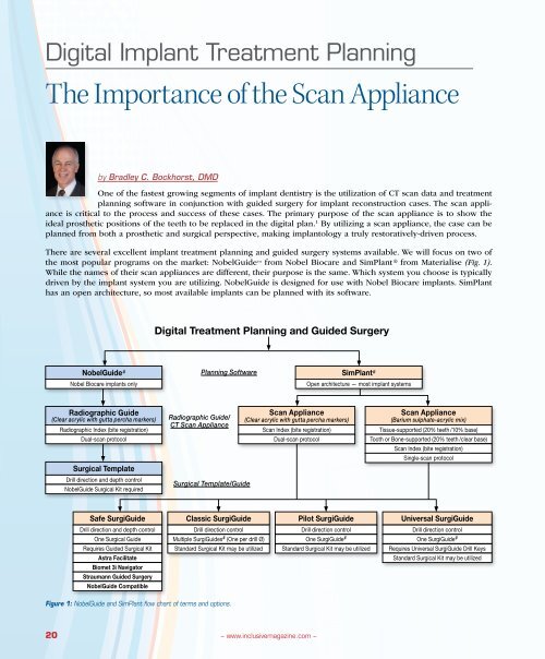
<strong>The</strong>re are several excellent implant treatment planning and guided surgery systems available. We will focus on two <strong>of</strong><br />
<strong>the</strong> most popular programs on <strong>the</strong> market: NobelGuide from Nobel Biocare and SimPlant ® from Materialise (Fig. 1).<br />
While <strong>the</strong> names <strong>of</strong> <strong>the</strong>ir scan appliances are different, <strong>the</strong>ir purpose is <strong>the</strong> same. Which system you choose is typically<br />
driven by <strong>the</strong> implant system you are utilizing. NobelGuide is designed for use with Nobel Biocare implants. SimPlant<br />
has an open architecture, so most available implants can be planned with its s<strong>of</strong>tware.<br />
Digital Treatment Planning and Guided Surgery<br />
NobelGuide #<br />
Nobel Biocare implants only<br />
Planning S<strong>of</strong>tware<br />
SimPlant #<br />
Open architecture — most implant systems<br />
Radiographic Guide<br />
(Clear acrylic with gutta percha markers)<br />
Radiographic Index (bite registration)<br />
Dual-scan protocol<br />
Surgical Template<br />
Drill direction and depth control<br />
NobelGuide Surgical Kit required<br />
Radiographic Guide/<br />
CT <strong>Scan</strong> <strong>Appliance</strong><br />
Surgical Template/Guide<br />
<strong>Scan</strong> <strong>Appliance</strong><br />
(Clear acrylic with gutta percha markers)<br />
<strong>Scan</strong> Index (bite registration)<br />
Dual-scan protocol<br />
<strong>Scan</strong> <strong>Appliance</strong><br />
(Barium sulphate -acrylic mix)<br />
Tissue-supported (20% teeth /10% base)<br />
Tooth or Bone-supported (20% teeth /clear base)<br />
<strong>Scan</strong> Index (bite registration)<br />
Single-scan protocol<br />
Safe SurgiGuide<br />
Classic SurgiGuide<br />
Pilot SurgiGuide<br />
Universal SurgiGuide<br />
Drill direction and depth control<br />
One Surgical Guide<br />
Requires Guided Surgical Kit<br />
Astra Facilitate<br />
Biomet 3i Navigator<br />
Straumann Guided Surgery<br />
NobelGuide Compatible<br />
Drill direction control<br />
Multiple SurgiGuides # (One per drill Ø)<br />
Standard Surgical Kit may be utilized<br />
Drill direction control<br />
One SurgiGuide #<br />
Standard Surgical Kit may be utilized<br />
Drill direction control<br />
One SurgiGuide #<br />
Requires Universal SurgiGuide Drill Keys<br />
Standard Surgical Kit may be utilized<br />
Figure 1: NobelGuide and SimPlant flow chart <strong>of</strong> terms and options.<br />
20<br />
– www.inclusivemagazine.com –
Role <strong>of</strong> <strong>the</strong> <strong>Scan</strong> <strong>Appliance</strong><br />
<strong>The</strong> primary purpose <strong>of</strong> <strong>the</strong> scan appliance is to show<br />
<strong>the</strong> ideal pros<strong>the</strong>tic positions <strong>of</strong> <strong>the</strong> tooth or teeth to be<br />
replaced. It provides an invaluable diagnostic tool to relate<br />
<strong>the</strong> tooth-to-bone relationship. 2 This is critical for <strong>the</strong><br />
planning process. While <strong>the</strong> SimPlant “virtual teeth” function<br />
is useful for short spans such as single tooth replacement<br />
(Fig. 2), an appliance in which <strong>the</strong> teeth have been<br />
set in <strong>the</strong> ideal position(s) provides <strong>the</strong> most accuracy.<br />
Both NobelGuide and SimPlant advocate a dual-scan protocol.<br />
In <strong>the</strong>se cases, <strong>the</strong> surgical guide is literally fabricated<br />
from <strong>the</strong> CT scan <strong>of</strong> <strong>the</strong> scan appliance (Fig. 3a-3d).<br />
Figure 2: “Virtual teeth” are inserted to represent teeth #8-10.<br />
Figure 3a: Radiographic Guide<br />
Figure 3b: Virtual rendering <strong>of</strong><br />
Radiographic Guide<br />
Figure 3c: Virtual rendering <strong>of</strong> Surgical<br />
Template<br />
Figure 3d: Surgical Template<br />
Types <strong>of</strong> <strong>Scan</strong> <strong>Appliance</strong>s<br />
<strong>The</strong> types <strong>of</strong> scan appliances can be split into single- and dual-scan protocols (Fig. 4a,<br />
4b). SimPlant traditionally utilizes a single-scan protocol. With <strong>the</strong> introduction <strong>of</strong> its<br />
dual-scan module, Materialise now recommends <strong>the</strong> dual scan. NobelGuide utilizes a<br />
dual-scan protocol.<br />
Single-<strong>Scan</strong> <strong>Appliance</strong>: Barium sulphate is mixed with acrylic to identify various<br />
structures radiographically. <strong>The</strong> teeth to be replaced contain a 20 percent barium sulphate<br />
mix. If <strong>the</strong> goal is to perform a flapless procedure on a fully edentulous case,<br />
<strong>the</strong> teeth are made with a 20 percent BaSO4 mix and <strong>the</strong> base a 10 percent mix. This<br />
allows <strong>the</strong> teeth to be identified as well as <strong>the</strong> s<strong>of</strong>t tissue in <strong>the</strong> CT study. 3 For partially<br />
edentulous cases, <strong>the</strong> scan appliance can be a flipper-type design or it can overlay <strong>the</strong><br />
remaining teeth, depending on <strong>the</strong> clinician’s preference.<br />
<strong>The</strong> types <strong>of</strong> scan<br />
appliances can basically<br />
be split into single- and<br />
dual-scan protocols.<br />
Figure 4a: Single-scan appliances<br />
Figure 4b: Dual-scan appliance<br />
Dual-<strong>Scan</strong> <strong>Appliance</strong>:<br />
Scatter from neighboring<br />
restorations can obscure<br />
<strong>the</strong> view <strong>of</strong> <strong>the</strong> teeth in <strong>the</strong><br />
scan appliance during single<br />
scans. Barium sulphate<br />
can also cause a minor<br />
amount <strong>of</strong> scatter and potentially<br />
obscure <strong>the</strong> view<br />
<strong>of</strong> vital structures. <strong>The</strong><br />
dual scan avoids this potential<br />
complication. <strong>The</strong><br />
patient is scanned with <strong>the</strong><br />
– Digital Implant Treatment Planning: <strong>The</strong> <strong>Importance</strong> <strong>of</strong> <strong>the</strong> <strong>Scan</strong> <strong>Appliance</strong> – 21
scan appliance, and <strong>the</strong>n <strong>the</strong> scan appliance is scanned alone. <strong>The</strong> dual-scan appliance is typically fabricated from clear<br />
acrylic with approximately eight 1 mm to 1.5 mm spherical gutta percha markers. <strong>The</strong> planning s<strong>of</strong>tware converts <strong>the</strong><br />
CT scan files and merges <strong>the</strong> two scans by matching <strong>the</strong> gutta percha markers, aligning <strong>the</strong> radiopaque markers so that<br />
<strong>the</strong> pros<strong>the</strong>sis will be visible over <strong>the</strong> available osseous anatomy. 4<br />
Utilizing <strong>the</strong> Patient’s Existing Denture as a <strong>Scan</strong> <strong>Appliance</strong><br />
While utilizing <strong>the</strong> patient’s existing denture as a scan appliance is possible, it can be problematic. Gutta percha markers<br />
can be added to <strong>the</strong> patient’s existing denture, or <strong>the</strong> denture can be duplicated and utilized only if it is well-fitting<br />
Figure 5a: View <strong>of</strong> well-fitting scan appliance<br />
Figure 5b: Radiolucent area shows crestal and facial ridge atrophy. <strong>The</strong> Radiographic<br />
Guide should be hard relined and <strong>the</strong> case rescanned.<br />
and <strong>the</strong> teeth are in <strong>the</strong> correct positions (Fig. 5a, 5b). If<br />
<strong>the</strong> denture is worn down and has a poor fit, a new appliance<br />
should be fabricated. For fully edentulous cases,<br />
this involves <strong>the</strong> same steps as fabricating a new denture<br />
including wax rims to establish <strong>the</strong> occlusal records and<br />
a wax try-in to evaluate <strong>the</strong> VDO, CR, tooth set-up and<br />
es<strong>the</strong>tics. (NOTE: This set-up will not only be used to create a<br />
proper scan appliance, it can also be used as a guide for <strong>the</strong><br />
provisional and final pros<strong>the</strong>ses.) By receiving <strong>the</strong> patient’s<br />
acceptance <strong>of</strong> <strong>the</strong> pros<strong>the</strong>sis before CT scans are done,<br />
<strong>the</strong> potential for success is also increased. 5<br />
Figure 6: <strong>The</strong> patient’s existing denture was used as <strong>the</strong> scan appliance. <strong>The</strong> thin<br />
flanges will result in fenestrations and potential weak areas in <strong>the</strong> surgical guide.<br />
Ano<strong>the</strong>r issue that can arise when utilizing <strong>the</strong> patient’s existing denture is <strong>the</strong> thickness, particularly in <strong>the</strong> flange areas.<br />
Dentures, by design, are thin for patient comfort. <strong>The</strong> scan appliance should ideally be about 3 mm thick. <strong>The</strong> use <strong>of</strong> a<br />
thin denture could result in fenestrations and potential weak areas in <strong>the</strong> surgical guide (Fig. 6).<br />
Verify Fit <strong>of</strong> <strong>the</strong> <strong>Scan</strong> <strong>Appliance</strong><br />
Once <strong>the</strong> scan appliance has been fabricated it should<br />
be tried-in to verify fit (Fig. 7). If <strong>the</strong> lab did not provide<br />
one, a scan index should be fabricated. <strong>The</strong> scan index,<br />
or Radiographic Index as it is called in <strong>the</strong> NobelGuide<br />
system, is a bite registration <strong>of</strong> <strong>the</strong> scan appliance to <strong>the</strong><br />
opposing dentition (Fig 8). <strong>The</strong> purpose <strong>of</strong> <strong>the</strong> scan index<br />
is to ensure that <strong>the</strong> appliance remains completely seated<br />
during <strong>the</strong> scan. It must be made out <strong>of</strong> a radiolucent<br />
material so that it does not block out <strong>the</strong> teeth. We have<br />
Figure 7: Try-in <strong>of</strong> <strong>the</strong> scan appliance<br />
to verify fit. Note: Inspection windows<br />
can be used as an aid in partially<br />
edentulous cases.<br />
Figure 8: <strong>Scan</strong> appliance and scan<br />
index on articulated models.<br />
22<br />
– www.inclusivemagazine.com –
Figure 9a: SimPlant <strong>Scan</strong> protocol<br />
Figure 9b: NobelGuide <strong>Scan</strong> protocol<br />
found Capture ® Clear Bite (<strong>Glidewell</strong> Laboratories) to be an acceptable material.<br />
Once <strong>the</strong> fit <strong>of</strong> <strong>the</strong> scan appliance and scan index are verified, <strong>the</strong> patient can<br />
be sent for <strong>the</strong> CT scan.<br />
<strong>The</strong> CT scan protocol for <strong>the</strong> system you intend to utilize should be followed<br />
(Fig. 9a, 9b). <strong>The</strong> scan protocols for NobelGuide and SimPlant can be found at<br />
inclusivedental.com. <strong>The</strong> scan appliance and scan index must be fully seated<br />
for <strong>the</strong> patient scan. If a dual scan is being utilized (Fig. 10a), <strong>the</strong> second scan<br />
is taken <strong>of</strong> <strong>the</strong> scan appliance alone (without <strong>the</strong> scan index). <strong>The</strong> appliance<br />
should be up <strong>of</strong>f <strong>the</strong> metal table or bar in <strong>the</strong> same orientation as it was in <strong>the</strong><br />
patient’s mouth (Fig. 10b). <strong>The</strong> standard files generated by a CT scan are called<br />
DICOM files (Digital Imaging Communication <strong>of</strong> Medical Images). <strong>The</strong> radiologist<br />
should reconstruct <strong>the</strong> scan per <strong>the</strong> protocol. Typically, <strong>the</strong> radiology lab<br />
burns <strong>the</strong> CT scan data onto a CD. In order to avoid confusion, request that <strong>the</strong><br />
radiologist create two folders: one containing <strong>the</strong> DICOM files <strong>of</strong> <strong>the</strong> patient<br />
scan and, if a dual scan was done, a second folder <strong>of</strong> <strong>the</strong> scan appliance. If you<br />
are using <strong>the</strong> NobelGuide or SimPlant s<strong>of</strong>tware yourself or are utilizing <strong>Glidewell</strong><br />
Laboratories’ Digital Implant Treatment Planning Services, <strong>the</strong> DICOM files<br />
are converted and utilized to virtually plan <strong>the</strong> case.<br />
<strong>The</strong> scan appliance<br />
and scan index<br />
must be fully seated<br />
for <strong>the</strong> patient scan.<br />
<strong>Scan</strong> 1 <strong>Scan</strong> 2<br />
Patient<br />
Radiographic<br />
Guide<br />
Radiographic<br />
Index<br />
Figure 10a, 10b: Dual scan: <strong>The</strong> patient is scanned with <strong>the</strong> scan appliance and scan index. A second scan <strong>of</strong> <strong>the</strong> scan appliance alone is performed.<br />
– Digital Implant Treatment Planning: <strong>The</strong> <strong>Importance</strong> <strong>of</strong> <strong>the</strong> <strong>Scan</strong> <strong>Appliance</strong> – 23
Single Tooth<br />
Figure 11a: Plan for replacement <strong>of</strong> tooth #19. <strong>The</strong> cross-sectional slice<br />
through tooth #19 can be seen on <strong>the</strong> right side <strong>of</strong> <strong>the</strong> screen.<br />
Figure 11b: Virtual plan to replace congenitally missing lateral incisors.<br />
Multiple Teeth<br />
Full Arch<br />
Figure 11c: Multiple implants can be planned with appropriate implant-implant<br />
and implant-root spacing, as can areas to be grafted.<br />
Figure 11d: Case planned for eight implants for a fixed restoration.<br />
Figure 11e: Case plan for a mandibular All-on-4 restoration.<br />
24<br />
– www.inclusivemagazine.com –
Restoratively Driven Treatment Planning<br />
<strong>The</strong> whole point <strong>of</strong> <strong>the</strong> diagnostic work-up and utilization <strong>of</strong> <strong>the</strong> scan appliance is to show <strong>the</strong> ideal positions <strong>of</strong> <strong>the</strong><br />
tooth or teeth to be replaced. While digital treatment planning guided surgery protocol was initially developed for <strong>the</strong><br />
fully edentulous patient, it has advantages for partially edentulous patients as well. 6 Whe<strong>the</strong>r you are replacing one<br />
tooth, multiple teeth or a full arch, digital treatment planning allows you to virtually plan <strong>the</strong> case from both a surgical<br />
and pros<strong>the</strong>tic perspective in a 3-D environment (Fig. 11a-e). This allows you to make almost all <strong>of</strong> <strong>the</strong> clinical decisions<br />
up front. <strong>The</strong> result is implants that are more ideally placed and, <strong>the</strong>refore, simpler pros<strong>the</strong>tics and superior restorations.<br />
In appropriate cases, <strong>the</strong> pros<strong>the</strong>sis can be prefabricated for an immediately loaded restoration. 7 If you plan to<br />
immediately load <strong>the</strong> case, we recommend you deliver a provisional restoration at <strong>the</strong> time <strong>of</strong> surgery and <strong>the</strong>n proceed<br />
to <strong>the</strong> definitive pros<strong>the</strong>sis at a later date.<br />
Fabrication <strong>of</strong> <strong>the</strong> Surgical Guide<br />
Once completed, <strong>the</strong> virtual plan can be transferred to <strong>the</strong><br />
clinical setting through <strong>the</strong> use <strong>of</strong> a surgical guide. <strong>The</strong><br />
guide produced for NobelGuide is referred to as a Surgical<br />
Template (Fig. 12a). <strong>The</strong> guide from Materialise (Sim-<br />
Plant) is called a SurgiGuide (Fig. 12b). Using <strong>the</strong> surgical<br />
guides allows <strong>the</strong> surgeon to place <strong>the</strong> implants according<br />
to a restoratively driven treatment plan. 8 We will review<br />
<strong>the</strong> various types <strong>of</strong> guides in a future article.<br />
Conclusion<br />
Figure 12a: NobelGuide Surgical<br />
Template<br />
Figure 12b: Materialise Simplant<br />
SurgiGuide<br />
Digital treatment planning and guided surgery is rapidly gaining popularity and is becoming a standard <strong>of</strong> care. A key<br />
benefit to this approach is that <strong>the</strong> case can be planned from <strong>the</strong> beginning with <strong>the</strong> final restoration in mind. <strong>The</strong> scan<br />
appliance can be utilized to its fullest by:<br />
• Working with an experienced lab to ensure it is properly fabricated<br />
• Verifying clinically that it and <strong>the</strong> scan index fit well and are completely seated for <strong>the</strong> CT scan<br />
• Ensuring <strong>the</strong> radiologist is familiar with <strong>the</strong> scan protocol.<br />
By understanding <strong>the</strong> role and proper use <strong>of</strong> <strong>the</strong> scan appliance in <strong>the</strong> digital implant treatment planning and guided<br />
surgery process, <strong>the</strong> clinician can take full advantage <strong>of</strong> this technology. n<br />
References<br />
1. Kosinski T. Optimizing implant placement and aes<strong>the</strong>tics: technology to <strong>the</strong> rescue! Dentistry Today Aug 1009.<br />
2. Ganz S. CT scan technology; an evolving tool for avoiding complications and achieving predictable implant placement and restoration. International Magazine <strong>of</strong> Oral<br />
Implantology-1/2001.<br />
3. Rosenfeld A, Mandelaris G. Pros<strong>the</strong>tically directed implant placement using computer s<strong>of</strong>tware to ensure precise placement and predictable pros<strong>the</strong>tic outcomes.<br />
Int J Periodontics Restorative Dent. 2006 Jun; 26(3):215-21.<br />
4. Moy P, Palacci P, Ericson I. Immediate function & es<strong>the</strong>tics in implant dentistry. Odont PhD, Quintessence Publishing Co. Ltd.<br />
5. Amet E, Ganz S. Implant treatment planning using a patient acceptance pros<strong>the</strong>sis, radiographic record base, and surgical template. Part 1: Presurgical Phase.<br />
Implant Dent 1997;6: p.193-197.<br />
6. Balshi T, Balshi S. CT-generated surgical guides and flapless surgery. Int J Oral Maxill<strong>of</strong>ac Implants 2008; Vol 23, Num 2.<br />
7. Marchack C. An immediately loaded CAD/CAM-guided definitive pros<strong>the</strong>sis: A clinical report. J Pros<strong>the</strong>tic Dent 2005;93:8-12.<br />
8. Orentlicher G, Goldsmith D, Horowitz A. Compendium Apr 2009, Vol. 30, Num 3.<br />
– Digital Implant Treatment Planning: <strong>The</strong> <strong>Importance</strong> <strong>of</strong> <strong>the</strong> <strong>Scan</strong> <strong>Appliance</strong> – 25


