PDF - Neural Development
PDF - Neural Development
PDF - Neural Development

You also want an ePaper? Increase the reach of your titles
YUMPU automatically turns print PDFs into web optimized ePapers that Google loves.
Kizil et al. <strong>Neural</strong> <strong>Development</strong> 2012, 7:27 Page 3 of 13<br />
http://www.neuraldevelopment.com/content/7/1/27<br />
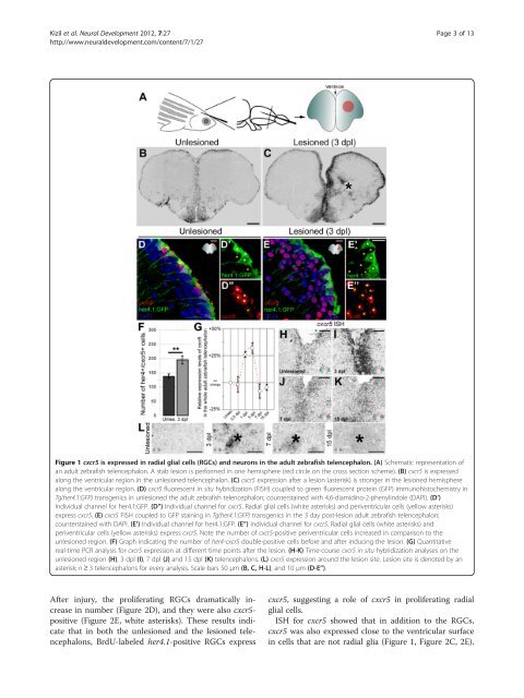
Figure 1 cxcr5 is expressed in radial glial cells (RGCs) and neurons in the adult zebrafish telencephalon. (A) Schematic representation of<br />
an adult zebrafish telencephalon. A stab lesion is performed in one hemisphere (red circle on the cross section scheme). (B) cxcr5 is expressed<br />
along the ventricular region in the unlesioned telencephalon. (C) cxcr5 expression after a lesion (asterisk) is stronger in the lesioned hemisphere<br />
along the ventricular region. (D) cxcr5 fluorescent in situ hybridization (FISH) coupled to green fluorescent protein (GFP) immunohistochemistry in<br />
Tg(her4.1:GFP) transgenics in unlesioned the adult zebrafish telencephalon; counterstained with 4,6-diamidino-2-phenylindole (DAPI). (D’)<br />
Individual channel for her4.1:GFP. (D”) Individual channel for cxcr5. Radial glial cells (white asterisks) and periventricular cells (yellow asterisks)<br />
express cxcr5. (E) cxcr5 FISH coupled to GFP staining in Tg(her4.1:GFP) transgenics in the 3 day post-lesion adult zebrafish telencephalon;<br />
counterstained with DAPI. (E’) Individual channel for her4.1:GFP. (E”) Individual channel for cxcr5. Radial glial cells (white asterisks) and<br />
periventricular cells (yellow asterisks) express cxcr5. Note the number of cxcr5-positive periventricular cells increased in comparison to the<br />
unlesioned region. (F) Graph indicating the number of her4-cxcr5 double-positive cells before and after inducing the lesion. (G) Quantitative<br />
real-time PCR analysis for cxcr5 expression at different time points after the lesion. (H-K) Time-course cxcr5 in situ hybridization analyses on the<br />
unlesioned region (H), 3 dpl (I), 7 dpl (J) and 15 dpl (K) telencephalons. (L) cxcr5 expression around the lesion site. Lesion site is denoted by an<br />
asterisk; n ≥ 3 telencephalons for every analysis. Scale bars 50 μm (B, C, H-L), and 10 μm (D-E”).<br />
After injury, the proliferating RGCs dramatically increase<br />
in number (Figure 2D), and they were also cxcr5-<br />
positive (Figure 2E, white asterisks). These results indicate<br />
that in both the unlesioned and the lesioned telencephalons,<br />
BrdU-labeled her4.1-positive RGCs express<br />
cxcr5, suggesting a role of cxcr5 in proliferating radial<br />
glial cells.<br />
ISH for cxcr5 showed that in addition to the RGCs,<br />
cxcr5 was also expressed close to the ventricular surface<br />
in cells that are not radial glia (Figure 1, Figure 2C, 2E).