Dynamics of glutamatergic signaling in the mushroom ... - HAL - ESPCI
Dynamics of glutamatergic signaling in the mushroom ... - HAL - ESPCI
Dynamics of glutamatergic signaling in the mushroom ... - HAL - ESPCI

Create successful ePaper yourself
Turn your PDF publications into a flip-book with our unique Google optimized e-Paper software.
S<strong>in</strong>akevitch et al. Neural Development 2010, 5:10<br />
http://www.neuraldevelopment.com/content/5/1/10<br />
RESEARCH ARTICLE<br />
Open Access<br />
<strong>Dynamics</strong> <strong>of</strong> <strong>glutamatergic</strong> <strong>signal<strong>in</strong>g</strong> <strong>in</strong> <strong>the</strong><br />
<strong>mushroom</strong> body <strong>of</strong> young adult Drosophila<br />
Ir<strong>in</strong>a S<strong>in</strong>akevitch 1,2,5* , Yves Grau 3 , Nicholas J Strausfeld 4 , Serge Birman 1,2*<br />
Abstract<br />
Background: The <strong>mushroom</strong> bodies (MBs) are paired bra<strong>in</strong> centers located <strong>in</strong> <strong>the</strong> <strong>in</strong>sect protocerebrum <strong>in</strong>volved<br />
<strong>in</strong> olfactory learn<strong>in</strong>g and memory and o<strong>the</strong>r associative functions. Processes from <strong>the</strong> Kenyon cells (KCs), <strong>the</strong>ir<br />
<strong>in</strong>tr<strong>in</strong>sic neurons, form <strong>the</strong> bulk <strong>of</strong> <strong>the</strong> MB’s calyx, pedunculus and lobes. In young adult Drosophila, <strong>the</strong> last-born<br />
KCs extend <strong>the</strong>ir processes <strong>in</strong> <strong>the</strong> a/b lobes as a th<strong>in</strong> core (a/b cores) that is embedded <strong>in</strong> <strong>the</strong> surround<strong>in</strong>g matrix<br />
<strong>of</strong> o<strong>the</strong>r mature KC processes. A high level <strong>of</strong> L-glutamate (Glu) immunoreactivity is present <strong>in</strong> <strong>the</strong> a/b cores (a/<br />
bc) <strong>of</strong> recently eclosed adult flies. In a Drosophila model <strong>of</strong> fragile X syndrome, <strong>the</strong> ma<strong>in</strong> cause <strong>of</strong> <strong>in</strong>herited mental<br />
retardation, treatment with metabotropic Glu receptor (mGluR) antagonists can rescue memory deficits and MB<br />
structural defects.<br />
Results: To address <strong>the</strong> role <strong>of</strong> Glu <strong>signal<strong>in</strong>g</strong> <strong>in</strong> <strong>the</strong> development and maturation <strong>of</strong> <strong>the</strong> MB, we have compared<br />
<strong>the</strong> time course <strong>of</strong> Glu immunoreactivity with <strong>the</strong> expression <strong>of</strong> various <strong>glutamatergic</strong> markers at various times,<br />
that is, 1 hour, 1 day and 10 days after adult eclosion. We observed that last-born a/bc KCs <strong>in</strong> young adult as well<br />
as develop<strong>in</strong>g KCs <strong>in</strong> late larva and at various pupal stages transiently express high level <strong>of</strong> Glu immunoreactivity <strong>in</strong><br />
Drosophila. One day after eclosion, <strong>the</strong> Glu level was already markedly reduced <strong>in</strong> <strong>the</strong> a/bc neurons. Glial cell<br />
processes express<strong>in</strong>g glutam<strong>in</strong>e syn<strong>the</strong>tase and <strong>the</strong> Glu transporter dEAAT1 were found to surround <strong>the</strong> Gluexpress<strong>in</strong>g<br />
KCs <strong>in</strong> very young adults, subsequently enwrapp<strong>in</strong>g <strong>the</strong> a/b lobes to become distributed equally over<br />
<strong>the</strong> entire MB neuropil. The vesicular Glu transporter DVGluT was detected by immunosta<strong>in</strong><strong>in</strong>g <strong>in</strong> processes that<br />
project with<strong>in</strong> <strong>the</strong> MB lobes and pedunculus, but this transporter is apparently never expressed by <strong>the</strong> KCs<br />
<strong>the</strong>mselves. The NMDA receptor subunit dNR1 is widely expressed <strong>in</strong> <strong>the</strong> MB neuropil just after eclosion, but was<br />
not detected <strong>in</strong> <strong>the</strong> a/bc neurons. In contrast, we provide evidence that DmGluRA, <strong>the</strong> only Drosophila mGluR, is<br />
specifically expressed <strong>in</strong> Glu-accumulat<strong>in</strong>g cells <strong>of</strong> <strong>the</strong> MB a/bc immediately and for a short time after eclosion.<br />
Conclusions: The distribution and dynamics <strong>of</strong> <strong>glutamatergic</strong> markers <strong>in</strong>dicate that newborn KCs transiently<br />
accumulate Glu at a high level <strong>in</strong> late pupal and young eclosed Drosophila, and may locally release this am<strong>in</strong>o acid<br />
by a mechanism that would not <strong>in</strong>volve DVGluT. At this stage, Glu can b<strong>in</strong>d to <strong>in</strong>tr<strong>in</strong>sic mGluRs abundant <strong>in</strong> <strong>the</strong><br />
a/bc KCs, and to NMDA receptors <strong>in</strong> <strong>the</strong> rest <strong>of</strong> <strong>the</strong> MB neuropil, before be<strong>in</strong>g captured and metabolized <strong>in</strong><br />
surround<strong>in</strong>g glial cells. This suggests that Glu acts as an autocr<strong>in</strong>e or paracr<strong>in</strong>e agent that contributes to <strong>the</strong><br />
structural and functional maturation <strong>of</strong> <strong>the</strong> MB dur<strong>in</strong>g <strong>the</strong> first hours <strong>of</strong> Drosophila adult life.<br />
Background<br />
The neurotransmitter L-glutamate (Glu) plays essential<br />
roles <strong>in</strong> various bra<strong>in</strong> functions <strong>in</strong> mammals, such as<br />
motor control, synaptic plasticity, learn<strong>in</strong>g and memory,<br />
cognition, and bra<strong>in</strong> maturation dur<strong>in</strong>g development<br />
[1-5]. Disruption <strong>of</strong> Glu <strong>signal<strong>in</strong>g</strong> is central to epilepsy<br />
[6,7] and major neurological and psychiatric disorders,<br />
* Correspondence: is<strong>in</strong>akev@asu.edu; serge.birman@espci.fr<br />
1 Laboratoire de Neurobiologie, CNRS UMR 7637, <strong>ESPCI</strong> ParisTech, 10 rue<br />
Vauquel<strong>in</strong>, 75231 Paris cedex 5, France<br />
<strong>in</strong>clud<strong>in</strong>g Alzheimer’s and Park<strong>in</strong>son’s diseases, schizophrenia,<br />
mood disorders, depression, anxiety, and stressand<br />
trauma-related disorders [3,8-10]. Glu acts by b<strong>in</strong>d<strong>in</strong>g<br />
to specific ion channel-coupled ionotropic (iGluRs)<br />
or G prote<strong>in</strong>-coupled metabotropic (mGluRs) membrane<br />
receptors. Glu receptors are implicated <strong>in</strong> processes <strong>of</strong><br />
learn<strong>in</strong>g and memory through long-term potentiation, a<br />
form <strong>of</strong> synaptic streng<strong>the</strong>n<strong>in</strong>g that follows brief, high<br />
frequency stimulation [11-14] and long-term depression,<br />
a long last<strong>in</strong>g reduction <strong>in</strong> synaptic transmission<br />
© 2010 S<strong>in</strong>akevitch et al; licensee BioMed Central Ltd. This is an Open Access article distributed under <strong>the</strong> terms <strong>of</strong> <strong>the</strong> Creative<br />
Commons Attribution License (http://creativecommons.org/licenses/by/2.0), which permits unrestricted use, distribution, and<br />
reproduction <strong>in</strong> any medium, provided <strong>the</strong> orig<strong>in</strong>al work is properly cited.
S<strong>in</strong>akevitch et al. Neural Development 2010, 5:10<br />
http://www.neuraldevelopment.com/content/5/1/10<br />
Page 3 <strong>of</strong> 20<br />
[12,15,16]. Glu release from nerve end<strong>in</strong>gs or astrocytes<br />
[17,18] requires previous uptake and concentration <strong>in</strong><br />
synaptic vesicles by vesicular Glu transporters [19,20].<br />
However, Glu can also be released by non-vesicular<br />
mechanisms [21] and exerts a paracr<strong>in</strong>e action on neuronal<br />
migration [22,23].<br />
In Drosophila and o<strong>the</strong>r arthropods, Glu is well characterized<br />
as <strong>the</strong> excitatory neurotransmitter <strong>of</strong> <strong>the</strong> neuromuscular<br />
junction [24-28]. However, this am<strong>in</strong>o acid<br />
has important <strong>signal<strong>in</strong>g</strong> functions <strong>in</strong> <strong>the</strong> Drosophila<br />
bra<strong>in</strong> as well [29-33]. The Drosophila genome was<br />
predicted to encode 30 iGluR subtypes, <strong>in</strong>clud<strong>in</strong>g<br />
3 AMPA- and 15 ka<strong>in</strong>ate-like, 2 NMDA-like, 4 δ-like and<br />
6 divergent receptors [34]. For now, <strong>the</strong> best characterized<br />
<strong>of</strong> <strong>the</strong>se are <strong>the</strong> postsynaptic iGluRs expressed at <strong>the</strong><br />
neuromuscular junction [25]. Drosophila NMDA-like<br />
receptors are expressed <strong>in</strong> <strong>the</strong> central nervous system<br />
[35] and have been implicated <strong>in</strong> learn<strong>in</strong>g and memory<br />
[36] and locomotor control [37]. The Drosophila genome<br />
encodes a s<strong>in</strong>gle functional mGluR, DmGluRA, an ortholog<br />
<strong>of</strong> vertebrate group II mGluRs [38]. This mGluR is<br />
presynaptic and expressed at <strong>the</strong> periphery <strong>of</strong> <strong>the</strong> active<br />
zones at <strong>the</strong> <strong>glutamatergic</strong> neuromuscular junctions,<br />
where it modulates both synapse excitability and f<strong>in</strong>e<br />
structure [24]. DmGluRA is also expressed <strong>in</strong> <strong>the</strong> bra<strong>in</strong>,<br />
<strong>in</strong> particular <strong>in</strong> lateral clock neurons, where it regulates<br />
circadian locomotor behavior [39].<br />
The <strong>mushroom</strong> bodies (MBs) are paired centers<br />
located <strong>in</strong> <strong>the</strong> protocerebrum <strong>of</strong> Drosophila and o<strong>the</strong>r<br />
dicondylic <strong>in</strong>sects that play essential roles <strong>in</strong> olfactory<br />
learn<strong>in</strong>g and memory [40] and o<strong>the</strong>r bra<strong>in</strong> functions,<br />
such as <strong>the</strong> control <strong>of</strong> locomotor activity [41], courtship<br />
behavior [42], courtship condition<strong>in</strong>g [43], visual context<br />
generalization [44], and sleep [45]. The <strong>in</strong>tr<strong>in</strong>sic structure<br />
<strong>of</strong> <strong>the</strong> MB is provided by <strong>the</strong> Kenyon cells (KCs),<br />
which have <strong>the</strong>ir cell bodies <strong>in</strong> <strong>the</strong> bra<strong>in</strong> cortex and<br />
<strong>the</strong>ir dendrites <strong>in</strong> <strong>the</strong> MB calyx, where <strong>the</strong>y receive<br />
<strong>in</strong>put from <strong>the</strong> antennal lobe projection neurons. Axonlike<br />
processes <strong>of</strong> KCs project anteriorly and ventrally <strong>in</strong><br />
<strong>the</strong> peduncle to form <strong>the</strong> vertical and medial lobes,<br />
which are subdivided <strong>in</strong>to discrete parallel entities, <strong>the</strong><br />
vertical a, a’ and <strong>the</strong> medial b, b’ and g lobes. In addition<br />
to <strong>the</strong> KCs, <strong>the</strong>re are o<strong>the</strong>r MB <strong>in</strong>tr<strong>in</strong>sic neurons<br />
and several classes <strong>of</strong> MB extr<strong>in</strong>sic neurons that connect<br />
<strong>the</strong> MB to o<strong>the</strong>r areas <strong>of</strong> <strong>the</strong> bra<strong>in</strong> neuropil [33,46-48].<br />
Emerg<strong>in</strong>g evidence suggests that different subtypes <strong>of</strong><br />
MB KCs may be <strong>in</strong>volved <strong>in</strong> dist<strong>in</strong>ct mechanisms <strong>of</strong><br />
memory formation due to <strong>the</strong>ir connections to different<br />
MB extr<strong>in</strong>sic neurons [49-53].<br />
Developmental studies have shown that <strong>the</strong> KCs are<br />
produced <strong>in</strong> each hemisphere <strong>of</strong> <strong>the</strong> bra<strong>in</strong> by <strong>the</strong> division<br />
<strong>of</strong> four neuroblasts born early dur<strong>in</strong>g <strong>the</strong> embryonic<br />
stage. The division <strong>of</strong> <strong>the</strong>se neuroblasts sequentially<br />
produces <strong>the</strong> three morphologically and spatially dist<strong>in</strong>ct<br />
subtypes <strong>of</strong> KCs: g, a’/b’ and a/b [54,55]. The g<br />
neurons are generated up to <strong>the</strong> mid-third <strong>in</strong>star larval<br />
stage; <strong>the</strong>y form <strong>the</strong> larval dorsal and medial lobe<br />
[55,56]. The next KC subtype to be generated is <strong>the</strong><br />
a’/b’ neuron, which cont<strong>in</strong>ues to be produced until<br />
puparium formation. Lastly, <strong>the</strong> a/b neurons are generated<br />
from <strong>the</strong> time <strong>of</strong> puparium formation until adult<br />
eclosion. In <strong>the</strong> a/b lobes, <strong>the</strong> KCs are organized <strong>in</strong><br />
concentric layers. The youngest axon-like processes<br />
situated <strong>in</strong> <strong>the</strong> <strong>in</strong>ner layer <strong>of</strong> <strong>the</strong> lobes are successively<br />
displaced outwards as <strong>the</strong>y differentiate and newer a/b<br />
processes are added to <strong>the</strong> structure from <strong>the</strong> most<br />
recently born KCs [47]. This volume <strong>of</strong> <strong>the</strong> a/b lobes<br />
<strong>in</strong>to which grow <strong>the</strong> last-born axons conta<strong>in</strong>s densely<br />
packed and extremely th<strong>in</strong> fibers that are rich <strong>in</strong> act<strong>in</strong><br />
filaments. This subset <strong>of</strong> processes has been named <strong>the</strong><br />
a/b core (a/bc) [33,47].<br />
An <strong>in</strong>creased response to mGluR activation may play a<br />
prom<strong>in</strong>ent role <strong>in</strong> <strong>the</strong> fragile X syndrome (FXS), <strong>the</strong><br />
most common form <strong>of</strong> <strong>in</strong>herited mental retardation and<br />
<strong>the</strong> predom<strong>in</strong>ant cause <strong>of</strong> autism [57]. Mutations <strong>in</strong><br />
dFmr1, <strong>the</strong>Drosophila homologue <strong>of</strong> <strong>the</strong> gene implicated<br />
<strong>in</strong> FXS, lead both to learn<strong>in</strong>g deficits and altered<br />
development <strong>of</strong> <strong>the</strong> MB, <strong>of</strong> which <strong>the</strong> most common<br />
feature is a failure <strong>of</strong> b lobes to stop at <strong>the</strong> bra<strong>in</strong> midl<strong>in</strong>e<br />
[58]. These behavioral and developmental phenotypes<br />
can be successfully rescued <strong>in</strong> Drosophila by treatment<br />
with mGluR antagonists [59], implicat<strong>in</strong>g Glu <strong>in</strong> <strong>the</strong><br />
pathology, as is <strong>the</strong> case <strong>in</strong> mammalian models [60].<br />
Recent studies showed that dFmr1 <strong>in</strong>teracts with<br />
DmGluRA <strong>in</strong> <strong>the</strong> regulation <strong>of</strong> synaptic architecture and<br />
excitability at <strong>glutamatergic</strong> synapses [61,62]. However,<br />
until now <strong>the</strong> precise role <strong>of</strong> Glu and mGluRs <strong>in</strong> FXS<br />
and MB development has rema<strong>in</strong>ed obscure.<br />
HerewepresentevidencethatGluanditsreceptor<br />
DmGluRA are directly <strong>in</strong>volved <strong>in</strong> construction <strong>of</strong> <strong>the</strong><br />
MB neural circuits. Previous studies suggested that <strong>the</strong><br />
Drosophila last-born a/bc KCs are immunoreactive to<br />
anti-Glu antibodies [32,33]. In <strong>the</strong> present study, we<br />
show that <strong>the</strong>se neurons express a high level <strong>of</strong> Glu-like<br />
immunoreactivity <strong>in</strong> newly eclosed adult flies. Interest<strong>in</strong>gly,<br />
newborn KCs <strong>in</strong> late larval and pupal stages also<br />
appear to express as a rule a high level <strong>of</strong> Glu. To<br />
understand fur<strong>the</strong>r <strong>the</strong> role and fate <strong>of</strong> Glu dur<strong>in</strong>g KC<br />
maturation, we analyzed <strong>the</strong> dynamics <strong>of</strong> Glu,<br />
DmGluRA and o<strong>the</strong>r Glu <strong>signal<strong>in</strong>g</strong>-associated prote<strong>in</strong>s<br />
<strong>in</strong> <strong>the</strong> MB <strong>of</strong> young adult Drosophila from <strong>the</strong> time <strong>of</strong><br />
<strong>the</strong>ir eclosion until 10 days post-eclosion. Our results<br />
<strong>in</strong>dicate that a transient Glu release likely regulates<br />
functional maturation <strong>of</strong> newborn KCs by a paracr<strong>in</strong>e<br />
action dur<strong>in</strong>g Drosophila post-embryonic development<br />
and <strong>the</strong> first hours after adult eclosion.
S<strong>in</strong>akevitch et al. Neural Development 2010, 5:10<br />
http://www.neuraldevelopment.com/content/5/1/10<br />
Page 4 <strong>of</strong> 20<br />
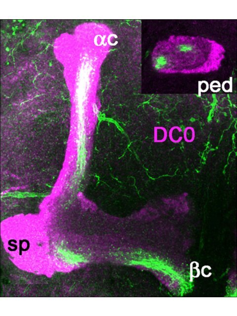
Figure 1 Glutamate and DVGluT immunoreactivities <strong>in</strong> <strong>the</strong> Drosophila <strong>mushroom</strong> bodies after adult eclosion. (A1, A2) DC0 marks <strong>the</strong><br />
whole MB neuropil that consists <strong>of</strong> <strong>the</strong> calyx (ca) and pedunculus (ped), and <strong>the</strong> vertical (a, a’) and horizontal (b, b’ and g) lobes. The spur (sp)<br />
area <strong>of</strong> <strong>the</strong> pedunculus conta<strong>in</strong>s axons <strong>of</strong> <strong>the</strong> g lobe. (A3) Schemes <strong>of</strong> <strong>the</strong> adult MB just before eclosion. The last born KCs extend axons <strong>in</strong> <strong>the</strong><br />
cores <strong>of</strong> <strong>the</strong> a and b lobes (ac, bc). Arrow <strong>in</strong>dicates <strong>the</strong> position <strong>of</strong> a transverse section <strong>of</strong> <strong>the</strong> pedunculus on <strong>the</strong> right show<strong>in</strong>g <strong>the</strong> layered<br />
and concentric organization <strong>of</strong> <strong>the</strong> KC axons. (B, C) Frontal sections <strong>of</strong> <strong>the</strong> MB lobes labeled with anti-Glu antibodies. One hour after adult<br />
eclosion, Glu can be detected at a high level <strong>in</strong> <strong>the</strong> lobes but only <strong>in</strong> <strong>the</strong> ac and bc axons. Upper <strong>in</strong>sert <strong>in</strong> (B) shows a transverse section <strong>of</strong> <strong>the</strong><br />
pedunculus with strong Glu sta<strong>in</strong><strong>in</strong>g <strong>in</strong> <strong>the</strong> a/bc KCs. The lower <strong>in</strong>sert is <strong>the</strong> control for Glu immunosta<strong>in</strong><strong>in</strong>g after preabsorption <strong>of</strong> <strong>the</strong> diluted<br />
antibodies with 10 -4 M conjugated Glu. (D1, D2) In 10-day-old flies, Glu immunoreactivity (green) is absent from <strong>the</strong> DC0-sta<strong>in</strong>ed KCs (magenta).<br />
Glu is distributed at this stage <strong>in</strong> scattered patterns <strong>in</strong> <strong>the</strong> a and g lobes and <strong>in</strong> <strong>the</strong> spur (sp), and likely orig<strong>in</strong>ates from MB extr<strong>in</strong>sic neurons.<br />
(E-J) DVGluT immunoreactivity <strong>in</strong> <strong>the</strong> MB with<strong>in</strong> 1 hour after after eclosion. Confocal optical sections from <strong>the</strong> frontal to <strong>the</strong> posterior parts <strong>of</strong><br />
<strong>the</strong> protocerebrum from 201Y-GAL4; UAS-mCD8::GFP flies. Note that <strong>the</strong> <strong>in</strong>tr<strong>in</strong>sic a/bc KCs are not positive for DVGluT. Sta<strong>in</strong>ed processes from<br />
extr<strong>in</strong>sic neurons are found <strong>in</strong> <strong>the</strong> a and g lobes and <strong>in</strong> <strong>the</strong> spur (sp) region. Scale bars: 20 μm.
S<strong>in</strong>akevitch et al. Neural Development 2010, 5:10<br />
http://www.neuraldevelopment.com/content/5/1/10<br />
Page 5 <strong>of</strong> 20<br />
Figure 2 Commonly used <strong>mushroom</strong> body drivers do not target <strong>the</strong> last born Kenyon cells just after adult eclosion. (A) In 17d-GAL4;<br />
UAS-mCD8::GFP adults just after eclosion, GFP is not present <strong>in</strong> <strong>the</strong> <strong>in</strong>ner axons <strong>of</strong> <strong>the</strong> a/bc neurons as seen <strong>in</strong> <strong>the</strong> lobe (arrow) or <strong>in</strong> <strong>the</strong><br />
pedunculus (arrowheads <strong>in</strong> <strong>the</strong> <strong>in</strong>sert). (B) Ten days later, all <strong>the</strong> a/bc KCs express GFP as shown <strong>in</strong> <strong>the</strong> lobes and pedunculus (<strong>in</strong>sert). (C, D)<br />
Similar observations with <strong>the</strong> c739-GAL4 driver l<strong>in</strong>e that does not express GFP <strong>in</strong> <strong>the</strong> a/bc neurons right after eclosion (C), but target all a/b KCs<br />
10 days later (D). sp, spur. (E, F) Similar observations with <strong>the</strong> 201Y-GAL4 driver l<strong>in</strong>e that targets both <strong>the</strong> a/b and g neurons. This l<strong>in</strong>e does not<br />
target <strong>the</strong> last born a/bc KCs just after eclosion as well. Scale bars: 20 μm.<br />
Results<br />
High Glu levels <strong>in</strong> last-born Kenyon cells<br />
Diverse subtypes <strong>of</strong> KCs form <strong>the</strong> Drosophila MB neuropil<br />
and <strong>the</strong>ir axons extend <strong>in</strong> <strong>the</strong> pedunculus and <strong>in</strong><br />
<strong>the</strong> medial and vertical lobes [33,48]. The whole Drosophila<br />
MB structure can be revealed with anti-DC0<br />
(PKA-C1) antibodies that label all parts <strong>of</strong> <strong>the</strong> KCs with<br />
different <strong>in</strong>tensity (Figure 1A1, A2): <strong>the</strong> cell bodies (K),<br />
<strong>the</strong> dendrites <strong>in</strong> <strong>the</strong> calyx (ca), and <strong>the</strong> axons <strong>in</strong> <strong>the</strong> pedunculus<br />
(ped) and lobes. In <strong>the</strong> g lobe, KCs extend only<br />
one axonal branch, form<strong>in</strong>g a medial lobe and <strong>the</strong> spur<br />
(sp), whereas <strong>the</strong> axons <strong>of</strong> o<strong>the</strong>r KCs divide at <strong>the</strong> level<br />
<strong>of</strong> <strong>the</strong> spur to give rise to vertical and medial axonal<br />
branches, thus form<strong>in</strong>g <strong>the</strong> a’/b’ and a/b divisions <strong>of</strong><br />
<strong>the</strong> lobes. The KC axons are organized <strong>in</strong> concentric<br />
strata <strong>in</strong> <strong>the</strong> pedunculus and a/b lobes. This is schematically<br />
depicted <strong>in</strong> Figure 1A3, <strong>the</strong> first-born cells, that<br />
is, <strong>the</strong> g neurons, be<strong>in</strong>g <strong>the</strong> most external, and <strong>the</strong> lastborn<br />
cells, <strong>the</strong> a/bc neurons, be<strong>in</strong>g embedded with<strong>in</strong><br />
<strong>the</strong> surround<strong>in</strong>g processes <strong>of</strong> a/b neurons. Like <strong>the</strong><br />
o<strong>the</strong>r KCs, <strong>the</strong>se last-born a/bc neurons orig<strong>in</strong>ate from<br />
four neuroblasts, which provide four identical axonal<br />
bundles <strong>in</strong> <strong>the</strong> pedunculus and throughout <strong>the</strong> MBs<br />
(Figure 1A3).<br />
The a/bc KCs present a high level <strong>of</strong> Glu-like immunoreactivity<br />
<strong>in</strong> <strong>the</strong>ir newborn axons immediately after<br />
adult eclosion (Figure 1B, C). Figure 1B (<strong>in</strong>sert) shows<br />
that Glu-like immunoreactivity <strong>in</strong> <strong>the</strong> pedunculus is<br />
restricted to four th<strong>in</strong> axonal bundles <strong>of</strong> a/bc KCs<br />
embedded with<strong>in</strong> <strong>the</strong> a/b axons. In contrast, no Glulike<br />
immunoreactivity can be detected <strong>in</strong> <strong>the</strong> older<br />
neurons <strong>in</strong> <strong>the</strong> core <strong>of</strong> <strong>the</strong> a/b lobes <strong>in</strong> 10-day-old<br />
adult flies (Figure 1D1, D2). At this age, we observed<br />
Glu-like immunoreactivity scattered <strong>in</strong> <strong>the</strong> a and g<br />
lobes and <strong>in</strong> <strong>the</strong> spur. This pattern is likely to represent<br />
<strong>the</strong> distribution <strong>of</strong> Glu-like immunoreactivity
S<strong>in</strong>akevitch et al. Neural Development 2010, 5:10<br />
http://www.neuraldevelopment.com/content/5/1/10<br />
Page 6 <strong>of</strong> 20<br />
Figure 3 Rapid disappearance <strong>of</strong> Glu immunoreactivity from <strong>the</strong> <strong>mushroom</strong> body a/b cores follow<strong>in</strong>g adult eclosion. (A-C) Transverse<br />
sections <strong>of</strong> <strong>the</strong> MB pedunculus from 17d-GAL4 (A), c739-GAL4 (B) and 201Y-GAL4 (C) adult flies express<strong>in</strong>g mCD8::GFP and labeled for Glu at<br />
different ages. Similar results were obta<strong>in</strong>ed with <strong>the</strong> three driver l<strong>in</strong>es. (A-C, a1-a3) With<strong>in</strong> 1 hour after eclosion, <strong>the</strong> core neurons express Glu<br />
but not GFP. Only th<strong>in</strong> processes <strong>in</strong> <strong>the</strong> outer layers <strong>of</strong> <strong>the</strong> pedunculus are positive for both. (A-C, b1-b3) Twenty-four hours after eclosion, <strong>the</strong><br />
<strong>in</strong>tensity <strong>of</strong> Glu label<strong>in</strong>g already dramatically decreased while <strong>the</strong> expression <strong>of</strong> GFP started to appear <strong>in</strong> <strong>the</strong> core neurons. (A-C, c1-c3) Ten days<br />
after eclosion, <strong>the</strong>re is no detectable Glu <strong>in</strong> <strong>the</strong> core neurons and GFP is strongly expressed <strong>in</strong> <strong>the</strong>se cells. Scale bars: 10 μm.
S<strong>in</strong>akevitch et al. Neural Development 2010, 5:10<br />
http://www.neuraldevelopment.com/content/5/1/10<br />
Page 7 <strong>of</strong> 20<br />
Figure 4 Newborn Kenyon cells express high levels <strong>of</strong> Glu <strong>in</strong> <strong>the</strong> Drosophila <strong>mushroom</strong> body <strong>in</strong> late larval stage and dur<strong>in</strong>g pupal<br />
development. (A, B) Transverse section <strong>of</strong> <strong>the</strong> MB pedunculus from 201Y-GAL4 (A) and 17d-GAL4 (B) pharate adults collected a few hours<br />
before eclosion and express<strong>in</strong>g mCD8::GFP. (A1-A3) GFP and Glu expression <strong>in</strong> 201Y-GAL4 are located both <strong>in</strong> <strong>the</strong> a/bc and g lobes. Most <strong>of</strong> <strong>the</strong><br />
GFP-positive a/bc neurons do not co-localize with Glu and only few axons express both Glu and GFP at <strong>the</strong> border <strong>of</strong> Glu-conta<strong>in</strong><strong>in</strong>g a/bc<br />
neurons. Agarose section <strong>of</strong> <strong>the</strong> pedunculus close to <strong>the</strong> lobes. (B1-B3) In 17d-GAL4, most Glu-conta<strong>in</strong><strong>in</strong>g a/bc neurons do not express GFP and<br />
only <strong>the</strong> border axons are both GFP- and Glu-positive. Agarose section <strong>of</strong> <strong>the</strong> pedunculus close to <strong>the</strong> calyx. In (A, B), Glu immunoreactivity <strong>in</strong><br />
<strong>the</strong> g lobe most likely orig<strong>in</strong>ates from extr<strong>in</strong>sic neurons. (C-I) Glu immunoreactivity monitored at late larval and pupal stages <strong>in</strong> <strong>the</strong> MB <strong>of</strong> wildtype<br />
Drosophila. (C, D). Transverse sections through <strong>the</strong> calyx and lobes <strong>of</strong> wander<strong>in</strong>g third <strong>in</strong>star larva. The four bundles <strong>of</strong> Glu-positive<br />
processes orig<strong>in</strong>ate from four clusters <strong>of</strong> KCs. Only one cluster is shown (on <strong>the</strong> left side <strong>in</strong> (C)) and arrows show bundles orig<strong>in</strong>at<strong>in</strong>g from <strong>the</strong><br />
o<strong>the</strong>r Glu-positive clusters. The four Glu-positive bundles extend <strong>in</strong> <strong>the</strong> core area <strong>of</strong> <strong>the</strong> pedunculus (arrowheads), <strong>in</strong>dicat<strong>in</strong>g that <strong>the</strong>y<br />
correspond to newborn KCs. As shown <strong>in</strong> (D), <strong>the</strong>se Glu-positive fibers also project <strong>in</strong>to <strong>the</strong> a/bc region <strong>of</strong> <strong>the</strong> larval MB (bc is not visible on this<br />
agarose section). The contour <strong>in</strong>dicates <strong>the</strong> shape <strong>of</strong> <strong>the</strong> larval MB vertical lobe (a) and spur (sp) region. Note that extr<strong>in</strong>sic <strong>glutamatergic</strong> cells<br />
are also present at this stage <strong>in</strong> <strong>the</strong> spur area. (E) In pupa 48 hours after puparium formation (APF) at 22°C (stages P5 to P6 <strong>of</strong> Ba<strong>in</strong>bridge and<br />
Bownes [100]), clusters <strong>of</strong> Glu-positive KCs are still present but <strong>the</strong>ir bundles at <strong>the</strong> base <strong>of</strong> <strong>the</strong> pedunculus (arrowheads) are less <strong>in</strong>tensely<br />
labeled compared to larval and o<strong>the</strong>r pupal stages. (F, G) Pupa 72 hours APF (stage P8). At this developmental stage, four groups <strong>of</strong> newborn<br />
KCs are brightly Glu-positive and project axons <strong>in</strong> <strong>the</strong> pedunculus (arrowheads <strong>in</strong> (G)). O<strong>the</strong>r Glu-immunoreactive processes <strong>in</strong> <strong>the</strong> g lobe area <strong>of</strong><br />
<strong>the</strong> pedunculus likely correspond to fibers from extr<strong>in</strong>sic cells as only cell bodies <strong>of</strong> newborn KCs express high Glu-immunoreactivity. (H, I) In<br />
pupa 96 hours APF at 22°C (stages P11 to P12), four clusters <strong>of</strong> Glu-immunopositive newborn KCs (arrows <strong>in</strong> (H)) project <strong>the</strong>ir axons to <strong>the</strong><br />
pedunculus (arrowheads <strong>in</strong> (I)). K, Kenyon cell bodies; ca, calyx, ped, pedunculus; sp, spur. Scale bars: 10 μm.
S<strong>in</strong>akevitch et al. Neural Development 2010, 5:10<br />
http://www.neuraldevelopment.com/content/5/1/10<br />
Page 8 <strong>of</strong> 20<br />
<strong>in</strong> <strong>the</strong> processes <strong>of</strong> MB extr<strong>in</strong>sic neurons ra<strong>the</strong>r than<br />
<strong>in</strong> KCs.<br />
Last-born Kenyon cells do not express DVGluT<br />
The accumulation <strong>of</strong> Glu <strong>in</strong> <strong>the</strong> young a/bc KCssuggests<br />
that this am<strong>in</strong>o acid could be transiently used as<br />
a neurotransmitter by <strong>the</strong>se cells. The vesicular Glu<br />
transporter DVGluT is <strong>in</strong>volved <strong>in</strong> Glu synaptic vesicle<br />
storage, prior to neurotransmitter release, and can be<br />
used as a marker <strong>of</strong> <strong>glutamatergic</strong> neurons <strong>in</strong> Drosophila<br />
[29,30]. Therefore, we performed anti-DVGluT<br />
immunosta<strong>in</strong><strong>in</strong>g immediately after adult eclosion, at a<br />
time when <strong>the</strong> Glu level is high <strong>in</strong> <strong>the</strong> MB core neurons.<br />
The DVGluT antibodies strongly labeled <strong>the</strong> protocerebrum<br />
and antennal lobe neuropils. In contrast,<br />
<strong>in</strong> <strong>the</strong> MB neuropil, nei<strong>the</strong>r <strong>the</strong> core neurons nor any<br />
<strong>in</strong>tr<strong>in</strong>sic KCs were found to be immunopositive for<br />
DVGluT (Figure 1E-J). The DVGluT immunoreactivity<br />
observed <strong>in</strong> <strong>the</strong> MBs, particularly <strong>in</strong> <strong>the</strong> spur <strong>of</strong> <strong>the</strong><br />
pedunculus and <strong>in</strong> <strong>the</strong> g and a lobes (Figure 1E-J),<br />
most likely corresponds to synapses from extr<strong>in</strong>sic <strong>glutamatergic</strong><br />
neurons. This suggests that Glu <strong>in</strong> <strong>the</strong><br />
young a/bc KCs ei<strong>the</strong>r is not stored <strong>in</strong> a vesicular pool<br />
or is stored <strong>in</strong> vesicles by ano<strong>the</strong>r transporter not yet<br />
identified.<br />
MB driver expression <strong>in</strong> last-born KCs<br />
The previous results suggest that <strong>the</strong> last-born MB core<br />
neurons mature with<strong>in</strong> just a few days after eclosion<br />
from an early Glu-express<strong>in</strong>g state to a differentiated<br />
cell <strong>in</strong> which Glu no longer accumulates. We asked<br />
whe<strong>the</strong>r o<strong>the</strong>r MB markers could be used to differentiate<br />
between <strong>the</strong>se two maturation states. The enhancer<br />
trap l<strong>in</strong>es 17d-, c739-, and 201Y-GAL4 mimic <strong>the</strong><br />
expression <strong>of</strong> MB markers and are commonly used as<br />
MB-specific drivers. We crossed each <strong>of</strong> <strong>the</strong>se l<strong>in</strong>es to<br />
UAS-mCD8::GFP flies to characterize <strong>the</strong>ir expression<br />
pattern <strong>in</strong> young adults. When flies were collected<br />
immediately after eclosion, we found every time that <strong>the</strong><br />
Glu-express<strong>in</strong>g a/bc neurons consist <strong>of</strong> two populations:<br />
<strong>in</strong>ner a/bc neurons, which corresponds to <strong>the</strong> younger<br />
cells that do not express green fluorescent prote<strong>in</strong><br />
(GFP); and outer a/bc neurons, which do express GFP<br />
(Figure 2A-C and <strong>in</strong>serts). In contrast, <strong>in</strong> 10-day-old<br />
flies, we observed that <strong>the</strong>se three drivers express<br />
mCD8-GFP <strong>in</strong> all <strong>the</strong> a/bc KCs: 17d-GAL4 <strong>in</strong> <strong>the</strong> a/bc<br />
neurons only (Figure 2D), c739-GAL4 <strong>in</strong> <strong>the</strong> a/b and a/<br />
bc cells (Figure 2E), and 201Y-GAL4 <strong>in</strong> <strong>the</strong> a/bc andg<br />
lobe neurons (Figure 2F). In each <strong>of</strong> <strong>the</strong>se l<strong>in</strong>es, GFP<br />
immunosta<strong>in</strong><strong>in</strong>g arranges <strong>in</strong> a specific way revealed by<br />
<strong>the</strong> pedunculus sections shown <strong>in</strong> <strong>the</strong> Figure 2 <strong>in</strong>serts:<br />
17d-GAL4 expresses GFP <strong>in</strong> four <strong>in</strong>ner bundles correspond<strong>in</strong>g<br />
to <strong>the</strong> a/bc cells, c739-GAL4 <strong>in</strong> <strong>the</strong> whole<br />
area <strong>of</strong> <strong>the</strong> pedunculus that conta<strong>in</strong>s <strong>the</strong> a/b axons, and<br />
201Y-GAL4 <strong>in</strong> <strong>the</strong> four a/bc bundles and a peripheral<br />
surround<strong>in</strong>g area that conta<strong>in</strong>s <strong>the</strong> g lobe axons. Thus,<br />
at least three different MB markers are not expressed <strong>in</strong><br />
<strong>the</strong> immature last-born a/bc KCsatatimewhen<strong>the</strong>se<br />
cells show high levels <strong>of</strong> Glu immunoreactivity, while<br />
<strong>the</strong>se markers are strongly expressed <strong>in</strong> <strong>the</strong>se same cells<br />
<strong>in</strong> 10-day-old flies.<br />
Rapid disappearance <strong>of</strong> Glu from last-born Kenyon cells<br />
We <strong>the</strong>n precisely compared <strong>the</strong> temporal patterns <strong>of</strong><br />
Glu and GFP expression <strong>in</strong> <strong>the</strong> a/bc KCs<strong>of</strong>MBdriver<br />
l<strong>in</strong>es express<strong>in</strong>g mCD8-GFP by look<strong>in</strong>g at <strong>the</strong> pedunculus<br />
<strong>in</strong> frontally cut agarose sections (Figure 3). In all<br />
three GAL4 l<strong>in</strong>es (17d-, c739-, and 201Y-GAL4) early<br />
after eclosion, <strong>the</strong> a/bc neurons could be divided <strong>in</strong>to<br />
three subtypes: <strong>the</strong> most <strong>in</strong>ner a/bc neurons that<br />
express Glu but not GFP; more peripheral a/bc neurons<br />
that co-express glutamate and GFP; neurons <strong>in</strong><br />
<strong>the</strong> a/bc outer area that express only GFP. With<strong>in</strong> 1<br />
hour after eclosion, only <strong>the</strong> outer parts <strong>of</strong> <strong>the</strong> a/bc<br />
region were simultaneously positive for both GFP and<br />
Glu <strong>in</strong> each driver l<strong>in</strong>e (Figure 3A-C, panels a1-a3).<br />
Because <strong>the</strong> border <strong>of</strong> <strong>the</strong> core regions co-localizes<br />
with GFP, this suggests that <strong>the</strong> matur<strong>in</strong>g a/bc neurons<br />
beg<strong>in</strong> to express GFP at <strong>the</strong> time when Glu is<br />
still present. Twenty-four hours after eclosion, GFP<br />
starts to be expressed <strong>in</strong> most a/bc neurons.Thisis<br />
when Glu-like immunoreactivity was already becom<strong>in</strong>g<br />
dramatically reduced (Figure 3A-C, panels b1-b3). Ten<br />
days after eclosion, <strong>the</strong>re is no detectable Glu immunoreactivity<br />
<strong>in</strong> <strong>the</strong> a/bc KCs, which all express GFP at<br />
a high <strong>in</strong>tensity (Figure 3A-C, panels c1-c3). Therefore,<br />
maturation <strong>of</strong> <strong>the</strong> last-born KCs appears to be a fast<br />
process <strong>in</strong> Drosophila that is concluded <strong>in</strong> a few hours<br />
only after adult eclosion.<br />
WeobservedthatGluisalreadypresent<strong>in</strong><strong>the</strong>a/bc<br />
axons <strong>of</strong> pharate adults, removed from <strong>the</strong> pupal cases a<br />
few hours before eclosion (Figure 4A, B). At this stage,<br />
Glu immunoreactivity is also detectable <strong>in</strong> <strong>the</strong> g region<br />
<strong>of</strong> <strong>the</strong> MB pedunculus. Representative pictures are<br />
shown <strong>in</strong> Figure 4A1-A3, section close to <strong>the</strong> lobes, and<br />
Figure 4B1-B3, section close to <strong>the</strong> calyx. The MB drivers<br />
201Y-GAL4 and 17d-GAL4 do not express GFP <strong>in</strong><br />
<strong>the</strong> Glu-express<strong>in</strong>g a/bc neurons <strong>of</strong> pupal flies, except<br />
for a few neurons at <strong>the</strong> border <strong>of</strong> <strong>the</strong> core. In 201Y-<br />
GAL4, GFP is also present <strong>in</strong> <strong>the</strong> g area <strong>in</strong> part <strong>of</strong> <strong>the</strong><br />
pedunculus. The level <strong>of</strong> Glu immunoreactivity <strong>in</strong> <strong>the</strong> g<br />
lobe varied significantly amongst flies sampled before<br />
eclosion, probably depend<strong>in</strong>g on <strong>the</strong> exact developmental<br />
progress <strong>of</strong> each pupal animal. This signal probably<br />
orig<strong>in</strong>ates from extr<strong>in</strong>sic glutametergic neurons because<br />
we found that only cell bodies <strong>of</strong> newborn KCs express
S<strong>in</strong>akevitch et al. Neural Development 2010, 5:10<br />
http://www.neuraldevelopment.com/content/5/1/10<br />
Page 9 <strong>of</strong> 20<br />
Figure 5 <strong>Dynamics</strong> <strong>of</strong> glutam<strong>in</strong>e syn<strong>the</strong>tase expression <strong>in</strong> <strong>mushroom</strong> body glial cells after adult eclosion. (A) A mouse monoclonal<br />
antibody raised aga<strong>in</strong>st sheep glutam<strong>in</strong>e syn<strong>the</strong>tase (GS) recognizes a s<strong>in</strong>gle band <strong>in</strong> a western blot <strong>of</strong> Drosophila head and bra<strong>in</strong>. The apparent<br />
size <strong>of</strong> <strong>the</strong> prote<strong>in</strong> (approximately 42 kDa) closely corresponds to <strong>the</strong> predicted size <strong>of</strong> Drosophila GS2 (41 kDa). (B) Localization <strong>of</strong> GS<br />
immunoreactivity <strong>in</strong> glial cells that express <strong>the</strong> Glu transporter dEAAT1 <strong>in</strong> 10-day-old adult fly MBs. Triple sta<strong>in</strong><strong>in</strong>g <strong>of</strong> bra<strong>in</strong>s from dEAAT1-GAL4;<br />
UAS-dEAAT1::GFP Drosophila with anti-GS (magenta), anti-GFP (green) and anti-DC0 (blue, for MB sta<strong>in</strong><strong>in</strong>g). (B1-B3) GS co-localizes with dEAAT1-<br />
positive glial cell bodies and processes <strong>in</strong> <strong>the</strong> spur (sp) region <strong>of</strong> <strong>the</strong> MB neuropil (merged magenta and green produces a white color). (C)<br />
Localization <strong>of</strong> GS <strong>in</strong> <strong>the</strong> MB with respect to <strong>glutamatergic</strong> synapses. Triple sta<strong>in</strong><strong>in</strong>g <strong>of</strong> wild-type adult bra<strong>in</strong>s with anti-DVGluT (green), anti-GS<br />
(magenta), and anti-GFP (blue) <strong>in</strong> agarose sections. (C1) GS is distributed all over <strong>the</strong> lobes (arrowheads) except <strong>the</strong> core neurons. (C2) DVGluTpositive<br />
<strong>glutamatergic</strong> synapses are present on <strong>the</strong> a lobe (arrow). (C3) GS immunoreactivity lies <strong>in</strong> close vic<strong>in</strong>ity to <strong>glutamatergic</strong> synapses<br />
(arrow). In <strong>the</strong> b lobe, GS is present (arrowheads) but DVGluT is absent. (D-I) Sections <strong>of</strong> Drosophila bra<strong>in</strong>s sta<strong>in</strong>ed with anti-GS (magenta) at<br />
different adult ages show<strong>in</strong>g <strong>the</strong> MB pedunculus (D, F, H) and <strong>the</strong> medial lobe region (E, G, I). MB a/b KCs are positive for GFP (green) <strong>in</strong> c739-<br />
GAL4; UAS-mCD8::GFP flies (D-G) or DC0 (green) <strong>in</strong> wild-type flies (H-I). In <strong>the</strong> pedunculus, GS is found <strong>in</strong> glial cells that surrounds <strong>the</strong> a/b axons<br />
ei<strong>the</strong>r with<strong>in</strong> 1 hour after adult eclosion (D), 24 hours later (F) or at 10 days old (adult) (H). (E) With<strong>in</strong> 1 hour after eclosion, GS-immunoreactive<br />
processes enwrap <strong>the</strong> MB medial b and g lobes. GS is also detected <strong>in</strong> many pr<strong>of</strong>iles <strong>in</strong>side <strong>the</strong> b and g lobes, except <strong>in</strong> <strong>the</strong> regions<br />
correspond<strong>in</strong>g to <strong>the</strong> core neurons that express nei<strong>the</strong>r GFP nor GS. (G) Twenty-four hours after eclosion, GS-positive glial processes distribute<br />
evenly <strong>in</strong> <strong>the</strong> whole b lobe area, <strong>in</strong>clud<strong>in</strong>g <strong>the</strong> b core, which at this time expresses GFP. (I) Ten days after eclosion, GS is expressed at a high<br />
level <strong>in</strong> glial cells that surround <strong>the</strong> b lobes. Scale bars: 20 μm.
S<strong>in</strong>akevitch et al. Neural Development 2010, 5:10<br />
http://www.neuraldevelopment.com/content/5/1/10<br />
Page 10 <strong>of</strong> 20<br />
high Glu-immunoreactivity (see Figure 4C, I and text<br />
below).<br />
Newborn Kenyon cells express Glu dur<strong>in</strong>g larval and<br />
pupal stages<br />
In order to explore if Glu is similarly present <strong>in</strong> develop<strong>in</strong>g<br />
KCs dur<strong>in</strong>g earlier phases <strong>of</strong> MB development, we<br />
performed Glu immunosta<strong>in</strong><strong>in</strong>g on bra<strong>in</strong> agarose sections<br />
<strong>in</strong> late third <strong>in</strong>star larva and at various stages <strong>of</strong><br />
pupal development. At <strong>the</strong>se stages, we generally found<br />
four clusters <strong>of</strong> Glu-immunoreactive KC bodies that<br />
send four Glu-positive bundles <strong>in</strong> <strong>the</strong> pedunculus and<br />
lobes (Figure 4C, I). Strik<strong>in</strong>gly, <strong>the</strong>se Glu-positive bundles<br />
are always located <strong>in</strong> <strong>the</strong> core areas <strong>of</strong> <strong>the</strong> pedunculus<br />
(arrowheads <strong>in</strong> Figure 4C, E, G, I), <strong>in</strong>dicat<strong>in</strong>g that<br />
<strong>the</strong>y correspond to newborn cells. A Glu signal was also<br />
detected <strong>in</strong> <strong>the</strong> g lobe area <strong>in</strong> late pupae (Figure 4G)<br />
that most likely orig<strong>in</strong>ates from extr<strong>in</strong>sic neurons as <strong>the</strong><br />
only Glu-positive KC bodies are those <strong>of</strong> newborn cells.<br />
Therefore, although more work is needed to draw a precise<br />
picture <strong>of</strong> Glu expression <strong>in</strong> MB dur<strong>in</strong>g metamorphosis,<br />
it seems to be a rule that develop<strong>in</strong>g KCs<br />
transiently express high levels <strong>of</strong> Glu dur<strong>in</strong>g <strong>the</strong> formation<br />
and/or maturation <strong>of</strong> <strong>the</strong>ir neural circuits <strong>in</strong> <strong>the</strong><br />
Drosophila MB.<br />
Mushroom body-associated glial cells express glutam<strong>in</strong>e<br />
syn<strong>the</strong>tase<br />
Because we suspected that <strong>the</strong> Glu accumulated <strong>in</strong> <strong>the</strong><br />
last-born immature KCs could be released and play a<br />
role <strong>in</strong> KC maturation, we looked for expression <strong>of</strong><br />
fur<strong>the</strong>r <strong>glutamatergic</strong> markers <strong>in</strong> <strong>the</strong> vic<strong>in</strong>ity <strong>of</strong> <strong>the</strong> a/bc<br />
<strong>in</strong> newly eclosed flies. In mammalian nervous systems,<br />
subtypes <strong>of</strong> glial cells are equipped with specific prote<strong>in</strong>s<br />
to ensure Glu recapture, metabolism and recycl<strong>in</strong>g.<br />
These <strong>in</strong>clude high affnity Glu transporters [63,64] and<br />
<strong>the</strong> enzyme glutam<strong>in</strong>e syn<strong>the</strong>tase (GS), which converts<br />
<strong>the</strong> captured Glu <strong>in</strong>to glutam<strong>in</strong>e [65-67]. Such glial cells<br />
can extend cytoplasmic processes close to Glu neurotransmitter<br />
release sites. Similarly, <strong>in</strong> <strong>the</strong> Drosophila<br />
central nervous system, <strong>the</strong> cell surface Glu transporter<br />
dEAAT1 is expressed by peripheral glia <strong>of</strong> <strong>the</strong> cell body<br />
cortex and addressed to glial processes that <strong>in</strong>vade <strong>the</strong><br />
neuropil proper [31], and <strong>the</strong> fly glutam<strong>in</strong>e syn<strong>the</strong>tase<br />
orthologue GS2 is expressed <strong>in</strong> glial cell subsets [68-70]<br />
(T Rival and S Birman, unpublished).<br />
A monoclonal antibody raised aga<strong>in</strong>st sheep bra<strong>in</strong> GS<br />
recognizes only one band on western blots <strong>of</strong> Drosophila<br />
bra<strong>in</strong> prote<strong>in</strong>s, which migrates close to <strong>the</strong> predicted<br />
size <strong>of</strong> GS2 (Figure 5A). In situ immunolabel<strong>in</strong>g revealed<br />
that <strong>the</strong> fly GS is present <strong>in</strong> dEAAT1-express<strong>in</strong>g glial<br />
cells that surround <strong>the</strong> whole MB (Figure 5B). These<br />
glial cells also extend elaborate velate process <strong>in</strong>side <strong>the</strong><br />
MB neuropil (Figure 5B, C). We observed <strong>in</strong> our preparations<br />
that GS-positive glial processes primarily surround<br />
<strong>the</strong> a/b neurons and enwrap <strong>the</strong>ir axons <strong>in</strong> <strong>the</strong><br />
pedunculus and lobes, separat<strong>in</strong>g <strong>the</strong>m from <strong>the</strong> o<strong>the</strong>r<br />
(g, a’/b’) divisions <strong>of</strong> <strong>the</strong> lobes. These glial processes orig<strong>in</strong>ate<br />
from cells that lie on <strong>the</strong> surface over <strong>the</strong> MBs as<br />
well as between <strong>the</strong> lobes, <strong>the</strong>reby enclos<strong>in</strong>g and isolat<strong>in</strong>g<br />
<strong>the</strong> MB from <strong>the</strong> surround<strong>in</strong>g protocerebral neuropils.<br />
Co-immunolabel<strong>in</strong>g aga<strong>in</strong>st GS and DVGluT<br />
showed that some GS-positive glial processes lie close to<br />
<strong>glutamatergic</strong> synapses, particularly <strong>in</strong> <strong>the</strong> a lobes (Figure<br />
5C). However, a large network <strong>of</strong> GS-positive glial<br />
processes can be observed throughout <strong>the</strong> entire<br />
MB neuropil, <strong>in</strong>clud<strong>in</strong>g volumes such as <strong>the</strong> b lobe<br />
where DVGluT-positive processes are obviously absent<br />
(Figure 5C).<br />
<strong>Dynamics</strong> <strong>of</strong> glutam<strong>in</strong>e syn<strong>the</strong>tase expression dur<strong>in</strong>g<br />
<strong>mushroom</strong> body maturation<br />
We found that GS expression is highly dynamic <strong>in</strong> <strong>the</strong><br />
MB region dur<strong>in</strong>g <strong>the</strong> first days <strong>of</strong> Drosophila adult life.<br />
The network <strong>of</strong> GS-positive glial pr<strong>of</strong>iles <strong>in</strong> <strong>the</strong> a/b lobe<br />
area becomes progressively dense and elaborated with<br />
time. Immediately after eclosion, GS is expressed by<br />
glial cells that surround <strong>the</strong> whole MB and send processes<br />
<strong>in</strong>to it. These glial processes branch <strong>in</strong>to <strong>the</strong> a/b<br />
region <strong>of</strong> <strong>the</strong> pedunculus and lobes and envelop <strong>the</strong> a/b<br />
axons. Surpris<strong>in</strong>gly, no glial branch<strong>in</strong>g occurs <strong>in</strong> <strong>the</strong><br />
a/bc areas; <strong>the</strong>se are merely surrounded by <strong>the</strong> GS-positive<br />
processes <strong>in</strong> newly eclosed flies (Figure 5D, E).<br />
Twenty-four hours after eclosion, glial cell processes<br />
also beg<strong>in</strong> to <strong>in</strong>vade <strong>the</strong> core neuropil <strong>of</strong> <strong>the</strong> pedunculus<br />
and a/b lobes. Processes also distribute equally throughout<br />
<strong>the</strong> lobes, with <strong>the</strong> highest density <strong>of</strong> processes at<br />
<strong>the</strong> lobe’s periphery. The GS-express<strong>in</strong>g glial cells form<br />
a thick layer that surrounds <strong>the</strong> a/b lobes and generate<br />
a f<strong>in</strong>e mesh-like network everywhere <strong>in</strong>side <strong>the</strong>se lobes,<br />
<strong>in</strong>clud<strong>in</strong>g <strong>the</strong> a/bc area(Figure5F,G).In10-day-old<br />
flies, glial cells express high levels <strong>of</strong> GS immunoreactivity<br />
and surround all <strong>the</strong> MB lobes, form<strong>in</strong>g much more<br />
elaborate and extensive distributions that <strong>in</strong> newly<br />
eclosed flies (Figure 5H, I).<br />
NMDA receptor localization <strong>in</strong> <strong>the</strong> <strong>mushroom</strong> body<br />
region<br />
Next, we searched for <strong>the</strong> presence and distribution <strong>of</strong><br />
Glu receptors potentially <strong>in</strong>volved <strong>in</strong> learn<strong>in</strong>g and memory<br />
<strong>in</strong> <strong>the</strong> Drosophila MB. The NMDA receptors<br />
(NMDARs) are subtypes <strong>of</strong> iGluRs composed <strong>of</strong> an<br />
essential NR1 subunit and variable NR2 subunits. We<br />
applied antibodies aga<strong>in</strong>st <strong>the</strong> Drosophila dNR1 prote<strong>in</strong><br />
on whole-mount or frontal agarose sections <strong>of</strong> <strong>the</strong> fly<br />
bra<strong>in</strong>. The immunosta<strong>in</strong><strong>in</strong>g on whole mounts revealed a
S<strong>in</strong>akevitch et al. Neural Development 2010, 5:10<br />
http://www.neuraldevelopment.com/content/5/1/10<br />
Page 11 <strong>of</strong> 20<br />
brightly labeled cluster <strong>of</strong> neurosecretory neurons, <strong>in</strong><br />
agreement with a previous report [36]. Additionally, we<br />
found dNR1 sta<strong>in</strong><strong>in</strong>g <strong>in</strong> <strong>the</strong> MB lobes that was only<br />
detectable on agarose sections. In newly eclosed flies,<br />
significant dNR1 immunoreactivity was observed <strong>in</strong> <strong>the</strong><br />
MB pedunculus and lobes, with a higher level <strong>of</strong> sta<strong>in</strong><strong>in</strong>g<br />
<strong>in</strong> <strong>the</strong> g axons, specifically on <strong>the</strong> surface neurons<br />
that make up <strong>the</strong> spur region <strong>of</strong> <strong>the</strong> MB neuropil. In<br />
contrast, <strong>the</strong> a/bc neurons were not sta<strong>in</strong>ed with <strong>the</strong>se<br />
antibodies (Figure 6A, B).<br />
The dNR1 immunosta<strong>in</strong><strong>in</strong>g appeared somewhat different<br />
<strong>in</strong> flies exam<strong>in</strong>ed 10 days after eclosion (Figure<br />
6C-E). The lateral horn <strong>of</strong> <strong>the</strong> protocerebrum exhibited<br />
a high level <strong>of</strong> dNR1 immunoreactivity. Some KCs<br />
express dNR1 but <strong>the</strong> MB calyx showed only sparse<br />
sta<strong>in</strong><strong>in</strong>g, suggest<strong>in</strong>g that <strong>the</strong> NMDAR subunit is ma<strong>in</strong>ly<br />
addressed to cell bodies and axons <strong>of</strong> KCs <strong>in</strong>vad<strong>in</strong>g<br />
any <strong>of</strong> <strong>the</strong> MB lobes except those formed by <strong>the</strong> a/bc<br />
neurons (Figure 6C-E). We observed that <strong>the</strong> sta<strong>in</strong><strong>in</strong>g<br />
<strong>in</strong> <strong>the</strong> MB lobes and pedunculus was dramatically<br />
reduced <strong>in</strong> 10-day-old flies compared to <strong>the</strong> sta<strong>in</strong><strong>in</strong>g <strong>of</strong><br />
<strong>the</strong> same areas <strong>in</strong> flies collected with<strong>in</strong> 1 hour after<br />
eclosion. The spur area <strong>of</strong> <strong>the</strong> pedunculus had <strong>the</strong><br />
highest level <strong>of</strong> dNR1 immunoreactivity (Figure 6E).<br />
Interest<strong>in</strong>gly, <strong>the</strong> spur region is also <strong>the</strong> area <strong>of</strong> <strong>the</strong><br />
MB that receives <strong>the</strong> highest density <strong>of</strong> DVGluTimmunoreactive<br />
pr<strong>of</strong>iles (Figure 1E-J). The dNR1-positive<br />
processes might also be associated with extr<strong>in</strong>sic<br />
neurons <strong>of</strong> <strong>the</strong> MBs.<br />
DmGluRA-GAL4 transiently expresses <strong>in</strong> last-born<br />
Kenyon cells<br />
DmGluRA is <strong>the</strong> only G-prote<strong>in</strong>-coupled mGluR <strong>in</strong> Drosophila.<br />
This receptor has been shown to localize <strong>in</strong> <strong>the</strong><br />
presynaptic site at <strong>the</strong> neuromuscular junction [24] and<br />
it is also expressed <strong>in</strong> <strong>the</strong> bra<strong>in</strong> - for example, <strong>in</strong> clock<br />
neurons, where it regulates circadian locomotor behavior<br />
[39]. The GAL4 enhancer trap technique is a widely<br />
used method for analyz<strong>in</strong>g tissue-specific gene expression<br />
patterns <strong>in</strong> Drosophila. To generate <strong>the</strong> <strong>in</strong>sertion <strong>of</strong><br />
a GAL4-conta<strong>in</strong><strong>in</strong>g P element <strong>in</strong> <strong>the</strong> regulatory region<br />
<strong>of</strong> <strong>the</strong> DmGluRA gene, localized on <strong>the</strong> fourth chromosome,<br />
we used a targeted transposition strategy as<br />
described by Sepp and Auld [71]. This technique<br />
<strong>in</strong>duces <strong>the</strong> precise replacement <strong>of</strong> one P element for<br />
ano<strong>the</strong>r. Start<strong>in</strong>g from <strong>the</strong> l<strong>in</strong>e 39C42, <strong>in</strong> which an outmoded<br />
P element lack<strong>in</strong>g any expression reporter is<br />
<strong>in</strong>serted 5.94 kb upstream <strong>of</strong> <strong>the</strong> translation <strong>in</strong>itiation<br />
codon <strong>of</strong> DmGluRA [24], we generated a new stra<strong>in</strong> <strong>in</strong><br />
which this P element was replaced with a P(Gal4)<br />
enhancer trap element, here called DmGluRA-GAL4.<br />
We used this driver l<strong>in</strong>e to monitor <strong>the</strong> expression <strong>of</strong><br />
<strong>the</strong> DmGluRA receptor <strong>in</strong> <strong>the</strong> MBs, with mCD8::GFP as<br />
Figure 6 Localization <strong>of</strong> <strong>the</strong> NMDA receptor subunit dNR1 <strong>in</strong><br />
<strong>the</strong> <strong>mushroom</strong> body <strong>of</strong> young adult Drosophila. Double<br />
sta<strong>in</strong><strong>in</strong>gs with anti-dNR1 (magenta) and anti-GFP (green) <strong>in</strong> bra<strong>in</strong><br />
sections <strong>of</strong> c739-GAL4, UAS-mCD8::GFP flies. (A, B) In <strong>the</strong> MBs <strong>of</strong><br />
recently eclosed flies, dNR1 is detected <strong>in</strong> processes <strong>of</strong> <strong>the</strong><br />
pedunculus and lobes correspond<strong>in</strong>g to <strong>the</strong> a/b and g cells,<br />
whereas <strong>the</strong> core region is not labeled. (C) In <strong>the</strong> MBs <strong>of</strong> 10-day-old<br />
flies, some KC bodies are positive for dNR1 (K) but <strong>the</strong> calyx (ca)<br />
shows a low level <strong>of</strong> immunoreactivity. In contrast, a high level <strong>of</strong><br />
dNR1 is present <strong>in</strong> <strong>the</strong> lateral horn (LH) area where <strong>the</strong> projection<br />
neurons from <strong>the</strong> antennal lobes end. (D, E) In 10-day-old flies, anti-<br />
NR1 sta<strong>in</strong><strong>in</strong>g is scattered with<strong>in</strong> <strong>the</strong> a/b and g areas <strong>of</strong> <strong>the</strong><br />
pedunculus (D) and with high <strong>in</strong>tensity <strong>in</strong> <strong>the</strong> spur (sp) region <strong>of</strong><br />
<strong>the</strong> MB (E). Scale bars: 20 μm.
S<strong>in</strong>akevitch et al. Neural Development 2010, 5:10<br />
http://www.neuraldevelopment.com/content/5/1/10<br />
Page 12 <strong>of</strong> 20<br />
a reporter gene. Strik<strong>in</strong>gly, we found that <strong>the</strong> last-born<br />
KCs located <strong>in</strong> <strong>the</strong> <strong>in</strong>ner part <strong>of</strong> <strong>the</strong> a/bc neurons<br />
express GFP <strong>in</strong> <strong>the</strong>ir cell bodies, dendrites, and axons <strong>in</strong><br />
<strong>the</strong> pedunculus and lobes (Figure 7A). Accord<strong>in</strong>g to<br />
<strong>the</strong>ir position, <strong>the</strong>se cells undoubtedly correspond to<br />
Glu-express<strong>in</strong>g immature a/bc neurons. Although<br />
expression <strong>of</strong> <strong>the</strong> GAL4 reporter may, <strong>in</strong> part, differ<br />
from <strong>the</strong> mGluR pattern, such a precise localization suggests<br />
that <strong>the</strong> new born KCs express <strong>the</strong> DmGluRA<br />
receptor as well.<br />
Twenty-four hours after eclosion, GFP immunosta<strong>in</strong><strong>in</strong>g<br />
is still bright <strong>in</strong> labeled a/bc cell bodies and axons<br />
and<strong>in</strong>dividualaxonalbranches are clearly visible at <strong>the</strong><br />
end <strong>of</strong> <strong>the</strong> a and b lobes (Figure 7B). All around <strong>the</strong><br />
labeled axons <strong>in</strong> <strong>the</strong> lobes, one can notice GFP-positive<br />
dots, which may represent sections <strong>of</strong> smaller axonal<br />
branches derived from <strong>the</strong> ma<strong>in</strong> axons (Figure 7B). Ten<br />
days later, GFP expression is dramatically reduced <strong>in</strong> <strong>the</strong><br />
MB a/bc neurons (Figure 7C). Remarkably, only two<br />
bundles out <strong>of</strong> <strong>in</strong>itially four are still GFP-positive, as can<br />
Figure 7 DmGluRA-GAL4 expresses selectively <strong>in</strong> <strong>the</strong> <strong>mushroom</strong> body core neurons. Double sta<strong>in</strong><strong>in</strong>gs for DC0 (magenta) and GFP (green)<br />
<strong>in</strong> bra<strong>in</strong> sections <strong>of</strong> DmGluRA-GAL4, UAS-mCD8::GFP young adult flies. (A) With<strong>in</strong> 1 hour after eclosion, anti-GFP brightly sta<strong>in</strong>s <strong>the</strong> a/bc KCs,<br />
reveal<strong>in</strong>g <strong>the</strong>ir cell bodies and dendrites <strong>in</strong> <strong>the</strong> calyx (ca) (Aa) and <strong>the</strong>ir axons <strong>in</strong> <strong>the</strong> pedunculus (ped) (<strong>in</strong>sert <strong>in</strong> (Ab)) and lobes (Ab). GFP is<br />
apparently also present <strong>in</strong> outer areas <strong>of</strong> <strong>the</strong> lobes. (B) Twenty-four hours after eclosion, <strong>the</strong> <strong>in</strong>tensity <strong>of</strong> anti-GFP sta<strong>in</strong><strong>in</strong>g appears still high <strong>in</strong><br />
<strong>the</strong> a/bc KCs. (C) Ten days after eclosion, GFP expression is less <strong>in</strong>tense but still present <strong>in</strong> <strong>the</strong> a/bc neurons <strong>of</strong> <strong>the</strong> Drosophila MB. Not all axons<br />
from a/bc KCs but only two out <strong>of</strong> four clonal units are sta<strong>in</strong>ed (arrows show <strong>the</strong> absence <strong>of</strong> <strong>the</strong> GFP <strong>in</strong> two groups <strong>of</strong> cells). So only part <strong>of</strong><br />
<strong>the</strong> sta<strong>in</strong><strong>in</strong>g is ma<strong>in</strong>ta<strong>in</strong>ed <strong>in</strong> <strong>the</strong> calyx (Ca) whereas <strong>in</strong>tensity appears to still be high <strong>in</strong> <strong>the</strong> pedunculus and lobes (Cb). sp, spur. Scale bars:<br />
20 μm.
S<strong>in</strong>akevitch et al. Neural Development 2010, 5:10<br />
http://www.neuraldevelopment.com/content/5/1/10<br />
Page 13 <strong>of</strong> 20<br />
be seen <strong>in</strong> <strong>the</strong> calyx and pedunculus presented <strong>in</strong> Figure<br />
7C. GFP immunoreactivity associated with dendritic<br />
fields <strong>in</strong> <strong>the</strong> calyx and axons <strong>in</strong> <strong>the</strong> lobes is dramatically<br />
reduced. In addition, any bright axonal branches and<br />
processes have vanished, compared to <strong>the</strong> sta<strong>in</strong><strong>in</strong>g<br />
observed <strong>in</strong> newly eclosed flies. Such a dramatic change<br />
suggests that DmGluRA expression <strong>in</strong> a/bc neuronsis<br />
transient and does not persist for long after eclosion.<br />
The immunoreactivity still detectable at 10 days might<br />
be <strong>the</strong> remnant <strong>of</strong> earlier expression and accumulation<br />
<strong>of</strong> <strong>the</strong> stable GFP prote<strong>in</strong> when <strong>the</strong> DmGluRA promoter<br />
was still active.<br />
High ubiquit<strong>in</strong> levels <strong>in</strong> last-born Kenyon cells <strong>of</strong> young<br />
eclosed flies<br />
Ubiquit<strong>in</strong> is a small prote<strong>in</strong> that b<strong>in</strong>ds covalently to specific<br />
prote<strong>in</strong>s and ei<strong>the</strong>r marks <strong>the</strong>m for degradation by<br />
<strong>the</strong> proteasome or modifies <strong>the</strong>ir activity [72]. Substrate<br />
prote<strong>in</strong> ubiquit<strong>in</strong>ation plays important roles <strong>in</strong> neuronal<br />
differentiation [73,74] and synaptic plasticity [75,76]. We<br />
found selectively high levels <strong>of</strong> ubiquit<strong>in</strong> immunoreactivity<br />
<strong>in</strong> <strong>the</strong> a/bc neurons <strong>of</strong> just eclosed flies, as shown <strong>in</strong><br />
Figure 8A1, A2. The spur region <strong>of</strong> <strong>the</strong> g lobe was also<br />
labeled but with lower <strong>in</strong>tensity than <strong>the</strong> core neurons.<br />
In 10-day-old flies, ubiquit<strong>in</strong> immunoreactivity was<br />
found to be homogenous all over <strong>the</strong> MB lobes, with<br />
higher <strong>in</strong>tensity <strong>in</strong> <strong>the</strong> spur region (Figure 8B). This<br />
result fur<strong>the</strong>r <strong>in</strong>dicates that <strong>the</strong> a/bc neurons<strong>of</strong>newly<br />
eclosed flies are <strong>in</strong> a transient condition <strong>of</strong> active metabolism<br />
related to <strong>the</strong>ir maturation.<br />
Discussion<br />
One <strong>in</strong>trigu<strong>in</strong>g question <strong>in</strong> neuroscience is how newborn<br />
neurons establish a functional network dur<strong>in</strong>g<br />
<strong>the</strong>ir period <strong>of</strong> growth and maturation. Here we studied<br />
<strong>in</strong> Drosophila <strong>the</strong> late maturation <strong>of</strong> a subset <strong>of</strong> <strong>the</strong> a/b<br />
<strong>in</strong>tr<strong>in</strong>sic MB KCs, <strong>the</strong> a/bc neurons, dur<strong>in</strong>g a short period<br />
after adult eclosion. In a previous study, Glu-like<br />
immunoreactivity was observed <strong>in</strong> <strong>the</strong> <strong>in</strong>growth lam<strong>in</strong>a<br />
<strong>of</strong> <strong>the</strong> cockroach MB, which conta<strong>in</strong>s axons <strong>of</strong> <strong>the</strong><br />
youngest KCs [77]. Similarly <strong>in</strong> Drosophila, Glu accumulates<br />
<strong>in</strong> <strong>the</strong> a/bc, which conta<strong>in</strong>s newly generated neurons,<br />
whereas taur<strong>in</strong>e-express<strong>in</strong>g neurons were found <strong>in</strong><br />
<strong>the</strong> outer a/bc and aspartate-express<strong>in</strong>g neurons <strong>in</strong> <strong>the</strong><br />
rest <strong>of</strong> <strong>the</strong> a/b lobes [33]. It has been shown <strong>in</strong> vertebrates<br />
that Glu can have a strong <strong>in</strong>fluence on cone<br />
motility [78] and <strong>in</strong>duce rapid filopodia protrusion from<br />
hippocampal neurites [79] or cultured astrocytes [80]. In<br />
<strong>the</strong> present study, we performed an extensive analysis <strong>of</strong><br />
<strong>the</strong> distribution <strong>of</strong> various <strong>glutamatergic</strong> markers <strong>in</strong> <strong>the</strong><br />
MBs <strong>of</strong> young adult Drosophila. Our results suggest that<br />
<strong>the</strong> a/bc neurons are not simply <strong>glutamatergic</strong>. Ra<strong>the</strong>r,<br />
<strong>the</strong> evidence provided here <strong>in</strong>dicates that <strong>the</strong>se newborn<br />
KCs may transiently use Glu asaparacr<strong>in</strong>eagentto<br />
favor <strong>in</strong>teractions with glial cell processes and become<br />
mature neurons form<strong>in</strong>g functional circuits (Figure 9).<br />
Glutamate and vesicular Glu transporter<br />
Although <strong>the</strong> last-born a/bc KCs show a high level <strong>of</strong><br />
Glu immunoreactivity a few hours prior and after adult<br />
eclosion, Glu immunosta<strong>in</strong><strong>in</strong>g is dramatically reduced <strong>in</strong><br />
<strong>the</strong>se cells 24 hours after eclosion and is entirely absent<br />
a few days later. Disappearance <strong>of</strong> this signal could<br />
result from <strong>the</strong> release or <strong>in</strong>tracellular metabolism <strong>of</strong><br />
this am<strong>in</strong>o acid. Similarly, it was observed <strong>in</strong> cockroach<br />
MBs that newborn KCs loose Glu immunoreactivity<br />
when <strong>the</strong>y become mature and establish contacts with<br />
extr<strong>in</strong>sic neurons [77,81]. Here we also present <strong>the</strong> first<br />
Figure 8 Ubiquit<strong>in</strong> immunoreactivity <strong>in</strong> <strong>the</strong> MB lobes. (A) Double sta<strong>in</strong><strong>in</strong>g with anti-ubiquit<strong>in</strong> (magenta) and anti-GFP (green) <strong>in</strong> bra<strong>in</strong><br />
sections <strong>of</strong> 201Y-GAL4; UAS-mCD8::GFP. A high level <strong>of</strong> ubiquit<strong>in</strong> sta<strong>in</strong><strong>in</strong>g is detected <strong>in</strong> <strong>the</strong> a/bc KCs just after eclosion. (B) Ten days later, <strong>the</strong><br />
ubiquit<strong>in</strong> signal is not bright any more <strong>in</strong> <strong>the</strong> core neurons and is distributed all over <strong>the</strong> MB lobe neuropils, with higher <strong>in</strong>tensity <strong>in</strong> <strong>the</strong> spur<br />
region (sp). ped, pedunculus. Scale bars: 20 μm.
S<strong>in</strong>akevitch et al. Neural Development 2010, 5:10<br />
http://www.neuraldevelopment.com/content/5/1/10<br />
Page 14 <strong>of</strong> 20<br />
Figure 9 <strong>Dynamics</strong> <strong>of</strong> glutamate expression and glial cell processes <strong>in</strong> <strong>the</strong> matur<strong>in</strong>g <strong>mushroom</strong> body a/b lobe. The a/b lobe is <strong>the</strong> last<br />
born doma<strong>in</strong> <strong>of</strong> <strong>the</strong> MB, formed dur<strong>in</strong>g pupation. This lobe is entirely covered by glial cell processes that express Glu <strong>signal<strong>in</strong>g</strong> molecules<br />
(dEAAT1, GS). The cell bodies <strong>of</strong> <strong>the</strong>se glial cells reside outside <strong>the</strong> MBs. The a/bc consists <strong>of</strong>: newborn KCs (magenta bright: Glu+++), youngest<br />
KCs (pale magenta: Glu+), and mature a/bc KCs (green: Glu-). (A) At <strong>the</strong> moment <strong>of</strong> eclosion and <strong>the</strong> first hour <strong>the</strong>reafter, newborn axons from<br />
<strong>the</strong> a/bc are strongly Glu-positive (Glu+++, magenta bright). These grow down through <strong>the</strong> surround<strong>in</strong>g, and older, a/bc neurons: older<br />
processes are shown green displaced by <strong>the</strong> youngest a/bc axons (pale magenta). After eclosion, <strong>the</strong> only volume <strong>of</strong> <strong>the</strong> a/b lobe that does<br />
not have glial processes is that <strong>in</strong> which occur newborn axons <strong>of</strong> Glu-conta<strong>in</strong><strong>in</strong>g a/bc neurons (bright magenta). (B) Twenty-four hours after<br />
eclosion, Glu expression <strong>in</strong> <strong>the</strong> core neurons has a lower <strong>in</strong>tensity (pale magenta: Glu+) and at <strong>the</strong> same time delicate extensions <strong>of</strong> glial cells<br />
are seen penetrat<strong>in</strong>g <strong>in</strong>to <strong>the</strong> a/bc. (C) Ten days after eclosion: glial cells provide a mesh like network <strong>in</strong>side <strong>the</strong> a/b lobes and Glu<br />
immunoreactivity is no longer detectable <strong>in</strong> <strong>the</strong> core neurons. ped, pedunculus.<br />
evidence that Glu transiently accumulates at a high level<br />
<strong>in</strong> develop<strong>in</strong>g newborn KCs <strong>of</strong> Drosophila <strong>in</strong> late larva<br />
and dur<strong>in</strong>g pupal stages. Therefore, transient Glu<br />
expression could correlate with KC growth and maturation<br />
not only <strong>in</strong> <strong>the</strong> a/bc around eclosion time but also<br />
<strong>in</strong> o<strong>the</strong>r lobes dur<strong>in</strong>g earlier stages <strong>of</strong> MB development.<br />
Three subtypes <strong>of</strong> vesicular Glu transporters (VGluTs)<br />
have been identified <strong>in</strong> <strong>the</strong> mammalian nervous system<br />
with similar Glu transport functionality [20]. Two <strong>of</strong><br />
<strong>the</strong>se (VGluT1 and VGluT2) present complementary<br />
distribution <strong>in</strong> central <strong>glutamatergic</strong> neurons [82,83].<br />
The third is<strong>of</strong>orm, VGluT3, appears to be primarily<br />
expressed <strong>in</strong> neurons that release ano<strong>the</strong>r transmitter<br />
(seroton<strong>in</strong>, dopam<strong>in</strong>e, acetylchol<strong>in</strong>e or GABA), where it<br />
may be required for efficient synaptic transmission<br />
[84,85]. In <strong>the</strong> present study, nei<strong>the</strong>r <strong>the</strong> a/bc neurons<br />
nor any o<strong>the</strong>r <strong>in</strong>tr<strong>in</strong>sic MB KCs were found to express<br />
<strong>the</strong> Drosophila vesicular transporter DVGluT. This may<br />
<strong>in</strong>dicate that <strong>the</strong> Glu that is accumulated <strong>in</strong> <strong>the</strong> <strong>in</strong>ner<br />
a/bc neurons is not stored <strong>in</strong> synaptic vesicles. However,<br />
we cannot exclude <strong>the</strong> possibility that <strong>the</strong>se cells express<br />
ano<strong>the</strong>r vesicular Glu transporter not yet identified <strong>in</strong><br />
Drosophila. We did observe DVGluT immunoreactivity<br />
<strong>in</strong> <strong>the</strong> MBs, particularly <strong>in</strong> <strong>the</strong> g lobe and spur region<br />
and <strong>in</strong> <strong>the</strong> a lobes (Figure 1E-J), but <strong>the</strong> punctuate<br />
label<strong>in</strong>g and localization suggest that this distribution<br />
corresponds to <strong>glutamatergic</strong> synapses belong<strong>in</strong>g to<br />
extr<strong>in</strong>sic neurons.<br />
Glu transporter and glutam<strong>in</strong>e syn<strong>the</strong>tase<br />
Can <strong>the</strong> Glu transiently stored <strong>in</strong> <strong>the</strong> newborn MB neurons<br />
be released <strong>in</strong>to <strong>the</strong> extracellular space? In <strong>the</strong><br />
absence <strong>of</strong> DVGluT or ano<strong>the</strong>r similar transporter, this<br />
could <strong>in</strong>volve a non-vesicular release <strong>of</strong> Glu. Non-conventional<br />
release <strong>of</strong> Glu from immature neurons has<br />
been previously demonstrated <strong>in</strong> <strong>the</strong> develop<strong>in</strong>g rat hippocampus<br />
[21] where Glu release exerts a paracr<strong>in</strong>e<br />
action that seems to particularly affect <strong>the</strong> migration <strong>of</strong><br />
neighbor<strong>in</strong>g matur<strong>in</strong>g neurons [22,23]. To address this<br />
question <strong>in</strong>directly, we looked for <strong>the</strong> presence <strong>in</strong> <strong>the</strong><br />
MB <strong>of</strong> o<strong>the</strong>r prote<strong>in</strong>s known to be <strong>in</strong>volved <strong>in</strong> <strong>the</strong> recycl<strong>in</strong>g<br />
and degradation <strong>of</strong> Glu at glutamergic synapses.<br />
An important role <strong>of</strong> glial cells is to capture Glu<br />
released from <strong>the</strong> synapse with specific transporters and<br />
<strong>the</strong>n convert Glu to glutam<strong>in</strong>e with GS. The only Drosophila<br />
high-aff<strong>in</strong>ity Glu transporter, dEAAT1, is<br />
expressed <strong>in</strong> subtypes <strong>of</strong> glial cells and is associated<br />
with Glu-release sites [28,31,86]. GS2 is similarly<br />
expressed <strong>in</strong> glial cells <strong>in</strong> <strong>the</strong> Drosophila nervous system<br />
[70] (T Rival and S Birman, unpublished). We show<br />
here for <strong>the</strong> first time that glial cells express<strong>in</strong>g<br />
dEAAT1 and GS surround <strong>the</strong> Drosophila MB lobe
S<strong>in</strong>akevitch et al. Neural Development 2010, 5:10<br />
http://www.neuraldevelopment.com/content/5/1/10<br />
Page 15 <strong>of</strong> 20<br />
neuropiles, closely enwrapp<strong>in</strong>g <strong>the</strong> a/b lobes, thus isolat<strong>in</strong>g<br />
<strong>the</strong>m from o<strong>the</strong>r lobes, and send<strong>in</strong>g a mesh-like<br />
system <strong>of</strong> extensions <strong>in</strong>side <strong>the</strong>se lobes. Enwrapp<strong>in</strong>g and<br />
<strong>in</strong>vad<strong>in</strong>g <strong>of</strong> <strong>the</strong> MB b lobes by glia was also observed to<br />
occur <strong>in</strong> cockroach MBs, where glial cells are implicated<br />
<strong>in</strong> <strong>the</strong> removal <strong>of</strong> degenerat<strong>in</strong>g transient KC processes<br />
that occur dur<strong>in</strong>g <strong>the</strong>ir establishment <strong>of</strong> mature connections<br />
with extr<strong>in</strong>sic cell dendrites [81]. The high levels<br />
<strong>of</strong> glial dEAAT1 and GS with<strong>in</strong> <strong>the</strong> Drosophila MB<br />
lobes suggest that this neuropil is tightly cordoned <strong>of</strong>f<br />
from o<strong>the</strong>r parts <strong>of</strong> <strong>the</strong> bra<strong>in</strong> and regulates <strong>the</strong> extracellular<br />
Glu level between <strong>the</strong> axons.<br />
Our data show that GS expression is highly dynamic<br />
<strong>in</strong> <strong>the</strong> MB dur<strong>in</strong>g <strong>the</strong> first day <strong>of</strong> adult life, suggest<strong>in</strong>g<br />
that glial cells play a role <strong>in</strong> establish<strong>in</strong>g <strong>the</strong> MB’s functional<br />
network. Dur<strong>in</strong>g <strong>the</strong> first hour after eclosion, <strong>the</strong><br />
meshwork <strong>of</strong> glial processes express<strong>in</strong>g Glu <strong>signal<strong>in</strong>g</strong>associated<br />
molecules (GS and dEAAT1) is not present<br />
<strong>in</strong> <strong>the</strong> <strong>in</strong>ner a/bc region, but with<strong>in</strong> 24 hours this area<br />
becomes covered by glial extensions (Figure 9). These<br />
glial elements are possibly guided towards <strong>the</strong> a/bc area<br />
by <strong>the</strong> gradient <strong>of</strong> Glu released by <strong>the</strong> last born KCs.<br />
Glia could be <strong>in</strong>volved <strong>in</strong> reduc<strong>in</strong>g Glu concentration <strong>in</strong><br />
this area and play a role <strong>in</strong> axonal guidance and f<strong>in</strong>al<br />
maturation <strong>of</strong> KCs. Evidence that Glu transporters are<br />
required for coord<strong>in</strong>ated bra<strong>in</strong> development has been<br />
previously reported for mice: <strong>the</strong> absence <strong>of</strong> two glial<br />
Glu transporters resulted <strong>in</strong> excess <strong>of</strong> extracellular Glu<br />
and abnormal formation <strong>of</strong> <strong>the</strong> neocortex [87].<br />
NMDA receptor<br />
Assum<strong>in</strong>g Glu is released by <strong>the</strong> newborn MB neurons,<br />
it has to <strong>in</strong>teract with specific receptors. Therefore, we<br />
searched for <strong>the</strong> expression <strong>of</strong> Glu receptors <strong>in</strong> MB<br />
neuropiles <strong>of</strong> young adult Drosophila, particularly<br />
those receptors that are likely to regulate neuronal<br />
growth and maturation through second-messenger<br />
pathways. Once activated by simultaneous Glu b<strong>in</strong>d<strong>in</strong>g<br />
and membrane depolarization, <strong>the</strong> NMDAR channel<br />
allows calcium <strong>in</strong>flux <strong>in</strong>to <strong>the</strong> postsynaptic cell, where<br />
this ion triggers a cascade <strong>of</strong> biochemical events result<strong>in</strong>g<br />
<strong>in</strong> synaptic maturation and plasticity [88]. We used<br />
available antibodies aga<strong>in</strong>st <strong>the</strong> constitutive dNR1 subunit<br />
<strong>of</strong> <strong>the</strong> Drosophila NMDAR [36]. Immediately after<br />
eclosion, many processes <strong>in</strong> <strong>the</strong> MB neuropil were<br />
found to be dNR1-positive, with <strong>the</strong> exception <strong>of</strong> <strong>the</strong><br />
a/bc neurons. The Glu released from ei<strong>the</strong>r <strong>the</strong>se a/bc<br />
neurons, or <strong>the</strong> surround<strong>in</strong>g glial cells, or extr<strong>in</strong>sic MB<br />
<strong>glutamatergic</strong> neurons may activate <strong>the</strong>se NMDAR<br />
receptors. Thus, a widespread localization <strong>of</strong> NMDAR<br />
characterizes <strong>the</strong> MB immediately after eclosion, at <strong>the</strong><br />
beg<strong>in</strong>n<strong>in</strong>g <strong>of</strong> adult life when <strong>the</strong> MB is expected to<br />
receive <strong>the</strong> least <strong>in</strong>puts from sensory <strong>in</strong>terneurons.<br />
Subsequently, with <strong>in</strong>creas<strong>in</strong>g sensory data be<strong>in</strong>g<br />
received and relayed to projection neurons, <strong>the</strong>re is a<br />
dramatic and concomitant restructur<strong>in</strong>g <strong>of</strong> NMDAR<br />
<strong>signal<strong>in</strong>g</strong>: <strong>the</strong> majority <strong>of</strong> MB neurons no longer<br />
express <strong>the</strong>se receptors. It is only those neurons that<br />
receive constant <strong>glutamatergic</strong> <strong>signal<strong>in</strong>g</strong> that still<br />
address <strong>the</strong> dNR1 subunit <strong>in</strong> <strong>the</strong> vic<strong>in</strong>ity <strong>of</strong> <strong>glutamatergic</strong><br />
synapses express<strong>in</strong>g DVGluT. This occurs <strong>in</strong> particular<br />
with<strong>in</strong> <strong>the</strong> spur region <strong>of</strong> <strong>the</strong> MB and <strong>the</strong> lateral<br />
horn. Such developmentally related regulation <strong>of</strong><br />
NMDAR expression <strong>in</strong> <strong>the</strong> MBs <strong>of</strong> young adult flies<br />
may relate to adaptations <strong>of</strong> synaptic activity <strong>in</strong><br />
response to sensory experience [88].<br />
Metabotropic Glu receptor<br />
mGluRs are neuromodulatory G-prote<strong>in</strong>-coupled receptors<br />
that are <strong>in</strong>volved <strong>in</strong> many aspects <strong>of</strong> bra<strong>in</strong> physiology,<br />
<strong>in</strong>clud<strong>in</strong>g neuronal development, synaptic plasticity,<br />
and neurological diseases [8,89]. Whereas eight dist<strong>in</strong>ct<br />
mGluRs are present <strong>in</strong> <strong>the</strong> mammalian genome, a s<strong>in</strong>gle<br />
functional mGluR is expressed <strong>in</strong> Drosophila,<br />
DmGluRA. The fly mGluR is structurally and pharmacologically<br />
closer to <strong>the</strong> mammalian group II mGluRs,<br />
which are ma<strong>in</strong>ly presynaptic receptors negatively<br />
coupled to adenylate cyclase. Our attempts to locate<br />
DmGluRA with <strong>the</strong> commercially available monoclonal<br />
antibody 7G11 [90] were not successful because <strong>the</strong><br />
antibody produced by <strong>the</strong> hybridoma clone recently lost<br />
its b<strong>in</strong>d<strong>in</strong>g specificity. To monitor DmGluRA distribution,<br />
we used a new GAL4 l<strong>in</strong>e that carries an enhancer<br />
trap <strong>in</strong>sertion close to <strong>the</strong> mGluR start site <strong>of</strong> transcription,<br />
keep<strong>in</strong>g <strong>in</strong> m<strong>in</strong>d that expression <strong>of</strong> this GAL4<br />
reporter may, <strong>in</strong> part, differ from <strong>the</strong> mGluR pattern.<br />
Strik<strong>in</strong>gly, <strong>the</strong> DmGluRA-GAL4 l<strong>in</strong>e was found to<br />
express GFP selectively <strong>in</strong> <strong>the</strong> Glu-accumulat<strong>in</strong>g a/bc<br />
KCs <strong>of</strong> newly eclosed adult flies. This is <strong>in</strong> contrast to<br />
commonly used MB GAL4 driver l<strong>in</strong>es (17d-, c739- and<br />
201Y-GAL4) that do not express GFP <strong>in</strong> <strong>the</strong>se neurons<br />
immediately after eclosion. Ten days later, <strong>the</strong> GFP<br />
sta<strong>in</strong><strong>in</strong>g <strong>in</strong> <strong>the</strong> DmGluRA-GAL4 l<strong>in</strong>e appeared strongly<br />
reduced <strong>in</strong> <strong>the</strong> a/bc; <strong>in</strong> contrast, <strong>the</strong> MB drivers now<br />
expressed GFP <strong>in</strong> <strong>the</strong>se neurons.<br />
Because <strong>the</strong> GAL4 reporter method reveals whole neurons,<br />
we could not determ<strong>in</strong>e where <strong>the</strong> receptor is<br />
addressed restrictively <strong>in</strong> cell bodies, dendrites or axons.<br />
A previous study performed with an active lot <strong>of</strong> 7G11<br />
antibody <strong>in</strong>dicated that DmGluRA is present <strong>in</strong> nearly all<br />
neuropiles <strong>of</strong> <strong>the</strong> mature adult fly bra<strong>in</strong>, <strong>in</strong>clud<strong>in</strong>g <strong>the</strong><br />
MB calyces, but not <strong>in</strong> <strong>the</strong> MB lobes [91]. However, this<br />
article did not report on <strong>the</strong> localization <strong>of</strong> DmGluRA <strong>in</strong><br />
newly eclosed Drosophila. Fur<strong>the</strong>r work is required to<br />
precisely locate <strong>the</strong> subcellular localization <strong>of</strong> DmGluRA<br />
<strong>in</strong> <strong>the</strong> newborn a/bc neurons, ei<strong>the</strong>r with a new antibody<br />
or a DmGluRA-GFP fusion gene. The source <strong>of</strong> Glu<br />
b<strong>in</strong>d<strong>in</strong>g to this mGluR receptor may be <strong>the</strong> neighbor<strong>in</strong>g
S<strong>in</strong>akevitch et al. Neural Development 2010, 5:10<br />
http://www.neuraldevelopment.com/content/5/1/10<br />
Page 16 <strong>of</strong> 20<br />
glial cells or newborn KCs <strong>the</strong>mselves, or both. Through<br />
activation <strong>of</strong> <strong>the</strong>se receptors, Glu is likely to have a transient<br />
paracr<strong>in</strong>e action on <strong>the</strong> a/bc neurons dur<strong>in</strong>g <strong>the</strong><br />
first day after eclosion that could be required for dendrite<br />
growth or synaptic maturation [92].<br />
Although <strong>the</strong> a/bc KCs represent a m<strong>in</strong>or part <strong>of</strong> <strong>the</strong><br />
a/b lobe neurons, <strong>the</strong> maturation <strong>of</strong> <strong>the</strong>se cells appears<br />
to be essential for proper MB function<strong>in</strong>g. Selective<br />
expression <strong>of</strong> <strong>the</strong> rutabaga (rut)-encoded adenylate<br />
cyclase <strong>in</strong> <strong>the</strong> a/bc neurons with 17d-GAL4 was shown<br />
to partially restore olfactory learn<strong>in</strong>g and memory <strong>in</strong><br />
2-to 5-day-old rut mutant flies [49]. In contrast, no rescue<br />
<strong>of</strong> <strong>the</strong> rut defect was observed with c739-GAL4,<br />
which expresses <strong>in</strong> more peripheral a/b neurons at this<br />
stage. Therefore, <strong>the</strong> network <strong>in</strong>volved <strong>in</strong> olfactory<br />
learn<strong>in</strong>g and memory apparently requires <strong>the</strong> a/bc neurons<br />
and is already functional <strong>in</strong> 2- to 5-day-old flies.<br />
Fur<strong>the</strong>rmore, treatment with mGluR antagonists<br />
restored courtship behavior, memory deficits and MB<br />
structural defects <strong>in</strong> DFmr1 mutants, a Drosophila<br />
model <strong>of</strong> FXS. These positive effects are even stronger<br />
when <strong>the</strong> pharmacological treatment is applied both<br />
dur<strong>in</strong>g larval development and after eclosion [59]. This<br />
suggests that <strong>the</strong>se behavioral defects relate to an abnormally<br />
high level or prolonged duration <strong>of</strong> DmGluRA<br />
expression <strong>in</strong> <strong>the</strong> a/bc neurons <strong>of</strong> DFmr1 mutants [93].<br />
Fur<strong>the</strong>r study should determ<strong>in</strong>e <strong>the</strong> distribution <strong>of</strong> Glu<br />
and DmGluRA dur<strong>in</strong>g MB development <strong>in</strong> Drosophila<br />
FXS models.<br />
Ubiquit<strong>in</strong><br />
The ubiquit<strong>in</strong>-proteasome system is one <strong>of</strong> <strong>the</strong> major<br />
conserved cellular pathways controll<strong>in</strong>g prote<strong>in</strong> turnover<br />
<strong>in</strong> eukaryotic cells. Substrate prote<strong>in</strong> ubiquit<strong>in</strong>ation<br />
plays important roles <strong>in</strong> neuronal differentiation [73,74],<br />
axonal target<strong>in</strong>g [94], synapse formation and plasticity<br />
[75,76,95]. In addition to strong Glu immunolabel<strong>in</strong>g <strong>in</strong><br />
<strong>the</strong> <strong>in</strong>ner a/bc KCs,wealsoobservedahighlevel<strong>of</strong><br />
anti-ubiquit<strong>in</strong> immunoreactivity <strong>in</strong> <strong>the</strong>se neurons immediately<br />
after eclosion. Such a high sta<strong>in</strong><strong>in</strong>g level was no<br />
longer detected <strong>in</strong> 10-day-old flies. In contrast, <strong>the</strong> spur<br />
region <strong>of</strong> <strong>the</strong> MB showed a constant high ubiquit<strong>in</strong><br />
immunoreactivity that did not change with <strong>the</strong> age <strong>of</strong><br />
<strong>the</strong> animal. This could suggest that synaptic plasticity is<br />
particularly active <strong>in</strong> this MB area.<br />
Similarly, label<strong>in</strong>g <strong>of</strong> <strong>the</strong> cockroach MB b lobe with<br />
anti-ubiquit<strong>in</strong> showed, at specific stages <strong>in</strong> each developmental<br />
<strong>in</strong>star, as well as at an early adult stage, consistent<br />
sta<strong>in</strong><strong>in</strong>g <strong>of</strong> newly generated KC axons. Anti-ubiquit<strong>in</strong><br />
also labeled <strong>the</strong> extend<strong>in</strong>g transiently Glu-immunoreactive<br />
collateral processes from develop<strong>in</strong>g KCs <strong>in</strong> <strong>the</strong><br />
<strong>in</strong>growth zone, <strong>the</strong> hemimetabolous homologue <strong>of</strong> Drosophila’s<br />
core KCs [81]. These authors showed that ubiquit<strong>in</strong><br />
expression precedes degeneration <strong>of</strong> <strong>the</strong>se collaterals<br />
and <strong>the</strong>ir subsequent removal by scaveng<strong>in</strong>g glial cells.<br />
Glu receptors can be endocytosed by an ubiquit<strong>in</strong>-dependent<br />
mechanism [75]. The down-regulation <strong>of</strong> Glu and<br />
its receptor prote<strong>in</strong>, possibly mediated by ubiquit<strong>in</strong>, thus<br />
appear to be important steps <strong>in</strong> <strong>the</strong> maturation and<br />
differentiation <strong>of</strong> <strong>the</strong> a/bc KCs.<br />
Conclusions<br />
The present study suggests that <strong>the</strong> Glu accumulated <strong>in</strong><br />
<strong>the</strong> a/bc KCs<strong>of</strong>youngadultDrosophila is used for cell<br />
growth and maturation ra<strong>the</strong>r than for neurotransmission.<br />
The distribution and dynamics <strong>of</strong> <strong>glutamatergic</strong><br />
markers <strong>in</strong>dicates that Glu released from newborn KCs<br />
can b<strong>in</strong>d to <strong>in</strong>tr<strong>in</strong>sic mGluRs <strong>in</strong> <strong>the</strong> a/bcores and to<br />
NMDARs <strong>in</strong> <strong>the</strong> rest <strong>of</strong> <strong>the</strong> MB neuropil before be<strong>in</strong>g<br />
captured and metabolized by surround<strong>in</strong>g glial cells. As<br />
an autocr<strong>in</strong>e or paracr<strong>in</strong>e agent, Glu is likely to play a<br />
role <strong>in</strong> pathway f<strong>in</strong>d<strong>in</strong>g with<strong>in</strong> <strong>the</strong> lobe (Figure 9),<br />
namely, <strong>in</strong>teractions between matur<strong>in</strong>g KCs and extr<strong>in</strong>sic<br />
neuron dendrites, guidance <strong>of</strong> glial cell outgrowth<br />
and glial process targets <strong>in</strong>to and around <strong>the</strong> relevant<br />
lobes, and maturation <strong>of</strong> synaptic networks required for<br />
a functional MB. Fur<strong>the</strong>r study <strong>of</strong> <strong>the</strong> paracr<strong>in</strong>e function<br />
<strong>of</strong> Glu <strong>in</strong> wild-type flies and <strong>in</strong> <strong>the</strong> Drosophila FXS<br />
model may shed light on similar actions <strong>of</strong> this neurotransmitter<br />
<strong>in</strong> <strong>the</strong> develop<strong>in</strong>g human bra<strong>in</strong> <strong>in</strong> normal<br />
and pathological conditions.<br />
Methods<br />
Drosophila culture and stra<strong>in</strong>s<br />
Fly stocks and crosses were ma<strong>in</strong>ta<strong>in</strong>ed at 25°C on standard<br />
corn meal-yeast-agar medium supplemented with<br />
methyl-4-hydroxy-benzoate as a mold protector. The<br />
follow<strong>in</strong>g stra<strong>in</strong>s were used: wild-type Oregon R; UASmCD8::GFP<br />
to express cell surface membrane-associated<br />
GFP [55]; <strong>the</strong> MB drivers 17d-GAL4 [96], c739-GAL4<br />
and 201Y-GAL4 [97]; dEAAT1-GAL4, which targets<br />
Glu transporter-express<strong>in</strong>g glial cells [31]; and<br />
DmGluRA-GAL4, a GAL4 enhancer trap <strong>in</strong>serted<br />
upstream and close to <strong>the</strong> DmGluRA transcription start<br />
site (Y Grau, unpublished). This l<strong>in</strong>e was generated by a<br />
P-replacement method (see below).<br />
Targeted transposition<br />
To generate <strong>the</strong> DmGluRA-GAL4 driver, we mobilized<br />
<strong>the</strong> orig<strong>in</strong>al P element 39C42, w + , localized on <strong>the</strong><br />
fourth chromosome, 5.94 kb upstream <strong>of</strong> <strong>the</strong> translation<br />
<strong>in</strong>itiation codon <strong>of</strong> DmGluRA [24]. Among <strong>the</strong> white<br />
revertant l<strong>in</strong>es analyzed by PCR us<strong>in</strong>g genomics primers<br />
spann<strong>in</strong>g 39C42, one deletion event that removed most<br />
<strong>of</strong> <strong>the</strong> P element, <strong>in</strong>clud<strong>in</strong>g <strong>the</strong> m<strong>in</strong>iwhite gene, but not<br />
<strong>the</strong> P element <strong>in</strong>verted repeats, was recovered; this is<br />
called <strong>the</strong> P-XVII3:w - l<strong>in</strong>e. This homozygous viable l<strong>in</strong>e<br />
was used as a target to isolate replacement events <strong>of</strong> <strong>the</strong>
S<strong>in</strong>akevitch et al. Neural Development 2010, 5:10<br />
http://www.neuraldevelopment.com/content/5/1/10<br />
Page 17 <strong>of</strong> 20<br />
residual P element by P(GAL4, w + ). Df(4)O2 has been<br />
described <strong>in</strong> Bogdanik et al. [24]. Df(4)G(spa - )islethal<br />
over Df(4)O2 and was obta<strong>in</strong>ed from <strong>the</strong> Bloom<strong>in</strong>gton<br />
stock center. These and o<strong>the</strong>r used mutations are<br />
described <strong>in</strong> FlyBase. The donor stra<strong>in</strong> carry<strong>in</strong>g a P<br />
(GAL4, w + ) located on <strong>the</strong> X chromosome was provided<br />
by Dr J-M Dura (CNRS, Montpellier, France).<br />
The P-XVII3 enhancer trap targeted transposition<br />
screen consisted <strong>of</strong> <strong>the</strong> cross <strong>of</strong> wP(Gal4, w + ); SbD2-3/+;<br />
P-XVII3:w - /Df(4)O2 males to yw; Df(4)O2/Ci D spa<br />
females. w + ;Sb+;Ci D males were collected and mated<br />
to yw; Df(4)O2/Ci D spa as s<strong>in</strong>gle-pair mat<strong>in</strong>g. Then, s<strong>in</strong>gle<br />
w + ;Ci D males were crossed to yw; Df(4)G/Ci D spa<br />
females. Tubes where all Ci D spa+ progeny was w + <strong>in</strong>dicated<br />
a mobilization <strong>of</strong> <strong>the</strong> P(GAL4, w + )transposonto<br />
<strong>the</strong> fourth chromosome. These l<strong>in</strong>es were established as<br />
stable stocks and fur<strong>the</strong>r analyzed by PCR to determ<strong>in</strong>e<br />
whe<strong>the</strong>r P(GAL4, w + ) had <strong>in</strong>deed replaced <strong>the</strong> P-XVII3:<br />
w - element. We determ<strong>in</strong>ed whe<strong>the</strong>r <strong>the</strong> donor P element<br />
had been replaced <strong>in</strong> <strong>the</strong> appropriate location by<br />
molecular verification <strong>of</strong> targeted transpositions events.<br />
PCR analysis was performed on genomic DNA with primers<br />
specific for <strong>the</strong> DmGluRA gene surround<strong>in</strong>g <strong>the</strong> P-<br />
<strong>in</strong>sertion site as described <strong>in</strong> Bogdanik et al. [24] as well<br />
as primers <strong>in</strong>ternal to <strong>the</strong> GAL4 enhancer trap construct<br />
(Plac1 and Pry2; described by <strong>the</strong> Berkeley Drosophila<br />
Genome Project). All PCR reactions were done as<br />
recommended by <strong>the</strong> manufacturer (Takara, Kyoto). To<br />
determ<strong>in</strong>e <strong>the</strong> precise location <strong>of</strong> <strong>the</strong> P(GAL4, w + ) <strong>in</strong>sertion<br />
<strong>in</strong> <strong>the</strong> obta<strong>in</strong>ed l<strong>in</strong>es, small regions spann<strong>in</strong>g <strong>the</strong><br />
junction between <strong>the</strong> gene and <strong>in</strong>serted P element ends<br />
were amplified us<strong>in</strong>g PCR and sequenced. We thus<br />
found that one l<strong>in</strong>e, called R39-GAL4, was <strong>the</strong> result <strong>of</strong><br />
a targeted transposition event that successfully converted<br />
<strong>the</strong> P-XVII3 <strong>in</strong>sertion to a P-GAL4 located 5.6 kb<br />
upstream <strong>of</strong> <strong>the</strong> DmGluRA translation start.<br />
Western blot analysis<br />
Prote<strong>in</strong>s from Drosophila head and dissected bra<strong>in</strong><br />
homogenates were separated on 7.5% SDS-PAGE and<br />
transferred onto 0.45 μm nitrocellulose membranes<br />
(Bio-Rad Laboratories, Hercules, CA, USA) <strong>in</strong> 25 mM<br />
Tris, 192 mM glyc<strong>in</strong>e, 15% methanol at 0.6 A for 2 h at<br />
4°C. Membranes were blocked for 1 hour <strong>in</strong> phosphatebuffered<br />
sal<strong>in</strong>e (PBS) buffer conta<strong>in</strong><strong>in</strong>g 10% low-fat<br />
powdered milk at room temperature and <strong>in</strong>cubated with<br />
mouse monoclonal anti-GS antibodies (Chemicon, Millipore<br />
MAB302, Billerica, MA, USA) at 1:2,000 <strong>in</strong> PBS<br />
plus 5% milk overnight <strong>in</strong> <strong>the</strong> cold room. Follow<strong>in</strong>g four<br />
5-m<strong>in</strong>ute washes <strong>in</strong> PBS, membranes were <strong>in</strong>cubated<br />
with anti-mouse horse radish peroxidase-conjugated IgG<br />
(Jackson Immunoresearch Laboratories, West Grove,<br />
PA, USA) at 1:7,000 <strong>in</strong> PBS plus 5% milk for 2 h. Membranes<br />
were washed four times <strong>in</strong> PBS and developed<br />
us<strong>in</strong>g chemilum<strong>in</strong>escence as described by <strong>the</strong> manufacturer<br />
(SuperSignal West Pico Chemilum<strong>in</strong>escent Substrate,<br />
Pierce Thermo Scientific, Rockford, IL, USA).<br />
Several exposures were captured on Kodak BioMax MR<br />
films.<br />
Immunohistochemistry<br />
For <strong>in</strong> situ immunosta<strong>in</strong><strong>in</strong>g, adult male flies <strong>of</strong> three different<br />
ages were collected: ei<strong>the</strong>r immediately (with<strong>in</strong> 1<br />
hour), 24 hours, or 10 days after eclosion. For each condition,<br />
at least three <strong>in</strong>dependent experiments were conducted<br />
with three to five flies processed each time for<br />
bra<strong>in</strong> dissection, agarose section<strong>in</strong>g and immunolabel<strong>in</strong>g.<br />
The follow<strong>in</strong>g primary antibodies were used: rabbit<br />
Glu antisera and mouse monoclonal anti-Glu (Gemacbio,<br />
Sa<strong>in</strong>t Jean d’Illac, France; diluted 1:1,000); rabbit<br />
DVGluT antisera (gift <strong>of</strong> Aaron DiAntonio; 1:1,000)<br />
[31,33]; mouse monoclonal anti-GS (Chemicon, Millipore<br />
MAB302; 1:500) [62]; rabbit polyclonal anti-dNR1<br />
(NMDAR subunit; gift <strong>of</strong> Ann-Shyn Chiang; 1:500) [36];<br />
rabbit polyclonal anti-ubiquit<strong>in</strong> (Dako Z0458, Glostrup,<br />
Denmark; 1:100) [73]; chicken polyclonal anti-GFP<br />
(Abcam ab13970, Cambridge, MA, USA; 1:1,000); rabbit<br />
polyclonal anti-DC0 (alias PKA-C1, catalytic subunit <strong>of</strong><br />
Drosophila prote<strong>in</strong> k<strong>in</strong>ase A; gift <strong>of</strong> Daniel Kalderon;<br />
1:1,000) [98]. The secondary antibodies were used at<br />
1:500 dilution: goat anti-rabbit, Cy5-conjugated (Fab’)<br />
fragment <strong>of</strong> IgG (Jackson ImmunoResearch), goat antimouse<br />
IgG conjugated to Alexa Fluor 555 or goat antichicken<br />
Alexa Fluor 488 (Invitrogen Molecular Probes,<br />
Life Technologies, Carlsbad CA, USA). Preparations<br />
<strong>in</strong>cubated <strong>in</strong> <strong>the</strong> absence <strong>of</strong> primary antiserum showed<br />
no label<strong>in</strong>g.<br />
The protocol for Glu immunosta<strong>in</strong><strong>in</strong>g toge<strong>the</strong>r with<br />
controls <strong>of</strong> Glu specificity were described <strong>in</strong> detail previously<br />
[32,33,77]. For controls <strong>of</strong> immunosta<strong>in</strong><strong>in</strong>g<br />
(Figure 1B, <strong>in</strong>serts), L-glutamic acid (Sigma Aldrich<br />
G-6904, Sa<strong>in</strong>t-Louis, MO, USA) was conjugated to bov<strong>in</strong>e<br />
serumalbum<strong>in</strong>(BSA)viaglutaraldehyde Glu-G-BSA<br />
follow<strong>in</strong>g a previously described protocol [99]. After<br />
adsorption <strong>of</strong> <strong>the</strong> Glu antiserum with Glu-G-BSA (10 -4<br />
M with respect to <strong>the</strong> am<strong>in</strong>o acid), sections <strong>of</strong> bra<strong>in</strong>s<br />
showed no label<strong>in</strong>g. Drosophila bra<strong>in</strong>s were removed <strong>in</strong><br />
fixative conta<strong>in</strong><strong>in</strong>g 2.5% paraformaldehyde plus 1.5% glutaraldehyde<br />
(both from Electron Microscopy Sciences,<br />
Hatfield, PA, USA), 1% sodium metabisulfite (Na 2 S 2 O 5 ;<br />
SMB; Sigma) <strong>in</strong> 0.1 M sodium cacodylate buffer (pH 7.0),<br />
and postfixed <strong>in</strong> <strong>the</strong> same solution overnight at 4°C. The<br />
fixed whole bra<strong>in</strong>s were <strong>in</strong>cubated for 15 m<strong>in</strong>utes <strong>in</strong> 0.05<br />
M Tris-HCl, pH 7.5 buffer supplemented with 0.5% SMB<br />
(Tris-SMB)and0.5%sodiumborohydride(NaBH4)to<br />
saturate double bonds. After a wash <strong>in</strong> Tris-SMB, bra<strong>in</strong>s<br />
were embedded <strong>in</strong> 8% agarose and cut <strong>in</strong> 60-μm serial<br />
sections with a vibratom (Leica). Sections were washed <strong>in</strong>
S<strong>in</strong>akevitch et al. Neural Development 2010, 5:10<br />
http://www.neuraldevelopment.com/content/5/1/10<br />
Page 18 <strong>of</strong> 20<br />
Tris-SMB supplemented with 0.5% (v/v) Triton X-100<br />
(Tris-SMB-Tx), <strong>the</strong>n pre0<strong>in</strong>cubated with 5% (v/v) normal<br />
sw<strong>in</strong>e serum (Dako, Glostrup, Denmark) <strong>in</strong> Tris-SMB-Tx<br />
for 1 hour. Bra<strong>in</strong> sections were <strong>the</strong>n simultaneously <strong>in</strong>cubated<br />
with rabbit Glu antiserum (1:1,000) and chicken<br />
anti-GFP (1:1,000) <strong>in</strong> Tris-SMB-Tx overnight at room<br />
temperature. After a wash <strong>in</strong> Tris-Tx, sections were <strong>in</strong>cubated<br />
overnight at room temperature with conjugated<br />
secondary antibodies diluted 1:250. After a f<strong>in</strong>al wash <strong>in</strong><br />
Tris buffer, sections were embedded <strong>in</strong> 80% glycerol.<br />
For marker prote<strong>in</strong>s, Drosophila bra<strong>in</strong>s were dissected<br />
and fixed overnight <strong>in</strong> 4% paraformaldehyde <strong>in</strong> PBS<br />
made from tablets (Sigma P4417). The fixed bra<strong>in</strong>s were<br />
washed <strong>in</strong> PBS, embedded <strong>in</strong> 8% agarose and cut <strong>in</strong><br />
serial 60-μm sections. Sections were washed four times<br />
<strong>in</strong> PBS conta<strong>in</strong><strong>in</strong>g 0.5% (v/v) Triton X-100 (PBS-Tx),<br />
<strong>the</strong>n block<strong>in</strong>g solution was applied consist<strong>in</strong>g <strong>of</strong> 10% (v/v)<br />
sw<strong>in</strong>e serum <strong>in</strong> PBS-Tx. After one night <strong>in</strong>cubation with<br />
primaryantibodies<strong>in</strong>PBS-Tx,<strong>the</strong>sectionswerethoroughly<br />
washed <strong>in</strong> PBS and <strong>the</strong>n <strong>in</strong>cubated with <strong>the</strong><br />
secondary antibodies at 1:500. After a f<strong>in</strong>al wash <strong>in</strong> PBS,<br />
sections were embedded <strong>in</strong> 80% glycerol.<br />
Microscopy<br />
Images were collected on a Zeiss LSM 510 confocal<br />
microscope (Carl Zeiss, Oberkochen, Germany). Series<br />
<strong>of</strong> four to ten 0.5-μm optical sections (1,024 × 1,024 at<br />
12-bit color depth) were scanned us<strong>in</strong>g 40 × 1.0 or 63 ×<br />
1.4 oil iris Plan-Apochromat objectives. The images<br />
were stored as TIFF files and edited <strong>in</strong> Adobe Photoshop<br />
CS2. The Glu and dNR1 antibodies gave only<br />
superficial sta<strong>in</strong><strong>in</strong>g <strong>in</strong> sections so <strong>the</strong> images were composed<br />
<strong>of</strong> two to five optical sections for <strong>the</strong>se antigens.<br />
For DVGluT, GS and ubiquit<strong>in</strong>, images were made from<br />
up to ten optical sections from a stack. The contrast<br />
and <strong>in</strong>tensity were adjusted accord<strong>in</strong>gly.<br />
Abbreviations<br />
a/bc: a/b core; BSA: bov<strong>in</strong>e serum album<strong>in</strong>; FXS: fragile X syndrome; GFP:<br />
green fluorescent prote<strong>in</strong>; Glu: L-glutamate; GS: glutam<strong>in</strong>e syn<strong>the</strong>tase; iGluR:<br />
ionotropic receptor; KC: Kenyon cell; MB: <strong>mushroom</strong> body; mGluR:<br />
metabotropic receptor; NMDAR: NMDA receptor; PBS: phosphate-buffered<br />
sal<strong>in</strong>e; SMB: sodium metabisulfite; VGluT: vesicular Glu transporter.<br />
Acknowledgements<br />
We thank Drs G Romano, L Restifo and J-M Dura for fly stocks, Drs A<br />
DiAntonio, AS Chiang and D Kalderon for antibodies, and Drs SM Brown and<br />
M-T Besson for valuable discussions. We are grateful to to Drs. M. Adams, B.<br />
H. Smith, C. Durch and to J Patterson (Ms) for provid<strong>in</strong>g technical and<br />
material support to IS for <strong>the</strong> experiments <strong>of</strong> Figure 4. This work benefited<br />
from a two-year Programme d’Urgence FRM/Postdocs UFP20060306548<br />
from <strong>the</strong> Fondation pour la Recherche Médicale to IS and a research grant<br />
MNM2-11627 from <strong>the</strong> Association Française contre les Myopathies to SB.<br />
Author details<br />
1 Laboratoire de Neurobiologie, CNRS UMR 7637, <strong>ESPCI</strong> ParisTech, 10 rue<br />
Vauquel<strong>in</strong>, 75231 Paris cedex 5, France.<br />
2 Institut de Biologie du<br />
Développement de Marseille-Lum<strong>in</strong>y, CNRS UMR 6216, Université de la<br />
Méditerranée, Campus de Lum<strong>in</strong>y case 907, 13288 Marseille cedex 9, France.<br />
3 Institut de Génomique Fonctionnelle, CNRS UMR 5203, INSERM U661,<br />
Université de Montpellier I et II, 141 rue de la Cardonille, 34094 Montpellier<br />
cedex 5, France.<br />
4 Center for Insect Science and Department <strong>of</strong> Neuroscience,<br />
University <strong>of</strong> Arizona, Tucson, Arizona 85721, USA.<br />
5 Current address: Arizona<br />
State University, School <strong>of</strong> Life Sciences, Tempe, AZ 85287-4501, USA.<br />
Authors’ contributions<br />
IS designed and performed <strong>the</strong> immunohistochemistry experiments,<br />
analyzed <strong>the</strong> data and wrote <strong>the</strong> manuscript. YG performed <strong>the</strong> targeted<br />
transposition. NJS supervised <strong>in</strong>itial experiments and wrote <strong>the</strong> manuscript.<br />
SB supervised <strong>the</strong> experiments and wrote <strong>the</strong> manuscript. All authors<br />
approved <strong>the</strong> f<strong>in</strong>al manuscript.<br />
Compet<strong>in</strong>g <strong>in</strong>terests<br />
The authors declare that <strong>the</strong>y have no compet<strong>in</strong>g <strong>in</strong>terests.<br />
Received: 12 September 2009 Accepted: 6 April 2010<br />
Published: 6 April 2010<br />
References<br />
1. D<strong>in</strong>gled<strong>in</strong>e R, McBa<strong>in</strong> CJ: Glu and aspartate. Basic Neurochemistry:<br />
Molecular, Cellular and Medical Aspects Philadelphia: Lipp<strong>in</strong>cott-Raven<br />
PublishersSiegel GJ, Agran<strong>of</strong>f BW, Albers RW, Fisher SK, Uhler MD , 6 1999,<br />
315-333.<br />
2. Fonnum F: Glutamate: a neurotransmitter <strong>in</strong> mammalian bra<strong>in</strong>.<br />
J Neurochem 1984, 42:1-11.<br />
3. Mattson MP: Glutamate and neurotrophic factors <strong>in</strong> neuronal plasticity<br />
and disease. Ann N Y Acad Sci 2008, 1144:97-112.<br />
4. Meldrum BS: Glutamate as a neurotransmitter <strong>in</strong> <strong>the</strong> bra<strong>in</strong>: review <strong>of</strong><br />
physiology and pathology. J Nutr 2000, 130:1007S-1015S.<br />
5. Nicholls DG: The <strong>glutamatergic</strong> nerve term<strong>in</strong>al. Eur J Biochem 1993,<br />
212:613-631.<br />
6. Chapman AG: Glutamate and epilepsy. J Nutr 2000, 130:1043S-1045S.<br />
7. Demarque M, Villeneuve N, Manent JB, Becq H, Represa A, Ben-Ari Y,<br />
Aniksztejn L: Glutamate transporters prevent <strong>the</strong> generation <strong>of</strong><br />
seizures <strong>in</strong> <strong>the</strong> develop<strong>in</strong>g rat neocortex. J Neurosci 2004,<br />
24:3289-3294.<br />
8. Benarroch EE: Metabotropic glutamate receptors: synaptic modulators<br />
and <strong>the</strong>rapeutic targets for neurologic disease. Neurology 2008,<br />
70:964-968.<br />
9. Sanacora G, Zarate CA, Krystal JH, Manji HK: Target<strong>in</strong>g <strong>the</strong> <strong>glutamatergic</strong><br />
system to develop novel, improved <strong>the</strong>rapeutics for mood disorders. Nat<br />
Rev Drug Discov 2008, 7:426-437.<br />
10. Sodhi M, Wood KH, Meador-Woodruff J: Role <strong>of</strong> glutamate <strong>in</strong><br />
schizophrenia: <strong>in</strong>tegrat<strong>in</strong>g excitatory avenues <strong>of</strong> research. Expert Rev<br />
Neuro<strong>the</strong>r 2008, 8:1389-1406.<br />
11. Baudry M, Lynch G: Remembrance <strong>of</strong> arguments past: how well is <strong>the</strong><br />
glutamate receptor hypo<strong>the</strong>sis <strong>of</strong> LTP hold<strong>in</strong>g up after 20 years?<br />
Neurobiol Learn Mem 2001, 76:284-297.<br />
12. Kullmann DM, Lamsa KP: Long-term synaptic plasticity <strong>in</strong> hippocampal<br />
<strong>in</strong>terneurons. Nat Rev Neurosci 2007, 8:687-699.<br />
13. Miyamoto E: Molecular mechanism <strong>of</strong> neuronal plasticity: <strong>in</strong>duction and<br />
ma<strong>in</strong>tenance <strong>of</strong> long-term potentiation <strong>in</strong> <strong>the</strong> hippocampus. J Pharmacol<br />
Sci 2006, 100:433-442.<br />
14. Rao VR, F<strong>in</strong>kbe<strong>in</strong>er S: NMDA and AMPA receptors: old channels, new<br />
tricks. Trends Neurosci 2007, 30:284-291.<br />
15. Bellone C, Luscher C, Mameli M: Mechanisms <strong>of</strong> synaptic depression<br />
triggered by metabotropic glutamate receptors. Cell Mol Life Sci 2008,<br />
65:2913-2923.<br />
16. Cho K, Bashir ZI: Cooperation between mGlu receptors: a depress<strong>in</strong>g<br />
mechanism? Trends Neurosci 2002, 25:405-411.<br />
17. Montana V, Ni Y, Sunjara V, Hua X, Parpura V: Vesicular glutamate<br />
transporter-dependent glutamate release from astrocytes. J Neurosci<br />
2004, 24:2633-2642.<br />
18. Ni Y, Malarkey EB, Parpura V: Vesicular release <strong>of</strong> glutamate mediates<br />
bidirectional <strong>signal<strong>in</strong>g</strong> between astrocytes and neurons. J Neurochem<br />
2007, 103:1273-1284.<br />
19. Hisano S: Vesicular glutamate transporters <strong>in</strong> <strong>the</strong> bra<strong>in</strong>. Anat Sci Int 2003,<br />
78:191-204.
S<strong>in</strong>akevitch et al. Neural Development 2010, 5:10<br />
http://www.neuraldevelopment.com/content/5/1/10<br />
Page 19 <strong>of</strong> 20<br />
20. Takamori S: VGLUTs: ‘excit<strong>in</strong>g’ times for <strong>glutamatergic</strong> research? Neurosci<br />
Res 2006, 55:343-351.<br />
21. Demarque M, Represa A, Becq H, Khalilov I, Ben-Ari Y, Aniksztejn L:<br />
Paracr<strong>in</strong>e <strong>in</strong>tercellular communication by a Ca2+- and SNARE<strong>in</strong>dependent<br />
release <strong>of</strong> GABA and glutamate prior to synapse formation.<br />
Neuron 2002, 36:1051-1061.<br />
22. Manent JB, Demarque M, Jorquera I, Pellegr<strong>in</strong>o C, Ben-Ari Y, Aniksztejn L,<br />
Represa A: A noncanonical release <strong>of</strong> GABA and glutamate modulates<br />
neuronal migration. J Neurosci 2005, 25:4755-4765.<br />
23. Manent JB, Represa A: Neurotransmitters and bra<strong>in</strong> maturation: early<br />
paracr<strong>in</strong>e actions <strong>of</strong> GABA and glutamate modulate neuronal migration.<br />
Neuroscientist 2007, 13:268-279.<br />
24. Bogdanik L, Mohrmann R, Ramaekers A, Bockaert J, Grau Y, Broadie K,<br />
Parmentier ML: The Drosophila metabotropic glutamate receptor<br />
DmGluRA regulates activity-dependent synaptic facilitation and f<strong>in</strong>e<br />
synaptic morphology. J Neurosci 2004, 24:9105-9116.<br />
25. DiAntonio A: Glutamate receptors at <strong>the</strong> Drosophila neuromuscular<br />
junction. Int Rev Neurobiol 2006, 75:165-179.<br />
26. Jan LY, Jan YN: L-glutamate as an excitatory transmitter at <strong>the</strong> Drosophila<br />
larval neuromuscular junction. J Physiol 1976, 262:215-236.<br />
27. Johansen J, Halpern ME, Johansen KM, Keshishian H: Stereotypic<br />
morphology <strong>of</strong> <strong>glutamatergic</strong> synapses on identified muscle cells <strong>of</strong><br />
Drosophila larvae. J Neurosci 1989, 9:710-725.<br />
28. Rival T, Soustelle L, Cattaert D, Strambi C, Iche M, Birman S: Physiological<br />
requirement for <strong>the</strong> glutamate transporter dEAAT1 at <strong>the</strong> adult<br />
Drosophila neuromuscular junction. J Neurobiol 2006, 66:1061-1074.<br />
29. Daniels RW, Coll<strong>in</strong>s CA, Gelfand MV, Dant J, Brooks ES, Krantz DE,<br />
DiAntonio A: Increased expression <strong>of</strong> <strong>the</strong> Drosophila vesicular glutamate<br />
transporter leads to excess glutamate release and a compensatory<br />
decrease <strong>in</strong> quantal content. J Neurosci 2004, 24:10466-10474.<br />
30. Daniels RW, Gelfand MV, Coll<strong>in</strong>s CA, DiAntonio A: Visualiz<strong>in</strong>g <strong>glutamatergic</strong><br />
cell bodies and synapses <strong>in</strong> Drosophila larval and adult CNS. J Comp<br />
Neurol 2008, 508:131-152.<br />
31. Rival T, Soustelle L, Strambi C, Besson MT, Iche M, Birman S: Decreas<strong>in</strong>g<br />
glutamate buffer<strong>in</strong>g capacity triggers oxidative stress and neuropil<br />
degeneration <strong>in</strong> <strong>the</strong> Drosophila bra<strong>in</strong>. Curr Biol 2004, 14:599-605.<br />
32. S<strong>in</strong>akevitch-Pean I, Geffard M, Plotnikova SI: Localization <strong>of</strong> glutamate <strong>in</strong><br />
<strong>the</strong> nervous system <strong>of</strong> Drosophila melanogaster: immunocytochemical<br />
study. Zh Evol Biokhim Fiziol 2001, 37:64-68.<br />
33. Strausfeld NJ, S<strong>in</strong>akevitch I, Vil<strong>in</strong>sky I: The <strong>mushroom</strong> bodies <strong>of</strong> Drosophila<br />
melanogaster : an immunocytological and golgi study <strong>of</strong> Kenyon cell<br />
organization <strong>in</strong> <strong>the</strong> calyces and lobes. Microsc Res Tech 2003, 62:151-169.<br />
34. Littleton JT, Ganetzky B: Ion channels and synaptic organization: analysis<br />
<strong>of</strong> <strong>the</strong> Drosophila genome. Neuron 2000, 26:35-43.<br />
35. Volkner M, Lenz-Bohme B, Betz H, Schmitt B: Novel CNS glutamate<br />
receptor subunit genes <strong>of</strong> Drosophila melanogaster. J Neurochem 2000,<br />
75:1791-1799.<br />
36. Xia S, Miyashita T, Fu TF, L<strong>in</strong> WY, Wu CL, Pyzocha L, L<strong>in</strong> IR, Saitoe M, Tully T,<br />
Chiang AS: NMDA receptors mediate olfactory learn<strong>in</strong>g and memory <strong>in</strong><br />
Drosophila. Curr Biol 2005, 15:603-615.<br />
37. Cattaert D, Birman S: Blockade <strong>of</strong> <strong>the</strong> central generator <strong>of</strong> locomotor<br />
rhythm by non-competitive NMDA receptor antagonists <strong>in</strong> Drosophila<br />
larvae. J Neurobiol 2001, 48:58-73.<br />
38. Parmentier ML, P<strong>in</strong> JP, Bockaert J, Grau Y: Clon<strong>in</strong>g and functional<br />
expression <strong>of</strong> a Drosophila metabotropic glutamate receptor expressed<br />
<strong>in</strong> <strong>the</strong> embryonic CNS. J Neurosci 1996, 16:6687-6694.<br />
39. Hamasaka Y, Rieger D, Parmentier ML, Grau Y, Helfrich-Forster C, Nassel DR:<br />
Glutamate and its metabotropic receptor <strong>in</strong> Drosophila clock neuron<br />
circuits. J Comp Neurol 2007, 505:32-45.<br />
40. Heisenberg M: Mushroom body memoir: from maps to models. Nat Rev<br />
Neurosci 2003, 4:266-275.<br />
41. Mart<strong>in</strong> JR, Ernst R, Heisenberg M: Mushroom bodies suppress locomotor<br />
activity <strong>in</strong> Drosophila melanogaster. Learn Mem 1998, 5:179-191.<br />
42. Ferveur JF, Stortkuhl KF, Stocker RF, Greenspan RJ: Genetic fem<strong>in</strong>ization <strong>of</strong><br />
bra<strong>in</strong> structures and changed sexual orientation <strong>in</strong> male Drosophila.<br />
Science 1995, 267:902-905.<br />
43. McBride SM, Giuliani G, Choi C, Krause P, Correale D, Watson K, Baker G,<br />
Siwicki KK: Mushroom body ablation impairs short-term memory and<br />
long-term memory <strong>of</strong> courtship condition<strong>in</strong>g <strong>in</strong> Drosophila<br />
melanogaster. Neuron 1999, 24:967-977.<br />
44. Liu L, Wolf R, Ernst R, Heisenberg M: Context generalization <strong>in</strong> Drosophila<br />
visual learn<strong>in</strong>g requires <strong>the</strong> <strong>mushroom</strong> bodies. Nature 1999, 400:753-756.<br />
45. Jo<strong>in</strong>er WJ, Crocker A, White BH, Sehgal A: Sleep <strong>in</strong> Drosophila is regulated<br />
by adult <strong>mushroom</strong> bodies. Nature 2006, 441:757-760.<br />
46. Ito K, Suzuki K, Estes P, Ramaswami M, Yamamoto D, Strausfeld NJ: The<br />
organization <strong>of</strong> extr<strong>in</strong>sic neurons and <strong>the</strong>ir implications <strong>in</strong> <strong>the</strong> functional<br />
roles <strong>of</strong> <strong>the</strong> <strong>mushroom</strong> bodies <strong>in</strong> Drosophila melanogaster Meigen. Learn<br />
Mem 1998, 5:52-77.<br />
47. Kurusu M, Awasaki T, Masuda-Nakagawa LM, Kawauchi H, Ito K, Furukubo-<br />
Tokunaga K: Embryonic and larval development <strong>of</strong> <strong>the</strong> Drosophila<br />
<strong>mushroom</strong> bodies: concentric layer subdivisions and <strong>the</strong> role <strong>of</strong> fascicl<strong>in</strong><br />
II. Development 2002, 129:409-419.<br />
48. Tanaka NK, Tanimoto H, Ito K: Neuronal assemblies <strong>of</strong> <strong>the</strong> Drosophila<br />
<strong>mushroom</strong> body. J Comp Neurol 2008, 508:711-755.<br />
49. Akalal DB, Wilson CF, Zong L, Tanaka NK, Ito K, Davis RL: Roles for<br />
Drosophila <strong>mushroom</strong> body neurons <strong>in</strong> olfactory learn<strong>in</strong>g and memory.<br />
Learn Mem 2006, 13:659-668.<br />
50. Isabel G, Pascual A, Preat T: Exclusive consolidated memory phases <strong>in</strong><br />
Drosophila. Science 2004, 304:1024-1027.<br />
51. Krashes MJ, Keene AC, Leung B, Armstrong JD, Waddell S: Sequential use<br />
<strong>of</strong> <strong>mushroom</strong> body neuron subsets dur<strong>in</strong>g Drosophila odor memory<br />
process<strong>in</strong>g. Neuron 2007, 53:103-115.<br />
52. Pascual A, Preat T: Localization <strong>of</strong> long-term memory with<strong>in</strong> <strong>the</strong><br />
Drosophila <strong>mushroom</strong> body. Science 2001, 294:1115-1117.<br />
53. Zars T, Fischer M, Schulz R, Heisenberg M: Localization <strong>of</strong> a short-term<br />
memory <strong>in</strong> Drosophila. Science 2000, 288:672-675.<br />
54. Ito K, Awano W, Suzuki K, Hiromi Y, Yamamoto D: The Drosophila<br />
<strong>mushroom</strong> body is a quadruple structure <strong>of</strong> clonal units each <strong>of</strong> which<br />
conta<strong>in</strong>s a virtually identical set <strong>of</strong> neurones and glial cells. Development<br />
1997, 124:761-771.<br />
55. Lee T, Lee A, Luo L: Development <strong>of</strong> <strong>the</strong> Drosophila <strong>mushroom</strong> bodies:<br />
sequential generation <strong>of</strong> three dist<strong>in</strong>ct types <strong>of</strong> neurons from a<br />
neuroblast. Development 1999, 126:4065-4076.<br />
56. Awasaki T, Tatsumi R, Takahashi K, Arai K, Nakanishi Y, Ueda R, Ito K:<br />
Essential role <strong>of</strong> <strong>the</strong> apoptotic cell engulfment genes draper and ced-6<br />
<strong>in</strong> programmed axon prun<strong>in</strong>g dur<strong>in</strong>g Drosophila metamorphosis. Neuron<br />
2006, 50:855-867.<br />
57. Bear MF, Huber KM, Warren ST: The mGluR <strong>the</strong>ory <strong>of</strong> fragile X mental<br />
retardation. Trends Neurosci 2004, 27:370-377.<br />
58. Michel CI, Kraft R, Restifo LL: Defective neuronal development <strong>in</strong> <strong>the</strong><br />
<strong>mushroom</strong> bodies <strong>of</strong> Drosophila fragile X mental retardation 1 mutants.<br />
J Neurosci 2004, 24:5798-5809.<br />
59. McBride SM, Choi CH, Wang Y, Liebelt D, Braunste<strong>in</strong> E, Ferreiro D, Sehgal A,<br />
Siwicki KK, Dockendorff TC, Nguyen HT, McDonald TV, Jongens TA:<br />
Pharmacological rescue <strong>of</strong> synaptic plasticity, courtship behavior, and<br />
<strong>mushroom</strong> body defects <strong>in</strong> a Drosophila model <strong>of</strong> fragile X syndrome.<br />
Neuron 2005, 45:753-764.<br />
60. Dolen G, Osterweil E, Rao BS, Smith GB, Auerbach BD, Chattarji S, Bear MF:<br />
Correction <strong>of</strong> fragile X syndrome <strong>in</strong> mice. Neuron 2007, 56:955-962.<br />
61. Gatto CL, Broadie K: Temporal requirements <strong>of</strong> <strong>the</strong> fragile X mental<br />
retardation prote<strong>in</strong> <strong>in</strong> <strong>the</strong> regulation <strong>of</strong> synaptic structure. Development<br />
2008, 135:2637-2648.<br />
62. Repicky S, Broadie K: Metabotropic glutamate receptor-mediated usedependent<br />
down-regulation <strong>of</strong> synaptic excitability <strong>in</strong>volves <strong>the</strong> fragile<br />
X mental retardation prote<strong>in</strong>. J Neurophysiol 2009, 101:672-687.<br />
63. Benarroch EE: Glutamate transporters: diversity, function, and<br />
<strong>in</strong>volvement <strong>in</strong> neurologic disease. Neurology 2010, 74:259-264.<br />
64. Danbolt NC: Glutamate uptake. Prog Neurobiol 2001, 65:1-105.<br />
65. Barnett NL, Pow DV, Rob<strong>in</strong>son SR: Inhibition <strong>of</strong> Muller cell glutam<strong>in</strong>e<br />
syn<strong>the</strong>tase rapidly impairs <strong>the</strong> ret<strong>in</strong>al response to light. Glia 2000,<br />
30:64-73.<br />
66. Derouiche A, Frotscher M: Astroglial processes around identified<br />
<strong>glutamatergic</strong> synapses conta<strong>in</strong> glutam<strong>in</strong>e syn<strong>the</strong>tase: evidence for<br />
transmitter degradation. Bra<strong>in</strong> Res 1991, 552:346-350.<br />
67. Pow DV, Rob<strong>in</strong>son SR: Glutamate <strong>in</strong> some ret<strong>in</strong>al neurons is derived<br />
solely from glia. Neuroscience 1994, 60:355-366.<br />
68. Caggese C, Barsanti P, Viggiano L, Bozzetti MP, Caizzi R: Genetic, molecular<br />
and developmental analysis <strong>of</strong> <strong>the</strong> glutam<strong>in</strong>e syn<strong>the</strong>tase isozymes <strong>of</strong><br />
Drosophila melanogaster. Genetica 1994, 94:275-281.
S<strong>in</strong>akevitch et al. Neural Development 2010, 5:10<br />
http://www.neuraldevelopment.com/content/5/1/10<br />
Page 20 <strong>of</strong> 20<br />
69. Freeman MR, Delrow J, Kim J, Johnson E, Doe CQ: Unwrapp<strong>in</strong>g glial<br />
biology: Gcm target genes regulat<strong>in</strong>g glial development, diversification,<br />
and function. Neuron 2003, 38:567-580.<br />
70. Thomas GB, van Meyel DJ: The glycosyltransferase Fr<strong>in</strong>ge promotes<br />
Delta-Notch <strong>signal<strong>in</strong>g</strong> between neurons and glia, and is required for<br />
subtype-specific glial gene expression. Development 2007, 134:591-600.<br />
71. Sepp KJ, Auld VJ: Conversion <strong>of</strong> lacZ enhancer trap l<strong>in</strong>es to GAL4 l<strong>in</strong>es<br />
us<strong>in</strong>g targeted transposition <strong>in</strong> Drosophila melanogaster. Genetics 1999,<br />
151:1093-1101.<br />
72. Dhananjayan SC, Ismail A, Nawaz Z: Ubiquit<strong>in</strong> and control <strong>of</strong> transcription.<br />
Essays Biochem 2005, 41:69-80.<br />
73. Chisholm AD, J<strong>in</strong> Y: Neuronal differentiation <strong>in</strong> C. elegans. Curr Op<strong>in</strong> Cell<br />
Biol 2005, 17:682-689.<br />
74. Segref A, Hoppe T: Th<strong>in</strong>k locally: control <strong>of</strong> ubiquit<strong>in</strong>-dependent prote<strong>in</strong><br />
degradation <strong>in</strong> neurons. EMBO Rep 2009, 10:44-50.<br />
75. DiAntonio A, Hicke L: Ubiquit<strong>in</strong>-dependent regulation <strong>of</strong> <strong>the</strong> synapse.<br />
Annu Rev Neurosci 2004, 27:223-246.<br />
76. Hegde AN, Upadhya SC: The ubiquit<strong>in</strong>-proteasome pathway <strong>in</strong> health<br />
and disease <strong>of</strong> <strong>the</strong> nervous system. Trends Neurosci 2007, 30:587-595.<br />
77. S<strong>in</strong>akevitch I, Farris SM, Strausfeld NJ: Taur<strong>in</strong>e-, aspartate- and glutamatelike<br />
immunoreactivity identifies chemically dist<strong>in</strong>ct subdivisions <strong>of</strong><br />
Kenyon cells <strong>in</strong> <strong>the</strong> cockroach <strong>mushroom</strong> body. J Comp Neurol 2001,<br />
439:352-367.<br />
78. Zheng JQ, Wan JJ, Poo MM: Essential role <strong>of</strong> filopodia <strong>in</strong> chemotropic<br />
turn<strong>in</strong>g <strong>of</strong> nerve growth cone <strong>in</strong>duced by a glutamate gradient.<br />
J Neurosci 1996, 16:1140-1149.<br />
79. Smith SJ, Jahr CE: Rapid <strong>in</strong>duction <strong>of</strong> filopodial sprout<strong>in</strong>g by application<br />
<strong>of</strong> glutamate to hippocampal neurons. The Nerve Growth C New York:<br />
RavenLetourneau PC, Kater SB, Macagno ER 1992, 19-26.<br />
80. Cornell-Bell AH, Thomas PG, Caffrey JM: Ca2+ and filopodial responses to<br />
glutamate <strong>in</strong> cultured astrocytes and neurons. Can J Physiol Pharmacol<br />
1992, 70(Suppl):S206-218.<br />
81. Brown SM, Strausfeld NJ: Development-dependent and -<strong>in</strong>dependent<br />
ubiquit<strong>in</strong> expression <strong>in</strong> divisions <strong>of</strong> <strong>the</strong> cockroach <strong>mushroom</strong> body.<br />
J Comp Neurol 2006, 496:556-571.<br />
82. Kaneko T, Fujiyama F: Complementary distribution <strong>of</strong> vesicular glutamate<br />
transporters <strong>in</strong> <strong>the</strong> central nervous system. Neurosci Res 2002, 42:243-250.<br />
83. Kaneko T, Fujiyama F, Hioki H: Immunohistochemical localization <strong>of</strong><br />
candidates for vesicular glutamate transporters <strong>in</strong> <strong>the</strong> rat bra<strong>in</strong>. J Comp<br />
Neurol 2002, 444:39-62.<br />
84. Gras C, Amilhon B, Lepicard EM, Poirel O, V<strong>in</strong>atier J, Herb<strong>in</strong> M, Dumas S,<br />
Tzavara ET, Wade MR, Nomikos GG, Hanoun N, Saur<strong>in</strong>i F, Kemel ML,<br />
Gasnier B, Giros B, El Mestikawy S: The vesicular glutamate transporter<br />
VGLUT3 synergizes striatal acetylchol<strong>in</strong>e tone. Nat Neurosci 2008,<br />
11:292-300.<br />
85. Seal RP, Amara SG: Excitatory am<strong>in</strong>o acid transporters: a family <strong>in</strong> flux.<br />
Annu Rev Pharmacol Toxicol 1999, 39:431-456.<br />
86. Soustelle L, Besson MT, Rival T, Birman S: Term<strong>in</strong>al glial differentiation<br />
<strong>in</strong>volves regulated expression <strong>of</strong> <strong>the</strong> excitatory am<strong>in</strong>o acid transporters<br />
<strong>in</strong> <strong>the</strong> Drosophila embryonic CNS. Dev Biol 2002, 248:294-306.<br />
87. Matsugami TR, Tanemura K, Mieda M, Nakatomi R, Yamada K, Kondo T,<br />
Ogawa M, Obata K, Watanabe M, Hashikawa T, Tanaka K: Indispensability<br />
<strong>of</strong> <strong>the</strong> glutamate transporters GLAST and GLT1 to bra<strong>in</strong> development.<br />
Proc Natl Acad Sci USA 2006, 103:12161-12166.<br />
88. Cohen S, Greenberg ME: Communication between <strong>the</strong> synapse and <strong>the</strong><br />
nucleus <strong>in</strong> neuronal development, plasticity, and disease. Annu Rev Cell<br />
Dev Biol 2008, 24:183-209.<br />
89. Conn PJ, P<strong>in</strong> JP: Pharmacology and functions <strong>of</strong> metabotropic glutamate<br />
receptors. Annu Rev Pharmacol Toxicol 1997, 37:205-237.<br />
90. Panneels V, Eroglu C, Cronet P, S<strong>in</strong>n<strong>in</strong>g I: Pharmacological characterization<br />
and immunoaff<strong>in</strong>ity purification <strong>of</strong> metabotropic glutamate receptor<br />
from Drosophila overexpressed <strong>in</strong> Sf9 cells. Prote<strong>in</strong> Expr Purif 2003,<br />
30:275-282.<br />
91. Devaud JM, Clouet-Redt C, Bockaert J, Grau Y, Parmentier ML: Widespread<br />
bra<strong>in</strong> distribution <strong>of</strong> <strong>the</strong> Drosophila metabotropic glutamate receptor.<br />
Neuroreport 2008, 19:367-371.<br />
92. P<strong>in</strong>heiro PS, Mulle C: Presynaptic glutamate receptors: physiological<br />
functions and mechanisms <strong>of</strong> action. Nat Rev Neurosci 2008, 9:423-436.<br />
93. Pan L, Woodruff E, Liang P, Broadie K: Mechanistic relationships between<br />
Drosophila fragile X mental retardation prote<strong>in</strong> and metabotropic<br />
glutamate receptor A <strong>signal<strong>in</strong>g</strong>. Mol Cell Neurosci 2008, 37:747-760.<br />
94. Fulga TA, Van Vactor D: Synapses and growth cones on two sides <strong>of</strong> a<br />
highwire. Neuron 2008, 57:339-344.<br />
95. Patrick GN: Synapse formation and plasticity: recent <strong>in</strong>sights from <strong>the</strong><br />
perspective <strong>of</strong> <strong>the</strong> ubiquit<strong>in</strong> proteasome system. Curr Op<strong>in</strong> Neurobiol<br />
2006, 16:90-94.<br />
96. Melzig J, Re<strong>in</strong> KH, Schafer U, Pfister H, Jackle H, Heisenberg M, Raabe T:<br />
A prote<strong>in</strong> related to p21-activated k<strong>in</strong>ase (PAK) that is <strong>in</strong>volved <strong>in</strong><br />
neurogenesis <strong>in</strong> <strong>the</strong> Drosophila adult central nervous system. Curr Biol<br />
1998, 8:1223-1226.<br />
97. Yang MY, Armstrong JD, Vil<strong>in</strong>sky I, Strausfeld NJ, Kaiser K: Subdivision <strong>of</strong><br />
<strong>the</strong> Drosophila <strong>mushroom</strong> bodies by enhancer-trap expression patterns.<br />
Neuron 1995, 15:45-54.<br />
98. Skoulakis EM, Kalderon D, Davis RL: Preferential expression <strong>in</strong> <strong>mushroom</strong><br />
bodies <strong>of</strong> <strong>the</strong> catalytic subunit <strong>of</strong> prote<strong>in</strong> k<strong>in</strong>ase A and its role <strong>in</strong><br />
learn<strong>in</strong>g and memory. Neuron 1993, 11:197-208.<br />
99. Geffard M, Buijs RM, Seguela P, Pool CW, Le Moal M: First demonstration<br />
<strong>of</strong> highly specific and sensitive antibodies aga<strong>in</strong>st dopam<strong>in</strong>e. Bra<strong>in</strong> Res<br />
1984, 294:161-165.<br />
100. Ba<strong>in</strong>bridge SP, Bownes M: Stag<strong>in</strong>g <strong>the</strong> metamorphosis <strong>of</strong> Drosophila<br />
melanogaster. J Embryol Exp Morphol 1981, 66:57-80.<br />
doi:10.1186/1749-8104-5-10<br />
Cite this article as: S<strong>in</strong>akevitch et al.: <strong>Dynamics</strong> <strong>of</strong> <strong>glutamatergic</strong><br />
<strong>signal<strong>in</strong>g</strong> <strong>in</strong> <strong>the</strong> <strong>mushroom</strong> body <strong>of</strong> young adult Drosophila. Neural<br />
Development 2010 5:10.<br />
Submit your next manuscript to BioMed Central<br />
and take full advantage <strong>of</strong>:<br />
• Convenient onl<strong>in</strong>e submission<br />
• Thorough peer review<br />
• No space constra<strong>in</strong>ts or color figure charges<br />
• Immediate publication on acceptance<br />
• Inclusion <strong>in</strong> PubMed, CAS, Scopus and Google Scholar<br />
• Research which is freely available for redistribution<br />
Submit your manuscript at<br />
www.biomedcentral.com/submit