Dynamics of glutamatergic signaling in the mushroom ... - HAL - ESPCI
Dynamics of glutamatergic signaling in the mushroom ... - HAL - ESPCI
Dynamics of glutamatergic signaling in the mushroom ... - HAL - ESPCI

Create successful ePaper yourself
Turn your PDF publications into a flip-book with our unique Google optimized e-Paper software.
S<strong>in</strong>akevitch et al. Neural Development 2010, 5:10<br />
http://www.neuraldevelopment.com/content/5/1/10<br />
Page 12 <strong>of</strong> 20<br />
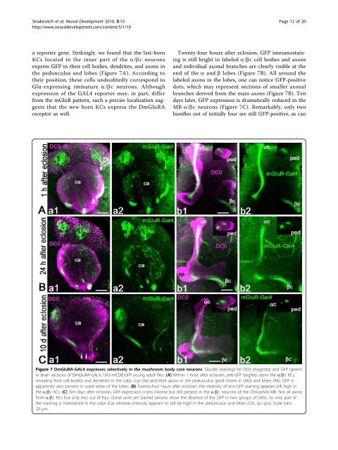
a reporter gene. Strik<strong>in</strong>gly, we found that <strong>the</strong> last-born<br />
KCs located <strong>in</strong> <strong>the</strong> <strong>in</strong>ner part <strong>of</strong> <strong>the</strong> a/bc neurons<br />
express GFP <strong>in</strong> <strong>the</strong>ir cell bodies, dendrites, and axons <strong>in</strong><br />
<strong>the</strong> pedunculus and lobes (Figure 7A). Accord<strong>in</strong>g to<br />
<strong>the</strong>ir position, <strong>the</strong>se cells undoubtedly correspond to<br />
Glu-express<strong>in</strong>g immature a/bc neurons. Although<br />
expression <strong>of</strong> <strong>the</strong> GAL4 reporter may, <strong>in</strong> part, differ<br />
from <strong>the</strong> mGluR pattern, such a precise localization suggests<br />
that <strong>the</strong> new born KCs express <strong>the</strong> DmGluRA<br />
receptor as well.<br />
Twenty-four hours after eclosion, GFP immunosta<strong>in</strong><strong>in</strong>g<br />
is still bright <strong>in</strong> labeled a/bc cell bodies and axons<br />
and<strong>in</strong>dividualaxonalbranches are clearly visible at <strong>the</strong><br />
end <strong>of</strong> <strong>the</strong> a and b lobes (Figure 7B). All around <strong>the</strong><br />
labeled axons <strong>in</strong> <strong>the</strong> lobes, one can notice GFP-positive<br />
dots, which may represent sections <strong>of</strong> smaller axonal<br />
branches derived from <strong>the</strong> ma<strong>in</strong> axons (Figure 7B). Ten<br />
days later, GFP expression is dramatically reduced <strong>in</strong> <strong>the</strong><br />
MB a/bc neurons (Figure 7C). Remarkably, only two<br />
bundles out <strong>of</strong> <strong>in</strong>itially four are still GFP-positive, as can<br />
Figure 7 DmGluRA-GAL4 expresses selectively <strong>in</strong> <strong>the</strong> <strong>mushroom</strong> body core neurons. Double sta<strong>in</strong><strong>in</strong>gs for DC0 (magenta) and GFP (green)<br />
<strong>in</strong> bra<strong>in</strong> sections <strong>of</strong> DmGluRA-GAL4, UAS-mCD8::GFP young adult flies. (A) With<strong>in</strong> 1 hour after eclosion, anti-GFP brightly sta<strong>in</strong>s <strong>the</strong> a/bc KCs,<br />
reveal<strong>in</strong>g <strong>the</strong>ir cell bodies and dendrites <strong>in</strong> <strong>the</strong> calyx (ca) (Aa) and <strong>the</strong>ir axons <strong>in</strong> <strong>the</strong> pedunculus (ped) (<strong>in</strong>sert <strong>in</strong> (Ab)) and lobes (Ab). GFP is<br />
apparently also present <strong>in</strong> outer areas <strong>of</strong> <strong>the</strong> lobes. (B) Twenty-four hours after eclosion, <strong>the</strong> <strong>in</strong>tensity <strong>of</strong> anti-GFP sta<strong>in</strong><strong>in</strong>g appears still high <strong>in</strong><br />
<strong>the</strong> a/bc KCs. (C) Ten days after eclosion, GFP expression is less <strong>in</strong>tense but still present <strong>in</strong> <strong>the</strong> a/bc neurons <strong>of</strong> <strong>the</strong> Drosophila MB. Not all axons<br />
from a/bc KCs but only two out <strong>of</strong> four clonal units are sta<strong>in</strong>ed (arrows show <strong>the</strong> absence <strong>of</strong> <strong>the</strong> GFP <strong>in</strong> two groups <strong>of</strong> cells). So only part <strong>of</strong><br />
<strong>the</strong> sta<strong>in</strong><strong>in</strong>g is ma<strong>in</strong>ta<strong>in</strong>ed <strong>in</strong> <strong>the</strong> calyx (Ca) whereas <strong>in</strong>tensity appears to still be high <strong>in</strong> <strong>the</strong> pedunculus and lobes (Cb). sp, spur. Scale bars:<br />
20 μm.