Dynamics of glutamatergic signaling in the mushroom ... - HAL - ESPCI
Dynamics of glutamatergic signaling in the mushroom ... - HAL - ESPCI
Dynamics of glutamatergic signaling in the mushroom ... - HAL - ESPCI

You also want an ePaper? Increase the reach of your titles
YUMPU automatically turns print PDFs into web optimized ePapers that Google loves.
S<strong>in</strong>akevitch et al. Neural Development 2010, 5:10<br />
http://www.neuraldevelopment.com/content/5/1/10<br />
Page 7 <strong>of</strong> 20<br />
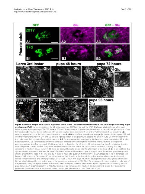
Figure 4 Newborn Kenyon cells express high levels <strong>of</strong> Glu <strong>in</strong> <strong>the</strong> Drosophila <strong>mushroom</strong> body <strong>in</strong> late larval stage and dur<strong>in</strong>g pupal<br />
development. (A, B) Transverse section <strong>of</strong> <strong>the</strong> MB pedunculus from 201Y-GAL4 (A) and 17d-GAL4 (B) pharate adults collected a few hours<br />
before eclosion and express<strong>in</strong>g mCD8::GFP. (A1-A3) GFP and Glu expression <strong>in</strong> 201Y-GAL4 are located both <strong>in</strong> <strong>the</strong> a/bc and g lobes. Most <strong>of</strong> <strong>the</strong><br />
GFP-positive a/bc neurons do not co-localize with Glu and only few axons express both Glu and GFP at <strong>the</strong> border <strong>of</strong> Glu-conta<strong>in</strong><strong>in</strong>g a/bc<br />
neurons. Agarose section <strong>of</strong> <strong>the</strong> pedunculus close to <strong>the</strong> lobes. (B1-B3) In 17d-GAL4, most Glu-conta<strong>in</strong><strong>in</strong>g a/bc neurons do not express GFP and<br />
only <strong>the</strong> border axons are both GFP- and Glu-positive. Agarose section <strong>of</strong> <strong>the</strong> pedunculus close to <strong>the</strong> calyx. In (A, B), Glu immunoreactivity <strong>in</strong><br />
<strong>the</strong> g lobe most likely orig<strong>in</strong>ates from extr<strong>in</strong>sic neurons. (C-I) Glu immunoreactivity monitored at late larval and pupal stages <strong>in</strong> <strong>the</strong> MB <strong>of</strong> wildtype<br />
Drosophila. (C, D). Transverse sections through <strong>the</strong> calyx and lobes <strong>of</strong> wander<strong>in</strong>g third <strong>in</strong>star larva. The four bundles <strong>of</strong> Glu-positive<br />
processes orig<strong>in</strong>ate from four clusters <strong>of</strong> KCs. Only one cluster is shown (on <strong>the</strong> left side <strong>in</strong> (C)) and arrows show bundles orig<strong>in</strong>at<strong>in</strong>g from <strong>the</strong><br />
o<strong>the</strong>r Glu-positive clusters. The four Glu-positive bundles extend <strong>in</strong> <strong>the</strong> core area <strong>of</strong> <strong>the</strong> pedunculus (arrowheads), <strong>in</strong>dicat<strong>in</strong>g that <strong>the</strong>y<br />
correspond to newborn KCs. As shown <strong>in</strong> (D), <strong>the</strong>se Glu-positive fibers also project <strong>in</strong>to <strong>the</strong> a/bc region <strong>of</strong> <strong>the</strong> larval MB (bc is not visible on this<br />
agarose section). The contour <strong>in</strong>dicates <strong>the</strong> shape <strong>of</strong> <strong>the</strong> larval MB vertical lobe (a) and spur (sp) region. Note that extr<strong>in</strong>sic <strong>glutamatergic</strong> cells<br />
are also present at this stage <strong>in</strong> <strong>the</strong> spur area. (E) In pupa 48 hours after puparium formation (APF) at 22°C (stages P5 to P6 <strong>of</strong> Ba<strong>in</strong>bridge and<br />
Bownes [100]), clusters <strong>of</strong> Glu-positive KCs are still present but <strong>the</strong>ir bundles at <strong>the</strong> base <strong>of</strong> <strong>the</strong> pedunculus (arrowheads) are less <strong>in</strong>tensely<br />
labeled compared to larval and o<strong>the</strong>r pupal stages. (F, G) Pupa 72 hours APF (stage P8). At this developmental stage, four groups <strong>of</strong> newborn<br />
KCs are brightly Glu-positive and project axons <strong>in</strong> <strong>the</strong> pedunculus (arrowheads <strong>in</strong> (G)). O<strong>the</strong>r Glu-immunoreactive processes <strong>in</strong> <strong>the</strong> g lobe area <strong>of</strong><br />
<strong>the</strong> pedunculus likely correspond to fibers from extr<strong>in</strong>sic cells as only cell bodies <strong>of</strong> newborn KCs express high Glu-immunoreactivity. (H, I) In<br />
pupa 96 hours APF at 22°C (stages P11 to P12), four clusters <strong>of</strong> Glu-immunopositive newborn KCs (arrows <strong>in</strong> (H)) project <strong>the</strong>ir axons to <strong>the</strong><br />
pedunculus (arrowheads <strong>in</strong> (I)). K, Kenyon cell bodies; ca, calyx, ped, pedunculus; sp, spur. Scale bars: 10 μm.