Dynamics of glutamatergic signaling in the mushroom ... - HAL - ESPCI
Dynamics of glutamatergic signaling in the mushroom ... - HAL - ESPCI
Dynamics of glutamatergic signaling in the mushroom ... - HAL - ESPCI

Create successful ePaper yourself
Turn your PDF publications into a flip-book with our unique Google optimized e-Paper software.
S<strong>in</strong>akevitch et al. Neural Development 2010, 5:10<br />
http://www.neuraldevelopment.com/content/5/1/10<br />
Page 6 <strong>of</strong> 20<br />
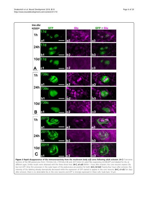
Figure 3 Rapid disappearance <strong>of</strong> Glu immunoreactivity from <strong>the</strong> <strong>mushroom</strong> body a/b cores follow<strong>in</strong>g adult eclosion. (A-C) Transverse<br />
sections <strong>of</strong> <strong>the</strong> MB pedunculus from 17d-GAL4 (A), c739-GAL4 (B) and 201Y-GAL4 (C) adult flies express<strong>in</strong>g mCD8::GFP and labeled for Glu at<br />
different ages. Similar results were obta<strong>in</strong>ed with <strong>the</strong> three driver l<strong>in</strong>es. (A-C, a1-a3) With<strong>in</strong> 1 hour after eclosion, <strong>the</strong> core neurons express Glu<br />
but not GFP. Only th<strong>in</strong> processes <strong>in</strong> <strong>the</strong> outer layers <strong>of</strong> <strong>the</strong> pedunculus are positive for both. (A-C, b1-b3) Twenty-four hours after eclosion, <strong>the</strong><br />
<strong>in</strong>tensity <strong>of</strong> Glu label<strong>in</strong>g already dramatically decreased while <strong>the</strong> expression <strong>of</strong> GFP started to appear <strong>in</strong> <strong>the</strong> core neurons. (A-C, c1-c3) Ten days<br />
after eclosion, <strong>the</strong>re is no detectable Glu <strong>in</strong> <strong>the</strong> core neurons and GFP is strongly expressed <strong>in</strong> <strong>the</strong>se cells. Scale bars: 10 μm.