Impact Of Host Plant Xylem Fluid On Xylella Fastidiosa Multiplication ...
Impact Of Host Plant Xylem Fluid On Xylella Fastidiosa Multiplication ...
Impact Of Host Plant Xylem Fluid On Xylella Fastidiosa Multiplication ...

Create successful ePaper yourself
Turn your PDF publications into a flip-book with our unique Google optimized e-Paper software.
IMPACT OF HOST PLANT XYLEM FLUID ON XYLELLA FASTIDIOSA<br />
MULTIPLICATION, AGGREGATION, AND ATTACHMENT<br />
Project Leaders:<br />
Nick Toscano<br />
Dept. of Entomology<br />
University of California<br />
Riverside, CA 92521<br />
Cooperators:<br />
Jian Bi<br />
Dept. of Entomology<br />
University of California<br />
Riverside, CA<br />
Donald Cooksey<br />
Dept. of <strong>Plant</strong> Pathology<br />
University of California<br />
Riverside, CA 92521<br />
Korsi Dumenyo<br />
Dept. of <strong>Plant</strong> Pathology<br />
University of California<br />
Riverside, CA<br />
Rufina H. Martinez<br />
Dept. of <strong>Plant</strong> Pathology<br />
University of California<br />
Riverside, CA<br />
Reporting Period: The results reported here are from work conducted from October 2003 to August 2004.<br />
ABSTRACT<br />
Research in Temecula Valley indicated that the proximity of citrus groves to vineyards has influenced the incidence and<br />
severity of Pierce’s disease (PD), <strong>Xylella</strong> fastidiosa (Xf), in grapes. Although the glassy-winged sharpshooter (GWSS) feeds<br />
on and moves back and forth between Temecula citrus groves and vineyards, there are no visible <strong>Xylella</strong> fastidiosa (Xf)<br />
symptoms in the citrus. This implies that citrus trees are resistant or tolerant to the Xf but may be a reservoir to harbor the<br />
pathogen for GWSS acquisition while grape vines are susceptible. We investigated the mechanisms of host plant<br />
resistance/susceptibility by examining the impact of xylem fluid of grapefruit, orange, lemon and grape on Xf multiplication,<br />
aggregation and attachment as well as the related xylem fluid chemistry. Our laboratory experiments revealed that xylem<br />
fluid of grapefruit, orange and lemon caused an aggregation of Temecula PD cells to form large white clumps while grape<br />
xylem fluid did not cause visible clumping, but created a visible thick biofilm. The numbers of Xf cells in grapefruit xylem<br />
fluid treatment were significantly higher at 6, 8 and 9 days after culture compared with those in grape xylem fluid treatment.<br />
The numbers of Xf cells in orange or lemon xylem fluid tests were generally lower than those in grape xylem fluid treatment.<br />
Citrus xylem fluid significantly inhibited Xf biofilm formation compared to grape xylem fluid. The content of total amino<br />
acids in grape xylem fluid was near 9-fold higher than that in grapefruit xylem fluid. Sugar contents were 1.4- to 5.5-fold<br />
higher in grape xylem fluid than those in grapefruit xylem fluid. Peroxidase and total thiol levels were also higher in grape<br />
xylem fluid than in citrus xylem fluid. Our results indicate that the differences between citrus and grape plants in their<br />
responses to <strong>Xylella</strong> may be due to differences in their xylem fluid chemistry.<br />
INTRODUCTION<br />
<strong>Xylella</strong> fastidiosa (Xf) is a xylem-limited, plant pathogenic bacterium that causes Pierce’s disease (PD) in grapes (Purcell,<br />
1981). Xf is mainly vectored by the glassy-winged sharpshooter (GWSS), Homalodisca coagulata, in Southern California.<br />
Although a comprehensive list of suitable hosts for the GWSS has been identified, comprising 75 plant species in 35 families<br />
(Turner and Pollard, 1959), the major crop hosts in Temecula Valley are citrus and grapes. Previous studies in California<br />
have identified 94 plant species in more than 28 of plant families as host of Xf (Freitg, 1951; Raju et al, 1983; Raju et al.,<br />
1980). Most identified Xf hosts show no symptoms but serve as inoculum sources of Xf for vector acquisition. Perring et al<br />
(2001) studied the incidence of PD in the Temecula Valley and found that proximity of citrus groves to vineyards has<br />
influenced the incidence and severity of PD in grapes. The PD infection is most severe when the grape vines are adjacent to<br />
citrus, and that the damage declines as one moves away from citrus (Perring et al., 2001). Although the GWSS feeds on and<br />
moves back and forth between citrus trees and grape vines, there is generally no Xf caused disease symptom in citrus in the<br />
area. This implies that citrus trees are resistant or tolerant to the Xf, but may be a reservoir to harbor the pathogen for GWSS<br />
acquisition and transmission while grape vines are susceptible. Little is known about the biochemical mechanisms involved<br />
in host plant resistance/susceptibility to Xf in the system. Additional information is required to determine if citrus can be<br />
suitable reservoirs for Xf. Elucidation of the biochemical mechanisms may be useful for developing host plant resistance in<br />
grapes as a sustainable component of integrated pest management program.<br />
Xf aggregates to form biofilm inside its host plants and insect vectors. The biofilm formation is considered as a major<br />
virulence factor of PD (Marques and Ceri, 2002). Biofilm is defined as structured communities of sessile microbial<br />
aggregates enclosed in a self produced polymeric matrix and attached to a surface (Costerton et al., 1995). It was recently<br />
reported that a defined medium with some components based on susceptible grape cultivar “Chardonnay” xylem fluid<br />
chemistry better supports Xf growth and stimulates Xf aggregation and biofilm formation in vitro (Leite et al. 2004).<br />
However, the effect of citrus xylem fluid on Xf multiplication, aggregation and biofilm formation remains unknown.<br />
Xf is a nutritionally fastidious bacterium (Wells et al. 1987). In defined medium certain amino acids are essential for Xf<br />
growth, glucose stimulates the growth while fructose and sucrose have inhibiting effect (Wells et al. 1987; Chang and<br />
Donaldson, 2000). It is not known whether differences in contents of amino acids and the sugars in the xylem fluid of citrus<br />
- 60 -
and grape may differentially affect growth of Xf. Redox status also likely affects the tendency for Xf aggregation and biofilm<br />
formation. Adding reducing agents such as glutathione to artificial medium promotes Xf aggregation and biofilm formation<br />
(Leite et al., 2004). It was reported that thiols mediate the aggregation and adhesion of Xf (Leite et al., 2002). Thiolcontaining<br />
compounds in xylem fluid include cysteine, methionine and glutathione. The redox status in citrus and grape<br />
xylem fluid and its role in Xf aggregation and biofilm formation, and host plant resistance/susceptibility to Xf need to be<br />
further investigated.<br />
OBJECTIVES<br />
1. Investigate the effect of host plant xylem fluid on Xf multiplication, aggregation and attachment.<br />
2. Determine the biochemical mechanisms of host xylem fluid influence on Xf multiplication, aggregation and attachment.<br />
RESULTS<br />
Commercial citrus (lemon, orange and grapefruit) groves in proximity to vineyards were selected in the Temecula Valley,<br />
California. Three blocks of 30 citrus and 30 grape vines were used. A minimum of 15 citrus trees and 15 vines were<br />
randomly selected from each block (making a total of 15 trees or vines from each plant species) to extract xylem fluid.<br />
Terminal shoots from each plant were used for xylem extraction with a pressure bomb apparatus (Anderson et al., 1989).<br />
Upon collection, the xylem fluid was immediately placed on dry ice before final storage in a -80 o C freezer. The samples<br />
were used to test the impact of these xylem fluid on Xf resistance and chemical analyses of soluble carbohydrates, free amino<br />
acids, and redox status.<br />
Effects of xylem fluid of each plant species on Xf attachment were evaluated on the biofilm formation. Formation of biofilm<br />
on the abiotic surfaces was assessed as described by Espinosa-Urgel et al. (2000). The analyses of Xf multiplication and<br />
aggregation were based on the fact that optical density (540 nm) is correlated with bacterial cell numbers and aggregation<br />
state as described by Burdman et al. (2000).<br />
Our data indicated that, when the xylem fluid of grapefruit, orange and lemon was added to the PD Temecula strain of Xf in<br />
PD3 medium in glass culture tubes, there were heavy Xf cell aggregations to form large white clumps in suspension of the<br />
culture and the culture fluid was clear with no significant turbidity; in contrast, grape xylem fluid added to the same Xf<br />
culture did not cause visible clumping, but rather a visible thick biofilm was formed on the surface of glass tube and the<br />
culture was turbid (Figure 1). After homogenization of the culture, we found that the numbers of Xf cells in the grapefruit<br />
xylem fluid treatment were significantly higher at 6, 8 and 9 days after culture compared with those in the grape xylem fluid<br />
treatment (Figure 2). The numbers of Xf cells in orange or lemon xylem fluid treatments were generally lower than those in<br />
grape xylem fluid treatment (Figure 3). These data suggest that the citrus species, especially grapefruit, are suitable hosts for<br />
Xf growth and may serve as a great reservoir of the pathogen for GWSS acquisition. Our assay results revealed that xylem<br />
fluid of the citrus species significantly inhibited Xf biofilm formation compared to that of grape (Figure 4). Our attempt to<br />
investigate the biochemical mechanisms likely to be involved indicated that 96% of amino acids in grape xylem fluid was<br />
comprised of glutamine, while 47% of amino acids in grape fruit xylem fluid was proline (Figure 5). The content of total<br />
amino acids in grape xylem fluid was near 9-fold higher than that in grapefruit xylem fluid (Figure 5). Sugar contents were<br />
1.4- to 5.5-fold higher in grape xylem fluid than those in grapefruit xylem fluid (Figure 6). Peroxidase and total thiol levels<br />
were also higher in grape xylem fluid than in citrus xylem fluid (Figures 7 and 8).<br />
CONCLUSIONS<br />
<strong>Xylem</strong> fluid of grapefruit, orange and lemon caused PD Temecula strain of Xf cells to aggregate and form large white clumps<br />
but inhibited the attachment. In contrast, grape xylem fluid did not cause visible clumping but led to heavy attachment.<br />
Grapefruit xylem fluid significantly increased multiplication of Xf cells compared with grape xylem fluid. Citrus species,<br />
especially grapefruit, appear to be suitable hosts for Xf growth and may serve as a reservoir of the pathogen for GWSS<br />
acquisition and transmission to grape vines. Further research is underway to elucidate the biochemical mechanisms.<br />
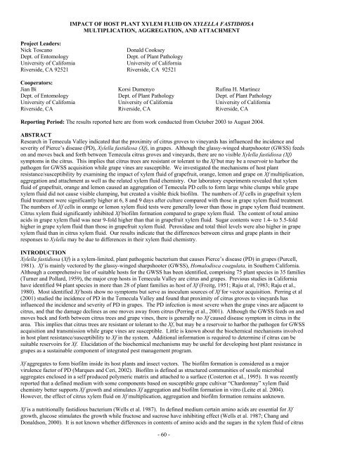
Figure 1. Effect of host plant xylem fluid on Xf aggregation. A, treatment with grape xylem fluid. B, treatment with grapefruit xylem<br />
fluid. C, treatment with orange xylem fluid. D, treatment with lemon xylem fluid. Note that white clumps of Xf aggregates are formed in<br />
the grapefruit, orange and lemon xylem fluid treatments.<br />
- 61 -
0.18<br />
600<br />
Optical density<br />
0.16<br />
0.14<br />
Grape<br />
0.12<br />
Grape fruit<br />
0.1<br />
Control<br />
0.08<br />
0.06<br />
0.04<br />
4 5 6 7 8 9 10<br />
Days of culture<br />
Figure 2. Effect of host plant xylem fluid on Xf growth.<br />
nM / ml xylem fluid<br />
500<br />
400<br />
300<br />
200<br />
100<br />
0<br />
Glu Gln Pro Total<br />
Grape<br />
Grape Fruit<br />
Optical Density<br />
0.18<br />
0.16<br />
0.14<br />
0.12<br />
0.1<br />
0.08<br />
0.06<br />
0.04<br />
4 5 6 7 8 9 10<br />
Days of culture<br />
Figure 3. Effect of host plant xylem fluid on Xf growth.<br />
Optical density<br />
0.9<br />
0.8<br />
0.7<br />
0.6<br />
0.5<br />
0.4<br />
0.3<br />
0.2<br />
0.1<br />
0<br />
2 4 6<br />
Days of culture<br />
Figure 4. Effect of host plant xylem fluid on Xf biofilm<br />
formation.<br />
Grape<br />
Orange<br />
Lemon<br />
Grape<br />
Grape Fruit<br />
Orange<br />
Lemon<br />
Figure 5. Some amino acid contents in grape and grape<br />
fruit xylem fluid.<br />
nM / ml xylem fluid<br />
4000<br />
3500<br />
3000<br />
2500<br />
2000<br />
1500<br />
1000<br />
500<br />
0<br />
Glucose Fructose Sucrose<br />
Grape<br />
Grape Fruit<br />
Figure 6. Sugar contents in grape and grape fruit xylem fluid.<br />
Change of OD/ml xylem fluid<br />
3<br />
2.5<br />
2<br />
1.5<br />
1<br />
0.5<br />
0<br />
Grape<br />
Grape<br />
Fruit<br />
Orange<br />
Lemon<br />
OD412 / ml xylem fluid<br />
2<br />
1.5<br />
1<br />
0.5<br />
0<br />
Grape<br />
Grape<br />
Fruit<br />
Orange<br />
Lemon<br />
Figure 7. Peroxidase levels in host xylem fluid.<br />
Figure 8. Total thiol contents in host xylem fluid.<br />
- 62 -
REFERENCES<br />
Anderson, P. C., B. V. Brodbeck and R. F. Mizell III. 1989. Metabolism of amino acids, organic acids, and sugars extracted<br />
from the xylem fluid of four host plants by adult Homalodisca coagulata. Entomol. Exp. Appl. 50-149-160.<br />
Anderson, P. C. 2002. Biologival, cultural, genetic, and chemical control of Pierce’s disease: xylem fluid chemistry<br />
mediation of resistance to Pierce’s disease, pp. 43-44, in Pierce’s Disease Research Symposium, 2002. CDFA.<br />
Burdman, S., E. Jurkevitch, M. E. Soria-Diaz, A. M. G. Serrano, and Y. Okon. 2000. Extracellular polysaccharide<br />
composition of Azospirillum brasilense and its relation with cell aggregation. FEMs Microbiol. Lett. 189:259-264.<br />
Chang, C. J., and R. C. Donaldson. 2000. Nutritional requirements of <strong>Xylella</strong> fastidiosa, which causes Pierce’s disease in<br />
grapes. Can. J. Microbiol. 46: 291-293.<br />
Costerton, J. W. Z. Lewandowski, D. E. Caldwell, D. R. Corber, and A. M. Lappin-Scott. 1995. Microbial biofilms. Annu.<br />
Rev. Microbiol. 49:711-745.<br />
Espinosa-Urgel, M., A. Salido, and J. L. Ramos. 2000. Genetic analysis of functions involved in adhesion of Pseudomonas<br />
putida to seeds. J. Bacteriol. 182:2363-2369.<br />
Freitg, J. H. 1951. <strong>Host</strong> range of Pierce’s disease virus of grapes as determined by insect transmission. Phytopathology 41:<br />
920-934.<br />
Leite, B., P. C. Anderson, and M. L. Ishida. 2004. Colony aggregation and biofilm formation in xylem chemistry-based<br />
media for <strong>Xylella</strong> fastidiosa. FEMS Microbiol. Lett. 230:283-290.<br />
Marques, L. L. R. and H. Ceri. 2002. Charaterization of biofilm formation by <strong>Xylella</strong> fastidiosa in vitro. <strong>Plant</strong> Disease.<br />
86:633-638.<br />
Leite, B., P. C. Anderson, and M. L. Ishida. 2004. Colony aggregation and biofilm formation in xylem chemistry-based<br />
media for <strong>Xylella</strong> fastidiosa. FEMS Microbiol. Lett. 230:283-290.<br />
Perring, T. M., C. A. Farrar and M. J. Blua. 2001. Proximity to citrus influences Pierce’s disease in Temecula Valley<br />
vineyards. Calif. Agr. 55:13-18.<br />
Purcell, A. H. 1981. Pierce’s disease. In Flaherty, D. L., Jenson, F. L. Kasamatis, A. N. et al., (eds). Grape Pest<br />
Management. UC Berkeley. Division of Agricultural Science. No. 4105. p 62-9<br />
Raju, B. C., A. C. Goheen, and N. W. Frazier. 1983. Occurrence of Pierce’s disease bacteria in plants and vectors in<br />
California. Phytopathology 73: 1309-1313.<br />
Raju, B. C., S. F. Nome, D. M. Docampo, D. M. Goheen, A. C. Nyland, and S. K. Lowe. 1980. Alternative hosts of Pierce’s<br />
disease of grapevines that occur adjacent to grape growing areas in California. Am. J. Enol. Vitic. 31: 144-148.<br />
Turner, W. F. and H. N. Pollard. 1959. Life histories and behavior of five insect vectors of phony peach disease. USDA<br />
Tech. Bul. 1188.<br />
Wells, J. M., B. C. Raju, H. Y. Huang, W. G. Weiburg, L. Mandelcopaul, and D. J. Brenner. 1987. <strong>Xylella</strong> fastidiosa gen.<br />
Nov.: Gram negative, xylem limited fastidious plant bacteria related to Xanthomonas spp. Int. J. Syst. Bacteriol. 36: 136-<br />
143.<br />
FUNDING AGENCY<br />
Funding for this project was provided by the University of California Pierce’s Disease Grant Program.<br />
- 63 -
OPTIMIZING MARKER-ASSISTED SELECTION FOR RESISTANCE TO XYLELLA FASTIDIOSA<br />
TO ACCELERATE BREEDING OF PIERCE’S DISEASE RESISTANT GRAPES OF HIGH FRUIT QUALITY<br />
Project Leaders:<br />
Andrew Walker<br />
Dept. of Viticulture and Enology<br />
University of California<br />
Davis, CA 95616<br />
awalker@ucdavis.edu<br />
Alan Krivanek<br />
Dept. of Viticulture and Enology<br />
University of California<br />
Davis, CA 95616<br />
Summaira Riaz<br />
Dept. of Viticulture and Enology<br />
University of California<br />
Davis, CA 95616<br />
Reporting Period: The results reported here are from work conducted from October 2003 to October 2004. Research on<br />
this project was initiated under the “Genetics of Resistance to Pierce’s Disease” of the Long-term American Vineyard<br />
Foundation Pierce’s Disease Project.<br />
ABSTRACT<br />
Efforts at identifying molecular markers linked to <strong>Xylella</strong> fastidiosa (Xf) resistance are continuing. Our primary focus is on<br />
resistance derived from b43-17, a Vitis arizonica/candicans type collected near Monterrey, Nuevo Leon, Mexico. The<br />
‘9621’ V. rupestris x V. arizonica hybrid mapping family (PD resistant D8909-15 x PD resistant F8909-17) was used to<br />
localize PdR1, a primary PD resistance locus within the linkage map of the male parent F8909-17 (progeny of b43-17) and<br />
identify candidate linked resistance markers. In more recent research, a comparative mapping strategy between the ‘9621’<br />
linkage map and other SSR maps within Vitis was used to identify 9 SSR markers within 10 cM of the resistance locus.<br />
Resistance from the female parent D8909-15 has not yet been localized to a genetic map. The strategy of bulk segregant<br />
analysis (BSA) in concert with the AFLP marker system has been initiated to saturate the region around the resistance locus<br />
and is expected to yield an additional 20 to 50 markers linked to the resistance trait. All candidate resistant markers have<br />
been and will continue to be applied to breeding populations derived from ‘8909’ x V. vinifera and (‘8909’ x V. vinifera) x<br />
V. vinifera back-cross generations in order to confirm resistance marker effectiveness in V. vinifera backgrounds and<br />
continue with marker assisted selection for development of high quality PD resistant grapes.<br />
INTRODUCTION<br />
Several American Vitis species are native to the regions where PD is endemic, and resistance from these sources has been<br />
introgressed into many different cultivars grown in the south-eastern United States. The acceptance of the new hybrid<br />
cultivars has been limited due in part to some undesirable non-vinifera fruit quality traits. The development of high quality<br />
PD resistant cultivars will be facilitated by the use of molecular markers to achieve a more precise introgression of the<br />
resistance genes into domesticated backgrounds and avoid introgression of undesirable traits (Figure 1). Backcross<br />
introgression via molecular markers has been accomplished successfully in other crops (Young and Tanksley 1989). This<br />
type of introgression is generally termed Marker Assisted Selection (MAS), whereby indirect selection on a trait of interest<br />
(such as disease resistance) is made by screening for the presence of a DNA marker allele tightly linked to the trait. MAS for<br />
disease resistance can also be used to eliminate susceptible genotypes in a breeding population early in the selection process,<br />
which allows for evaluation of much larger effective populations. Larger effective population sizes increase the opportunity<br />
to identify genotypes with high disease resistance and good horticultural qualities (such as good flavor traits, color, berry and<br />
cluster size. etc.). Other key aspects of the MAS process include avoiding confounding environmental effects on the trait<br />
phenotype and accelerating breeding progress while saving space and time, allowing for more efficient use of resources<br />
(Paterson et al. 1991, Kelly 1995). Rapid screening time is particularly valuable when applied to perennial crops such as<br />
grape with relatively long generation times (Alleweldt 1988, Striem et al. 1994). To effectively use linked markers in MAS<br />
only requires that the markers be highly reproducible, linked in coupling phase i.e. on the same homologous chromosome,<br />
and within 5 centimorgan (cM) mapping units of the resistance locus (Kelly 1995).<br />
Within grapevines, markers linked to powdery mildew resistance (Dalbo et al. 2001, Pauquet et al. 2001), downy mildew<br />
resistance (Luo et al. 2001) and seedlessness (Lahogue 1998) have been published. In the case of powdery mildew<br />
resistance, MAS has already been successfully utilized for screening a grape breeding population. We are successfully<br />
developing a MAS system for screening PD resistant genotypes that will greatly benefit our breeding of PD resistant wine<br />
grapes.<br />
OBJECTIVES<br />
Our overall objective is to identify DNA markers that are tightly linked to the primary locus or loci required for complete<br />
resistance to PD within Vitis. Research will focus on PD resistance as inherited from V. arizonica and will utilize an<br />
established V. rupestris x V. arizonica genetic map. These markers will be utilized for MAS to eliminate susceptible seedling<br />
progeny our continuing PD resistance breeding program.<br />
Sub-objectives<br />
1. Continue with a comparative mapping strategy between the V. rupestris x V. arizonica 9621 (D8909-15 x F8909-17)<br />
linkage map and other SSR maps within Vitis in order to identify additional SSR markers linked to resistance.<br />
- 64 -
2. Utilize Bulk Segregant Analysis (BSA) with the AFLP marker system to saturate with markers the region around the<br />
previously mapped Xf resistance locus and eventually convert confirmed candidate markers to stable SCAR primers.<br />
3. Confirm candidate marker linkage to resistance within families derived from resistant by susceptible crosses such as the<br />
‘8909’ x V. vinifera and (‘8909’ x V. vinifera) x V. vinifera back-cross generations.<br />
RESULTS AND CONCLUSIONS<br />
Sub-objective 1.<br />
Initial mapping of the PD resistance locus PdR1 in the male parent F8909-17 of the 9621 family localized it to chromosome<br />
14, and identified 6-8 SSR markers on the same linkage group. Marker placement on published SSR linkage maps of Vitis<br />
were used to preferentially target chromosome 14, bringing the total number of SSR markers on the linkage group up to 30.<br />
Approximately 9 SSR markers are localized within a 10 cM distance of the resistance gene. These SSR markers are reliable<br />
and are the easiest of the molecular markers to incorporate within a MAS breeding program. Correlation tests of these<br />
candidate markers to PD resistance when functioning within a V. vinifera genetic background are underway and described in<br />
sub-objective 3. The SSR marker analysis has allowed us to confirm that marker alleles linked in coupling to PD resistance<br />
alleles of the PdR1 locus in another PD resistant progeny of b43-17 (F8909-08) are different than the alleles linked in<br />
coupling the resistance alleles in F8909-17. It is apparent from these results that b43-17 is homozygous resistant for the<br />
PdR1 locus, and that F8909-17<br />
inherited its resistance allele from one<br />
chromosome 14 and F8909-08 inherited<br />
its resistance allele from the<br />
homologous chromosome 14. In either<br />
case the markers linked to resistance<br />
will function for MAS, however,<br />
different alleles linked in coupling to<br />
the resistance alleles will have to be<br />
followed through the downstream MAS<br />
process. Placement of SSR markers to<br />
chromosome 14 via the comparative<br />
mapping strategy continue as the<br />
markers become available, however, the<br />
number of SSR markers that can be<br />
targeted to a specific chromosomal<br />
region via comparative mapping is<br />
limited.<br />
Figure 1<br />
Resistant Variety<br />
Poor Fruit Characters<br />
R<br />
Resistant Variety<br />
Mix of Fruit Characters<br />
R<br />
Breeding PD resistant grapes<br />
Resistant Variety<br />
Excellent Fruit Characters<br />
R<br />
Sub-objective 2.<br />
For high density marker saturation within a narrow window around the PdR1 locus, a bulk segregant analysis (BSA) strategy<br />
(Michelmore et al. 1991) in concert with the AFLP marker system was chosen as the method of choice. Initial BSA was<br />
attempted within the 9621 family, however, confounding effects of the resistance loci within the D8909-15 parent made the<br />
attempt more difficult than expected. To avoid confounding affects from resistance inherited from other genetic backgrounds<br />
and focus the BSA procedure only on the PdR1 locus, work has begun within two segregating families from susceptible by<br />
resistant crosses. The first family, 99217 (C8909-07 x F8909-08) consists of 33 genotypes, has been screened for PD<br />
resistance (Krivanek et al. submitted) and segregates 1:1 resistant to susceptible (Table 1). DNA has been extracted from<br />
these genotypes, flanking SSR markers were run and a good correlation between resistance and resistance marker alleles has<br />
been established (Table 1). A bulk of the DNA from the 12 most susceptible and a bulk of the DNA from the 12 most<br />
resistant genotypes are in process and will be tested for AFLP polymorphisms utilizing florescent primers and visualized on a<br />
PE 3100 sequencer. The second family derived from a susceptible by resistant cross is a V. vinifera x F8909-08 family; it<br />
consists of 40 genotypes and has been designated as 0062. Testing of this family for PD resistance is currently underway via<br />
our standard greenhouse testing procedure (Krivanek et al. in press; Krivanek and Walker in press). It is expected that the<br />
progeny in this family will segregate in a 1:1 manner, and if so, DNA extraction and BSA procedures will be undertaken as<br />
with the 99217 family. Candidate AFLP markers will be converted to stable and more reliable SCAR primers before<br />
incorporation into the MAS program.<br />
Sub-objective 3.<br />
Work is progressing with two distinct breeding populations for testing of candidate resistance markers and initial application<br />
of those markers to MAS. <strong>On</strong>e family is a cross of the PD resistant F8909-08 to a female V. vinifera wine grape F2-7<br />
(Cabernet Sauvignon x Carignane) and designated as the 0062 family. A second breeding population consists of a cross of<br />
F8909-08 to several elite V. vinifera table grape genotypes (the 500 series). A subset of the 500 series has been screened for<br />
PD resistance and screened for markers flanking the PdR1 locus. Five confirmed resistant genotypes have been utilized in<br />
the development of the first backcross generations BC1 (backcrossed to additional elite V. vinifera genotypes). The BC1<br />
population (25000 series) consists of approximately 200 individuals and was planted in the field in 2003. Marker analysis for<br />
flanking markers to the PdR1 locus has been completed for the 25000 series and the marker information was utilized in<br />
selection of genotypes for the spring of 2004 crosses for the development of the BC2 generations. Subsets of candidate<br />
- 65 -<br />
X<br />
Marker Assisted<br />
Breeding<br />
Susceptible Variety<br />
Excellent Fruit Characters
esistant and susceptible genotypes within the 25000 series have shown improved fruit quality (Figure 2) and are currently<br />
being screened to confirm the correlation between the resistance markers and the PD resistance trait. We are also utilizing<br />
these populations to confirm the effectiveness and economics of the MAS relative to our greenhouse screening procedure.<br />
Table 1. Resistance classification and marker genotypes for the individuals of the full-sib family derived from the<br />
susceptible by resistant cross of C8909-07 x F8909-08. * = Genotypes selected for Bulk Segregant Analysis procedure.<br />
Genotype<br />
Overall<br />
resistance<br />
level to PD<br />
Mean<br />
natural log<br />
(cells/ml)<br />
Mean<br />
CMI<br />
score<br />
Mean %<br />
leaf<br />
scorch<br />
Alleles of SSR<br />
markers flanking the<br />
PdR1 resistance<br />
99217-21 * Resistant 9.51 1.00 58.3 Rr / Rr<br />
99217-40 * Resistant 9.70 1.33 75.0 rr / Rr<br />
99217-18 * Resistant 9.77 2.75 95.0 Rr / Rr<br />
99217-41 * Resistant 10.19 4.25 76.3 Rr / Rr<br />
99217-35 * Resistant 10.55 1.33 100.0 rr / Rr<br />
99217-19 * Resistant 11.08 2.50 76.7 rr / Rr<br />
99217-01 * Resistant 11.52 2.25 90.0 rr / Rr<br />
99217-23 * Resistant 11.57 3.00 87.5 Rr / Rr<br />
99217-34 * Resistant 11.83 3.75 65.0 Rr / Rr<br />
99217-46 Resistant 11.87 5.75 100.0 Rr / Rr<br />
99217-27 * Resistant 12.20 4.25 100.0 Rr / rr<br />
99217-22 * Resistant 12.29 4.00 100.0 Rr / Rr<br />
99217-12 * Resistant 12.50 4.00 95.0 Rr / Rr<br />
99217-38 ? 12.69 5.00 100.0 Rr / Rr<br />
99217-36 ? 13.09 5.00 100.0 rr / rr<br />
99217-50 ? 13.52 4.25 83.8 Rr / Rr<br />
99217-14 Susceptible 14.06 5.50 88.8 rr / Rr<br />
99217-07 Susceptible 14.87 5.50 100.0 rr / rr<br />
99217-04 * Susceptible 15.42 6.00 100.0 rr / rr<br />
99217-33 * Susceptible 15.59 5.75 100.0 rr / rr<br />
99217-06 * Susceptible 15.80 5.25 68.3 rr / rr<br />
99217-09 * Susceptible 15.81 5.75 100.0 rr / rr<br />
99217-10 Susceptible 15.82 4.75 100.0 rr / rr<br />
99217-13 * Susceptible 15.84 5.50 100.0 rr / rr<br />
99217-42 Susceptible 15.85 4.25 75.0 rr / Rr<br />
99217-15 * Susceptible 15.87 5.25 100.0 rr / rr<br />
99217-32 * Susceptible 15.87 5.50 100.0 rr / rr<br />
99217-28 * Susceptible 15.91 5.75 100.0 rr / rr<br />
99217-05 * Susceptible 15.91 5.75 100.0 rr / rr<br />
99217-37 * Susceptible 15.92 5.25 100.0 rr / rr<br />
99217-26 * Susceptible 15.95 5.50 100.0 rr / rr<br />
99217-24 * Susceptible 16.04 6.00 100.0 rr / rr<br />
Figure 2.<br />
Vitis arizonica PD<br />
Resistant poor fruit<br />
quality<br />
Hybrid BC1-25017 with<br />
flanking PD resistance markers<br />
Improved fruit quality<br />
Vitis vinifera PD<br />
Susceptible Excellent fruit<br />
quality<br />
- 66 -
REFERENCES<br />
Alleweldt G. and Possingham J.V. 1988. Progress in grapevine breeding. Theor. Appl. Genet. 75:669-673.<br />
Dalbo M.A., Ye G.N., Weeden N.F., Wilcox W., and Reisch B.I. 2001, Marker-assisted selection for powdery mildew<br />
resistance in grapes. J. Am. Soc. Hort. Sci. 126: 83-89.<br />
Kelly J.D. 1995. Use of random amplified polymorphic DNA markers in breeding for major gene resistance to plant<br />
pathogen. HortScience 30:461-465.<br />
Krivanek A.F., Famula T.R., Tenscher A. and Walker M.A. (Submitted) Inheritance of resistance to Pierce's disease within a<br />
Vitis rupestris x Vitis arizonica hybrid population. Theor Appl Genet.<br />
Krivanek A.F., Stevenson J.F. and Walker M.A. (In Press) Development and comparison of symptom indices for quantifying<br />
grapevine resistance to Pierce's disease. Phytopathology.<br />
Krivanek A.F. and Walker M.A. (In Press) Vitis resistance to Pierce's disease is characterized by differential <strong>Xylella</strong><br />
fastidiosa populations in stems and leaves. Phytopathology.<br />
Lahogue F., This P. and Bouquet A. 1998. Identification of a codominant SCAR marker linker to the seedlessness character<br />
in grapevine. Theor. Appl. Genet. 97: 950-959.<br />
Luo Su-Lan, He Pu-Chao, Zhou P. and Zheng Xue-Qin. 2001. Identification of molecular genetic markers tightly linked to<br />
downy mildew resistant genes in grape. Acta Genet. Sinica 28: 76-82.<br />
Michelmore R.W., Paran I. and Kesseli R.V. 1991. Identification of markers linked to disease-resistance genes by bulked<br />
segregant analysis: a rapid method to detect markers in specific genomic regions by using segregating populations. Proc<br />
Natl Acad Sci USA 88:9828-9832<br />
Paterson A.H., Tanksley S.D. and Sorrells M.E. 1991. DNA markers in plant improvement. Adv. Agr. 46:39-90.<br />
Pauquet J., Bouquet A., This P. and Adam-Blondon A-F. 2001. Establishment of a local map of AFLP markers around the<br />
powdery mildew resistance gene Run1 in grapevine and assessment of their usefulness for marker assisted selection.<br />
Theor. Appl. Genet. 103:1201-1210.<br />
Striem M.J., Ben-Hayyim G. and Spiegel-Roy P. 1994. Developing molecular genetic markers for grape breeding, using<br />
polymerase chain reaction procedures. Vitis 33:53-54.<br />
Young N.D. and Tanksley S.D. (1989) RFLP analysis of the size of chromosomal segements retained around the Tm-2 locus<br />
of tomato during backcross breeding. Theor Appl Genet 77:353-359.<br />
FUNDING AGENCIES<br />
Funding for the 2004-2005 funding year was received in mid-September 2004. This proposal was not submitted to other<br />
funding agencies. However, it is linked to the Walker/Tenscher Pierce’s disease resistance breeding project funded by the<br />
CDFA Pierce’s Disease and Glassy-winged Sharpshooter Board (and formerly by the California Table Grape Commission<br />
and the California Raisin Advisory Board), and the Walker/Riaz mapping project. This project was initiated through funding<br />
by the American Vineyard Foundation and CDFA for the Genetics of Resistance to Pierce’s disease, a project that developed<br />
a framework map for the 9621 population. Funding from the Louis P. Martini Endowed Chair in Viticulture has also<br />
supported Pierce’s disease mapping and marker development projects.<br />
- 67 -
MAP BASED IDENTIFICATION AND POSITIONAL CLONING OF XYLELLA FASTIDIOSA RESISTANCE<br />
GENES FROM KNOWN SOURCES OF PIERCE’S DISEASE RESISTANCE IN GRAPE<br />
Project Leaders:<br />
Andrew Walker<br />
Dept. of Viticulture and Enology<br />
University of California<br />
Davis, CA 95616<br />
awalker@ucdavis.edu<br />
Summaira Riaz<br />
Dept. of Viticulture and Enology<br />
University of California<br />
Davis, CA 95616<br />
Reporting Period: The results reported here are from work conducted from November 2003 to October 2004.<br />
ABSTRACT<br />
Development of an SSR genetic linkage map based on the 9621 family is continuing. The family segregates for PD<br />
resistance and is based on the cross of PD resistant D8909-15 x PD resistant F8909-17. We expanded the mapping<br />
population size from 116 to 188 genotypes. The current genetic linkage map consists of 217 non-AFLP markers (SSR, EST-<br />
SSR and ESTP) in 19 linkage groups. The PD resistance locus PdR1 maps to linkage group 14 of the male parent (F8909-<br />
17), which now consists of 30 markers, 9 of which are localized within 10 cM of PdR1. To avoid confounding affects from<br />
resistance inherited from D8909-15 additional families derived from a susceptible by resistant cross are currently being<br />
evaluated for map based cloning of the PdR1 locus. A family from the cross of F2-7 (a cross of two V. vinifera wine grapes,<br />
Cabernet Sauvignon x Carignane) x F8909-08 (a PD resistant sibling of F8909-17) has been made and is currently being<br />
screened for PD resistance via our standard greenhouse testing procedure. To saturate a narrow region around the resistance<br />
locus with molecular markers, bulk segregant analysis (BSA) in concert with the AFLP marker system has been initiated in<br />
cooperation with our report titled “Optimizing marker-assisted selection (MAS) for resistance to <strong>Xylella</strong> fastidiosa to<br />
accelerate breeding of PD resistant grapes.”<br />
INTRODUCTION<br />
This project expands upon and continues a genetic mapping effort initiated with funding from the California Grape Rootstock<br />
Improvement Commission, the Fruit tree, Nut tree and Grapevine Improvement Advisory Board, the California Table Grape<br />
Commission and the American Vineyard Foundation. The project has been mapping resistance to Xiphinema index, the<br />
dagger nematode, and <strong>Xylella</strong> fastidiosa (Xf) in an “F2” population designated as the 9621 family (D8909-15 x F8909-17). A<br />
genetic map of 116 individuals from the 9621 population was created primarily with AFLP markers (Doucleff et al. 2004).<br />
Our efforts were expanded to informative markers, such as microsatellites or simple sequence repeats (SSR) for two main<br />
reasons. First, a genetic map based on SSR markers provides a reliable and repeatable framework for initial mapping of<br />
candidate genes and quantitative trait loci (QTLs). Secondly, SSR markers tightly linked to resistance and phenotypic traits<br />
of interest are ideal for marker-assisted selection due to their applicability across different genetic backgrounds and ease of<br />
use. The grape genetic research community formed the International Grape Genome Program (IGGP) to increase<br />
coordination and cooperation and to enhance knowledge of the grape genome. Use of the SSR marker system is common<br />
among the different research groups so that our mapping efforts can be linked to others. Integrating the 9621 genetic linkage<br />
map to other mapping populations will facilitate targeting genomic regions that harbor quantitative trait loci. Comparison to<br />
other maps will allow us to identify more markers that are linked to Xf resistance and optimize marker-assisted selection<br />
strategies applied to breeding programs. For fine scale mapping a narrow region around the primary resistance locus, we<br />
include procedures here. The proposal will expand to include construction and utilization of a genomic library of a resistant<br />
parental genotype for eventual cloning of the PD resistance gene.<br />
OBJECTIVES<br />
1. Increase the base population from 116 to 188 genotypes within the 9621 family and expand to a family based on a<br />
susceptible by resistant cross of 2,000 to 4,000 genotypes.<br />
2. Increase the number of SSR and EST markers on the core genetic linkage map from 100 to 300 markers.<br />
3. Screen an additional 100-150 EST derived SSR markers for which functions are known after their comparison to<br />
homologues in available EST databases.<br />
4. Develop core framework map with an average distance of 2 to 5 cM between markers and utilize Bulk Segregant<br />
Analysis (BSA) with the AFLP marker system to saturate a 1 cM region around the PdR1 resistance locus.<br />
RESULTS AND CONCLUSIONS<br />
Objective 1<br />
The original starting material for this project was a molecular marker linkage map of the 9621 population based on 116<br />
individuals (Doucleff et al. 2004). We expanded the core set of individuals from the 9621 to 188 genotypes to take<br />
advantage of 96-well plate based techniques and to increase resolution on the map to improve marker association with PD<br />
resistance. A second family derived from a susceptible by resistant cross of F2-7 (a V. vinifera wine grape, Cabernet<br />
Sauvignon x Carignane) x F8909-08 (a PD resistant sibling of F8909-17) has been made, and 40 individuals are currently<br />
being screened for PD resistance via our standard greenhouse testing procedure. An expansion of the family was made in the<br />
- 68 -
Spring 2004 and a total of 4,500 seeds have been collected and placed into cold stratification. Should the initial subset of the<br />
family segregate in a 1:1 resistant to susceptible ratio as expected the expanded family of approximately 2,000 to 3,000<br />
genotypes will be an excellent choice for fine resolution placement of the PdR1 resistance gene. This would be the first step<br />
toward placement of resistance markers (flanking the PdR1 locus) onto a bacterial artificial chromosome (BAC) within a<br />
genomic library in a procedure termed “chromosome landing” (Tanksley et al. 1995). Plans for construction of the library are<br />
underway.<br />
Objective 2<br />
The original genetic linkage map was based primarily on AFLP markers with 375 placed on the map, with an additional 32<br />
ISSR, 25 RAPD and 9 SSR markers (Doucleff et al. 2004). Our efforts expanded to more reliable SSR markers in order to<br />
construct a repeatable framework map useful for more precise placement of primary resistance genes, QTL analysis and<br />
marker-assisted selection. Among the marker classes added to the map 310 SSR markers have been tested, 155 were<br />
polymorphic in the parents and all have been added to the map; 90 EST derived SSR markers have been tested, 60 of them<br />
were polymorphic and 46 have been added to the map; 20 EST markers (provided by Doug Adams) have been tested and 16<br />
were added to the map (Table 1). A total of 217 markers (SSR, EST-SSR and ESTP) tested on 188 genotypes have now been<br />
utilized for map construction.<br />
The 217 SSR markers included some that have been previously published and many that were developed by Vitis<br />
Microsatellite Consortium and are as yet unpublished. All markers were tested on a small set of 8 DNA samples including<br />
both parents and run on 6 % polyacrylamide gels. DNA on the gels was visualized by silver staining with a commercial kit<br />
(Promega). We have tested and used all available informative genomic microsatellite markers for the 9621 population.<br />
Meanwhile, we also initiated collaboration efforts with the research group at INRA (Montpellier, France) to obtain primer<br />
sequences of SSR markers developed at their facility.<br />
To develop ESTP (expressed sequence tagged polymorphism) markers, sequences of grape cDNA were obtained from Dr.<br />
Doug Adams (Department of Viticulture and Enology, UC Davis). Potential PCR primers were designed using the computer<br />
program PRIMER 0.5. Primers were selected to have similar properties to facilitate standard conditions for PCR reactions.<br />
Primers are 20 to 23 nucleotides long with GC contents of 50-60% and melting temperature ranging from 59-64°C.<br />
Amplification and polymorphism for each EST was tested on 2% agarose gels. If length base polymorphisms were not<br />
revealed, then a set of 10 different restriction enzymes (HindIII, EcoRI, Ava II, BstNI, DraI, Hae III, Hinf1, Msp I, EcoRV,<br />
Rsa I) were tested to find restriction site based polymorphism among parents D89090-15 and F8909-17.<br />
Objective 3<br />
There are now a large number of EST derived SSR markers available, in addition to the genomic SSR markers from the Vitis<br />
Microsatellite Consortium. The EST derived SSR markers are more valuable if the cDNA sequence from which the EST was<br />
derived has a known function as determined by comparisons with homologs from other EST databases. We plan on selecting<br />
EST-SSR markers that show homology to genes which control disease resistance along with those that control other<br />
important morphological, physiological and agronomic traits. So far we have tested 90 EST-SSR markers from three<br />
different sources (Table 1) and 45 of informative markers were added to the entire core set of 9621 population. Our goal is to<br />
screen an additional 100-150 EST-SSR markers with putative known function and we are adding to the map as they are<br />
completed.<br />
Objective 4<br />
In order to develop the core framework map based on SSR markers, preliminary linkage analysis for each parent was carried<br />
out with MAPMAKER 2.0. Each segregating locus was paired with a “dummy” locus, resulting in a doubled data set. Linkage<br />
groups obtained from the doubled data set were then divided into two symmetrical sets of groups and one set was chosen for<br />
further detail. The "first order" and "compare" commands were used to determine the probable order of all markers in each<br />
linkage group. The integrated linkage analysis to obtain the sex-average map was performed with JOINMAP 2.0 (LOD 5.0 and<br />
recombination frequency 0.45). Using the fixed sequence command, the order of markers was determined relative to the<br />
established order obtained from the initial MAPMAKER analysis. Map units in centimorgans (cM) were derived from the<br />
Kosambi (K) mapping function. The integrated consensus map analysis was carried out with JOINMAP 3.0. The consensus<br />
linkage map was developed with 217 markers (155 SSR markers, 45 EST-SSR, 16 ESTP markers and the Pierce’s disease<br />
resistance locus). A total of 214 markers fall in 19 linkage groups and only 3 markers were unlinked. Total map length is<br />
1300 cM with average distance between markers of 5.9 cM. All markers were evenly distributed. The current map is<br />
depicted in Figure 1. The largest linkage group was comprised of 30 markers and smallest group consisted of 4 markers<br />
(Table 2). The locus for Pierce’s disease resistance mapped to linkage group 14 with flanking markers on each side (Figure<br />
1). Many additional markers have been added but have not been included on the map.<br />
To saturate a narrow region around the PdR1 locus resistance locus with molecular markers, the strategy of bulk segregant<br />
analysis (BSA) (Michelmore et al. 1991) in concert with the AFLP marker system has been initiated in cooperation with our<br />
report titled “Optimizing marker-assisted selection (MAS) for resistance to <strong>Xylella</strong> fastidiosa to accelerate breeding of PD<br />
resistant grapes.” Work has begun within two segregating families from susceptible by resistant crosses. <strong>On</strong>e family,<br />
C8909-07 by F8909-08, segregates 1:1 resistant to susceptible and a good correlation between resistance and resistance<br />
marker alleles has been established. A bulk of the DNA from the 12 most susceptible and a bulk of the DNA the 12 most<br />
- 69 -
esistant genotypes are in process and will be tested for AFLP polymorphisms utilizing florescent primers and visualized on a<br />
PE 3100 sequencer.<br />
Table 1. Data on number of markers mapped for the 9621 (D8909-15 x F8909-17) mapping population.<br />
Molecular Markers<br />
Genomic SSR VMC published/unpublished 134<br />
VVMD 10<br />
VVS 2<br />
INRA 9<br />
EST derived SSR Southern Cross University, Australia 4<br />
INRA, France 7<br />
Genome Facility (U.C. Davis) 35<br />
ESTP markers Doug Adams/NCBI data base 16<br />
Grand Total 217<br />
Table 2. Details of the 9621 genetic linkage map.<br />
Linkage groups 19<br />
Linked markers 214<br />
Total map length<br />
1300 cM<br />
Average distance between markers 5.98 cM<br />
Largest group (PD linkage group) 30 markers 80cM (group14)<br />
Smallest group 4 markers 18cM (group 15)<br />
Figure 1a.<br />
Riaz & Walker2004 SSR based genetic linkage map of 9621 (8909-15 X8909-17)<br />
- 70 -
Figure 1b.<br />
Riaz & Walker2004 SSR based genetic linkage map of 9621 (8909-15 X8909-17)<br />
REFERENCES<br />
Doucleff M, Jin Y, Gao F, Riaz S, Krivanek AF, Walker MA (2004) A genetic linkage map of grape utilizing Vitis rupestris<br />
and Vitis arizonica. Theor Appl Genet (Published on-line Aug 4).<br />
Michelmore RW, Paran I, Kesseli RV 1991. Identification of markers linked to disease-resistance genes by bulked segregant<br />
analysis: a rapid method to detect markers in specific genomic regions by using segregating populations. Proc Natl Acad<br />
Sci USA 88:9828-9832<br />
Tanksley SD, Ganal MW, Martin GB (1995) Chromosome landing: a paradigm for map-based gene cloning in plants with<br />
large genomes. Trends Genet 11:63-68.<br />
FUNDING AGENCIES<br />
Funding for this project was provided by the CDFA Pierce’s Disease and Glassy-winged Sharpshooter Board. Previous<br />
mapping efforts upon which this research is based received funding from the American Vineyard Foundation, the California<br />
Grape Rootstock Improvement Commission, and the Louis P. Martini Endowed Chair in Viticulture.<br />
- 71 -
BREEDING PIERCE’S DISEASE RESISTANT WINEGRAPES<br />
Project Leaders:<br />
Andrew Walker<br />
Dept. of Viticulture and Enology<br />
University of California<br />
Davis, CA 95616-8749<br />
awalker@ucdavis.edu<br />
Alan Tenscher<br />
Dept. of Viticulture and Enology<br />
University of California<br />
Davis, CA 95616-8749<br />
Reporting Period: The results reported here are from work conducted from November 2003 through October 2004.<br />
ABSTRACT<br />
Strong and continued progress is being made in breeding Pierce’s disease (PD) resistant grapes. Fruit quality has markedly<br />
improved while maintaining high levels of PD resistance. We continue to make many crosses, produce thousands of seeds,<br />
and plant about two thousand plants in the field each year. We have been increasing the number of seedlings and high fruit<br />
quality selections we test under our greenhouse screen. This screening is very severe, but material that passes the screen is<br />
reliably resistant and dramatically restricts <strong>Xylella</strong> fastidiosa (Xf) movement. We are also co-screening for powdery mildew<br />
resistance. The heritability of Xf resistance from a range of resistant southeast US (SEUS) cultivar and species parents is not<br />
consistent – some parents produce few resistant offspring, while others produce a large percentage – making careful parental<br />
screening very important. We have been able to expand our Xf screening the past few years and have tested hundreds of<br />
potential parents before we need to make breeding decisions the following year.<br />
INTRODUCTION<br />
Renewed and intensified PD outbreaks in historic PD zones in wine regions around the state and the introduction of GWSS<br />
into the southern San Joaquin Valley demonstrate the vulnerability of V. vinifera wine grape culture in California. All of<br />
California’s wine grapes are susceptible to PD and no effective prevention or cure currently exists. Under severe PD<br />
pressure, culture of V. vinifera grapes is not possible. We are currently breeding PD-resistant wine grape cultivars for<br />
localized use in traditional PD “hot-spots” that are common in the North Coast, and it is likely that acceptable white and red<br />
wine grapes for these areas can be produced in two generations of crosses with our current Xf resistant selections. To further<br />
improve the utility of these Xf resistant cultivars, we are co-selecting for high levels of powdery mildew resistance. Unlike<br />
wine varieties for widespread use where the need for “pure V. vinifera” cultivars is enforced by marketing, given adequate<br />
quality (neutrality, color, season, cultural characteristics) varieties for localized use should prove useful to industry as<br />
blenders and by keeping “hot-spot” vineyard acreage in production. Our concurrent efforts to identify Xf resistance genes<br />
(see companion proposal – Walker and Riaz) will make it possible in the future to transform wine grapes with grape-derived<br />
resistance genes. Using grape genes to transform grapes should help overcome public reluctance about GM grapes and<br />
provide durable PD resistance.<br />
PD resistance exists in a number of Vitis species and in the related genus, Muscadinia. Resistant cultivars have been<br />
developed in public and private breeding programs across the southeastern United States (SEUS). These cultivars have high<br />
PD resistance, but relatively low fruit quality relative to V. vinifera grapes. In the southeastern US, they must also resist<br />
downy and powdery mildew, black rot and anthracnose, which have as great an effect on viticulture in the southeast as PD<br />
does. Most of these diseases are not found in California, allowing breeders to incorporate more high quality V. vinifera into<br />
their breeding efforts and enabling the production of much higher quality PD resistant cultivars in a shorter time span. We<br />
have characterized (see past reports) and employed a wide range of PD resistant germplasm from the collections at the<br />
National Clonal Germplasm Repository, Davis; selections obtained from breeders in the southeastern U.S.; from V. rupestris<br />
x V. arizonica selections that have exceptional PD resistance; and from several V. vinifera x M. rotundifolia hybrid<br />
winegrape types that have some fertility. These breeding efforts have already resulted in relatively high quality selections<br />
with excellent PD resistance.<br />
At UC Davis we are uniquely poised to undertake this important breeding effort. We have developed rapid screening<br />
techniques for Xf resistance and have optimized ELISA and PCR detection of Xf (Buzkan et al. 2003, Buzkan et al. 2004,<br />
Krivanek et al. 2004, Krivanek and Walker 2004). We have unique and highly resistant V. rupestris x V. arizonica<br />
selections, as well as an extensive collection of southeastern grape hybrids, that offer the introduction of extremely high<br />
levels of Xf resistance into commercial grapes. We also have several years’ worth of seedlings in the ground that need<br />
evaluation as winegrape types.<br />
OBJECTIVES<br />
The objectives of our PD breeding project are divided into two primary parts. The first is the breeding of Xf resistant wine<br />
grapes through backcross techniques using V. vinifera wine grapes and Xf resistant selections and sources characterized from<br />
our previous breeding efforts. The second is the continuing characterization of Xf resistance and winegrape quality traits<br />
(color, tannin, ripening dates, flavor, productivity, etc.) in novel germplasm sources, in our breeding populations, and in our<br />
genetic mapping populations. These efforts support both the breeding program and the genetic mapping program.<br />
- 72 -
Completion of these objectives is tied to the speed with which seedlings can be produced, fruited and evaluated and<br />
subsequent generations produced.<br />
• Develop multiple lines of Xf resistant wine grapes using 8909 (V. rupestris x V. arizonica selections; Xf resistant breeder<br />
selections (DC1-39, Zehnder selections, etc); and southern grape species (V. arizonica, V. champinii, V. shuttleworthii, V.<br />
simpsonii, M. rotundifolia, and others).<br />
• Continue backcross generations with 8909-08, DC1-39, and other lines to advanced vinifera selections and select for high<br />
quality wine grape characteristics.<br />
• Continue to identify and characterize additional sources of Xf resistance with high levels of powdery mildew resistance.<br />
• Maintain current and produce additional populations for genetic mapping efforts aimed at characterizing Xf resistance<br />
genes, and identifying and mapping fruit quality traits such as color, tannin content, flavor, production, etc. in Xf resistant<br />
backgrounds.<br />
• Study the inheritance of Xf resistance from a broad range of resistance sources.<br />
RESULTS AND CONCLUSIONS<br />
Shift From Table Grape Breeding to Wine Types<br />
Because the California Table Grape Commission’s decision to not fund the breeding of PD resistant grapes, as of May 2004<br />
we are now solely breeding PD resistant wine grapes. This year we evaluated 4,042 seedlings from 39 different crosses made<br />
in the last three years for use as wine grapes. From this number, four subgroups based on different resistance source were<br />
identified as particularly promising (Table 1). Promise was based on resistance to Xf and powdery mildew, fruit quality<br />
parameters, and viticultural characteristics such as yield and growth habit.<br />
Evaluation of Fruit Quality<br />
Within a cross we observed useful segregation of wine grape quality factors such as quality and quantity of color, acidity, pH,<br />
flavor, and skin and seed tannin. Table 2A and 2B present data for typical genotypes from three of the four resistance<br />
groups. These were harvested on August 26, 2004. Figure 1 displays clusters from two of the four promising Xf resistance<br />
subgroups listed in Table 1. Their morphology is becoming very vinifera-like in the first generation. Figure 2 displays juice<br />
extracted from some of the Xf resistant crosses in comparison with the juices from Cabernet Sauvignon and Pinot noir.<br />
There are a wide variety of colors that should allow matching enological needs with our selection process.<br />
<strong>Plant</strong>ing of 2003 Crosses<br />
Table 3 summarizes the field planting of wine crosses made in 2003. We did not germinate the 2,150 seeds of the cross of a<br />
SEUS cultivar by Syrah since our GH screening of progeny from the same SEUS female by pure V. vinifera indicated only 1<br />
in 12 of the seedlings was likely to be resistant. Crosses made in Spring 2003 contained efforts directed at table and raisin<br />
grape production. This year’s crosses were entirely devoted to wine grape efforts.<br />
Wine Crosses Made in 2004<br />
Table 4 details the wine grape crosses made during Spring 2004. We were able to tailor our choices for PD resistant parents<br />
with our previous experiences directed at table grape breeding. The assays of subsets of progeny from crosses with various<br />
parental sources found that the expression of PD resistance in progeny varies. Vitis arizonica/candicans selections from near<br />
Monterey, Mexico (b43-17, b43-36, and b43-56) produced 100% resistant progeny in the testing of the subset and should<br />
therefore be homozygous resistant. F8909-08 and F8909-17 were both derived from b43-17. The heritability of selections<br />
from Florida varied: BO2SG, BD5-117 and Midsouth produced 50% resistant progeny; while only 20% of the progeny of<br />
BO3SG was resistant, so progeny from it will be planted sparingly. NC-11J x UCD0124-01 represents a resistant x resistant<br />
cross from two different resistant backgrounds. B55-1 and NC6-15 are opportunities to ingress resistance from Muscadinia<br />
rotundifolia into wine crosses. We plan to plant between two and three thousand of the most promising seedlings from the<br />
crosses detailed above in Spring 2005.<br />
Greenhouse Screen Results<br />
We screened 474 genotypes with our greenhouse screen. The tested genotypes included cultivars and species from the<br />
SEUS, many Olmo Vinifera/Rotundifolia (VR) hybrids with potential PD resistance and for use as parents, table and wine<br />
grape crosses, and possible Xf resistant wine grape selections from a private breeder in North Carolina. Several promising<br />
Xf-resistant SEUS genotypes were identified. Six of 19 Olmo VR hybrids tested resistant. Two may be promising parents.<br />
None of the wine grape selections from North Carolina proved to be adequately resistant.<br />
Table 5 presents the ratio of resistant to susceptible (R:S) progeny from crosses of highly susceptible V. vinifera parents<br />
crossed with a variety of Xf resistance sources. <strong>On</strong>e V. smalliana and one V. champinii F1 hybrid progeny had R:S ratios of<br />
close to 1:1, suggesting that the resistance in these parents was heterozygous and controlled by a single gene. Other parents<br />
had ratios ranging from 1:3 through 1:11. Details are summarized in Table 5. We made crosses onto the V. champinii hybrid<br />
this year and they will be tested to see if the inheritance ratio remains 1:1, as does our F8909-17 resistance source (see<br />
Walker-Krivanek report). In other backgrounds, resistance seems to erode with continued backcrossing to V. vinifera, thus<br />
these stable resistance sources are very valuable and are easily adapted to marker-assisted selection.<br />
- 73 -
Progeny from crosses of field resistant parents, like JS23-416 – judged resistant in Florida (Herb Barrett, personal<br />
communication) yet has been susceptible in our greenhouse tests, to V. vinifera do not seem to be resistant (
Table 2A. Analytical evaluation of representative progeny from three different sources of Xf resistance.<br />
Genotype Species or Cross Cluster Wt. (g) Brix pH TA (g/L) Berry Wt. (g) Est. Yield (gal/ton)<br />
BO2SG V. smalliana 45 24.5 3.28 19.7 0.3 129<br />
BO3SG V. smalliana-simpsonii 66 25.0 3.53 12.1 0.3 90<br />
Cab Sauv V. vinifera 269 23.0 3.52 6.8 1.0 160<br />
Pinot noir V. vinifera 299 25.5 3.72 6.1 1.2 182<br />
J13-09 BO2SG x Melissa 184 24.2 3.16 12.1 1.3 160<br />
J13-13 BO2SG x Melissa 62 25.5 3.22 9.8 1.4 162<br />
J14-09 BO2SG x C1020 90 25.2 3.36 9.1 1.2 176<br />
J14-12 BO2SG x C1020 125 27.0 3.46 8.3 1.0 167<br />
J14-16 BO2SG x C1020 120 26.0 3.38 9.8 1.4 170<br />
J17-3 BO3SG x C67-129 100 25.0 3.32 7.1 1.3 150<br />
J17-06 BO3SG x C67-129 102 25.8 3.53 6.4 1.4 149<br />
J17-08 BO3SG x C67-129 117 26.5 3.43 7.7 1.0 135<br />
J17-14 BO3SG x C67-129 200 27.0 3.68 5.9 0.9 148<br />
J17-24 BO3SG x C67-129 224 26.0 3.62 6.7 1.1 137<br />
J17-25 BO3SG x C67-129 70 27.0 3.65 5.9 1.0 146<br />
J17-36 BO3SG x Melissa 110 26.5 3.76 4.5 0.9 154<br />
J17-39 BO3SG x Melissa 70 25.0 3.33 7.4 0.8 176<br />
J17-50 BO3SG x Melissa 185 24.0 3.32 6.8 1.2 165<br />
J18-18 BO3SG x Melissa 195 23.0 3.14 9.8 1.1 143<br />
J18-24 BO3SG x Melissa 60 26.5 3.54 5.5 1.1 148<br />
J18-35 BO3SG x Melissa 93 26.2 3.55 6.2 0.9 152<br />
J18-37 BO3SG x Melissa 100 23.5 3.14 9.7 0.7 158<br />
J18-38 BO3SG x Melissa 101 25.0 3.23 8.6 1.0 154<br />
J27-03 Midsouth x B90-116 99 23.5 3.85 8.3 1.2 168<br />
J27-06 Midsouth x B90-116 125 25.0 3.76 5.2 1.2 145<br />
Table 2B. Sensory evaluation of representative progeny from three different sources of Xf resistance.<br />
Skin Tannin Seed<br />
Juice Color<br />
Genotype Species or Cross<br />
Intensity a Color b Juice Hue<br />
Juice Flavor<br />
Intensity<br />
BO2SG V. smalliana 2 4 red dark fruity, peppery<br />
BO3SG V. smalliana-simpsonii 1 4 red dark fruity, peppery<br />
Cab Sauv V. vinifera 3 2.5 pink light slightly vegetal<br />
Pinot noir V. vinifera 1 4 pink very light fruity<br />
J13-09 BO2SG x Melissa 2 4 red medium + tart, red fruit<br />
J13-13 BO2SG x Melissa 2.5 4 red-purple medium + fruity, slight hot pepper<br />
J14-09 BO2SG x C1020 2 4 red medium tart, jammy, very slight hot pepper<br />
J14-12 BO2SG x C1020 2 4 pink light slightly jammy, broad fruity<br />
J14-16 BO2SG x C1020 2 4 green green pepper, hot pepper<br />
J17-3 BO3SG x C67-129 1.5 4 red-purple medium + slightly fruity, hot pepper<br />
J17-06 BO3SG x C67-129 2 3.5 pink-red medium hay, hot pepper<br />
J17-08 BO3SG x C67-129 1.5 4 pink-orange light + vinifera-like, acidic, hot pepper<br />
J17-14 BO3SG x C67-129 2 4 red medium slightly jammy, fruity<br />
J17-24 BO3SG x C67-129 4 4 red medium + fruity, hot pepper<br />
J17-25 BO3SG x C67-129 1.5 4 red medium very slightly vegetal-herbal<br />
J17-36 BO3SG x Melissa 2 4 pink medium - slight hay, hot pepper<br />
J17-39 BO3SG x Melissa 2 4 red medium +<br />
tart, raspberry, very slight hot<br />
pepper<br />
J17-50 BO3SG x Melissa 2 4 pink-red medium simple fruit, berry<br />
J18-18 BO3SG x Melissa 3 4 pink-red medium - slight hay, canned<br />
J18-24 BO3SG x Melissa 2 4 red medium slight hay, fruity<br />
J18-35 BO3SG x Melissa 2 3.5 pink-red medium - hay, hot pepper<br />
J18-37 BO3SG x Melissa 2 4 pink-brown light tart berry, slightly buttery<br />
J18-38 BO3SG x Melissa 1 4 red medium - berry, slight hot pepper<br />
J27-03 Midsouth x B90-116 1 4 purple dark current, vegetal<br />
J27-06 Midsouth x B90-116 1 4 red medium- strawberry, herbal<br />
- 75 -
a = (1=low, 4= high); b = (1=green, 4= brown)<br />
Table 3. UC Davis field plantings of wine crosses made in 2003. F2-7 and F2-35 are respectively a black and<br />
a white female seedling of the cross Cabernet Sauvignon x Carignane. B34-82 is a USDA cross.<br />
Cross Resistance Source Seedlings <strong>Plant</strong>ed<br />
F2-7 x F8909-08 V. arizonica 10<br />
F2-35 x F8909-08 V. arizonica 38<br />
F2-35 x BD5-117 SEUS complex 164<br />
F2-7 x BD5-117 SEUS complex 149<br />
BD5-117 x B34-82 SEUS complex 141<br />
Total 502<br />
Table 4. Wine grape crosses made at UCD in 2004.<br />
Female Parent Male Parent Resistance Source # Seeds<br />
BO2SG Cabernet Sauvignon V. smalliana 376<br />
BO2SG Carignane V. smalliana 196<br />
BO2SG Sauvignon blanc V. smalliana 404<br />
BO3SG Chambourcin V. smalliana-simpsonii 412<br />
BO3SG Petite Sirah V. smalliana-simpsonii 419<br />
BO3SG Cabernet Sauvignon V. smalliana-simpsonii 371<br />
BO3SG Carignane V. smalliana-simpsonii 350<br />
BO3SG Sauvignon blanc V. smalliana-simpsonii 223<br />
F2-7 (CabS x Carig.) BD5-117 SEUS complex 1131<br />
F2-7 Midsouth V. champinii 522<br />
F2-7 F8909-08 V. arizonica - candicans 4,500<br />
F2-7 F8909-17 V. arizonica - candicans 300<br />
F2-35 (CabS x Carig.) B55-1 M. rotundifolia 18<br />
F2-35 B43-17 V. arizonica-candicans 323<br />
F2-35 B43-36 V. arizonica 141<br />
F2-35 B43-56 V. arizonica 56<br />
F2-35 BD5-117 SEUS complex 783<br />
F2-35 Midsouth V. champinii 522<br />
NC-11J UCD0124-01 M. rotundifolia-SEUS complex 175<br />
Midsouth Midsouth V. champinii 500<br />
NC6-15 Sauvignon blanc M. rotundifolia 50<br />
Total 11,772<br />
Table 5. Ratios of Xf-resistant: susceptible (R:S) progeny in populations from various resistance sources by V. vinifera<br />
parents based on a greenhouse screen. Resistance is defined as a mean value less than 100,000 cfu/ml (colony forming units<br />
per ml).<br />
Resistant Parent Resistance Source<br />
Number<br />
Resistant<br />
Number<br />
Tested<br />
Percent<br />
Resistant<br />
Approx: R/S<br />
ratio<br />
Midsouth V. champinii 9 17 53% 1:1<br />
BO2SG V. smalliana 11 23 48% 1:1<br />
Cha3-48 V. champinii 8 26 31% 1:2<br />
DC1-39 Complex 9 33 27% 1:3<br />
BO3SG V. smalliana-simpsonii 1 6 17% 1:5<br />
F901 V. shuttleworthii 1 7 14% 1:6<br />
AW c52-94 V. simpsoni 2 15 13% 1:6<br />
Z 71-50-1 Complex 2 25 8% 1/11<br />
AT0023-019 V. arizonica (La Paz) 2 29 7% 1/11<br />
F902 V. shuttleworthii 0 16 0% -<br />
Roucaneuf Complex 0 22 0% -<br />
Villard blanc Complex 0 6 0% -<br />
JS23-416 Susceptible 0 19 0% -<br />
Total 244<br />
- 76 -
Figure 1. Representative<br />
clusters from two<br />
promising Xf resistance<br />
source subgroups. BO2SG<br />
and BO3SG are the<br />
resistant female parents.<br />
Cabernet Sauvignon and<br />
Pinot noir are shown for<br />
size/shape comparisons.<br />
Crosses to BO2SG are in<br />
the top row while crosses to<br />
BO3SG are in the bottom<br />
row. The other clusters are<br />
from first generation<br />
crosses. Analytical details<br />
can be found in Table 2.<br />
Figure 2. Juice<br />
extracted from selected<br />
clusters of Xf-resistant<br />
crosses shown in<br />
Figure 1 and detailed<br />
in Table 2. Note the<br />
high quantity of red<br />
color and the variation<br />
in hue from some of<br />
the crosses. This<br />
variation allows for<br />
tailoring varieties to<br />
meet particular<br />
enological needs. Juice<br />
from Cabernet<br />
Sauvignon and Pinot<br />
noir are on the left in<br />
the first two vials<br />
respectively.<br />
- 77 -
- 78 -
Section 2:<br />
Vector Biology<br />
and Ecology<br />
- 79 -
- 80 -
PLANT AND PREDATOR EFFECTS ON INTERPLANT MOVEMENT BY THE<br />
GLASSY-WINGED SHARPSHOOTER<br />
Project Leaders:<br />
Christine Armer and Sharon Strauss<br />
Ecology and Evolution<br />
University of California<br />
Davis, CA 95616<br />
Cooperator:<br />
David Morgan<br />
California Dept. of Food and Agriculture<br />
Mount Rubidoux Field Station<br />
Riverside, CA 92501<br />
Reporting period: The results reported here are from work conducted from May 2004 through September 2004.<br />
ABSTRACT<br />
Adult GWSS in caged habitats were monitored hourly to determine the effects of plant species availability and predator<br />
presence on intra- and inter-plant movement, as these factors are directly related to the acquisition and spread of Pierce’s<br />
Disease. GWSS were placed in caged habitats with either a monoculture of beans or polyculture of bean, sunflower, and tree<br />
tobacco, and either with or without spiders, in a 2x2 factorial design. Origin of the GWSS (field-caught or laboratory-reared)<br />
was also included as a third factor in the multi-factor MANOVA to determine the importance of each treatment on GWSS<br />
feeding, resting, and intra- and inter-plant movement. Approximately 85-90% of the day was spent feeding or resting on<br />
plants. <strong>On</strong>ly 0.5-1.5% of the observations recorded flying GWSS, and another 1-2% found GWSS walking between plants.<br />
More insects moved between plants in the mixed-plant cages than in the bean-only cages, suggesting the GWSS are able to<br />
detect the presence of other species of plants in the vicinity. This increase in interplant movement would probably<br />
correspond to an increase in Pierce’s disease transmission. Field-collected insects spent less time feeding and more time<br />
resting on plants than did laboratory-reared insects. Both sets of insects spent more time feeding in bean-only cages than in<br />
mixed-plant cages. Beans may not have provided optimal nutrients, and GWSS may have moved to other plants to<br />
supplement nutrient intake. GWSS fed on sunflower and tobacco readily, although preferences have not yet been calculated.<br />
No predator-mediated spread of Pierce’s Disease is expected to occur, as the presence, activity levels, and predation by<br />
spiders had no affect on GWSS behavior. Further analysis of feeding times and movement between plant species may clarify<br />
the relative importance of toxin dilution (nicotine from tree tobacco) and nutrient balancing from bean and sunflower plants.<br />
INTRODUCTION<br />
The glassy-winged sharpshooter (GWSS) Homalodisca coagulata Say, is primarily of economic importance because it<br />
vectors the Pierce’s disease-causing bacterium, <strong>Xylella</strong> fastidiosa (Blua et al. 1999). The insect feeds on hundreds of species<br />
of plants (Adlerz 1980; Hoddle et al. 2003), many of which harbor asymptomatic populations of X. fastidiosa (Purcell and<br />
Hopkins 1996). Every time a GWSS moves to a new plant to feed, the chances of acquiring and transmitting Pierce’s<br />
Disease increase. Therefore, the factors causing GWSS to move between plants are directly related to the spread of Pierce’s<br />
disease.<br />
Generalist herbivores such as the GWSS may move to new plants to balance nutrients, to avoid intra- or inter-specific<br />
competition, to dilute plant defensive toxins, or to avoid predation. GWSS feeds primarily, if not exclusively, on the xylem,<br />
where nutrients are very dilute (Andersen et al. 2003). The nutritional requirements of GWSS have been determined<br />
(Andersen et al. 1992; Brodbeck et al. 1996), and only cowpea and soybean have been found to reliably sustain GWSS<br />
throughout a complete generation (D.J.W. Morgan, pers. comm.; Brodbeck et al. 1999). However, why GWSS move<br />
between plants, especially when a nutritionally adequate host such as bean is available, is unknown. Interspecific<br />
competition is rarely a concern for GWSS, as few other organisms feed on the xylem on the host plants on which GWSS can<br />
feed. Intraspecific competition may occur, as GWSS move off plants when present in very high densities (Armer, pers. obs.),<br />
but these densities will not occur frequently when biological control is in place. <strong>Plant</strong> defensive compounds are not common<br />
in the xylem (Raven 1983), but alkaloids and quinones are present in certain plant families and may be more prevalent than<br />
scientists have previously expected. For example, solanaceous plants carry defensive compounds from synthesis sites in the<br />
roots to the leaves via the xylem. Tree tobacco is one such solanaceous plant, which contains nicotine in the xylem. Finally,<br />
predators may affect herbivore behavior, as some herbivores can detect and respond to the presence of predators by halting<br />
feeding or altering host plant selection (Schmitz et al. 1997; Schmitz and Suttle, 2001). Alternately, an herbivore that moves<br />
frequently between plants to optimize feeding may be more apparent to visual predators.<br />
OBJECTIVE<br />
Determine the effect of plant species variety and predators on GWSS interplant movement.<br />
RESULTS<br />
Caged habitats of 0.56m 2 contained 6 plants in soil. <strong>Plant</strong>s and predators were set up in a 2x2 factorial design, with either a<br />
monoculture (all bean plants) or polyculture (2 bean, 2 sunflower, and 2 tree tobacco plants) and with or without spiders.<br />
Sixteen adult GWSS were placed in each cage and their location and behavior were monitored every hour throughout as<br />
daylight was available, for 10-14 hours. The behaviors are shown on the x-axis of Figure 1. The percent of adult GWSS in a<br />
cage performing each activity was averaged over all hours observed. The data were compared by a 3-factor MANOVA (SAS<br />
- 81 -
v.8) for differences due to the plant availability (beans-only or mixed plants), spiders (presence or absence), and whether the<br />
GWSS were field-collected as adults or lab-reared. Adults that had been reared from birth only on bean plants in laboratory<br />
colonies were used in 27 cages, and GWSS that had been captured in the wild as adults were used in 9 cages. <strong>On</strong>e behavior<br />
was omitted from the analysis to allow independence of the observations (see Cisneros and Rosenheim 1998).<br />
proportion of time spent on behavior<br />
0.8<br />
0.7<br />
0.6<br />
0.5<br />
0.4<br />
0.3<br />
0.2<br />
0.1<br />
0<br />
% feeding % resting<br />
on plant<br />
%<br />
walking<br />
on plant<br />
%walking<br />
on cage<br />
or soil<br />
%resting<br />
on cage<br />
or soil<br />
%flying<br />
% feeding % resting<br />
on plant<br />
%<br />
walking<br />
on plant<br />
bean only<br />
bean+spider<br />
mix only<br />
mix+spider<br />
%walking<br />
on cage<br />
or soil<br />
%resting<br />
on cage<br />
or soil<br />
%flying<br />
field-caught adults GWSS<br />
lab-reared adult GWSS<br />
Figure 1. Behaviors performed by caged GWSS adults observed during daylight hours. The average time spent by<br />
individuals in each cage on each behavior is shown; error bars indicate standard error.<br />
GWSS spent nearly all of their time either feeding or resting on plants (Figure 1). About 2-5% of the time was devoted to<br />
walking on a plant, 1-5% to walking on the cage or soil, 2-5% to resting on the cage or soil, and 0-2% to flying. <strong>Plant</strong><br />
treatment (bean-only or mixed species) affected all behaviors (F=13.87, df=5,132, P
moving an estimated 28±3% (mean±SE) of the observation period in both treatments. Spiders in the bean treatment caught<br />
and fed on 0.22±.07 GWSS per day, whereas those in the mixed-plant treatment fed on 0.33±.09 GWSS.<br />
All GWSS were sexed after observation, and data were examined for possible behavioral differences. However, there were<br />
no differences between the sexes in terms of their behavior (MANOVA with sex and plant-spider treatment as the factors;<br />
F=1.29, df=5,276, p=0.27).<br />
CONCLUSIONS<br />
The availability of multiple plant species increased GWSS interplant movement, and feeding times were reduced in these<br />
cages, suggesting GWSS 1) can detect the presence of other host species in the vicinity, probably through olfaction, and 2)<br />
that diet-mixing helps GWSS obtain needed nutrients more rapidly. However, the increased movement between plants also<br />
may correspond to an increased in acquisition and spread of the bacterium that causes Pierce’s Disease. The effects of<br />
potentially toxic plants, such as tree tobacco, are not currently understood on GWSS interplant movement. Further data<br />
analysis should help clarify the insects’ response. Spiders did not affect GWSS feeding and intra- and inter-plant behavior in<br />
the observations described here. Thus, these (and possibly other arthropod) predators should not affect the GWSS’<br />
acquisition and spread of Pierce’s Disease.<br />
REFERENCES<br />
Adlerz, W.C. (1980) Ecological observations on two leafhoppers that transmit the Pierce’s disease bacterium. Proc. Flor.<br />
State Hort. Soc. 93:115-120.<br />
Andersen, P.C., Brodbeck, B.V., and Mizell, R.F. III (1992) Feeding by the leafhopper, Homalodisca coagulata, in relation<br />
to xylem fluid chemistry and tension. J. Insect Physiol. 38:611-622.<br />
Andersen, P.C., Brodbeck, B.V., and Mizell, R.F. III (2003) <strong>Plant</strong> and insect characteristics in response to increasing<br />
densities of Homalodisca coagulata on three host species: a quantification of assimilate extraction. Entomol. Exp. Appl.<br />
107:57-68.<br />
Blua, M.J., Phillips, P.A., and Redak, R.A. (1999) A new sharpshooter threatens both crops and ornamentals. Calif. Agric.<br />
53:22-25.<br />
Brodbeck, B.V., Andersen, P.C., and Mizell, R.F. III (1996) Utilization of primary nutrients by the polyphagous xylophage,<br />
Homalodisca coagulata, reared on single host species. Arch. Insect Biochem. Physiol. 32:65-83.<br />
Brodbeck, B.V., Andersen, P.C., and Mizell, R.F. III (1999) Effects of total dietary nitrogen and nitrogen form on the<br />
development of xylophagous leafhoppers. Arch. Insect Biochem. Physiol. 42:37-50.<br />
Cisneros, J.J. and Rosenheim, J.A. (1998) Changes in the foraging behavior, within-plant vertical distribution, and<br />
microhabitat selection of a generalist insect predator: an age analysis. Environ. Entomol. 27:949-957.<br />
Hoddle, M.S., Triaptisyn, S.V., and Morgan, D.J.W. (2003) Distribution and plant association records for Homalodisca<br />
coagulata (Hemiptera: Cicadellidae) in Florida. Florida Entomol. 86:89-91.<br />
Purcell, A.H. and Hopkins, D.L. (1996) Fastidious xylem-limited bacterial plant pathogens. Ann. Rev. Phytopathol. 34:131-<br />
151.<br />
Raven, J.A. (1983) Phytophages of xylem and phloem: a comparison of animal and plant sap-feeders. Adv. Ecol. Res.<br />
13:135-234.<br />
Schmitz, O.J., Beckerman, A.P., and O’Brien, K.M. (1997) Behaviorally mediated trophic cascades: effects of predation risk<br />
on food web interactions. Ecology 78:1388-1399.<br />
Schmitz, O.J. and Suttle, K.B. (2001) Effects of top predator species on direct and indirect interactions in a food web.<br />
Ecology 82:2072-2081.<br />
FUNDING AGENCIES<br />
Funding for this project was provided by an Invasive Species NSF-IGERT postdoctoral fellowship to C. Armer at the<br />
University of California, Davis.<br />
- 83 -
SHARPSHOOTER FEEDING BEHAVIOR IN RELATION TO TRANSMISSION<br />
OF THE PIERCE’S DISEASE BACTERIUM<br />
Project Leader:<br />
Elaine Backus<br />
USDA, ARS, PWA<br />
Exotic & Pests Dis.<br />
Parlier, CA 93648<br />
Researchers:<br />
P. Houston Joost<br />
Dept. of Entomology<br />
University of California<br />
Riverside, CA 92521<br />
Javad Habibi<br />
Dept. of Entomology<br />
University of Missouri<br />
Columbia, MO 65211<br />
Holly Shugart<br />
USDA, ARS, PWA<br />
Exotic & Pests Dis.<br />
Parlier, CA 93648<br />
Cooperators:<br />
David Morgan<br />
Calif. Dept. of Food &<br />
Agric.<br />
Mt. Rubidoux Field Stn.<br />
Riverside, CA 92501<br />
William H. Bennett<br />
(Private consultant)<br />
7441 Elkstown Rd.<br />
Otterville, MO 65348<br />
Edwin Civerolo<br />
USDA, ARS, PWA<br />
Exotic & Pests Dis.<br />
Parlier, CA 93648<br />
Russell Groves<br />
USDA, ARS, PWA<br />
Exotic & Pests Dis.<br />
Parlier, CA 93648<br />
Reporting Period: The results reported here are from work conducted from November 1, 2003 to September 30, 2004.<br />
ABSTRACT<br />
Progress this year consisted of completing past projects as well as building infrastructure for future research. Backus’s new<br />
lab in Parlier was renovated, upgraded and equipped with state-of-the-art facilities for electrical penetration graph (EPG)<br />
monitoring of insect feeding and histology of plant and insect tissues. Extensive colonies of glassy-winged, smoke tree,<br />
green, and red-headed sharpshooters were established in Fresno and Parlier (with R. Groves, ARS Parlier). New personnel<br />
were hired; data was intensively analyzed and grant proposals written. Much effort was also expended in developing new<br />
protocols and preliminary findings for feeding waveform correlations with bacterial expulsion and muscle contraction, as<br />
well as AC and DC waveforms for several species in colony. Stylet activities and salivary sheath-cell type correlations for<br />
the major GWSS waveforms were completed (Objective 1), as was all of the plant histology for the GWSS inoculation test<br />
(Objective 2). Results to date support a modified version of last year’s hypothesis for the mechanism of Xf inoculation to<br />
grape. Xf bacteria may exit the stylets during brief stylet activities represented by the B1spikelet burst, B1-like portions of N<br />
and/or C, probably within seconds of the first puncture of any penetrated cell, both along the pathway to and within xylem.<br />
Proper placement of the bacteria appears to be crucial; placement in xylem leads to growth of the bacteria sufficient for<br />
detection by less sensitive methods such as culturing. Otherwise, when more sensitive detection methods such as<br />
immunocytochemistry of the tissues immediately surrounding the salivary sheath are used, they can detect Xf in non-xylem<br />
tissues. Three papers from this research are in preparation for submission in late 2004 – early 2005. This work will help<br />
solve the PD/GWSS problem by identifying the mechanism of Xf inoculation and crucial aspects of inoculation efficiency,<br />
and eventually aid host plant resistance through the development of the Stylet Penetration Index.<br />
INTRODUCTION<br />
Almost nothing was known, until this work, about the stylet penetration behaviors of the glassy-winged sharpshooter<br />
(GWSS), and how they interact with populations of <strong>Xylella</strong> fastidiosa (Xf) to facilitate transmission to grapevine. This<br />
project is combining the three most successful methods of studying leafhopper feeding (i.e. histology of fed-upon plant<br />
tissues, videotaping of feeding on transparent diets, and electrical penetration graph [EPG] monitoring) to identify most<br />
details of feeding.<br />
OBJECTIVES<br />
1. Identify and quantify all feeding behaviors of GWSS on grapevine, and correlate them with location of mouthparts<br />
(stylets) in the plant and presence/ population size of Xf in the foregut.<br />
2. Identify the role of specific stylet activities in Xf transmission, including both the mechanisms of acquisition and<br />
inoculation, and their efficiency. This project’s emphasis is on inoculation.<br />
3. Begin to develop a simple, rapid method to assess feeding, or detect the likelihood of X. fastidiosa transmission (an<br />
“inoculation-behavior detection method”), for future studies.<br />
RESULTS<br />
During the first six months of this reporting period (Nov. 2003 – April 2004), Backus’s new lab at USDA-ARS in Parlier was<br />
closed due to extensive renovation construction underway. Notwithstanding this delay, we made significant progress on<br />
several sharpshooter research fronts during this time. We hired new personnel (a post-doc and a second technician),<br />
purchased many supplies and pieces of equipment (including a new confocal microscope), and trained in the use of the<br />
equipment. Also, we received CDFA importation permits and permission for a GWSS maintenance colony to be established<br />
in Fresno Co., at a site on the campus of CSU-Fresno. A trailer was rented, retrofitted for quarantine infrastructure, and<br />
inspected by officers of the Fresno Co. Agricultural Commissioner’s office. Insect maintenance and research rooms were<br />
built and outfitted with lighted shelves, cages, growth chambers, and research equipment. Also, a contract was arranged by<br />
Groves and Civerolo with Morgan to supply greenhouse-reared GWSS on a monthly basis. Acquisition of insects began in<br />
- 84 -
September 2004. The new USDA-ARS/CSU-Fresno Insect Maintenance and Research facility went into full operation in<br />
October 2004. Also during this time we established colonies in the greenhouse in Parlier of the following species: smoke<br />
tree sharpshooter, H. liturata (STSS), as well as (with Groves) red-headed sharpshooter, Xyphon fulgida (RHSS), green<br />
sharpshooter, Draeculacephala minerva (GSS) and three-cornered alfalfa hopper, Spissistilus festinus (3CAH) (collected<br />
locally). Preliminary studies of the feeding behavior and EPG waveforms of all of these species are underway.<br />
In addition to major infrastructure improvements in the first 6 months, we also analyzed past data, and Joost performed<br />
extensive preliminary tests to develop new protocols in electromyography and real-time imaging of sharpshooter muscles<br />
controlling feeding. We also wrote papers, and reviewed and wrote grant proposals. Among these were revisions of the<br />
Almeida & Backus paper on blue-green sharpshooter waveforms, now in print [1] and a newly funded UC PD proposal to<br />
continue research on mechanisms of Xf transmission and details of ingestion behavior. <strong>On</strong>ce we had moved back into the lab<br />
and set up, progress resumed on existing objectives during the last four months of the reporting period (July – October 2004).<br />
Objective 1 - Waveform Correlations<br />
Experiment 1: AC-DC Correlation Monitor<br />
Significant progress was made this year in the continuing development of this technology. Bennett built two new prototype<br />
monitors, the last of which included design suggestions developed by Backus in consultation with W. F. Tjallingii,<br />
Wageningen Agricultural University, The Netherlands. These prototypes for the first time succeeded in achieving waveform<br />
fidelity with the original, separate AC and DC waveforms, a goal sought for the last two years of work developing these<br />
instruments [2].<br />
Experiment 2: Salivary Sheath-Cell Type Correlation<br />
Backus analyzed histological images produced last year by Habibi from recordings made by Yan (see methods and<br />
preliminary findings in [2, 3]). Preliminary findings and waveform appearances are the same as those pictured in the 2002<br />
and 2003 progress reports [2, 3], but waveform names are as in [3]. Results show that early pathway activities, especially<br />
A1, occur in the shallow epidermal/parenchyma tissues, A2 and continuous B1 usually occur in the parenchyma peripheral to<br />
the vascular bundle (although the sample size of tissues collected for B1 is very small). B2 usually occurs in the parenchyma<br />
or phloem, and is often associated with a large deposit of sheath saliva sometimes at a branching point in the sheath. The<br />
number of B2 events is also correlated with the number of sheath branches. Short, early C and N events can occur variably,<br />
in parenchyma, phloem or xylem; however, longer later C and N events are almost always in mature xylem cells. It is still<br />
uncertain whether B1 or C may represent the first penetration of a xylem cell. Correlations were completed and a manuscript<br />
is in prep for submission in late November [4]. Appendix Table A further summarizes the plant tissue/cell correlations<br />
known at the end of the reporting period (late Sept. 2003).<br />
Experiment 3: Stylet Activities Correlation<br />
Joost analyzed the videomicrography data collected by Yan of the stylet activities in artificial diet (see methods and<br />
preliminary findings in [2, 3], as well as a schematic of the equipment in the Backus et al. 2004 poster). Stylets could clearly<br />
be seen performing stereotypical behaviors during three waveform types frequently seen on grape, i.e. A1, A2 and B1.<br />
Results are summarized in Figures 1 – 4 below, Table A and in the Backus et al. 2004 poster. They reveal for the first time<br />
that A1 represents the primary formation of the salivary sheath (Figures 1, 2), B1 represents stylet tip fluttering (Figures 1, 3),<br />
and B2 represents stylet sawing through the hardened sheath (and, we speculate, perhaps also through tough plant material)<br />
(Figures 1, 4). It is particularly interesting that the B1 spikelet burst is dispersed intermittently throughout other pathway<br />
waveforms, e.g. between peaks of A1 (Figure 2), as well as in continuous durations by itself (Figure 3). This dispersion, plus<br />
last year’s Experiment 4 finding [3] that B1 was the only pathway waveform associated with Xf inoculation, suggest that the<br />
spikelet bursts might represent precibarial valve movement, an important component of a hypothesized inoculation behavior<br />
[4]. A manuscript describing these results is in prep for submission in late November [5].<br />
Objective 2 - Inoculation Behavior:<br />
Experiment 4: EPG Waveforms Associated with Inoculation<br />
Habibi completed sectioning and photomicrography of the remaining grape tissues probed by EPG-recorded GWSS, i.e.<br />
those during the short probe treatment (see the 2003 progress report [3] for methods and preliminary findings). Results from<br />
each of the three bacterial detection methods used (Table 1) continue to support that immunocytochemistry may be the most<br />
sensitive detection method; 56% of probes showed positive detection of Xf near the salivary sheath, while 45% were positive<br />
with PCR, and only 10% with culturing. These findings continue to support the interpretations discussed in the 2003<br />
Table 1: Number of EPG-GWSS-probed grape samples that was<br />
positive for Xf near the probe out of the total number tested, for<br />
each of the three bacterial detection methods.<br />
Probing Treatment PCR Culture Immunocyt.<br />
3 short probes 5/10 0/10 3/8<br />
1 long probe 4/10 1/8 6/8<br />
- 85 -<br />
progress report [3]. Unlike PCR,<br />
immunocytochemistry results suggest that detectable<br />
bacteria are inoculated more often during long than<br />
short probes (Table 1). However, it will be important<br />
to determine how many insects were actually<br />
inoculative before we can state that conclusively. We<br />
have begun to dissect the fixed, dried heads of the<br />
recorded sharpshooters for scanning electron<br />
microscopy, to determine how many of them<br />
contained Xf and in exactly which areas in the<br />
precibarium/cibarium. This information will be
correlated with all other findings to determine how often the inoculation behavior, when performed by bacteria-laden insects,<br />
actually results in expulsion of Xf. Present findings [3] still implicate waveforms B1, C and N, especially during long probes.<br />
All data analysis will be completed and a manuscript submitted in early 2005 [6].<br />
Figure 1. Waveform of GWSS probe in artificial diet<br />
compressed 35 times. Box labels indicate where Figures.<br />
3-5 were taken from this trace.<br />
Figure 2. A1 waveforms were correlated with GWSS<br />
stylet activities in artificial diet. Top panel trace contains<br />
an A1 waveform compressed 5 times. The middle panel is<br />
an uncompressed A1 waveform trace that corresponds to<br />
the boxed waveform trace in the top panel. Subdivisions,<br />
a-h, in middle panel are correlated with stylet activities in<br />
the bottom panel with the same subdivision letters. Time<br />
marks in the lower right hand corner of the top and middle<br />
panel equal one second.<br />
Figure 3. Correlation of B1 waveforms with GWSS stylet<br />
activities in artificial diet. Top panel is a waveform trace<br />
with B1 compressed 5 times. The middle panel is an<br />
uncompressed B1 waveform trace that corresponds to the<br />
boxed waveform portion in the top panel. The boxed<br />
waveform portion of the middle panel is a B1 spikelet<br />
burst and correlates with the stylet activities in the bottom<br />
panel. Time marks in the lower right hand corner of the top<br />
and middle panel equal one second.<br />
Figure 4. Correlation of B2 waveform with GWSS<br />
stylet activities in artificial diet. Top panel is a B2<br />
waveform trace compressed 5 times. The middle panel is<br />
an uncompressed B2 waveform trace that corresponds to<br />
the boxed portion of the waveform in the top panel. The<br />
bottom panel are the stylet activities that were observed<br />
at the onset of the B2 waveform and through out the<br />
waveform. Time marks in the lower right hand corner of<br />
the top and middle panel equal one second.<br />
- 86 -
CONCLUSIONS<br />
These findings will help solve the PD/GWSS problem by:<br />
• Identifying the mechanism of Xf inoculation and using EPG to observe it real-time as it occurs,<br />
• Identifying one determinant of inoculation efficiency, i.e. the role(s) of inoculation behavior vs. bacterial presence and/or<br />
detachment in the foregut,<br />
• Developing protocols for further tests of transmission biology and efficiency, especially with respect to acquisition.<br />
• Developing a Stylet Penetration Index for testing among host and non-host species or cultivars, diets, etc. for<br />
performance of transmission behaviors, ultimately leading to improved host plant resistance.<br />
REFERENCES<br />
1. Almeida, R. and E.A. Backus. 2004. Stylet penetration behaviors of Graphocephala atropunctata (Say): EPG waveform<br />
characterization and quantification. Ann. Entomol. Soc. Amer. 97: 838-851.<br />
2. Backus, E.A. 2002. Sharpshooter feeding behavior in relation to transmission of Pierce's Disease bacterium. Proceedings<br />
of the 2002 Pierce's Disease Research Symposium. pp. 67-69.<br />
3. Backus, E.A. 2003. Sharpshooter feeding behavior in relation to transmission of the Pierce's Disease bacterium.<br />
Proceedings of the 2003 Pierce's Disease Research Symposium. pp. 127-131.<br />
4. Backus, E.A., J. Habibi, and F. Yan. 2005. Stylet penetration behaviors of the glassy-winged sharpshooter on grape: AC<br />
and DC EPG waveform characterization, tissue correlation, and possible role in <strong>Xylella</strong> transmission. Special Feature<br />
edition of Ann. Entomol. Soc. Am., in honor of L. R. Nault and A. H. Purcell.: In prep. for Nov. submission.<br />
5. Joost, P.H., E.A. Backus, and F. Yan. 2005. Specific stylet activities by the glassy-winged sharpshooter, Homalodisca<br />
coagulata (Say), are correlated with AC EPG waveforms. Environ. Entomol.: In prep. for Nov. submission.<br />
6. Backus, E.A., F. Yan, J. Habibi, and H. Shugart. 2005. Certain stylet penetration behaviors recorded by EPG are<br />
correlated with inoculation of <strong>Xylella</strong> fastidiosa into grape by the glassy-winged sharpshooter. Entomol. Exp. Appl.: In<br />
prep. for early 2005 submission.<br />
Appendix Table A. Current definitions of the AC EPG waveform phases, families and types of GWSS on grape.<br />
Waveform Waveform Waveform<br />
Proposed Biological Meanings<br />
Phase Family Type Waveform Characteristics <strong>Plant</strong> Tissue/Cell Insect Activity<br />
Pathway A A1 Highest amplitude, hump-like Parenchyma or Major salivary sheath formwaveform<br />
at beginning of probe; mesophyll ation; deep extension/<br />
usually with spike at the top retraction of stylets; some<br />
watery salivation<br />
A2 Medium amplitude, variable slope; Parenchyma or Lengthening and/or hardenirregular,<br />
high frequency with occa- mesophyll ing of salivary sheath; cell<br />
sional trenches and/or potential drops membrane breakage; some<br />
watery salivation<br />
B B1 Short, single- or multi-peak "spikelet Parenchyma or Stylet tip fluttering; possible<br />
bursts" (20-28 Hz) separated by flatter, xylem or pith internal muscle/valve movewave-like<br />
sections ment; involved in inoculation<br />
B2 Extremely regular, stereotypical pattern Parenchyma or Stylet sawing through salivary<br />
of peaks (6 Hz), with distinct phrases xylem or pith sheath or tough wood; sheath<br />
branching; sheath salivation<br />
Ingestion C C (to be Very regular, low rep. rate (3 Hz) with Parenchyma or Trial (short) or sustained (long)<br />
subdivided) with distinct phrases xylem or pith ingestion (watery excretory<br />
droplets correlated)<br />
Interruption N N (to be Irregular, appearing A-like at times, but Parenchyma or Sheath or watery salivation in<br />
subdivided) interrupting continuous C; ave. dur. xylem or pith ingestion cell; sheath extension<br />
16 sec.<br />
FUNDING AGENCIES<br />
Funding for this project was provided by the University of California Pierce’s Disease Grant Program.<br />
- 87 -
EFFECTS OF FEEDING SUBSTRATE ON RETENTION AND TRANSMISSION OF<br />
XYLELLA FASTIDIOSA STRAINS BY THE GLASSY-WINGED SHARPSHOOTER<br />
Project Leaders:<br />
Heather S. Costa<br />
Dept. of Entomology<br />
University of California<br />
Riverside, CA 92521<br />
Donald A. Cooksey<br />
Dept. of <strong>Plant</strong> Pathology<br />
University of California<br />
Riverside, CA 92521<br />
Cooperator:<br />
Blake Bextine<br />
Dept. of Entomology<br />
University of California<br />
Riverside, CA 92521<br />
Reporting Period: The results reported here are from work conducted from October 2003 to September 2004.<br />
ABSTRACT<br />
In this project we are testing the effects of feeding substrate on the acquisition and retention of <strong>Xylella</strong> fastidiosa by the<br />
glassy-winged sharpshooter (GWSS), Homalodisca coagulata. We are using two strains of X. fastidiosa that are present in<br />
California: a Pierce’s disease (PD) strain that infects grape, and an oleander leaf scorch (OLS) strain that infects oleander. A<br />
series of experiments were conducted to compare the retention of PD or OLS strains after acquisition, when insects were<br />
subsequently maintained on a plant species that was either a host or non-host of that particular strain. In these studies, we<br />
found no significant difference in the mean proportion of insects testing positive for the PD or OLS strains, regardless of<br />
whether the insects were subsequently fed on either a host or a non-host of the PD or OLS strain. Thus, retention of a<br />
particular strain of the pathogen by an individual insect does not appear to be dependant on the xylem content of the plant<br />
host on which it is feeding. In a second study transmission efficiency of adult GWSS fed for 24 h on X. fastidiosa-infected<br />
plants was compared to those fed for 24 h on X. fastidiosa from pure media-grown cultures delivered through a cut stem<br />
system. In these experiments insects transmitted PD and OLS strains when they acquired the bacteria from a plant, but did<br />
not transmit either strain when media-grown bacteria were delivered through the cut-stem system.<br />
INTRODUCTION<br />
The glassy-winged sharpshooter (GWSS) is capable of acquiring and transmitting several different strains of X. fastidiosa<br />
from a variety of host plants. In this project we are testing the effects of feeding substrate on the acquisition, retention and<br />
transmission of X. fastidiosa by GWSS. Two strains of the pathogen present in California are being used in these<br />
experiments: a Pierce’s disease (PD) strain that infects grapevine, and an oleander leaf scorch (OLS) strain that infects<br />
oleander. These two strains have different host ranges; the PD strain does not infect oleander, and the OLS strain does not<br />
infect grape.<br />
OBJECTIVES<br />
1. Compare retention times of X. fastidiosa when infected glassy-winged sharpshooter (GWSS) are subsequently fed on<br />
plants that are either hosts or non-hosts of the strain they carry.<br />
2. Compare acquisition and transmission efficiency of insects fed on infected plants to those fed on media-grown cultures<br />
delivered through cut stems.<br />
3. Compare retention times of two strains of X. fastidiosa in GWSS when simultaneously acquired through cut stems, then<br />
subsequently fed on either (a) a non-host of both strains, (b) on a host of only one strain, or (c) alternating hosts of each<br />
strain.<br />
4. Test the effects of antibacterial materials on acquisition and transmission of X. fastidiosa by GWSS.<br />
5. Test the effects of variation in substrate pH and free ion availability on the acquisition and transmission of X. fastidiosa<br />
by GWSS.<br />
RESULTS<br />
Objective 1<br />
We began by comparing the relative proportion of insects that tested positive after acquisition of a given strain of X.<br />
fastidiosa, when they were subsequently maintained on a plant species that was either a host or non-host of that strain. Grape<br />
plants (Vitis spp.) infected with a Pierce’s disease (PD) strain of <strong>Xylella</strong> fastidiosa, and oleander plants (Nerium oleander)<br />
infected with an oleander leaf scorch (OLS) strain were used as sources of inoculum. The strain of X. fastidiosa infecting<br />
plants was confirmed by PCR. Groups of GWSS adults were caged on either an OLS infected oleander plant, or a PD<br />
infected grapevine for 2 days. Insects were then moved to an uninfected plant of the same species as the source plant<br />
(oleander or grape), or to a non-host of the strain (chrysanthemum). Samples of insects were collected at 1, 3, and 7 days after<br />
transfer to uninfected hosts and frozen. Insects were subsequently tested for the presence of X. fastidiosa using PCR.<br />
- 88 -
Proportion of sharpshooters that tested positive<br />
1<br />
0.8<br />
0.6<br />
PD Strain<br />
0.4<br />
Grape<br />
0.2<br />
Chrysanthemum<br />
0<br />
0 2 4 6 8<br />
1<br />
0.8<br />
0.6<br />
0.4<br />
0.2<br />
OLS Strain<br />
Oleander<br />
Chrysanthemum<br />
0<br />
0 2 4 6 8<br />
Days after transfer from source plant<br />
Results from retention experiments using the OLS strain acquired from<br />
oleander showed no significant difference in the mean proportion of<br />
insects testing positive when insects were subsequently fed for on<br />
either a host (oleander), or a non-host (chrysanthemum) of the OLS<br />
strain for 1, 3, or 7 days after acquisition. Similarly experiments using<br />
the PD strain acquired from grapevine also found no significant<br />
difference in the mean proportion of insects testing positive at 1, 3 or 7<br />
d after acquisition regardless of whether the insects were subsequently<br />
fed on either a host (grapevine) or a non-host (chrysanthemum) of the<br />
PD strain. Thus, both PD and OLS strains of X. fastidiosa remained<br />
detectable in GWSS, even when the insects fed on a non-host of the<br />
strain for 7 d.<br />
Objectives 2 and 3<br />
To test if feeding substrate can influence the ability of insects to<br />
acquire and transmit a particular strain of X. fastidiosa, we plan to use a<br />
pathogen delivery system to allow us to either maintain or manipulate<br />
the feeding substrate as desired. The method described by Bextine and<br />
Miller (2002) was originally used for an Alcaligenes sp. of bacteria.<br />
This technique was modified to provide an environment suitable to<br />
survival of <strong>Xylella</strong> fastidiosa and the test plants used. We are using<br />
sections of chrysanthemum stem about 10 cm long that are connected<br />
by tubing to a syringe with a suspension of X. fastidiosa in PBS. The distal end of the stem is also cut and left open. The<br />
syringe is depressed until liquid is extruded from the distal end of the cut stem. Then GWSS are allowed to feed on these<br />
stems.<br />
To demonstrate that live X. fastidiosa cells could survive movement through a cut stem, X. fastidiosa was suspended in a PBS<br />
buffer, and the syringe was depressed until liquid was extruded from the distal end of the cut stem. Droplets forming on the<br />
distal end were collected and analyzed using PCR to determine if X. fastidiosa cells were present. In all cases, <strong>Xylella</strong> was<br />
detected within the first 10 drops extruded. Thus, in these experiments, material was injected into stems until at least 10 drops<br />
of material was extruded from the distal cut end to ensure that the bacteria have been moved the entire distance of the stem.<br />
In transmission experiments, adult insects were fed for 24 hours on either infected plants, or media-grown bacteria delivered<br />
through the cut stem system as described above. Adults were then individually moved to uninfected test plants and allowed to<br />
feed for 4 d. When GWSS adults were fed on PD-infected grapevines, 12/26 (46%) transmitted the pathogen to healthy<br />
grapevine test plants. In contrast, when insects were fed on media-grown PD bacteria through the cut stem method, no<br />
individuals (0/48) transmitted the pathogen to test pants. Similar results were found with OLS-infected plants (9/37,or 24%<br />
of individuals transmitted) compared to media-grown OLS delivered through cut stems (0/22 transmitted). Thus, insects did<br />
not transmit PD or OLS strains when media-grown bacteria were delivered through the cut-stem system. Purcell et al.<br />
(personal communication) found similar results when leafhoppers were fed X. fastidiosa through parafilm sachets.<br />
Additional studies are being conducted to determine why insects are unable to transmit the pathogen from the cut stem<br />
delivery system. For example, a recent study demonstrated that X. fastidiosa cultures will produce different levels of<br />
“biofilm formation” when grown on different types of media (Leite et al. 2004). We will test if growing our strains on<br />
different media may help induce transmissibility by insects. In addition, we will conduct further studies to determine the<br />
pathway of the bacteria through the system. For example, by testing the honeydew of insects feeding on the cut stem system,<br />
we can determine if the bacteria are successfully passing through the insect. In the interim, work on the remaining objectives<br />
will continue using insects fed on PD and OLS infected plants.<br />
CONCLUSIONS<br />
In retention experiments (Objective 1) for both the PD and OLS strains, we found the proportion of insects retaining the<br />
pathogen was the same, regardless of whether insects subsequently fed on a host or a non-host of that strain. This indicates<br />
that retention of a particular strain of the pathogen by an individual insect is not dependant on host-specific xylem content of<br />
the plant on which it is feeding. In transmission experiments (Objectives 2 and 3) insects successfully transmitted the PD and<br />
OLS when they acquired the pathogen from infected grapevine and oleander plants respectively, but did not transmit either<br />
the PD or OLS strains when the media-grown bacteria were delivered through the cut-stem system. This could be the result<br />
of biological characteristics of media-grown bacteria that contribute to non-transmissibility by insects, or failure of the cut<br />
stem system to properly deliver bacteria to the insect. Further experiments are being conducted to determine the basis for<br />
lack of transmission of media-grown bacteria by GWSS.<br />
- 89 -
REFERENCES<br />
Bextine, B. and T. Miller. 2002. Insect-symbiotic bacteria inhibitory to <strong>Xylella</strong> fastidiosa in sharpshooters: pressure bomb<br />
extraction of xylem fluid to improve bacterial detection of <strong>Xylella</strong> in plants. Symposium Proceedings: Pierce’s Disease<br />
Research Symposium, Dec. 2002, California Department of Food and Agriculture, Sacramento. Pp. 29-30.<br />
Leite, B., P. C. Andersen, and M. L. Ishida. 2004. Colony aggregation and biofilm formation in xylem chemistry-based<br />
media for <strong>Xylella</strong> fastidiosa. FEMS Microbiol. Lett. 230: 283-290.<br />
FUNDING AGENCIES<br />
Funding for this project was provided by the CDFA Pierce’s Disease and Glassy-winged Sharpshooter Board.<br />
- 90 -
DEVELOPMENT OF AN ARTIFICIAL DIET FOR THE GLASSY-WINGED SHARPSHOOTER<br />
Project Leader:<br />
Thomas A. Coudron<br />
USDA, ARS, BCIRL<br />
Columbia, MO 65203<br />
Collaborators:<br />
Walker A. Jones<br />
USDA, ARS, KDLG Subtropical Agric.<br />
Res. Center<br />
Beneficial Insects Research Unit<br />
Weslaco, TX 78596<br />
Researcher:<br />
Cynthia L. Goodman<br />
USDA, ARS, BCIRL<br />
Columbia, MO 65203<br />
Elaine Backus<br />
USDA, ARS, PWA<br />
SJV Agricultural Sciences Center<br />
Parlier, CA 93648<br />
Wayne Hunter<br />
USDA, ARS<br />
U.S. Horticultural Research Laboratory<br />
Subtropical Insects Research<br />
Ft. Pierce, FL 34945<br />
Reporting Period: Funding for the study was initiated in October, 2004 and the project is in the start-up phase at the time of<br />
this reporting.<br />
ABSTRACT<br />
The intent of this project is to develop an artificial rearing system for the glassy-winged sharpshooter (Homalodisca<br />
coagulata) (GWSS), the primary vector of Pierce’s Disease (<strong>Xylella</strong> fastidiosa) (PD). In order to accomplish this, a diet<br />
delivery system will first be developed and then used to test artificial diets. Diet formulations will be based, in part, on<br />
previous studies performed by Cohen (2002) using GWSS, as well as on artificial diets developed for other Hemiptera<br />
(Mitsuhashi, 1979; Coudron et al., 2002) and on the xylem chemistry of GWSS host plants (Andersen, et al., 1992). Diets<br />
will be evaluated based on their effects on life history analyses, reproductive rate and intrinsic rate of increase of GWSS.<br />
Another aspect of our project involves investigating nitrogen source(s) for GWSS, as that may represent a nutrient limitation<br />
for xylem feeders. Two potential sources for nitrogen, i.e. proteins or peptides, will be studied by determining the fate of<br />
dietary proteins/peptides (Brandt, et al., 2004) and the ability of salivary and midgut proteolytic enzymes to digest<br />
proteins/peptides (Wright, et al., 2004). In this way, we will identify the role(s) proteins and peptides play in GWSS nutrition<br />
and their potential uses in artificial diet formulations.<br />
INTRODUCTION<br />
The formulation of an artificial diet for GWSS will greatly enhance the ability of researchers to rear this insect. Presently, the<br />
rearing of GWSS is labor-intensive and costly because of its dependence on the propagation of appropriate host plants, with<br />
researchers often needing to propagate several species of plants to enable them to rear GWSS under optimal conditions. The<br />
development of an artificial diet would likely be more cost effective and portable, increasing the availability of high quality<br />
insects for Pierce’s disease researchers and decreasing the costs and time-constraints associated with maintaining the insect in<br />
culture. The increased accessibility of GWSS to researchers can lead to more rapid developments in novel control measures<br />
for this major vector of PD, with these new measures being directly applied by growers. Furthermore, the coupling of an<br />
artificial diet with a suitable delivery system can lead to an improved understanding of the relationship between GWSS<br />
nutrition and other PD-related issues (including GWSS’ varying abilities to acquire/maintain/transmit infectious Xf under<br />
different circumstances, e.g., via artificial membranes vs. plants, Redak et al., 2004). In addition, the diet delivery system<br />
alone would have other potential uses such as in studying the interactions between GWSS, Xf, and the host plant, as well as in<br />
testing potential anti-GWSS and anti-Xf control agents. This could be accomplished by incorporating into the feeding<br />
system: 1) selected host plant-associated compounds; 2) media containing the causative agent of PD (<strong>Xylella</strong> fastidiosa, Xf)<br />
(although some studies have suggested that Xf acquired via an artificial membrane by GWSS may not be infectious, Redak et<br />
al., 2004); 3) control agents including anti-GWSS or -Xf compounds (such as proteins to be engineered into host plants to<br />
control either GWSS or Xf; Dandekar et al., 2003; Lin, 2003; Meredith and Dandekar, 2003; Reisch et al., 2003) or anti-<br />
GWSS microbials (Kaya, 2003; Mizell & Boucias, 2003). In summary, the development of an artificial diet and a<br />
corresponding delivery system for GWSS could lead to insights that can be used to generate improved methods for<br />
controlling GWSS and, therefore, Pierce’s disease.<br />
An important part of our project also involves gaining a better understanding of the digestive physiology of GWSS. This will<br />
be investigated by focusing on the role proteins and peptides play in GWSS nutrition, as these or similar compounds have<br />
been isolated from some xylem fluids (Cohen, 2002; Jain and Basha, 2003; Rep et al., 2003). We will accomplish this by<br />
determining the extent to which GWSS can digest proteins and peptides, as well as elucidating the fate of specific ingested<br />
proteins in GWSS. This information will be directly used in the generation of an optimal artificial diet for GWSS.<br />
Furthermore, GWSS’ ability to degrade proteins/peptides will also shed light on the degree to which GWSS can disable<br />
defensive proteins/peptides in plants, which is important when dealing with salivary enzymes that are secreted into plant<br />
tissues and could alter anti-Xf defense components (e.g., either naturally occurring or genetically engineered<br />
proteins/peptides; Lin, 2003; Meredith and Dandekar, 2003; Reisch et al., 2003). This knowledge could be used when<br />
modifying target plants such as grapevines to improve their resistance against Pierce’s disease (PD). Therefore, our<br />
investigation into nutritional requirements will not only aid us in the development of a suitable artificial diet for GWSS, but<br />
- 91 -
will also provide insights into the potential efficacies of anti-PD plant modifications.<br />
OBJECTIVES<br />
1. Develop an artificial diet delivery system for rearing the glassy-winged sharpshooter (GWSS), Homalodisca coagulata.<br />
2. Formulate and evaluate an artificial diet for the development and reproduction of GWSS.<br />
3. Investigate the utilization of proteinaceous components in the food stream of GWSS in order to refine and improve the<br />
artificial diet using physiological and proteomic/genomic approaches.<br />
RESULTS AND CONCLUSIONS<br />
This project has just been funded. Preparation of quarantine facilities is complete and the identification of insect cultures to<br />
be used in our studies is underway. The process to hire an additional researcher has been initiated. Preliminary experiments,<br />
in collaboration with Jones and Setamou at ARS in Weslaco, have demonstrated continuous feeding by adult GWSS for over<br />
30 days on artificial diets presented through a specialized feeding tube. Additionally, differences in survival have been noted<br />
as a result of changes in amino acid concentration and composition within the diet.<br />
REFERENCES<br />
Anderson PC, Brodbeck BV, Mizell RF, 1992. J. Insect Physiol. 38:611-612.<br />
Brandt SL, Coudron TA, Habibi J, Brown GR, Ilagan OM, Wright MK, Wagner RM, Backus EA, Huesing JE, 2004. Curr.<br />
Microbiol. 48:1-9.<br />
Cohen AC, 2002. Proc. Pierce’s Disease Research Symp., pp. 83-85.<br />
Coudron TC, Wittmeyer JL, Kim Y, 2002. J. Econ. Entomol. 95(6), 1159-1168.<br />
Dandekar A, Gupta G, McDonald K, Hong-Geller E., 2003. Proc. Pierce’s Disease Research Symp., pp. 87-88.<br />
Jain AK, Basha SM, 2003. African J. Biotechnol. 2:66-70.<br />
Kaya H., 2003. Proc. Pierce’s Disease Research Symp., pp. 263-264.<br />
Lin H, 2003. Proc. Pierce’s Disease Research Symp., 158-159.<br />
Meredith C, Dandekar A, 2003. Proc. Pierce’s Disease Research Symp., pp. 23-25.<br />
Mitsuhashi J, 1979. In: Leafhopper Vectors and <strong>Plant</strong> Disease Agents (Maramarosch, K, Harris, KF, eds). Academic Press:<br />
NY; pp. 369-412.<br />
Mizell RF, Boucias DG, 2003. Proc. Pierce’s Disease Research Symp., pp. 274-275.<br />
Reisch B, Walker A, Kikkert JR, 2003. Proc. Pierce’s Disease Research Symp., pp. 30-32.<br />
Rep M, Dekker HL, Vossen JH, de Boer AD, Houterman PM, de Koster CG, Cornelissen BJC, 2003. FEBS Letters 534, 82-<br />
86.<br />
Wright, M.K., Wagner, R.M., Huesing, J.E., Brandt, S.L., Habibi, J., Coudron, T.A., Thoma, R., Backus, E.A. 2004. J. Insect<br />
Physiol. (submitted).<br />
FUNDING AGENCIES<br />
Funding for this project was provided by the CDFA Pierce’s Disease and Glassy-winged Sharpshooter Board.<br />
- 92 -
BIOLOGY AND ECOLOGY OF THE GLASSY-WINGED SHARPSHOOTER IN THE SAN JOAQUIN VALLEY<br />
Project Leaders:<br />
Kent M. Daane<br />
Division of Insect Biology, Dept. of Environmental<br />
Science, Policy and Management<br />
University of California,<br />
Berkeley, CA 94720<br />
Marshall W. Johnson<br />
Dept. of Entomology<br />
University of California<br />
Kearney Agricultural Center<br />
Parlier, CA 93648<br />
Cooperators:<br />
Glenn Yokota<br />
Division of Insect Biology<br />
University of California<br />
Berkeley, CA<br />
Alexander Purcell<br />
Division of Insect Biology<br />
University of California, Berkeley, CA<br />
Russell Groves<br />
USDA-ARS, PWA, SJVASC<br />
Parlier, CA<br />
Jennifer Hashim<br />
University of California CE<br />
Kern County<br />
Bakersfield, CA<br />
James Hagler<br />
USDA-ARS, WCRL<br />
Phoenix, AZ<br />
Robert Luck<br />
Dept. of Entomology<br />
Univ. of California<br />
Riverside CA<br />
Elaine Shapland<br />
Division of Insect Biology<br />
Univ. of California<br />
Berkeley, CA<br />
Valerie Fournier<br />
University of California, Berkeley<br />
(at the USDA-ARS, WCRL)<br />
Youngsoo Son<br />
University of California<br />
KAC, Parlier, CA<br />
Reporting Period: The results reported here are from work conducted from November 1, 2002 to October 1, 2004.<br />
ABSTRACT<br />
We followed glassy-winged sharpshooter (GWSS) preference and age structure on ornamental host plants in Bakersfield,<br />
California. Results of an urban survey showed GWSS host utilization varied greatly. This was especially true during the<br />
growing season when the mobile GWSS nymphs and adults would frequently shift amongst abutted host plants. While host<br />
plant utilization was dynamic, yet there were clear seasonal patterns. In late-fall through mid-winter, GWSS were most<br />
commonly found on privet, oleander, and citrus. In late-winter through spring, the preferred hosts were Xylosma, photinia,<br />
and flowering pear. In summer, host utilization was most dynamic and often dependent on host condition (such as irrigation).<br />
Nevertheless, GWSS adult and nymph summer and early-fall populations were consistently found on Xylosma, photinia,<br />
oleander, star jasmine, and Crape myrtle. Controlled experiments with potted host plants found similar results and highlight<br />
differences in GWSS feeding and oviposition preferences. Throughout all studies, we sampled the numbers of predators and<br />
parasitoids. Emerged parasitoids show Gonatocerus ashmeadi and G. triguttatus were reared from egg masses collected on<br />
most host plants, and accounted for a large percentage of summer GWSS mortality. Predators were present, especially<br />
spiders, and often observed feeding on GWSS. However, our data has not yet found any one predator species to be<br />
consistently associated with GWSS or with a reduction in GWSS densities. Collected predators are being analyzed using<br />
immunologically-based assays that employ pest-specific monoclonal antibodies (MAbs) to help identify the key predators of<br />
GWSS. During the urban surveys, we collected plant material (e.g., potential vector host plants) and potential insect vectors<br />
to determine the incidence of X. fastidiosa. This material was processed in the laboratory using “immunocapture DNA<br />
extraction” to determine the presence of X. fastidiosa. Results show that GWSS collected in urban regions often (>10%)<br />
carry <strong>Xylella</strong> fastidiosa, however, it is not the strain that cause PD.<br />
INTRODUCTION<br />
The primary focus of this research is the description of glassy-winged sharpshooter (GWSS), Homalodisca coagulata, GWSS<br />
preference, egg deposition, age structure, population dynamics and levels of natural regulation on different host plants in the<br />
urban / agricultural interface in the San Joaquin Valley (SJV). Currently, such a description of GWSS biology and ecology in<br />
the SJV is lacking. The developed information from this research will help understand GWSS seasonal movement and<br />
infestation foci. <strong>Of</strong> primary concern to regional control programs is whether or not untreated urban GWSS populations serve<br />
as an inoculum source for either the insect vector or the bacterial pathogen, <strong>Xylella</strong> fastidiosa (Xf).<br />
To develop a more complete description of host plant influence on GWSS age structure and natural enemy impact, we<br />
conducted both urban surveys and manipulative experiments. Specifically, we sought to determine the potential of common<br />
plant species used in residential landscaping to either reduce or increase GWSS densities. We further screened common<br />
plants and GWSS collected for the presence of <strong>Xylella</strong> fastidiosa. When completed, information on the abundance, host plant<br />
use, and seasonal dispersal patterns of GWSS and natural enemies in urban better enable researchers to determine GWSS<br />
movement and host plant succession in the SJV, and the data may be useful for modification of surrounding vegetation, such<br />
as trap crops, to suppress GWSS movement into a vineyard.<br />
- 93 -
OBJECTIVES<br />
1. Determine glassy-winged sharpshooter biology and ecology throughout the season, particularly its age structure on and<br />
utilization of the different host plants that represent common breeding or dispersion refuges for glassy-winged<br />
sharpshooter in the San Joaquin Valley.<br />
2. Determine the contribution of resident natural enemies on glassy-winged sharpshooter mortality and whether natural<br />
enemy abundance or species composition varies significantly on different GWSS host plants or ecosystems in the San<br />
Joaquin Valley.<br />
3. Determine the presence of <strong>Xylella</strong> fastidiosa in glassy-winged sharpshooter collected from different host plant species<br />
and in selected ecosystems in the San Joaquin Valley.<br />
RESULTS<br />
Objective 1 - Survey.<br />
GWSS numbers, age structure and natural enemies were surveyed in residential areas in Bakersfield, California. In the 2003-<br />
2004 season, six residential sites were sampled. Each site was selected for its combination of different GWSS and Xf host<br />
plants; most of the sampled sites had 3-8 individual plants of each plant species, with 3 or more GWSS host plant species in<br />
close proximity. <strong>Host</strong> plants surveyed included: carob, rose, star jasmine, Chinese elm, flowering pear, apple, escallonia,<br />
pink lady, ivy, nectarine, photinia, citrus, gardenia, privet, euonymous, hibiscus, agapanthus (lily of the Nile), grape, crape<br />
myrtle, eucalyptus, mock orange, oleander, Xylosma and Wheeler’s dwarf. Each month, samples were taken for GWSS and<br />
natural enemies. We also recorded plant condition. From April 2003 to October 2004, we made >3000 plant samples<br />
(sample plant × sample date).<br />
A thorough analysis of this data set will be made at the end of the residential survey (April 2005) when we project to have<br />
>5000 samples, each with information on host plant species, condition and phenology; GWSS density and age structure; and<br />
potential natural enemies present. An initial analysis show strong host plant preferences GWSS adults and nymphs,<br />
especially towards oleander, crape myrtle and Xylosma during the spring and summer months (Figure 1). <strong>Host</strong> plant<br />
preference for adult and nymph feeding sites was not always the same as those preferred for egg deposition – especially with<br />
respect to oleander, as reported by other researchers.<br />
The seasonal population dynamics showed a strong spring GWSS population on all hosts followed by a summer decline,<br />
which is largely attributed to egg parasitism of the summer brood. We believe that the winter period is critical for GWSS<br />
population dynamics as this period represents the low point in the population density. Oleander and privet may be the most<br />
important overwintering hosts in the urban regions. In contrast, host plants as crape myrtle and crabapple are dormant<br />
throughout winter and, according to our samples, play no role in the GWSS overwintering. However, they are excellent hosts<br />
for oviposition and nymphal development during late spring and summer time. For some host, GWSS are confined to<br />
specific sections. For example, the flowering pear trees brake dormancy early in the year and start blooming by the first<br />
week of February. GWSS adults have been found on the twig tips in the middle of the winter in these trees. It is unknown<br />
whether they survive the entire winter in this plant or the early physiological activity of the flowering pear attracts the<br />
GWSS. We also found GWSS overwintering exclusively on the “suckers” of the following tree species: eucalyptus, carob<br />
tree, Chinese elm, and olive.<br />
Objective 1 – Manipulative Experiments<br />
To categorize GWSS age structure, ecology, and resident natural enemies (particularly predators) on different host plants<br />
common in urban areas, potted (6.6 L) plants were used to provide a replicated array of similarly-conditioned (e.g., age, size,<br />
irrigation) GWSS host plant species. These preference studies were conducted in an unsprayed, GWSS infested citrus<br />
orchard, and two unsprayed residential areas in Bakersfield, California. Perennial species included ivy, photinia, citrus,<br />
gardenia, privet, euonymous, hibiscus, agapanthus (lily of the Nile), grapevine, crape myrtle, eucalyptus, and oleander.<br />
Annual (or weed) species included prickly lettuce, little mallow, annual sowthistle, coast fiddleneck, common groundsel,<br />
London rocket, fox tail brome, lambsquarters, blue grass, and shepperd purse. Both perennial and annual species were set in<br />
a randomized block design. Results show GWSS seasonal-long densities were influenced by host plant species, with a<br />
significant difference (ANOVA, P < 0.001) among host plants, for both perennial and annual categories (Daane et al. 2003.<br />
2004a). Results are provided for perennial host plants in the citrus orchard (Figure 2), which shows a 20-fold difference in<br />
the number of GWSS on ivy, the least preferred host planted tested, and grape, the most preferred. We found a relatively<br />
similar pattern in the 2002/03 and 2003/04 seasons. Interestingly, GWSS egg mass density was not related to adult or<br />
2<br />
2<br />
nymphal densities (P = 0.25, rP<br />
P= 0.03; P = 0.35, rP<br />
P= 0.01, respectively). As with the urban survey, we conclude that GWSS<br />
adults have oviposition preferences that may be different from the nymphal feeding preference. We believe this difference is<br />
a result of both GWSS adults and nymphs switching among host plants, and to a disparate level of predator and parasitoid<br />
activity.<br />
In a second experiment, we manipulated combinations of GWSS host plant species in cages. Four plant species have been<br />
planted in different combinations (e.g., citrus only, citrus and oleander, oleander only, oleander, citrus and crape myrtle), with<br />
a total of 7 plant species (4 replicates). Initial progress was slowed by the difficulty we encountered in transferring field-<br />
- 94 -
collected GWSS material to the experimental site – basically, many of the GWSS nymphs died or left the tested host plant<br />
almost immediately after being transfer. We are currently improving inoculation techniques.<br />
Objective 2 – Natural Enemies<br />
During the surveys of GWSS population dynamics in non-agricultural regions, described previously, we collected<br />
information on GWSS natural enemies, using sampling techniques such as GWSS egg mass collections (>100 leaves per<br />
perennial plant species per collection) and potential GWSS predator collections (beat and sweep samples). As in all studies,<br />
we recorded host plant species and seasonal period. We found Gonatocerus ashmeadi and G. triguttatus (Triapitsyn et al.<br />
1998) comprised about 95 and 4%, respectively, of collected parasitoids. As has been suggested, these parasitoids kill >90%<br />
of the summer GWSS population. Parasitoid numbers drop during the winter, when most GWSS are in the adult stage –<br />
although large nymphs were present as well. No egg masses or recently hatched nymphs were found from November<br />
through February. The first fresh egg masses were collected in April (2003) and March (2004), and we found parasitized eggs<br />
within as soon as April (2004). Our results suggest that egg parasitoids are the primary biological control factor. Combined<br />
with the winter / spring area wide insecticide control programs (which dramatically reduce the over-wintered population on<br />
citrus, the primary GWSS host plant during this period, and lower the overall GWSS population levels in the SJV) the egg<br />
parasitoids reduce the GWSS population in the urban regions to such an extent that GWSS can be difficult to find in large<br />
numbers in late summer samples.<br />
Predators may play a small role controlling GWSS nymphs. Spiders were the most common predator found, and there was a<br />
2<br />
significantly positive relationship between the number of spiders found and the number of GWSS egg masses (P < 0.001, rP<br />
P=<br />
0.28). Still, there has not yet been any concrete evidence that links these generalist predators with the regulation or<br />
suppression of GWSS. During the GWSS urban surveys, predators were collected, identified to family or genus, and stored<br />
at -80ºC. These specimens have been shipped to the Western Cotton Research Laboratory, where the predator gut content is<br />
being assayed with immunologically-based assays that employ pest-specific monoclonal antibodies (MAbs) for the presence<br />
of GWSS egg protein using the ELISA by Drs. Hagler, Fournier and Leon (Hagler et al. 2003). These studies will provide<br />
direct evidence of predation by generalist predators.<br />
Objective 3 - <strong>Xylella</strong><br />
How important are glassy-winged sharpshooter populations in the urban regions as vectors of Xf in nearby agricultural areas?<br />
First, GWSS population densities have been relatively low in the SJV urban centers, as previously described. Second, GWSS<br />
has a relatively low Xf transmission efficiency. Together, the low density and poor transmission efficiency would suggest<br />
few GWSS would have Xf in their mouthparts and play any role in the movement of the pathogen. We tested adult GWSS<br />
collected from ornamental plants in Bakersfield and, to our surprise, found Xf in GWSS (mouthparts) collected from<br />
oleander, Xylosma, and Chinese elm. The positive results do not necessarily mean that the GWSS acquired the Xf from the<br />
plants that they were collected on as the adults move between host plants often.<br />
How important are GWSS nymphs in the movement of Xf among ornamental plants and to vineyards? Nymphs shed the<br />
lining of their gut with each molt before adulthood, loosing any Xf living there and therefore provide a better indication of<br />
acquisition. The initial screening of GWSS nymphs used a “presence” or “absence” of groups of nymphs collected and<br />
therefore data are presented as such, rather than a percentage. In the initial collections, Xf was found only in GWSS nymphs<br />
collected from oleander (in the Bakersfield region). It is also important to note that all GWSS samples testing positive for Xf<br />
were analyzed for bacterial strain differences and analyses showed that the bacteria present are not of the PD type, but could<br />
be oleander, almond, oak, peach or plum. Most likely the Xf is oleander strain, which does not pose an immediate threat to<br />
nearby vineyards because this strain does not cause PD in grapes<br />
CONCLUSIONS<br />
We have described GWSS population density and age structure on ornamental plants common in residential landscaping in<br />
the SJV. We have further described natural enemy presence. This research can be added to information collected in<br />
Riverside and Ventura counties to help predict GWSS movement and develop control programs. The research has broader<br />
implications for use of ornamental landscape and riparian plants within agricultural settings (e.g., landscaping around farm<br />
buildings and homes). <strong>Plant</strong>s which act as preferred hosts for both vector and pathogen can be target for control. By testing<br />
GWSS for the presence of Xf, researchers will identify potential sources of the pathogen, thereby preventing potential<br />
epidemic spread of Pierce’s disease causing Xf throughout a reservoir of ornamental host plants. To see a list of host plants,<br />
for both Xf and GWSS) go to: TUhttp://nature.berkeley.edu/xylellaUT.<br />
- 95 -
GWSS per 30 s sweep (seasonal average)<br />
4<br />
3<br />
2<br />
1<br />
0<br />
Pink Lady<br />
Kaiser Permanente 2003-04<br />
Wheeler’s dwarf<br />
GWSS 1 st –2 nd instars<br />
GWSS 3 rd –4 th instars<br />
GWSS 5 th instars<br />
GWSS adults<br />
Escallonia<br />
Star jasmine<br />
Abelia<br />
Flowering Pear<br />
Crab Apple<br />
Oleander<br />
Xylosma<br />
Figure 1. The seasonal average for host plant preference<br />
GWSS adults and nymphs was clearly towards oleander<br />
and Xylosma at this sampling site. Data of the seasonal<br />
average are skewed by the large spring GWSS<br />
population density.<br />
GWSS per plant<br />
5<br />
4<br />
3<br />
2<br />
1<br />
0<br />
a<br />
Ivy<br />
Figure 1<br />
a<br />
Gardenia<br />
a<br />
ab<br />
ab<br />
ab<br />
ab<br />
Privet<br />
Hibiscus<br />
Photinia<br />
Euonymus<br />
Citrus<br />
bc<br />
cd<br />
d<br />
Agapanthus<br />
Eucalyptus<br />
Oleander<br />
d<br />
d<br />
Crape Myrtle<br />
Grapes<br />
Figure 2. Average densities (± SEM) of GWSS<br />
(nymphs and adults) were significantly<br />
different among perennial host plants, Tukey’s<br />
HSD at P < 0.05. Data are seasonal averages,<br />
and biased towards host species preferred in<br />
June and July, when GWSS densities were the<br />
highest.<br />
REFERENCES<br />
Hagler, J., K. Daane, and H. Costa. 2003. Progress on the development of a monoclonal antibody specific to glassy-winged<br />
sharpshooter egg protein: a tool for predator gut analysis and early detection of pest infestation. In M. Athar Tariq et al.<br />
[eds.], Proceedings, Pierce’s Disease Research Symposium. Calif. Dept. Food and Agricul., Digital Logistix, Sacramento,<br />
CA.<br />
Daane, K. M., M. W. Johnson 2003. Biology and Ecology of the glassy-winged sharpshooter in the San Joaquin Valley In M.<br />
Athar Tariq et al. [eds.], Proceedings, Pierce’s Disease Research Symposium. Calif. Dept. Food and Agricul., Digital<br />
Logistix, Sacramento, CA.<br />
Daane, K. M., M. W. Johnson, T. Ruiz, T., and J. Hashim. 2004a. Research shows GWSS have their urban preferences.<br />
Kern/Tulare GWSS Update. March 5, 2004.<br />
Daane, K. M., E. Shapland, T. Ruiz, M. W. Johnson, and J. Hashim. 2004b. Identifying the role of weeds in the<br />
overwintering of <strong>Xylella</strong>. Kern/Tulare GWSS Update. April 20, 2004.<br />
Shapland, E., K. M. Daane, G. Y. Yokota, A. H. Purcell, C. Wistrom, M. W., Johnson. 2004c. Researchers follow <strong>Xylella</strong><br />
pathways. Kern/Tulare GWSS Update. August 6, 2004.<br />
Triapitsyn S. V., R. F. Mizell III, J. L. Bossart, and C. E. Carlton. 1998. Egg parasitoids of Homalodisca coagulata<br />
(Homoptera: Cicadellidae). Fl. Entomol. 81: 241-243.<br />
FUNDING AGENCIES<br />
Funding for this project was provided by the CDFA Pierce’s Disease and Glassy-winged Sharpshooter Board.<br />
- 96 -
IDENTIFYING KEY PREDATORS OF THE VARIOUS GLASSY-WINGED SHARPSHOOTER LIFESTAGES<br />
Project Leaders:<br />
Valerie Fournier<br />
Division of Insect Biology<br />
University of California<br />
Berkeley, CA 94720<br />
Jesus de León<br />
Beneficial Insects Research Unit<br />
USDA, ARS<br />
Weslaco, TX 78596<br />
Cooperators:<br />
Nilima Prabhaker<br />
Dept. of Entomology<br />
University of California<br />
Riverside CA 92521<br />
James Hagler<br />
Western Cotton Research Lab<br />
USDA-ARS<br />
Phoenix, AZ 85040<br />
Russell Groves<br />
Exotic & Invasive Diseases & Pests Lab<br />
USDA, ARS<br />
Parlier, CA 93648<br />
Heather Costa<br />
Dept. of Entomology<br />
University of California<br />
Riverside CA 92521<br />
Kent Daane<br />
Division of Insect Biology<br />
University of California<br />
Berkeley, CA 94720<br />
Reporting Period: The results reported here are from work conducted from November 1, 2003 to October 1, 2004.<br />
ABSTRACT<br />
Glassy-winged sharpshooter (GWSS) egg-specific monoclonal antibody (MAb) and GWSS-specific genetic markers have<br />
been developed for use as diagnostic tools for predator gut content analysis. Feeding trials were conducted to determine how<br />
long a MAb-based ELISA can detect GWSS remains in the guts of Chrysoperla carnea and Harmonia axyridis. We found<br />
that C. carnea can yield positive ELISA reaction for the presence of GWSS egg antigen for up to 24 hours after eating an<br />
egg. Further results showed that the detection period of GWSS egg antigen in H. axyridis is less than 6 hours. Using<br />
mitochondrial COII primers specific to GWSS, we obtained successful amplification of GWSS DNA fragments from H.<br />
axyridi that consumed six GWSS eggs. Optimization tests are underway to increase the efficacy of GWSS-specific genetic<br />
primers to detect pest DNA in predator guts. Feeding trials with additional predators (Zelus renardii, Sinea diadema, and<br />
several spider species) are currently being performed.<br />
INTRODUCTION<br />
Effective control of GWSS will require an areawide integrated pest management approach (AW-IPM). A major component<br />
of AW-IPM is the exploitation of the pest’s natural enemies, which, when utilized to their greatest potential, can increase the<br />
effectiveness of other control tactics. Unfortunately, very little information exists on GWSS’s predaceous natural enemies.<br />
Evidence of predation of GWSS eggs and adults has been observed in the field (JH pers. obs.); however, the composition of<br />
the predator complex, and the relative impact of each predator on GWSS mortality is unknown. A major obstacle is the<br />
difficulty of studying predators in their natural environment. Unlike parasitoids, predators rarely leave evidence of attack.<br />
Laboratory experiments can be used to evaluate the suitability of particular prey and the rates of predation. However, lab<br />
studies seldom translate to field situations. Direct field observations are sometimes used to identify predators of key pests,<br />
but the small size and cryptic nature of predators and GWSS make direct observations difficult and laborious. Predator gut<br />
content analysis represents a valid approach to investigate predation. Currently, the state-of-the-art predator stomach content<br />
assays include enzyme-linked immunosorbant assays (ELISA) for the detection of prey-specific proteins (Hagler 1998;<br />
Hagler & Naranjo 1994ab) and polymerase chain reaction (PCR) assays for the detection of prey-specific DNA (Symondson<br />
2002). To this end, we have developed GWSS egg-specific MAbs (Hagler et al. 2002; Fournier et al. submitted) and GWSSspecific<br />
primers (de León & Jones 2004). Both assays provide an avenue to qualitatively assess the impact of predator<br />
species on GWSS populations.<br />
OBJECTIVES<br />
Our main objective is to identify the composition of the GWSS predator complex using pest-specific ELISA and PCR assays.<br />
However, several optimization studies are needed (e.g. detectability half-life) before these assays can be used to examine<br />
field-collected predators. Here we report results of laboratory tests on detection periods of GWSS egg antigen in the guts of<br />
two generalist predators, the green lacewing, Chrysoperla carnea Stephens (Neuroptera: Chrysopidae) and the multicolored<br />
Asian lady beetle, Harmonia axyridis (Pallas) (Coleoptera: Coccinellidae) using a GWSS egg-specific ELISA. We also<br />
present preliminary results on predator gut content analysis using PCR.<br />
RESULTS:<br />
ELISA Response to Lacewing that Consumed GWSS Eggs<br />
Predators were placed individually in Petri dishes and starved for 36 h. Lacewings were then fed one or two GWSS eggs<br />
(within a 30-min time frame) and isolated from food for 0, 6, 9, 12, 24, or 36h at 25°C, photoperiod of 16:8h (L:D), and then<br />
frozen (-80ºC). Negative controls were individuals that did not eat any GWSS eggs. Each lacewing was analyzed by indirect<br />
- 97 -
ELISA for the presence of GWSS egg antigen (methods described in Hagler et al. 2002). Data indicate that the number of<br />
ELISA positive reactions decreased over time (Table 1). All negative controls yielded negative ELISA absorbance values.<br />
Significant differences between the mean absorbance of values of the lacewings fed GWSS eggs and their negative control<br />
counterparts was found in all post-feeding time intervals, except for time=24 and 36 h.<br />
Table 1. ELISA results testing for the presence of GWSS egg antigen in the guts of Chrysoperla carnea (3 rd instar larva).<br />
Treatments a Negative Control Lacewing fed with GWSS eggs<br />
Absorbance at<br />
405 nm,<br />
mean ± SD<br />
Critical<br />
value b<br />
% positive<br />
reactions<br />
(N) c<br />
Absorbance<br />
at 405 nm,<br />
mean ± SD<br />
% positive<br />
reactions<br />
(N)<br />
Significance<br />
d<br />
0h 0.089±0.003 0.098 0 (15) 0.526±0.488 95 (19) ***<br />
6h 0.072±0.006 0.090 0 (22) 0.176±0.142 62 (21) ***<br />
9h 0.076±0.004 0.088 0 (19) 0.197±0.167 76 (21) **<br />
12h 0.074±0.007 0.095 0 (21) 0.147±0.149 43 (23) *<br />
24h 0.077±0.008 0.101 0 (14) 0.170±0.180 36 (22) N.S.<br />
36h 0.073±0.005 0.088 0 (22) 0.072±0.011 0 (22) N.S.<br />
a post-GWSS egg consumption intervals (hour).<br />
b Mean + 3SD of the negative controls (Sutula et al. 1986).<br />
c Based on the critical value of the negative control predators. N=total no. of individuals assayed for each treatment.<br />
d Significant differences (t test) between negative control predators and their counterparts fed GWSS eggs: ***, P < 0.001;<br />
**, P < 0.01; *, P < 0.05; N.S., not significant.<br />
ELISA Response to Multicolored Asian Lady Beetle that Consumed GWSS Eggs<br />
Adult beetles were placed in individual Petri dishes and starved for 36 h. Each adult was fed six GWSS eggs (within a 60-<br />
min time frame) and isolated from food for 0 or 6h and then frozen (-80ºC). Negative controls were individuals that did not<br />
eat any GWSS eggs. We analyzed the dissected gut of each individual by indirect ELISA for the presence of GWSS egg<br />
antigen. All negative controls yielded negative ELISA absorbance values. We found that 65% of the individuals that ate<br />
GWSS eggs scored positive at time=0 h, and 8% at time=6h. A significant difference between the mean absorbance values of<br />
the beetles fed GWSS eggs and their negative control counterparts only occurred for the time=0h treatment.<br />
Predator Gut Content Analysis Using PCR Assays<br />
We are currently optimizing a PCR assay to detect GWSS DNA in the guts of various species of predators. Several pairs of<br />
primers were designed to amplify GWSS-specific fragments from: (1) randomly amplified polymorphic DNA (RAPD) based<br />
on sequence characterized amplified regions (SCAR); and (2) the mitochondrial cytochrome oxydase subunit I (COI) and<br />
subunit II (COII) genes (de León & Jones 2004). The size of amplified fragments of GWSS DNA varies from 166 to 302 bp.<br />
Adult H. axyridis fed six GWSS eggs were immediately frozen (-80ºC) after eating. Negative controls were beetles that did<br />
not eat any GWSS eggs. Each individual was homogenized in a lysis buffer solution, DNA was extracted using a DNeasy kit<br />
(Qiagen Inc., Valencia CA) and subjected to PCR using GWSS-specific COII primers. GWSS DNA was successfully<br />
amplified from H. axyridis extracts (Figure 1). Further tests are underway comparing the efficacy of different primer sets and<br />
determining the half-life detection interval of GWSS DNA in the guts of several predator species (C. carnea, Z. renardii, S.<br />
diadema, and several species of spiders).<br />
- 98 -
GWSS Harm-1 Harm-2 Harm-neg<br />
Figure 1. PCR assays were performed using GWSS-specific COII primers on Harmonia axyridis. This 2% agarose gel<br />
shows that GWSS DNA fragment (178bp) was amplified from the following samples (duplicates): positive control (GWSS),<br />
predators fed six GWSS eggs (Harm-1, Harm-2). No amplification occurred for the H. axyridis individual that did not<br />
consumed any GWSS eggs (Harm-neg).<br />
CONCLUSIONS<br />
We showed that molecular gut content assays can be used to detect GWSS remains in the guts of predators. <strong>On</strong>ce<br />
optimization tests are complete we will assay extensive numbers of field-collected predators. We will be able to distinguish<br />
specimens that preyed upon immature and adult life stages of the GWSS via the PCR assay and those that consumed eggs via<br />
the ELISA assay. An understanding of the key natural enemies of GWSS will contribute to an areawide IPM approach for<br />
GWSS control. <strong>On</strong>ce key predators are identified they can be better exploited for conservation and augmentative biological<br />
control programs.<br />
REFERENCES<br />
de León, J. H., & W. A. Jones. 2004. Detection of DNA Polymorphisms in Homalodisca coagulata (Homoptera:<br />
Cicadellidae) by Polymerase Chain Reaction-based DNA Fingerprinting Methods. Ann. Entomol. Soc. Am. 97: 574-<br />
585.<br />
Hagler, J.R. 1998. Variation in the efficacy of several predator gut content immunoassays. Biol. Cont. 12: 25-32.<br />
Hagler, J.R., K. Daane, & H. Costa. 2002. Progress on the development of a monoclonal antibody specific to glassy-winged<br />
sharpshooter egg protein: A tool for predator gut analysis and early detection of pest infestation. Pierce’s Disease<br />
Symposia Proceedings, CDFA Sacramento. pp. 79-80.<br />
Hagler, J.R. & S.E. Naranjo. 1994a. Determining the frequency of heteropteran predation on sweetpotato whitefly and pink<br />
bollworm using multiple ELISAs. Entomol. Exp. et Applicata. 72: 59-66.<br />
Hagler, J.R. & S.E. Naranjo. 1994b. Qualitative survey of two Coleopteran predators of Bemisia tabaci (Homoptera:<br />
Aleyrodidae) and Pectinophora gossypiella (Lepidoptera: Gelechiidae) using a multiple prey gut content ELISA. Biol.<br />
Cont. 23:193-197.<br />
Sutula, C.L., J.M. Gillett, S.M. Morrissey, & D.C. Ramsdell. 1986. Interpreting ELISA data and establishing the positivenegative<br />
threshold. <strong>Plant</strong> Dis. 70: 722-726.<br />
Symondson, W.O.C. 2002. Molecular identification of prey in predator diets. Mole. Ecol. 11: 627-641.<br />
FUNDING AGENCIES<br />
Funding for the project was provided by the CDFA Pierce’s Disease and Glassy-winged Sharpshooter Board, the University<br />
of California’s Pierce’s Disease Grant Program, and the USDA Agricultural Research Service.<br />
- 99 -
Project Leader:<br />
Thomas P. Freeman<br />
Electron Microscopy Laboratory<br />
North Dakota State University<br />
Fargo, ND 58105<br />
ULTRASTRUCTURAL CONTRIBUTIONS TO THE STUDY OF THE<br />
GLASSY-WINGED SHARPSHOOTER AND PIERCE’S DISEASE<br />
Cooperators:<br />
Roger A. Leopold, Dennis R. Nelson, James S. Buckner<br />
USDA, ARS, Biosciences Research Laboratory<br />
Fargo, ND 58105<br />
Thomas J. Henneberry<br />
Western Cotton Research Laboratory<br />
Phoenix, AZ 85040<br />
Reporting Period: The results reported here are from work conducted from October 2003 to October 2004.<br />
ABSTRACT<br />
A variety of microscopic techniques including light microscopy, confocal scanning light microscopy, transmission electron<br />
microscopy, and scanning electron microscopy are helping to elucidate the structure and function of the mouthparts and the<br />
salivary sheath of the glassy-winged sharpshooter, a vector of Pierce’s disease.<br />
OBJECTIVES<br />
1. Describe the morphology and ultrastructure of the glassy-winged sharpshooter mouthparts.<br />
2. Describe stylet penetration and the function of each stylet pair during feeding.<br />
3. Ascertain the path of mouthparts from the epidermal layer to the vascular tissue of the host plant, and to ascertain if the<br />
sharpshooter has fed in parenchymatous or phloem tissue en route to xylem tissue.<br />
4. Determine the ultrastructure of the salivary sheath and its association with all plant tissues encountered from the<br />
epidermal layer to the xylem tissue.<br />
RESULTS AND CONCLUSIONS<br />
The glassy-winged sharpshooter (GWSS) has a significant economic impact as the vector for the transmission of <strong>Xylella</strong><br />
fastidiosa, which causes Pierce’s disease in grapes, leaf scorch in oleander and almonds, and variegated chlorosis in citrus.<br />
Different strains of the bacterium also cause diseases of avocados, peaches, plums, apricot, cherries, and many other trees and<br />
ornamentals (Purcell and Saunders 1999, Purcell et al. 1999). The GWSS feeds primarily on the xylem fluid of more than<br />
100 different host plants from more than 35 plant families.<br />
In response to the tremendous economic importance of this insect, a variety of research avenues are under investigation to<br />
develop control or management strategies. <strong>On</strong>e important research area that has not received adequate attention is the<br />
interaction between the GWSS and the host plants. Until very recently we knew very little regarding the structure of the<br />
GWSS mouthparts, and simply assumed that they were similar to those of other leafhoppers. During the last two years, we<br />
have provided extensive ultrastructural descriptions of the GWSS mouthparts, including several new sensory structures<br />
associated with the sharpshooter stylets and labium (Leopold et al. 2003, Freeman et al. 2002, 2003).<br />
Many unbranched salivary sheaths and branches of very complex sheaths, formed by nymph and adult sharpshooters, do not<br />
always extend directly from the host-plant epidermis to the xylem tissue. GWSS stylets may penetrate only as far as the<br />
vessel element wall or they may actually fragment the lignified wall and enter the cell lumen (Figures 1-4). Several vessel<br />
elements in a vascular bundle or secondary xylem may be damaged during a single sharpshooter probe (Figure 1).<br />
Fragmented vessel elements (Figures 2-4) would change the dynamics of water translocation. Penetrated vessel elements are<br />
only infrequently surrounded by salivary sheath material, which raises questions as to the function of the sheath in reducing<br />
or preventing cavitation. Penetrated vessel elements can, however, become partially or completely occluded with GWSS<br />
salivary sheath material (Figures 1-3), a situation that would also disrupt water translocation even in the absence of X.<br />
fastidiosa.<br />
The glassy-winged sharpshooter ingests large volumes of xylem fluid during feeding, most of which is quickly excreted. We<br />
have noted that both nymph and adult sharpshooters produce exudates during probes that do not reach the xylem, suggesting<br />
that they may be feeding in host cells located between the epidermal layer and the xylem. The transfer of <strong>Xylella</strong> to<br />
parenchyma cells outside of the xylem (Backus et al. 2003) might be another indicator that sharpshooters are feeding in nonxylem<br />
tissues. With a high assimilation efficiency of carbon (Brodbeck et al. 1993, 1995, 1996), there may be a nutritive<br />
advantage for even limited feeding in parenchymatous tissues. We now have preliminary data showing that first, second, and<br />
third-instar nymphs successfully feed on sunflower stems where the xylem is located too distant from the epidermis to be<br />
reached by the length of their stylets. We note that less than 50% of first and second instars have salivary sheaths terminating<br />
in the xylem even when the xylem is within the reach of their stylets. Third and fourth instars are only slightly more<br />
successful.<br />
- 100 -
Figures 1, 2. Confocal scanning light micrographs. Figure 1. Several vessel elements damaged by a single GWSS stylet<br />
probe.<br />
Figure 2. Salivary sheath material occluding a fragmented vessel element<br />
Figures 3, 4. Transmission electron micrographs showing fragmented vessel element walls (arrows) and salivary sheath<br />
occlusions (s).<br />
In our greenhouse and laboratory studies, host plants fed on by sharpshooters for several days to weeks begin to show<br />
symptoms similar to those of plants infected with the bacterium X. fastidiosa. These symptoms occur in our host plants even<br />
though the sharpshooters we are studying are free of <strong>Xylella</strong>. Previous reports indicated that the symptoms of Pierce’s<br />
disease may occur very shortly after inoculation with X. fastidiosa, long before there is a significant increase in the<br />
population of the bacteria to a level believed necessary to produce symptoms (Labavitch et al. 2002). Many plant species<br />
infected by strains of X. fastidiosa show no symptoms of Pierce’s disease (Purcell and Saunders 1999). Our research is<br />
ongoing to determine the correlation of mechanical damage and occlusion of vessel elements to the onset of symptoms in<br />
non-infected host plants<br />
REFERENCES<br />
Backus, E. A., F. Yan, J. Habibi, W. Bennett, M. Blua, A. Purcell, and E. Civerolo. 2003. Sharpshooter feeding behavior in<br />
relation to transmission of the Pierce’s disease bacterium. Pierce’s Disease Research Symposium. Dec. 8-11. Coronado,<br />
CA.<br />
Brodbeck, B.V., R.F. Mizell III, and P.C. Andersen. 1993. Physiological and behavioral adaptations of three species of<br />
leafhoppers in response to dilute nutrient content of xylem fluid. J. Insect Physiol. 39:73-81.<br />
Brodbeck, B.V., P.C. Andersen, and R.F. Mizell III. 1995. Differential utilization of nutrients during development by the<br />
xylophagus leafhopper, Homalodisca coagulata. Entomol. Expt. et Applicata 75:279-289.<br />
Brodbeck, B.V., R.F. Mizell III, and P.C. Andersen. 1996. Utilization of primary nutrients by the polyphagus xylophage,<br />
Homalodisca coagulata, reared on single host species. Arch. Insect. Biochem. Physiol. 32:65-83.<br />
Freeman, T. P., R. A. Leopold, J. S. Buckner, and D. R. Nelson. 2002. Ultrastructural contributions to the study of the<br />
glassy-winged sharpshooter and Pierce’s disease. Proceedings of the Pierce’s Disease Research Symposium, Dec. 15-18.<br />
Coronado, CA, pp 116-118.<br />
- 101 -
Freeman, T. P., R. A. Leopold, D. R. Nelson, and J. S. Buckner. 2003. Ultrastructural contributions to the study of the<br />
glassy-winged sharpshooter and Pierce’s disease. Proceedings of the Pierce’s Disease Research Symposium, Dec. 8-11.<br />
Coronado, CA, pp 215-216.<br />
Leopold, R. A., T. P. Freeman, J. S. Buckner, and D. R. Nelson. 2003. Mouthpart morphology and stylet penetration of host<br />
plants by the glassy-winged sharpshooter, Homalodisca coagulata, (Homoptera; Cicadellidae). Arthropod Struct. &<br />
Develop. 32:189-199.<br />
Labavitch, J. M., M. A. Matthews, L. C. Greve. 2002. The development of Pierce’s disease in xylem: The role of vessel<br />
cavitation, cell wall metabolism, and vessel occlusion. Proceedings of the Pierce’s Disease Research Symposium, Dec.<br />
15-18. Coronado, CA<br />
Purcell, A.H., and S. R. Saunders. 1999. Fate of Pierce’s disease strains of <strong>Xylella</strong> fastidiosa in common riparian plants in<br />
California. <strong>Plant</strong>-dis. 83:825-830.<br />
Purcell, A.H., S.R. Saunders, M. Henderson, M.E. Grebus, and M.J. Henry. 1999. Causal role of <strong>Xylella</strong> fastidiosa in<br />
oleander leaf scorch disease. Phytopath. 89:53-58.<br />
FUNDING AGENCIES<br />
Funding for this project was provided by the University of California Pierce’s Disease Grant Program.<br />
- 102 -
EPIDEMIOLOGY OF PIERCE’S DISEASE IN THE CENTRAL SAN JOAQUIN VALLEY OF CALIFORNIA:<br />
FACTORS AFFECTING PATHOGEN DISTRIBUTION AND MOVEMENT<br />
Project Leaders:<br />
Russell Groves<br />
USDA, ARS, SJVASC<br />
Parlier, CA 93648<br />
Cooperators:<br />
Marshall Johnson<br />
Dept. of Entomology<br />
University of California, Riverside<br />
Kearney Agricultural Center<br />
Parlier, CA 93648<br />
Dennis Haines<br />
Tulare County Agric. Commissioner<br />
Tulare, CA 93274<br />
Jianchi Chen<br />
USDA, ARS, SJVASC<br />
Parlier, CA 93648<br />
Kent Daane<br />
Division of Insect Biology<br />
Dept. of Environmental Science Policy and Management<br />
University of California<br />
Berkeley, CA 94720<br />
Dan Bigham<br />
Tulare County Agric. Commissioner<br />
Tulare, CA 93274<br />
Reporting Period: The results reported here are from work conducted November 2003 to September 2004.<br />
ABSTRACT<br />
The primary objective of this research was to characterize the seasonal abundance, dispersal, and overwintering biology of<br />
the glassy-winged sharpshooter (GWSS), a primary vector of <strong>Xylella</strong> fastidiosa (Xf). Moreover, to identify where the<br />
vector(s) acquire the pathogen, to determine when vectors move into vineyards and transmit the pathogen to grapes, and to<br />
genetically characterize the populations of Xf isolated from GWSS collected in different perennial cultivated and noncultivated<br />
plant species. Based on results of seasonal plant utilization by GWSS in our study through the winter of 2003-04<br />
and into the subsequent growing season, we conclude that host plant species can significantly influence GWSS population<br />
biology. GWSS adult, nymph, and egg mass densities varied among perennial, cultivated crop plant species and noncultivated<br />
weed species examined in this study. Perennial crop species examined included sweet cherry, navel, lemon, olive,<br />
avocado, peach, plum, pomegranate, pistachio, and grape. Adult GWSS dispersed into and fed upon a wide range of these<br />
crop species with the largest dispersing populations observed in citrus (lemon and navel) and pomegranate, similar to our<br />
findings in 2003. Adult GWSS were also regularly collected from and observed feeding upon a wide range of non-crop weed<br />
species within and surrounding experimental orchard crops. Nymph populations were not equally represented across all<br />
perennial tree crops with increased populations collected from citrus, pomegranate, and also non-crop annual weed species.<br />
Overwintering adult GWSS were consistently collected in relatively low population densities on citrus, pomegranate,<br />
avocado, plum, peach, and non-crop annual weed species. Patterns of adult GWSS capture among the distances sampled<br />
along linear transects extending into perennial crops were dissimilar among perennial crops. The presence of Xf in a<br />
subsample of vectors collected from different perennial crops and on non-crop species is underway using a multiplex PCR<br />
protocol to differentiate genomic populations.<br />
INTRODUCTION<br />
The glassy-winged sharpshooter (GWSS), Homalodisca coagulata, was introduced into Southern California in the late<br />
1980’s and later identified in 1994 (Blua et al. 1999). The insect regularly occurs in most of Southern California and has<br />
become established along eastern portions of the San Joaquin Valley of central California. Large populations of the GWSS<br />
are becoming widely distributed and will reportedly feed and oviposit on a wide range of perennial crop and ornamental plant<br />
species as well as numerous non-crop wild plant species (Adlerz and Hopkins 1979, Daane and Johnson 2003). This<br />
sharpshooter has continued to expand its range in the state and is expected to affect the overall increase in plant diseases<br />
caused by <strong>Xylella</strong> fastidiosa (Xf) (Purcell and Saunders 1999a). Strains of Xf have a complex pathogenic relationship with a<br />
diverse host range including members of both monocots and dicots (Chen et al. 2000). Analyses of the genetic diversity of Xf<br />
have begun to elucidate differences between many of the strains (Chen et al 1995, Hendson et al. 2001, Pooler and Hartung<br />
1995). Knowledge of the genetic diversity of strains that comprise the population of Xf in the central San Joaquin Valley<br />
(SJV) of CA, especially as it relates to insect vectors, will help in devising effective strategies for managing Pierce’s disease<br />
(PD), as well as other diseases caused by this bacterium.<br />
<strong>Xylella</strong> fastidiosa is transmitted by xylem feeding sharpshooters (Cicadellidae) and spittlebugs (Cercopidae) (Hill and Purcell<br />
1997, Purcell and Frazier 1985). In California, there are at least 20 species capable of transmitting the pathogen, although<br />
only four species are considered to be epidemiologically important in grapes (Pearson and Goheen 1988). Based on the<br />
population dynamics of native sharpshooter species in coastal California vineyards, much of the spread of Xf, especially early<br />
in the season when it is most damaging to grapevines, are by adults that move into the vineyard from outside host sources<br />
(Purcell and Saunders 1999b). Knowledge of which vector species transmit Xf in the central SJV, where they acquire the<br />
- 103 -
pathogen, when they move into vineyards, and when they spread the pathogen to grapes is critical to understanding and<br />
managing the spread of PD in this area.<br />
OBJECTIVES<br />
1. To identify and characterize the seasonal abundance of the primary vectors of Xf and seasonal patterns of insect<br />
dispersal.<br />
2. Compare the genetic structure of Xf strains isolated from GWSS collected from perennial, cultivated and non-cultivated<br />
plant species.<br />
RESULTS<br />
Objective 1<br />
Examination of the seasonal host utilization patterns and dispersal biology of the glassy-winged sharpshooter, Homalodisca<br />
coagulata (GWSS) within and among a variety of perennial crop plant species has been monitored through the winter (2003-<br />
04) and following spring and summer seasons of 2004. Experimental sites are located in GWSS-infested areas of Tulare<br />
County, California. The results of these studies continue to provide valuable insight into the relative importance of different<br />
crop types as predominant overwintering habitats, ovipositional substrates, and preferred feeding hosts for GWSS. Patterns<br />
of crop utilization were monitored within perennial crop species including grape, citrus (navel and lemon), stonefruit (sweet<br />
cherry, peach, and plum), olive, and avocado at each of three locations for each crop type. Additionally, non-crop weed<br />
vegetation was monitored throughout the season at three experimental sites along with riparian vegetation. <strong>Host</strong> utilization<br />
was assessed monthly at each of three locations for each crop type based on sweep/beat-net sampling for adult and immature<br />
GWSS and visual inspections for GWSS egg masses. Results from our second year again indicate that host plant species<br />
influences GWSS population biology. Similar to our findings in 2003, the largest mean number of adult GWSS were<br />
collected from citrus (navel and lemon) and pomegranate whereas mean nymphal population densities were lower than the<br />
previous season. More nymphs were present in navel orange and pomegranate with fewer nymphs collected in olive,<br />
avocado, cherry, plum, and peach. Non-crop plant species upon which adult and nymphal GWSS were collected included<br />
red-root pigweed, prickly lettuce, annual sowthistle, little mallow, lambsquarters, field bindweed, blue morning glory, curly<br />
dock, evening primrose, johnsongrass, and ground cherry. The greatest mean number of GWSS egg masses were collected<br />
from both citrus and pomegranate.<br />
Seasonal dispersal of adult GWSS was again monitored within and among the previously indicated perennial crop plant<br />
species Traps were suspended 2 m above the ground between tree canopies along 4 linear transects at each of 3 experimental<br />
locations for each crop sampled. Beginning November 2003, a total of 11,677 adult GWSS, 29 green sharpshooters (GSS,<br />
Draeculacephala minerva), and 351 spittlebugs (Cercopidae) were captured on yellow sticky cards. Temporal patterns of<br />
GWSS capture were similar in citrus and pomegranate throughout the 2004 sampling season representing dispersal of both<br />
overwintered and 1 st generation adult GWSS. Seasonal patterns of GWSS capture in olive, avocado, and plum was dissimilar<br />
to that of either citrus or pomegranate similar to the patterns observed in 2003. Beginning November 2003, we have begun to<br />
closely monitor the overwintering host utilization patterns of adult GWSS among the variety of perennial crop and non-crop<br />
weed species previously listed. Overwintering adult GWSS have been sampled monthly (Nov – Feb, 2003) in perennial tree<br />
crops by beating/shaking all scaffolds over two, 80 ft 2 white, PVC tarps that flank both sides of the tree stem and in non-crop<br />
weed species using sweep net collections described previously. Adult GWSS have been collected overwintering on citrus<br />
(lemon and navel), pomegranate, peach, plum, and avocado averaging 0.2, 0.4, 0.9, 0.02, 0.05, and 0.5 adult GWSS/tree,<br />
respectively, over the four month sample interval. Mean populations of adult GWSS swept from non-crop annual vegetation<br />
have averaged 1.1, 2.4, 0.9, and 0.3 adult GWSS/50-sweep sample over the four month sample interval, respectively. To<br />
examine the seasonal population biology of GWSS utilizing non-crop host species, GWSS, native sharpshooters, and all<br />
spittlebugs have been sampled monthly from the ground cover and surrounding vegetation at each of the 3 experimental<br />
locations with high populations of GWSS present in 2003.<br />
At each location, sharpshooter and spittlebug adults and<br />
C1 D1 A1 D2 D3 D4 C2 A2 B1<br />
nymphs associated with the ground cover and surrounding<br />
non-crop vegetation are sampled using a standard sweep net<br />
A type<br />
(100 sweeps at each of 10 sites per location for ground<br />
G type<br />
cover).<br />
Objective 2<br />
The presence of Xf in a subsample of vectors captured<br />
among the different perennial crops and on non-crop species<br />
has begun using PCR. Genomic DNA is first isolated and<br />
initially screened against RST 31/33 universal primers to<br />
detect all Xf strains. The diversity of the chosen Xf isolates<br />
will be assessed using RAPD-based protocols and single<br />
nucleotide polymorphisms (SNPs) from genome loci of<br />
taxonomic importance deduced from the available genome<br />
sequences. Previous studies have demonstrated that these<br />
C1 D1 A1 D2 D3 D4 D5 C2 A2 B1<br />
A type<br />
G type<br />
Figure 1. Polymerase chain reaction products from 18 Xf isolations<br />
collected in 2003 illustrating 2 genotypes (A = almond; G = grape).<br />
- 104 -
protocols generate sufficient polymorphisms within Xf to enable grouping of strains according to host associations. SNP<br />
analyses represent one of the most recent technologies used for comparative studies of closely related bacteria. Based on<br />
published genomic information, strain specific primers recently will be used to investigate the pathotype profile using the 16S<br />
rDNA intergenic region. Results from our current season’s research indicate that this multiplex PCR protocol can<br />
differentiate genomic populations which might co-exist in infectious vectors (Fig. 1). Here again, attempts will also be made<br />
to quantify Xf in selected insect vectors to identify the population dynamics of Xf within a vector population.<br />
CONCLUSIONS<br />
The results obtained from the second year of this project remains consistent with our first year observations and has<br />
generated significant new information regarding the seasonal host utilization patterns, dispersal, and overwintering biology of<br />
GWSS in the central SJV of California. This information will improve our understanding of the epidemiology of Pierce’s<br />
disease which will also be useful in understanding the epidemiology of other economically important diseases caused by Xf<br />
for which GWSS may become an important vector. This objective directly addresses gaps in our present understanding that<br />
must be filled in order to develop comprehensive PD and GWSS management strategies. This research has expanded on<br />
previous work by documenting important aspects of the population biology of GWSS in the agricultural landscape of the<br />
central San Joaquin Valley of California. An improved knowledge of the genetic diversity of strains that comprise the<br />
population of Xf detected from potentially infectious GWSS will further help in devising effective strategies for managing<br />
Pierce’s Disease, as well as other important diseases caused by this bacterium.<br />
REFERENCES<br />
Adlerz, W.C., and Hopkins, D.L. 1979. Natural infectivity of two sharpshooter vectors of Pierce's disease in Florida. J. Econ.<br />
Entomol. 72: 916-919.<br />
Blua, M. J., Phillips, P. A., and Redak, R. A. 1999. A new sharpshooter threatens both crops and ornamentals. Calif. Agric.<br />
53:22-25.<br />
Chen, J., Lamikanra, O., Chang, C.J., and Hopkins, D.L. 1995. Randomly amplified Polymorphic DNA analysis of <strong>Xylella</strong><br />
fastidiosa Pierce’s disease and oak leaf scorch pathotypes. Appl. Environ. Microbiol. 61: 1688-1690.<br />
Chen, J., Banks, D., Jarret, R.L., Chang, C.J., and Smith, B.J. 2000. Use of 16S rDNA sequences as signature characters to<br />
identify <strong>Xylella</strong> fastidiosa. Curr. Microbiol. 40: 29-33.<br />
Daane, K. M. and Johnson, M. W. 2003. Biology and ecology of the glassy-winged sharpshooter in the San Joaquin Valley,<br />
pp. 247-249. In Proceedings, Pierce’s Disease Research Symposium, M. Athar Tariq, S. Oswalt, P. Blincoe, R. Spencer,<br />
L. Houser, A. Ba, and T. Esser (eds.), California Dept. of Food and Agric., 8-11 Dec 2003, San Diego, CA.<br />
Hendson, M., Purcell, A.H., Chen, D., Smart, C., Guilhabert, M., and Kirkpatrick, B. 2001. Genetic diversity of Pierce’s<br />
disease strains and other pathotypes of <strong>Xylella</strong> fastidiosa. Appl. Environ. Microbiol. 67:895-903.<br />
Hill, B.L., and Purcell, A.H. 1997. Populations of <strong>Xylella</strong> fastidiosa in plants required for transmission by an efficient vector.<br />
Phytopath. 87: 1197-1201.<br />
Pearson, R.C., and Goheen, A.C., eds. 1988. Compendium of Grape Diseases. American Phytopathological Society, St.<br />
Paul, MN. 93 pp.<br />
Pooler, M.R., and Hartung, J.S. 1995. Genetic relationships among strains of <strong>Xylella</strong> fastidiosa from RAPD-PCR data.<br />
Curr. Microbiol. 31:134-137.<br />
Purcell, A.H. and Frazier, N.W. 1985. Habitats and dispersal of the principal leafhopper vectors of Pierce's disease in the San<br />
Joaquin Valley. Hilgardia. 53:1-32.<br />
Purcell, A.H. and Saunders, S.R. 1999a. Glassy-winged sharpshooters expected to increase plant disease. Calif. Agric. 53:26-<br />
27.<br />
Purcell, A.H. and Saunders, S.R. 1999b. Fate of Pierce's disease strains of <strong>Xylella</strong> fastidiosa in common riparian plants in<br />
California. <strong>Plant</strong> Dis. 83:825-830.<br />
FUNDING AGENCIES<br />
Funding for this project was provided by the University of California Pierce’s Disease Grant Program.<br />
- 105 -
A NOVEL IMMUNOLOGICAL APPROACH FOR QUANTIFYING PREDATION RATES<br />
ON GLASSY-WINGED SHARPSHOOTER<br />
Project Leaders:<br />
James Hagler and Thomas Henneberry<br />
USDA, ARS, Western Cotton Research Lab<br />
Phoenix, AZ 85040<br />
Cooperators:<br />
Nilima Prabhaker, Heather Costa, and Mark Hoddle<br />
University of California<br />
Riverside, CA 92521<br />
Kent Daane and Valerie Fournier<br />
University of California<br />
Berkeley, CA 94720<br />
Russell Groves<br />
USDA, ARS<br />
Parlier, CA 93648<br />
Reporting Period: The results reported here are from work conducted from August 15, 2004 to October 12, 2004.<br />
ABSTRACT<br />
A glassy-winged sharpshooter (GWSS) protein marking system is being developed for use as a diagnostic tool for predator<br />
gut content analysis. We determined that GWSS can be marked with 100% efficiency for at least 7 days after feeding on<br />
protein-marked plant material or spraying with a topical protein solution. Moreover, feeding trials have shown that protein<br />
marked insects can be detected by a protein-specific ELISA in the guts of predators that consumed them. Field studies are<br />
being initiated that will quantify the predation rates of an assemblage of predators on GWSS using a multitude of proteinspecific<br />
ELISAs.<br />
INTRODUCTION<br />
Very little information exists on predaceous natural enemies of GWSS. While predaceous arthropods are important<br />
regulators of arthropod populations (Luff, 1983; Sabelis, 1992; Symondson et al., 2002); identifying the feeding choices and<br />
amount of prey consumed by generalist predators is very difficult. Predators and GWSS are small, elusive, cryptic (Hagler et<br />
al., 1991), and the predators may feed exclusively at night (Pfannenstiel & Yeargan, 2002). Hence, visual field observations<br />
of predation are extraordinarily difficult to obtain. Moreover, predators do not leave evidence of attack. Perhaps the most<br />
frequently used experimental approach for evaluating natural enemies in the field are through studies conducted in field cages<br />
(Luck et al., 1988). Such studies require manipulation of either the natural enemy or the targeted prey population(s) within<br />
the cage (e.g., the removal or introduction of the organism of interest). Mortality of the pest can be estimated based on the<br />
presence or absence of the pest (Smith & De Bach, 1942; Leigh & Gonzalez, 1976; Luck et al., 1988; Lang, 2003). Such<br />
studies have documented the qualitative impact of manipulated predator assemblages on many types of pests, but they do not<br />
provide quantitative information on predation rates or evidence of which predator in the assemblage is exerting the greatest<br />
biological control. <strong>Of</strong>ten the only direct evidence of arthropod predation can be found in the stomach contents of predators.<br />
Currently, the state-of-the-art predator stomach content assays include enzyme-linked immunosorbent assays (ELISA) for the<br />
detection of pest-specific proteins (Hagler, 1998) and PCR assays for the detection of pest-specific DNA (Agustí et al.; 1999;<br />
Symondson, 2002; Greenstone & Shufran, 2003).<br />
ELISAs have been widely used to identify key predators of certain pests, including GWSS (Ragsdale et al., 1981; Sunderland<br />
et al., 1987, Hagler et al., 1992, 1993, 1994; Hagler & Naranjo, 1994ab; Bacher et al., 1999; Fournier et al., in prep). The<br />
simplicity and low cost of conducting an ELISA lends itself to the efficient screening of hundreds of field-collected predators<br />
per day. However, polyclonal antibody-based ELISAs often lack species specificity and monoclonal antibody-based ELISAs<br />
are too technically difficult, costly, and time consuming to develop for wide scale appeal (Greenstone, 1996). Moreover,<br />
pest-specific ELISAs share the same limitation as the other predator evaluation methods; the quantification of predation rates<br />
is impossible (see Hagler & Naranjo, 1996; Naranjo & Hagler, 1998 for reviews). PCR assays using pest-specific DNA<br />
probes might be less expensive to develop (Greenstone & Shufran, 2003), but PCR assays are also not quantifiable and they<br />
are more costly, technical, tedious, and time consuming to conduct than ELISAs (pers. obs.).<br />
Due to the reasons discussed above, quantifying predation rates is extremely difficult. These difficulties have resulted in a<br />
dearth of information on the quantitative impact that generalist predators have on suppressing pest populations. The many<br />
shortcomings of each method of predator assessment described above were the impetus for us to develop a technique to<br />
quantify predator activity. The technique combines our previous research using pest-specific MAb-based ELISAs to detect<br />
predation (Hagler et al., 1991, 1993, 1994, 2003) with protein marking ELISAs we developed to study arthropod dispersal<br />
(Hagler & Miller, 2002; Hagler, 1997a, b; Hagler & Naranjo, 2004; Hagler & Jackson, 1998; Hagler et al., 2002). Here we<br />
describe a technique for marking individual GWSSs, each with a unique protein. In turn, the gut contents of each predator in<br />
the assemblage can be examined by a multitude of protein-specific ELISAs to determine how many GWSS were consumed<br />
and which predator species consumed them. The advantages of immunomarking prey over prey-specific ELISAs are: (1)<br />
prey-specific antibodies (or PCR probes) do not need to be developed, (2) the protein-specific sandwich ELISAs are more<br />
sensitive than the indirect prey-specific ELISAs (Hagler et al., 1997), (3) a wide variety of highly specific protein/antibody<br />
complexes are available, (4) the specificity of each antibody to its target protein facilitates the marking and examination of<br />
- 106 -
the gut contents of every predator in the assemblage by a myriad of protein-specific ELISAs, and (5) all of the proteins and<br />
their complimentary antibodies are commercially available at an affordable price.<br />
OBJECTIVES<br />
We are in the preliminary phase of a research project dedicated to quantifying predation rates on GWSS nymphs and adults<br />
and qualifying predation on eggs. There are enough protein/antibody complexes commercially available that each GWSS in<br />
a field cage can be marked with a specific protein. We will mark individuals (e.g. adults and nymphs) and release them for 6<br />
hours into a cage containing an assemblage of predators. The experiment will contain a day and night treatment. Observed<br />
mortality for each GWSS life stage will be determined by simply counting the number of GWSSs remaining in each cage.<br />
Each predator will then be examined by a multitude of protein-specific ELISAs to determine which predators ate GWSS<br />
nymphs and adults and how many each predator consumed. Then, each predator will be examined by a GWSS egg-specific<br />
ELISA to determine the frequency of predation on GWSS eggs (see Fournier et al. in this volume). Specifically, this study<br />
will: (1) quantify predation on GWSS nymphs and adults, (2) qualify predation on GWSS eggs, and (3) determine the<br />
circadian feeding activity of predators. Results obtained from this research will enhance our basic understanding of predatorprey<br />
interactions and aid in evaluating the efficacy of generalist predators for a conservation biological control program or an<br />
inundative biological control program.<br />
RESULTS<br />
We (JRH) conducted feasibility studies to determine if protein markers can be substituted for pest-specific MAbs for the<br />
immunological detection of prey in predator guts. In a series of lab studies, we fed a wide variety of predators (e.g., chewing<br />
and piercing/sucking type predators) both large and small prey marked with rabbit immunoglobulin G (IgG). In turn, the gut<br />
contents of each predator was analyzed by a rabbit IgG-specific ELISA. The results showed that, regardless of the predator<br />
species and the size of prey consumed, the rabbit IgG ELISA could easily detect the mark in the predator’s stomach for at<br />
least 6 hours after feeding (Figure 1).<br />
2.0<br />
Earwig Retention of Marked PBW<br />
100%<br />
100%<br />
2.0<br />
Orius Retention of Marked Parasitoid<br />
ELISA Reading<br />
1.5<br />
1.0<br />
1.5<br />
1.0<br />
100%<br />
0.5<br />
0.5<br />
100%<br />
100%<br />
0.0<br />
0%<br />
Negative Earwig O Hour 24 Hour<br />
Time after feeding (h)<br />
0%<br />
0.0<br />
Negative Orius 0 3 6<br />
Time after feeding (h)<br />
Figure 1. Mean (±SD) ELISA readings for the retention of rabbit IgG in the gut of two types of predators that consumed<br />
either a single 2 nd instar pink bollworm larva or an adult parasitoid (Eretmocerus emiratus) marked with 5.0 mg/mL of rabbit<br />
IgG. The numbers above the error bars are the percentage of individuals positive for rabbit IgG. The negative predators<br />
consumed unmarked prey. Note: these data were chosen for display because they represent the extreme case scenarios (e.g.,<br />
a large chewing predator eating a relatively large marked prey and a small piercing/sucking predator eating a very small<br />
marked prey). Similar studies are being conducted on GWSS.<br />
The next study was designed to determine if we could mark adult GWSSs. In a pilot study, we marked (internally and<br />
externally) adult GWSS with rabbit IgG protein using the techniques described below.<br />
Internal Marking<br />
GWSSs were provided a chrysanthemum (mum) that was previously marked with a topical spray of a 5.0 mg/mL rabbit IgG<br />
solution. Individuals were allowed to feed on a protein-marked mum for 48 h. The GWSSs were removed from the proteinmarked<br />
mum and placed on unmarked mums for 3, 5, or 7 days after marking and then analyzed for the presences of rabbit<br />
IgG by the anti-rabbit IgG ELISA described by Hagler (1997a). The efficacy of the marking procedure is given in Figure 2.<br />
External Marking<br />
We applied an external mark to individual GWSSs by spraying them with 1.0 ml of a 0.5 mg/mL rabbit IgG solution using a<br />
medical nebulizer (Hagler 1997b). The GWSS were air-dried for 1 h and then placed on mums for 3, 5, or 7 days after<br />
marking and then analyzed for the presence of rabbit IgG by ELISA. The efficacy of the marking procedure is given in<br />
Figure 2.<br />
- 107 -
1.6<br />
GWSS Adults That Fed on Protein Marked Mums (Internal Mark)<br />
1.6<br />
GWSS Sprayed With Protein (Topical Mark)<br />
1.4<br />
1.4<br />
1.2<br />
1.2<br />
ELISA Value<br />
1.0<br />
0.8<br />
0.6<br />
0.4<br />
0.2<br />
1.0<br />
0.8<br />
0.6<br />
0.4<br />
0.2<br />
0.0<br />
Unmarked 3 5 7<br />
Negative<br />
Days Feeding on Marked Diet<br />
Control<br />
0.0<br />
Unmarked 3 5 7<br />
Negative<br />
Days After Marking<br />
Control<br />
Figure 2. The efficacy of the internal (left graph) and external marking procedure (right graph) (n=8 to 16 per treatment). All<br />
of the GWSSs assayed 3, 5, and 7 days after marking yielded positive ELISA responses for the presence of rabbit IgG. All of<br />
the unmarked GWSSs yielded negative ELISA responses.<br />
Results indicate that the protein marking procedure works for at least 7 days after marking GWSS. The next phase of our<br />
research (in progress) will be to mark individual GWSSs using the methods described above. Specifically, 10 individual<br />
GWSSs will be marked, each with a unique protein (see Table 1). The 10 GWSSs will then be placed in a field cage<br />
containing various predator species. The predator assemblage examined will represent those predators commonly found in<br />
areas inhabited by GWSS (JRH, pers. obs.). A partial list of the predator assemblage that will be examined and their<br />
probable feeding behaviors is given in Table 2. After 6 h in the cage, every remaining predator will be collected and<br />
analyzed by 10 different protein-specific ELISAs. A hypothetical example of the data we will generate over the next year is<br />
given in Table 3.<br />
Table 1. A listing of the proteins that will be used to mark 10 individual GWSS.<br />
Individual GWSS Protein marker<br />
1 Rabbit IgG<br />
2 Guinea pig IgG<br />
3 Equine IgG<br />
4 Mouse IgG<br />
5 Dog IgG<br />
6 Pig IgG<br />
7 Bovine IgG<br />
8 Cat IgG<br />
9 Rat IgG<br />
10 Sheep IgG<br />
Table 2. A listing of the arthropod assemblage to be examined.<br />
Species Stage \1 Classification \2 Likely GWSS prey \3<br />
H. convergens Adult/immature Carnivore Egg<br />
Zelus renardii Adult/immature Carnivore Nymph/Adult<br />
Geocoris punctipes Adult Omnivore Egg/early instar nymph<br />
Spiders<br />
Adult/immature Carnivore Nymph/Adult<br />
Salticidae<br />
Clubionidae<br />
Agelenidae<br />
Araneidae<br />
Earwig Adult/immature Omnivore Egg, nymph, adult<br />
Chrysoperla carnea Immature Carnivore Egg<br />
Preying mantis Adult/immature Carnivore Nymph, adult<br />
Syrphid fly Immature Carnivore Egg<br />
Coccinella septempunctata Adult/immature Carnivore Egg<br />
1/ The predator life stage that will be examined.<br />
2/ The primary feeding habit of each species.<br />
3/ The most likely GWSS life stage that will be attacked.<br />
- 108 -
Table 3. A hypothetical example of results yielded from a multitude of IgG-specific gut content ELISAs conducted on an<br />
individual predator (e.g., Zelus renardii). The number of positives yielded in all the assays indicates the number of prey<br />
consumed by this single predator.<br />
Protein-Specific<br />
ELISA<br />
1 Rabbit IgG Anti-Rabbit IgG -<br />
2 Guinea pig IgG Anti-Guinea pig IgG -<br />
3 Equine IgG Anti-Equine IgG -<br />
4 Mouse IgG Anti-Mouse IgG -<br />
5 Dog IgG Anti-Dog IgG -<br />
6 Pig IgG Anti-Pig IgG +<br />
7 Bovine IgG Anti-Bovine IgG -<br />
8 Cat IgG Anti-Cat IgG +<br />
9 Rat IgG Anti-Rat IgG -<br />
Predator Targeted GWSS Protein marker<br />
designated in Table 1<br />
Z. renardii<br />
ELISA<br />
result /1<br />
10 Sheep IgG Sheep IgG -<br />
1/ This individual predator scored positive in the anti-pig and anti-cat ELISAs; therefore it consumed 2 marked<br />
GWSSs.<br />
CONCLUSIONS<br />
Although it is widely accepted that predators play a role in pest regulation, we still have an inadequate understanding of, and<br />
ability to predict their impact in cropping systems. Frequently parasitoids are given major credit for suppressing pest<br />
populations; however, the impact that predators have on suppressing GWSS populations goes unrealized due to the<br />
difficulties of assessing arthropod predation as discussed above. The prey marking technique described here circumvents<br />
many of the shortcomings of the current methods used to study predation. The preliminary studies described here prove that<br />
prey marking can be a powerful method for the immunological detection of predation and can be used to study various<br />
aspects of predator feeding behavior. Over the next 2 years we plan to quantify predation rates on GWSS. Ultimately, this<br />
information can be used to improve the efficacy of conservation and inundative biological control of GWSS. This research is<br />
designed to determine which predators are exerting the greatest biological control on GWSS eggs, nymphs and adults. This<br />
information can then be used to develop a comprehensive biological control program that better conserves the populations of<br />
those predators exerting the greatest control on the various GWSS life stages.<br />
REFERENCES<br />
Agustí, N., M.C. De Vicente & R. Gabarraa. 1999. Development of sequence amplified characterized region (SCAR)<br />
markers of Helicoverpa armigera: A new polymerase chain reaction-based technique for predator gut analysis. Mole.<br />
Ecol. 8: 1467-1474.<br />
Bacher, S., K. Schenk & H. Imboden. 1999. A monoclonal antibody to the shield beetle Cassida rubiginosa: A tool for<br />
predator gut analysis. Biol. Cont. 16: 299-309.<br />
Greenstone, M.H. 1996. Serological analysis of arthropod predation: Past, present and future. In: The Ecology of<br />
Agricultural Pests: Biochemical Approaches. W.O.C. Symondson, W.O.C. & J.E. Liddell, eds. Chapman & Hall, New<br />
York, NY. Pp. 265-300.<br />
Greenstone, M.H. & K.A. Shufran. 2003. Spider predation: Species-specific identification of gut contents by polymerase<br />
chain reaction. J. Arachnology. 31: 131-134.<br />
Hagler, J. R. 1997a. Field retention of a novel mark-release-recapture method. Environ. Entomol. 26:1079-1086.<br />
Hagler, J. R. 1997b. Protein marking insects for mark-release-recapture studies. Trends Entomol. 1:105-115.<br />
Hagler, J. R. 1998. Variation in the efficacy of several predator gut content immunoassays. Biol. Control 12:25-32.<br />
Hagler, J. R. & C. G. Jackson. 1998. An immunomarking technique for labeling minute parasitoids. Environ. Entomol.<br />
27:1010-1016.<br />
Hagler, J.R. & E. Miller. 2002. An alternative to conventional insect marking procedures: Detection of a protein mark on<br />
pink bollworm by ELISA. Entomol. Exp. et Appl. 103: 1-9.<br />
Hagler, J. R. & S. E. Naranjo. 1994a. Determining the frequency of Heteropteran predation on sweetpotato whitefly and pink<br />
bollworm using multiple ELISAs. Entomol. Exp. et Appl. 72:59-66.<br />
Hagler, J. R. & S. E. Naranjo. 1994b. Qualitative survey of two Coleopteran predators of Bemisia tabaci (Homoptera:<br />
Aleyrodidae) and Pectinophora gossypiella (Lepidoptera: Gelechiidae) using a multiple prey gut content ELISA. Biol.<br />
Cont. 23:193-197.<br />
Hagler, J. R. & S. E. Naranjo. 1996. Using gut content immunoassays to evaluate predaceous biological control agents: a case<br />
study. In: The Ecology of Agricultural Pests. W.O.C Symondson & J. E. Liddell, eds. Chapman & Hall, New York, NY.<br />
pp. 383-399<br />
Hagler, J. R. & S. E. Naranjo. 1997. Measuring the sensitivity of an indirect predator gut content ELISA: Detectability of<br />
prey remains in relation to predator species, temperature, time, and meal size. Biol. Cont. 9:112-119.<br />
- 109 -
Hagler, J.R. & S.E. Naranjo. 2004. A multiple ELISA system for simultaneously monitoring intercrop movement and feeding<br />
activity of mass-released predators. International Journal of Pest Management. 50: 199-207.<br />
Hagler, J. R., A. C. Cohen, F. J. Enriquez & D. Bradley-Dunlop. 1991. An egg-specific monoclonal antibody to Lygus<br />
hesperus. Biol. Cont. 1:75-80.<br />
Hagler, J. R., S. E. Naranjo, D. Bradley-Dunlop, F. J. Enriquez & T. J. Henneberry. 1994. A monoclonal antibody to pink<br />
bollworm (Lepidoptera: Gelechiidea) egg antigen: A tool for predator gut analysis. Ann. Entomol. Soc. Am. 87:85-90.<br />
Hagler, J. R., A. G. Brower, Z. Tu, D. N. Byrne, D. Bradley-Dunlop & F. J. Enriquez. 1993. Development of a monoclonal<br />
antibody to detect predation of the sweetpotato whitefly, Bemisia tabaci. Entomol. Exp. et Appl. 68:231-236.<br />
Hagler, J. R., A. C. Cohen, D. Bradley-Dunlop, and F. J. Enriquez. 1992. Field evaluation of predation on Lygus hesperus<br />
(Hemiptera: Miridae) using a species- and stage-specific monoclonal antibody. Environ. Entomol. 21:896-900.<br />
Hagler, J.R., C.G. Jackson, T.J. Henneberry & J.R. Gould. 2002. Parasitoid mark-release-recapture techniques. II.<br />
Development and application of a protein marking technique for Eretmocerus spp., parasitoids of Bemisia argentifolii.<br />
Biocont. Sci. Tech. 12: 661-675.<br />
Hagler, J.R., D. Daane, & H. Costa. 2003. A monoclonal antibody specific to glassy-winged sharpshooter egg protein.<br />
Proceeding, Pierce’s Disease Research Symposium, 9-11 December 2003, San Diego, CA. Complied by M. Athar Tariq,<br />
Stacie Oswalt, Peggy Blincoe, and Tom Esser, Sacramento, CA. pp. 188-191.<br />
Lang, A. 2003. Intraguild interference and biocontrol effects of generalist predators in a winter wheat field. Oecologia. 134:<br />
144-153.<br />
Leigh, T.F. & D. Gonzalez. 1976. Field cage evaluation of predators for control of Lygus hesperus Knight on cotton.<br />
Environ. Entomol. 5: 948-952.<br />
Luff, M.L. 1983. The potential of predators for pest control. Agric. Ecosystems Environ. 10:159-181.<br />
Luck, R.F., B.M. Shepard & P.E. Kenmore. 1988. Experimental methods for evaluating arthropod natural enemies. Ann.<br />
Rev. Entomol. 33: 367-391.<br />
Pfannenstiel, R.S. & K.V. Yeargan. 2002. Identification and diel activity patterns of predators attacking Helicoverpa zea eggs<br />
in soybean and sweet corn. Environ. Entomol. 31: 232-241.<br />
Ragsdale, D.W., A.D. Larson & L.D. Newsome. 1981. Quantitative assessment of the predators of Nezara virdula eggs and<br />
nymphs within a soybean agroecosystem using ELISA. Environ. Entomol. 10: 402-405.<br />
Sabelis, M. 1992. Predatory arthropods. In: Natural Enemies: The Population Biology of Predators, Parasites, and Diseases.<br />
In: Crawley, M.J., ed. Blackwell Scientific Publishing. pp. 225-264.<br />
Smith, H.S. & P. De Bach. 1942. The measurement of the effect of entomophagous insects on population densities of their<br />
hosts. J. Econ. Ent. 35: 845-849.<br />
Sunderland, K.D., N.E. Crook, D.L. Stacey & B.J. Fuller. 1987. A study of feeding by polyphagous predators on cereal<br />
aphids using ELISA and gut dissection. J. Appl. Ecol. 24: 907-933.<br />
Symondson, W.O.C. 2002. Molecular identification of prey in predator diets. Molecular Ecol. 11: 627-641.<br />
Symondson, W.O.C., K.D. Sunderland & M. Greenstone. 2002. Can generalist predators be effective biological control<br />
agents? Ann. Rev. Entomol. 47: 561-594.<br />
FUNDING AGENCIES<br />
Funding for this project was provided by the University of California’s Pierce’s Disease Grant Program and the USDA-<br />
Agricultural Research Service.<br />
- 110 -
Project Leader:<br />
Mark S. Hoddle<br />
Dept. of Entomology<br />
University of California<br />
Riverside, CA 92521<br />
IDENTIFICATION OF THE NATIVE PARASITOID FAUNA ASSOCIATED WITH<br />
GRAPHOCEPHALA ATROPUNCTATA AND HOST SPECIFICITY TESTING OF<br />
GONATOCERUS ASHMEADI ON HOMALODISCA LITURATA<br />
Cooperators:<br />
Elizabeth A. Boyd<br />
Dept. of Entomology<br />
University of California<br />
Riverside, CA 92521<br />
Serguei Triapitsyn<br />
Dept. of Entomology<br />
University of California<br />
Riverside, CA 92521<br />
Reporting Period: The results reported here are from work conducted from May 2003 to October 2004<br />
ABSTRACT<br />
To determine the oviposition preference of female blue-green sharpshooters (BGSS), Graphocephala atropunctata (Signoret)<br />
(Hemiptera: Cicadellidae), a survey was conducted on southern California wild grape, Vitis californica Benth (Vitaceae)<br />
growing near Temecula, California in August 2003 where populations of BGSS were known to occur. Female BGSS<br />
oviposited into new growth, primarily the succulent tendrils and stems. The under sides of small leaves and petioles were<br />
also used for oviposition, but to a lesser extent. Mature stems, large and medium sized leaves and petioles were not utilized<br />
for oviposition. Two parasitoids, Gonatocerus latipennis Girault and a Polynema sp. (Hymenoptera: Mymaridae) were<br />
reared from BGSS eggs. Literature reviews revealed a deficiency of known natural enemies for G. atropunctata. A sentinel<br />
plant study was conducted to further confirm the parasitization of BGSS eggs by these parasitoids. Collectively the<br />
Polynema sp. and Gonatocerus latipennis constitute the first documented parasitic natural enemies of BGSS eggs. A further<br />
examination, commencing in January 2004, of the activity of BGSS and its parasitoids in southern California is currently<br />
underway. Blue-green sharpshooter adult activity reached its peak in July while bi-weekly samples of wild grape canes and<br />
tendrils revealed peak emergence of blue-green nymphs and parasitoids occurred from mid-July to mid-August. No-choice<br />
tests with Gonatocerus ashmeadi Girault, a parasitoid of the galssy-winged sharpshooter, Homalodisca coagulata, and BGSS<br />
eggs as part of a non-target impact assessment have yielded few results thus far. However, no-choice tests with G. ashmeadi<br />
and the native smoke-tree sharpshooter (STSS), Homalodisca liturata Ball, yielded no significant differences in percent<br />
parasitism of eggs when compared to the GWSS control.<br />
INTRODUCTION<br />
The native BGSS has been a threat to California grape growers for nearly a century due to its excellent transmission<br />
efficiency (Hill and Purcell 1995) of the bacterium that causes Pierce’s Disease, a severe malady of commercially grown<br />
grapes. While much research has been devoted to epidemiologically related issues concerning this insect, little has been done<br />
to examine some of the most fundamental life history traits of this native pest, specifically oviposition preference (Severin<br />
1949) and the native Californian parasitoids attacking the eggs of this pest. Further, we intend to investigate possible nontarget<br />
effects of the exotic egg parasitoids that have been released to control another hemipteran pest, the GWSS, on BGSS<br />
and other native California sharpshooters and to identify the native parasitoid fauna associated with these native sharpshooter<br />
species. To address these issues, we need to know the oviposition preferences of native sharpshooters associated with<br />
particular host plants and their respective natural enemy fauna attacking oviposited eggs. The studies outlined below have<br />
determined the oviposition preferences of BGSS on wild grape, have documented its associated egg parasitoids, and provide<br />
data on host specificity of G. ashmeadi, a parasitoid being used as part of the classical biological control program against<br />
GWSS on the targets congener, the native STSS.<br />
OBJECTIVES<br />
1. Classify the native egg parasitoid fauna in California associated with sharpshooters native to California, primarily the<br />
smoke-tree sharpshooter (STSS): Homalodisca liturata Ball (Hemiptera: Clypeorrhyncha: Cicadellidae: Cicadellinae:<br />
Proconiini), blue-green sharpshooter (BGSS): Graphocephala atropunctata (Signoret), red-headed sharpshooter (RHSS):<br />
Xyphon fulgida (Nottingham), and green sharpshooter (GSS): Draeculocephala minerva Ball (the latter three, all<br />
Hemiptera: Clypeorrhyncha: Cicadellidae: Cicadellinae: Cicadellini).<br />
2. Assess the possible non-target impacts of Gonatocerus ashmeadi, G. trigutattus, and G. fasciatus, parasitioids being used<br />
for the classical biological control of GWSS, on the above mentioned native sharpshooters.<br />
- 111 -
RESULTS:<br />
Oviposition Survey<br />
Wild grape plant material collected on 5 August 2003 consisted of: 50 canes (terminal 25 cm of cane), 50 tendrils, 100 large,<br />
100 medium, and 100 small leaves with petioles. The tendrils and small leaves with petioles were selected from the terminal<br />
25 cm sections of the canes. Each of the 50 canes was cut into thirds: upper, middle and lower. No insects emerged from<br />
large or medium leaves and their petioles and are thus excluded from further discussion. A total of 49 insects (26 G.<br />
atropunctata, 18 Polynema sp. and five G. latipennis parasitoids, Figures. 1 and 2) emerged from plant material collected.<br />
The highest percentage of BGSS nymph emergence (18%) occurred in the apical-most portion of the stem, with less<br />
emerging from tendrils (14%), and middle (10%) and lower (2%) stems, respectively. A very small percentage of G.<br />
atropunctata nymphs emerged from small leaves and their petioles. For the parasitoids the highest percent emergence<br />
occurred from the tendrils (38%). Collectively, the tendrils and stems yielded the greatest emergence (Figure 3).<br />
Figures. 1 and 2. Parasitoids of the BGSS.<br />
♀<br />
♀<br />
60%<br />
G. latipennis<br />
Polynema sp.<br />
BGSS nymphs + parasitoids<br />
BGSS Nymph and Parasitoid<br />
Percentage<br />
50%<br />
40%<br />
30%<br />
20%<br />
Figure 3. Total emergence expressed as percentage<br />
of BGSS nymphs and parasitoids per substrate type<br />
(n = 50) from plant material collected on 5 August<br />
2003. “Parasitoids” refers to the combined<br />
emergence of G. latipennis and Polynema sp.<br />
10%<br />
0%<br />
Tendrils Upper 8.33<br />
cm Stems<br />
Middle 8.33<br />
cm Stems<br />
Lower 8.33<br />
cm Stems<br />
Small<br />
Upperside<br />
Leaves<br />
Small<br />
Underside<br />
Leaves<br />
Small<br />
Upperside<br />
Petioles<br />
Small<br />
Underside<br />
Petioles<br />
Substrate Type<br />
- 112 -
Ten entire grape canes were sampled on 14 August 2003 to account for any possible oviposition substrate not sampled in the<br />
previous survey. These canes were cut into thirds (apical, middle and basal), then placed into 10 cm of water in a Mason jar<br />
which left approximately 25 cm of cane exposed for emergence of nymphs and parasitoids. Canes and mason jars were then<br />
placed into three separate cages, according to their stem position. Cane sections were examined daily for emergence. In<br />
total, two BGSS nymphs and 16 Polynema sp. emerged from the canes. As there were so few insects emerged from these<br />
cane sections, the stems, leaves, petioles and tendrils were examined under the microscope for recent emergence holes from<br />
both BGSS nymph and parasitoids. A total of 65 emergence holes were counted. The majority of emergence holes were on<br />
the apical stems (n = 37) and on tendrils (n = 6, 13, 7, for apical, middle and basal portions, respectively) occurring along the<br />
length of the entire canes. <strong>On</strong>ly two emergence holes were counted from leaf petioles and none were counted from middle<br />
and basal stems and leaves.<br />
Sentinel <strong>Plant</strong> Study<br />
To confirm the host association of the emerged parasitoids with the BGSS, three sweet-basil, a chrysanthemum and two wild<br />
grape plants were exposed to BGSS lab colonies for 3 days to allow for oviposition. <strong>Plant</strong>s were removed from the colonies<br />
and transported to the oviposition survey site to allow for parasitization of BGSS eggs. After three days, the plants were<br />
brought back from field, cleaned of any insects and placed into separate cages. <strong>Plant</strong>s were observed daily for any emerging<br />
insects. A combined total of 197 BGSS and Polynema sp. emerged from the five sentinel plants. <strong>Of</strong> these, 55 were BGSS<br />
nymphs and 142 were Polynema sp. (54 males, 88 females). Parasitism rates of BGSS eggs by Polynema sp. ranged from<br />
33% on the mum to 78% and 86% on wild grape and basil, respectively.<br />
BGSS and Parasitoid Activity<br />
A total of 12 yellow sticky card traps (11 x 15 cm), were placed at the 2003 oviposition survey site to monitor BGSS adult<br />
and parasitoid flight activity. Traps were set up on 9 January 2004 and collected at bi-weekly intervals. Peak trap catch of<br />
BGSS adults occurred over the two week period of 11 June to 25 June 2004. Additionally, as soon as wild grape had<br />
sprouted and was available for collection, starting on 16 April 2004, twelve 30 cm cane sections were collected at the same<br />
bi-weekly sampling intervals. Tendrils were severed from the cane and placed into individual Petri dishes while stems were<br />
placed into dual 50 dram vials (25 cm of cane above water to allow for emergence). <strong>Plant</strong> material was checked daily for<br />
emergences of nymphs and parasitoids. Peak emergence of BGSS nymphs and parasitoids was spread over a four week<br />
period from 24 July to 20 August 2004. Data compilation is still in progress, however some of the results are shown below in<br />
Figure 4.<br />
Number Trapped<br />
800<br />
700<br />
600<br />
500<br />
400<br />
300<br />
200<br />
100<br />
Figure 4. BGSS adult,<br />
nymph and parasitoid<br />
activity.<br />
BGSS adults<br />
Parasitoids<br />
Nymphs<br />
grape sampling start<br />
30<br />
25<br />
20<br />
15<br />
10<br />
5<br />
Number Emerged<br />
0<br />
0<br />
1/23/2004 1/23/2004<br />
2/20/2004 2/20/2004<br />
3/19/2004 3/19/2004<br />
4/16/2004 4/16/2004<br />
5/14/2004 5/14/2004<br />
6/11/2004 6/11/2004<br />
7/9/2004 7/9/2004<br />
8/6/2004 8/6/2004<br />
Two-week Sample sample Periodperiod<br />
9/3/2004 9/3/2004<br />
10/1/2004 10/1/2004<br />
- 113 -
<strong>Host</strong> specificity testing: No-choice tests were conducted with G. ashmeadi and STSS eggs. Single, one day old, mated, fed<br />
G. ashmeadi were exposed to STSS (n = 40 egg masses) and control (GWSS, n = 7 egg masses) eggs on chrysanthemum<br />
leaves in individual 100 x 15 mm Petri dishes. Each wasp was supplied one egg mass less than 48 hours of age and allowed<br />
24 hr to parasitize the eggs before removal from the dish. The number of eggs per egg mass ranged from 2-14 ( x = 5.65) for<br />
STSS and 2-19 ( x = 5.89) for GWSS. Percent parasitism of egg masses ranged from 0-100% for both STSS ( x = 84.58%)<br />
and GWSS ( x = 71.43%) and was not found to be significantly different (Figure 5, Student’s t-test, alpha = 0.05, P =<br />
0.37702).<br />
100<br />
80<br />
Percentage<br />
60<br />
40<br />
20<br />
0<br />
GWSS<br />
STSS<br />
Figure 5: Percent parasitism of STSS and GWSS eggs by G. ashmeadi in Petri dish no-choice studies.<br />
CONCLUSIONS<br />
Clearly we now know BGSS oviposition preference on wild grape is for new growth, consisting primarily of the terminal 25<br />
cm of succulent stems and tendrils that occur along the entire length of the grape cane. Additionally we have confirmed two<br />
new natural enemy host associations for the BGSS, G. latipennis and Polynema sp. While these studies were conducted on<br />
wild grape, the information acquired may have implications in developing a more complete IPM program involving this<br />
native pest species and its associated natural enemies. Overall, the new knowledge of BGSS oviposition preference provides<br />
essential information for conducting future non-target effect studies involving the exotic GWSS egg-parasitoids which we<br />
have started to investigate. Peak BGSS adult activity measured through trap catches occurred from mid-June to early August<br />
while peak emergence of nymphs and parasitoids was spread over a four week period from 24 July to 20 August 2004.<br />
Another peak of adult activity may be expected in October once the nymphs have matured into adults. No-choice tests with<br />
G. ashmeadi and the STSS yielded no significant differences in percent parasitism as compared with GWSS control. It is<br />
likely there will be non-target impacts by G. ashmeadi in STSS habitats where this parasitoid is able to successfully infiltrate<br />
and compete with other resident natural enemies such as Ufens and Zagella sp. (both Trichogrammatidae)<br />
REFERENCES<br />
Hill, B. L., Purcell, A. H. (1995). Acquisition and retention of <strong>Xylella</strong> fastidiosa by an efficient vector. Phytopath. 85: 209-<br />
212.<br />
Severin, H.H.P. (1949) Life history of the blue green sharpshooter, Neokolla circellata. Hilgardia 19: 187-189.<br />
FUNDING AGENCIES<br />
Funding for this project was provided by the University of California Agriculture and Natural Resources.<br />
- 114 -
IS THE GLASSY-WINGED SHARPSHOOTER PARASITOID GONATOCERUS MORRILLI ONE SPECIES<br />
OR A COMPLEX OF CLOSELY RELATED SIBLING SPECIES?<br />
Project Leaders:<br />
Mark Hoddle<br />
Dept. of Entomology<br />
University of California<br />
Riverside, CA 92521<br />
Richard Stouthamer<br />
Dept. of Entomology<br />
University of California<br />
Riverside, CA 92521<br />
Cooperator:<br />
Serguei Triapitsyn<br />
Dept. of Entomology<br />
University of California<br />
Riverside, CA 92521<br />
Reporting period: The results reported here are from work conducted from July 2004 to October 2004.<br />
INTRODUCTION<br />
This is a new proposal that was officially funded in July 2004. This project objective is to determine the status of different<br />
Gonatocerus morrilli populations. We intend to use three approaches to determine the species identity of different G.<br />
morrilli populations: (1) Reassessment of key morphological features using scanning electron microscopy to determine if<br />
subtle morphological differences exist between G. morrilli populations which could possibly indicate species differences<br />
(Triapitsyn to conduct this work). (2) Conduct mating compatibility studies to determine if different populations of G.<br />
morrilli are reproductively isolated, or if mating occurs, whether offspring are viable thereby defining species groups on the<br />
basis of successful interbreeding (Hoddle). (3) To determine if molecular differences exist between G. morrilli populations<br />
collected from different regions by comparing mitochondrial and ribosomal DNA sequences. Molecular dissimilarities of<br />
key regions could potentially indicate the existence of different species (Stouthamer). Results from these three areas<br />
(morphology, behavior, and molecular) of investigation will be evaluated together to determine whether G. morrilli as it is<br />
currently viewed is a valid species or whether it is an aggregate of morphologically similar cryptic species.<br />
A classical biological control program is currently underway for glassy-winged sharpshooter (GWSS), which is an exotic<br />
pest in California. The native range of GWSS is the southeastern United States and northeastern Mexico (Triapitsyn &<br />
Phillips, 2000). GWSS is thought to have invaded California around 1990 as egg masses that were accidentally imported on<br />
ornamental plants from Florida. Species of GWSS egg parasitoids not present in California are currently being prospected<br />
for in the native range of GWSS. Promising candidate natural enemy species that attack eggs are being imported and<br />
released in California for GWSS control (Triapitsyn et al., 1998; Triapitsyn & Hoddle, 2001). Interestingly, one species of<br />
egg parasitoid associated naturally with GWSS in California, Gonatocerus morrilli (Howard) (Hymenoptera: Mymaridae), is<br />
also widely distributed in the home range of GWSS, but at the time of its initial discovery in California, G. morrilli had not<br />
been intentionally released here and was thought to be native to California. A potential host for G. morrilli in California<br />
prior to the arrival of GWSS could have been the native Homalodisca liturata (Ball) which has had unidentified Gonatocerus<br />
spp. reared from its egg masses collected in the San Diego area (Powers, 1973). The presence of G. morrilli in Riverside in<br />
1980-1984 has been documented (Huber 1988). Gonatocerus morrilli is now the second most important natural enemy of<br />
GWSS egg masses in California (Al-Wahaibi, 2004).<br />
The success and failure of a number of biological control projects against insect pests and weeds has hinged on the correct<br />
taxonomic identification of the target and its natural enemies (Gordh and Beardsley, 1999). Incorrect understanding of the<br />
taxonomy and subsequent interrelationships between the target and its natural enemy guild are serious impediments to an<br />
efficacious biological control program. For example, Trichogramma minutum and T. platneri are important commercially<br />
available biological control agents that are morphologically indistinguishable but reproductively incompatible (Nagarkatti,<br />
1975). Experimental work and subsequent modeling with these two species of Trichogramma has indicated that because premating<br />
isolation mechanisms are absent (e.g., pre-mating courtship behaviors that prevent coupling of males and females<br />
from different species) severe negative effects on biological control can occur. Negative effects manifest themselves because<br />
females that mate with males from different species fail to produce female offspring. This occurs because Trichogramma like<br />
Gonatocerus are haploid-diploid parasitic Hymenoptera. In this haplo-diploid system, fertilized eggs produce female<br />
offspring and unfertilized eggs produce male offspring. In situations where incompatible interspecies matings are occurring<br />
both species fail to produce females and the potential population growth of both parasitoid species is reduced to levels below<br />
the growth rate expected for either species in the absence of the other (Stouthamer et al., 2000).<br />
If different populations of morphologically similar G. morrilli from Florida, Louisiana, Texas, and Mexico are indeed valid<br />
species that lack pre-mating isolation mechanisms, then the current biological control program against GWSS in California<br />
that is attempting to establish these new agents may reduce the current level of control achieved by the precinctive<br />
populations of G. morrilli in California. This could occur because of male-biased offspring production resulting from<br />
incompatible matings across species. The rationale for introducing new strains or races of G. morrilli into California is based<br />
on the idea that different biotypes of this parasitoid may exist and fill niches not currently occupied by the strain of G.<br />
morrilli already present in California.<br />
In this grant we propose to determine if geographically distinct populations of G. morrilli are part of one continuous<br />
interbreeding population or if populations of G. morrilli are separate species that can’t be easily separated on the basis of<br />
- 115 -
currently employed morphological characters. To do this we intend to combine three separate approaches to determine the<br />
species identity of different G. morrilli populations: First, we’ll reassess key morphological features used to characterize G.<br />
morrilli with scanning electron microscopy to determine if subtle morphological differences exist between G. morrilli<br />
populations which could possibly indicate species differences. Such differences - should they exist - may not be easily<br />
observed with light microscopy. Second, we’ll conduct mating compatibility studies to determine if different populations of<br />
G. morrilli are reproductively isolated, or if mating occurs, whether offspring are viable thereby defining species groups on<br />
the basis of successful interbreeding. Third, we’ll determine if molecular differences exist between different G. morrilli<br />
populations by comparing mitochondrial and ribosomal DNA sequences. Molecular dissimilarities of key regions could<br />
potentially indicate the existence of different species, and at the same time allow their identification. Results from these three<br />
areas (morphology, behavior, and molecular avenues) of investigation will be evaluated together to determine whether G.<br />
morrilli as it is currently viewed is a valid species or whether it is an aggregate of morphologically indistinguishable cryptic<br />
species.<br />
RESULTS<br />
This project has not commenced. The reason for this is that the recruitment of the post-doc has taken some time. We expect<br />
the post-doc to be on-line in early December 2004. We will be formally requesting a no-cost extension for this project.<br />
FUNDING AGENCIES<br />
Funding for this project was provided by the University of California Pierce’s Disease Grant Program.<br />
- 116 -
SPATIAL POPULATION DYNAMICS AND OVERWINTERING BIOLOGY OF THE<br />
GLASSY-WINGED SHARPSHOOTER IN CALIFORNIA’S SAN JOAQUIN VALLEY<br />
Project Leaders:<br />
Marshall W. Johnson<br />
Dept. of Entomology<br />
University of California, Riverside<br />
Kearney Agricultural Center<br />
Parlier, CA 93648<br />
Russell Groves<br />
USDA, ARS, PWA, SJVASC<br />
Parlier, CA 93648<br />
Collaborators:<br />
Youngsoo Son<br />
Dept. of Entomology<br />
University of California<br />
Kearney Agricultural Center<br />
Parlier, CA 93648<br />
Kent M. Daane<br />
Division of Insect Biology<br />
Dept. of Environmental Science<br />
Policy and Management<br />
University of California<br />
Berkeley, CA 94720<br />
David Morgan<br />
CDFA Mount Rubidoux Field Station<br />
Riverside, CA 92501<br />
Elaine Backus<br />
USDA, ARS, PWA, SJVASC<br />
Parlier, CA 93648<br />
Andrew Larson<br />
Dept of <strong>Plant</strong> Science<br />
California State University<br />
Fresno, CA 93740-8033<br />
Reporting Period: The results reported here are from work conducted from June 2004 through September 2004.<br />
ABSTRACT<br />
The purpose of this project is to define specific environmental constraints that influence glassy-winged sharpshooter (GWSS)<br />
population dynamics and overwintering success. We are beginning experiments to determine the temperature-dependent<br />
feeding biology of GWSS in temperature-controlled chambers. Experiments are underway in the recently established GWSS<br />
Experimental Laboratory on the campus of California State University, Fresno. Adult GWSS feeding and survival under<br />
different combinations of host plant type and temperature regimes will be monitored to determine the temperature thresholds<br />
for adult feeding activity. Complementary experiments measuring honeydew excretion rates have begun to determine the<br />
amounts of excreta collected upon exposed surface(s) of water-sensitive paper and will be compared among different<br />
temperature and exposure regimes. Electro-penetration feeding monitoring assays are underway at different temperatures on<br />
individually tethered and feeding GWSS adults. Time course examinations of waveforms reveal the frequency and duration<br />
of insect feeding behavior under varying environmental conditions. The seasonal population dynamics of GWSS will be<br />
monitored on selected host plants placed in different micro-climatic areas of the San Joaquin Valley. Results from these<br />
experiments will be coupled with climatological data to help to spatially define where GWSS can be expected to persist in the<br />
agricultural landscape and identify where continued management efforts should be directed to limit introductions into<br />
currently non-infested areas.<br />
INTRODUCTION<br />
The bacteria <strong>Xylella</strong> fastidiosa (Xf) causes economically important diseases of several agronomic, horticultural, and<br />
landscape ornamental crops (Pearson and Goheen 1988). The bacterium is transmitted by xylem feeding sharpshooters<br />
(Cicadellidae) and spittlebugs (Cercopidae) (Adlerz and Hopkins 1979, Purcell and Frazier 1988). In California, Pierce’s<br />
disease incidence has been exacerbated following the introduction, establishment and continued spread of the glassy-winged<br />
sharpshooter (GWSS), Homalodisca coagulata, which is an effective vector of Xf. GWSS was first detected in southern<br />
California in the early 1990’s and populations have since become established in many locations throughout southern portions<br />
of the state. First detected in Kern County in 1998, GWSS is now present in the San Joaquin Valley. However, the rapid<br />
population expansion first observed in southern California appears to be constrained to discrete regions within agricultural<br />
areas of the San Joaquin Valley and incipient, localized populations in urban areas of Fresno, Sacramento, Chico, and San<br />
Jose. The continued spread of GWSS into other California localities will almost certainly threaten the economic viability of<br />
grapes and other crop species susceptible to infection by various Xf strains.<br />
Climate appears to play a significant role in the geographic distribution of diseases caused by Xf strains in California and<br />
throughout the southeastern U.S. (Purcell 1977, 1980, 1997). Similarly, populations of GWSS in the southeastern US appear<br />
to be constrained by climatic factors that limit the pest’s establishment and persistence (Pollard and Kaloostian 1961, Hoddle<br />
2004). Presently, limited information exists on the overwintering biology and ecology of GWSS in the San Joaquin Valley of<br />
California. An emerging hypothesis is that GWSS may be limited by certain temperature thresholds at, or below, which<br />
feeding may be discontinued. In turn, we are designing experiments to carefully determine the thresholds below which<br />
feeding discontinues. Additionally, we will determine the critical duration of time spent in this non-feeding state, which may<br />
result in increased mortality. The results of the outlined experiments will advance our ability to define the specific<br />
environmental constraints that influence GWSS population dynamics and overwintering success. This information will by<br />
- 117 -
increase our present understanding of the overwintering requirements of GWSS with a focus on critical environmental and<br />
host species factors that may limit population distribution in the Central Valley of California.<br />
OBJECTIVES<br />
1. Identify the critical environmental constraints that influence the spatial population dynamics and overwintering success<br />
of GWSS in California’s Central Valley.<br />
2. Characterize the impact of host plant species succession on the overwintering survivorship of GWSS populations that<br />
constrain the insect’s ability to become established and persist throughout the San Joaquin Valley.<br />
RESULTS<br />
Objective 1<br />
Experiments designed to define the temperature-dependent feeding biology of GWSS are underway at the GWSS<br />
Experimental Laboratory on the campus of California State University Fresno (CSUF). Colonies of adult GWSS are<br />
maintained at this newly established USDA-ARS research facility in cooperation with research personnel from CSUF, the<br />
University of California (Riverside, Berkeley), and the California Department of Food and Agriculture. Plans are to<br />
characterize adult GWSS feeding and survival in climate-controlled growth chambers to determine the temperature threshold<br />
for adult feeding activity under different combinations of host type and temperature regimes. Adult insects from the rearing<br />
colonies, as well as field collected insects in reproductive diapause, will be caged on selected plant species at varying<br />
temperatures for different exposure periods in environmental chambers. At the completion of the exposure period(s), the<br />
three infested treatments of each plant species will be removed from the chamber and adult GWSS performance and<br />
survivorship monitored through the remainder of the adult insect life on the respective test plants in individual screen cages.<br />
In preliminary trials designed to indirectly measure feeding rates, water<br />
sensitive paper placed under caged adult GWSS on cowpea collected varying<br />
levels of excreta at temperatures of 15.6, 10.0, and 4.6 o C (Figure 1). Water<br />
sensitive paper strips (2” X 3”), which collect excreted honeydew, are placed<br />
adjacent to the plant stem and immediately below a 2” diameter cylindrical<br />
Lexan ® cage in which adult GWSS are confined on a test plant. In future<br />
experiments, the paper will be notched and fit to the plant stem and will be<br />
manually replaced on a 4 hour interval over 24 hour intervals. Over the 24 h<br />
observations, 12 honeydew clocks will be used for each variety at each of 3<br />
start times corresponding to 0600, 1400, and 2200 h to determine any<br />
influence of time of day (Padgham and Woodhead 1988). The amount of<br />
excreta collected upon the exposed surface(s) of water-sensitive paper will<br />
be compared among different, replicated temperature and exposure regimes to better refine the environmental conditions in<br />
which GWSS feeding is restricted or discontinued.<br />
A third set of laboratory experiments are underway using an electropenetration<br />
feeding (EPG) monitoring apparatus to perform waveform<br />
analysis at different temperatures. Ten day old adult female GWSS<br />
are used in these EPG experiments and are initially placed in separate<br />
acclimation cages for 2 hours at the appropriate temperature upon<br />
which they will be tested. Preliminary results illustrate differences in<br />
the frequency and duration of probing events (green-shaded boxes) of<br />
adult GWSS held at temperatures of 15.6, 10.0, and 4.6 o C for 12 hour<br />
testing intervals on cowpea test plants (Figure 2). Waveform excerpts<br />
were taken approximately 225 seconds after the recording began and<br />
compressed 2000 times to represent 6.5 hours of recording. These<br />
preliminary results indicate that temperature grossly affects GWSS<br />
probing behavior between 4.4-15.5 o C. In planned experiments, a<br />
total of 5 tethered insects will be simultaneously monitored as<br />
experimental replicates at temperatures of 12.2, 10.0, 8.9, and 6.7 o C<br />
for exposure intervals of 6, 12, and 24 hour periods. Time course<br />
examination of waveforms will reveal the frequency and duration of insect feeding behavior and will help to accurately<br />
define the temperature threshold at which ingestion and other waveforms are halted (Serrano et al. 2000).<br />
Objective 2<br />
Seasonal population dynamics of GWSS will be monitored on selected host plants placed in different micro-climatic areas of<br />
the San Joaquin Valley: 1) the citrus-growing, foothill region of Tulare County; and 2) a GWSS-infested region of the valley<br />
floor just west of Porterville in Tulare County. In these experiments, we will examine GWSS survivorship in caged<br />
experiments on a selected host plant species. In each cage, fifty second generation GWSS adults, nearing reproductive<br />
diapause in the fall season, will be collected from natural infestations and released onto caged plants in late summer. Insects<br />
- 118 -<br />
15.6 o C 10.0 o C 4.6 o C<br />
Figure 1. Adult GWSS honeydew<br />
collected on water-sensitive paper<br />
at varying temperatures over a 24<br />
h interval.<br />
15.6 o C<br />
12.4 o C<br />
10.0 o C<br />
Figure 2. Waveform excerpts (ca. 6.5 h) from a preliminary<br />
experimental replicates illustrating the effects of<br />
temperature on GWSS probing behavior.
will be introduced onto potted plants placed in cages and populations monitored monthly throughout the winter period and in<br />
the subsequent spring. At each location, four caged replicates of host plant species including the plant species navel orange,<br />
grape, and peach will be evaluated individually and in combination. A detailed record of adult GWSS feeding and resting<br />
preference will be observed twice monthly throughout the 20 week duration of the experiment beginning November and<br />
lasting through March.<br />
CONCLUSIONS<br />
We believe that this recently funded project has a high probability of success both in terms of generating significant new<br />
information regarding the overwintering population dynamics of GWSS in California and in providing practical guidance<br />
towards management of this pathosystem. This information will further be useful in accurately identifying specific regions of<br />
the Central Valley where GWSS overwintering<br />
survivorship is greatest and a significant threat of<br />
reinfestation is posed. Our research will expand on<br />
previous work that has characterized the role of climatic<br />
factors in the distribution of Xf diseases by defining the<br />
specific environmental constraints that influence GWSS<br />
population dynamics. Moreover, results from these<br />
experiments will be coupled with climatological data in<br />
an effort to spatially define those locations where<br />
GWSS populations may be unable to successfully<br />
overwinter or conversely where populations may find<br />
overwintering refuges from extended periods of<br />
temperatures that limit adult feeding (Figure 3).<br />
Combined with our findings in laboratory bioassays,<br />
high resolution (i.e., 1 km scale) raster-based data can<br />
be queried to generate predictive maps revealing areas<br />
within the Central Valley that may function as “thermal<br />
islands”, which could favorably support GWSS<br />
overwintering populations compared to adjacent<br />
Number of 48 Hour Periods of Below 50 deg F.<br />
Temperatures for January 1993 in California<br />
Number of 96 Hour Periods of Below 50 deg F.<br />
Temperatures for January 1993 in California<br />
agricultural landscapes. As an example, Figure 3 illustrates results of a raster file generated from data collected in January<br />
1993 portraying the number of occurrences where daily maximum temperatures never exceeded 10 o C (50 o F) for periods of 48<br />
and 96 hours, respectively. With an improved understanding of the climatological limits of GWSS overwintering<br />
survivorship, these data can help to spatially define where GWSS can be expected to persist in the agricultural landscape and<br />
identify where continued management efforts should be directed to limit introductions into currently non-infested areas. The<br />
proposed research will generate critical new information about GWSS spatial population dynamics, thereby contributing<br />
towards the development of long-term, economically, and environmentally sustainable management solutions that will<br />
directly benefit agricultural producers, crop consultants, and other stakeholders.<br />
REFERENCES<br />
Adlerz, W.C., and Hopkins, D.L. 1979. Natural infectivity of two sharpshooter vectors of Pierce's disease in Florida. J. Econ.<br />
Entomol. 72: 916-919.<br />
Hoddle, M. S. 2004. The potential adventive geographic range of glassy-winged sharpshooter, Homalodisca coagulata and<br />
the grape pathogen <strong>Xylella</strong> fastidiosa: implications for California and other grape growing regions in the world. Crop<br />
Protec. 23:691-699.<br />
Padgham, D. E. and Woodhead, S. 1988. Variety-related feeding patterns in the brown planthopper, Nilaparvata lugens<br />
(Stål) (Hemiptera: Delphacidae), on its host, the rice plant. Bull. Ent. Res. 78:339-349.<br />
Pearson, R.C., and Goheen, A.C., eds. 1988. Compendium of Grape Diseases. American Phytopathological Society, St.<br />
Paul, MN. 93 pp.<br />
Pollard, H. N., and Kaloostian, G. H. (1961). Overwintering habits of Homalodisca coagulata, the principal natural vector of<br />
phony peach disease. J. Econ. Entomol. 54: 810-811.<br />
Purcell, A. H. (1977). Cold therapy of Pierce's disease of grapevines. <strong>Plant</strong> Dis. Reptr. 61: 514-518.<br />
Purcell, A. H. (1980). Environmental therapy for Pierce's disease of grapevines. <strong>Plant</strong> Dis. 64: 388-390.<br />
Purcell, A.H. and Frazier, N.W. 1985. Habitats and dispersal of the principal leafhopper vectors of Pierce's disease in the San<br />
Joaquin Valley. Hilgardia. 53:1-32.<br />
Purcell, A. H. (1997). <strong>Xylella</strong> fastidiosa, a regional problem or global threat? J. <strong>Plant</strong> Path. 79: 99-105.<br />
Serrano, M. S., Backus, E. A., and Cardona, C. 2000. Comparison of AC electronic monitoring and field data for estimating<br />
tolerance to Empoasca kraemeri (Homoptera: Cicadellidae) in common bean genotypes. J. Econ. Entomol. 93: 1796-<br />
1809.<br />
FUNDING AGENCIES<br />
Funding for this project was provided by the CDFA Pierce’s Disease and Glassy-winged Sharpshooter Board.<br />
Number of<br />
Occurrences<br />
0<br />
1<br />
2<br />
3<br />
4<br />
5<br />
6 - 10<br />
11 - 30<br />
±<br />
100 0 100 200<br />
50 Miles<br />
Number of<br />
Occurrences<br />
0<br />
1<br />
2<br />
3<br />
4<br />
5<br />
6 - 10<br />
11 - 30<br />
±<br />
100 0 100 200<br />
50 Miles<br />
Figure 3. Extended intervals of cool temperatures (< 50 o F) January 1993<br />
illustrating microclimatic differences in the San Joaquin Valley<br />
- 119 -
BIOLOGY AND MORPHOMETRIC ANALYSIS OF<br />
GLASSY-WINGED SHARPSHOOTERS REARED ON COWPEA<br />
i<br />
Project Leader:<br />
Walker A. Jones<br />
USDA, ARS<br />
Beneficial Insects Research Unit<br />
Weslaco, Texas 78596<br />
Cooperator:<br />
Mamoudou Sétamou<br />
USDA, ARS<br />
Beneficial Insects Research Unit<br />
Weslaco, Texas 78596<br />
Reporting period: The results reported here are from work conducted from October 1, 2003 to September 30, 2004.<br />
ABSTRACT<br />
Stage specific survival, growth, developmental biology, and morphometric analysis of individual glassy-winged<br />
sharpshooter (GWSS), Homalodisca coagulata (Say), were studied in the laboratory at 27 ± 1 ºC, 65 ± 5 RH and 14:10 L:D<br />
photoperiod regime, on excised cowpea leaves and stems. Embryonic development of eggs was completed in 7.1 days with<br />
92.6% of the eggs incubated being fertile. The total nymphal period for females (61 + 3.0 days) was significantly longer than<br />
that of males (53 + 1.5 days). Significant differences were observed between the duration of the 5 nymphal stages, with the<br />
2 nd being the shortest and the last (5 th ) the longest for both sexes. Stage specific mortality was similar between instars, ≈<br />
36.4% of the nymphs reached adult stage, and adult sex ratio was not different from a 1:1 ratio. Based on a cohort of 15<br />
pairs, analysis of life table parameters indicated that populations of H. coagulata increased at a rate of 1.045 per day and<br />
doubled within 15.6 days. Biometric data comprising body length, head capsule width and hind tibia length were recorded on<br />
a total of 276 individuals. The different growth stages were well described by the three biometric parameters. However,<br />
analysis of frequency distribution showed that head capsule width was the most suitable parameter for distinguishing the<br />
immature developmental stages of GWSS.<br />
INTRODUCTION<br />
The glassy-winged sharpshooter (GWSS), Homalodisca coagulata (Say), is a highly polyphagous xylem-feeder that is<br />
indigenous to the southern United States, from Florida to Texas, and northerneastern Mexico (Turner and Pollard 1959).<br />
Other than being a minor nuisance in urban environments, the glassy-winged sharpshooter itself causes relatively little direct<br />
economic damage or plant loss except for the cosmetic damage to citrus fruits from egg masses deposited into fruits when<br />
populations of H. coagulata are high (Hix et al. 2003). The most destructive characteristic of GWSS lies in its ability to<br />
transmit a plant bacterial pathogen, <strong>Xylella</strong> fastidiosa, one of the causal agents of Pierce's disease (PD) (Redak et al. 2004).<br />
However, the recent invasion and establishment of H. coagulata in California has dramatically changed the ecology of X.<br />
fastidiosa and the epidemiology of Pierce’s disease (Almeida and Purcell 2003).<br />
Despite the importance and vector status of GWSS, few studies have evaluated its reproductive biology. Little is known<br />
about its life table statistics, as published biological studies have not covered the entire life cycle of GWSS. The reasons of<br />
the paucity of knowledge on the reproductive biology of GWSS might be the lack of artificial diet-based rearing method for<br />
GWSS, as well as the different nutrient requirements of nymphs and adult (Brodbeck et al. 1996).<br />
The present study is focused on developing a simple rearing method for following the development of individual GWSS from<br />
egg to adult emergence. We also recorded the longevity and fecundity of adults, and determined the life table statistics of<br />
GWSS. Life tables and fertility tables are powerful tools for analyzing and understanding the impact that an external factor<br />
has on growth, survival, reproduction, and rate of increase of an insect population (Bellows et al. 1992). As the GWSS<br />
undergoes five ecdyses during its development (Turner and Pollard 1959, Brodbeck et al. 1999), it is of significant<br />
importance to develop reliable morphological criteria for distinguishing the various nymphal stages.<br />
OBJECTIVES<br />
1. Develop a simple method for rearing individual GWSS from egg to adult on cowpea.<br />
2. Determine the survivorship, egg to adult development time, and reproduction potential of GWSS on cowpea.<br />
3. Examine the growth pattern of this sharpshooter based on three selected biometric parameters that could be used to<br />
distinguish the different developmental stages.<br />
RESULTS AND CONCLUSIONS<br />
Biology and Life Table Statistics<br />
The ultimate survivorship of H. coagulata on cowpea was 36.4% (Figure 1). The duration of the five instars ranged from 6<br />
to 24 d and was significantly affected by nymphal stage, sex and the sex by developmental stage interaction (Table 1).<br />
Within each sex group, the first three instars had the shortest development time, while the last instar (5 th ) took the longest<br />
time to complete for females only (Table 1). The mean total nymphal period of H. coagulata on cowpea was 8 d longer for<br />
females (61 d) than males (53 d) (Table 2). Out of the 32 H. coagulata adults that emerged, 18 were females but the sex ratio<br />
was not different from a 1:1 ratio.<br />
- 120 -
Adult longevity was comparable for males (47 d) and females (52 d). For both males and females, no mortality occurred<br />
until 20 d after adult emergence. There was a 5 d pre-oviposition period (3 - 9 d) and a 3 d post-oviposition period (0 - 7 d).<br />
A high proportion of females (88%) deposited eggs, with a mean total of 194 eggs per female. The eggs were deposited in<br />
clusters under the epidermis layer of cowpea leaves and were mostly in even numbers (93%). Most of the eggs incubated<br />
(92.6%) were fertile, and took from 5 to 8 d, with a mean value of 7.1 d, to emerge at 27 ºC.<br />
Life table statistics of GWSS on cowpea are presented on Table 2. Populations of GWSS could multiply at a rate of 33.6<br />
times per generation on cowpea, thus doubling in 15.6 d. Analysis of natality pattern of GWSS revealed that the number of<br />
offspring per female was independent of female age, suggesting that food availability might determine the fecundity potential<br />
of females.<br />
The successful completion of GWSS life cycle on cowpea suggests that the xylem fluid of this plant has a nutrient profile<br />
suitable for both immature and adult stages. The rearing approach used here is quite sample and allowed us to follow each<br />
individual GWSS during its development.<br />
Biometric analysis<br />
Values of the three biometric parameters, BDL, HTL, and HCW, varied significantly with the developmental stage (Table 1).<br />
<strong>On</strong>ly the grouping for the HCW did not overlap between nymphal stages as indicated by the mean comparison and the<br />
distribution of frequency analysis (Table 1, Figure 2). Thus, the HCW could be used as a reliable parameter for<br />
distinguishing the five nymphal stages of GWSS.<br />
Survivorship of H. coagulata<br />
1.0<br />
0.8<br />
0.6<br />
0.4<br />
0.2<br />
instar 1<br />
instar 2<br />
instar 3<br />
instar 4<br />
instar 5<br />
Adults emerged<br />
0.0<br />
0 10 20 30 40 50 60<br />
Days<br />
Figure 1. Survival of H. coagulata nymphal stages on excised cowpea leaves maintained at 27 °C.<br />
- 121 -
Table 1. Mean a developmental duration and size of three biometric parameters of immature stages of GWSS reared on<br />
excised cowpea leaves.<br />
Instar Immature duration ± SE (days) Biometric parameter ± SE (mm)<br />
N Female Male N HCW BDL HTL<br />
1 90 10.8 ± 0.9 a BC 10.1 ± 0.9 a BC 76 0.63 ± 0.004 g 2.30 ± 0.03 f 0.86 ± 0.01 g<br />
2 76 6.1 ± 0.5 a C 5.8 ± 0.8 a C 21 0.82 ± 0.015 f 2.98 ± 0.0.09 f 1.19 ± 0.02 f<br />
3 57 8.2 ± 0.9 a BC 8.9 ± 1.2 a BC 27 1.33 ± 0.034 e 5.61 ± 0.24 e 2.03 ± 0.08 e<br />
4 40 12.1 ± 0.7 a B 12.9 ± 0.9a AB 27 1.87 ± 0.013 d 8.63 ± 0.14 d 3.01 ± 0.04 d<br />
5 32 23.7 ± 2.5 a A 14.6 ± 1.8 b A 40 2.13 ± 0.031 c 9.58 ± 0.15 cd 3.19 ± 0.04 d<br />
Total 32 60.9 ± 2.9 a 53.0 ± 1.5b - - - -<br />
a Means followed by the same small case letter within each row and by the same capital letter within each column are not<br />
significantly different (P > 0.05), Ryan-Einot-Gabriel-Welsch multiple range test (REGWQ). N, represents the sample size.<br />
Table 2. Fecundity and life table parameters of GWSS reared on excised cowpea leaves.<br />
Parameter n Fecundity* r m R o G DT λ<br />
Mean 15 193.7 0.044 33.6 79.3 15.6 1.045<br />
95% LCI 154.2 0.040 22.38 74.7 14.1 1.041<br />
95% UCI<br />
233.2 0.049 44.75 83.8 17.0 1.050<br />
* Mean fecundity of gravid females only, i.e., 13 females; n, number of pairs included in analysis; r m , jackknife estimate of<br />
the intrinsic rate of increase; R o , net reproductive rate; G, mean generation time (in days); DT, population doubling time (in<br />
days); and λ, finite rate of increase; LCI = lower confidence limits and UCI = upper confidence limit<br />
30 Instar 1<br />
0.48-0.66<br />
25<br />
Number of individuals<br />
20<br />
15<br />
10<br />
Instar 2<br />
0.75-0.90<br />
Instar 3<br />
1.01-1.50<br />
Instar 4<br />
1.70-1.94<br />
Instar 5<br />
1.98-2.20<br />
Adult<br />
2.30-3.02<br />
0.56<br />
0.76<br />
0.96<br />
1.16<br />
1.36<br />
1.56<br />
1.76<br />
1.96<br />
2.16<br />
2.36<br />
2.56<br />
2.76<br />
2.96<br />
3.16<br />
5<br />
0<br />
Head capsule width (mm)<br />
Figure 2. Distribution of head capsule widths of GWSS nymphs and adults.<br />
- 122 -
REFERENCES<br />
Almeida, R. P. P. and A. H. Purcell. 2003. Transmission of <strong>Xylella</strong> fastidiosa to grapevines by Homalodisca coagulata<br />
(Hemiptera: Cicadellidae). J. Econ. Entomol. 96: 264–271.<br />
Bellows, T. S. Jr., R. C. Vab Driesche, and J. S. Elkinton. 1992. Life table construction and analysis in the evaluation of<br />
natural enemies. Annu. Rev. Entomol. 37: 587–614.<br />
Brodbeck, B. V., P C. Andersen, and R. F. Mizell, III. 1999. Effects of total dietary nitrogen and nitrogen form on the<br />
development of xylophagous leafhoppers. Arch. Insect Biochem. Physiol. 42: 37–50.<br />
Brodbeck, B. V., P C. Andersen. , and R. F. Mizell, III. 1996. Utilization of primary nutrients by the polyphagous xylophage,<br />
Homalodisca coagulata, reared on single host species. Arch. Insect Biochem. Physiol. 32: 65–83.<br />
Hix, R. L., M. L. Arpaia, B. Grafton-Cardwell, C. Lovatt, and P. Phillips. 2003. Glassy-winged sharpshooter impact on<br />
orange yield, fruit size, and quality. In: Pierce’s Disease Program: Symposium Proceeding, Coronado, CA.<br />
Redak, R. A., A. H. Purcell, J. R. S. Lopes, M. J. Blua, R. F. Mizell, III, and P. C. Andersen. 2004. The biology of xylem<br />
fluid-feeding insect vectors of <strong>Xylella</strong> fastidiosa and their relation to disease epidemiology. Annu. Rev. Entomol. 49:<br />
243–270.<br />
Turner, W. F., and H. N. Pollard. 1959. Life histories and behavior of five insect vectors of phony peach disease. U.S. Dep.<br />
Agric. Tech. Bull. 1188:28.<br />
FUNDING AGENCIES<br />
Funding for this project was provided by the USDA Agricultural Research Service.<br />
- 123 -
EFFECTS OF USING CONSTANT AND CYCLICAL STEPWISE-INCREASING TEMPERATURES ON<br />
PARASITIZED AND UNPARASITZED EGGS OF THE GLASSY-WINGED SHARPSHOOTER<br />
DURING COLD STORAGE<br />
Project Leader:<br />
Roger A. Leopold<br />
USDA, ARS<br />
Biosciences Research Laboratory<br />
Fargo, ND 58105<br />
Cooperators:<br />
Wenlong Chen<br />
Dept. of Entomology<br />
North Dakota State University<br />
Fargo, ND<br />
George D. Yocum<br />
USDA, ARS<br />
Biosciences Research Laboratory<br />
Fargo, ND<br />
Reporting Period: The results reported here are from work conducted from December 1, 2003 to October 1, 2004.<br />
ABSTRACT<br />
Glassy-winged Sharpshooter (GWSS) egg masses, deposited on Euonymus japonica cuttings, were stored 1d after<br />
oviposition at either a constant temperature of 12ºC or under a regime that cycled daily, stepwise, (10, 11, 12, 13ºC @ 6h<br />
intervals) under an 8L:16D photoperiod. After storage under the cycled temperature regime for 15 and 20d, the hatch was<br />
74 and 63%, respectively. Control hatch at 20d was about 80% and 50% after storage at a constant 12ºC. The survival to<br />
adulthood, length of the nymphal stage, and the fecundity of the adult females were all affected by cold storage during the<br />
egg stage, regardless whether the temperature was held constant or cycled. Survival to adulthood was reduced 30 to 40%<br />
and the time required to complete the nymphal stages was significantly longer than the control. The number of eggs<br />
oviposited by females and length of the ovipositional period after being held at 12ºC during the egg stage was about onehalf<br />
that of the control group, while the values for the 20d cycled group are yet to be determined. The rates of parasitism<br />
and emergence by Gonatocerus ashmeadi decreased with the length of time that 1-d-old unparasitized GWSS eggs were<br />
stored under the cycled regime. When held up to 25d in storage, parasitism by wasps and emergence of their progeny<br />
remained statistically similar. After 50d of storage, parasitism and progeny emergence dropped 30% and 20%,<br />
respectively. After a storage period of 25d, parasitoid emergence from parasitized eggs stored at a constant 4.5ºC was<br />
significantly higher than those stored similarly at 4ºC. The cycled stepwise-increasing temperature regime of 4.5, 6.0, and<br />
7.5ºC changing at 8h intervals yielded a significantly higher parasitoid emergence than a cycled regime of 4, 6, and 8ºC.<br />
When stored under the regime starting at 4.5ºC, for 10, 20 and 25d, the emergence of wasps was 66%, 59% and 59%,<br />
respectively. Parasitized eggs stored under this regime for 80d produced no wasps.<br />
INTRODUCTION<br />
Studies on cold storage of insects and their eggs have shown that developmental age, storage temperature, time in storage,<br />
and inherent species tolerance are the factors which influence survival after a cold storage period (Leopold 1998). The<br />
most effective temperature for storage of GWSS eggs was determined to be 12ºC (Leopold et al. 2003). Storage of 1-d-old<br />
GWSS eggs at 10ºC resulted in no survival after only 8d period. Storage at 13 and 14ºC resulted in high survival and<br />
parasitism by Gonatocerus ashmeadi and G. triguttatus at 20d, but in-storage hatching of the GWSS eggs occurs after 30d<br />
and successful parasitism by the wasps decreases under these constant temperature regimes. The within-host cold<br />
tolerance of the Gonatocerus spp. is significantly greater than that of the unparasitized GWSS eggs. Emergence of the<br />
wasps occurs at temperatures > 5ºC when the parasitized eggs are stored < 20d. Since certain conditions, such as<br />
temperature variation and fluctuation and high or low humidities have been reported to enhance survival of insects and<br />
their parasites during cold storage (Iacob and Iacob 1972, Gautum 1986, Liu and Tian 1987, Leopold et al. 1998), the<br />
present study was initiated to determine whether we could lengthen the survival time of GWSS eggs and the egg parasitoid<br />
by varying the temperature while in storage. We were especially interested in determining whether any latent damaging<br />
effects of chilling would be expressed, beyond diminished emergence, that might affect the quality of previously coldstored<br />
insects.<br />
OBJECTIVES<br />
1. Compare the cold tolerance of GWSS eggs stored at a constant temperature with eggs stored under a cycled stepwise<br />
temperature regime and evaluate the post storage developmental time of nymphs and reproduction of adults.<br />
2. Compare the effects of cold storage of unparasitized GWSS eggs under constant and cycled stepwise low temperatures<br />
regimes on the subsequent parasitism and emergence of G. ashmeadi.<br />
3. Determine whether a cycled stepwise cold temperature regime enhances the shelf-life of parasitoids while in host eggs.<br />
RESULTS AND CONCLUSIONS<br />
Cold storage of Unparasitized GWSS Eggs<br />
GWSS egg masses deposited on Euonymus cuttings were stored in incubators set at constant (12ºC) and cycling stepwiseincreasing<br />
temperatures (10, 11, 12, and 13 ºC @ 6h intervals) under an 8L:16D photoperiod for varying lengths of time.<br />
After removal from storage, the cuttings bearing GWSS egg masses were incubated at room temperature (ca. 22 ºC) to<br />
record egg hatch. After storage at 12ºC for 30d, 52.7 ± 10.2% of 1-d-old eggs (n = 102), 50.7 ± 7.1% of 3-d-old eggs (n =<br />
87) and 44.7 ± 5.1% of 5-d-old eggs (n =61) hatched. However, no hatching was observed after 30d storage. When stored<br />
at the stepwise cycling temperature (10-13 ºC) for 15, 20, and 25d, the hatch of 1-d-old eggs was 73.9 ± 11.1% (n = 142),<br />
- 124 -
62.6 ± 9.1% (n = 98) and 44.6 ± 9.1% (n = 104), respectively. There was a significant difference in percentage hatch of 1-<br />
d-old GWSS eggs between the control eggs (83.0 ± 7.4%, n = 317) and those eggs stored for 25d (F = 3.939, df = 3,45, P =<br />
0.014), but no significant differences were found between those groups stored in the cold for 15 or 20 days and the control.<br />
After storage for 80d under the daily cycled regime, no hatching was observed.<br />
To determine effect of cold storage during GWSS egg stage on nymphal development and adult reproduction, newly<br />
hatched nymphs from eggs stored at 12 ºC for 20 days, and at the daily cycled temperature regime for 15 and 20 days were<br />
reared on sunflower plants until they emerged as adults. When the characteristic patch of brochosomes was observed on<br />
the forewings of the adult females (brochosomes were considered as the sign that females had mated), they were then<br />
individually maintained on sunflower plants and their egg mass output recorded until death occurred. Our preliminary data<br />
(Table 1) shows that 50% of nymphs from eggs stored at 12ºC for 20 d and 50% and 40% of nymphs held under the<br />
stepwise temperature regime for 15 days and 20 days, respectively, successfully developed into adults. In comparison with<br />
the control groups, GWSS males and females from those eggs that had been exposed to either cold storage regime took<br />
significantly longer to complete their nymphal stages (Table 1). There were no differences in male and female<br />
developmental times among the nymphs that hatched from GWSS eggs that had undergone cold storage. The number of<br />
eggs produced/female and the ovipositional period was considerably greater for the control groups and approached 2-fold<br />
differences.<br />
Effects of Cold Storage of GWSS Eggs on Parasitism and Emergence by G. ashmeadi<br />
Following storage in incubators set at a constant 12 or 12.5ºC and also at the stepwise cycled regime as described above,<br />
GWSS egg masses were exposed to caged G. ashmeadi colonies for 2 days at room temperature (ca. 22 ºC) and under an<br />
10L:14D photoperiod. Before statistical analysis, the data recorded for parasitism and emergence were square-root<br />
transformed to correct non-normality because the number of eggs/mass was not constant.<br />
After storage at 12ºC for 30d, 69.6 ± 11.7% of the 3-d-old GWSS eggs (n = 90) and 47.7 ± 11.7% (n = 106) of the 1-d-old<br />
eggs were successfully parasitized by G. ashmeadi. The percentage wasp emergence was 68.5 ± 11.3 for 3 day-old eggs<br />
and 35.3 ± 10.0% for the 1 day-old eggs. There were no significant differences in the incidence of parasitism, as<br />
determined by egg dissection, (F = 4.034, df = 1,14, P = 0.066) and emergence (F = 1.728, df = 1,14, P = 0.211). Further,<br />
G. ashmeadi successfully parasitized about 77% of the 4-d-old , 52% of the 5-d-old, and 45% of the 3-d-old GWSS eggs<br />
stored at 12.5ºC for 30d, and 46% of 3-d-old eggs stored for 50d. As above, there were no significant differences between<br />
parasitism and emergence in any of the comparable groups (data not shown).<br />
When stored under the cycled stepwise temperature regime (10-13 ºC), the parasitism (F = 14.934, df = 8,137, P < 0.001) and<br />
emergence (F = 13.661, df = 8,137, P < 0.001) of 1-d-old GWSS eggs by G. ashmeadi varied significantly with storage time<br />
(Table 2). More than 75% of GWSS eggs stored up to 25d were successfully parasitized and there was no significant<br />
difference in the incidence of parasitism between the control (92.1 ± 9.9%, n = 172) and the eggs stored for 15, 20 or 25d (F<br />
= 1.764, df = 3,35, P = 0.172). However, percentage emergence for the eggs stored for 25d was significantly lower than that<br />
for the control (91.7 ± 2.7%, n =172) (F = 3.250, df = 3,35, P < 0.033). Further, there were no significant differences in<br />
percentage emergence between the control eggs and the eggs stored for 15 or 20d (P = 0.099). After storage for 65d, < 44%<br />
eggs were parasitized by G. ashmeadi, and about 23% of wasps emerged, which was significantly lower than for eggs for<br />
stored for 25d or less. When stored for over 80d, the percentage parasitism and emergence were less than 12% and 7%,<br />
respectively. When these data were analyzed via a regression analysis, the percentage parasitism and emergence vs. storage<br />
time was found to be inversely correlated (Figures 1 and 2).<br />
Cold Storage of GWSS Eggs Parasitized by G. ashmeadi<br />
The experimental conditions for this study consisted of a constant 4 or 4.5ºC storage temperature and 2 daily cycled<br />
stepwise-increasing regimes (4, 6, and 8ºC or 4.5, 6, and 7.5ºC - each temp. changing at 8h intervals) under an 8 L: 16 D<br />
photoperiod. After the parasitized eggs were stored at 4 ºC for 10d, only 7.2 ± 5.0% (n = 85) of the wasps emerged, which<br />
was significantly lower than those parasitoids similarly stored at 4.5ºC (33.5 ± 7.2%, n = 280), 20 days (33.9 ± 6.9%, n =<br />
114) or 25 days (21.7 ± 5.2%, n = 125) (F = 11.962, df = 4,66, P < 0.001). No parasitoids (n = 164) emerged from host eggs<br />
stored at 4ºC for 20d (Figure 3). When parasitoids were stored within hosts under the cycled stepwise temperature regime<br />
starting at 4 ºC, percentage emergence was 42% (n =126) at 10 d, 8 % (n = 420) at 20d and 0% (n = 184) at 25d (Figure 4).<br />
However, for parasitized eggs stored at the other cycled regime starting at 4.5ºC, the wasp emergence was at or above 60%<br />
throughout the 25d of storage. Thus, the percentage emergence for the parasitoids stored under the stepwise regime starting<br />
at 4.5ºC for 10-25d was significantly higher than that for the eggs stored for 15d under the regime starting at 4ºC (F = 48.237,<br />
df = 5, 114, P < 0.0001). Parasitoids within GWSS eggs did not emerge after storage for 80 days, but further research is<br />
needed to ascertain if maintenance of the Euonymus cuttings that bear the egg masses during the storage period is causing a<br />
problem.<br />
- 125 -
REFERENCES<br />
Gautam, R. D. 1986. Effect of cold storage on the adult parasitoid Telenomus remus Nixon (Scelionidae: Hymenoptera) and<br />
the parasitized eggs of Spodoptera litura (Fabr.) (Noctuidae: Lepidopetra). J. Entomol. Res. 10: 125-31<br />
Iacob, M. and N. Iacob. 1972. Influence of temperature variation on the resistance of Trichogramma evanescens Westw to<br />
storage with a view to field release. Anal. Inst.de Cercetari pentru Protctia <strong>Plant</strong>elor 8: 191-199.<br />
Leopold, R. A. 1998. Cold storage of insects for integrated pest management. In: Temperature sensitivity in insects and<br />
application in integrated pest management. G. J. Hallman and D. L. Denlinger (eds.). Westview Press, Boulder. pp. 235-<br />
267.<br />
Leopold, R.A., R.R. Rojas, & P.W. Atkinson. 1998. Post pupariation cold storage of three species of flies: increasing chilling<br />
tolerance by acclimation and recurrent recovery periods. Cryobiology 36: 213-224.<br />
Leopold, R. A, W. Chen , D. J. W. Morgan & G. D. Yocum. 2003. Cold storage of parasitized and unparasitized eggs of the<br />
glassy-winged sharpshooter, Homalodisca coagulata. pp 221-224. In M. Athar Tariq, S. Oswalt, P. Blincoe & T. Esser<br />
(eds). Proc. of Pierce’s Disease Research Symposium, Dec. 8-11, San Diego<br />
Liu, J.J. & Y. Tian. 1987. Cold storage of Encarsia Formosa Gahn. Chin. J. Biol. Cont. 36:4-6.<br />
FUNDING AGENCIES<br />
Funding for this project was provided by the USDA Animal and <strong>Plant</strong> Health Inspection Service and the USDA Agricultural<br />
Research Service.<br />
% Parasitism<br />
110<br />
100<br />
90<br />
80<br />
70<br />
60<br />
50<br />
40<br />
30<br />
20<br />
10<br />
0<br />
0 20 40 60 80 100 120 140 160<br />
Storage time (d)<br />
% Emergence<br />
110<br />
100<br />
90<br />
80<br />
70<br />
60<br />
50<br />
40<br />
30<br />
20<br />
10<br />
0<br />
0 20 40 60 80 100 120 140 160<br />
Storage time (d)<br />
Figure 1. Relationship of the % parasitism (y) of G.<br />
ashmeadi to storage time (x) of the GWSS eggs at<br />
stepwise temperatures (10~13ºC)(y = 5.10 +<br />
1393.18/x, r = 0.58) (F =68.24, df =136, P < 0.001)<br />
Figure 2. Relationship of the % emergence (y) of G.<br />
ashmeadi to storage time (x) of the GWSS eggs at<br />
stepwise temperatures (10~13ºC) (y = -0.35 + 1286.50/x,<br />
r = 0.59) (F = 79.01, df = 136, P < 0.001).<br />
Table 1. Egg hatch, development time of nymphs and reproduction of adults for GWSS eggs stored under different<br />
temperature conditions (mean ± SE).<br />
Storage conditions Egg hatch Development time of nymphal stage<br />
Adult reproduction<br />
(%) %<br />
survival<br />
Male<br />
(d)<br />
Female<br />
(d)<br />
No. eggs/<br />
female<br />
Ovipositional<br />
period (d)<br />
Control (25ºC) 82.9 ± 7.4 80.2 35.9 ± 0.5 a 35.3 ± 0.6 a 1068.8 ± 187.7 113.4 ± 49.6<br />
12ºC for 20 d 52.7 ±10.2 50.0* 43.9 ± 0.9 b 42.5 ± 0.7 b 589.3 ± 81.9 65.7 ± 26.0<br />
10-13ºC for 15 d 73.9 ± 5.4 50.0* 43.0 ± 0.7 b 41.0 ± 1.2 b 662.7 ± 111.1 65.0 ± 11.2<br />
10-13ºC for 20 d 62.6 ±10.3 40.0* 43.0 ± 3.5 b 41.9 ± 0.4 b In progress In progress<br />
<strong>On</strong>ly 1 replicate. Means within a column followed by different letters were significantly different at the significant level of<br />
0.05 (SAS Proc GLM with LSD). Data for egg hatch were square-root transformed before analysis.<br />
- 126 -
Table 2. Parasitism and emergence by G. ashmeadi on the GWSS eggs exposed to the daily stepwise temperature regime (10,<br />
11, 12, 13ºC - changing at 6h intervals) for 15 to 140 d.<br />
Storage time No. egg masses No. eggs Parasitism<br />
(mean % ±SE)<br />
Emergence<br />
(mean % ± SE)<br />
15 d 7 88 74.99 ± 3.20 a 68.25 ± 3.11 a<br />
20 d 11 106 76.98 ± 9.26 a 67.18 ± 9.23 ab<br />
25 d 6 69 77.76 ± 6.58 a 57.15 ±13.49 ab<br />
50 d 21 226 47.75 ± 8.15 b 41.89 ± 8.05 bc<br />
60 d 23 208 37.27 ± 7.49 b 28.69 ± 6.61 c<br />
65 d 13 126 44.36 ± 8.69 b 22.58 ± 7.11 c<br />
80 d 31 253 11.79 ± 4.68 c 7.31 ± 3.84 d<br />
95 d 17 193 4.25 ± 2.40 c 1.90 ± 0.89 d<br />
140 d 9 96 2.02 ± 1.38 c 2.02 ± 1.38 d<br />
F = 14.934 F = 13.661<br />
df = 8,137 df = 8,137<br />
P < 0.001 P < 0.001<br />
Means within a column followed by different letters were significantly different at the significant level of 0.05 (SAS Proc<br />
GLM with LSD). Data were square-root transformed before analysis.<br />
% Emergence<br />
45<br />
40<br />
35<br />
30<br />
25<br />
20<br />
15<br />
10<br />
5<br />
0<br />
4 ºC<br />
4.5 ºC<br />
*<br />
10d 20d 25d<br />
Storage time (d)<br />
% Emergence<br />
80<br />
70<br />
60<br />
50<br />
40<br />
30<br />
20<br />
10<br />
0<br />
4: 6: 8 ºC = 8: 8: 8 h<br />
4.5: 6.0: 7.5 ºC = 8 : 8: 8<br />
10 d 15d 20d 25d<br />
Storage time (d)<br />
Figure 3. Percentage emergence of G. ashmeadi from<br />
GWSS eggs stored at constant temperatures for 10-25 d.<br />
Bar marked by an asterisk represents a significant<br />
difference (P < 0.05).<br />
Figure 4. Percentage emergence of G. ashmeadi from the<br />
GWSS eggs stored at stepwise temperatures for 10-25 d.<br />
Bar marked by an asterisk represents a significant difference<br />
(P < 0.05).<br />
- 127 -
PARASITISM OF THE GLASSY-WINGED SHARPSHOOTER: FUNCTIONAL RESPONSES AND SUPER-<br />
PARASITISM BY THE EGG PARASITOID GONATOCERUS ASHMEADI.<br />
Project Leader:<br />
Roger A. Leopold<br />
USDA, ARS<br />
Biosciences Research Laboratory<br />
Fargo, ND 58105<br />
Cooperators:<br />
Wenlong Chen<br />
Dept. of Entomology<br />
North Dakota State University<br />
Fargo, ND<br />
David J. Morgan<br />
CDFA<br />
Mt. Rubidoux Field Station<br />
Riverside, CA 92504<br />
Reporting Period: The results reported here are from work conducted from December 1, 2003 to October 1, 2004.<br />
ABSTRACT<br />
The functional responses and super-parasitism by the egg parasitoid, Gonatocerus ashmeadi, on Homalodisca coagulata<br />
eggs were related to host age and density when studied under laboratory conditions. Parasitism of Glassy-winged<br />
Sharpshooter (GWSS) eggs, 1-, 3-, 5-, 7- and 9-d-old, was measured at 22 ± 1ºC and under 10L:14D regime. For each host<br />
age, 10-60 eggs were exposed to an individual parasitoid for 24 h. The functional responses for the parasitoids to host eggs<br />
of all age groups most closely fit the type II and III models of Hollings (1959) and Hassell (1978) which relate to the<br />
elapsed time for accomplishing the behavioral events associated with parasitism of the host as modified by host density.<br />
The instantaneous attack rate by parasitoids on 1-d-old host eggs, as specified in the type III model, was significantly<br />
greater from that of the other ages. This rate was also greater in the type II model but was not statistically significant. The<br />
total number of host eggs parasitized varied significantly with host density and age of the eggs, but not when analyzed by a<br />
host x density interaction. <strong>Host</strong> age and density, as well as the host x density interaction, contributed significantly to the<br />
differences found in length of development time of G. ashmeadi within host eggs. The wasps exhibited a tendency towards<br />
super-parasitism at relatively high parasitoid-to-host ratios. The maximum number of parasitoid eggs found in a single host<br />
egg was 18. The development time and eclosion of the parasitoids had no correlation with parasitoid-to-host ratios.<br />
Frequencies of super-parasitism for G. ashmeadi displayed an aggregated distribution over all observed host densities.<br />
INTRODUCTION<br />
The effectiveness of parasitoids in regulation of a pest population is highly dependent on their ability to search for and<br />
handle hosts in a varying ecosystem. This effectiveness has been traditionally related to the functional response of a<br />
parasite or predator (Hassell 1978, Fujii et al. 1986). The functional response is defined as the relationship between the<br />
numbers of prey taken by the predator as a function of prey density (Holling 1959). The functional response is an essential<br />
component of the dynamics of host-parasitoid relationship, and is an important determinant of the stability of the system<br />
(Oaten and Murdoch 1975). Functional response analyses are commonly used to help predict the potential for parasitoids to<br />
regulate host population (Solomon 1949, Oaten and Murdoch 1975). Successful parasitoids have the ability to discriminate<br />
among parasitized eggs, avoid super-parasitism and minimize the waste of time and energy associated with their searching<br />
and parasitizing behaviors (Godfray 1994). However, under certain circumstances, superparasitism might be adaptive (van<br />
Alphen & Visser 1990). Further, when mass-rearing solitary parasitoids for use in an augmentative release program, superparasitism<br />
represents a waste of the production colony’s potential output. This report presents the progress on<br />
investigations determining certain aspects of the functional responses and super-parasitism by the parasitoid, G. ashmeadi.<br />
OBJECTIVES<br />
1. Investigate the response of G. ashmeadi to GWSS eggs of different ages and determine the effects of host egg age on<br />
functional response parameters and parasitism.<br />
2. Determine effect of host densities and ages with respect to developmental time of wasps.<br />
3. Investigate relationship between super-parasitism by the wasp at different host densities and effect of super-parasitism on<br />
wasp emergence and development time.<br />
RESULTS<br />
Functional Responses<br />
There was a significant increase in the numbers of H. coagulata eggs of different ages parasitized by egg parasitoid, G.<br />
ashmeadi, with an increase in host density (Table 1). At the host densities of 40, 50, and 60, the numbers of eggs<br />
parasitized were significantly higher than that of relatively low densities of 10 and 20 over all host ages. The number of<br />
1-d-old eggs parasitized was slightly greater than that of 5-, 7- and 9-d–old-eggs. A two-way ANOVA, with age and<br />
density as factors, revealed that the number of eggs parasitized varied significantly with host age (F = 3.64, df = 4,299, P<br />
= 0.007) as well as host densities (F = 88.43, df = 5,299, P < 0.0001). There was no significant effect of age × density<br />
interaction on the number of host eggs parasitized (F = 0.44, df = 20, 299, P = 0.899).<br />
The functional responses of G. ashmeadi parasitizing host eggs at the various ages showed that the shape of the<br />
functional response curves were affected by differences in the parasitization rates of G. ashmeadi. At all host ages, the G.<br />
ashmeadi functional response data most closely fit the type II and III models. Coefficients of determination (r 2 values)<br />
for type II and III curves were very similar (Table 2). The instantaneous attack rates (a) and handling time (T h ) estimated<br />
- 128 -
from type II functional response models varied slightly but were not significantly different among host ages (Table 3).<br />
The a value for 1-d-old eggs was slightly higher than that for other ages when data were fit to a type II functional<br />
response model. The estimate for handling time (time spent on eggs) by the wasps for all host egg ages did not vary<br />
significantly. When the data were fitted to a type III functional response model, the a value estimated for 1-d-old eggs<br />
was significantly higher than that for host eggs of 3-, 5-, 7- and 9-d-old. However, the handling time of G. ashmeadi for<br />
all egg ages was similar, ranging from the value of 0.032 to that of 0.040.<br />
Effect of <strong>Host</strong> age on Parasitoid Development Time<br />
The development time of G. ashmeadi within host eggs varied significantly with host density and host age (Table 4).<br />
Within the 1-, 3-, 5-, 7- and 9-d-old host eggs, the mean development time (± SE) of the parasitoid was 16.0 ±1.0 d (n =<br />
1435), 18.9 ± 1.8 d (n = 996), 18.3 ± 1.5 d (n = 1181), 17.6 ± 1.2 d (n = 961) and 17.8 ± 1.5 d (n = 1254), respectively.<br />
The parasitoid within 1-d-old sharpshooter eggs developed significantly faster than that within other ages (F = 766.41, df<br />
= 5, 5826, P < 0.0001). A two way ANOVA further showed that host age (F= 999.47, df = 4, 5826, P < 0.0001) and<br />
density (F = 58.26, df = 5, 5826, P < 0.0001) contributed significantly to the development time of G.ashmeadi. The<br />
significant interactive effect on development time occurred between host age and density (F = 62.82, df = 20, 5826, P <<br />
0.0001).<br />
Super-parasitism.<br />
Maximum number of parasitoid eggs in one host egg was 18. The level of super-parasitism of G. ashmeadi (Table 5)<br />
varied significantly with increasing host density (F = 225.17, df = 5,549, P < 0.0001). The mean number of parasitoid<br />
eggs per sharpshooter egg at 1:1 parasitoid-to-host ratio is significantly greater than that at other ratios. When the<br />
parasitoid-to-host ratio increased to > 1:15, host eggs pooled from each host density were almost all parasitized. There<br />
was a significant positive correlation between the number of parasitoid eggs per host egg and parasitoid-to-host ratio (F<br />
= 1231.69, df = 548, r = 0.8319, r 2 = 0.692, P < 0.0001). G. ashmeadi is a solitary parasitoid and normally only one wasp<br />
emerges from each egg of its host. In treatments with high host densities such as at 1:1 and 1:5 parasitoid ratios, the<br />
percentage of parasitoid eclosion was significantly higher than in low-density treatments (F = 3.996, df =4,243, P =<br />
0.004)(Table 5). However, there is no correlation between parasitoid-to-host ratio and percentage of parasitoid eclosion<br />
(F = 3.29, df = 242, r = 0.1140, r 2 = 0.013, P = 0.071). Although there was a significant statistical difference in<br />
development time of the parasitoid within the host egg among different parasitoid-to-host ratios (F = 46.851, df = 4,<br />
1862, P < 0.0001), the maximum difference was only about 0.7d.<br />
For G. ashmeadi, x 2 goodness-of-fit analyses of parasitoid egg numbers per host egg revealed that frequencies of superparasitism<br />
were significantly different from the expected Poison distribution over all host densities (x 2 = 231.291, df=4, P<br />
< 0.0001). The relationship between the variances (S 2 ) and means (m) was described by Taylor’s power law (Taylor<br />
1961) as: log S 2 = -0.4384 + 1.0288 logm (r 2 = 0.604, df = 28, F = 42.78, P < 0.0001, where b = 1.0288 > 1, indicating<br />
an aggregated distribution of super-parasitism for G. ashmeandi over all experimental parasitoid-to-host ratios.<br />
CONCLUSIONS<br />
The studies on the functional responses of G. ashmeadi to GWSS eggs of different ages and densities in the laboratory have<br />
improved our understanding of the interactions between the parasitoid and host egg. Because this parasitoid fits the II and III<br />
functional response models in relation to different host ages, it further confirms that the wasp has the capacity of effectively<br />
parasitizing eggs throughout most of the embryonic development of the GWSS. Further, studies on super-parasitism of G.<br />
ashmeadi provide valuable information for the mass-rearing and field release of this parasitoid. Our results indicate that<br />
super-parasitism occurs when the parasitoid-to-host ratio is greater than 1:15. Super-parasitism results in a waste of the<br />
reproductive potential of this species because G. ashmeadi is a solitary-developing wasp and usually only one parasitoid<br />
emerges from one GWSS egg.<br />
REFERENCES<br />
Fujii, K., C. S. Holling and P. M. Mace. 1986. A simple generalized model attack by predators and parasites. Ecol. Res. 1:<br />
141-156.<br />
Godfray, H. C. J. 1994. Parasitoids. Behavioral and evolutionary ecology. Princeton University Press, Princeton, NJ.<br />
Hassell, P. M. 1978. The dynamics of arthropod predator-prey systems. Princeton University Press, Princeton, NJ.<br />
Holling, C. S. 1959. Some characteristics of simple types of predation and parasitism. Can. Entomol. 91: 317-324.<br />
Oaten, A. and W. W. Murdoch. 1975. Functional response and stability in predator-prey systems. Am. Nat. 109: 289-298.<br />
Solomon, M. E. 1949. The natural control of animal populations. J. Anim. Ecol. 18: 1-45.<br />
Taylor, L.R. 1961. Aggregation, variance and the mean. Nature. 189: 732-735.<br />
van Alphen, J. J. M., and M. E. Visser. 1990. Superparasitism as an adaptive strategy for insect parasitoids. Ann. Rev.<br />
Entomol. 35: 59-79<br />
- 129 -
Table 1. Parasitism by G.ashmeadi on H. coagulata eggs of different ages at varying densities.<br />
Mean No. Parasitized (SE)<br />
Density 1d 3d 5d 7d 9d<br />
10 9.5(1.3) a 8.7(2.2) a 8.9(1.6) a 9.0(2.2) a 9.1(1.1) a<br />
20 18.1(1.6) b 15.5(3.2) b 14.8(3.4) b 14.6(3.9) ab 14.7(3.3) ab<br />
30 22.9(3.0) c 17.9(8.4) b 22.0(5.8) c 19.8(7.7) bc 18.7(4.1) b<br />
40 26.5(4.7) cd 22.2(9.8) bc 25.1(7.3) cd 22.7(5.6) c 25.8(6.2) c<br />
50 30.3(7.5) d 25.6(10.0) cd 29.4(5.1) de 23.9(11.9) cd 29.5(13.1) c<br />
60 34.8(4.7) e 30.7(6.9) d 32.2(4.5) e 29.9(7.3) d 30.1(3.4) c<br />
F = 43.12 F = 11.02 F = 31.69 F = 10.59 F = 16.96<br />
df = 5,59 df = 5,59 df = 5,59 df = 5,59 df = 5,59<br />
P
Table 4. Development time of G. ashmeadi within H. coagulata eggs of different ages when parasitized at varying densities.<br />
Development time (SE) at age:<br />
Density 1d 3d 5d 7d 9d<br />
10 15.9(0.6) d 21.0(2.1) a 17.9(1.6) c 15.7(1.4) e 17.6(1.4) c<br />
20 16.5(0.8) a 18.5(1.6) c 18.0(1.1) c 18.3(0.9) a 18.3(0.8) b<br />
30 16.5(0.7) b 18.6(2.2) c 18.8(1.3) b 18.1(0.9) ab 19.1(1.2) a<br />
40 16.1(0.8) c 18.3(2.2) c 17.8(1.5) c 18.0(1.0) bc 16.9(1.6) d<br />
50 16.0(1.4) cd 18.6(1.5) c 19.5(1.2) a 17.8(0.6) c 17.4(1.4) c<br />
60 15.5(0.7) e 19.4(1.0) b 17.4(1.0) d 17.2(1.2) d 18.1(1.2) b<br />
F = 45.39 F = 37.00 F = 88.13 F = 84.08 F = 73.93<br />
df = 5,1434 df = 5,995 df = 5,1180 df = 5,960 df = 5,1253<br />
P
Project Leader:<br />
Robert F. Luck<br />
Dept. of Entomology<br />
University of California-Riverside<br />
Riverside, CA 92521<br />
GLASSY-WINGED SHARPSHOOTER’S POPULATION DYNAMICS AS A TOOL<br />
FOR ERADICATING GLASSY-WINGED SHARPSHOOTER POPULATIONS<br />
Cooperators:<br />
Carlos E. Coviella<br />
Laboratorio de Ecología<br />
Universidad Nacional de Luján<br />
Luján, Argentina<br />
Peter Andersen<br />
Southern Region Pest Management Center<br />
University of Florida<br />
Quincy, FL 32351<br />
Mark Hoddle<br />
Dept. of Entomology<br />
University of California<br />
Riverside, CA 92521<br />
Russell Mizell<br />
Southern Region Pest Management Center<br />
University of Florida<br />
Quincy, FL 32351<br />
Reporting Period: The results reported here are from work conducted from July 2003 to July 2004.<br />
ABSTRACT<br />
Our results indicate that 1) GWSS populations in untreated areas have been declining steadily during the last three years.<br />
Current populations are only 10 to 20% as dense as those during 2001-2002. 2) Forecast analysis indicates that, if the current<br />
trend is extrapolated, GWSS populations in untreated areas should decrease to negligible numbers some time after winter,<br />
2008, and before summer 2013, depending on Citrus species. However, 3) analyses of the data sets currently available, show<br />
that adult GWSS densities are cycling around a possible equilibrium level of 600 adults in Valencias and 950 adults in<br />
lemons, when left untreated. The period encompassed by the data sets for Tangerines and Grapefruit is still too short for this<br />
type of analysis. 4) Overall, less than 30% of the first instar nymphs survive to the fifth instar nymphs, and less than 15% of<br />
these nymphs survive to become adults. 5) During this last winter (2003-2004), overwintering adult densities declined in<br />
grapefruit, tangerines, and oranges but they increased in lemons, in the absence of any significant production of nymphs. The<br />
latter suggests that adult GWSS were moving among trees and cultivars due to changes in the nutritional and/or moisture<br />
status of these trees. We will use the xylem fluid samples currently being analysed, to test this hypothesis.<br />
INTRODUCTION<br />
It is widely recognized that disrupting <strong>Xylella</strong> transmission and preventing Pierce’s disease (PD) epidemics requires Glassywinged<br />
sharpshooter (GWSS) population levels to be exceedingly scarce. Recognizing critical points in GWSS’ annual<br />
population cycles will allow us to identify the spatial and temporal scales during which GWSS populations are vulnerable to<br />
control measures timed to coincide with critical densities in its populations that can drive its local populations nearly extinct.<br />
In addition, determining whether GWSS populations will continue to decrease and eventually stabilize in the absence of<br />
pesticides but in the presence of parasitoids is of the utmost importance. Currently, almost all citrus groves infested with<br />
GWSS in California are treated. The groves at Agricultural Operations, University of California Riverside, are an exception.<br />
Our work in these untreated groves provides a means of exploring the dynamics of GWSS populations in untreated citrus<br />
groves exposed to egg parasitism. The results from these studies might also suggest the expected dynamics of GWSS<br />
populations inhabiting urban environments where GWSS is under little or no control except by egg parasitoids.<br />
Our results to date suggest that GWSS has a major reproductive period during the spring and a second reproductive period<br />
during autumn. This autumn generation involves a dense egg population laid by the GWSS arising from the spring<br />
generation but very few of these eggs mature to become adult GWSS. Furthermore, nymphal mortalities are quite high, only<br />
about 30% of the first instar nymphs reach the last nymphal stage, and less than 15% of these first instar nymphs survive to<br />
become adults, but this varies between Citrus varieties. Although the source of this egg and nymphal loss still needs to be<br />
explored, we have measured egg parasitism ranging from 78% to 92% during the second half of the year. It is at this point<br />
that the GWSS may be vulnerable to a selective control measures. Our studies also showed an 80 to 90% decline during the<br />
last three years in valencias and lemons. The period of one year during which we have been sampling tangerines and<br />
Grapefruit is still too short to conduct a worthwhile analysis for these varieties (See figures 1 to 4). Next year’s samples from<br />
the four citrus varieties will be crucial in testing whether the pattern in GWSS’ dynamics continues or is transient.<br />
OBJECTIVES<br />
This project seeks to characterize GWSS’ spatial and temporal dynamics involved in its annual population cycles on its<br />
dominant host, i.e. Citrus sp. We seek to identify periods in this cycle during which selective control measures, appropriately<br />
- 132 -
timed might drive the GWSS population below its critical density, thus leading to its local extinction. To fulfill this goal, we<br />
propose the following objectives:<br />
1- Expand our current studies to follow GWSS population dynamics at a landscape level, including urban areas, using our<br />
whole host plant sampling technique.<br />
2- Determine the relative contribution of the principal host plants to the adult GWSS production in each generation.<br />
3- Determine whether correlations exist between GWSS’ population dynamics on a given host tree and the host’s xylem<br />
chemistry and whether this correlation explains GWSS’ variable performance seasonally on different host plants.<br />
4- Use this information to identify critical periods during GWSS’ annual population cycle where selective control strategies<br />
might drive its local populations nearly extinct.<br />
RESULTS<br />
The number of adult GWSS in untreated valencia and lemon trees at the Agricultural Operations fields, University of<br />
California, Riverside has declined during the two and a half years of our study (Figure 1 through 4). GWSS densities on<br />
Tangerines and Grapefruit trees involves one and a half GWSS generations and, thus, is too short a period for a meaningful<br />
analysis of GWSS on these citrus varieties. Figures 1 and 2 show the mean number of adult GWSS obtained from three<br />
valencias and three lemons per sampling date, during the two and a half year sampling period. It is clear that a significant<br />
downward trend has occurred in the number of GWSS adults during the two and a half years. Peak densities have decreased<br />
by 67% for Valencias and 75% for lemons between 2002 and 2003. At the time of this report, we had not reached the peak<br />
adult densities for 2004, which typically occur in late August to early September. The GWSS samples from Tangerines and<br />
Grapefruit also show a decreasing trend. The average number of new adults produced in the three Valencia and the three<br />
lemon trees per sampling date also declined during the two and a half year study (Figure 1 & 2).<br />
Valencias<br />
Lemons<br />
7000<br />
7000<br />
6000<br />
6000<br />
5000<br />
5000<br />
4000<br />
4000<br />
3000<br />
3000<br />
2000<br />
2000<br />
1000<br />
1000<br />
0<br />
0<br />
11/28/2001<br />
1/28/2002<br />
3/28/2002<br />
5/28/2002<br />
7/28/2002<br />
9/28/2002<br />
11/28/2002<br />
1/28/2003<br />
3/28/2003<br />
5/28/2003<br />
7/28/2003<br />
9/28/2003<br />
11/28/2003<br />
1/28/2004<br />
3/28/2004<br />
5/28/2004<br />
7/28/2004<br />
11/21/2001<br />
1/21/2002<br />
3/21/2002<br />
5/21/2002<br />
7/21/2002<br />
9/21/2002<br />
11/21/2002<br />
1/21/2003<br />
3/21/2003<br />
5/21/2003<br />
7/21/2003<br />
9/21/2003<br />
11/21/2003<br />
1/21/2004<br />
3/21/2004<br />
5/21/2004<br />
Total adults<br />
New adults (per date)<br />
Total adults<br />
New adults (per date)<br />
Figure 1. Actual adult GWSS densities (solid line)<br />
and newly produced adults per date (dotted line) in an<br />
untreated Valencia grove.<br />
Figure 2. Actual adult GWSS densities (solid) and<br />
newly produced adults per date (dotted) in an<br />
untreated Lemon grove.<br />
Tangerines<br />
Grapefruit<br />
4000<br />
7000<br />
3500<br />
3000<br />
2500<br />
2000<br />
1500<br />
1000<br />
6000<br />
5000<br />
4000<br />
3000<br />
2000<br />
500<br />
1000<br />
0<br />
11/20/2001<br />
1/20/2002<br />
3/20/2002<br />
5/20/2002<br />
7/20/2002<br />
9/20/2002<br />
11/20/2002<br />
1/20/2003<br />
3/20/2003<br />
5/20/2003<br />
7/20/2003<br />
9/20/2003<br />
11/20/2003<br />
1/20/2004<br />
3/20/2004<br />
5/20/2004<br />
7/20/2004<br />
0<br />
11/20/2001<br />
1/20/2002<br />
3/20/2002<br />
5/20/2002<br />
7/20/2002<br />
9/20/2002<br />
11/20/2002<br />
1/20/2003<br />
3/20/2003<br />
5/20/2003<br />
7/20/2003<br />
9/20/2003<br />
11/20/2003<br />
1/20/2004<br />
3/20/2004<br />
5/20/2004<br />
7/20/2004<br />
Total adults<br />
New adults (per date)<br />
Total adults<br />
New adults (per date)<br />
Figure 3. Actual adult GWSS density since Fall 2003<br />
in an untreated Tangerine grove.<br />
Figure 4. Actual adult GWSS density since Fall<br />
2003 in an untreated Grapefruit grove.<br />
- 133 -
A more interesting analysis using the population samples from Valencia and Lemon trees is presented in Figures 5 and 6. We<br />
plotted the total adult and the newly emerged (red-veined) adult density using a logarithmic scale. We then used a forecasting<br />
technique on these data for Valencia and Lemons separately, i.e. the lines in Figures 5 and 6 which show what would happen<br />
if the current trend is extrapolated until it reaches zero. Although it is unlikely that GWSS will ever reach zero, we use these<br />
plots to estimate a minimum and a maximum date when we expect these populations to reach their minimum. These two<br />
dates are estimated by the lines crossing the X-axis in each graph and encompass the time period during which we estimate<br />
that GWSS adult populations will reach their minimum.<br />
Valencias GWSS long-term forecast<br />
Lemons GWSS long-term forecast<br />
4<br />
4<br />
3.5<br />
3.5<br />
3<br />
3<br />
2.5<br />
2.5<br />
2<br />
2<br />
1.5<br />
1.5<br />
1<br />
1<br />
0.5<br />
0<br />
04/19/01 09/01/02 01/14/04 05/28/05 10/10/06 02/22/08 07/06/09<br />
Log total adults Log new adults Linear (Log total adults) Linear (Log new adults)<br />
Figure 5. Logarithm of total and new adults in<br />
Valencias with trend lines showing expected “zero<br />
density” dates.<br />
0.5<br />
0<br />
12/06/99 04/19/01 09/01/02 01/14/04 05/28/05 10/10/06 02/22/08 07/06/09 11/18/10 04/01/12 08/14/13 12/27/14<br />
Log total adults Log new adults Linear (Log total adults) Linear (Log new adults)<br />
Figure 6. Logarithm of total and new adults in<br />
lemon with trend lines showing expected “zero<br />
density” dates.<br />
If the current trend continues for several years the adult GWSS will reach their minimum densities within the next three to six<br />
years. However, as new data are collected and plotted on these graphs a more refined minimum density will be obtained but<br />
it is extremely unlikely that the GWSS densities will become extinct. A second and even more powerful technique can be<br />
used to analyze the GWSS dynamics (figures 7 and 8). These figures need some explanation. What they show is a plot of<br />
GWSS adult densities at any a specific date, as a function of the density at a previous time interval. In our case, it is the<br />
density of adult GWSS at a given week, as a function of the density two weeks previously. In a sense, it explores the effect<br />
on a given date’s density, of the density two weeks prior. When plotted in this manner, we get a phase diagram that shows<br />
whether the GWSS population density is cycling and, if it is cycling, it shows the density around which the population is<br />
likely to be cycling. Figure 7 shows the phase diagram for Valencias. The point, at which the two diagonal lines cross, shows<br />
the density around which adult GWSS population cycles, generation after generation. This does not mean that the population<br />
will reach an equilibrium density at exactly that density. Rather, it indicates the density around which the population will<br />
cycle. For Valencias, this equilibrium density is about 600 adults per tree, and for lemons, it is about 950 adults per tree.<br />
Thus, this analysis suggests that GWSS will never reach “zero density,” but will alternatively reach densities above and<br />
below the cycling density at different times of the year and in different years. The data sets for tangerines and grapefruit do<br />
not encompass a sufficient enough period of time to allow this kind of analysis. We will need at least another year of GWSS<br />
data before we can conduct this analysis using the forecasting technique. At the same time, a longer dataset for Valencias<br />
and lemons will likely improve the accuracy of this analysis.<br />
7000<br />
6000<br />
6000<br />
5000<br />
Crossing lines indicate the<br />
likely equilibrium density (~950)<br />
around which the population is cycling<br />
5000<br />
4000<br />
3000<br />
Crossing lines indicate the likely<br />
equilibrium density (~600) around which<br />
the population is cycling<br />
4000<br />
3000<br />
2000<br />
2000<br />
1000<br />
1000<br />
0<br />
0 1000 2000 3000 4000 5000 6000 7000<br />
0<br />
0 1000 2000 3000 4000 5000 6000<br />
Figure 7. Phase diagram for adult GWSS dynamics<br />
in GWSS Valencias (see text).<br />
Figure 8. Phase diagram for adult dynamics in<br />
Lemons (see text).<br />
- 134 -
CONCLUSIONS<br />
Our work in untreated citrus groves has enabled us to explore what happens to uncontrolled GWSS populations. After an<br />
additional year of data, the GWSS densities on valencias and lemons are sufficient to allow us to tentatively forecast the time<br />
at which the GWSS will attain their minimum densities on each host cultivar. The analyses show that GWSS are decreasing<br />
at a rate that, if sustained, may drive GWSS populations to very low levels. The first technique used predicts minimum<br />
densities for GWSS to be achieved during the next three to six years. The second technique, the phase diagram, indicates that<br />
an extinction of GWSS is unlikely, and that the populations on valencias and lemons are each cycling around an equilibrium<br />
point. During periods when populations are above their equilibrium density, we are likely to see GWSS densities above 1000<br />
adults per tree. In addition, we have shown that GWSS populations manifest different dynamics in different places. As the<br />
populations become less dense, their dynamics will bring stability, allowing GWSS to recolonize areas where densities are<br />
low when GWSS adults move from areas where GWSS densities remain high (see figure 4, grapefruits as an example). This<br />
type of behavior, called metapopulation dynamics at it is known to bring stability in a wide range of biological systems were<br />
animals can readily move from one place to another. This appears to be the case for the GWSS and we expect to see these<br />
type of dynamics to emerge in the next few years.<br />
FUNDING AGENCIES<br />
Funding for this project was provided by CDFA Pierce’s Disease and Glassy-winged Sharpshooter Board.<br />
- 135 -
MYCOPATHOGENS AND THEIR EXOTOXINS INFECTING THE GLASSY-WINGED SHARPSHOOTER:<br />
SURVEY, EVALUATION, AND STORAGE<br />
Project Leaders:<br />
Russell F. Mizell, III,<br />
University of Florida<br />
NFREC-Quincy<br />
Quincy, FL 32351<br />
Drion G. Boucias<br />
University of Florida<br />
Dept. of Entomology and Nematology<br />
Gainesville Fl.32611<br />
Cooperator:<br />
Chris Tipping<br />
University of Florida<br />
NFREC-Quincy<br />
Quincy, FL 32351<br />
Reporting Period: The results reported here are from work conducted from December 2003-October 2004<br />
ABSTRACT<br />
A species of Hirsutella, the primary pathogen of GWSS in the southeastern US, has been the major focus of our research this<br />
past year. Due to the fastidious growth requirements of this fungus and the presence of numerous saprobic fungi associated<br />
with mycosed GWSS, a major effort has been made to design a series of gene-specific primers to be used to detect these<br />
diseases in field collected samples. Molecular-based diagnosis is being used to examine the hundreds of mycosed insects<br />
collected during the 2003 and 2004 regional surveys. A second effort has been directed at examining the seasonal incidence<br />
of this disease in an experimental crape myrtle plot. A number of parameters such as crape myrtle variety, host density, mist<br />
irrigation (humidity) have been found to influence the onset of Hirsutella in GWSS populations. Current laboratory research<br />
is being directed at examining transmission of the lab culture to both GWSS and to alternate insect hosts. In addition, culture<br />
filtrates of all of the fungi collected from GWSS are being assessed for the presence of active metabolites.<br />
INTRODUCTION<br />
We are not aware of any studies that have examined the insect pathogens associated with populations of GWSS. In general,<br />
the lack of pathogens (viral, bacterial, or protozoa) in leafhopper populations may be related to their piercing-sucking feeding<br />
behavior. In most cases, these pathogen groups are transmitted orally and would likely need to inhabit the xylem tissue to<br />
infect leafhoppers. Pathogens that are transmitted per os are typically affiliated with insects with chewing mouthparts. Thus,<br />
entomopathogenic fungi, which do not need to be ingested in order to infect insects, are considered to contain the primary<br />
pathogens of sucking insects. Indeed, the primary pathogens operating against insects such as whiteflies, scales, aphids,<br />
spittlebugs, plant hoppers, and leafhoppers are insect fungi (for listing see USDA-ARS Collection of Entomopathogenic<br />
Fungal Cultures at http://www.ppru.cornell.edu/mycology/catalogs/catalog). We commonly observe all mobile stages of<br />
GWSS exhibiting mycoses in north Florida and we are identifying them and assessing their impact.<br />
OBJECTIVES<br />
1. Identify and archive all the major pathogens affiliated with GWSS populations.<br />
2. Estimate the distribution, frequency and seasonality of the major diseases of GWSS.<br />
3. Screen the pathogens for exotoxins with potential toxicity to GWSS and other arthropods.<br />
4. Confirm infectivity of the isolates and the exotoxins and determine which if any pathogens may serve as microbial<br />
controls of GWSS and other leafhopper vectors.<br />
RESULTS<br />
Pathogen Distribution<br />
In the past field season we continued to survey the incidence of disease in GWSS populations in the Southeast. The purpose<br />
of this survey was twofold: first, to piece together a better picture of the distribution of the Glassy-winged Sharpshooter in<br />
the area. Secondly, it gave us the opportunity to investigate the varieties and incidence of fungal pathogens associated with<br />
this host. The survey area encompassed four states, Mississippi, Louisiana, Alabama, and Texas. A series of live GWSS and<br />
a total of 95 mummified GWSS were collected from sites in these states. In most cases, the external characters mimicked<br />
those observed on the cadavers collected from sites in Georgia, South Carolina, and Florida in 2003. The presence of various<br />
opportunistic fungi on field-collected samples has limited our abilities to culture the more fastidious slow growing species of<br />
Hirsutella, Sporothrix, and Pseudogibellula. The aforementioned fungi were identified last year to be key entomopathogens<br />
isolated from GWSS populations. After multiple cycles of isolation we were able to isolate target fungi from only about 10%<br />
of these insects, the vast majority of cultures contained saprobic fungi. In order to confirm the presence of the Hirsutella (the<br />
primary pathogen) we have developed and optimized PCR primers within unique intron motifs of both the actin and tubulin<br />
genes that have been matched with primers from the open-reading frame. Control reactions have demonstrated that these<br />
primer combinations are able to specifically amplify the GWSS Hirsutella from DNA extracted from mummies. This<br />
- 136 -
technology is being used to screen the more than 250 DNA samples extracted from mycosed GWSS collected from<br />
throughout the southeastern US. This work will be summarized and submitted for publication in December 2004.<br />
Analysis of the Dynamics of the Hirsutella in GWSS Populations<br />
A field plot containing 14 cultivars of crape myrtle (total 224 trees) was established at the NFREC. Four subplots, each<br />
containing 40 trees, were established within this stand. Two subplots were fitted with an overhead mist irrigation system that<br />
was operated 15 minutes every hour, 24 hours a day. Throughout the summer, trees were sampled by counting both the live<br />
GWSS and number of mycosed GWSS. Mycosed GWSS were flagged and their positions on the trees were noted. It should<br />
be noted that throughout the season the species of Hirsutella accounted for virtually 100% of the disease on the GWSS.<br />
Preliminary analysis demonstrated a non-uniform distribution of live GWSS and mycosis GWSS in the plot. In part this<br />
could be related to both the cultivar and/or to the presence the misting irrigation system. The cultivars attractive to GWSS<br />
(‘Osage’, ‘Miami’, ‘Tonto’) contained higher levels of mycosed GWSS. Irrigated crape myrtle, regardless of the cultivar,<br />
contained significantly higher mycosed GWSS than did the non-irrigated trees. Currently, the field data from this season is<br />
being combined with the positional (cardinal orientation) data and will be subjected to additional statistical analysis<br />
CONCLUSIONS<br />
We have identified and have in culture several isolates of a primary pathogen and potential GWSS biological control agent,<br />
Hirsutella sp. Molecular methods have been established and are being used to diagnosis GWSS collected from sites<br />
throughout the southeastern US. This past field season the dynamics of Hirsutella has been examined in replicated crape<br />
myrtle plots.<br />
FUNDING AGENCIES<br />
Funding for this project was provided by the University of California Pierce’s Disease Grant Program.<br />
- 137 -
POPULATION DYNAMICS AND INTERACTIONS BETWEEN THE GLASSY-WINGED SHARPSHOOTER AND<br />
ITS HOST PLANTS IN RESPONSE TO CALIFORNIA PHENOLOGY<br />
Project Leaders:<br />
Phil A Phillips<br />
University of California<br />
Cooperative Extension<br />
Ventura, CA 93003<br />
Cooperators:<br />
Pascal Oevering<br />
UC Cooperative Extension<br />
Ventura, CA 93003.<br />
Brent V Brodbeck<br />
University of Florida. NFREC-Quincy<br />
Quincy, FL 32351<br />
Gary S. Bender<br />
UC Cooperative Extension.<br />
San Diego CA, 92123<br />
Peter C. Andersen<br />
University of Florida. NFREC-Quincy<br />
Quincy, FL 32351<br />
Kent M. Daane<br />
Division of Insect Biology<br />
University of California<br />
Berkeley, CA 94720<br />
Ben Faber<br />
UC Cooperative Extension<br />
Ventura, CA 93003<br />
Russell F. Mizell, III<br />
University of Florida. NFREC-Quincy<br />
Quincy, FL 32351<br />
Marshall W. Johnson<br />
Kearney Agricultural Station<br />
Parlier, CA 93648<br />
Peggy Mauk<br />
UC Cooperative Extension<br />
Moreno Valley CA, 92557<br />
Reporting period: The results reported here are from work conducted from November 2003 to October 2004.<br />
INTRODUCTION<br />
The focus of this research is to determine the relative phenology (the timing of biological events as influenced by the<br />
environment and intrinsic biological phenomena) of host plant use by glassy-winged sharpshooter (GWSS), other leafhopper<br />
vectors and natural enemies, and Xf in ornamental, agricultural and CA native host plants in key CA locations in climatically<br />
different regions: Coastal (Piru, Ventura County), Inland (Redlands, San Bernadino County), and South (Pauma Valley, San<br />
Diego County). As year 1 of a 3 year study, we plan to replicate this years’ observations (only if continued CDFA funding is<br />
reinstalled and received) using fresh host plants at the same locations, and full analyses of results will not be available until<br />
after all data is collected. The findings of this first season are therefore presented as preliminary results.<br />
This research will be used to develop a GWSS performance database on the host plant species that are identified as truly<br />
critical to GWSS survival, which is needed to fully support decision making, and to supplement what is observed in the field.<br />
Currently, no quantitative data is available on the relative suitability of single or multiple hosts most relevant in Southern<br />
California’s agriculture, landscape or native vegetation, to GWSS growth and development. This project will provide this<br />
baseline information, identify host plant limitations at different life stages and will ultimately identify key nutrients<br />
responsible for this phenomenon.<br />
OBJECTIVES<br />
Use 25 different host plant species in 4 replicates per location at three locations: Coastal (Piru, Ventura County), Inland<br />
(Redlands, San Bernadino County), and South (Pauma Valley, San Diego County) to:<br />
1. Determine the age structure and utilization of GWSS on the host plants throughout the season<br />
2. Determine the GWSS egg parasitization and mortality, together with the presence of general predators on the host plants<br />
throughout the season<br />
3. Determine GWSS fecundity and feeding rate on selected host plants<br />
4. Determine the presence of XF in host plants at three times during the season<br />
5. Determine the chemical composition of the host plant xylem fluids at tree times during the season.<br />
RESULTS<br />
From April onwards, the GWSS age structure and resident generalist predators on 25 different host plants were observed<br />
weekly. In four replications, 25 potted (5gal) host plants were used to test the preference of resident GWSS at 3 Southern<br />
California locations within unsprayed citrus orchards. For each replication 25 plant pots were placed in a completely<br />
randomized block design within the rows. Each block was enclosed in a 5x5ft square pen made with chicken wire. <strong>Plant</strong>s<br />
were hand watered 2-3 times per week. The plant species were selected for their common ornamental or agricultural use or<br />
their status as orchard weeds or their occurrence in foothill and riparian environments in Southern California (Table 1).<br />
Batch samples from each of the host plant species were tested for the presence of Xf on three occasions between April and<br />
July. With the exception of one H. helix batch sample in May, all batch samples tested negative. In follow-up tests of single<br />
H. helix plants, no individual plant tested positive for Xf.<br />
- 138 -
Table 1 Mean number of egg masses, adults and nymphs recorded per GWSS host plant species in Piru, Redlands and Pauma<br />
Valley, California.<br />
<strong>Plant</strong> <strong>Plant</strong> name Common name Egg masses 1 Adults 2 Nymphs 3<br />
1 Hibiscus sp. 'Mrs. J. E. Hendrey' hibiscus 3.42 ± 1.064 abc 10.50 ± 4.265 a 3.42 ± 0.908 ab<br />
2 Lagerstroemia indica Crape Myrtle 9.58 ± 1.607 de 34.25 ± 20.350 a 17.92 ± 5.113 d<br />
3 Nerium oleander Oleander (white) O 19.75 ± 8.294 a 10.17 ± 2.925 bc<br />
4 Gardenia jasminoides ‘Mystery’ Gardenia 1.50 ± 0.832 ab 0.42 ± 0.193 a 2.17 ± 0.842 ab<br />
5 Citrus sp. Valencia Orange 2.42 ± 1.314 abc 13.15 ± 3.175 a 11.17 ± 3.164 c<br />
6 Photinia sp. Red Tip Photinia 6.67 ± 2.021 cd 2.08 ± 0.763 a 4.92 ± 1.681 abc<br />
7 Eucalyptus cinerea Silver Dollar Tree 0.50 ± 0.167 a 0.33 ± 0.188 a 0.50 ± 0.289 a<br />
8 Vitis vinifera Thompson Seedless Grape 11.17 ± 2.49 e 14.42 ± 3.019 a 29.75 ± 6.516 e<br />
9 Euonymus japonica Silver Queen 1.92 ± 0.654 ab 0.92 ± 0.358 a 0.25 ± 0.131 a<br />
10 Ligustrum japonicum ‘Texanum' Wax Leaf Privet 1.58 ± 0.617 ab 1.25 ± 0.494 a 3.25 ± 0.970 ab<br />
11 Agapanthus africanus Lily of the Nile 2.00 ± 0.834 ab 1.08 ± 0.336 a 0.42 ± 0.193 a<br />
12 Hedera helix English ivy 0.33 ± 0.243 a 1.08 ± 0.763 a 0.83 ± 0.297 a<br />
13 Sonchus oleraceus Sowthistle O O 0.08 ± 0.083 a<br />
14 Chenopodium berlandieri Lambsquarter O 0.33 ± 0.188 a 0.33 ± 0.256 a<br />
15 Malva neglecta Cheeseweed O O 0.92 ± 0.288 a<br />
16 Senecio vulgaris Common Groundsel O O O<br />
17 Rhus integrifolia* Lemonade Berry 0.33 ± 0.263 a 0.58 ± 0.193 a 1.17 ± 0.767 a<br />
18 Heteromeles arbutifolia* Toyon 2.00 ± 0.872 ab 0.33 ± 0.188 a 0.67 ± 0.497 a<br />
19 Baccharis pilularis* Coyote Brush 1.25 ± 0.740 ab 0.92 ± 0.609 a 1.42 ± 0.434 a<br />
20 Lonicera subspicata* Honeysuckle 0.08 ± 0.083 a 0.17 ± 0.112 a 0.08 ± 0.083 a<br />
21 Opuntia basilaris* Beavertail Cactus O O 0.33 ± 0.333 a<br />
22 Oenothera speciosa Mexican Evening Primrose 0.33 ± 0.067 a 0.25 ± 0.131 a 1.42 ± 0.452 a<br />
23 Populus candicans Cottonwood 4.92 ± 1.493 bc 205.67 ± 96.643 b 54.25 ± 8.927 f<br />
24 Platanus occidentalis “Bloodgood” Sycamore 13.33 ± 3.404 e 12.75 ± 4.961 a 6.58 ± 1.694 abc<br />
25 Prunus subhirtella Akebone Ornamental Cherry 13.83 ± 4.606 e 17.08 ± 8.164 a 4.67 ± 1.689 abc<br />
* California native plant<br />
O life stage not recorded on host plant species<br />
1<br />
Mean number of egg masses recorded on host plant species over all three locations (different letters indicate<br />
significant differences, Kruskal Wallis t=133.69, P
No egg masses were recorded on oleander, sowthistle, cheeseweed, lambsquarter, common groundsel and beavertail cactus.<br />
Over all sites the mean number of egg masses recorded was largest on sycamore, cherry and grape, followed by crape myrtle<br />
and photinia (Table 1). The number of egg masses per host plant species differed significantly for crape myrtle, eucalyptus,<br />
grape, primrose and cottonwood on which fewer egg masses were found in Piru and Redlands than in Pauma (results not<br />
shown). In Piru, most egg masses were recorded on sycamore and cherry, followed by grape. In Redlands, most egg masses<br />
were recorded on grape, followed by crape myrtle and photinia, which had more egg masses than sycamore and cherry. In<br />
Pauma most eggmasses were recorded on crape myrtle, grape, sycamore and cherry followed by photinia. Because of<br />
unequal variances Kruskal Wallis was used for these analyses with P
hosted egg masses but not the other life stages. The suitability of the host plants for these GWSS life stages may be linked to<br />
the chemical composition of the xylem fluids (Andersen et al. 1989, 1992, Brodbeck et al. 1990, 1993, 1995, 1996, 1999),<br />
data for which will be provided by the xylem analyses. Sowthistle, common groundsel, lambsquarter, cheese weed, primrose<br />
and beavertail were not hosting large GWSS numbers, if any, and may be discarded or replaced for next season.<br />
This season, the location seems to influence the size of GWSS egg masses (larger egg masses in the south), survival (lower in<br />
the south) and parasitism (lower in the south). The underlying factors may be related to temperature and humidity which<br />
have been recorded but have not been correlated to the findings yet. The major difference between the coastal and inland<br />
locations at similar latitude is the number of second generation adults, and all life stages from the second generation are<br />
responsible for most of the location differences. Aside from the egg masses, there are no obvious differences in the other life<br />
stages recorded in the coastal and southern location.<br />
Further conclusions cannot be drawn without the data that is still being taken in the fecundity and feeding studies and the<br />
chemical xylem composition of the host plants. For full understanding of the climatic influences behind these observations,<br />
multiple year data are needed and need to be analyzed for temporal and spatial differences, for which two additional years of<br />
funding will hopefully be forthcoming from the CDFA.<br />
REFERENCES<br />
Andersen, P.C., B.V. Brodbeck & R..F. Mizell, III. 1989. Metabolism of amino acids, organic acids and sugars extracted<br />
from the xylem fluid of four host plants by adult Homalodisca coagulata. Entomol. Exp. & App. 50: 149-59.<br />
Andersen, P. C., B. Brodbeck and R. F. Mizell, III. 1992. Feeding by the leafhopper, Homalodisca coagulata. in relation to<br />
xylem fluid chemistry and tension. J. Insect Physiol. 38: 611-622.<br />
Brodbeck, B., R. F. Mizell, III & P.C. Andersen. 1990. Amino acids as determinants of host preference for the xylem-feeding<br />
leafhopper, Homalodisca coagulata. Oecologia 83:338-345.<br />
Brodbeck, B. V., P. C. Andersen & R. F. Mizell, III. 1993. Physiological and behavioral adaptations of three species of<br />
leafhoppers in response to the dilute nutrient content of xylem fluid. J. Ins. Physiol. 39:73-81.<br />
Brodbeck, B.V., P.C. Andersen & R.F. Mizell, III. 1995. Differential utilization of nutrients during development by the<br />
xylophagous leafhopper, Homalodisca coagulata. Ent. Exp. & Appl. 75:279-289.<br />
Brodbeck, B., P. C. Andersen & R. F. Mizell, III. 1996. Utilization of primary nutrients by the polyphagous xylophage,<br />
Homalodisca coagulata, reared on single host species. Arch. Biochem. and Physiol. 32:65-83.<br />
Brodbeck, B.V., P. C. Andersen & R. F. Mizell, III. 1999. The effects of total dietary nitrogen and amino acid balance on the<br />
development of xylophagous leafhoppers. Arch. of Ins. Biochem. & Physiol. 42:37-50.<br />
FUNDING AGENCIES<br />
Funding for this project was provided by the CDFA Pierce’s Disease and Glassy-winged Sharpshooter Board.<br />
- 141 -
EXPLORATION FOR FACULTATIVE ENDOSYMBIONTS OF SHARPSHOOTERS<br />
Principle Investigator:<br />
Alexander H. Purcell<br />
Division of Insect Biology<br />
University of California<br />
Berkeley, CA 94720-3112<br />
purcell@nature.berkeley.edu<br />
Cooperators:<br />
Eoin Brody<br />
ESPM<br />
University of California<br />
Berkeley, CA 94720-3112<br />
Researcher:<br />
Clytia Montllor Curley<br />
Division of Insect Biology<br />
University of California<br />
Berkeley, CA 94720-3112<br />
Chris Carlton<br />
Dept. of Entomology<br />
Louisiana State University<br />
Baton Rouge, LA 70803<br />
Russell Mizell<br />
University of Florida<br />
NFREC<br />
Monticello, FL 32351-5677<br />
Reporting Period: The results reported here are from work conducted from July 1, 2002 to June 30, 2004.<br />
ABSTRACT<br />
Glassy-winged sharpshooters (GWSS) were collected in California and several states in the southeastern United States in<br />
2002 and 2003 to search for pathogenic or beneficial endosymbiotic bacteria of these insects. Various tissues were examined<br />
for the presence of bacteria by PCR: hemolymph, eggs, and bacteriomes. A subset of hemolymph and egg samples were<br />
cloned and sequenced based on unique digest patterns of their extracted 16s rDNA, or analyzed by restriction digest patterns<br />
of sample compared to known bacterial DNA. Most cloned sequences were identified as Baumannia (one of the primary<br />
symbionts of GWSS), and Wolbachia (a common secondary symbiont in a majority of insect taxa investigated). In addition,<br />
we isolated bacteria that were most closely related (by 16S rDNA sequence) to the following genera: Acinetobacter,<br />
Stenotrophomonas, Pseudomonas, Burkholderia. All are common bacteria that are found in soil, water, or plant surfaces, and<br />
also in insect guts or surfaces.<br />
INTRODUCTION<br />
We have surveyed populations of the glassy-winged sharpshooter (GWSS), Homalodisca coagulata, for bacterial symbionts<br />
that might be exploited to manipulate the biology of this insect vector of <strong>Xylella</strong> fastidiosa (Xf) (Purcell and Feil 2001).<br />
Pathogens or other microbial associates of GWSS have not been employed to date as biological control agents or contributors<br />
to the control of these pests largely because none are known, although some efforts to discover viruses of GWSS have been<br />
made. Although endosymbiotic bacterial associates of leafhoppers are little-understood and unexploited to date, their<br />
potential importance is well worth exploring. The first step has been to look for and identify any naturally occurring bacteria<br />
in GWSS populations from a wide geographical range.<br />
<strong>Of</strong> particular interest to us in this study were bacterial associates that are facultative (also referred to as “secondary”), i.e.,<br />
that occur in some individuals or populations but are not required by their hosts; and that could be introduced into, or<br />
augmented in pest populations. We use the term symbiont here in the biological sense of “living together” and do not imply<br />
mutual benefit (Douglas 1994). Facultative bacterial associates have been described in a variety of homopterans including<br />
leafhoppers (Swezy and Severin 1930, Schwemmler 1974, McCoy et al. 1978, Purcell et al. 1986). The only leafhopper<br />
facultative symbiont studied in some depth is BEV, a bacterium that occurs in Euscelidius variegatus in France, but<br />
apparently not in California (Purcell et al. 1986). Uninfected females of E. variegatus inoculated with cultures of BEV<br />
transmitted the bacteria transovarially (“vertically”) to their offspring, with resulting deleterious effects (Purcell et al. 1986,<br />
Purcell and Suslow 1987). This bacterium could also be transmitted horizontally between leafhoppers feeding on the same<br />
plant; hence it could persist in the population in spite of its negative fitness effects.<br />
It is clear from our studies of facultative bacteria in aphids (Chen et al. 2000, Montllor et al. 2002) as well as from the study<br />
of BEV, that endosymbiotic associations are complex and have critically important effects, both positive and negative, on the<br />
physiology, population biology and vector potential of their hosts. Some of the most extensive studies on the effects of<br />
facultative symbionts on insect hosts involve Wolbachia, a transovarially transmitted bacterium that occurs in 20-76% of<br />
investigated insect species (Weeks et al. 2002) with a range of interesting effects (e.g.,Werren 1994, Stouthammer et<br />
al.1999). Wolbachia has recently been described from GWSS (Moran et al. 2003), though its effects remain unknown.<br />
Although Wolbachia has “helped raise the awareness of the potential contribution of endosymbionts...it is important not to<br />
discard other alternatives” (Weeks et al. 2002). Our approach was to investigate whether other alternatives existed for<br />
GWSS.<br />
- 142 -
OBJECTIVES<br />
1. Survey glassy-winged sharpshooter and other sharpshooters in California and the southeastern United States for<br />
facultative bacterial endosymbionts and determine by DNA sequencing the identity of any bacteria discovered.<br />
2. Depending on type of microorganism and relative frequency in surveyed insects, select candidate symbionts to determine<br />
biological effects on GWSS.<br />
RESULTS<br />
We collected GWSS from various locations in California and in Louisiana and Florida in spring and summer 2002. In June<br />
2003 we collected GWSS from Louisiana, Mississippi, Alabama and Florida. Four other species of sharpshooter were also<br />
collected in California in summer 2002 and fall 2003. Some field collected GWSS from selected locations were brought<br />
back to the lab and caged together for one to several weeks in order to facilitate exchange of any potentially horizontally<br />
transmitted facultative symbionts. In several cases, long-term lab colonies were established from field populations, and could<br />
be repeatedly sampled. Laboratory-reared GWSS were also obtained from the California Department of Food and<br />
Agriculture rearing facility in Bakersfield, California on several occasions in 2003.<br />
DNA from three types of tissue from sharpshooters collected in 2002 and 2003 were extracted: hemolymph, eggs, and<br />
bacteriocytes. Over 400 extractions have been made and analyzed for bacterial DNA. Hemolymph is known to contain<br />
bacterial endosymbionts in aphids (e.g., Chen et al. 1996) and leafhoppers (e.g., Purcell et al. 1986) and is a logical place to<br />
sample. Approximately 2- 4 uL of hemolymph was removed by puncturing the abdomen with a glass needle, and was then<br />
added to 20 uL phosphate buffered saline (PBS) and stored frozen until analysis. After extraction, we amplified the DNA of<br />
the 16S ribosomal DNA with “universal” bacterial primers, digested any bacterial DNA with restriction enzymes, and looked<br />
for different patterns that might indicate the presence of more than one type of bacteria. A subset of bacterial 16S rDNA was<br />
cloned in E.coli, reanalyzed with restriction enzymes (e.g., Table 1), and sequenced if deemed appropriate. This procedure<br />
was also applied to eggs (dissected from gravid females or removed from leaves after being laid) in which we expected to<br />
find any vertically transmitted endosymbionts, such as the primary symbionts, Baumannia, but perhaps other symbionts as<br />
well.<br />
Forty-five percent (126/281) of hemolymph samples from all localities tested positive for bacterial 16S rDNA by PCR.<br />
Twenty-six individuals of another four species of sharpshooters from California were also tested for bacteria in hemolymph,<br />
of which five (19%) were positive by PCR. We have not yet analyzed these further. DNA from a total of 25 GWSS tissue<br />
samples from 17 individuals was chosen for cloning, and 19 produced multiple transformed E. coli colonies with bacterial<br />
16S rDNA inserts. DNA from 45 of these colonies was chosen for sequencing, and others were identified by restriction<br />
digest analysis. The most common sequence was identical to that of Baumannia, a bacteriome-associated symbiont of the<br />
GWSS (Moran et al. 2003) (Table 1). Like other bacteriome inhabitants, Baumannia is presumably transovarially<br />
transmitted from mother to offspring via hemolymph (Buchner 1965). Wolbachia, a commonly found facultative symbiont<br />
of many insects, including GWSS (Moran et al. 2003), was also cloned or commonly found facultative symbiont of many<br />
insects, including GWSS (Moran et al. 2003), was also cloned or otherwise identified from hemolymph and eggs of<br />
California, Florida and Louisiana GWSS. In addition, we surveyed extracted DNA that was positive for 16S rDNA for<br />
Wolbachia by PCR. Wolbachia has been described from GWSS (Moran et al. 2003), but its prevalence and the existence of<br />
strain differences has not been documented. We found Wolbachia in 10% (8/84) of hemolymph samples and 59% (19/32) of<br />
egg samples. These figures are probably conservative, and indicate that Wolbachia is a very common bacterium associated<br />
with GWSS. Baumannia was amplified from 67% (60/89) of hemolymph samples by PCR.<br />
Table 1. Cloned bacterial DNA from GWSS tissue samples. Bau=Baumannia; Wol=Wolbachia,<br />
Aci=Acinetobacter; Pseu=Pseudomonas; Burk=Burkholderia.<br />
Collection location<br />
(sample / no. clones sequenced or<br />
digested)<br />
GWSS tissue<br />
16s rDNA identity<br />
of inserts (by sequencing or<br />
restriction digest analysis)<br />
Bakersfield<br />
Hemolymph<br />
Eggs<br />
Bau, Wol, Aci<br />
Wol, un-id<br />
CDFA<br />
Hemolymph<br />
Bau, Wol, un-id<br />
Eggs<br />
Louisiana State Univ<br />
Hemolymph<br />
Eggs<br />
Bau, Sten, Pseu<br />
Bau<br />
Crestview FL<br />
Hemolymph<br />
Bau, Wol, Aci, Pseu<br />
Pearl River LA<br />
Eggs<br />
Hemolymph<br />
Eggs<br />
Bau, Wol<br />
Bau, Wol, un-id<br />
Bau, Wol, Burk<br />
- 143 -
Although Baumannia and Wolbachia were the most common bacteria found, a few other 16S rDNA of bacteria not<br />
previously described from GWSS were also cloned from GWSS samples (Table 1). Some samples are still being analyzed to<br />
determine the identity (“un-id” in Table 1) or close relationship of the bacteria represented. Among those isolated were<br />
bacteria with identity similar to Acinetobacter, Stenotrophomonas, Pseudomonas and Burkholderia. All are aerobic γ-<br />
Proteobacteria, and not uncommon as environmental contaminants and nosocomial pathogens (e.g., Towner et al. 1991,<br />
Ribbeck et al. 2003). However, Acinetobacter and Stenotrophomonas have also been isolated from ticks and fleas (Murrell et<br />
al. 2003); and Stenotrophomonas, among other bacteria, was isolated from the guts of ants, where it was presumed to provide<br />
nutrients and to be passed to offspring (Jaffe et al. 2001). Stenotrophomonas was also described as an endosymbiont of a fly<br />
(Otitidae), which did not develop properly without its complement of bacteria (Wozniak and Hinz 1995). Burkholderia, a<br />
pseudomonad, was isolated from termite guts (Wertz et al. 2003), and was able to colonize a variety of aquatic invertebrates<br />
both externally and internally (McEwen et al. 2001).<br />
We did not detect any bacteria in PBS buffer alone. Bacteria were detected in 4 of 12 buffer samples that were pipetted onto<br />
the outside surfaces of 12 different insects. We were only able to clone one of these DNA samples because subsequent PCRs<br />
of the other three were negative for 16S DNA. The cloned sample contained 16S DNA similar to that of Pseudomonas,<br />
Acinetobacter, and Methylobacterium. It is not yet possible, therefore, to determine whether Acinetobacter and<br />
Pseudomonas cloned from hemolymph samples came from the insect surface, the hemolymph, or both.<br />
CONCLUSIONS<br />
A wide-ranging search for secondary symbionts of the GWSS did not identify good candidates for studies on biological<br />
effects on this insect. Some bacteria we identified were possibly from insect external surfaces. The prevalence of a<br />
Wolbachia species, and the well-known importance of Wolbachia to other insect hosts make it the best candidate to pursue in<br />
further studies.<br />
REFERENCES<br />
Chen, D-Q., Montllor, C. B. and Purcell, A. H. (2000) Fitness effects of two facultative endosymbiotic bacteria on the pea<br />
aphid, Acyrthosiphon pisum, and the blue alfalfa aphid, A. kondoi. Entomol. Exp. Appl. 95, 315-323.<br />
Douglas, A. E. (1994). Symbiotic Interactions. Oxford University Press, New York.<br />
Jaffe, K. Caetano, F. H., Sanchez, P., Hernandez, J. V., Caraballo, L., Vitelli-Flores, J., Monsalve, W., Dorta, B., and<br />
Lemoine, V. R. (2001). Sensitivity of ant (Cephalotes) colonies and individuals to antibiotics implies feeding symbiosis<br />
with gut microorganisms. Can. J. Zool. 79, 1120-1124.<br />
McCoy, R. E., Thomas, D. L. Tsai, J. H. and French, W. J. (1978). Periwinkle wilt, a new disease associated with xylem<br />
delimited rickettsia-like bacteria transmitted by a sharpshooter. <strong>Plant</strong> Dis. Rep., 1022-1026.<br />
McEwen, H.A. and Leff, L.G. (2001). Colonization of stream macroinvertebrates by bacteria. Archiv. Fuer. Hydrobiologie<br />
151, 51-65.<br />
Montllor, C. B., Maxmen, A. and Purcell, A. H. (2002). Facultative bacterial endosymbionts benefit pea aphids<br />
Acyrthosiphon pisum under heat stress. Ecol. Entomol. 27, 189-195<br />
Moran, N. A., Dale, C., Dunbar, H., Smith, W. A., and Ochman, O. (2003) Intracellular symbionts of sharpshooters<br />
(Insecta:Hemiptera:Cicadellinae) form a distinct clade with a small genome. Environ. Microbiol. 5, 116-126.<br />
Murrell, A., Dobson, S. J., Yang, X., Lacey, L. and Barker, S. C. (2003). A survey of bacterial diversity in ticks, lice and<br />
fleas from Australia. Parasitol. Res. 89, 326-334.<br />
Purcell, A. H., and Feil, H. (2001). Glassy-winged sharpshooter. Pesticide Outlook 12, 199-203.<br />
Purcell, A. H., Steiner, T. and Megraud, F. (1986). In vitro isolation of a transovarially transmitted bacterium from the<br />
leafhopper Euscelidius variegatus (Hemiptera: Cicadellidae). J. Invert. Pathol. 48, 66-73<br />
Purcell, A. H., and Suslow, K. G. (1987). Pathogenicity and effects on transmission of a mycoplasmalike organism of a<br />
transovarially infective bacterium on the leafhopper Euscelidius variegatus (Homoptera: Cicadellidae). J. Invert. Pathol.<br />
50, 285-290.<br />
Ribbeck, K., Roder, A., Hagemann, M. and Berg, G. (2003). Stenotrophomonas maltophilia: a mutualistic nosocomial<br />
pathogen from the rhizosphere? J. Appl. Microbiol. 95, 656.<br />
Russell, J. A., Latorre, A., Sabater-Munoz, B, Moya, A and Moran, M. (2003) Side-stepping secondary symbionts:<br />
widespread horizontal transfer across and beyond the Aphidoidea. Molecular Ecol. 12, 1061-1075.<br />
Schwemmler, W. (1974). Studies on the fine structure of leafhopper intracellular symbionts during their reproductive cycles<br />
(Hemiptera:Deltocephalidae). Jpn. J. Entomol. Zool. 9, 215-224.<br />
Stouthammer, R., J. A. J. Breeuwer, and G. D. D. Hurst. (1999) Wolbachia pipientis: microbial manipulator of arthropod<br />
reproduction. Annual Review of Microbiology 53, 71-102.<br />
Swezy, O. and Severin, H. H. P. (1930). A rickettsia-like microorganism in Eutettix tenellus (Baker), the carrier of curly top<br />
of sugar beets. Phytopathology 20, 169-178.<br />
Towner, K. J., Bergogne-Berezin, E. and Fewson, C. A. (1991) The biology of Acinetobacter. FEMS Symposium no. 57.<br />
Weeks, A. R., Reynolds, K. T., and Hoffman, A. R. (2002) Wolbachia dynamics: what has (and has not) been demonstrated?<br />
Trends Ecol. Evol. 17, 257-262.<br />
Werren, J. H. (1994). Genetic invasion of the insect body snatchers. Nat. Hist. 1994, 36-38.<br />
- 144 -
Wertz, J.T., Stevenson, B.S. and Breznak, J.A. (2003). Abstracts of the General Meeting of the American Society for<br />
Microbiology 103, N-223.<br />
Wozniak C. A. and Hinz S. E. Stenotrophomonas maltophilia: An endosymbiont of the sugarbeet root maggot, Tetanops<br />
myopaeformis (Diptera: Otitidae), and a rhizospheric commensal of sugarbeet. [Meeting] Abstracts of the General<br />
Meeting of the American Society for Microbiology. 95(0). 1995. 333.<br />
FUNDING AGENCIES<br />
Funding for this project was provided by the University of California’s Pierce’s Disease Grant Program and the University<br />
of California, Berkeley College of Natural Resources ARE Institute.<br />
- 145 -
EFFECTS OF SUBLETHAL DOSES OF IMIDACLOPRID ON VECTOR TRANSMISSION<br />
OF XYLELLA FASTIDIOSA<br />
Project Leader:<br />
Alexander H. Purcell<br />
Division of Insect Biology<br />
University of California<br />
Berkeley, CA 94720<br />
Researchers:<br />
Keiko Okano<br />
Division of Insect Biology<br />
University of California<br />
Berkeley, CA 94720<br />
Alexey Aleshin<br />
Division of Insect Biology<br />
University of California<br />
Berkeley, CA 94720<br />
Reporting Period: The results reported here are from work conducted from November 2003 to October 2004.<br />
ABSTRACT<br />
A computer-monitored flight mill was developed to study the effects on insect flight of sub-lethal dosages of soil-applied<br />
imidacloprid (Admire 2F, 21.4% AI) to glassy-winged sharpshooters (GWSS) in laboratory cages. Adult sharpshooters were<br />
glued to a 10 cm radius plastic arm that rotated on a pivot. The rotations per minute were recorded and tabulated by<br />
computer. The range of distances flown on flight mills by adult GWSS not exposed to insecticide treatment (negative<br />
controls) ranged from 8 m to 6,843 meters and averaged 3,853 m for males and 2,537 m for females. Over 90% of males and<br />
females flew at least 60 m (“fliers”) during the 6-12 hour flight trials. More than 9 % of total distances flown by individual<br />
fliers occurred within 4 hours. Imidacloprid at sub-lethal dosages (9% mortality in 24 hours vs. 3% of untreated controls)<br />
that inhibit feeding did not reduce flight performance significantly, but dosages that killed 33% of the GWSS in 24 hours<br />
reduced flight in the surviving insects. Insects that had fed on insecticide-treated plants for 24 hours flew much less (fewer<br />
fliers), yet among those that did fly, the differences were not statistically significant. At 3.2 mg imidacloprid in 500 g soil, on<br />
average, one-third were killed after 24 hours, and less than 50 % of the survivors flew. However, there were occasional<br />
“outliers” that could fly just as well as, or sometimes more than, the control insects. Whether these individuals were resistant<br />
to imidacloprid or survived and flew as a result of uneven uptake of the insecticide by different replicate plants was not clear.<br />
There were no significant differences in flight distances of GWSS exposed to a dose of 0.1 mg in 500g soil.<br />
INTRODUCTION<br />
The systemic insecticide imidacloprid (Admire 2F, Bayer Co., Kansas City, MO) has been used to control glassy-winged<br />
sharpshooter (Homalodisca coagulata, GWSS) in citrus and grapes, mainly as a killing agent (Bethke et al. 2001). The main<br />
effect of insecticides in reducing the spread of Pierce’s disease is to decrease the numbers of insects entering and remaining<br />
in vineyards. But beyond the numbers of GWSS, disease spread also depends on the level of infectivity of GWSS with<br />
<strong>Xylella</strong> fastidiosa, vector transmission efficiency to grape, and movements of the vector from plant to plant (Purcell 1981).<br />
GWSS movements from vine to vine should be especially important if this is the main mode by which GWSS establishes<br />
new infections of grape, as circumstantial evidence suggests (Perring et al. 2001; Purcell and Saunders 2001). Sub-lethal<br />
(low lethality) dosages may persist in treated crops longer than highly lethal dosages, as plant growth dilutes insecticide<br />
concentrations and the insecticide deteriorates to less toxic or non-toxic forms. Identifying the effects of sub-lethal dosages<br />
on the behavior of a plant disease vector is especially important because non-lethal doses of insecticide may repel some<br />
insects and increase plant-to-plant movements, leading to increased disease spread by surviving vectors. Our previous<br />
studies suggested that imidacloprid does not repel the GWSS or promote their small scale plant-to-plant movement.<br />
Our objectives were to establish the effects of sub-lethal dosages of imidacloprid on GWSS transmission efficiency and<br />
movement. As we previously reported (Purcell 2003), systemic imidacloprid (soil applications) in grape reduced GWSS<br />
transmission of X. fastidiosa to grape, but the effects might have been mostly due to insect mortality rather than by affecting<br />
GWSS feeding behavior in such as way as to reduce vector transmission. Dosages that did not kill more than 10% of GWSS<br />
significantly reduced feeding by GWSS, but imidacloprid did not repel GWSS or blue-green sharpshooters in lab trials in<br />
which a documented repellent, Surround, did repel sharpshooters from plants (Purcell 2003).<br />
We tested various dosages of imidacloprid that caused reduced GWSS feeding to determine the effects of the insecticide<br />
exposure on the flight performance of GWSS on flight mills. Computer-monitored flight mills have been used to study flight<br />
performance in other leafhoppers (Gorder 1990; Taylor et al. 1992), and we adopted a previously described flight mill design<br />
(Gorder 1990; Schumacher et al. 1997) to assess the flight performance of GWSS with or without exposure to imidacloprid<br />
treatments of grape. Flight mill performance usually requires about 30% of the power required for free flight (Riley et al.<br />
1997), so flight mills underestimate free flight distances.<br />
OBJECTIVES<br />
1. Understand basic performance characteristics of GWSS flight.<br />
2. Determine the effect of various doses of imidacloprid on the flight performance of GWSS in the context of Pierce’s<br />
disease epidemiology.<br />
- 146 -
RESULTS<br />
Objective 1. Understand the Basic Characteristics of GWSS’s Flight.<br />
Flight mills were constructed as outlined by Schumacher et al. (1997), with slight modification. The rotating flight mill arm<br />
was a 20cm plastic drinking straw rotating on a jewel bearing fitted with a steal shaft. Custom computer software counted<br />
the number of revolutions in successive 60-second intervals and generated data on flight distance, duration, and velocity. For<br />
each trial, 3 replications of 4 to 10 GWSSs per cage were allowed to feed on grape for 24 hours. The prothorax of each insect<br />
was glued to a standard insect pin using water-soluble Styrofoam glue, and the insect pin (with the insect attached) was then<br />
inserted into the arm of the mill. Flight trials lasted for 12 hours, later reduced to 4 hours, during the day. GWSS were<br />
classified as “fliers” if they flew a total distance of 100 rotations (63 m) and “non-fliers” if they failed to complete 100<br />
rotations. Table 1 summarizes the flight mill performance of GWSS from untreated plants. Males consistently flew longer<br />
and more frequently than females (Figure 1), so data for males only (Table 2) were summarized for comparisons of GWSS<br />
from treated and untreated grape. Figure 2 illustrates a typical flight profile for GWSS males from untreated (Figure 2A) and<br />
high dosage plants (Figure 2B).<br />
Objective 2. Examine the Effect of Various Doses of Imidacloprid on the Flight Performance of GWSS in the Context of<br />
Pierce’s Disease Epidemiology<br />
To quantify the effects of sub-lethal dosages of Admire on GWSS flight performance, we measured the flight performance of<br />
insects exposed to both treated and untreated grape vines. Imidacloprid treatments were dilutions of a standard 3.2 mg in 500<br />
g of soil. Dilutions used were 1/4, 1/8, 1/16, 1/32 of the standard dose; controls were untreated vines. The plants were<br />
allowed one week for pesticide uptake before caging the insects on them for 24 hours and then monitoring their flight mill<br />
performance. The 1/32 nd dilution caused 9% mortality over a 24-hour period, compared to controls (3%) and did not<br />
significantly reduce total distance flown. A higher dose (1/4 of standard) did kill significantly more GWSS (33%) within 24<br />
hours and reduced the numbers of surviving insects classified as “fliers”, but some individual GWSS from the 1/4 th dosage<br />
plants flew as well as those from untreated plants (Table 2). This may have been because of physiological variation among<br />
individuals or the amount of imidacloprid taken up by plants on which the insects had fed. We collected and froze xylem<br />
saps to compare imidacloprid concentrations from each plant to the flight performance of the GWSS that fed on them before<br />
flight mill assays but have not yet analyzed these samples for imidacloprid content.<br />
Distribution of Flight by Insect<br />
Flight Distance (rev, 1 rev<br />
= .63m)<br />
12000<br />
10000<br />
8000<br />
6000<br />
4000<br />
2000<br />
0<br />
1 3 5 7 9 111315171921232527<br />
GWSS (arranged in Desending Order)<br />
Male<br />
Female<br />
Figure 1. The flight distances of GWSS male (diamonds) and female (squares) from untreated plants.<br />
Table 1. Flight mill performance of GWSS from untreated grape (control).<br />
Performance characteristics Range Average Stand. dev.<br />
Total Revolutions Males 12-10,826 3,853 3,085<br />
Females 72 - 8,557 2,537 2,410<br />
Total flight events Males 17-200 75 42<br />
Females 13-207 79 57<br />
Distance of longest flight event<br />
Males 6-1258 meters 358 m 359<br />
Females 6-495 m 149 m 140<br />
Average distance per flight event<br />
Males 6-178 m 70 m 46.9<br />
Females 6-151 37 40.8<br />
- 147 -
Table 2. Mortality and flight performance of GWSS males after a 24-hour exposure to untreated grape or grape<br />
with imidacloprid applied at 1/4 th or 1/32 nd of a standard dose (3.2 mg/500 g soil) 10 days previously.<br />
Performance characteristic<br />
(Males <strong>On</strong>ly)<br />
Sample<br />
size<br />
Range Average * Stand. dev.<br />
Mortality 1/4 th dose 57 0- 100% 33% a 0.34<br />
1/32 nd dose 48 0 - 25% 9% b 0.09<br />
untreated 48 0 - 20% 3% b 0.08<br />
Percentage of surviving non-fliers<br />
1/4 th dose 38 0 - 100% 59% a 0.38<br />
1/32 nd dose 44 0 - 22% 7% b 0.09<br />
untreated 46 0 - 20% 3% b 0.08<br />
*Numbers in a column followed by the same letter were not significantly different using chi-squared with Yates’ correction<br />
and ANOVA.<br />
The flight performance assays of GWSS exposed to 1/8 th and 1/16 th dilutions of the standard dosage of imidacloprid are still<br />
in progress. Preliminary indications are that the 1/8 th dilution may reduce average flight activity but with some individuals<br />
flying as far as fliers from untreated plants.<br />
Unreported Results that were Pending Last Year.<br />
The effects of the insect-repellent kaolin clay (Surround) and Admire applied to potted grapevines were assessed in cages for<br />
possible repellency effects to GWSS and BGSS (Purcell 2003). In general Surround was repellent, whereas Admire was not.<br />
The test plants used in these behavioral experiments were saved for diagnosis for PD, as all sharpshooters used in the<br />
experiments had been exposed to plants infected with X. fastidiosa. Unfortunately, transmission rates in all treatments<br />
(including untreated controls) were too low (3% per plant for GWSS, 9-21% for BGSS) to be of value in assessing the effects<br />
of Admire or Surround applications on the vector transmission of X. fastidiosa where the insects had a choice of treated vs.<br />
untreated plants. This lower than normal transmission rate was probably due to low populations of X. fastidiosa in the PDgrapes<br />
used for acquisition feeding.<br />
CONCLUSIONS<br />
GWSS flew on flight mills for up to 4. 2 miles (6.8 km), averaging over 1.5 miles in a 4 hr period. Soil-applied imidacloprid<br />
(Admire) dosages that caused 33% mortality during a 24-hr exposure to treated plants reduced average flight performance of<br />
surviving GWSS, but some of the insects that survived this exposure flew almost normally. Dosages that caused about 10%<br />
mortality and that have been shown to drastically reduce GWSS feeding did not significantly reduce flight on flight mills.<br />
Admire treatments probably reduce long distance movements of GWSS from treated crops having sap concentrations of<br />
imidacloprid that kill at least 30% of the GWSS within 24 hours.<br />
Sep 11, 2004; Control # 3 Male, 8078 rev (5.08km)<br />
140<br />
120<br />
100<br />
80<br />
60<br />
40<br />
20<br />
0<br />
1 61 121 181<br />
Figure 2A. Flight (flight mill rotations per minute) of a control GWSS (no insecticide); horizontal axis = minutes.<br />
- 148 -
Sep 21, 2004; 1/4 Admire # 5 No Big, 2722 rev (1.7km)<br />
140<br />
120<br />
100<br />
80<br />
60<br />
40<br />
20<br />
0<br />
1 61 121 181<br />
Figure 2B. Flight of a surviving GWSS fed on grape treated with 1/4 of standard dose. Note flights are fewer and shorter<br />
than untreated insects.<br />
REFERENCES<br />
Bethke, J. A., Blua, M. J., Redak, R. A. 2001. Effect of selected insecticides on Homalodisca coagulata (Homoptera:<br />
Cicadellidae) and transmission of oleander leaf scorch in a greenhouse study. J. Econ. Entomol. 94: 1031-1036.<br />
Gorder, N. K. 1990. Flight behavior and physiology of the beet leafhopper, Circulifer tenellus (Baker) (Homoptera:<br />
Cicadellidae) in California. PhD dissertation, University of California, Berkeley.<br />
Perring, T. M., et al. 2001. Proximity to citrus influences Pierce's disease in Temecula Valley vineyards. Calif. Agric. 55: 13-<br />
18.<br />
Purcell, A. H. 1981. Vector preference and inoculation efficiency as components of resistance to Pierce' disease in European<br />
grape Vitis vinifera Cultivars. Phytopath. 71: 429-435.<br />
Purcell, A. H. 2003. Effects of sub-lethal doses of imidacloprid on vector transmission of <strong>Xylella</strong> fastidiosa. 2003 Proceeding<br />
of the Pierce’s Disease Research Symposium, San Diego.<br />
Purcell, A. H. and Saunders, S. R. 1999. Glassy-winged sharpshooters expected to increase plant disease. Calif. Agric. 53:<br />
26-27.<br />
Riley, J. R., Downham, M. C. A. and Cooter, R. J. 1997. Comparison of the performance of Cicadulina leafhoppers on flight<br />
mills with that to be expected in free flight. Entomol. Exp. et Appl. 83: 317-322.<br />
Schumacher, P., A. Weyeneth, D. C. Weber & S. Dorn, 1997. Long flights in Cydia pomonella L. (Lepidoptera: Tortricidae)<br />
measured by a flight mill: influence of sex, mated status and age. Physiol. Entomol. 22: 149–160.<br />
FUNDING AGENCIES<br />
Funding for this project was provided by the CDFA Pierce’s Disease and Glassy-winged Sharpshooter Board and the<br />
University of California, Berkeley College of Natural Resources ARE Institute.<br />
- 149 -
A NOVEL METHOD TO INDUCE OVIPOSITION IN THE GLASSY-WINGED SHARPSHOOTER<br />
Project Leader:<br />
Chris Tipping<br />
University of Florida, NFREC<br />
Quincy, FL 32351<br />
Project Directors:<br />
Russell F. Mizell, III and Peter C. Andersen<br />
University of Florida, NFREC<br />
Quincy, FL 32351<br />
Cooperators:<br />
Brent Brodbeck<br />
University of Florida, NFREC<br />
Quincy, FL 32351<br />
Wayne Hunter<br />
U.S. Hort. Research Lab.<br />
Ft. Pierce, FL 34945<br />
Rolando Lopez<br />
Clemson University<br />
Charleston, SC 29414<br />
Reporting period: The results reported here are from work conducted from June 2003 to June 2004.<br />
ABSTRACT<br />
Gravid Homalodisca coagulata females were induced into ovipositing a significantly greater proportion of their eggs 24h<br />
after desiccation treatment with a directed flow of warm air (40°C, 5.0 meters per second for 15 m) compared to untreated<br />
females. Treated and untreated females oviposited 54.5% and 28.2% of their eggs, respectively, regardless of host plant.<br />
INTRODUCTION<br />
Accidental introductions of H. coagulata into regions of California have prompted researchers to begin a classical biological<br />
control program using egg parasitoids in the genus Gonatocerus (Jones 2001). Initiation and maintenance of large cultures of<br />
H. coagulata for egg production for culture of Gonatocerus parasitoids is difficult and time consuming because few host<br />
species adequately support all life stages of H. coagulata (Brodbeck et al. 2004). Currently, augmented releases of<br />
Gonatocerus parasites are an important component of long-term management of H. coagulata in California.<br />
The phenomenon of death stress oviposition was first reported by DeCoursey and Webster (1952) who indicated that a<br />
variety of chemical agents, including pesticides, could produced various levels of stress to gravid female mosquitoes<br />
Ochlerotatus sollicitans (Walker) and gravid Angoumois grain moth, Sitotroga cerealella (Oliver). Individuals that were<br />
stressed deposited a greater amount of eggs than untreated controls.<br />
<strong>On</strong>e of the objectives of our research project is to determine the behavioral and physiological mechanisms associated with the<br />
overwintering of Gonatocerus eggs parasitoids, an important natural enemy of H. coagulata. Efficient acquisition of evenaged<br />
cohorts of H. coagulata eggs is crucial to this project. For nearly 20 years, our research group has been involved in the<br />
study of many life history characteristics of H. coagulata, including oviposition behavior.<br />
OBJECTIVES<br />
The main objective of this study was to determine and manipulate the environmental conditions conducive to inducing<br />
oviposition of gravid H. coagulata females.<br />
RESULTS<br />
Twenty gravid females were field-collected from crape myrtle, Lagerstroemia indica L. by sweep net. Ten females were<br />
placed immediately into a cage that was provisioned with either one three-week old cotton plant, (Gossypium hirsutum (L.)<br />
‘Deltapine 88’), or one glabrous soybean plant, (Glycine max (L.) ‘D90-9216’). Ten females were stressed with a direct flow<br />
of warm air (40°C, 5.0 meters per second) for 15m (Fig 1). After airflow treatment, females were placed into a cage with a<br />
plant as described previously. <strong>Plant</strong>s were examined for egg masses the next morning. Females were dissected and numbers<br />
of mature, chorionated oocytes in the lateral and median oviducts were counted. Tests with each host plant were replicated<br />
three times. <strong>Host</strong> plant effects on oviposition were analyzed by ANOVA (SAS 1990). We defined the experimental unit as<br />
total eggs per plant, as we could not accurately quantify eggs per female. Paired comparison t-tests were used to compare the<br />
differences between the total eggs, number of eggs oviposited, and of mature chorionated oocytes not oviposited between<br />
treated and control females.<br />
<strong>Host</strong> plant had no effect on oviposition of stressed (F = 0.84; df = 1, 4 P < 0.42) or unstressed females (F = 0.03; df = 1, 4 P <<br />
0.88). Data from the six replications were then combined for t-test analysis. Field-collected gravid H. coagulata oviposited a<br />
significantly higher proportion of their eggs following stress treatment compared to unstressed controls (Table 1.). Targeted<br />
dissections indicated that stressed females had fewer chorionated oocytes within reproductive structures than females that<br />
were not stressed.<br />
- 150 -
Figure 1. Airflow apparatus used to induce desiccation stress in gravid female H. coagulata.<br />
Table 1. Means (+SE) of number of eggs oviposited and or retained by stressed and unstressed gravid H. coagulata.<br />
Values across rows followed by different letters are significantly different; P |t|<br />
54.4+4.4a 28.2+5.3b 0.002<br />
Eggs oviposited per<br />
female<br />
13.7+2.3a 5.9+1.2b 0.015<br />
Total oviposited +<br />
mature oocytes<br />
a n=six replications<br />
194+18.9a 187.2+17.1a 0.696<br />
CONCLUSIONS<br />
A broad ovipostional host range may not necessarily be disadvantageous to the neonates of H. coagulata, as we have recently<br />
documented adaptations that allow the immature stages to efficiently relocate to suitable hosts (Tipping et al. 2004). Stressinduced<br />
oviposition thus appears consistent with both the reproductive physiology and the nutritional ecology of H.<br />
coagulata due to the inability of females to reabsorb oocytes and the high vagility of immatures.<br />
The phenomenon of death stress oviposition, or induced oviposition, in H. coagulata can be a valuable tool for researchers<br />
who require large numbers of uniform aged eggs essential for nymphal development studies. Additionally, this technique can<br />
be useful for maintaining cultures of Gonatocerus parasitoids. Finally, collection of many egg masses in a short period of<br />
time may also be instrumental in the creation or augmentation of existing cultures of H. coagulata.<br />
- 151 -
REFERENCES<br />
Brodbeck, B. V., P. C. Andersen, R. F. Mizell III and S. Oden. 2004. Comparative nutrition and developmental biology of<br />
xylem-feeding leafhoppers reared on four genotypes of Glycine max. Environ. Entomol. 33: 165-173.<br />
De Coursey, J. D. and A. P. Webster. 1952. Effect of insecticides and other substances on oviposition by Aedes sollicitans.<br />
J. Econ. Entomol. 45: 1030-1034.<br />
Jones, W. A. 2001. Classical biocontrol of the glassy-winged sharpshooter, pp. 50-51. In Proceedings of the Pierce’s<br />
Disease Research Symposium, December 5-7, 2001, Coronado Island Mariott Resort, San Diego, California. California<br />
Department of Food and Agriculture, Copeland Printing, Sacramento, California, 141 pp.<br />
SAS Institute Inc. 1990. SAS/STAT User’s Guide. SAS Institute, Inc, Cary, NC.<br />
Tipping, C., R. F. Mizell III and P. C. Andersen. 2004. Dispersal adaptations of immature stages of three species of<br />
leafhopper (Hemiptera: Auchenorrhyncha: Cicadellidae). Florida Entomol. In Press.<br />
FUNDING AGENCIES<br />
Funding for this project was provided by the CDFA Pierce’s Disease and Glassy-winged Sharpshooter Board.<br />
- 152 -
OVERWINTERING BIOLOGY OF THE GLASSY-WINGED SHARPSHOOTER<br />
AND GONATOCERUS ASHMEADI<br />
Project Leader:<br />
Chris Tipping<br />
University of Florida, NFREC<br />
Quincy, FL 32351<br />
Project Directors<br />
Russell F. Mizell, III and Peter C. Andersen<br />
University of Florida, NFREC<br />
Quincy, FL 32351<br />
Cooperator:<br />
Brent Brodbeck<br />
University of Florida, NFREC<br />
Quincy, FL 32351<br />
Reporting period: The results reported here are from work conducted June 2003 to September 2004.<br />
ABSTRACT<br />
The Glassy-winged Sharpshooter, Homalodisca coagulata (Say), is found throughout southeastern US and regions of<br />
California. It has 2 distinct generations per season. The majority of adult females overwinter in a reproductive diapause.<br />
Targeted dissections of female H. coagulata reared at a photoperiod of 13:11 at 23-29 o C indicated all females were in<br />
reproductive diapause. Seventy-five percent of females reared at a photoperiod of 13.5:10.5 at 23-29 o C entered reproductive<br />
diapause, perhaps indicating that photoperiod can be modified by temperature as the trigger responsible for physiological<br />
changes associated with reproductive diapause. Diapause can be broken by placing females at a 11:13 photoperiod (15-17°<br />
C) for 21d followed by exposure to mid-summer environmental conditions. Additionally, parasitism of H. coagulata eggs by<br />
Gonatocerus spp. peaked sharply in early April 2004 and remained at 100% until the last week of September 2004. Finally,<br />
short-day photoperiod did not effect development or host seeking behavior of G. ashmeadi.<br />
INTRODUCTION<br />
The overwintering biology of the Glassy-winged Sharpshooter, Homalodisca coagulata (Say) is an important component of<br />
seasonal population dynamics. In the southeastern US, host plant preferences of adult H. coagulata during spring, summer,<br />
and fall months are predictable and intimately associated with nutrition (Mizell and French 1987, Brodbeck et al. 1995).<br />
Mixed hardwoods and citrus are the preferred overwintering hosts for H. coagulata in its endemic and parts of its introduced<br />
range in California, respectively (Pollard and Kaloostian 1961, Blua and Morgan 2003). In most years, females break<br />
diapause during early to mid-March and begin to oviposit on a variety of plants (Turner and Pollard 1959). Presently, the<br />
physiology associated with the overwintering biology of H. coagulata is poorly understood.<br />
Gonatocerus ashmeadi Girault is one of several egg parasitoids that are key natural enemies of H. coagulata. Little is known<br />
about their overwintering biology. Lopez et al. (2004) report G. ashmeadi could potentially overwinter in the eggs of their<br />
host. A greater understanding of the life history of G. ashmeadi is essential to maximizing their utility as classical biocontrol<br />
agents.<br />
Diapause is loosely defined as a temporary inactivation or reduction of one or several physiological processes triggered by an<br />
environmental cue (Lees 1966). Arthropods enter diapause to survive adverse environmental conditions (Masaki 1980).<br />
Photoperiod is often the primary cue that triggers physiological changes associated with diapause, however, other<br />
environmental factors including temperature and nutrition can have a modifying effect. In the southeast US, H. coagulata<br />
overwinters primarily in the adult stage (Turner and Pollard 1959). However, 5 th instar nymphs and viable eggs can<br />
occasionally be found in north Florida during the winter months.<br />
OBJECTIVES<br />
The environmental conditions that are responsible for initiation and cessation of reproductive diapause in H. coagulata are a<br />
major focus of this research project. Additionally, the effects of photoperiod and temperature on the development and<br />
behavior of G. ashmeadi were also investigated.<br />
RESULTS<br />
Because diapausing individuals are unidentifiable from non-diapausing individuals, we have developed and refined a<br />
protocol for targeted dissections to accurately determine the reproductive status of female H. coagulata. Leafhoppers were<br />
immobilized with gentle pinch to the head, placed in a paraffin filled dissecting dish and viewed under a stereoscope. The<br />
wings and telson were carefully removed with fine jewelers forceps followed by small incisions along the pleural membrane<br />
of the abdomen. The abdominal terga were then removed to facilitate examination of four Malpighian tubules, which lie<br />
dorsally in loops above the mid and hindgut. Fat body was generally concentrated in the first through fourth abdominal<br />
segments. Ovarioles were examined after portions of the gut tract were teased out of the body cavity. Ovarioles, ova, fat<br />
body, and Malpighian tubules were rated on the scale described in Table 1.<br />
- 153 -
Cohorts H. coagulata neonates were reared to adult on lemon basil, Ocimum basilicum L. ‘Lemon’, glabrous soy, Glycine<br />
max (L.) ‘D90-9216’, and cotton, Gossypium hirsutum L. ‘Deltapine 88’ in environmental chambers programmed with<br />
photoperiods of 13.5:10.5, or 13:11 at 23-29 o C. Females were dissected and rated as described previously, 15-28d post<br />
eclosion. Additionally, cohorts of H. coagulata were reared under ambient lighting in a greenhouse during summer and<br />
winter months and dissected. Targeted dissections revealed that all female H. coagulata reared under the 13:11 photoperiod<br />
were in reproductive diapause when compared to individuals reared in winter conditions (Table 1). Dissections of females<br />
reared under the 13.5:10.5 photoperiod indicated that 25% (5 of 20) were reproductively active when compared with cohorts<br />
reared under early summer conditions (Table 1).<br />
Female H. coagulata in reproductive diapause can be manipulated into becoming reproductively active. Cultures of<br />
overwintering H. coagulata were maintained in screen cages in a greenhouse at ambient light and temperatures. <strong>On</strong> January<br />
20, 2004, cohorts of leafhoppers were placed into an environmental chamber with a programmed photoperiod of 11:13 (15-<br />
17°C) for 21d. They were then moved to a greenhouse set for summer conditions (14:10, 32°C). After 12-14d, brochosomes<br />
were observed on the forewings of many of the females. Egg masses were usually present two days later. Five cohorts of<br />
leafhoppers were treated as described previously with the same results.<br />
A glabrous soy plant with approximately 20 H. coagulata egg masses was exposed to a culture cage of G. ashmeadi for 24h.<br />
The plant was then placed into an environmental chamber programmed with an 11:14 photoperiod (26°C). Parasites were<br />
observed emerging from parasitized egg masses after 14d. The plant was removed and egg masses evaluated for parasitism.<br />
All eggs were parasitized and all adult G. ashmeadi had successfully eclosed. Two additional plants with egg masses were<br />
treated as described previously with similar results. Additionally, adult G. ashmeadi that eclosed in the chamber were<br />
provided with a new soy plant with approximately 15 H. coagulata egg masses. After 14d, adults were observed emerging<br />
from the egg masses indicating short-day photoperiod had no effect on their life history.<br />
Single potted cotton or glabrous soy plants with H. coagulata egg masses were placed in the field on a weekly schedule<br />
beginning the first week of March 2004. After 15d all egg masses were checked for signs of Gonatocerus parasitoids.<br />
Seasonal parasitism peaked sharply in early April and fell sharply in late September 2004 (Table 1).<br />
CONCLUSIONS<br />
Examination of the ovarioles, ova, fat body, and Malpighian tubules can provide an accurate indication of the reproductive<br />
status of female H. coagulata. We conclude there is a critical photoperiod important for the initiation of reproductive<br />
diapause in H. coagulata. However, we have not determined the sensitive life stage to these diapausing inducing cues. We<br />
have also determined the environmental conditions important for the termination of reproductive diapause. Additionally, G.<br />
ashmeadi does not appear to modify its life history when reared under short-day photoperiods in an environmental chamber.<br />
Since populations of H. coagulata are reproductively active in north Florida for a relatively short period of four months,<br />
overwintering and diapause play a critical role in population dynamics of these insects. Understanding environmental cues<br />
critical to reproductive diapause initiation and termination are also essential for researchers attempting to rear these insects<br />
throughout the year.<br />
The photoperiod responsible for reproductive diapause of all female H. coagulata corresponds to August 24 in north Florida.<br />
During this time of year and several weeks later, temperature, rainfall, and host plant availability remain adequate for an<br />
additional generation of H. coagulata. We propose that this early seasonal reproductive diapause of H. coagulata is a lifehistory<br />
response to predation pressure by Gonatocerus spp. egg parasitoids.<br />
- 154 -
Table 1. Results of targeted dissections of internal reproductive morphology of H. coagulata reared under several<br />
photoperiod and temperature regimes.<br />
Photoperiod and Temperature a n Ovarioles Ova Fat body Brochosomes<br />
13.5:10.5 (Aug 5) 15 2 0 2.5-3 1<br />
23-29 o C 5 3 2 2.5-3 3<br />
13:11 (Aug 24) 18 2 0 3 1<br />
23-29 o C 6 2 0 2.5 1<br />
Greenhouse (May19-Jun29) 15 3 2 2.5-3 3<br />
13h 13m – 14h 5m (photophase)<br />
31-37 o C<br />
Greenhouse (Jan6-Feb26) 15 2 0 3 1<br />
10h 16m – 11h 16m (photophase)<br />
16.7-27.2 o C<br />
a Photoperiod and date for latitude of Tallahassee, FL.<br />
Key:<br />
Ovarioles<br />
1=not developed<br />
2=fully developed; no ova<br />
3=fully developed with ova<br />
Fat body<br />
1=minimal<br />
2=medium<br />
3=heavy<br />
Ova<br />
0=none<br />
1=single ova per ovariole<br />
2=two ova per ovariole<br />
Brochosomes (within Malpighian tubules)<br />
1=small; tubule translucent<br />
2=medium; tubule filled opaque white<br />
3=large; tubule swollen opaque white<br />
Parasitism 2004<br />
100<br />
80<br />
% parasitism<br />
60<br />
40<br />
20<br />
0<br />
50 100 150 200 250 300<br />
Julian Date<br />
Figure 1. Seasonal parasitism of H. coagulata eggs by Gonatocerus spp. in north Florida.<br />
- 155 -
REFERENCES<br />
Blua, M. J. and D. J. W. Morgan. 2003. Dispersion of Homalodisca coagulata (Hemiptera: Cicadellidae), a vector of <strong>Xylella</strong><br />
fastidiosa, into vineyards in southern California. J. Econ. Entomol. 96: 1369-1374.<br />
Brodbeck, B. V., P. C. Andersen, and R. F. Mizell. 1995. Differential utilization of nutrients during development by the<br />
xylophagous leafhopper, Homalodisca coagulata. Entomol. Exp. Appl. 75: 279-289.<br />
Lees, A. D. 1966. Photoperiodic timing mechanisms in insects. Nature, Lond. 210: 986-989.<br />
Lopez, R., R. F. Mizell III, P. C. Andersen, and B. V. Brodbeck. 2004. Overwintering biology, food supplementation and<br />
parasitism of eggs of Homalodisca coagulata (Say) (Homoptera: Cicadellidae) by Gonatocerus ashmeadi Girault and<br />
Gonatocerus morrilli (Howard) (Hymenoptera: Mymaridae).<br />
Masaki, S. 1980. Summer diapause. Ann. Rev. Entomol. 25: 1-25.<br />
Mizell, R. F., and W. J. French. 1987. Leafhopper vectors of phony peach disease: Feeding site preference and survival on<br />
infected and uninfected peach, and seasonal response to selected host plants. J. Entomol. Sci. 22: 11-22.<br />
Pollard, H. N, and G. H. Kaloostian. 1961. Overwintering habits of Homalodisca coagulata, the principle vector of phony<br />
peach disease virus. J. Econ Entomol. 54: 810-811.<br />
Turner, W. F. and H. N. Pollard. 1959. Life histories and behaviors of five insect vectors of phony peach disease. USDA<br />
Tech. Bull. 1188. 28 pages.<br />
FUNDING AGENCIES<br />
Funding for this project was provided by the CDFA Pierce’s Disease and Glassy-Winged Sharpshooter Board.<br />
- 156 -
Project Leader:<br />
Ed Weber<br />
UC Cooperative Extension<br />
Napa, CA 94559<br />
EVALUATION OF BLUE-GREEN SHARPSHOOTER FLIGHT HEIGHT<br />
Reporting period: The results reported here are from work conducted from February 2004 to September 2004.<br />
ABSTRACT<br />
Flight heights of blue-green sharpshooters between vineyards and riparian zones were monitored at eleven sites in Napa<br />
Valley in 2004 using pole towers to position yellow sticky cards up to 24 feet. At 10 of the towers, nearly 90% of catches<br />
from March-September were made at 15 feet or lower. At one tower, however, a large number of BGSS were caught in the<br />
upper traps in early March. This tower’s proximity to a Coast Live Oak (Quercus agrifolia) tree suggests that BGSS may<br />
reside at higher elevations in trees at some times of year.<br />
INTRODUCTION<br />
Where the blue-green sharpshooter (BGSS), Graphocephala atropunctata, is the primary vector of Pierce’s disease (PD),<br />
control measures should be aimed at reducing the number of BGSS entering vineyards (4), especially early in the growing<br />
season. Early-season infections (March-May) are responsible for most chronic cases of PD (6, 9). Those infections resulting<br />
from BGSS feeding later in the growing season are not likely to result in PD, because most will be eliminated with normal<br />
pruning. This is unlike the situation with PD caused by glassy-winged sharpshooter (GWSS) feeding, where chronic<br />
infections may occur nearly year-round (1).<br />
Vector control measures in the North Coast include the use of insecticides (4) as well as management of riparian plant<br />
communities to reduce the number of favorable BGSS breeding host plants (5).<br />
Another method of reducing vector numbers is to block their flight into vineyards through the use of physical barriers. This<br />
could include the use of tall fences made with insect screening materials, as well as natural barriers created by planting dense<br />
stands of conifers or other non-host tree species. Both of these approaches are already being employed in a few vineyards in<br />
the North Coast, although there are currently no data to show their impacts. The use of barriers has also been suggested as a<br />
management tactic to keep GWSS out of vineyards (2).<br />
For barriers to be effective, they would need to block the majority of BGSS from entering vineyards, since small numbers of<br />
insects can still lead to significant disease development (8). Unfortunately, little is known about the overwintering behavior<br />
of BGSS and its preferred winter plant hosts (7). Therefore, it is not clear how tall a barrier would need to be in order to be<br />
effective. Most trapping by both researchers and growers has been done from the ground at the 5-6 foot level. Monitoring of<br />
BGSS flight activity at higher elevations has not been reported.<br />
This project addresses the question of BGSS flight height by installing and monitoring pole towers that can accommodate<br />
yellow sticky card trapping up to a height of approximately 24 feet.<br />
OBJECTIVE<br />
1. Evaluate the predominant flight height of blue-green sharpshooters entering vineyards from adjacent riparian habitats<br />
through the use of yellow sticky cards positioned at heights from 5 to 24 feet.<br />
RESULTS<br />
Eleven pole towers were installed and monitored in the Napa Valley in 2004. Towers were positioned along riparian zones<br />
adjacent to vineyards with a history of Pierce’s disease. A diagram of a pole tower is shown in Figure 1. Towers were 25<br />
feet in height, constructed from Schedule 40 PVC pipe. Yellow sticky cards were attached to clips on rope at the following<br />
heights: 24 feet, 20 feet, 15 feet and 10 feet. An additional trap at 5 feet was mounted on a stake.<br />
24 feet<br />
20 feet<br />
Eight towers were installed in February 2004; the remaining three were installed prior<br />
to March 9. Traps were monitored on a weekly basis through September and numbers<br />
of BGSS were recorded. Traps were replaced every two weeks or as needed.<br />
15 feet<br />
10 feet<br />
5 feet<br />
Figure 1: Pole tower diagram.<br />
- 157 -
Figure 2 shows the average numbers of BGSS trapped at various heights during the early season period of March-May.<br />
Figure 3 shows the average numbers of BGSS trapped at various heights during the entire trapping period of March-<br />
September. Figures 2 and 3 include results for all towers except #10, which will be discussed separately.<br />
From March-May, each tower averaged 16.4 BGSS. <strong>Of</strong> these, 88.3% were caught at 15 feet or lower. For the entire season,<br />
each tower averaged 23.5 BGSS. <strong>Of</strong> these, 89.7% were caught at 15 feet or lower. The patterns of trap catches for the early<br />
part of the season and the full season were nearly identical.<br />
BGSS Flight Height<br />
Early Season trap catches: March-May 2004<br />
BGSS Flight Height<br />
Full Season trap catches: March-Sept. 2004<br />
24<br />
24<br />
Trap height (ft.)<br />
20<br />
15<br />
10<br />
Trap height (ft.)<br />
20<br />
15<br />
10<br />
5<br />
5<br />
0 2 4 6 8<br />
Ave. BGSS catches<br />
All towers except #10 (n=10)<br />
Figure 2. Figure 3.<br />
0 5 10<br />
Ave. BGSS catches<br />
15<br />
All towers except #10 (n=10)<br />
These data suggest that a 15-20 foot high barrier could be effective at greatly reducing the number of BGSS entering<br />
vineyards. However, previous work with insecticides showed that even with 70-90% reductions in BGSS trap counts, the<br />
incidence of PD was not significantly reduced in vineyards planted with highly sensitive varieties (8). With a 10-15 foot<br />
screen barrier, the number of BGSS flying over the top could still result in significant amounts of PD in an adjacent vineyard.<br />
Tower 10 had early season results very different than the others and is therefore considered separately. Figure 4 shows trap<br />
catches at Tower 10 during early March. Unlike the other towers, most BGSS were caught on the upper traps. However, for<br />
the rest of the season, the pattern of trap catches mirrored that of the other towers, albeit with greater numbers of BGSS<br />
(Figure 5).<br />
Tower 10 was installed adjacent to a Coast Live Oak (Quercus agrifolia) tree, an evergreen species. Most of the other trees<br />
and shrubs in the vicinity of Tower 10 were deciduous species. In early March, these plants were still dormant or just<br />
beginning to bud out. A record heat wave in early March led to daily high temperatures of 70-85°F for nearly 2 weeks. The<br />
estimated flight threshold temperature for BGSS is 58°F (2). This unseasonable heat wave led to significant BGSS flight<br />
activity in early March as evidenced by elevated trap numbers at Tower 10 and others (data not shown).<br />
The Coast Live oak tree adjacent to Tower 10 was apparently a preferred host plant at this time. If BGSS commonly reside<br />
in tall trees during the spring, then the effectiveness of barriers will likely be reduced. Additional studies are needed to better<br />
elucidate the early spring host preferences of BGSS in riparian zones, especially at higher elevations in the riparian canopy.<br />
BGSS Flight Height<br />
Tower #10: March 5-18, 2004<br />
BGSS Flight Height<br />
Tower #10: March 19-Sept. 2004<br />
24<br />
24<br />
Trap height (ft.)<br />
20<br />
15<br />
10<br />
Trap height (ft.)<br />
20<br />
15<br />
10<br />
5<br />
5<br />
0 5 10 15 20 25<br />
Total BGSS catches<br />
0 10 20 30 40 50<br />
Total BGSS catches<br />
Figure 4. Figure 5.<br />
- 158 -
CONCLUSIONS<br />
Nearly 90% of the BGSS trapped in this study were caught on traps at 15 feet or lower. This suggests that barriers could<br />
have a significant impact on reducing the numbers of BGSS entering vineyards. However, this may not be enough to have a<br />
major impact on reducing the incidence of PD. In addition, results from one tower indicated that BGSS may reside in some<br />
trees early in the season. This could allow for higher than normal flight activity, allowing more BGSS to enter vineyards by<br />
flying over a barrier. The effectiveness of barriers at reducing the incidence of PD will likely depend upon the nature of the<br />
adjacent riparian plant community, its mix of host plant species and the number of tall host trees.<br />
REFERENCES<br />
1. Almeida, R.P.P. and A.H. Purcell. 2003. Transmission of <strong>Xylella</strong> fastidiosa to grapevines by Homalodisca coagulata<br />
(Hemiptera: Cicadellidae). J. Econ. Ent. 96(2):264-271.<br />
2. Blua, M.J. and D.J.W. Morgan. 2003. Dispersion of Homalodisca coagulata (Hemiptera: Cicadellidae), a vector of<br />
<strong>Xylella</strong> fastidiosa, into vineyards in southern California. J. Econ. Ent. 96(5):1369-1374.<br />
3. Feil, H., et al. 2000. Effects of temperature on the flight activity of Graphocephala atropunctata (Hemiptera:<br />
Cicadellidae). Journal of Economic Entomology 93(1): 88-92.<br />
4. Goodwin, P., Purcell, A. H. 1992. Pierce's disease. Grape Pest Management, 2nd Edition. Oakland, University of<br />
California, Division of Agriculture and Natural Resources: 76-84.<br />
5. Insley, E., et al. 2000. Riparian vegetation management for Pierce’s disease in North Coast California Vineyards. An<br />
information manual from the North Coast PD Task Force. 46pp.<br />
6. Purcell, A.H. 1975. Role of the blue-green sharpshooter, Hordnia circellata, in the epidemiology of Pierce’s disease of<br />
grapevines. Env. Ent. 4:745-752.<br />
7. Purcell, A.H. 1976. Seasonal changes in host plant preference of the blue-green sharpshooter Hordnia circellata. Pan-<br />
Pacific Ent. 52:33-37.<br />
8. Purcell, A.H. 1979. Control of the blue-green sharpshooter and effects of spread of Pierce’s disease of grapevines. J.<br />
Econ. Ent. 72(6):887-892.<br />
9. Purcell, A.H. 1981. Vector preference and inoculation efficiency as components of resistance to Pierce’s disease in<br />
European grape cultivars. Phytopath. 71(4):429-435.<br />
FUNDING AGENCIES<br />
Funding for this project was provided by the CDFA Pierce’s Disease and Glassy-winged Sharpshooter Board.<br />
- 159 -
REPRODUCTIVE BIOLOGY AND PHYSIOLOGY OF FEMALE GLASSY-WINGED SHARPSHOOTERS<br />
Project Leaders:<br />
Frank G. Zalom<br />
Dept. of Entomology<br />
University of California<br />
Davis, CA 95616<br />
Cooperator:<br />
Nick C. Toscano<br />
Dept. of Entomology<br />
University of California<br />
Riverside, CA 92521<br />
Christine Y. S. Peng<br />
Dept. of Entomology<br />
University of California<br />
Davis, CA 95616<br />
Research Assistant:<br />
Natalie A. Hummel<br />
Dept. of Entomology<br />
University of California<br />
Davis, CA 95616<br />
Reporting Period: The results reported here are from work conducted from January 2004 to September 2004.<br />
ABSTRACT<br />
Female and male GWSS have been collected from July 2001 to September 2004 at monthly or bimonthly intervals from<br />
citrus hosts at UC Riverside Agricultural Operations. A sub-sample of 10 females per month was dissected to determine<br />
ovary rank of the specimens collected. Dissections of these female specimens reveal repeated patterns related to the<br />
proportion of previtellogenic females in the field. These patterns indicate two distinct generations each year with a possible<br />
third generation late in the season. Sampling will conclude in December 2004, and analysis will be completed to develop a<br />
model of female vitellogenesis cycles. A host plant study, completed in the summer of 2002, in which adult male and female<br />
GWSS were caged on grape, citrus, and oleander, has suggested differences in female fecundity and offspring survival. This<br />
study is currently being repeated. SEM studies have been completed and found a large number of sensilla on the female<br />
ovipositor. Morphology of these sensilla suggests that they may have mechanosensory or chemosensory functions.<br />
Histological studies of the female reproductive organs at varying stages of vitellogenesis are currently being analyzed.<br />
INTRODUCTION<br />
The glassy-winged sharpshooter (GWSS), Homalodisca coagulata (Say), is a serious pest of many tree and vine crops<br />
(Turner and Pollard 1959, Nielson 1968). The GWSS is of primary concern to California growers because of its capacity to<br />
vector the bacterium, <strong>Xylella</strong> fastidiosa, which causes vascular disease in a number of crops, including grapes, citrus and<br />
almonds, as well as landscape plants including oleander and mulberries (Meadows 2001, Hopkins 1989, Purcell and Hopkins<br />
1996). An adult GWSS need only acquire X. fastidiosa once while feeding on an infected plant to then become a vector of X.<br />
fastidiosa for the remainder of its life (Frazier 1965, Purcell 1979, and Severin 1949).<br />
Little is known about the reproductive biology of the GWSS. It has been reported that GWSS has two generations per year in<br />
Southern California (Blua et al. 1999). Oviposition occurs in late winter to early spring, and again in mid-to-late summer.<br />
Adult females can live several months and lay their eggs side by side in groups of about 10, ranging from 1 to 27 (Turner and<br />
Pollard, 1959). The greenish, sausage-shaped eggs are inserted into the leaf epidermis of the host plants.<br />
Our research is focused on the reproductive morphology and physiology of the GWSS. We are examining the seasonal<br />
differences in female GWSS reproduction between summer and overwintering populations by studying oögenesis cycles.<br />
This knowledge is important in determining how GWSS might choose plant hosts in the landscape, which host plants are<br />
particularly good for GWSS ovarian development and why they are good, and finally how control measures might best be<br />
implemented based upon season and stage of reproductive development. Better knowledge of reproductive biology might<br />
also lead to better decision support including improved choices and timing of chemical or non-chemical approaches to GWSS<br />
control.<br />
OBJECTIVES<br />
1. Collect and prepare GWSS specimens for studying the morphology and anatomy of females.<br />
2. Study and describe the sensory structures located on the female ovipositor.<br />
3. Characterize the reproductive cycle of female GWSS in Riverside, California.<br />
4. Study the effects of location on female GWSS reproductive cycle.<br />
5. Study the effect of host plant type on female GWSS fecundity.<br />
RESULTS<br />
Oögenesis study<br />
Female and male GWSS have been collected from July 2001 to September 2004. Samples were taken on monthly or<br />
bimonthly intervals. Dissections of female specimens collected from citrus hosts at UC Riverside Agricultural Operations<br />
have revealed repeated patterns related to the proportion of previtellogenic females in the field (Figure 1). In 2004,<br />
oviposition activity began in January with peaks in oviposition activity occurring in April and July. The proportion of young<br />
- 160 -
(previtellogenic) females peaked in June 2004. The proportion of postvitellogenic females was highest in January 2004,<br />
followed by peaks in May and September. The patterns in percentage of previtellogenic, vitellogenic, and postvitellogenic<br />
females are similar to those observed in 2002 and 2003. These data suggest that GWSS may have two distinct generations<br />
per year. Our observations also indicate that although vitellogenic activity decreases in December, there is not a clear<br />
reproductive diapause in the population of GWSS in Riverside, California. The majority of the female GWSS that<br />
overwinter are postvitellogenic, suggesting that they have matured and oviposited before entering a reproductive rest period.<br />
Histological studies of female oögenesis are being analyzed to verify the data collected from dissections. Morphological<br />
observations of the ovarioles are near completion, and the observations reveal that the ovarioles of the ovaries are the<br />
telotrophic type with asynchronous ovarioles.<br />
Effect of Location on Number of Generations Per Year<br />
We initiated sampling of GWSS populations in Tulare and Ventura Counties (California), but were unable to complete this<br />
objective due to strong eradication efforts which eliminated populations from our sampling sites.<br />
<strong>Host</strong> <strong>Plant</strong> Study<br />
The preliminary data of our host plant study in the summer of 2002 suggested that there is a potential difference in the female<br />
fecundity when caged on different plant species. For this study, adult female and male GWSS were caged on citrus, grape, or<br />
oleander, and allowed to mate and oviposit on the plants. We were successful in promoting GWSS oviposition and in rearing<br />
GWSS from egg to adult stage on all three host plant types. This experiment is currently being repeated with the late<br />
summer, overwintering generation of GWSS in citrus. Although the analysis is not yet complete, it appears that female<br />
fecundity patterns are different than those observed in the spring (early-summer) generation of 2002.<br />
Scanning Electron Microscopy Studies<br />
SEM study of the ovipositor has been carried out since September 2003. The SEM sessions have revealed sensory structures<br />
associated with the first, second, and third valvulae of the ovipositor. Many sensory hairs are also found to be located on the<br />
pygofer of the female. TEM studies are necessary to determine the exact type of sensillae present on the ovipositor. The<br />
external morphology revealed by SEM micrographs suggests that these structures include various types of mechanoreceptors<br />
and chemoreceptors.<br />
CONCLUSIONS<br />
It is too early this season to make any conclusions about host influences on female fecundity, but our prior data have<br />
indicated that female fecundity is influenced by host plant type. The observations suggest that it is feasible to target controls<br />
towards reproductive hosts (e.g. citrus) of GWSS in order to attempt to control future populations of GWSS. Although it<br />
appears that female fecundity varies between host plants, the fecundity may also depend on the generation (e.g. winter,<br />
spring, or early summer) being studied. Thus, it is important to avoid limiting year-long GWSS eradication efforts to those<br />
populations present on a single host plant type (e.g. citrus). In another experiment, we have successfully reared GWSS on a<br />
single host for two successive generations, under greenhouse rearing conditions. These greenhouse data suggest that multiple<br />
hosts are not necessary for the survival of GWSS. Thus, GWSS may not need to move between hosts in order to develop and<br />
reproduce. However, the pattern may change when GWSS are under field conditions where nutrients may be seasonally<br />
limiting.<br />
More research on female host selection for oviposition is needed. Now that we have located sensilla that may function as<br />
chemoreceptors, it appears likely that there is a chemical basis for GWSS host selection. These sensilla may only function at<br />
close range, thus this knowledge may not be useful for trap development. However, the finding of chemosensilla on the<br />
ovipositor could be useful for future development of artificial media for GWSS oviposition in colonies maintained for<br />
parasitoid rearing.<br />
Our study of the oögenesis cycle is defining the timing and number of generations of GWSS in California. This knowledge,<br />
combined with an understanding of female host selection, fecundity and offspring sex ratio, will result in a detailed<br />
understanding of host plant influences on female development and reproductive success. As indicated by somewhat<br />
conflicting results, based on the generation being studied, it is clear that the GWSS has complex reproductive patterns, and<br />
may have seasonally changing host preferences. Thus, it is important to modify eradication efforts based on the generation<br />
being controlled.<br />
We are also beginning to understand the way in which GWSS may sense the environment and may be able to manipulate this<br />
system for monitoring trap development.<br />
- 161 -
REFERENCES<br />
Blua, M. J., Phillips, P. A. and Redak, R. A. 1999. A new sharpshooter threatens both crops and ornamentals. California<br />
Agriculture. 53: 22-25.<br />
Frazier, N. W. 1965. Proceedings of the International Conference on Virus and Vector on Perennial <strong>Host</strong>s, with Special<br />
Reference to Vitis. Division of Agricultural Science, University of California, Davis. pp. 91-99.<br />
Hopkins, D. L. 1989. <strong>Xylella</strong> fastidiosa: xylem-limited bacterial pathogen of plants. Annual Review of Phytopathology 27:<br />
271-90.<br />
Meadows, R. 2001. Scientists, state aggressively pursue Pierce’s Disease. California Agriculture. 55(4): 8-11.<br />
Nielson, M. W. 1968. The leafhopper vectors of phytopathogenic viruses (Homoptera, Cicadellidae) taxonomy, biology, and<br />
virus transmission. USDA Technical Bulletin 1382.<br />
Purcell, A. H. 1979. Leafhopper vectors of xylem-borne plant pathogens. In. (K.E. Harris and K. Maramorosch, Eds.)<br />
Leafhopper Vectors and <strong>Plant</strong> Disease Agents. Academic Press, New York. pp. 603-625.<br />
Purcell, A. H. and Hopkins, D. L. 1996. Fastidious xylem-limiting bacterial plant pathogens. Annual Review of<br />
Phytopathology. 34:131-151.<br />
Severin, H. P. 1949. Transmission of the virus of Pierce’s Disease of grapevines by leafhoppers. Hilgardia 19: 190.<br />
Turner, W. F. and Pollard, H. N. 1959. Life histories and behavior of five insect vectors of phony peach disease. USDA<br />
Technical Bulletin 1188.<br />
100<br />
% Previtellogenic<br />
% Vitellogenic<br />
% Postvitellogenic<br />
90<br />
80<br />
70<br />
Percentage<br />
60<br />
50<br />
40<br />
30<br />
20<br />
10<br />
0<br />
Oct-01<br />
Dec-01<br />
Feb-02<br />
Apr-02<br />
Jun-02<br />
Aug-02<br />
Oct-02<br />
Dec-02<br />
Feb-03<br />
Apr-03<br />
Jun-03<br />
Aug-03<br />
Oct-03<br />
Dec-03<br />
Feb-04<br />
Apr-04<br />
Jun-04<br />
Aug-04<br />
Collection Date<br />
Figure 1: Percentage of previtellogenic, vitellogenic, and postvitellogenic adult female H. coagulata per month,<br />
according to dissections (October 2001 to September 2004), collected from citrus plants located at the University of<br />
California, Riverside, Agricultural Operations.<br />
FUNDING AGENCIES<br />
Funding for this project was provided by the University of California Pierce’s Disease Grant Program, and by F.G. Zalom<br />
and C.Y.S. Peng, Principal Investigators.<br />
- 162 -
GLASSY-WINGED SHARPSHOOTER IRIDOVIRUS PATHOGEN<br />
Project Leaders:<br />
Wayne Hunter<br />
USDA, ARS<br />
U.S. Horticultural Research Laboratory<br />
Ft. Pierce, FL 34945<br />
Ute Albrecht<br />
USDA, ARS<br />
U.S. Horticultural Research Laboratory<br />
Ft. Pierce, FL 34945<br />
Diann Achor<br />
University of Florida<br />
Lake Alfred, FL<br />
ABSTRACT<br />
Pierce’s disease of grapes, which is caused by the bacterial pathogen <strong>Xylella</strong> fastidiosa, threatens the national viticulture<br />
industry. The glassy-winged sharpshooter (GWSS) is the primary vector of Pierces disease which if not controlled threatens<br />
to completely eliminate the ability of the U.S. to compete in world markets. Viral pathogens of leafhoppers have yet to be<br />
examined as potential microbial control agents. Herein we examined the potential of a dsDNA virus, from the Iridoviridae,<br />
the iridescent insect infecting viruses, as a pathogenic agent of the GWSS. The GWSS adults were successfully infected with<br />
whitefly iridovirus, WFIV that had been propagated in Trichoplusia ni larvae. Virus infection caused reduced longevity and<br />
fecundity of GWSS. Adults were infected by microinjection and sprays. Infected individuals transmitted the virus to<br />
‘healthy’ cohorts when caged together, suggesting an aerosol mode of transmission. Detection of virus positive eggs<br />
suggests that WFIV may also have a transovarial mode of transmission. Leafhopper vectors of Pierce’s disease, such as the<br />
glassy-winged sharpshooter, Homalodisca coagulata, are susceptible to infection by iridescent insect viruses.<br />
- 163 -
- 164 -
Section 3:<br />
Pathogen Biology<br />
and Ecology<br />
- 165 -
- 166 -
SUPPLEMENTAL PLANT HOSTS FOR XYLELLA FASTIDIOSA NEAR FOUR<br />
TEXAS HILL COUNTRY VINEYARDS<br />
Project Leader:<br />
Mark C. Black<br />
Dept. of <strong>Plant</strong> Pathology & Microbiology<br />
Uvalde, TX 78801<br />
Researchers:<br />
Alfred M. Sánchez and James L. Davis<br />
Dept. of <strong>Plant</strong> Pathology & Microbiology<br />
Uvalde, TX 78801<br />
Project Director:<br />
James S. Kamas<br />
Dept. of Horticultural Sciences<br />
Texas Cooperative Extension<br />
Texas A & M University<br />
Fredericksburg, TX 78624<br />
Reporting period: The results reported here are from work conducted from June 2003 to August 2004.<br />
ABSTRACT<br />
Floras near four Texas Hill Country vineyards were surveyed for <strong>Xylella</strong> fastidiosa from late 2003 through mid 2004. Two<br />
vineyards had histories of Pierce’s disease (Gillespie County, Llano County) and two did not (Gillespie County; Travis<br />
County). In 2003, 526 plant samples representing 49 plant families were tested one or more times with serology (DAS<br />
ELISA) and 80 specimens were dilution plated in attempts to confirm positive serology reactions and estimate X. fastidiosa<br />
concentrations in plant tissue. Two perennial Asteraceae species were then surveyed in winter, spring, and early summer and<br />
serological detection was lowest in spring. Bacterial strain characterizations are underway. This study has implications for<br />
site selection, weed control in and near vineyards, rogueing of vineyards, and the need for pathogen-free planting stock.<br />
INTRODUCTION<br />
Pierce’s disease (PD), caused by the bacterial pathogen <strong>Xylella</strong> fastidiosa, is the greatest limiting factor for growing Vitis<br />
vinifera in most of Texas. Associations of X. fastidiosa, known vector glassy wing sharpshooter Homalodisca coagulata,<br />
other xylophagous insects, and numerous host plant species in warmer climates of Texas are apparently ancient and complex.<br />
Widespread death of European grape plants has been a common occurrence in much of Texas, perhaps since the first of many<br />
plant introductions 400 years ago. There are numerous scientific advantages to studying a biological system where pathogen,<br />
vectors, and host plants are native and endemic. However, little is known about the diversity of plants and the bacterium, or<br />
potential biocontrol agents in warmer regions of Texas.<br />
In the mid 90’s, the incidence and severity of Pierce’s disease escalated in the Texas Hill Country (west of Austin and north<br />
of San Antonio). While this area of Texas was once thought to be a PD risk transition zone, many established Hill Country<br />
vineyards have seen increased vine mortality due to PD. It is speculated that a series of warm winters allowed the pathogen<br />
to become more widely distributed throughout the native plant community, providing the initial inoculum for vine infections.<br />
While the disease is not known to occur in the northern Panhandle of the state, recent outbreaks at higher elevations in farwest<br />
Texas raise questions about pathogen survival and transport into commercial grape plantings.<br />
Variation exists within and among strains of X. fastidiosa with some degree of specialization to be more pathogenic on<br />
certain plants and less pathogenic on others (Hopkins 1984, Purcell and Hopkins 1996). However, wine grape plants<br />
inoculated with “citrus strain,” thought to be most different from “grape strain,” PD-like symptoms developed on grape (Li et<br />
al., 2002). Questions abound regarding plasticity of bacterial strains in response to changes in insect vectors, climate, plant<br />
species composition near vineyards, and grape cultivars.<br />
The greatest genetic variations within species of pathogens, vectors, and potential biocontrol agents typically occur where the<br />
species first evolved or coexisted. The X. fastidiosa center of origin probably includes the coastal areas of the U.S. near the<br />
Gulf of Mexico, including large areas of Texas. Various supplemental hosts may harbor diverse strains of X. fastidiosa,<br />
perhaps even mixed infections within a single plant. A non-native and highly susceptible species (e.g., V. vinifera) growing<br />
nearby may be repeatedly challenged by bacteria carried by xylem-feeding insects feeding on both weeds and the introduced<br />
plant. Numerous X. fastidiosa strains may have potential for some reproduction in European grape (Hopkins 1984, Li et al.,<br />
2002, Purcell and Hopkins, 1996), but the highly pathogenic populations that reproduced the most rapidly in wine grape<br />
xylem fluids and were vectored most efficiently quickly become predominant.<br />
OBJECTIVES<br />
Our objectives were to survey annuals, perennials, woody plants, and ornamentals near vineyards for colonization by X.<br />
fastidiosa using serology (ELISA) and dilution plating, and to collect isolates for European grape pathogenicity studies and<br />
other strain characterization.<br />
- 167 -
RESULTS<br />
Some plant families had no positive serology reactions and two native grape species and two other native Vitaceae species<br />
were never positive with either technique in 2003 (Table 1). <strong>Plant</strong> samples that reacted serologically for X. fastidiosa in 2003<br />
were from 12 plant families, but dilution plating (Hill and Purcell, 1995) with SCP buffer (Hopkins 1988) confirmed the<br />
bacterium in specimens from only eight families (Table 2). Identification of selected colonies was confirmed with serology.<br />
<strong>Xylella</strong> fastidiosa was detected in and cultured from weeds at three (two with PD histories, one with no PD history) of the<br />
four vineyards in 2003 (Tables 3, 4). Three weed host species were found at all four vineyards (Mexican hat, western<br />
ragweed, hierba del marrano). Two weed host species were found only at the two vineyards with PD histories (giant<br />
ragweed, common sunflower). Near one no-PD-history vineyard (Travis County), X. fastidiosa was in some nearby weeds,<br />
but weed control in the vineyard blocks was good and vineyard perimeters were closely and often mowed.<br />
Supplemental hosts of particular interest were five species in Asteraceae (Table 3). Two are perennials and three are annuals.<br />
Serological detection rates for two Asteraceae perennials were higher in summer and fall 2003 (aboveground plant parts,<br />
Table 3) and winter 2004 (belowground and soil surface-level plant parts) than in spring 2004 (belowground and soil surface,<br />
Table 5). Serology was not consistent among plant parts when petiole and root (Mexican hat) and underground stem,<br />
horizontal root and vertical root (perennial [western] ragweed) were tested separately. Overwintering X. fastidiosa may not<br />
be highly systemic on these species through winter and spring. Spittlebug nymphs (Cercopoidea) were frequently found on<br />
these two Asteraceae species in the spring, especially in riparian habitats. Fungal and bacterial contamination of dilution<br />
plates were much more pronounced in winter and spring from plant parts belowground or near the soil surface and X.<br />
fastidiosa concentrations could not be estimated.<br />
This bacterium was also detected and cultured from certain urban trees and shrubs in urban landscape situations in<br />
Fredericksburg, Uvalde and San Antonio in summer and fall (Table 1). Colonies of X. fastidiosa on sap dilution plates<br />
developed earlier for grape and redbud compared to sycamore and oleander in 2003. There were either too few positive<br />
samples for us to compare colony growth rates, or results were mixed among sample dates and locations for Mexican hat,<br />
western ragweed, hierba del marrano, western soapberry, cedar elm, giant ragweed, and common sunflower.<br />
CONCLUSIONS<br />
Knowledge of PD epidemics in Texas increases prospects for disease control in other wine grape production regions. This<br />
work focused on surveys for supplemental X. fastidiosa host plants at diverse vineyard sites. Future work will utilize the<br />
bacterial isolates and plant community data at PD and non-PD vineyards to explore new control strategies.<br />
A. H. Purcell described four requirements for a plant species to be an important source for X. fastidiosa acquisition by xylemfeeding<br />
insects: 1) frequently inoculated with X. fastidiosa; 2) attractive food host for the insect carrier; 3) X. fastidiosa<br />
spreads beyond the inoculation site [systemic spread]; and 4) ≥ 10 4 c.f.u./g of X. fastidiosa in xylem-containing plant tissue.<br />
Education efforts related to PD risk in European wine grapes grown in the Texas Hill Country include:<br />
A. Site selection. Avoid locating vineyards near riparian habitats because more weeds found there probably meet the four<br />
requirements listed above for important bacterial sources.<br />
B. <strong>Plant</strong> species composition. Based only on circumstantial evidence to date, presence of common sunflower and<br />
great (giant) ragweed may indicate higher site risk. This may be because of insect behavior on these two weeds.<br />
C. Weed control. Until Texas X. fastidiosa strains are characterized, broadleaf weed control within and near<br />
vineyards should remain a priority, including frequently mowed perimeters.<br />
D. Rogueing. Infected and symptomatic V. vinifera vines contain X. fastidiosa with high c.f.u./g. Early PD detection while<br />
incidence in still low, and immediate rogueing should be considered to help reduce vine-to-vine spread.<br />
E. <strong>Plant</strong>ing stock. Infected tolerant (few if any acute symptoms) cultivars grown in Texas and other southern states,<br />
including V. aestivalis, can be reservoirs of X. fastidiosa (L. Moreno, unpublished). Infected planting stocks of these<br />
varieties are potential sources of inoculum if planted adjacent to V. vinifera and in previously PD-free areas.<br />
Results are pending from 2004 greenhouse wine grape plant inoculations with X. fastidiosa isolates from grape, weeds and<br />
woody ornamentals to determine pathogenicity. Work in progress includes estimating frequency of selected plant species at<br />
four vineyards to learn more about high and low risk sites, and strain characterization in this and another laboratory.<br />
- 168 -
Table 1. Selected plant families negative for <strong>Xylella</strong> fastidiosa in one or more species with ELISA<br />
and in some cases, also with dilution plating in 2003.<br />
Family<br />
Number of plant specimens<br />
Cupressaceae 2<br />
Cyperaceae 14<br />
Euphorbiaceae 12<br />
Juncaceae 3<br />
<strong>On</strong>agraceae 12<br />
Poaceae 43<br />
Solanaceae 16<br />
Taxodiaceae 7<br />
Vitaceae z (excluding Vitis vinifera, V. aestivalis) 31<br />
z Cissus trifoliata (L.) L., Parthenocissus spp., V. mustangensis Buckl., V. berlandieri Planch.<br />
Table 2. <strong>Plant</strong> families with one or more species positive for <strong>Xylella</strong> fastidiosa with serology and<br />
dilution plating in 2003.<br />
Family<br />
Species<br />
Apocynaceae Oleander (Nerium oleander L.)<br />
Asteraceae [five species, see Table 3]<br />
Fabaceae<br />
Redbud (Cercis canadensis L.)<br />
Fagaceae<br />
Red oak (Quercus sp.) y<br />
Platanaceae Sycamore (Platanus occidentalis L.)<br />
Sapindaceae Western soapberry (Sapindus saponaria L.)<br />
Ulmaceae<br />
Cedar elm (Ulmus crassifolia Nutt.)<br />
Vitaceae<br />
European grape (Vitis vinifera L.) z<br />
y D. Appel, T. Kurdyla and M.Vest, unpublished data.<br />
z Also in 2004, V. aestivalis Michx L. Moreno, cv. Cynthiana/Norton, by serology and immunofluorescence;<br />
M. Black, cv. Lenoir (uncertain parentage) by serology with dilution plating pending.<br />
Table 3. Five weed species in Asteraceae collected near four vineyards and positive for <strong>Xylella</strong> fastidiosa<br />
with serology and dilution plating in summer and fall 2003.<br />
Percent positive<br />
Serology<br />
Common name Scientific name Longevity (ELISA) Dilution plating<br />
Perennial (western) Ambrosia psilostachya DC. Perennial 33% N=54 y 65% z N=17<br />
ragweed<br />
Red-spike Mexican Ratibida colunifera (Nutt.) Perennial 19% N=48 89% N=9<br />
hat<br />
Woot. & Standl.<br />
Hierba del marrano Symphyotrichum<br />
Annual 21% N=14 100% N=3<br />
(slim aster)<br />
divaricatum (Nutt.) Nesom<br />
Great (giant) ragweed Ambrosia trifida L. Annual 57% N=7 75% N=4<br />
Common sunflower Helianthus annuus L. Annual 25% N=12 33% N=3<br />
y Number of specimens tested.<br />
z Dilution plating usually done only with samples positive or questionable positive with serology.<br />
Table 4. <strong>Xylella</strong> fastidiosa c.f.u./g u estimates for wine grape and five Asteraceae weed species at four<br />
locations in the Texas Hill Country in 2003.<br />
Vineyard location and history<br />
<strong>Plant</strong> species Llano PD v Gillespie PD w Travis no PD v Gillespie no PD x<br />
Wine grape 10 6 -10 8 10 6 -10 7 - y -<br />
Perennial (western) ragweed 10 4 -10 6 10 6 -10 7 10 3 -10 6 0<br />
Mexican hat 10 6- 10 7 10 3 10 3 -10 6 0<br />
Great (giant) ragweed 10 6 - . z .<br />
Common sunflower 10 5 - . .<br />
Hierba del marrano 10 7 - 10 4 0<br />
u Colony forming units per gram of xylem-rich plant tissue.<br />
v Near riparian habitats.<br />
w Near smaller, varied, somewhat seasonal riparian habitats.<br />
x Not near significant riparian habitat.<br />
y Species found but not sampled, or ELISA-negative sample not dilution plated.<br />
z Species not found.<br />
- 169 -
Table 5. Winter, spring and summer 2004 survey of Mexican hat and perennial (western) ragweed for<br />
colonization by <strong>Xylella</strong> fastidiosa near four Texas Hill Country vineyards. Results of dilution plating on<br />
PWG semi-selective medium were all negative through August 2004.<br />
Location and PD history<br />
Season Gillespie PD Llano PD Gillespie No PD Travis No PD<br />
------------------- Positive samples, % (N=total number of samples) --------------------<br />
Winter (Feb, Mar) 17% (N=30) 20% (N=40) . z 43% (N=37)<br />
Spring (Apr, May) 9% (N=33) 5% (N=41) . 20% (N=41)<br />
Summer (Jun-Aug) 0% (N=6) 10% (N=10) 20% (N=4) 83% (N=5)<br />
z Site not sampled.<br />
REFERENCES<br />
Hill, B.L. and A. H. Purcell. 1995. Acquisition and retention of <strong>Xylella</strong> fastidiosa by an efficient vector, Graphocephala<br />
atropunctata. Phytopath. 85:209-212.<br />
Hopkins, D.L. 1984. Variability of virulence in grapevine among isolates of the Pierce’s disease bacterium. Phytopath.<br />
74:1395-1398.<br />
Hopkins, D.L. 1988. Production of diagnostic symptoms of blight in citrus inoculated with <strong>Xylella</strong> fastidiosa. <strong>Plant</strong> Dis.<br />
72:432-435.<br />
Li, W.-B., C.-H. Zhou, W. D. Pria, Jr., D. C. Teixeira, V. S. Miranda, E. O. Pereira, A. J. Ayers, C.-X. He, P. I. Costa, and J.<br />
S. Hartung. 2002. Citrus and coffee strains of <strong>Xylella</strong> fastidioa induce Pierce’s disease in grapevine. <strong>Plant</strong> Dis.<br />
86:1206-1210.<br />
Purcell, A. H. and D. L. Hopkins. 1996. Fastidious xylem-limited bacterial plant pathogens. Annu. Rev. Phytopathol.<br />
34:131-151.<br />
FUNDING AGENCIES<br />
Funding for this project was provided in part through a cooperative agreement between the USDA Animal and <strong>Plant</strong> Health<br />
Inspection Service and Texas A & M University.<br />
- 170 -
DEVELOPING A MICROARRAY-PCR-BASED IDENTIFICATION AND DETECTION SYSTEM<br />
FOR XYLELLA FASTIDIOSA STRAINS IMPORTANT TO CALIFORNIA<br />
Project Leaders:<br />
Jianchi Chen<br />
USDA, ARS, PWA<br />
San Joaquin Valley Agricultural Sciences Center<br />
Exotic and Invasive Diseases and Pests Research<br />
Parlier, CA 93648<br />
Edwin L. Civerolo<br />
USDA, ARS, PWA<br />
San Joaquin Valley Agricultural Sciences Center<br />
Exotic and Invasive Diseases and Pests Research<br />
Parlier, CA 93648<br />
Reporting Period: The results reported here are from work conducted from January 2004 to September 2004.<br />
ABSTRACT<br />
From the analysis of the 16S rDNA sequence of <strong>Xylella</strong> fastidiosa, we have identified four single nucleotide polymorphisms<br />
(SNPs). The combination of these four SNPs placed all of the known X. fastidiosa strains into four groups. With a few<br />
exceptions, the four SNP groups are very similar to those based on other genetic analyses such as RAPD analysis, whole 16S<br />
rDNA sequence analysis, and the combination of phenotypic characterization, particularly pathogenicity tests. <strong>Of</strong> particular<br />
interest is the PD group. All eight PD strain 16S rDNA sequences from different labs clustered into the same group<br />
characterized by two SNPs. Utilizing the SNP information, primer sets, Teme150fc-Teme454rg, specific to PD strain group,<br />
and Dixon454fa-Dixon1261rg, specific to non-PD almond leaf scorch group, were designed. More than 200 X. fastidiosa<br />
strains isolated from California have been tested for the specificity of these SNPs and the results were quite consistent. A<br />
microarray system, initially based on the characteristic SNPs from the 16S rRNA locus, is under construction. Coupled with<br />
PCR using universal 16S rDNA primers, the microarray-PCR based system has a high potential for quick and accurate X.<br />
fastdiosa strain identification.<br />
INTRODUCTION<br />
The need to accurately identify and differentiate X. fastidiosa strains is becoming more apparent considering the coexistence<br />
of different pathotypes in the same crop (Chen et al., 2004a, b). This prompted us to research on improvement of pathogen<br />
detection. Polymerase chain reaction (PCR) has been a common technique for X. fastidiosa identification. There are,<br />
however, technical problems limiting the application of PCR. False positive amplifications can occur among related<br />
organisms in the environment sharing similar genetic sequences. Specific primers may fail to amplify DNA from a particular<br />
isolate if there is a spontaneous mutation(s) in the primer-binding site, leading to a false negative result. The sensitivity and<br />
specificity of PCR amplification tend to be inversely related.<br />
The rationale of this project is to maximize the sensitivity of PCR technology. To increase pathogen detection specificity,<br />
microarray methodology based on the principle of DNA hybridization is applied to further confirm the accuracy of the<br />
amplified DNA fragments (Chen and Civerolo, 2003). Conceptually, the development of high-density oligonucleotide arrays<br />
allows massively parallel hybridizations to occur on the same surface, permitting high levels of probe redundancy and<br />
multiple independent detections of a diagnostic DNA sequence. Because of the taxonomic value and available large genomic<br />
sequence database, single nucleotide polymorphisms in the 16S rRNA gene are particularly useful. Other genes and<br />
intergenic regions could also be the targets due to the availability of complete genome sequences from four different X.<br />
fastidiosa strains.<br />
OBJECTIVES<br />
The overall goal of this project is to develop and evaluate a microarray-PCR-based system for accurate and quick<br />
identification of X. fastidiosa strains. A particular emphasis is on strains currently important in California. Two specific<br />
objectives are:<br />
1. Using the complete and annotated genome sequence of X. fastidiosa Temecula strain as a guide, select appropriate DNA<br />
sequences and evaluate their potential for pathotype / genotype identification. Design and construct a DNA microarray;<br />
and<br />
2. Evaluate the effectiveness of the constructed microarray through hybridization experiment. Using the microarray as a<br />
reference, analyze genomic variation of different pathotypes with multiple strains collected from broad geographical<br />
areas and hosts.<br />
RESULTS AND CONCLUSIONS<br />
Selected sequences in the genome of X. fastidiosa Temecula were used as preliminary queries to identify diagnostic<br />
sequences. Because of the sequence availability, most comparisons were made to the four complete genome sequences<br />
including PD-Temecula, citrus variegated chlorosis-9a5c, almond leaf scorch disease-Dixon and oleander leaf scorch disease-<br />
Ann-1. In general, the tested genome DNA sequences showed high level of similarity as expected. However, single<br />
nucleotide polymrphisms were found in most cases. Yet, the number of SNPs varied from gene to gene. Genes of<br />
evolutionary importance were particularly emphasized because they could provide a more stable and, therefore, a more<br />
consistent base for strain identification. Thus, special efforts were made on DNA sequences from rrn operons. In addition,<br />
- 171 -
16S rDNA is by far the most sequenced locus in bacteria including X. fastidiosa that has at least 38 sequences currently<br />
available. These 38 16S rDNA sequences from eight different sources were retrieved from the GenBank database. The<br />
sequences were aligned using CLUSTAL-W program. Nucleotide variations were examined manually. <strong>On</strong>ly the variations<br />
supported by multiple sequences were considered as true SNPs. The nucleotide order in the 16S ribosomal RNA gene,<br />
PD0048, in the X. fastidiosa strain Temecula genome sequence was used as reference to standardize the nucleotide number<br />
(Table 1).<br />
Currently, the microarray system is still being established. The evaluation of SNPs for strain identification was done using<br />
PCR methodology. The Primer 3 program was used to facilitate primer designs. All primers were designed with Tm = 60±3<br />
C. The basic strategy of primer design was to arrange the SNPs at the 3’ end of the oligo-primers. Two multiplex PCR<br />
formats were implemented. For the three primer format, primers Teme150fc - Teme478rg-XF16s1031r generated two<br />
dominant amplicons, a 348 bp band for the PD group, and a 700 bp band for non-PD group generated by A non-specific<br />
prime paired with Teme150fc. In the four primer format, two primer sets were used. The PD group specific primer set,<br />
Teme150fc-Teme454rg was the same as in the three primer format. The other primer set, Dixon454fa-Dixon1261rg<br />
generated an 847 bp amplicon for the non-PD almond leaf scorch disease (ALSD) group (Figure 1). For comparison purpose,<br />
primer set RST31-RST33 was also included. RST31-RST33 is the most commonly used primer set for PCR identification X.<br />
fastidiosa at the species level. Primer specificity was also compared to non-reduntant GenBank database through the BLAST<br />
network service.<br />
Efforts have also been made to obtain a comprehensive collection of X. fastidiosa strains in California with emphasis on<br />
grape and almond strains. Over 300 isolation attempts have been made from samples of grapes, almonds and other plants.<br />
Samples were collected form San Diego, Kern, Tulare, Kings, Fresno, Stanislaus, Butte, Alemeda and Solano counties.<br />
Strains were initially confirmed by biological characters such as slow growing and opalescence colony type and then by PCR<br />
with primer RST31/33. Over 200 strains were used to evaluate the specificity of the identified SNPs. Research results<br />
obtained by far consistently indicate that SNPs in the 16S rDNA sequence have high potential for X. fastidiosa strain<br />
differentiations. Current design strategy for microarry experiments is to place these SNPs in the center of the oligomers.<br />
Also as shown in Table 1, a total of four SNPs can be considered for oligomer designs to cover all the known strains of X.<br />
fastidiosa. The advantage of such a microarray identification system becomes even more obvious when 16S rDNA primers<br />
of different specificity levels, such as universal primers, are used to generate a large amount of target DNAs from a low titer<br />
of bacterial cells.<br />
Ann-1<br />
Baja<br />
Bakersfield<br />
Dixon<br />
Fresno<br />
Oak 92-8<br />
PD 1 FD<br />
R 65<br />
R 76<br />
rc75 Olive<br />
Temecula<br />
Figure 1. Representative results of multiplex PCR using the four primer format based on single nucleotide polymorphisms in<br />
the 16S rDNA sequence. The STRONG presence of the upper band (847 bp) indicates the almond leaf scorch strain group.<br />
The STRONG presence of the lower band (348 bp) indicates a grape Pierce’s disease strain group.<br />
- 172 -
REFERENCES<br />
Chen, J., and Civerolo, E. 2003. Developing a microarray-PCR-based identification and detection system for <strong>Xylella</strong><br />
fastidiosa strains important to California. Proceedings of Pierce’s disease research symposium. California Department of<br />
Food and Agriculture. Page 49-50. December 8-11, 2003. Coronado, California.<br />
Chen, J., Groves, R., Civerolo, E. L., Viveros, M., Freeman, M., and Y. Zheng. 2004a. Simultaneous association of more than<br />
one <strong>Xylella</strong> fastidiosa genotype with almond leaf scorch disease in California. Phytopath. 94:S17.<br />
Chen, J., Groves, R., Civerolo, E. L., Viveros, M., Freeman, M., and Y. Zheng. 2004b. Simultaneous association of more<br />
than one <strong>Xylella</strong> fastidiosa genotype with almond leaf scorch disease in California. (submitted)<br />
Table 1. List of four single nucleotide polymorphisms from 38 rDNA sequences of <strong>Xylella</strong> fastidosa and the related<br />
information.<br />
Strain Name <strong>Host</strong> Geographic Origin 150 454 1261 1338<br />
R116v11 Grape Georgia C G A C<br />
PCE-FG Grape Florida C G A C<br />
PD28-5 Grape Florida C G A C<br />
PCE-FF Grape Florida C G A C<br />
Temecula Grape California C G A C<br />
GR.8935 Grape Florida C G A C<br />
Temecula Grape California C G A C<br />
Temecula Grape California C G A C<br />
Mul-2 Mulberry Nebraska C G A C<br />
Dixon1 Almond California T A G C<br />
Dixon2 Almond California T A G C<br />
Ann-1 1 Oleander California T A G C<br />
Ann-1 2 Oleander California T A G C<br />
PWT-22 Periwinkle Florida T A G C<br />
PWT-100 Periwinkle Florida T A G C<br />
Mul1 Mulberry Massachusetts T A G C<br />
Elm Elm Washington DC T A G C<br />
OAK OAK Washington DC T A G C<br />
PLS2-9 Plum Georgia T A G C<br />
PLM G83 Plum Georgia T A G C<br />
PP4-5 Peach Georgia T A G C<br />
RGW-R Ragweed Florida T A G C<br />
ELM-1 Elm Washington DC T A G C<br />
ALS-BC Almond California T A A T<br />
MUL-3 Mulberry Massachusetts T A A T<br />
P3 Coffee Brazil T A A T<br />
B14 Citrus Brazil T A A T<br />
SL1 Citrus Brazil T A A T<br />
CRS2 Coffee Brazil T A A T<br />
CM1 Coffee Brazil T A A T<br />
CI.52 Citrus Brazil T A A T<br />
CO.01 Coffee Brazil T A A T<br />
CVC93-2 Citrus Brazil T A A T<br />
9a5c Citrus Brazil T A A T<br />
9a5c Citrus Brazil T A A T<br />
PE.PLS Pear Taiwan T A A C<br />
PL.788 Plum Georgia T A A C<br />
OSL92-3 Oak Florida T A A C<br />
FUNDING AGENCIES<br />
Funding for this project was provided by the University of California Pierce's Disease Grant Program.<br />
- 173 -
DNA MICROARRAY AND MUTATIONAL ANALYSIS TO IDENTIFY VIRULENCE GENES<br />
IN XYLELLA FASTIDIOSA<br />
Project Leader:<br />
Donald A. Cooksey<br />
Dept. of <strong>Plant</strong> Pathology<br />
University of California<br />
Riverside, CA 92521<br />
Collaborators:<br />
Korsi Dumenyo<br />
Dept. of <strong>Plant</strong> Pathology<br />
University of California<br />
Riverside, CA 92521<br />
Rufina Hernandez-Martinez<br />
Dept. of <strong>Plant</strong> Pathology<br />
University of California<br />
Riverside, CA 92521<br />
Reporting Period: The results reported here are from work conducted from October 2003 to September 2004.<br />
ABSTRACT<br />
The development of successful management and control strategies of Pierces disease of grape requires the identification of<br />
virulence and pathogenicity genes and determining how they functions to control the disease development process. Based on<br />
the presumption that biofilm formation is a major pathogenicity factor of <strong>Xylella</strong> and that it may play a major role in the<br />
disease causing process, we have been studying the factors – genetic and environmental that affect biofilm formation by<br />
<strong>Xylella</strong> fastidiosa. We have identified that, Bovine serum albumen, a component of PW medium specifically inhibits biofilm<br />
formation in X. fastidiosa and that this inhibition is BSA concentration dependent. Because of its effect on the biofilm<br />
formation in vitro, we are studying the expression profiles of X. fastidiosa genes in the presence and absence of BSA in the<br />
media. We have also identified a global regulatory gene, rsmA (rsm = regulator of secondary metabolism) that control<br />
biofilm. An rsmA-deficient strain of X. fastidiosa forms more biofilm in vitro than the wild type. In a preliminary nylon<br />
membrane DNA macroarray experiment using about a 100 select candidate pathogenicity genes, we have determined an<br />
increased expression of 15 genes in the mutant when compared to the wild type parent. We are now using full genome<br />
microarrays of <strong>Xylella</strong> fastidiosa to catalogue the genes whose expressions are controlled by either rsmA or BSA. The results<br />
from these ongoing analyses using both approaches should help us catalogue X. fastidiosa genes which may be involved in<br />
pathogenicity and biofilm formation. Subsequent genetic analysis of the genes to be identified should give us some<br />
understanding of not only how pathogenicity is regulated in this bacterium but also how to tackle the problems posed by<br />
Pierces disease.<br />
INTRODUCTION<br />
Although the exact mechanism of Piece’s disease is not completely understood, infected grape plants show symptoms<br />
resembling those of water-stress. Moreover, the xylem-limited <strong>Xylella</strong> fastidiosa bacterium produces biofilm in vitro and in<br />
planta (4, 9, 10, 12). Putting these two observations together, it has been suggested that this biofilm clogs up the vascular<br />
tissues of the plant and occlude water and nutrient transport. Because of this assumed importance of biofilm formation in the<br />
disease mechanism of <strong>Xylella</strong> fastidiosa, we have been studying signals and factors affecting biofilm formation in a bit to<br />
identify the regulators of pathogenicity in <strong>Xylella</strong> fastidiosa. rsmA is a post-transcriptional regulatory gene that controls<br />
pathogenicity and secondary metabolism in a wide group of bacteria including Gram positive and negative organisms (1, 3,<br />
11, 15). In Erwinia spp. and other related plant-associated bacteria, rsmA together with its regulatory noncoding RNA pair,<br />
rsmB control many phenotypes including pathogenicity, extracellular polysaccharide and enzyme production, and elicitation<br />
of hypersensitive response, pigment formation, motility and antibiotic biosynthesis). And in E. coli and related<br />
enterobacterial human pathogens, csrA and csrB, the homologues of rsmA and rsmB regulate, among others, glycogen<br />
biosynthesis and biofilm formation (6, 8, 17, 19). Because of the role of biofilm formation on the pathogenicity of many<br />
bacterial pathogens (5, 14), and the fact that rsmA or its homologs control both pathogenicity and biofilm formation in<br />
different bacteria, we wanted to determine the possible role of rsmA on biofilm formation in <strong>Xylella</strong>. We found that <strong>Xylella</strong><br />
fastidiosa strains vary widely in their biofilm forming abilities and this is influenced by the culture medium in which the<br />
assay is carried out.<br />
We report that BSA is the specific inhibitor of biofilm formation in PW medium and that the amount of biofilm the bacterium<br />
forms if inversely proportional to the concentration of BSA in the medium. Further, we show that biofilm formation is<br />
regulated by rsmA gene as rsmA - mutants form higher levels of biofilm than the wild type parent. We confirm this<br />
observation by showing that the heterogonous expression of <strong>Xylella</strong> fastidiosa rsmA in E. coli reduces biofilm formation in<br />
this bacterium. Put together, these suggest that rsmA may regulate pathogenicity in <strong>Xylella</strong> fastidiosa through its effects on<br />
factors such as biofilm formation in the plant.<br />
- 174 -
OBJECTIVES<br />
1. Use DNA microarray analysis to identify virulence and pathogenicity genes in <strong>Xylella</strong> fastidiosa through coordinate<br />
regulation with a known virulence factor or expression in planta during infection.<br />
2. Clone and mutate putative virulence genes and characterize virulence defects in a bid to understand the mechanism of<br />
virulence.<br />
RESULTS<br />
Cloning, Characterization of rsmA and the Construction of rsmA Mutant of <strong>Xylella</strong> fastidiosa<br />
As mentioned above, three observations let us to investigate the role of rsmA in pathogenicity and biofilm formation in<br />
<strong>Xylella</strong> fastidiosa: 1, the homologues of the gene are widely distributed in the prokaryotic world; 2, the gene controls<br />
pathogenicity and virulence in many phytobacteria and 3; in E. coli, the gene controls biofilm formation. To determine the<br />
role of rsmA in <strong>Xylella</strong>, we the cloned the gene and characterized it. The authenticity of the cloned gene was confirmed with<br />
DNA sequencing. <strong>Xylella</strong> fastidiosa rsmA is a small gene that encodes a predicted product is 72-amino acid with a putative<br />
RNA-binding protein. Heterologous expression of X. fastidiosa rsmA in a biofilm overproducing csrA mutant of E. coli<br />
resulted in reduced biofilm formation indicating that the gene does have a role in biofilm formation (Figure 1). After<br />
confirming that the cloned gene is indeed rsmA, we determined the effect of the mutation on biofilm formation in <strong>Xylella</strong>.<br />
The mutant and wild type were assayed for their ability of form biofilm in vitro. Observation show that, the mutant formed<br />
more biofilm that the parent (Figure 1). Since the ultimate goal is to identify virulence genes, we tested whether rsmA<br />
mutants are pleiotropically affected in the expression of any genes. For this, we used the nylon membrane DNA macroarrays<br />
of about 100 select pathogenicity genes based on the published genomic sequences (7, 16, 18). Hybridization of 32 P-labelled<br />
total cDNA reveal 15 genes which were more than 10-fold induced in the mutant (Table 1).<br />
A600<br />
A600<br />
3.5<br />
2.5<br />
3<br />
2.5<br />
2<br />
2<br />
1.5<br />
1<br />
0.5<br />
MG1655/pCL1920<br />
MG1655/pRSMA-9<br />
TRMG/pCL1920<br />
TRMG/pRSMA-9<br />
1.5<br />
1<br />
0.5<br />
RsmA19<br />
A05<br />
0<br />
12 Hr Average 24 Hr Average 48 Hr Average<br />
0<br />
PD3 PW PYG<br />
MG1655: Wt type E.coli<br />
TRMG: csrA - derivative<br />
pCL1920: Spc R pSC101 replicon plasmid<br />
pRSMA-9: X. fastidiosa rsmA clone in pCL1920<br />
A05: Wild type X. fastidiosa<br />
RsmA19: rsmA - derivative of A05<br />
PD3, PW, PYG: <strong>Xylella</strong> fastidiosa growth media<br />
Figure 1. Left. Biofilm formation by csrA - Ecoli is suppressed by X. fastidiosa rsmA and (Right), rsmA mutant of X.<br />
fastidiosa form more biofilm than their wilt type rsmA + parents.<br />
Identification of the PW Medium Component that Inhibits Biofilm Formation<br />
Because of the increasing evidence of links between biofilm formation and pathogenicity in many biofilm forming bacteria<br />
(2, 13), we were interested in identifying any possible factors that control biofilm formation. We had long observed that<br />
<strong>Xylella</strong> fastidiosa make more biofilm when grown in PD3 medium than in PW medium. We explored this difference between<br />
the two media by adding different components of PW media to PD3 medium in order to identify the component responsible<br />
for the inhibition of biofilm formation. Our result show when Bovine serum albumen (BSA) was added to PD3 medium,<br />
biofilm formation was reduced; implying that BSA is the inhibitor. We then wanted to see of this inhibition depends on the<br />
concentration of BSA present in the medium. Different concentrations of BSA were again supplemented into PD3 basal<br />
medium and the bacterium was assayed again for biofilm formation. Our results (Figure 2) again show that the bacteria<br />
formed less biofilm with increasing concentration of BSA. These results clearly indicate that BSA is a specific inhibitor of<br />
biofilm formation. We are now utilizing this information in our full genome microarrays experiments to determine identify<br />
the genes which are coordinately regulated with biofilm as has been done for another strain of <strong>Xylella</strong> fastidiosa (4).<br />
- 175 -
Inhibition of biofilm is BSA concentration dependent<br />
1.4<br />
1.2<br />
1<br />
Biofilm formation (A600)<br />
0.8<br />
0.6<br />
0.4<br />
0.2<br />
0<br />
0% 0.10% 0.20% 0.30% 0.50% 1.00%<br />
BSA concentration (%)<br />
Figure 2. Inhibition of biofilm formation is BSA concentration dependent<br />
Table 1. List of genes overexpresses at least 10-fold in RsmA19.<br />
Gene name<br />
Function<br />
Volume Ratio<br />
(A19/A05)<br />
brk BrkB protein 14.4<br />
pilE Type IV pilin 10.8<br />
chi Chitinase 12.0<br />
pcp or lpp<br />
Peptidoglycan-associated outer membrane lipoprotein<br />
precursor 25.7<br />
pilU Twitching motility protein 10.6<br />
vacB VacB protein 14.0<br />
algH Transcriptional regulator 18.3<br />
algU or algT RNA polymerase sigma-H factor 21.8<br />
ccmA Heme ABC transporter ATP-binding protein 14.5<br />
colS Two-component system, sensor protein 10.7<br />
tapB Temperature acclimation protein B 69.8<br />
fucA1 Alpha-L-fucosidase 10.9<br />
pilT Twitching motility protein 13.3<br />
gcvR Transcriptional regulator 12.9<br />
clpP or lopP ATP-dependent Clp protease proteolytic subunit 13.2<br />
CONCLUSIONS<br />
In conclusion, we have identified a genetic factor and an environmental factor, both of which control the important<br />
phenomenon of biofilm formation; a process that is tightly linked to pathogenicity of <strong>Xylella</strong> fastidiosa. rsmA - mutants of<br />
<strong>Xylella</strong> fastidiosa form more biofilm that the parents and the presence of BSA in the medium suppresses biofilm formation<br />
by the bacterium. We have identified 15 preliminary genes which are coordinately regulated with rsmA mutation and<br />
possibly, biofilm formation. We are using high density DNA microarrays to catalogue <strong>Xylella</strong> fastidiosa genes which are upor<br />
down-regulated with rsmA mutation and reduced biofilm formation due to BSA in the medium. This work will contribute<br />
significantly to fundamental information on the genetics and pathogenicity of <strong>Xylella</strong> fastidiosa. This information is essential<br />
for any attempt to design a management strategy for PD based on the disease mechanism. The identification of previously<br />
unknown virulence genes can also lead to recognition of new unforeseen targets for management strategies. In addition, the<br />
construction of a DNA microarray for this pathogen, and identification of genes differentially expressed during infection, will<br />
complement work by others on differential expression of grapevine genes during infection. This will open the door to<br />
“interactive genomic” studies that will enhance our understanding of the bacterial-plant interaction that leads to Pierce’s<br />
disease, and in the future, studies of interactions with its insect vectors.<br />
- 176 -
Work in Progress<br />
We have developed whole genome arrays of <strong>Xylella</strong> fastidiosa and are presently analyzing gene expression levels between<br />
the wild type and rsmA mutant, growth with and without BSA and in vivo versus in vitro conditions. We hope to catalogue<br />
the genes whose expressions are associated with biofilm formation, rsmA mutation and infection. Those genes which will<br />
overlap with more than one approach will be especially interesting for further analysis. Genetic analysis of these genes<br />
therefore should open a window for us into what goes on during the infection process. The rsmA mutant together with its<br />
parent is also being assayed for pathogenicity on grapes. In addition, we have constructed several mutants in a select<br />
candidate pathogenicity genes and are in the process of analysis these for the effects of the mutations and hence the roles of<br />
these genes in the bacterium.<br />
REFERENCES<br />
1. Blumer, C., and D. Haas. 2000. FEMS Microbiol Lett 187:53-58.<br />
2. Bonafonte, M. A., C. Solano, B. Sesma, M. Alvarez, L. Montuenga, D. Garcia-Ros, and C. Gamazo. 2000. FEMS<br />
Microbiol Lett 191:31-6.<br />
3. Cui, Y., L. Madi, A. Mukherjee, C. K. Dumenyo, and A. K. Chatterjee. 1996. Mol <strong>Plant</strong> Microbe Interact 9:565-73.<br />
4. de Souza, A. A., et al., 2004. FEMS Microbiol Lett 237:341-53.<br />
5. Deziel, E., Y. Comeau, and R. Villemur. 2001. J Bacteriol 183:1195-204.<br />
6. Gudapaty, S., P. Babitzke, and T. Romeo. 2000. Abstracts of the ASM General Meeting [print] 100:366.<br />
7. Hernandez-Martinez, R., C. K. Dumenyo, and D. A. Cooksey. 2002. Phytopath 92:S36.<br />
8. Jackson, D. W., K. Suzuki, L. Oakford, J. W. Simecka, M. E. Hart, and T. Romeo. 2002. J Bacteriol 184:290-301.<br />
9. Marques, L. L. R., H. Ceri, G. P. Manfio, D. M. Reid, and M. E. Olson. 2002. <strong>Plant</strong> Dis 86:633-638.<br />
10. Marques, L. L. R., G. P. Manfio, D. M. Reid, H. Ceri, and M. E. Olson. 2001. Phytopath 91:S58.<br />
11. Mukherjee, A., Y. Cui, Y. Liu, C. K. Dumenyo, and A. K. Chatterjee. 1996. Microbiol 142 ( Pt 2):427-34.<br />
12. Newman, K. L., R. P. Almeida, A. H. Purcell, and S. E. Lindow. 2004. Proc Natl Acad Sci U S A 101:1737-42.<br />
13. Osiro, D., L. A. Colnago, A. M. Otoboni, E. G. Lemos, A. A. de Souza, H. D. Coletta Filho, and M. A. Machado. 2004.<br />
FEMS Microbiol Lett 236:313-8.<br />
14. Parkins, M. D., H. Ceri, and D. G. Storey. 2001. Mol Microbiol 40:1215-26.<br />
15. Romeo, T. 1998. Mole Microbiol 29:1321-1330.<br />
16. Simpson, A. J. G., et al. 2000. Nature (London) 406:151-157.<br />
17. Throup, J. P., K. P. Francis, T. Romeo, P. Williams, and G. S. A. B. Stewart. 1997. Abstracts of the ASM General<br />
Meeting.<br />
18. Van Sluys, M. A., et al., 2003. J Bacteriol 185:1018-26.<br />
19. Yang, H., Y. Liu Mu, and T. Romeo. 1996. J Bacteriol 178:1012-1017.<br />
FUNDING AGENCIES<br />
Funding for this project was provided by the CDFA Pierce’s Disease and Glassy-winged Sharpshooter Board and the<br />
University of California Agricultural Experiment Station.<br />
- 177 -
CULTURE-INDEPENDENT ANALYSIS OF ENDOPHYTIC MICROBIAL COMMUNITIES<br />
IN GRAPEVINE IN RELATION TO PIERCE’S DISEASE<br />
Project Leaders:<br />
Donald A. Cooksey<br />
Dept. of <strong>Plant</strong> Pathology<br />
University of California<br />
Riverside, CA 92521<br />
James Borneman<br />
Dept. of <strong>Plant</strong> Pathology<br />
University of California<br />
Riverside, CA 92521<br />
Reporting Period: The results reported here are from work conducted from October 1, 2003 to September 30, 2004.<br />
ABSTRACT<br />
Culture-independent, nucleic acid-based methods of assessing microbial diversity in natural environments have revealed far<br />
greater microbial diversity than previously known through traditional plating methods. If true for grapevines, then this has<br />
important consequences for Pierce’s disease management strategies that involve the establishment of introduced bacteria<br />
systemically in the grapevine xylem. Such establishment will likely be influenced by the presence of yet uncharacterized<br />
microorganisms, and knowledge of endophytic communities and their dynamics will therefore be important to the successful<br />
implementation of these strategies. In addition, analysis of microbial community composition in different hosts and<br />
conditions could lead to the identification of new biological control agents. We are employing a novel method, called<br />
oligonucleotide fingerprinting of rRNA genes (OFRG), that was recently developed by the Co-PI for analyzing microbial<br />
community composition in environmental samples.<br />
INTRODUCTION<br />
In recent years, culture-independent, nucleic acid-based methods of assessing microbial diversity in natural environments<br />
have revealed far greater microbial diversity than previously known through traditional plating methods (Amann et al., 1995).<br />
This is true for water, soil, the plant rhizosphere, and the plant leaf surface (Yang et al. 2001). A recent culture-independent<br />
analysis of bacterial populations inside of citrus plants in relation to <strong>Xylella</strong> fastidiosa also suggested that bacterial<br />
endophytic populations are much more diverse than previously realized (Araújo et al., 2002). If true for grapevines, then this<br />
has important consequences for Pierce’s disease management strategies. Several strategies are being investigated to<br />
biologically control <strong>Xylella</strong> fastidiosa in grapevines, including the use of antibiotic-producing endophytes (Kirkpatrick et al.,<br />
2001), endophytes that disrupt cell-to-cell signaling by the pathogen (Lindow, 2002), endophytes that degrade xanthan gum<br />
(Cooksey, 2002a), and the use of nonpathogenic strains of <strong>Xylella</strong> for competitive exclusion of pathogenic strains (Cooksey,<br />
2002b). These strategies have in common the need to establish an introduced strain systemically in the grapevine xylem.<br />
Such establishment will likely be influenced by the presence of yet uncharacterized microorganisms, and knowledge of<br />
endophytic communities and their dynamics will therefore be important to the successful implementation of these strategies.<br />
In addition, analysis of microbial community composition in different hosts and conditions could lead to the identification of<br />
new biological control agents.<br />
We are employing a novel method that was recently developed by the Co-PI for analyzing microbial community composition<br />
in environmental samples. This method can be used to characterize both bacterial and fungal communities (Valinsky et al.,<br />
2002a; 2002b). Previous culture-independent methods, such as denaturing gradient gel electrophoresis (DGGE), generate<br />
only superficial descriptions of microbial community composition (Araújo et al., 2002). A far more complete view of total<br />
microbial community composition can be achieved by amplifying, cloning, and sequencing of conserved rRNA genes from<br />
the hundreds or thousands of microorganisms present in an environmental sample, but this is prohibitively expensive for any<br />
significant number of experiments. The new methodology, called oligonucleotide fingerprinting of rRNA genes (OFRG),<br />
represents a significant advance in providing a cost-effective means to extensively analyze microbial communities. The<br />
method involves the construction of clone libraries of rDNA molecules that are PCR amplified from environmental DNA,<br />
arraying of the rDNA clones onto nylon membranes or specially-coated glass slides, and subjecting the arrays to a series of<br />
hybridization experiments using 27 different end-labeled DNA oligonucleotide discriminating probes (Borneman et al.,<br />
2001). The process generates a hybridization fingerprint and identification for each clone that is essentially like sequencing<br />
the individual clones.<br />
The state of knowledge of the relationship between <strong>Xylella</strong> fastidiosa and the resident endophytic flora of grapevines is at a<br />
very early stage. Work to date has been limited to the culturing of endophytes from grapevines, but even this has led to the<br />
realization that grapevine xylem sap contains a complex community of microorganisms. Bell et al. (1995) cultured over 800<br />
bacterial strains from grapevine xylem fluid in Nova Scotia. Dr. Bruce Kirkpatrick has also isolated several hundred<br />
bacterial strains from grapevine xylem fluid in two counties of California (Kirkpatrick et al., 2001). In citrus, the cultureindependent<br />
DGGE method of microbial community analysis was compared with culturing of endophytes in relation to the<br />
citrus variegated chlorosis strain of X. fastidiosa (Araújo et al., 2002). It was found that DGGE detected the major bacteria<br />
that were cultured from citrus xylem, but it also detected other bacterial species that had not been cultured. In addition, this<br />
method showed differences in microbial communities in different plant varieties, and most importantly, between citrus that<br />
was infected vs. non-infected with X. fastidiosa. This provides support to our hypothesis that there are likely to be important<br />
- 178 -
interactions between <strong>Xylella</strong> and indigenous microflora in grapevines. With the greater resolving power of the<br />
oligonucleotide fingerprinting technique proposed in our study, we expect to make considerable advances in our knowledge<br />
of grapevine microbial communities and their interactions with <strong>Xylella</strong> or with other endophytes being considered for<br />
establishment as biological control agents.<br />
OBJECTIVES<br />
1. Characterize the diversity and community structure of endophytic microorganisms in healthy and infected grapevines.<br />
2. Compare endophytic microbial populations in different susceptible and tolerant grapevine cultivars, in different hosts that<br />
support high or low populations of <strong>Xylella</strong>, and in plants grown under different conditions.<br />
3. Characterize the potential interactions of endophytic populations with <strong>Xylella</strong> and introduced biological control agents<br />
through experimental manipulations.<br />
RESULTS<br />
Several DNA extraction and PCR amplification protocols were tested over the past year. Most procedures yielded too many<br />
clones that were of plant origin. Even extracted plant sap contained considerable plant DNA, of mitochondrial and<br />
chloroplast origin, that amplified with different versions of prokaryotic-specific ribosomal DNA primers. The use of<br />
filtration with various pore sizes to remove plant material from extracted sap also did not eliminate plant DNA from the<br />
samples. Finally, we recently succeeded in selectively extracting and amplifying bacterial DNA from grapevine sap using<br />
differential centrifugation to remove DNA of plant origin (naked or in organelles). <strong>Plant</strong> sap was extracted from grapevines<br />
with a pressure pump and centrifuged at 8,000 rpm for 1 hr. The pellet was suspended in 1 ml phosphate buffered saline and<br />
loaded onto a tube containing percoll. After centrifugation for 30 min at 22,000 rpm, fractions were collected and subjected<br />
to DNA isolation. Isolated DNA was amplified with rDNA primers and cloned (Table 1). Fractions containing bacteria<br />
yielded only one plant-derived DNA clone out of 58 in the first experiment, and similar results were obtained when the<br />
experiment was repeated. A full-scale extraction and amplification from symptomatic and asymptomatic grapevines from the<br />
field is in progress.<br />
Table 1. Bacterial species identified from rDNA sequences amplified from grapevine sap in preliminary tests.<br />
Acidovorax sp.<br />
Agrobacterium sp.<br />
Bacillus macroides<br />
Burkholderia sp.<br />
Caulobacter sp.<br />
Escherichia coli<br />
Escherichia fergusonii<br />
Pseudomonas putida<br />
Pseudomonas syringae<br />
Rhizobium tropici<br />
Shigella flexneri<br />
Teichococcus ludipueritiae<br />
<strong>Xylella</strong> fastidiosa<br />
Unidentified Acinetobacter<br />
Unidentified Proteobacterium<br />
Unidentified Sphingomonas<br />
CONCLUSIONS<br />
Most of the endophytic species that we detected through cloning of bacterial rDNA sequences were not detected in previous<br />
culture-based approaches to identify endophytes in grapevine (Bell et al., 1995; Kirkpatrick, 2003). Since the 16 species that<br />
we detected were identified among just 58 clones in our recent preliminary studies, we expect that our full surveys of<br />
endophytic bacteria in grapevine this year will yield a far greater diversity than previously known. Researchers working on<br />
biological control of the pathogen, as well as disease resistance in grapevine cultivars, will benefit from the information<br />
gained in this work. The work should enhance discovery of potential biological control agents for Pierce’s disease and the<br />
implementation of biological control efforts underway.<br />
REFERENCES<br />
Amann, R. Il, W. Ludwig, and K. H. Schleifer. 1995. Phylogenetic identification and in situ detection of individual microbial<br />
cells without cultivation. Microbiol. Rev. 59:143-169.<br />
Araújo, W. L., J. Marcon, W. Maccheroni, J. D. van Elsas, J. W. L. van Vuurde, and J. L. Azevedo. 2002. Diversity of<br />
endophytic bacterial populations and their interaction with <strong>Xylella</strong> fastidiosa in citrus plants. Appl. Environ. Microbiol.<br />
68:4906-4914.<br />
Bell, C. R.; G.A. Dickie, W.L.G. Harvey, and J.W.Y.F. Chan. 1995. Endophytic bacteria in grapevine. Can. J. Microbiol.<br />
41:46-53.<br />
- 179 -
Borneman, J., M. Chrobak, G. Della Vedova, A. Figueroa, and T. Jiang. 2001. Probe selection algorithms with applications in<br />
the analysis of microbial communities. Bioinformatics 17(Suppl. 1):S39-S48.<br />
Cooksey, D.A. 2002a. Biological control of Pierce’s disease through degradation of xanthan gum. Pages 25-26 IN:<br />
Proceedings of the Pierce’s Disease Research Symposium, December 15-18, 2002, San Diego, CA, California<br />
Department of Food and Agriculture.<br />
Cooksey, D.A. 2002b. Biological control of Pierce’s disease with non-pathogenic strains of <strong>Xylella</strong> fastidiosa. Pages 23-24<br />
IN: Proceedings of the Pierce’s Disease Research Symposium, December 15-18, 2002, San Diego, CA, California<br />
Department of Food and Agriculture.<br />
Kirkpatrick, B. 2003. Characterize and assess the biocontrol potential of bacterial endophytes of grapevines in California.<br />
Pages 99-100 IN: Proceedings of the Pierce’s Disease Research Symposium, December 8-11, 2003, San Diego, CA,<br />
California Department of Food and Agriculture.<br />
Kirkpatrick, B., E. A. Weber, P. C. Andersen, A. H. Purcell, and M. A. Walker. 2001. Biological, cultural, and chemical<br />
management of Pierce’s disease. Pages 52-57 IN: Proceedings of the Pierce’s Disease Research Symposium, December<br />
5-7, 2001, San Diego, CA, California Department of Food and Agriculture.<br />
Lindow, S. E. 2002. Management of Pierce’s disease of grape by interfering with cell-cell communication in <strong>Xylella</strong><br />
fastidiosa. Pages 20-22 IN: Proceedings of the Pierce’s Disease Research Symposium, December 15-18, 2002, San<br />
Diego, CA, California Department of Food and Agriculture.<br />
Valinsky, L., G. Della Vedova, A. J. Scupham, S. Alvey, A. Figueroa, B. Yin, J. Hartin, M. Chrobak, D. E. Crowley, T.<br />
Jiang, and J. Borneman. 2002a. Analysis of bacterial community composition by oligonucleotide fingerprinting of rRNA<br />
genes. Appl. Environ. Microbiol. 68:3243-3250.<br />
Valinsky, L., G. Della Vedova, T. Jiang, and J. Borneman. 2002b. Oligonucleotide fingerprinting of rRNA genes for analysis<br />
of fungal community composition. Appl. Environ. Microbiol. 68:5999-6004.<br />
Yang, C. H., D. E. Crowley, J. Borneman, and N. T. 2001. Microbial phyllosphere populations are more complex than<br />
previously realized. Proc. Natl. Acad. Sci. USA 98:3889-3894.<br />
FUNDING AGENCIES<br />
Funding for this project was provided by the University of California Pierce’s Disease Grant Program and the University of<br />
California Agricultural Experiment Station.<br />
- 180 -
IMPORTANCE OF GROUND VEGETATION IN THE DISPERSAL AND OVERWINTERING<br />
OF XYLELLA FASTIDIOSA<br />
Project Leaders:<br />
Kent M. Daane<br />
Division of Insect Biology, Dept. of Environmental<br />
Science, Policy, & Management<br />
University of California<br />
Berkeley, CA 94720<br />
Cooperators:<br />
Glenn Yokota<br />
Division of Insect Biology<br />
University of California<br />
Berkeley, CA 94720<br />
Christina Wistrom<br />
Division of Insect Biology<br />
University of California<br />
Berkeley, CA 94720<br />
Alexander Purcell<br />
Division of Insect Biology, Dept. of Environmental<br />
Science, Policy, & Management<br />
University of California<br />
Berkeley, CA 94720<br />
Elaine Shapland<br />
Division of Insect Biology<br />
University of California<br />
Berkeley, CA 94720<br />
Tarcisio Ruiz<br />
Division of Insect Biology<br />
University of California<br />
Berkeley, CA 94720<br />
Reporting Period: The results reported here are from work conducted from January 2004 to October 2004. The CDFA grant<br />
was awarded in June 2004.<br />
ABSTRACT<br />
The purpose of this project is to determine the ability of alternate host plants, specifically “weeds,” in almonds and vineyards<br />
to serve as reservoirs for <strong>Xylella</strong> fastidiosa (Xf) and for new inoculations by the glassy-winged sharpshooter (GWSS). We<br />
collected and analyzed weed and GWSS samples in and around commercial vineyard and almond fields for the presence of Xf<br />
on a monthly basis. Xf has been recovered from weeds collected during February and March, while no collected weeds tested<br />
positive for the presence of Xf between April and September. Monthly ground cover sampling will continue through the<br />
winter, as this time period may prove most important in the persistence of Xf over consecutive growing seasons. GWSS<br />
collected from alternate host plants have also been processed for Xf and have shown that adults collected on many species<br />
harbor Xf in their mouthparts. Results from these experiments will help to identify what time of year and what ground cover<br />
species are of most concern to growers wanting to control the spread of PD with minimal environmental impact.<br />
INTRODUCTION<br />
The economic viability of California’s vineyards and almonds has received considerable attention of late because of the<br />
expanding range of the glassy-winged sharpshooter (GWSS), Homalodisca coagulata , which can vector the xylem-limited<br />
bacterial pathogen, <strong>Xylella</strong> fastidiosa (Xf) (Goodwin & Purcell 1992, Redak et al. 2004). Xf is the causal agent of Pierce’s<br />
disease (PD) and almond leaf scorch (ALS) as well as other plant diseases. The arrival of GWSS has dramatically changed<br />
the epidemiology of Xf and its associated diseases in California (Redak et al. 2004). GWSS may not be an “efficient” vector<br />
of PD (Almeida & Purcell 2003a,b; Purcell & Saunders 1999a,b), but it presents a more serious threat, in part, because of its<br />
wide host range (Redak et al. 2004) and dispersal abilities (Blua et al. 2003). <strong>Of</strong> importance here is that the wide host range<br />
of Xf commonly overlaps with plant species visited by GWSS. Our proposed research will focus on the common host range<br />
of both vectors and pathogen, with an emphasis on potential annual weeds that may provide an overwintering reservoir for Xf<br />
and a spring feeding site for vectors of PD and ALS.<br />
How can this work impact control decisions? An excellent example of an<br />
overlooked insect-pathogen-host triangle is stinging nettle (Urtica urens),<br />
a common weed throughout the Central Valley. In our 2003 survey, we<br />
found that stinging nettle was a common host for GWSS in springtime,<br />
and recent DNA extraction showed the presence Xf in 60% of stinging<br />
nettle collected near a Kern County PD-infected vineyard. Whether or<br />
not Xf titer is high enough in these weeds for GWSS acquisition and<br />
transmission is not known, and is one aspect of the proposed study.<br />
Regardless, management of common hosts may be a critical component<br />
of epidemiology and area wide management of PD and ALS (Redak et al.<br />
2004). With over 145 natural or experimental host plants for Xf that can<br />
cause PD, the insect/pathogen relationship is far too diverse a subject for<br />
one study. For this reason, we are studying the common landscape and<br />
ground vegetation found near vineyards and almonds in the San Joaquin<br />
Valley.<br />
Stinging nettle, Kern Co., 1/2004<br />
Figure 1. Stinging nettle collected with the vine<br />
rows of a PD-infected vineyard showed 9 of 12<br />
samples positive for Xf.<br />
- 181 -
P<br />
(CFU<br />
P<br />
OBJECTIVE<br />
1. Determine the presence of <strong>Xylella</strong> fastidiosa in alternate host plants that are commonly visited by glassy-winged and<br />
native sharpshooters in selected ecosystems in the San Joaquin Valley; with samples representing different seasons and<br />
annual or perennial hosts.<br />
RESULTS<br />
Insect and <strong>Plant</strong> Samples<br />
GWSS and native sharpshooter (Feil and Purcell 2001) visitation on common non-agricultural crops were monitored to<br />
determine the importance of the seasonal period as a component of PD epidemiology. Newly molted adult vectors need to<br />
acquire Xf from overwintering reservoirs in order to spread PD. GWSS displays seasonal preference for different plant hosts<br />
(Daane et al. 2003, 2004), which are often related to host plant phenology or condition (Anderson et al. 1992). We have<br />
observed that in winter and early spring, GWSS preferentially feed on perennial weeds such as stinging nettle, filaree<br />
(Erodium sp.) and common groundsel (Senecio vulgaris) in or near vineyards.<br />
GWSS were collected in May, June, July and August from urban ornamental plants that may serve as a host for transferring<br />
Xf from cities to agricultural land. Insects analyzed for the presence of Xf in their mouthparts with the DNeasy Tissue Kit<br />
from Qiagen (Bextine 2004). Adult GWSS collected from oleander, xylosma, Chinese elm and riparian zone plants tested<br />
positive for Xf, while insects collected from crape myrtle tested negative for Xf. Nymphal GWSS testing positive for Xf were<br />
found only on oleander during the month of June. Nymhal GWSS testing positive for Xf indicate from which plant the<br />
insects are acquiring the bacteria, but will not pose a threat for long since with each successive molt, the insects loose their<br />
ability to transmit Xf. Adult GWSS testing positive are more of a concern, as an adult GWSS can move between many plants<br />
during its lifetime, feeding and spreading Xf.<br />
Presence of Pathogen<br />
Non-agricultural plants commonly visited by sharpshooters were screened for the presence of Xf. While lists of Xf and<br />
sharpshooter host plants are available, there are some basic questions that have not been addressed for the San Joaquin<br />
Valley: How common is Xf in non-agricultural plants? How often do GWSS feed on Xf hosts?<br />
Vineyards with heavy infestations of PD were sampled for ground vegetation weeds in and around the crops once a month<br />
from January through September. Collections focused on the most abundant variety of weeds, and three samples were taken<br />
from each weed species on each date. Samples were processed with either the selective media scheme of PWG and PD3, or<br />
with immunocapture DNA extraction and subjected to PCR with universal primers RST-31 and RST-33 (Minsavage 1994).<br />
Some weeds collected in January and February were found to contain Xf, but after early March, Xf was not detected in any<br />
weeds collected (Table 1).<br />
Pathogen Population Levels<br />
4<br />
For GWSS to acquire and transmit Xf, the titer of Xf within plants typically should be equal to or greater that log10P<br />
per g), the threshold population required for acquisition for most sharpshooters (Almeida & Purcell 2003a,b). For chronic<br />
PD and ALS to develop, Xf infections must survive the winter, which can vary depending on temperature and the degree of<br />
plant dormancy (Almeida & Purcell 2003c, Feil & Purcell 2001) and the plant species.<br />
Table 1. Winter/spring weed samples tested for the presence of <strong>Xylella</strong> fastidiosa.<br />
Date Abundant Weeds Xf<br />
4 February 2004 stinging nettle +<br />
11 February 2004 stinging nettle +<br />
3 March 2004 chickweed +<br />
bluegrass +<br />
shepherd's purse +<br />
filaree -<br />
alfalfa -<br />
10 March 2004 tall grass -<br />
bluegrass -<br />
Preliminary analysis of ground cover weeds was conducted using selective media PWG and PD3. However, due to the large<br />
amounts of naturally occurring bacteria in wild weeds, all samples were contaminated beyond our ability to count Xf colony<br />
growth. The same samples were then processed using immunocapture DNA extraction and PCR, which did detect Xf in some<br />
weeds. When we no longer detected Xf in weeds after mid-March, we then tested the sensitivity our extraction methods and<br />
-6<br />
PCR. We found that using the immunocapture DNA extraction protocol for plants, we are able to detect at least 1.43×10P<br />
CFU/g of Xf DNA, which was satisfactory in ruling out faulty DNA extraction methods. The sensitivity of PCR to detect Xf<br />
- 182 -
-5<br />
with RST-31 and RST-33 was also examined, and found to detect 6.5×10P<br />
P<br />
µg/mL of DNA. In addition, an internal set of<br />
primers was developed so that nested PCR is now possible for samples appearing negative with traditional methods.<br />
Pathogen Strain<br />
A simple assay was conducted to categorize Xf by its common strains. Recent genetic and cross-inoculation studies showed<br />
that Xf had genetically distinct strains in different host plants (e.g., oak, oleander, grapes) (Almeida & Purcell 2003c, Chen et<br />
al. 1995, Henderson et al. 2001). Typically, Xf isolates from one plant species are genetically similar, despite different<br />
geographical origins. However, Xf isolated from almonds can be genetically separated into three distinct strains – with one<br />
ALS strain recovered in orchards in the northern San Joaquin Valley (ALS-XfSJV) that is genetically more similar to grape<br />
strains than the two other ALS strains (ALS-Xf1, ALS-Xf2).<br />
The few weeds samples that returned positive results in the winter and spring were analyzed using restriction enzyme<br />
digestion, and have so far been found to be all of the northern San Joaquin Valley (ALS-XfSJV). The lack of positive results<br />
for Xf in vineyard weeds after mid-March prevented us from analyzing any changes (new inoculations) of Xf strains.<br />
However, we were able to analyze the strain of Xf in the mouthparts of the GWSS tested, and found that these insects were<br />
also found to be carrying Xf of the PD type. These results are consistent with previous findings that strains of Xf tend to be<br />
host-specific (Almeida and Purcell 2003c).<br />
CONCLUSIONS<br />
The results of this study indicate that the winter and spring weeds may be the most important reservoirs for Xf in vineyards<br />
infected with Pierce's Disease. We recovered Xf from four species of weeds that have either not been studied in depth<br />
(Stellaria sp. and Capsella sp.) or would benefit from further investigation (Erodium sp. and Poa annua). We seem to have<br />
caught the tail end of the season where Xf is abundant in weeds, so the next season's sampling scheme will focus more<br />
heavily on vineyard groundcover during the winter months of December, January and February. Future research along these<br />
lines could illuminate the importance of previously overlooked alternate host plant species.<br />
<strong>On</strong>e hypothesis for the importance of winter weeds for the persistence of Xf is that when symptomatic leaves senesce in late<br />
fall, they land directly on the groundcover, thus greatly enhancing the likelihood that any insect feeding there will transmit<br />
the bacteria to the weeds. Conclusive evidence of this hypothesis could provide a simple and low cost method for controlling<br />
the spread of PD.<br />
REFERENCES<br />
Almeida, RPP and AH Purcell. 2003a. Homalodisca coagulata transmission of <strong>Xylella</strong> fastidiosa to almond. <strong>Plant</strong> Disease<br />
87: 1255-1259.<br />
Almeida, RPP and AH Purcell. 2003b. Transmission of <strong>Xylella</strong> fastidiosa to grapevines by Homalodisca coagulata. J Econ<br />
Entomol 96: 264-271.<br />
Almeida, RPP and AH Purcell. 2003c. Biological traits of <strong>Xylella</strong> fastidiosa strains from grapes and almonds. Appl Environ<br />
Microbiol 69: 7447-7452.<br />
Andersen, PC, B Brodbeck and RF Mizell, III. 1992. Feeding by the leafhopper, Homalodisca coagulata, in relation to xylem<br />
fluid chemistry and tension. J Insect Physiol 38: 611-622.<br />
Bextine, B, S Tuan, H Shaikh, MJ Blua and TA Miller. 2004. Evaluation of methods for extracting <strong>Xylella</strong> fastidiosa DNA<br />
from the glassy-winged sharpshooter. J Econ Entomol 97: 757 - 763.<br />
Chen, J, O Lamikanra, CJ Chang and DL Hopkins. 1995. Randomly amplified Polymorphic DNA analysis of <strong>Xylella</strong><br />
fastidiosa Pierce’s disease and oak leaf scorch pathotypes. Appl Environ Microbiol 61: 1688-1690.<br />
Daane, KM and MW Johnson 2003. Biology and Ecology of the glassy-winged sharpshooter in the San Joaquin Valley In M.<br />
Athar Tariq et al. [eds.], Proc Pierce’s Disease Research Symposium. CDFA Digital Logistix, Sacramento, CA.<br />
Daane, KM, MW Johnson, T Ruiz and J Hashim. 2004. Research shows GWSS have their urban preferences. Kern/Tulare<br />
GWSS Update. March 5, 2004.<br />
Feil H and AH Purcell. 2001. Temperature-dependent growth and survival of <strong>Xylella</strong> fastidiosa in vitro and in potted<br />
grapevines. <strong>Plant</strong> Disease 85:1230-1234.<br />
Fukatsu, T. 1999. Acetone preservation: a practical technique for molecular analysis. Mol Ecol 8: 1935 – 1945.<br />
Goodwin, P and AH Purcell. 1992. Pierce’s disease, pp 76-84. In DL Flaherty et al. [eds.], Grape pest management. Univ<br />
Calif Div Agric Nat Res Publ 3343, Berkeley, CA.<br />
Hendson, M, AH Purcell, D Chen, C Smart, M Guilhabert and B Kirkpatrick. 2001. Genetic diversity of Pierce’s disease<br />
strains and other pathotypes of <strong>Xylella</strong> fastidiosa. Appl Environ Microbiol 67:895-903.<br />
Hoddle, M S, SV Trispitsyn and DJW Morgan. 2003. Distribution and plant association records for Homalodisca coagulate<br />
(Hemiptera: Cicadellidae) in Florida. Fl Entomol 86: 89-91.<br />
Perring, TM, CA Farrar and MJ Blua. 2001. Proximity to citrus influences Pierce's disease in Temecula Valley vineyards.<br />
Calif Agricul 55: 13-18.<br />
Purcell, AH 1990. Homopteran transmission of xylem-inhabiting bacteria, pp. 243-266 In: Advances in Disease Vector<br />
Research, vol., 6. Springer-Verlag, New York.<br />
Purcell, AH and NW Frazier. 1985. Habitats and dispersal of the leafhopper vectors of Pierce’s disease in the San Joaquin<br />
Valley. Hilgardia 53: 1-32.<br />
- 183 -
Purcell, AH and SR Saunders. 1999a. Glassy-winged sharpshooters expected to increase plant disease. Calif Agricul 53(2):<br />
26-27.<br />
Purcell, AH and SR Saunders. 1999b. Fate of Pierce's disease strains of <strong>Xylella</strong> fastidiosa in common riparian plants in<br />
California. <strong>Plant</strong> Disease 83: 825-830.<br />
Redak, RA, AH Purcell, JRS Lopes, MJ Blua, RF Mizell and CP Andersen. 2004. The biology of xylem-fluid feeding vectors<br />
of <strong>Xylella</strong> fastidiosa and their relation to disease epidemiology. Annu Rev Entomol 49: 243-270.<br />
FUNDING AGENCIES<br />
Funding for this project was provided by the University of California Pierce’s Disease Grant Program for fiscal year 2002-03,<br />
and by the CDFA Pierce’s Disease and Glassy-winged Sharpshooter Board for fiscal year 2004-05.<br />
- 184 -
ROLE OF TYPE I SECRETION IN PIERCE’S DISEASE<br />
Project Leader:<br />
Dean W. Gabriel<br />
Dept. of <strong>Plant</strong> Pathology<br />
University of Florida<br />
Gainesville, FL 32611<br />
Reporting period: The results reported here are from work conducted from November 8, 2003 to October 31, 2004.<br />
ABSTRACT<br />
<strong>Xylella</strong> fastidiosa Temecula sequence information reveals no type III, but two type I secretion systems, both dependent on a<br />
single tolC homologue. Marker exchange mutagenesis using pGEM-T as delivery vector and nptII as marker was employed<br />
to generate tolC disruptions. PCR and Southern blot analyses confirmed marker exchange at the tolC locus. Grape (var.<br />
Carignane) plants inoculated with mutant (tolC::nptII) strains exhibited no symptoms of PD, indicating that pathogenic<br />
ability of PD strains may be dependant on tolC and type I secretion. Complementation assays using tolC in the mutant strains<br />
are in progress to help confirm this hypothesis.<br />
INTRODUCTION<br />
<strong>Xylella</strong> fastidiosa (Xf) is a xylem-inhabiting Gram-negative bacterium that causes serious diseases in a wide range of plant<br />
species (Purcell & Hopkins, 1996). Two of the most serious of these are Pierce’s Disease (PD) of grape and Citrus<br />
Variegated Chlorosis (CVC). The entire genomes of both PD and CVC have been sequenced (Simpson et al., 2000).<br />
Availability of the complete genomic DNA sequence of both a PD and a CVC strain of Xf should allow rapid determination<br />
of the roles played by genes suspected of conditioning pathogenicity of CVC and/or PD. For example, analyses of the CVC<br />
and PD genomes showed that there was no type III secretion system, but there were at least two complete type I secretion<br />
systems present, together with multiple genes encoding type I effectors in the RTX (repeats in toxin) family of protein toxins,<br />
including bacteriocins and hemolysins. RTX proteins form pores in lipid bilayers of many prokaryotic and eukaryotic species<br />
and cell types; at least one is associated with pathogenicity in plants. However, lack of useful DNA cloning vectors and/or<br />
techniques for working with either CVC or PD strains have impeded progress in functional genomics analyses.<br />
Last year we focused on attempts to perform marker-interruption in the PD strains using various suicide vectors and<br />
techniques. Although marker-interruption using suicide vectors is normally an efficient, single crossover event in many<br />
bacteria, repeated marker-interruption attempts with X. fastidiosa in our lab and in others have failed (Feil et al., 2003;<br />
Gaurivaud et al., 2001; Guilhabert et al., 2001). Since marker-exchange has now been reported to be successful with X.<br />
fastidiosa (Feil et al., 2003), we report here the utility of marker-exchange to generate tolC interruption in X. fastidiosa PD<br />
strain and the role of tolC in pathogenicity.<br />
OBJECTIVES<br />
The primary objective of this work is to determine the effect of type I secretion gene knockouts on pathogenicity of a PD<br />
strain on grape.<br />
RESULTS<br />
X. fastidiosa strain Temecula (Guilhabert, 2001), was grown in PD3 (Davis et al., 1981) and confirmed to be pathogenic on<br />
Madagascar periwinkle and Grape (var. Carnignane). Symptoms appeared after 2 months. Marker-exchange mutagenesis of<br />
tolC was performed using pJR6.3. This plasmid carries an internal fragment of PD1964 (tolC of Temecula) interrupted at an<br />
internal BamHI site by an nptII gene from pKLN18 (kindly provided by K. Newman and S. Lindow). <strong>On</strong>e microgram of<br />
pJR6.3 DNA was use to transform electrocompetent cells (prepared by washing 10 ml of four day old PD3 broth culture of X.<br />
fastidiosa Temecula, serially with 10, 5, 2 ml of ice-cold deionized water and resuspending in 100 µl the same) by<br />
electroporation (1mm gap cuvettes; 1800 volts). Electroporated cells were allowed to recover in 1 ml of PD3 broth for 24<br />
hours at 28 o C and were spread on PD3 plates amended with kanamycin (50 µg/ml). Plates were incubated at 28 o C for 10<br />
days and single colonies were screened for interruption of tolC by PCR analysis and by Southern blot hybridization. The<br />
results (Figure 1) indicate that tolC gene can be disrupted and marker-exchange was efficient in generating gene-disruptions<br />
in X. fastidiosa.<br />
<strong>Plant</strong> inoculation assays were performed in collaboration with Dr. Don Hopkins, at the Mid-Florida Research and Education<br />
Center, Apopka, Florida. Grape plants (var. Carnignae) were inoculated with the wild-type X. fastidiosa Temecula strain and<br />
the mutant (tolC::nptII) strain in triplicates. The plants were maintained under green-house conditions and were evaluated for<br />
Pierce’s disease symptoms at 60 and 90 days after inoculation. The results (Figure 2) showed loss of pathogenicity of X.<br />
fastidiosa tolC::nptII mutants on grapes. All the three plants inoculated with the wild-type Temecula strain exhibited typical<br />
PD.<br />
- 185 -
Figure 1: Southern blot of tolC::aptII mutant (MI) and wild type total DNA cut with XhoI.<br />
XHO I is internal to the nptII gene. The probe was PD1964 (wild type tolC from Temecula,<br />
1459 bp).<br />
For complementation assays, PD1964 was amplified by PCR, cloned into pGEM-T, verified by sequencing and sub-cloned<br />
into pUFR47, a wide host range replicon based on repW (DeFeyter et al., 1993) and pBBR1MCS-5, a wide host range<br />
replicon based on a Bordatella replication origin (Kovach et al., 1995). pUFR47 and pBBR1MCS-5 containing the entire<br />
tolC gene are referred as pJR13.2 and pJR22.2 respectively. Non-pathogenic Temecula mutant M1 was transformed with<br />
pJR13.2 and pJR22.2 independently by electroporation as described above. The cells were recovered in 1 ml of PD3 broth<br />
for 6 hours and were spread on PD3 plates amended with Gentamycin (5 µg/ml). The plates were incubated at 28 o C for 10<br />
days and single colonies were screened for the presence of pJR13.2 /pJR22.2 and also for the integrity of nptII integration, by<br />
PCR assay. Grape plants (var Carnignane) were inoculated in triplicates with wild-type X. fastidiosa Temecula, mutant M1,<br />
M1/pJR13.2, and M1/pJR22.2 strains and are currently being monitored for Pierce’s disease symptoms. Preliminary results<br />
indicate possible complementation using both vectors. These results need to be repeated and confirmed, and these tests are<br />
currently in progress.<br />
Figure 2: Grape var. Camignane 90 days after inoculation with wild type Temecula (left) and<br />
tolC::nptII mutant M1 (right).<br />
CONCLUSIONS<br />
Type I secretion gene tolC (PD1964) of X. fastidiosa Temecula was disrupted by marker exchange<br />
mutagenesis. The mutant strains lost all pathogenicity, indicating a critical role of tolC in pathogenicity of X. fastidiosa on<br />
grape. Complementation assays are in progress and could result in a demonstration of a role of tolC in pathogenicity. If such<br />
a role can be confirmed, it would indicate several important molecular targets for potential PD control methods.<br />
- 186 -
REFERENCES<br />
1. Davis, M. J., W. J. French, and N. W. Schaad. 1981. Axenic culture of the bacteria associated with phony diseaseof peach<br />
and plum leaf scald. Curr. Microbiol. 6:309-314.<br />
2. De Feyter, R., Yang, Y., & Gabriel, D. W. 1993. Gene-for-genes interactions between cotton R genes and Xanthomonas<br />
campestris pv. malvacearum avr genes. Mole. <strong>Plant</strong>-Microbe Interact. 6:225-237.<br />
3. Feil H, Feil WS, Detter JC, Purcell AH, Lindow SE. 2003. Site-directed disruption of the fimA and fimF fimbrial genes of<br />
<strong>Xylella</strong> fastidiosa. Phytopath. 93: 675-682.<br />
4. Gaurivaud P, Souza LC, Virgilio AC, Mariano AG, Palma RR, Monteiro PB. 2001. Gene disruption by<br />
homologous recombination in the <strong>Xylella</strong> fastidiosa citrus variegated chlorosis strain. Appl Environ Microbiol. 68:4658-65.<br />
5. Guilhabert, M.R., Hoffman, L.M., Mills,D.A., & Kirkpatrick, B.C. 2001. Transposon mutagenesis of <strong>Xylella</strong> fastidiosa by<br />
electroporation of Tn5 synaptic complexes. Mole. <strong>Plant</strong>-Microbe Interact. 14:701-706.<br />
5. Kovach, M.E., Elzer, P.H., Hills, D.S., Robertson, G. T., Farris, M.A., Roop, R. M. and Peterson, K.M. 1995. Four new<br />
derivatives of the broad-host range cloning vector pBBR1MCS, carrying different antibiotic resistance cassettes. Gene<br />
166:175-176.<br />
6. Purcell, A. H. and Hopkins, D. L. 1996. Fastidious, xylem limited plant pathogens. Annual Review of<br />
Phytopathol. 34:131-151.<br />
7. Simpson, A. J. G. et al. 2000. The genome sequence of the plant pathogen <strong>Xylella</strong> fastidiosa. Nature 406:151-159.<br />
FUNDING AGENCIES<br />
Funding for this project was provided by the University of California Pierce’s Disease Grant Program.<br />
- 187 -
Project Leader:<br />
David Gilchrist<br />
Dept. of <strong>Plant</strong> Pathology<br />
University of California<br />
Davis, CA 95616<br />
ISOLATION AND FUNCTIONAL TESTING OF PIERCE’S DISEASE-SPECIFIC<br />
PROMOTERS FROM GRAPE<br />
Cooperators:<br />
Douglas Cook<br />
Dept. of <strong>Plant</strong> Pathology<br />
University of California<br />
Davis, CA 95616<br />
James Lincoln<br />
Dept. of <strong>Plant</strong> Pathology<br />
University of California<br />
Davis, CA 95616<br />
Reporting Period: This two-year project was initiated on October 1, 2004. Obviously, there are few results to report at this<br />
time. <strong>On</strong>ly a discussion of the justification, objectives, and timetable will be presented per request by the Pierce’s Disease<br />
Symposium organizers.<br />
ABSTRACT<br />
Among the potential solutions to Pierce’s disease in grapes are approaches based on gene transfer technology that focus on<br />
understanding the underlying biochemical and molecular mechanisms regulating PD. <strong>On</strong>e of the research priorities identified<br />
by the 2003 PD/GWSS project reviews and as indicated in the 2004 RFP was the need to identify, clone and characterize<br />
unique DNA sequences that specifically regulate the expression of grape genes in tissues that are infected with Xf. Emphasis<br />
was placed on the urgency and practical utility of isolating promoters of PD responsive genes. <strong>On</strong>e of the major bottlenecks<br />
in using transgenes, either expressed as proteins or as inhibiting RNAs in grape (or any plant) is the lack of suitable<br />
promoters to specifically drive the expression of a transgene on a specific trait (susceptibility to PD) in particular tissues (e.g.,<br />
vascular tissue) or in response to particular situations (e.g., sharpshooter feeding or <strong>Xylella</strong> infection). In the absence of tissue<br />
or response-specific promoters, transgenic strategies to either understand or control PD one can use only so-called<br />
constitutive promoters. The basic problem associated with the use of constitutive promoters is that the transgene is expressed<br />
in all cells all the time, not just in the tissue or cells where the gene is needed. Highly controlled induction is needed if the<br />
interest is in altering gene expression to avoid a cellular change (disease) that is initiated in one or a few isolated cells. The<br />
isolation and characterization of Xf-responsive promoters has immediate and direct application to several current PD projects<br />
that are studying the biochemical or molecular genetic basis of PD at the cellular and tissue levels in grape. It also is of<br />
practical importance that these promoters will be useful in either the up- or down-regulation of the expression of a specific<br />
gene-of-interest. The difference in presence or absence of the target gene product is determined by whether the promoter is<br />
used to drive a sense or an anti-sense construct of the gene of interest.<br />
INTRODUCTION<br />
The objective of promoter analysis is to identify and characterize cis-acting DNA (adjacent) sequences that, when induced,<br />
regulate PD-associated gene expression in grapes. Although regulatory sequences frequently occur just upstream of the<br />
transcription start site, they can also be found much further upstream. Transcript abundance can also be controlled posttranscriptionally,<br />
often by cis-acting sequences in the 3' untranslated region of a gene. Thus, the challenge in our studies is to<br />
demonstrate that the cis-acting sequences have a unique functional role in PD symptom development. It is not the goal of this<br />
proposal to understand mechanisms of transcriptional regulation, but rather to isolate and confirm sequences that are active in<br />
the regulation of gene expression when Xf is present as an inducer of a select set of genes.<br />
To test whether a particular DNA sequence, that lies adjacent to a gene of interest, is involved in the regulation of that gene,<br />
it is necessary to introduce such putative regulatory sequences into a cell and then determine if they are activated when the<br />
inducer (in our case, Xf) is introduced into the system. This is done by combining a regulatory sequence with a reporter<br />
sequence (in our case, GFP) that can be used to monitor the effect of the regulatory (promoter) sequences in the presence of<br />
Xf.<br />
We have identified a set of plant genes whose expression is correlated with infection by <strong>Xylella</strong> fastidiosa as part of a recent<br />
study of expressed sequence tags from Xf-infected and healthy V. vinifera plants in the Napa Valley. The genes are<br />
essentially off (silent) in plants that have not been exposed to the pathogen, but strongly induced in both natural field<br />
infections and greenhouse inoculated plants. Three of these genes are induced early during disease development, prior to the<br />
occurrence of symptoms, while the fourth gene is induced in symptomatic tissues only.<br />
In addition to their utility for engineering PD resistance in grape, the advent of Xf-induced reporter gene expression would<br />
provide an extremely powerful tool to examine other host responses in their intact cellular and tissue context. With such<br />
tools, it should be possible to examine the chemical and/or physical cues from the insect or pathogen that trigger host gene<br />
- 188 -
expression and the deleterious effect of the disease. Moreover, the recent development of Xf-GFP strains by Dr. Steven<br />
Lindow at UC Berkeley offers the possibility of dual labeling to simultaneously monitor pathogen spatial distribution and<br />
host gene expression. Such dual labeling experiments are made possible by the availability of multiple forms of GFP protein<br />
engineered to fluoresce with distinct spectral characteristics. It is conceivable, for example, that host genes might be induced<br />
specifically in live cells, adjacent to sites of pathogen colonization of xylem elements, and this technology would provide the<br />
means to test such hypotheses.<br />
OBJECTIVES<br />
1. Identify and determine sequence of promoters driving genes specifically transcribed in grape tissue or cells of plants<br />
infected with Xf.<br />
2. Construct transformation-ready vectors containing Vitis promoter-GFP reporter gene fusions that will be used for the<br />
functional assay of putative promoters. (GFP=green fluorescent protein) identified in (1)<br />
3. Conduct transient functional assays of the promoter-GFP fusions in stems, leaves and roots infected with Xf.<br />
4. Produce stable transgenic grape plants with promoters that functioned effectively in the transient assays and characterize<br />
the strength of the selected promoters using the GFP-reporter<br />
5. Distribute promoters to Pierce’s Disease research community to facilitate characterization of cloned grape genes<br />
suspected to be involved in PD susceptibility or resistant to Xf. These promoters will have application in situations where<br />
the goal is to either up- or down-regulate expression of a specific gene-of-interest; the latter by localized expression of<br />
anti-sense gene constructs.<br />
RESULTS<br />
Since this project just began October 1, 2004, there are few results to report. We have employed a postdoctoral researcher<br />
and are currently sequencing the BAC clones indicated in the objectives.<br />
Experimental Procedures to Accomplish Objectives<br />
I. DNA Sequencing and promoter identification:<br />
A. Isolation and characterization BAC clones containing the <strong>Xylella</strong>-induced genes.<br />
Bacterial Artificial Chromosome (BAC) libraries of V. vinifera are available as high density filters for gene<br />
identification in grapes through the UC Davis CA&ES Genomics Facility (http://cgf.ucdavis.edu/). High-density<br />
filter sets of the library were used for hybridization with 32 P-labeled probes corresponding to four <strong>Xylella</strong>-induced<br />
transcripts. A combination of restriction enzyme fingerprinting and DNA sequencing of BAC-derived PCR products<br />
was used to determine that each probe hybridized to a single genomic locus containing the gene of interest. <strong>On</strong>e<br />
BAC clone was selected for each transcript and used to prepare a sheared BAC sublibrary, which is currently being<br />
subject to random shotgun sequencing.<br />
B. Sequence the BAC clones to completion.<br />
Although our specific interest is in sequences immediately 5’ and 3’ to the candidate genes (maximum 10 kbp) we<br />
will sequence regions beyond where we believe the promoters to reside. The rationale derives from efficiencies and<br />
strategies of modern sequencing techniques; it is both faster and more cost effective to use the BAC shotgun<br />
strategy described below which automatically provides additional sequence information for less cost that if we were<br />
to attempt to focus on shorter regions immediately adjacent to either end of the candidate genes.<br />
C. Identify 5' promoter regions in the sequenced genomic clones based on comparison to cDNA sequences<br />
currently in hand for the four genes.<br />
We have complete cDNA sequences for each of the candidate genes that will facilitate annotation of the BAC clones<br />
and identification of regions immediately upstream and downstream of the transcription units. As described below,<br />
we will use PCR to isolate and clone these 5’ and 3’ regulatory sequences into transformation ready vector<br />
constructs (see below). Generally, we anticipate using conventional 3’ terminators, such as that from the<br />
Agrobacterium octopine synthase gene (ocs). However, one of the candidate genes (a small auxin upregulated,<br />
saur, mRNA homolog) is predicted to confer post-transcriptional regulatory properties that may be involved in<br />
<strong>Xylella</strong>-specific RNA levels. Thus, we will clone the 3’ region of this candidate gene and incorporate its structure<br />
into a subset of the transgene constructs described below.<br />
II. Construct transformation-ready vectors systems containing Vitis promoters fused to GFP.<br />
A set of plasmids has been constructed previously that allows the rapid assembly of novel binary plasmids in E. coli. <strong>On</strong>e<br />
is a low copy backbone plasmid with elements from Agrobacterium; the second is a high copy E. coli plasmid containing<br />
a cassette of T-DNA elements; and the third is a high copy E. coli plasmid comprised of a linker and many unique<br />
restriction sites for ease of cloning the several classes of sequences to be recovered and tested. These plasmids will be<br />
used to construct a collection of binary vectors containing grape 5’ promoters and 3’ sequences for expression of GFP<br />
genes. Analysis of the sequence of the appropriate BAC clones will allow the design of PCR primers to amplify and<br />
clone the 5’ promoter and 3’ sequences of the transcriptionally regulated grape genes into novel binary vectors. (Details<br />
of the plasmids are available upon request.)<br />
- 189 -
III. Production of transgenic plants and plant tissues of grape and application of transient assay of promoters<br />
We will employ three different but functionally related approaches to testing and characterizing the isolated promoter<br />
regions indicated above. All three of the approaches described below will be initiated simultaneously in the interest of<br />
time. Each of the promoters of the four genes will be assembled in several different configurations with the reporter gene<br />
(GFP) and will be evaluated in conjunction with a constitutive promoter (CaMV 35S or FMV 34S) giving a total number<br />
of 40 transgene constructs. Total costs will be minimized by terminating any of the whole plant transformants bearing<br />
promoter constructs that are demonstrated by the transient or A. rhizogenes assays to be unresponsive to the presence of<br />
Xf.<br />
A. Stable, full-plant grape transformation will be provided on a recharge basis by the Ralph M. Parsons Foundation<br />
<strong>Plant</strong> Transformation Facility at the rate of $2,000 per construct. This facility is located at UC Davis as a service<br />
oriented facility dedicated to providing cost effective plant transformation services for the University of California<br />
system and outside academic and industrial partners.<br />
B. Transient and root-specific stable transformations will be used for rapid identification of promoter specificity and<br />
relative strength. The intent is to decrease the number of whole plant transformations that need to be conducted --<br />
because whole plant transformation is labor intensive, time consuming and expensive. The transient assays using<br />
Agrobacterium tumefaciens and the root transformations by A. rhizogenes, bearing the test promoters and marker<br />
genes, will be conducted by techniques that have used successfully for several years in the Gilchrist Lab.<br />
C. A. rhizogenes-derived root transformations will be used for initial assay of the expression of transgenes in<br />
differentiated tissue with vascular connections to Xf-infected stem sections. A. rhizogenes effects stable<br />
transformation of plant tissues by transferring genes of interest to intact plants under controlled conditions. The<br />
inoculation with A. rhizogenes bearing a gene of interest leads directly to the formation of transformed roots, which<br />
appear within 2-3 weeks and at which point the pathogen can be introduced into the assay system. Our procedure will<br />
be to introduce the putative promoter sequence, coupled to GFP, into grape roots via transformation as indicated<br />
above. Our recent data obtained with the Xf-GFP indicates that the bacteria can move both up and down from the site<br />
of infection. Hence, the presence of the bacteria, either directly placed in the transformed tissue with the putative<br />
promoter constructs have a chance of responding to the direct presence of Xf (in the roots) or to distal signals from<br />
bacteria present in the stem. Not only will these assays indicate Xf responsive promoters, some information on the<br />
strength of the promoters but whether they are responsive to distance signals also. These are all procedures that have<br />
been developed in our lab with grape as recipient host tissues.<br />
IV. Characterization of GFP expression during <strong>Xylella</strong> infection and leafhopper feeding to identify desired promoter<br />
specificities.<br />
Confocal Microscopy. Real time, non-destructive images of the isolated promoters driving the expression of GFP in<br />
grape plants will be obtained using a laser activated confocal microscope (BioRad MRC1024) by excitation at 488nm<br />
with a Krypton/Argon 15 mW laser. The use of the laser allows non-destructive GFP detection in intact plant leaves and<br />
roots. For stem imaging, hand sectioning will be used. Three different fluorescent emissions can be detected<br />
simultaneously depending on the filter set used. Current configuration is with the following three filters: (emission filter<br />
578nm-618nm); (emission filter 506nm-538nm); and (emission filter 664nm-696nm).<br />
The first characterized promoters are expected to be available beginning in February 2006 with the final characterization<br />
and methods for expression completed by May of 2006. All promoters and characterization details will be available for<br />
research purposes at the conclusion of the two-year project.<br />
CONCLUSIONS<br />
The research envisioned will be accomplished by combining expertise and materials from two laboratories, active in PD<br />
research, to isolate and characterize PD-responsive promoters from grape. The current project led by Dr. Cook has already<br />
identified several genes that are expressed strongly in <strong>Xylella</strong>-infected tissues, but not in healthy counterparts. The project led<br />
by Dr. Gilchrist has developed both a transient leaf-based and a stable root-based grape assay and has identified putative anti-<br />
PD genes from grape. We are poised to isolate the promoters of the PD-responsive genes from BAC genomic DNA libraries<br />
of Cabernet Sauvignon in the Cook lab and functionally test them by techniques used in the Gilchrist lab.<br />
FUNDING AGENCIES<br />
Funding for this project was provided by the American Vineyard Foundation and the CDFA Pierce’s Disease and Glassywinged<br />
Sharpshooter Board.<br />
- 190 -
SCREENING OF GRAPE CDNA LIBRARIES AND FUNCTIONAL TESTING OF GENES<br />
CONFERRING RESISTANCE TO PIERCE’S DISEASE<br />
Project Leaders:<br />
David Gilchrist<br />
Dept. of <strong>Plant</strong> Pathology<br />
University of California<br />
Davis, CA 95616<br />
Cooperators:<br />
Andrew Walker<br />
Dept. of Viticulture and Enology<br />
University of California<br />
Davis, CA 95616<br />
James E. Lincoln,<br />
Dept. of <strong>Plant</strong> Pathology<br />
University of California<br />
Davis, CA 95616<br />
Steven Lindow<br />
Dept. of <strong>Plant</strong> and Microbial Biology<br />
University of California<br />
Berkeley, CA 94720<br />
Reporting Period: The results reported here are from work conducted from October 1, 2003 to October 1, 2004.<br />
ABSTRACT<br />
Our overall objective is to identify genes from cDNA libraries of either grape or heterologous plants that, when induced in<br />
grape, will disrupt infection, spread or symptom development by the xylem-limited bacteria, <strong>Xylella</strong> fastidiosa (Xf). We are<br />
interested in the effect of the genetic disruption of PD symptoms on the movement or establishment of the bacterium in the<br />
xylem of susceptible grape plants. Specific objectives are to: a) create cDNA libraries from several different grape<br />
backgrounds, including three with PD resistance; b) develop a functional A. rhizogenes-based cDNA screen in grape; and c)<br />
investigate the potential of blocking PD symptom expression and disease impact with anti-PCD (anti-apoptotic) transgenes.<br />
To these ends we have created full-length cDNA libraries from resistant and susceptible grape and developed an<br />
Agrobacterium rhizogenes-based transformation procedure that provides a functional screen for genes that alter the disease<br />
phenotype. Transformation of grape explants with A. rhizogenes results in the emergence of a transformed root containing a<br />
single new DNA insert, from which the transgene can be re-isolated for characterization. The identified genes will be those<br />
that directly affect the ability of the pathogen to cause disease and is not dependent on DNA sequence relationships.<br />
Pathogenicity tests with any isolated disease-disrupting cDNA will first involve a transient expression system using micropropagated<br />
(MP) plants that are vegetative clones of sterile grape plants in small plastic boxes that can be infected with Xf<br />
under sterile conditions. This ensures that these plants will have uniform physiology without confounding by stress<br />
inductions as would likely occur in the field or greenhouse grown plants. The MP plants show foliar symptoms typical of<br />
infected plants under field and greenhouse conditions. Transient assays with test genes involve infiltration of A. tumefaciens<br />
containing the gene of interest into MP leaf tissue. The bacteria transfer the test gene into leaf cells that are presymptomatic<br />
will determine if the expression of the transgene in the leaf can block PD symptoms.<br />
INTRODUCTION:<br />
Published information from our laboratories confirms that specific transgenes from homologous or heterologous plants, that<br />
block PCD during plant disease development (4), as well as chemical inhibitors of apoptotic proteases (3), can arrest both<br />
symptom development and microbial growth in planta in a range of plant-microbe interactions (3, 4, 5). The conserved<br />
genetically determined PCD process can be studied by biochemical, cytological and genetic techniques and can be<br />
transgenically manipulated by techniques developed in our laboratory (3, 4). Based on previous results we tested the effect<br />
of the p35 transgene from baculovirus on viability of roots, produced on Xf infected chardonnay and observed protection of<br />
the roots against death in the presence of Xf. We believe that the effect of specifically expressing anti-apoptotic transgenes in<br />
PD infected tissues on the development of death-related symptoms in grape will contribute significant information in terms of<br />
PD biology and physiology. In a longer time frame these data will likely yield genetic or chemical-based signaling strategies<br />
for protection of grape against infection by Xf in years not decades, perhaps similar to the effects we reported previously in<br />
tomato (4).<br />
OBJECTIVES<br />
1. Construct cDNA libraries from several different grape backgrounds including from lines with PD resistance and from<br />
infected and uninfected grape tissue.<br />
2. Conduct functional A. rhizogenes-based cDNA screen and clone genes that give altered phenotype in grape.<br />
3. Evaluate specific anti-apoptotic plant genes in grape for effect on Xf and PD symptoms.<br />
4. Determine the potential of blocking PD symptom expression with anti-apoptotic transgenes through chemical induction<br />
of such genes in transgenic grape tissue or by tissue-specific expression in roots or vegetative tissue of Xf infected<br />
grapes.<br />
5. Use a combination of genetic and signal molecule discovery tactics to elucidation of the molecular basis of susceptibility<br />
- 191 -
RESULTS<br />
Construction of cDNA Libraries<br />
The construction of a grape cDNA library initially proved much more difficult than we had experienced in making libraries<br />
from 4 other plant species. Isolation of mRNA was not difficult but the grape tissue contains high levels of phenolic<br />
compounds in an oxidative environment that contaminate the RNA, rendering it difficult to reverse transcribe. We now have<br />
an efficient protocol for generating full-length cDNA libraries from grape using an antioxidant cocktail during<br />
homogenization and CsCl gradient purification of RNA. The Hanes City (V. shuttworthii) and Chardonnay libraries are<br />
completed with 300,000 members each with an average insert size of 1000 bases . The tissue source was field grown plants<br />
provided by Dr. Walker. The susceptible Chardonnay is used as a recipient host to screen cDNA libraries. We have begun<br />
screening these libraries while continuing to develop libraries from Cowart (M. rotundifolia) and Dr. Walker’s resistant tester<br />
line 8909-15. The inserts for all libraries are cloned into the binary vector B5 for direct transformation into the A. rhizogenes<br />
functional screen in Chardonnay and a transient assay. The transient assay is based on a leaf infiltration approach that we<br />
have used successfully for tomato and tobacco disease assays of putative resistance genes. For transient assays, selected<br />
cDNA inserts in the B5 vector are used to transform Agrobacterium tumefaciens strain GV2260. The resulting GV2260<br />
transformed bacteria are then pressure infiltrated into attached pre-symptomatic leaves of Xf infected MP plants. The ability<br />
of the expressed gene to inhibit symptoms is then evaluated. As potential cloned resistance genes become available they also<br />
will be used to identify homologues from the Chardonnay cDNA library that may provide resistance by simple alteration in<br />
expression level within the homologous host in a time and tissue specific manner. These full-length cDNA libraries are<br />
available to all grape researchers in this program.<br />
Screening of cDNA Libraries<br />
The Agrobacterium rhizogenes-based transformation procedure results in the induction of transformed roots from infected (or<br />
healthy) vegetative tissue sections following co-cultivation with the transforming bacteria. Each emerging root is an<br />
independent transformation event, contains a single new DNA insert from which the transgene can be re-isolated by PCR for<br />
characterization. Figure 1 (below) illustrates the successful transformation of all emerging roots from a grape stem explant<br />
with the green fluorescent protein (GFP). This technique is a functional cDNA library screen<br />
(each root contains a different cDNA library member) for genes from grape libraries that block<br />
either bacterial multiplication, movement, or symptom expression. We previously determined<br />
that viable roots do not form on host tissue explants that are infected with Xf unless protected by<br />
transgenes. The genes that will be identified will be those that directly affect the ability of the<br />
Figure 1<br />
pathogen to cause disease and are not dependent on DNA<br />
sequence relationships. The library is being screened in sets of<br />
50,000 cDNAs to improve the efficiency in terms of handling<br />
numbers of symptom blocking cDNAs. Based on previous experience with tomato, we<br />
expect that less than 0.01% of the cDNAs will effectively protect against PCD and/or the<br />
disease development. This underscores the need for a highly effective functional screen. In<br />
order to provide sufficient Xf–infected tissue of similar physiological state for<br />
transformation, we developed a micro-propagation (MP) technique for producing clones of<br />
sterile grape plants in small plastic boxes that can be inoculated with Xf under sterile<br />
conditions illustrated in Figure 2 at the right. The MP plants show foliar symptoms typical<br />
of infected plants under field and greenhouse conditions (See leaf in foreground). <strong>Plant</strong>s<br />
produced under these same conditions also are the source of Xf infected stem sections used<br />
for transformation in the A. rhizogenes functional screen.<br />
Figure 2<br />
The major advantage of the MP plants is that they are much more efficiently transformed than the greenhouse-derived tissue,<br />
which tends to be more highly lignified and produces fewer transformed roots. As a means of fast tracking the cDNA screen<br />
while optimizing the grape transformation procedure, we have screened approximately 30,000 members of the Chardonnay<br />
cDNA library by A. rhizogenes transformation of tomato cotyledons. The resulting roots were subject to disease-dependent<br />
PCD induction by treatment with the pathogenic toxin FB1 (1, 2). PCR was used to amplify the Chardonnay cDNA insertion<br />
from the surviving tomato roots. The cDNA inserts were then cloned and sequenced. Using this analysis of the Chardonnay<br />
cDNA library, we so far have found several grape full-length cDNAs (encoding open reading frames) that protect tomato<br />
roots from disease-linked programmed cell death (PCD), a death process that is functionally equivalent to the death of cells in<br />
Xf infected grape. These grape genes are now being re-evaluated in the A. rhizogenes-grape system for protection of<br />
Chardonnay grape tissue against symptoms due to the presence of Xf in the xylem. Several potentially protecting cDNAs that<br />
protect roots are now in the queue to produce whole plant transgenics by the UCD <strong>Plant</strong> Transformation Facility (Table 1).<br />
The expression of these genes in the protected roots was confirmed by northern analysis (unpublished). Most of these genes<br />
share sequences homologous with animal genes known to block disease-linked PCD.<br />
- 192 -
Table 1. “Short list” of plant anti-apoptotic genes, derived from<br />
functional screen of cDNA libraries, for transformation into grape<br />
Name<br />
ID (putative)<br />
404 empty vector<br />
P35<br />
baculovirus p35<br />
G8<br />
glutathione-S-transferase<br />
G71<br />
cytokine-like protein<br />
P14LD<br />
pathogenesis related gene secretory form<br />
P14<br />
pathogenesis related gene non-secretory form<br />
MT<br />
metallothionine<br />
Y376<br />
mycorrhiza up regulated gene<br />
Y456<br />
nematode up regulated gene<br />
It is important to emphasize that this screen is not dependent on the presence or role of PCD in PD but will detect any gene<br />
that affects the integrity of the bacterium in the infected tissue or the ability of the bacterium to elicit symptoms of PD,<br />
regardless of whether the step being affected is strictly dependent on the induction of PCD.<br />
Two of the genes (P14LD and Y456) were constitutively expressed in grape by A. rhizogenes transformation. The<br />
transformed roots were protected against Xf -induced death, as were those Xf-infected grape explants from which the<br />
emerging grape roots transformed with the p35 gene. This indicates a role for PCD in PD and provides optimism that novel<br />
genetic determinants of resistance can be identified using this screen. Given the strategies used it is likely the genes will<br />
function in grape by altering the effect of Xf infection in grape through suppression of symptoms either directly on cell death<br />
or indirectly by modifying the behavior of the bacterial in the xylem. It should be emphasized that the effect of anti-apoptotic<br />
transgenes on plants is not to induce so-called systemic acquired resistance (SAR) as no markers of SAR are induced in the<br />
presence of anti-apoptotic genes such as the p35 gene (4) nor were they observed in the case of the P14LD and Y456.<br />
Our goal is to rapidly identify resistance genes in grape genotypes that block any one of<br />
several required steps in the Infection and spread of Xf in the xylem, steps which logically will<br />
include genetic factors regulating PCD induced by disease stress in grape. We have begun to<br />
evaluate the effect of experimental transgenes both from tomato and from grape on grape<br />
tissue bearing GFP-Xf in xylem elements with various cell death markers and GFP-marked<br />
bacteria. By using the GFP-tagged Xf, this also is a direct functional assay for genes that<br />
block bacterial movement or accumulation in the xylem of newly differentiated grape tissue Figure 3<br />
(6). <strong>Of</strong> particular interest is the possibility that PD blocking signals initiated with transgenes<br />
may move systemically through the vascular system from transformed rootstocks to upper regions of grafted cultivated grape<br />
tissue affording protection against systemic movement or activity of Xf without genetically engineering the cultivated grape.<br />
To this end, the MP plants provide an excellent experimental system by which transformed roots can be initiated on<br />
untransformed shoots. The fact that these transformed roots can be formed within 4-6 weeks means that any gene that<br />
protects roots can quickly be evaluated for systemic protection; protection from a transformed root stock (in the real world) to<br />
an untransformed susceptible fruit producing scion illustrated in Figure 3 above.<br />
RNA Induced Gene Silencing (RNAi)<br />
This same system will enable us to explore the potential for using RNA induced gene silencing (RNAi) (7,9), the expressed<br />
silencing small RNA molecules, comprised of small (21 bp) sequences derived from the gene to be silenced, are known to<br />
move systemically throughout the plant (8) and silence transgenes from roots to scions. The RNAi from RNAs expressed in<br />
the roots have the potential to silence any gene from our project or from other labs that is induced in either susceptible or<br />
resistant responses, and deemed to have a definitive role in disease. The small mobile silencing RNAs further have the<br />
potential to move systemically in the plant (8) to silence genetic determinants of susceptibility. If either signals from the<br />
transgenic roots (from cDNA library screen) or roots expressing RNAi were to provide protection against PD, the best case<br />
scenario would be to simply graft a transformed shoot onto an existing infected plant and block the disease without<br />
transforming either the roots or the scion. To this end we have developed a plant transformation vector capable of expressing<br />
a hairpin RNA. As proof of concept we have used this vector to construct a GFP RNAi expression vector and have shown it<br />
is capable of knocking out GFP expression in transient assays. We are currently using A. rhizogenes to produce GFP RNAi<br />
roots on GFP-expressing transgenic grape shoots to explore the ability of transgenic roots to knock out expression in the<br />
shoot.<br />
The research discussed herein has been reported at the Pierce’s Disease Symposium in San Diego and in annual reports to the<br />
CDFA Pierce’s Disease/GWSS Research Program. Manuscripts are being prepared on the various screens developed for the<br />
cDNA libraries and the construction of the libraries.<br />
- 193 -
CONCLUSIONS<br />
Genetic resistance and information characterizing the bacterial-plant interaction are high priority areas in the Pierce’s<br />
Disease/GWSS Research Program. The goal of this project is to identify novel genes from cDNA libraries of either grape or<br />
heterologous plants that, when expressed in grape, will disrupt infection, spread or symptom development by Xf. Published<br />
information from our laboratory established that specific transgenes from homologous or heterologous hosts that block<br />
programmed cell death (PCD) (1) during plant disease development (4), can arrest both symptom development and microbial<br />
growth in planta in a range of plant-microbe interactions (3, 4, 5). PCD is now considered as a key pathway involving many<br />
gene products in numerous diseases of animals and plants. Blockage of PCD can be achieved by expression of anti-apoptotic<br />
transgenes, RNAi suppression of endogenous genes, and by chemical inhibitors of PCD. Significantly we demonstrated that<br />
expression of the anti-apoptotic p35 gene in transgenic grape tissue blocked cell death and PD symptoms in Xf infected<br />
tissue. We believe that examination of the molecular basis of cell death in symptomatic tissues will be very informative in<br />
the short run in terms of PD biology and physiology. In a longer time frame these data will likely yield genetic or chemical<br />
strategies for protection of grape against infection by Xf in years not decades.<br />
REFERENCES<br />
1. Gilchrist, D. G. 1997. Mycotoxins reveal connections between plants and animals in PCD and ceramide signaling. Cell<br />
Death Differen. 4:1312-1317.<br />
2. Gilchrist, D. G. 1998. Programmed cell death in plant disease: the purpose and promise of cellular suicide. Ann. Rev.<br />
Phytopath. 36:393-414<br />
3. Richael, C., Lincoln, J., Bostock, R., and Gilchrist, D. G. 2001.Caspase inhibitors reduce symptom development in<br />
compatible plant-pathogen interactions and limit pathogen multiplication in planta. Physiol. Mol. <strong>Plant</strong> Pathol. 59(4)<br />
213-221.<br />
4. Lincoln J.E., Richael, C., Overduin, B., Smith, K., Hall, B.D. Bostock, R.M., and Gilchrist D. G. 2002. Expression of the<br />
anti-apoptotic baculovirus p35 gene in tomato results in inhibition of cell death and a decreased susceptibility to a variety<br />
of pathogens. Proc Natl Acad Sci . 99: 15217-15221.<br />
5. Gilchrist, D., Lincoln, J.E. and Richael, C. 2001. Inhibiting apoptosis in plants using a baculovirus p35 protease inhibitor<br />
gene. Patent # 6310273, United States Patent <strong>Of</strong>fice.<br />
6. Gilchrist, D, Lincoln, J., Walker, A., and Kirkpatick, B. 2002. Application of Agrobacterium rhizogenes-mediated<br />
transformation strategies for a rapid high throughput screen for genetic resistance to Pierce’s disease in grape that<br />
maintains the clonal integrity of the recipient host. Symposium Proceedings, p 11-12, Pierce’s Disease Research<br />
Symposium. San Diego, CA December 15-18.<br />
7. Lavorgana, G., et. al. 2004. In search of antisense. Trends in Biochemical Sciences. 29:88-94.<br />
8. Palauqui, J-C, et. al. 1997. Systemic acquired silencing: transgene-specific post-transcriptional silencing is transmitted<br />
by grafting from silenced stocks to non-silenced scions. The EMBO J. 16:4738-4745.<br />
9. Baulcombe, D. and English, J. 1996. Ectopic pairing of homologous DNA and post-transcriptional gene silencing in<br />
transgenic plants. Curr. Opinion Biotech. 7:173-180.<br />
FUNDING AGENCIES<br />
Funding for this project was provided by the American Vineyard Foundation and the CDFA Pierce’s Disease and Glassywinged<br />
Sharpshooter Board.<br />
- 194 -
UNDERSTANDING XYLELLA FASTIDIOSA COLONIZATION AND COMMUNICATION<br />
IN XYLEM LUMINA<br />
Project Leaders:<br />
Harvey C. Hoch and Thomas J. Burr<br />
Dept. of <strong>Plant</strong> Pathology<br />
Cornell University<br />
NYSAES<br />
Geneva, New York 14456<br />
Researcher:<br />
Yizhi Meng<br />
Dept. of <strong>Plant</strong> Pathology<br />
Cornell University<br />
NYSAES<br />
Geneva, New York 14456<br />
Cooperator:<br />
James N. Turner<br />
NY State Dept. of Health<br />
Wadsworth Center<br />
Albany, New York<br />
Reporting Period: The results reported here are from work conducted from October 2003 to October 2004.<br />
ABSTRACT<br />
Microfluidic chambers were fabricated using photolithographic and soft-molding methods. The chambers were made to<br />
mimic the physical parameters of grape xylem vessels in which <strong>Xylella</strong> cells were studied temporally and spatially for<br />
colonization and biofilm development. <strong>Xylella</strong> bacteria were observed to migrate by ‘twitching’ motility against a rapid<br />
flowing medium in microfludic chambers. Twitching motility is important in explaining how this pathogen is able to migrate<br />
against the flow of the plant’s transpiration stream to colonize previously non-invaded xylem vessel regions. Mutant strains<br />
with insertions in pilB, pilQ, and fimA genes established the roles of short pili, and longer type IV pili in biofilm development<br />
and long distance migration of the bacterium.<br />
INTRODUCTION<br />
Pierce’s disease of grape is generally recognized as being caused by restricted sap flow and resultant water stress due to<br />
plugging of xylem elements by live bacterial aggregates and associated mucilage. It is not clear whether the extracellular<br />
polymeric mucilage is of bacterial and/or plant origin. Based on the analysis of the complete genome sequence of <strong>Xylella</strong><br />
fastidiosa, gums produced by the X. fastidiosa are similar to the ‘xanthan gums’ produced by Xanthomonas campestris pv<br />
campestris, although they may be less viscous (Simpson et al., 2000). In addition, tylose development in xylem vessels in<br />
response to the presence of the bacterium further restricts sap flow (Mollenhauer and Hopkins, 1976). These general<br />
concepts regarding X. fastidiosa pathogenicity are readily recognized; although, it is not understood how the bacteria become<br />
established in the turbulent habitat of a ‘fluid conduit’ i.e., xylem vessels and tracheae, to form colonies. In addition, how the<br />
bacteria are disseminated throughout the xylem vessels from insect-vector feeding sites has long been a particularly puzzling<br />
and important question. Long-distance intra-plant migration of the bacteria is even more perplexing since xylem sap flow is<br />
always down the pressure gradient, viz., with the transpiration stream that flows toward the leaf. Even under nocturnal<br />
conditions when leaf stomates are mostly closed, cuticular transpiration maintains sap flow toward the leaf, albeit at slower<br />
rates. Sap flow is seldom stagnant, and rarely, if ever, moves in a reverse direction away from the leaves. Since X. fastidiosa<br />
is a non-flagellated bacterium, one hypothesis for its ability to migrate against the normal flow of the plant’s vascular system<br />
has been through the slow and incremental expansion of the bacterial colony through repeated cell division along xylem<br />
vessel walls. Another possibility is that occasional cavitation of the water column causes momentary reversal and short<br />
distance flow of the sap, thereby carrying the bacteria down the xylem elements. Neither of these scenarios satisfactorily<br />
explains colonization of upstream xylem regions.<br />
Investigations conducted during the last research period concentrated on understanding biofilm development and how <strong>Xylella</strong><br />
bacteria are able to colonize regions ‘upstream’ from their initial site of introduction. Toward this, we generated mutant<br />
strains to help answer these queries, and we used microfluidic chambers in which we were able to examine the temporal and<br />
spatial aspects of bacterial colonization.<br />
OBJECTIVES<br />
To understand how the physical parameters of xylem tracheae and vessels influence <strong>Xylella</strong> fastidiosa colonization. Toward<br />
this, we evaluated bacterial movement, colony formation, and biofilm development. Our approach has been to use<br />
microfabricated ‘artificial’ vessels that mimic topologies and chemistries of xylem vessels.<br />
RESULTS<br />
Development of Artificial <strong>Xylem</strong> Vessels (Microfluidic Chambers)<br />
Microfluidic chambers were fabricated from polydimethylsiloxane (PDMS), supported by a microscope slide with the<br />
channel side sealed with an air plasma treated cover glass. The mold for the PDMS device was prepared in silicon wafers<br />
using photolithographic procedures. ‘In’ and ‘out’ ports and tubing were sealed to the microfluidic chamber. Flow of media<br />
through the chambers was facilitated with a syringe pump. Chamber dimensions were as previously reported, but generally<br />
were 50-100 µm in width and depth, and up to 14 cm in length.<br />
- 195 -
Mutagenesis of <strong>Xylella</strong><br />
The EZ::TN Transposome system was used to generate X. fastidiosa mutants<br />
(Guilhabert et al., 2001). Two types of mutants were sought: biofilm modified<br />
mutants, and mutants deficient in ‘twitching’ (type-IV pili) movements. Ninety-six<br />
well polystyrene microtiter plates were used to screen for biofilm-modified mutants.<br />
The wild-type strain was used as a baseline control for biofilm development.<br />
Crystal violet, added to each well, served as an indicator for the presence of biofilm.<br />
Wells exhibiting either enhanced or decreased biofilm expression as compared to<br />
the wild-type strain were identified visually. Subsequently, biofilm development<br />
was assessed by dissolving similarly stained biofilms with DMSO and quantifying<br />
by absorbance (A620) in a microtiter plate reader. Screening for twitch minus<br />
mutants was performed on modified PW solid medium (Davis et al., 1981).<br />
Colonies with a peripheral fringe were designated as having a normal twitching<br />
phenotype characteristic of wild-type X. fastidiosa. Colonies lacking a peripheral<br />
fringe were designated as having a twitching defect.<br />
Light micrographs of wild-type and<br />
twitch-minus mutant (1A2) colonies<br />
on agar medium with and without a<br />
peripheral “fringe.”<br />
Movement and Biofilm Development of <strong>Xylella</strong> Bacteria<br />
Wildtype <strong>Xylella</strong> fastidiosa (Temecula) exhibited a colony morphology, viz.<br />
fringed margin, consistent with twitching motility that is observed in other<br />
bacterial species. Time-lapse imaging of bacteria at the colony edge, revealed<br />
both individual bacteria and aggregates of cells that migrated between 0.01-<br />
0.32 µm min -1 , generally in a direction away from the colony periphery. When<br />
the bacteria were introduced into a microfludic chamber, twitching movements<br />
propelled migration of individual cells in various directions depending on the<br />
rate and direction of medium flow. Under stagnant no-flow conditions, the<br />
cells exhibited no directional preference for migration. However, when the<br />
medium was passed through the chamber at approximately 20,000 µm min -1<br />
(volumetric flow rate = 0.20 µL min -1 ), a rate comparable to grapevine xylem<br />
sap flow under high transpiration conditions (Braun and Schmid, 1999a; Braun<br />
and Schmid, 1999b; Lascano et al., 1992; Peuke, 2000), the bacteria migrated<br />
predominately against the direction of flow. Under both flow and no-flow<br />
conditions the cells were either prostrate on the substratum or, often they were<br />
erect and attached at one pole. Maximum twitching speed for X. fastidiosa cells<br />
examined under flow conditions was 4.9 ±1.1 µm min -1 (n = 17), a speed<br />
comparable to the observed rate of bacterial spread within grapevines assessed<br />
through destructive sampling (Newman et al., 2004).<br />
(Also see, http://www.nysaes.cornell.edu/pp/faculty/hoch/movies/)<br />
Light micrographs of time-lapse series depicting<br />
paths of three (circled red, green, black) wildtype<br />
twitching bacteria in microfluidic channels<br />
under flow (left) and no flow (right) conditions.<br />
Scale bar, 10 µm. Time (h:min:sec). Lower<br />
figure, cumulative twitching motility paths for<br />
17 cells under corresponding conditions for 60<br />
min, respectively.<br />
A number of mutant strains were identified as twitching-minus mutants; two (1A2, 5A7) are reported here. Colony<br />
peripheries of 1A2 and 5A7 were well demarcated and without bacteria distinctly separated from the main colony mass (lack<br />
of peripheral fringe). Colony expansion for these two mutants occurred through repeated cell division and gradual spread as<br />
the cell mass increased. When examined in the microfluidic chambers, neither mutant strain exhibited migration, with or<br />
without medium flow. Both of these strains were biofilm enhanced. Another mutant, 6E11, was found to be biofilm<br />
deficient but still produced colonies with a peripheral fringe and exhibited active twitching, similar to that observed for the<br />
wild-type strain. Growth rates of all mutants were not significantly different from the wild-type strain. Sequence analysis of<br />
mutants 1A2, 5A7, and 6E11 indicated that transposon insertion occurred in ORFs PD1927, PD1691 and PD0062 of the<br />
Temecula genome corresponding to putative genes pilB, pilQ, and fimA,<br />
respectively. PilB is known to function as a nucleotide binding protein<br />
supplying energy for pilin subunit translocation and assembly, whereas<br />
PilQ is a multimeric outer membrane protein that forms gated pores,<br />
through which the pilus is extruded (Wall and Kaiser, 1999; Alm and<br />
Mattick, 1997; Strom and Lory, 1993). Mutants deficient in these proteins<br />
have smooth colony edge phenotypes, do not twitch, and are generally<br />
devoid of type IV pili (Kang et al., 2002; Huang and Whitchurch, 2003;<br />
Alm and Mattick, 1997; Strom and Lory, 1993). Disruption of<br />
fimA in X. fastidiosa (Feil et al., 2003) as well as in E. coli (Orndorff et al.,<br />
2004) indicates that the gene encodes for an essential protein of type-I pili<br />
that functions in surface attachment and biofilm formation.<br />
Biofilm formation by X. fastidiosa wild-type (T1)<br />
and mutant strains 1A2, 5A7, and 6E11 following<br />
7 days growth.<br />
- 196 -
Electron microscopy substantiated the presence of polar pili on the wildtype<br />
and many of the mutant strains.<br />
Negative staining of TEM preparations of the wild-type strain revealed an<br />
abundance of pili, the majority of which were 0.4-1.0 µm in length with<br />
many additional filaments 1.0-5.8 µm in length. Mutant strains 1A2 and<br />
5A7 had only the shorter class of pili, whereas strain 6E11 had<br />
predominantly long pili. The correlation between the presence of long<br />
and short pili on the wild-type X. fastidiosa strain, the occurrence of<br />
essentially only long pili on the twitching, biofilm-deficient strain (6E11),<br />
and the absence of long pili on the twitching-minus, biofilm-enhanced<br />
mutants (1A2 and 5A7), clearly relates to distinct functional roles for two<br />
length classes of pili.<br />
SEM and TEM of wild-type cells attached to the<br />
substratum at the pili-bearing polar ends.<br />
Mutant strains 1A2 and 6E11 depicting only<br />
short pili and only longer type-IV pili,<br />
respectively.<br />
CONCLUSIONS<br />
Microfabricated fluidic chambers were created to mimic plant xylem vessels, in which we studied the non-flagellated <strong>Xylella</strong><br />
fastidiosa bacterium. We discovered that the bacteria migrate ‘upstream’ by twitching motility, which explains, in part, how<br />
they are able to travel against the flow direction of xylem sap to invade non-colonized plant regions.<br />
REFERENCES<br />
Alm, R. A. and Mattick, J. S. 1997. Genes involved in the biogenesis and function of type-4 fimbriae in Pseudomonas<br />
aeruginosa. Gene 192:89-98.<br />
Braun, P. and Schmid, J. 1999a. Sap flow measurements in grapevines (Vitis vinifera L.) - 1. Stem morphology and use of the<br />
heat balance method. <strong>Plant</strong> Soil 215:39-45.<br />
Braun, P. and Schmid, J. 1999b. Sap flow measurements in grapevines (Vitis vinifera L.) - 2. Granier measurements. <strong>Plant</strong><br />
Soil 215:47-55.<br />
Davis, M. J., French, W. J. and Schaad, N. W. 1981. Axenic Culture of the Bacteria Associated with Phony Disease of Peach<br />
and Plum Leaf Scald. Curr. Microbiol. 6:309-314.<br />
Feil, H., et al. 2003. Site-directed disruption of the fimA and fimF genes of <strong>Xylella</strong> fastidiosa. Phytopathology 93:675-682.<br />
Guilhabert, M. R., Hoffman, L. M., Mills, D. A. and and Kirkpatrick, B. C. 2001. Transposon mutagenesis of <strong>Xylella</strong><br />
fastidiosa by electroporation of Tn5 synaptic complexes. Mol. <strong>Plant</strong> Microbe In. 14:701-706.<br />
Huang, B., Whitchurch, C. B. and Mattick, J. S. 2003. FimX, a multidomain protein connecting environmental signals to<br />
twitching motility in Pseudomonas aeruginosa. J. Bacteriol. 185:7068-7076.<br />
Kang, Y. W., et al. 2002. Ralstonia solanacearum requires type 4 pili to adhere to multiple surfaces and for natural<br />
transformation and virulence. Mol. Microbiol. 46:427-437.<br />
Lascano, R. J., Baumhardt, R. L. and Lipe, W. N. 1992. Measurement of water flow in young grapevines using the stem heat<br />
balance method. Am. J. Enol. Vitic. 43:159-165.<br />
Mollenhauer, H. H. and Hopkins, D. L. 1976. <strong>Xylem</strong> morphology of Pierce's disease-infected grapevines with different levels<br />
of tolerance. Physiol. <strong>Plant</strong> Pathol. 9:95-100.<br />
Newman, K. L., Almeida, R. P. P., Purcell, A. H. and Lindow, S. E. 2004. Cell-cell signaling controls <strong>Xylella</strong> fastidiosa<br />
interactions with both insects and plants. Proc. Natl. Acad. Sci. U.S.A. 101:1737-1742.<br />
Orndorff, P. E., et al. 2004. Immunoglobulin-mediated agglutination of and biofilm formation by Escherichia coli K-12<br />
require the type 1 pilus fiber. Infect. Immun. 72:1929-1938.<br />
Peuke, A. D. 2000. The chemical composition of xylem sap in Vitis vinifera L. cv. riesling during vegetative growth on three<br />
different franconian vineyard soils and as influenced by nitrogen fertilizer. Am. J. Enol. Vitic. 51:329-339.<br />
Simpson, A. J. G., et al. 2000. The genome sequence of the plant pathogen <strong>Xylella</strong> fastidiosa. Nature 406:151-157.<br />
Strom, M. S. and Lory, S. 1993. Structure-Function and Biogenesis of the Type Iv Pili. Annu. Rev. Microbiol. 47:565-596.<br />
Wall, D. and Kaiser, D. 1999. Type IV pili and cell motility. Mol. Microbiol. 32:1-10.R. Alm, R. A. and Mattick, J. S. 1997.<br />
Genes involved in the biogenesis and function of type-4 fimbriae in Pseudomonas aeruginosa. Gene 192:89-98.<br />
FUNDING AGENCIES<br />
Funding for this project was provided by the University of California Pierce’s Disease Grant Program.<br />
- 197 -
ISOLATION OF BACTERIOPHAGES SPECIFIC FOR XYLELLA FASTIDIOSA<br />
Project Leader:<br />
Michele M. Igo<br />
Section of Microbiology<br />
Division of Biological Sciences and<br />
California Agricultural Experimental Station<br />
University of California<br />
Davis, CA 95616<br />
Cooperators:<br />
Carol Lauzon<br />
Dept. of Biological Sciences<br />
California State University<br />
Hayward, CA 94542<br />
Bruce Kirkpatrick<br />
Dept. of <strong>Plant</strong> Pathology<br />
University of California<br />
Davis, CA 95616<br />
Reporting Period: Funding for this project was received in September 2004.<br />
ABSTRACT<br />
This report gives an overview of the project. The goal of this project is to isolate a collection of viruses (phages) that can<br />
infect and replicate in X. fastidiosa (Xf). This collection will then be screened to identify phage exhibiting useful biological<br />
properties.<br />
INTRODUCTION<br />
The causative agent of Pierce’s disease (PD) is the Gram-negative bacterium <strong>Xylella</strong> fastidiosa (Xf). Xf is highly specialized<br />
and is capable of multiplying in both the foregut of xylem-feeding insects, such as the glassy-winged sharpshooter and in the<br />
xylem system of the host plant (for recent reviews, see 4, 6, 7). The complex nature of the bacterial-host interactions that<br />
take place during the PD infectious cycle and the fastidious growth properties of Xf in the laboratory present a formidable<br />
challenge to researchers working with this bacterium. At present, there are only a few methods available to perform such<br />
basic operations as genetic exchange, mutant isolation, strain construction, and complementation. Further complications of<br />
working with Xf arise because of its slow generation time, its tendency to form aggregates, and its poor plating efficiency.<br />
Finally, few methods are available for disrupting the interaction between Xf and its hosts, which is a key component of the<br />
PD infectious cycle. As a result, there are currently no effective treatments to cure infected vines.<br />
In other Gram-negative bacteria, bacteriophages, phage derivatives and phage components have played a major role in<br />
overcoming these issues (1, 3, 8). For example, phages have been used to move genetic markers between strains, for<br />
complementation, and as cloning vectors. In addition, phages have been used as diagnostic reagents to detect pathogenic<br />
bacteria, and as therapeutic agents in bacterial infections. Unfortunately, since not all phages possess exploitable properties, it<br />
is usually necessary to isolate a collection of phages that infect the bacteria of interest and then to screen the individual<br />
phages for desirable properties.<br />
Based on studies of environmental samples, it has been estimated that there are >10 30 tailed phages in the biosphere and that<br />
phage typically outnumber bacterial cells 10 to 1 (2). These studies also revealed that phages could be found anywhere that<br />
their bacterial hosts are present. This observation has already proven true for Xf. Carol Lauzon and her colleagues have<br />
reported the presence of two phages associated with Xf from infected grapevines (5). The goal of this project is to isolate a<br />
collection of phages that are capable of infecting and replicating in Xf (Aim 1). These phages will then be screened<br />
individually to identify specific phages that have the potential to be used as genetic tools and for killing Xf en planta (Aim 2).<br />
Phages capable of moving genetic markers between Xf strains would give researchers in the field a powerful tool for<br />
investigating the properties of this unusual bacterium and establishing which parts of its genetic material make it such a<br />
deadly pathogen for certain varieties of grapes. Furthermore, phage or mixtures of phages capable of killing Xf would<br />
provide the tools necessary to determine the feasibility of using phage therapy to control the spread of PD.<br />
OBJECTIVES<br />
The primary goal of this project is to isolate a collection of phages as pure stocks and to screen this collection for phages that<br />
exhibit useful biological properties for studying and controlling the growth of Xf.<br />
Specific Aim 1: Generate a collection of pure phage stocks that infect Xf.<br />
1A) Collect environmental samples that potentially contain Xf specific phages.<br />
1B) Isolate and obtain pure stocks of phages from the samples.<br />
- 198 -
Specific Aim 2: Identify specific phage with potentially useful properties within our collection.<br />
2A) Screen the collection to identify virulent phages.<br />
2B) Screen the collection to identify generalizing transducing phages.<br />
RESULTS AND CONCLUSIONS<br />
The first goal of this project is to generate a collection of Xf-specific phages that exhibit different biological properties. To<br />
increase our chances of obtaining a diverse set of phages, we have collected samples from PD-infected grapevines growing in<br />
different vineyards in Northern California. Using infected grapevines as a source seems particularly promising based on the<br />
work of Dr. Lauzon and her colleagues (5). Our strategy has been to collect sap from infected vines and samples from the<br />
tissue of symptomatic plants. We have also collected soil samples from around infected grapevines to determine if the soil is<br />
a good source of Xf-specific phage. The next step in our analysis will be to determine if any of these samples contain phage<br />
that can infect Xf. As a starting point, we will use previously published protocols that have successfully been used to isolate<br />
phages from environmental samples for other Gram-negative bacteria.<br />
REFERENCES<br />
1. Ackerman, H., and M. DuBow 1987. Viruses of Prokaryotes, vol. I and II. CRC Press, Boca Raton.<br />
2. Brussow, H., and R. W. Hendrix 2002. Phage genomics: small is beautiful. Cell. 108:13-6.<br />
3. Hendrix, R. W. 2003. Bacteriophage genomics. Curr Opin Microbiol. 6:506-11.<br />
4. Hopkins, D. L., and A. H. Purcell 2002. <strong>Xylella</strong> fastidiosa: Cause of Pierce's disease of grapevine and other emergent<br />
diseases. <strong>Plant</strong> Disease. 86:1056-1066.<br />
5. Lauzon, C. R. 2001. A survey of insect vectors of Pierce's Disease (PD) and PD-infected <strong>Plant</strong>s for the presence of<br />
bacteriophages that infect <strong>Xylella</strong> fastidiosa, p. 62. Proceedings Pierce's Disease Research Symposium. California<br />
Department of Food and Agriculture, Coronado, California.<br />
6. Purcell, A. H. 1997. <strong>Xylella</strong> fastidiosa, a regional problem or global threat? Journal of <strong>Plant</strong> Pathol. 79:99-105.<br />
7. Purcell, A. H., and D. L. Hopkins 1996. Fastidious xylem-limited bacterial plant pathogens. Annu Rev Phytopathol.<br />
34:131-51.<br />
8. Smith, M., and C. Rees 1999. Exploitation of Bacteriophages and their Components. Methods Microbiol. 29:97- 132.<br />
FUNDING AGENCIES<br />
Funding for this project was provided by the CDFA Pierce’s Disease and Glassy-winged Sharpshooter Board.<br />
- 199 -
Project Leader:<br />
Michele M. Igo<br />
Section of Microbiology<br />
Division of Biological Sciences and<br />
California Agricultural Experimental Station<br />
University of California<br />
Davis, CA 95616<br />
THE XYLELLA FASTIDIOSA CELL SURFACE<br />
Cooperators:<br />
Andrew Walker<br />
Dept. of Viticulture and Enology<br />
University of California<br />
Davis, CA 95616<br />
Bruce Kirkpatrick<br />
Dept. of <strong>Plant</strong> Pathology<br />
University of California<br />
Davis, CA 95616<br />
Linda Bisson<br />
Dept. of Viticulture and Enology<br />
University of California<br />
Davis, CA 95616<br />
Reporting Period: The results reported here are from work conducted from October 1, 2003 to September 30, 2004.<br />
ABSTRACT<br />
A common response of Gram-negative bacteria to environmental stress is to change the composition of their cell surface,<br />
particularly the protein composition of their outer membrane. These changes are known to have a profound effect on the<br />
sensitivity of Gram-negative bacteria to detergents, antibiotics, and bacteriophages. The goal of this project is to determine<br />
how environmental changes influence the protein composition of the <strong>Xylella</strong> fastidiosa (Xf) outer membrane. Our strategy<br />
has been to isolate the outer membrane fraction from Xf cells grown under different environmental conditions. The proteins<br />
in this fraction are then separated by one- or two-dimensional gel electrophoresis and their identity established by peptide<br />
mass fingerprinting. In this report, I have focused on experiments that examine the Xf outer membrane protein profile using<br />
one-dimensional gel electrophoresis. This analysis has allowed us to assign three outer membrane proteins to specific genes<br />
on the Xf chromosome. These gels have also allowed us to examine how the composition of the Xf outer membrane changes<br />
in response to environmental signals and the physiological state of the bacterial cell.<br />
INTRODUCTION<br />
Pierce’s disease (PD) is a devastating disease of grapevines that is caused by the Gram-negative, endophytic bacterium<br />
<strong>Xylella</strong> fastidiosa (Xf). Although the specific details of the disease process are not fully understood, an important feature is<br />
the ability of this pathogen to colonize the xylem tissue of plants and the foregut of insect vectors (for a recent review, see 5).<br />
As with most pathogenic bacteria, successful colonization is dependent on the ability of planktonic Xf cells to adhere to the<br />
host cell surface and to form a microcolony (3, 4, 7). This surface-associated growth commonly leads to the formation of a<br />
biofilm. Biofilm-associated Xf bacteria constitute a major component of the bacterial biomass in the host tissue. In contrast,<br />
planktonic bacteria are less prevalent and are seen primarily as a mechanism for the bacteria to translocate from one surface<br />
to another.<br />
The transition of bacteria from the planktonic to the biofilm-associated state involves profound physiological changes (3).<br />
The most obvious change is the production of an exopolysaccharide matrix, one of the distinguishing characteristics of a<br />
bacterial biofilm. However, the matrix-enclosed mode of bacterial growth requires many other changes, including changes<br />
in the protein composition of the bacterial cell envelope. In Gram-negative bacteria, these changes include differences in<br />
both the relative abundance of some major outer membrane proteins and the appearance or disappearance of specific highaffinity<br />
receptor proteins. This differential expression allows the bacteria to cope with the new environmental condition<br />
and with alterations in the nutrient supply.<br />
Changes in the protein composition of the outer membrane are known to have a profound effect on the sensitivity of Gramnegative<br />
bacteria to detergents, antibiotics, and bacteriophages (8). As a result, strategies designed to attack planktonic cells<br />
are usually not effective against biofilm-associated cells (3). Therefore, in order to develop effective methods for controlling<br />
the spread of Xf, it is important to obtain information concerning the protein composition of the Xf outer membrane and how<br />
the composition of this membrane changes in response to environmental signals and the physiological state of the bacterial<br />
cell.<br />
OBJECTIVES<br />
The goal of this project is to analyze the outer membrane proteome of Xf and to determine how the outer membrane protein<br />
profile changes in response to various physiological and environmental conditions. Our experiments are designed to address<br />
two objectives:<br />
1. Identify the major outer membrane proteins of Xf and assign them to a specific gene on the Xf chromosome.<br />
2. Determine how the protein composition of the Xf outer membrane is influenced by environmental signals and signals<br />
from the infected grapevine.<br />
- 200 -
RESULTS<br />
The primary focus of our research during this reporting period has been to analyze the outer membrane proteome of Xf and to<br />
assign the outer membrane proteins to specific genes on the Xf chromosome. In last year’s Symposium Proceedings (6), we<br />
described our protocol for analyzing the protein profile of the Xf outer membrane. This protocol involves rupturing the Xf<br />
cells with a French pressure cell and isolating the outer membrane fractions by sucrose density gradient centrifugation. The<br />
proteins in this fraction are then analyzed using SDS-polyacrylamide (PAGE) gel electrophoresis. These gels have allowed<br />
us to quantitate the amount of the different proteins in the Xf outer membrane and to predict the sizes of the proteins based on<br />
their migration in the gels. Figure 1 shows a series of SDS-polyacrylamide gels, which reveal the outer membrane profile of<br />
<strong>Xylella</strong> fastidiosa strain Temecula 1. These Coomassie-stained gels indicate that there are at least 14-16 major proteins in the<br />
Xf outer membrane. The sizes of the outer membrane proteins range from 130K to 18K. (Proteins smaller than 18K would<br />
not have been detected in this series of experiments.).<br />
Figure 1: The outer membrane profile of <strong>Xylella</strong> fastidiosa strain Temecula 1.<br />
Proteins in these gels were identified using Coomassie blue stain. The numbers indicate the size of molecular<br />
weight standards and their migration on the different percentage gels (left lane). <strong>On</strong> each gel, the outer membrane<br />
proteins from Xf Temecula 1 are present in the right lane. The diamonds indicate the location of the MopB<br />
protein on the different percentage gels. The stars indicate the locations of the three excised bands, which<br />
contained a unique protein based on the MALDI-TOF spectra.<br />
The most abundant outer membrane protein is the MopB protein, which has been characterized by George Bruening and his<br />
colleagues (2). Using their purified MopB protein, we have been able to determine the location of the MopB protein relative<br />
to other proteins in our outer membrane profiles. (MopB is indicated by the diamonds in Figure 1). The next step in our<br />
analysis was to assign additional proteins to specific genes on the Xf genome. For these experiments, we separated the<br />
proteins in the outer membrane fractions on preparative SDS-PAGE gels and excised five distinct bands from the gels. The<br />
proteins in each band were then subjected to trypsin digestion and the resulting fragments were analyzed by MALDI-TOF-<br />
MS at the UC Davis Molecular Structure Facility. The resulting information was analyzed using MS-Fit at Protein<br />
Prospector (UCSF; http://prospector.ucsf.edu). Analysis of the bands at ~114K and ~104K indicated that more than one<br />
protein was present in the excised gel fragment. In contrast, the other three bands contained unique proteins. This allowed us<br />
to assign these three outer membrane proteins to specific genes on the Xf chromosome (10). The locations of the bands<br />
containing these proteins are indicated by the stars in Figure 1.<br />
The largest of the three proteins is ~108K and corresponds to PD1283. PD1283 is predicted to encode a 958 amino acid<br />
protein and has been classified as a TonB-dependent receptor protein. The second protein is ~98K and corresponds to<br />
PD0326. PD0326 is predicted to encode a 784 amino acid protein and shows homology to the outer membrane<br />
protein/protective antigen OMA87. Based on this homology, PD0326 is also called the oma gene in some databases. The<br />
third protein is ~90K and corresponds to PD0528. Interestingly, this gene is classified in many databases as an inner<br />
membrane. However, our analysis of this protein using relatively new computer algorithms suggests that PD0528 encodes a<br />
beta barrel outer membrane protein (1). This assignment is more consistent with our fractionation results, which indicate that<br />
the PD0528 protein is a major component of our Xf outer membrane fraction.<br />
Our analysis of the outer membrane fractions using one-dimensional (1-D) gels illustrates the validity and power of our<br />
approach for assigning outer membrane proteins to specific genes on the Xf chromosome. However, it was not possible to<br />
completely separate all of the outer membrane proteins using 1-D gels. To overcome this problem, we are analyzing our<br />
- 201 -
outer membrane fractions using two-dimensional (2-D) gel electrophoresis with the assistance of our cooperator Linda<br />
Bisson and a graduate student in her laboratory, Paula Mara. This technique separates proteins based on their isoelectric<br />
points (pI) and their apparent molecular weights. In our initial experiments, we identified over 40 well-separated spots and<br />
have analyzed these gels using Phoretix proteome analysis software. This software has allowed us to make a tentative<br />
assignment of molecular weights and isoelectric points to many of the predominant proteins. To confirm the identification of<br />
some of the ambiguous spots, we plan to cut out these spots and identify the proteins using MALDI-TOF-MS as described<br />
above. Although we are still working out some technical details, using 2-D gels will allow us to determine the relative<br />
abundance of each of the outer membrane proteins under different environmental conditions (the focus of Objective 2).<br />
These gels will also provide us with a proteome map for Xf Temecula 1 outer membrane, which we can then compare to the<br />
published whole-cell protein map for Xf CVC (9).<br />
CONCLUSIONS<br />
Proteins on the bacterial cell surface play an important role in the ability of pathogenic bacteria, such as Xf, to induce the<br />
disease state. During the past year, we have used one-dimensional gel electrophoresis to examine the Xf outer membrane<br />
profile and have assigned three proteins to specific genes on the Xf chromosome. We have also been developing a protocol<br />
for analyzing the Xf outer membrane proteome using two-dimensional gels. <strong>On</strong>ce these technical details have been worked<br />
out, we will be in the position to examine how different physiological and environmental signals affect the relative<br />
abundance of specific Xf outer membrane proteins. This information should provide valuable insights into the role of the<br />
outer membrane proteins in Xf virulence and identify potential new targets that may help in the development of effective<br />
strategies for controlling the spread of PD.<br />
REFERENCES<br />
1) Bagos, P. G., T. D. Liakopoulos, I. C. Spyropoulos, and Hamodrakas, S.J. 2004. A Hidden Markov Model method,<br />
capable of predicting and discriminating beta-barrel outer membrane proteins BMC Bioinformatics. 5:29.<br />
2) Bruening, G. 2003. Roles of <strong>Xylella</strong> fastidiosa proteins in virulence. Pierce's Disease Research Symposium. California<br />
Department of Food and Agriculture, Coronado, California, p. 135-137.<br />
3) Costerton, J. W., Stewart, P.S., and Greenberg, E. P. 1999. Bacterial biofilms: A common cause of persistent infections.<br />
Science 284: 1318-1322.<br />
4) Feil, H., Feil, W. S., Detter, J.C., Purcell, A. H., and Lindow, S. E. 2003. Site-directed disruption of the fimA and fimF<br />
fimbrial genes of <strong>Xylella</strong> fastidiosa. Phytopath. 93:675-682.<br />
5) Hopkins, D. L. and Purcell, A. H. 2002. <strong>Xylella</strong> fastidiosa: Cause of Pierce's disease of grapevine and other emergent<br />
diseases. <strong>Plant</strong> Dis. 86:1056-1066.<br />
6) Igo, M. 2003. The <strong>Xylella</strong> fastidiosa cell surface. Pierce's Disease Research Symposium. California Department of Food<br />
and Agriculture, Coronado, California, p. 56-58.<br />
7) Marques, L.L.R., Ceri, H., Manfio G. P., Reid, D.M., and Olson M.E. 2002. Characterization of biofilm formation by<br />
<strong>Xylella</strong> fastidiosa in vitro. <strong>Plant</strong> Dis. 86:633-638.<br />
8) Nikaido, H. 1996. Outer Membrane. In: F. C. Neidhardt, R. C. III, J. Ingraham, E. C. C. Lin, K. B. Low, B. Magasanik,<br />
W. Reznikoff, M. Riley, M. Schaechter, and H. E. Umbarger (eds), Escherichia coli and Salmonella: Cellular and<br />
Molecular Biology, vol. 1. American Society for Microbiology, Washington, D.C.<br />
9) Smolka, M. B. et al. 2003. Proteome analysis of the plant pathogen <strong>Xylella</strong> fastidiosa reveals major cellular and<br />
extracellular proteins and a peculiar codon bias distribution. Proteomics. 3:224-37.<br />
10) Van Sluys, M. A. et al. 2003. Comparative analyses of the complete genome sequences of Pierce's disease and citrus<br />
variegated chlorosis strains of <strong>Xylella</strong> fastidiosa. J. Bacteriol. 185:1018-26.<br />
FUNDING AGENCIES<br />
Funding for this project was provided by the University of California Pierce’s Disease Grant program.<br />
- 202 -
ANALYSIS OF XYLELLA FASTIDIOSA TRANSPOSON MUTANTS AND DEVELOPMENT<br />
OF PLASMID TRANSFORMATION VECTORS<br />
Project Leader:<br />
Bruce Kirkpatrick<br />
Dept. of <strong>Plant</strong> Pathology<br />
University of California<br />
Davis, CA 95616<br />
Cooperator:<br />
Magalie Guilhabert<br />
Dept. of <strong>Plant</strong> Pathology<br />
University of California<br />
Davis, CA 95616<br />
Reporting Period:<br />
ABSTRACT<br />
We screened over 1,000 random Tn5 <strong>Xylella</strong> fastidiosa (Xf) mutants in Chardonnay grapevines growing in the greenhouse in<br />
2003. Approximately 10 of the mutants exhibited a hypervirulent phenotype, i.e. vines inoculated with these mutants<br />
developed symptoms sooner and died sooner than vines inoculated with the wild type Xf parental strain. The identity of the<br />
Tn5 insertion sites in these mutants was reported at 2003 PD Symposium. In 2004 we re-inoculated these hypervirulent<br />
mutants into another set of Chardonnay, Chenin blanc and Thompson seedless vines and the hypervirulent phenotype was<br />
reproduced in all 3 varieties. Movement and populations assays showed that the hypervirulent mutants moved faster and<br />
reached higher populations than wild type Xf. In the first Chardonnay screen, we identified an unexpectedly high number of<br />
avirulent mutants. Because some of these may have been the result of poor inoculation we sequenced the DNA that flanked<br />
the Tn5 insertion in all the mutants. Those mutants with Tn5 insertions in genes other than “house keeping” genes were reinoculated<br />
into a new set of vines and their pathogenic phenotype is being determined. Additional small (1.3kb) native Xf<br />
plasmids were engineered as potential Xf/E. coli shuttle vectors. However, like our other similar constructs, these plasmids<br />
were not stably maintained without antibiotic selection, and not useful tools for in planta gene complementation studies.<br />
INTRODUCTION<br />
During the past 4 years one of the objectives of our research on Pierce's disease (PD) has involved the development of<br />
transformation and transposon mutagenesis systems for the bacterium that causes Pierce's disease (PD), <strong>Xylella</strong> fastidiosa<br />
(Xf). We developed a random transposon based mutagenesis system for Xf in 2001 (Guilhabert et. al., 2001). Recently, we<br />
developed two E.coli/Xf plasmid shuttle vectors, one based on the plasmid RSF1010 and the other based on a small cryptic<br />
plasmid found in one of the grapevine Xf strains, UCLA. Both those plasmid shuttle vectors replicate autonomously in Xf<br />
(Guilhabert and Kirkpatrick, 2003; Guilhabert and Kirkpatrick, manuscript submitted for publication). However these<br />
plasmids are only stably maintained in Xf cells that are kept under selection using the antibiotic, kanamycin. Therefore, these<br />
vectors will be useful for in vitro studies of Xf gene function; however they cannot be used to study the function of Xf genes<br />
in the plant host. We evaluated other plasmids that can be stably maintained in Xf cells inoculated into plant hosts.<br />
The complete genome sequence of a citrus (Simpson et al., 2000) and a grape (Van Sluys et al., 2002) strain of Xf have been<br />
determined. Analysis of their genomes revealed important information on potential plant pathogenicity and insect<br />
transmission genes. However, approximately one-half of the putative ORFs that were identified in Xf encode proteins with no<br />
assignable function. In addition, some of the putative gene functions assigned on the basis of sequence homology with other<br />
prokaryotes may be incorrect. For these reasons we felt that it was important to develop and assess the pathogenicity of a<br />
library of random Tn5 mutants in order to identify any gene that may influence or mediate Xf pathogenicity. Our group, as<br />
well as other PD researchers, is evaluating specific mutants in Xf genes that are speculated, based on homology with other<br />
gene sequences in the database, to be involved with pathogenicity. However, screening a random transposon (Tn) library of<br />
Xf, a strategy that has led to the identification of important pathogenicity genes in other plant pathogenic bacteria, may<br />
identify other novel genes, especially those that regulate the expression of pathogenicity/attachment genes that will be<br />
important in the disease process. Using Tn5 mutagenesis, there is a high probability that we can knock out and subsequently<br />
identify Xf genes that mediate plant pathogenesis. Proof that a particular gene is indeed mediating pathogenicity and/or<br />
insect transmission would be established by re-introducing a cloned wild type gene back into the Xf genome by homologous<br />
recombination, or more ideally, introduce the wild type gene back into Xf on the plant stable shuttle vector.<br />
OBJECTIVES<br />
1. Screen a library of Xf transposon mutants for Xf mutants with altered pathogenicity, movement or attachment properties.<br />
2. Identify and characterize anti-virulence Xf genes.<br />
3. Identify and characterize virulence Xf genes.<br />
4. Develop a Xf/E. coli transformation plasmid that is stable in planta<br />
RESULTS AND CONCLUSION<br />
Objective 1<br />
Using the transposome technology previously described (Guilhabert et al., 2001) we obtained 2000+ Xf Tn5 mutants, which<br />
should represent fairly random mutagenesis events throughout the Xf genome. During the spring and summer 2002, we<br />
inoculated 1,000 chardonnay plants with individual Xf Tn5 mutants using a pinprick inoculation procedure (Hill and Purcell,<br />
- 203 -
1995; Purcell and Saunders, 1999). The vines were grown in pots in a greenhouse using a nutrient-supplemented de-ionized<br />
drip irrigation system. The parental, Temecula strain served as a positive control and a water inoculation served as a negative<br />
control. Two months after inoculation, the vines were observed for symptom development approximately every two weeks<br />
for 6 more months (32 weeks total after inoculation). The symptoms were rated on a visual scale from 0 to 5, 0 being healthy<br />
and five being dead. Rating of 1 showed only one or two leaves with the scorching symptom starting on the margins of the<br />
leaves. Rating of 2, showed two to three leaves with more developed scorching. Rating of 3 showed all the leaves with some<br />
scorching and a few attached petioles whose leaf blades had abscised (match sticks). Rating of 4 showed all the leaves with<br />
heavy scorching and/or numerous match sticks.<br />
We successfully identified Xf mutants with altered virulence, confirming for the first time, that screening a library of Tn5 Xf<br />
mutants in susceptible hosts can identify genes mediating Xf pathogenicity. We also developed a two-step procedure, direct<br />
PCR on Xf colony and direct sequencing of the PCR product that can rapidly identify Xf Tn5 insertion sites.<br />
Objective 2<br />
Six months after inoculation (see objective 1), 10 of the inoculated Chardonnay vines showed hyper-virulence, i.e. more<br />
severe symptoms compared to the vines inoculated with wild type Xf cells. This phenotype was further confirmed in Chenin<br />
Blanc and Thompson Seedless grapevines. Further analysis demonstrated that all the hypervirulent Xf mutants tested showed<br />
i) earlier symptom development, ii) higher disease scores over a period of 32 weeks and iii) earlier death of inoculated<br />
grapevines than vines inoculated with wild type; thus demonstrating that the hypervirulence phenotype is correlated with<br />
earlier symptom development and earlier vine death in multiple Vitis vinifera cultivars. The hypervirulent mutants also<br />
moved faster than wild type in grapevines. These results suggest that i) wild type Xf attenuates its virulence in planta and ii)<br />
movement is important in Xf virulence. The mutated genes were sequenced and their insertion sites confirmed by PCR<br />
amplification and sequencing of PCR products. None of the mutated genes had been previously described as anti-virulence<br />
genes, although six of them showed similarity with genes of known functions in other organisms. The hypervirulent mutants<br />
were further characterized for in vitro and in planta attachment. <strong>On</strong>e of the hypervirulent mutants was altered in its<br />
Wild type<br />
Xf mutant microcolony formation and biofilm maturation within the xylem vessels (Figure 1). We<br />
are in the process of further characterizing the protein involved in Xf biofilm<br />
A<br />
B<br />
maturation.<br />
Table 1: Function categories of Xf DNA flanking Tn5 transposon<br />
insertion in putatively avirulent Xf mutants<br />
C<br />
E<br />
G<br />
D<br />
DFD<br />
D<br />
F<br />
H<br />
F<br />
Putative Gene function<br />
% of Mutants Affected<br />
Hypothetical protein 29<br />
House-keeping 26<br />
Phage-related protein 20<br />
Pathogenicity/virulence 10<br />
Intergenic region 6<br />
Surface protein 2<br />
Transporter 2<br />
Regulator of transcription 1<br />
Mobility 1<br />
Transposon elements 1<br />
Cell-Structure 1<br />
Undefined category 1<br />
Figure 1: A hypervirulent Xf mutant showns a lack of microcolony<br />
formation and biofilm formation. Panels A-G are Xf wild type cells; Panels<br />
B-H are Xf mutant cells. Panels A and B wild type and mutant cells,<br />
respectively, inoculated into PD3 medium in a 125 mL flask and placed on a<br />
shaker. The degree of self-aggregation was visualized after 10 days of<br />
incubation. Panels C and D wild type and mutant cells, respectively, plated<br />
onto PD3 medium plates. The colony morphology was examined after 10<br />
days of incubation. Panels E and F, wild type and mutant cells in xylem<br />
vessels. Note the lack of a three dimension array in the mutant compare to<br />
wild type. Panels G and H, close up of wild type and mutant cells in a<br />
biofilm. Note the wild type cells typically aggregated together side to side<br />
while the mutant cells did not aggregate in this manner. Scale bar equivalent<br />
to 5 microns in every panel.<br />
- 204 -
Objective 3<br />
Six months after inoculation (see objective 1), we also noticed an unexpectedly high percentage (35%) of inoculated vines<br />
that did not develop typical PD symptoms. <strong>On</strong>e might have expected no more than 5% or so of the mutants to be non<br />
pathogenic. We sequenced the Xf DNA, flanking the Tn5 element in order to determine the specific location of the Tn5<br />
insertion in each putatively “avirulent” mutant. Table 1 summarizes the categories of the genes that were knocked out in the<br />
avirulent Xf mutants. We then chose to further characterize insertions in open reading frames (ORFs) that code for proteins<br />
that have possible roles in Xf virulence/colonization or ORFs with no known function. Tn5 insertions in known “housekeeping”<br />
genes were not screened further. Three new Chardonnay grapevines growing in pots in the greenhouse were<br />
inoculated with each Xf mutant of interest as well as the appropriate controls. The experiment was done in duplicate. The<br />
rate of symptom development or lack there of, is being monitored as we described in objective 1. After 14 weeks, petiole<br />
samples at the point of inoculation (poi) and 12 inches above the poi will be taken from each mutant and control vines. Xf<br />
cells will be cultured from those samples in order to assess bacterial population and colonization. The insertion sites will be<br />
further confirmed by PCR.<br />
Objective 4: Develop a Xf/E. coli Shuttle that is Stable in planta.<br />
A plasmid DNA fraction was isolated from the UCLA strain of Xf and subjected to in vitro mutagenesis using the<br />
transposome technology that was previously used to create our Tn5 Xf library. This DNA was electroporated in the UCLA<br />
strain and 4 kan R colonies were obtained. These were sequenced and found to be insertions in the small 1.3kb plasmid that<br />
we previously attempted to develop as a Xf/E. coli shuttle vector. These Tn5 insertions were in different areas of the native<br />
plasmid so we tested the relative stability of these plasmids by culturing the transformants on PD3 medium with and without<br />
kanamycin. After 3 passages on non-selective media the colonies were transferred to PD3 media containing kanamycin and<br />
no colonies were observed on the plates. This indicates that the plasmids containing the Tn5 insertions were lost upon<br />
culture in non-selective medium, results that were the same as our previous attempts to engineer these small native plasmids<br />
as shuttle vectors. Future work will focus on a similar strategy to construct a shuttle vector from the 5.8kb plasmid in the<br />
UCLA strain, with the hope that this construct might be stably maintained in Xf without antibiotic selection.<br />
REFERENCES<br />
Guilhabert, M. R and Kirkpatrick, B. C. 2003. Transformation of <strong>Xylella</strong> fastidiosa with broad host range RSF1010<br />
derivative plasmids. Mol. <strong>Plant</strong> Path. 4, 279-285.<br />
Guilhabert, M. R., Hoffman, L. M., Mills, D. A. and Kirkpatrick, B. C. 2001.Transposon mutagenesis of <strong>Xylella</strong> fastidiosa by<br />
electroporation of Tn5 synaptic complexes. Mol. <strong>Plant</strong>-Microbe Interact. 14:701-706.<br />
Hill, B. L. and Purcell, A. H. 1995. <strong>Multiplication</strong> and movement of <strong>Xylella</strong> fastidiosa within grapevine and four other plants.<br />
Phytopath. 85, 1368-1372.<br />
Purcell, A. H. and Saunders, S. R. 1999. Fate of Pierce's disease strains of <strong>Xylella</strong> fastidiosa in common riparian plants in<br />
California. <strong>Plant</strong> Dis. 83, 825-830.<br />
Simpson, A. J. G. et al. 2000. The genome sequence of the plant pathogen <strong>Xylella</strong> fastidiosa. Nature 406: 151-157.<br />
Van Sluys et al., 2003. Comparative analyses of the complete genome sequences of Pierce’s disease and citrus variegated<br />
cholorosis strains of <strong>Xylella</strong> fastidiosa. J. Bacterial. 185, 1018-1026.<br />
FUNDING AGENCIES<br />
Funding of this project was provided by the CDFA Pierce’s Disease and Glassy-winged Sharpshooter Board and the<br />
University of California Pierce’s Disease Grant program.<br />
- 205 -
DEVELOPMENT OF SSR MARKERS FOR GENOTYPING AND ASSESSING THE GENETIC DIVERSITY<br />
OF XYLELLA FASTIDIOSA IN CALIFORNIA<br />
Project Leaders:<br />
Hong Lin<br />
Crop Diseases, Pests & Genetics<br />
SJVAC, USDA-ARS<br />
Parlier, CA 93648<br />
Andrew Walker<br />
Dept. of Viticulture & Enology<br />
University of California<br />
Davis, CA 95616<br />
Collaborator:<br />
Edwin Civerolo<br />
SJVAC, USDA-ARS<br />
Parlier, CA 93648<br />
Reporting period: The results reported here are from work conducted from March 2004 to September 2004.<br />
ABSTRACT<br />
Recently available genomic sequences of four <strong>Xylella</strong> fastidiosa strains (PD, CVCD, ALSD and OLSD) facilitate genome<br />
wide searches for identifying Simple Sequence Repeat (SSR) loci. Sixty SSR loci were selected for SSR marker<br />
development. We designed and validated 34 SSR primers with good reliability and specificity. These SSR primers showed<br />
various levels of polymorphism with average 11.3 alleles per locus among 43 <strong>Xylella</strong> fastidiosa isolates. These multi-locus<br />
SSR markers, distributed across the entire genome, are a useful tool for pathogen genotyping, population genetics and<br />
molecular epidemiology studies.<br />
INTRODUCTION<br />
<strong>Xylella</strong> fastidiosa (Xf) causes economically important diseases that results in significant losses in several agricultural,<br />
horticultural and landscape crops, including grape Pierce’s disease (PD), almond leaf scorch disease (ALSD), citrus<br />
variegated chlorosis disease (CVCD) and oleander leaf scorch disease (OLSD). Recent introduction and establishment of the<br />
invasive and more effective vector, the Glassy-winged Sharpshooter (Homalodisca coagulata, GWSS) has had a great impact<br />
on the California grape industry. <strong>Host</strong> plant resistance is a critical component of integrated crop management. If this insect<br />
becomes widely established, the use of resistant varieties may become the most reliable and effective way to control PD.<br />
However, the durability of resistant grape plants depends upon the variability and adaptability of the pathogen population and<br />
its interaction with the resistance genes of plants. Most resistance studies are performed by screening against a subpopulation<br />
of a given pathogen, and neglect that fact that changes in pathogen population structure that may lead to resistance<br />
breakdown.<br />
It is clear that pathogen populations with a high evolutionary potential are more likely to overcome host genetic resistance<br />
than pathogen populations with a low evolutionary potential (MacDonald and Linde, 2002). The risk becomes even greater<br />
with the recent establishment of a more effective vector, the GWSS, which dramatically increases the dispersal of Xf<br />
genes/genotypes. In California, information regarding the population structure and genetic diversity, as well as the genetic<br />
evolutionary and epidemiological relationships, among Xf strains in agricultural populations is not clear. In order to develop<br />
effective management strategies, it is critical to understand pathogen population structure and genetic diversity in the<br />
agricultural ecosystem. A tool is needed that is capable of precisely, powerfully, easily analyzing Xf diversity and genotyping<br />
strains. We developed multi-locus DNA markers to fill this need.<br />
OBJECTIVES<br />
1. Perform genome-wide sequence analysis to identify Simple Sequence Repeat (SSR) loci from four Xf genomic<br />
sequencing databases (PD, CVCD, ALSD and OLSD). Design and develop multi-locus SSR markers.<br />
2. Analyze genetic diversity and population structures of PD Xf statewide. Compile a large Xf allele frequency database for<br />
strain identification.<br />
3. Use the SSR Marker system to examine interactions between hosts and Xf including adaptation, host selection and<br />
pathogenecity of Xf strains<br />
RESULTS<br />
SSR Locus Identification and Primer Design<br />
1. A genome wide search was performed to identify SSR loci among all four Xf strains (CVC 9a5c 2.68Mbp, PD Temecula<br />
2.52Mbp, ALS Dixon 2.67Mbp, and OLS Ann-1 2.63Mbp). Figure 1 shows the distributions of SSR loci among four<br />
strains of Xf.<br />
2. We used the following criteria to select SSR loci for primer design; a) each locus has single hit per genome and b) each<br />
selected locus contains at least 5 or more of repeat unit lengths.<br />
- 206 -
3. Sequence alignment was then performed to remove redundant loci and to identify conserved flanking sequence regions<br />
across four strains for priming sites between 100-200 bp up/down stream of each repeat locus. This step ensures that<br />
primers designed will work for all Xf strains.<br />
4. BLAST analysis was performed to examine each selected locus against more than 300 microbial genomes in GeneBank<br />
to ensure selected loci are unique. No significant hits were found (E value 5.<br />
PD CVC ALS OLS<br />
Figure 2. Examples of SSR markers with primers CSSR6 (above) and OSSR9 (below) among 43 <strong>Xylella</strong><br />
fastidiosa isolates separated by 5% of polyacrylamide gel. A, T, C and G are molecular size markers.<br />
- 207 -
Strain<br />
Name<br />
<strong>Host</strong> of<br />
Origin<br />
County or state from which<br />
strain was collected<br />
PD-1 Grape Kern, CA<br />
PD-2 Grape Kern, CA<br />
PD-3 Grape Kern, CA<br />
PD-4 Grape Kern, CA<br />
PD-5 Grape Temecula, CA<br />
PD-6 Grape Temecula, CA<br />
PD-7 Grape Temecula, CA<br />
PD-8 Grape Kern, CA<br />
PD-9 Grape Kern, CA<br />
PD-10 Grape Kern, CA<br />
PD-11 Grape Kern, CA<br />
PD-12 Grape Baja, CA<br />
PD-13 Grape Kern, CA<br />
PD-14 Grape Kern, CA<br />
PD-15 Grape Napa, CA<br />
PD-16 Grape Napa, CA<br />
PD-17 Grape Napa, CA<br />
PD-18 Grape Napa, CA<br />
PD-19 Grape Napa, CA<br />
PD-20 Grape Napa, CA<br />
PD-21 Grape Napa, CA<br />
Grape<br />
PD-22 (Temecula)* Temecula, CA<br />
CVC-1 Citrus São Paulo, Brazil<br />
CVC-2 Citrus São Paulo, Brazil<br />
CVC-3 Citrus São Paulo, Brazil<br />
CVC-4 Citrus São Paulo, Brazil<br />
CVC-5 Citrus São Paulo, Brazil<br />
CVC-6 Citrus São Paulo, Brazil<br />
CVC-7 Citrus São Paulo, Brazil<br />
CVC-8 Citrus São Paulo, Brazil<br />
CVC-9 Citrus São Paulo, Brazil<br />
Citrus<br />
CVC-10 (9a5c)* Brazil<br />
ALS-1 Almond Tulare, CA<br />
ALS-2 Almond Contra Costa, CA<br />
ALS-3 Almond San Joaquin, CA<br />
ALS-4 Almond San Joaquin, CA<br />
ALS-5 Almond San Joaquin, CA<br />
ALS-6<br />
Almond<br />
(Dixon)* Solano, CA<br />
OLS-1 Oleander Riverside, CA<br />
OLS-2 Oleander CA<br />
OLS-3 Oleander CA<br />
OLS-4<br />
Oleander<br />
(Ann-1)* Riverside, CA<br />
OLS-5 Oleander CA<br />
Figure 3. Dendrogram shows genetic distance among<br />
the 43 isolates in table 1. Data was compiled from 356<br />
alleles generated by 34 SSR loci.<br />
Table 1. 43 X fastidiosa isolates were used for this<br />
study. *Labels in bold are the strains used for genome<br />
sequence.<br />
- 208 -
REFERENCES<br />
Belkum, A., Scherer, S., Alphen, L., and Verbrugh, H. 1998 Short-sequence DNA repeats in prokaryotic genomes.<br />
Microbiology and Molecular Biology Review. V. 62, 2:275-293.<br />
McDonald, B., Linde, C., 2002 Pathogen population genetics, evolutionary potential, and durable Resistance. Annu. Rev.<br />
Phytopathol. 40:349-379<br />
FUNDING AGENCIES<br />
Funding for this project was provided by the CDFA Pierce’s Disease and Glassy-winged Sharpshooter Board.<br />
- 209 -
ROLE OF ATTACHMENT OF XYLELLA FASTIDIOSA TO GRAPE AND INSECTS IN ITS<br />
VIRULENCE AND TRANSMISSIBILITY<br />
Project Leaders:<br />
Steven E. Lindow<br />
University of California<br />
Division of <strong>Plant</strong> and Molecular Biology<br />
Berkeley, CA 94720-3112<br />
icelab@socrates.berkeley.edu<br />
Alexander H. Purcell<br />
University of California<br />
Division of Insect Biology<br />
Berkeley, CA 94720-3112<br />
Cooperator:<br />
Helene Feil<br />
University of California<br />
Division of <strong>Plant</strong> and Molecular Biology<br />
Berkeley, CA 94720-3112<br />
icelab@socrates.berkeley.edu<br />
Reporting period: The results reported here are from work that commenced on June 1, 2004.<br />
ABSTRACT<br />
Attachment of <strong>Xylella</strong> fastidiosa to xylem vessels and insect vectors may be required for virulence and transmission;<br />
therefore we have individually disrupted fimA, fimF, xadA, and hecA to assess their role in adhesion to plants and in the<br />
disease process. We performed adhesion assays using each mutant and wild-type separately as well as combination of two of<br />
the mutants and observation of the phenotypes of these mutants under a scanning electron microscope is underway. Patterns<br />
of cell adhesion and aggregation of mutants on surfaces lead us to hypothesize that fimA and fimF are important in cell-to-cell<br />
aggregation while xadA and hecA are involved in the first steps of adhesion of bacteria to the plant host. Rooted grapevine<br />
cuttings were inoculated with FimA-, FimF-, XadA-, HecA-, and wild-type X. fastidiosa ‘Temecula’ or ‘STL’. A higher<br />
incidence and severity of disease was observed in vines inoculated with the wild-type X. fastidiosa strain compared with<br />
FimA-, FimF-, XadA- or HecA- mutant strains. Similarly, wild-type strain STL strain of X. fastidiosa resulted in more vines<br />
with symptoms than FimA-, FimF- or XadA- mutants of this strain indicating that the process of attachment appears to<br />
involve similar genes in both the Temecula and STL strains. It thus appears that successful colonization of plants by X.<br />
fastidiosa requires both cell-to-cell and cell-to-surface attachment. To distinguish the various mutants from each other in<br />
mixed inoculations and to determine what factors affect attachment of the mutants we have constructed disrupted fimA<br />
vectors for use in a gfp marked <strong>Xylella</strong> fastidiosa. This will allow us to distinguish the FimA- cells from other cells in a<br />
mixture adhesion assay using fluorescence microsopy and to follow these cells in grape following inoculation with these<br />
mutants. Because hecA is a large gene, we are also disrupting various locations within the HecA gene. We will test these<br />
different HecA- mutants in inoculation experiments to determine the role of HecA in virulence of X. fastidiosa to grape.<br />
INTRODUCTION<br />
Adhesion is a well-known strategy used by phytopathogenic bacteria to initiate colonization of their plant hosts and a<br />
precursor step to invasion (Romantschuk et al. 1994). <strong>Xylella</strong> fastidiosa possesses many genes involved in attachment or<br />
adhesion. Simpson et al. (2000) identified 26 genes encoding proteins involved in the biogenesis and function of Type 4<br />
fimbriae filaments (pilA, B, C…). We have focused on the fimbrial operon, which is composed of 6 genes (fimA, ecdD, fimC,<br />
D, E, and F). Even though the fimbrial mutant cells had less fimbriae than the wild type cells as seen in scanning electron<br />
micrographs, the cells seemed to still be able to attach to surfaces by another mechanism (Feil et al. 2003) (Figure 1A). This<br />
suggested that fimbriae are more important in cell-to-cell adhesion than in cell-to-surface adhesion. While FimA and FimF<br />
were found to be important in cell-to-cell aggregation (Feil et al. 2003) the initial attachment of X. fastidiosa to plants must<br />
involve other factors. The goal of this research was thus to assess the relative role of different fimbrial and non-fimbrial<br />
adhesins in the attachment process and to determine their role in the disease process. Among the afimbrial adhesins of X.<br />
fastidiosa we chose XadA and HecA to study because genes homologous to these in other bacteria were found to be virulence<br />
determinants.<br />
OBJECTIVES<br />
1. Determine the role of adhesins other than those found in the fimbrial operon, in particular of the adhesin XadA and<br />
hemagglutinin HecA in the attachment and virulence of X. fastidiosa in grape.<br />
2. Characterize the behavior of the fimbrial and adhesion mutants of <strong>Xylella</strong> fastidiosa in grape and to compare this<br />
behavior over time via expression analysis.<br />
3. Determine what factors affect attachment of wild-type or mutant cells to grape<br />
4. Determine if these mutants can attach to the insect vector and be transmitted to grape.<br />
- 210 -
RESULTS<br />
XadA and HecA mutants of the ‘Temecula’ strain of X. fastidiosa were produced using the method described previously (Feil<br />
et al. 2003). Characterization of HecA mutants was done by PCR and sequencing. To confirm that HecA was disrupted at<br />
the HecA site, 3 kb fragments of DNA from HecA- mutant cells containing the kan insert were sequenced. Using Blast<br />
search, we found that the sequences of the mutant were identical to those of HecA on one side and to N-<br />
manoacetyltransferase on the other, indicating that the kan gene was inserted in the HecA region we wanted to disrupt. There<br />
are four large HecA homologs in the X. fastidiosa genome. The HecA we mutated is the third from the origin of replication<br />
of the genome. Dr. Tom Burr group at Cornell University has mutated the 3’ HecA homolog using transposon mutagenesis<br />
and is characterizing this mutant. We compared wild-type to FimA-, FimF-, XadA- , and HecA- cells using the adhesion<br />
assay on silicon surfaces and SEM. We have performed adhesion assays using each mutant and wild-type separately as well<br />
as combination of two of the mutants.<br />
We have found that XadA appears to play a major role in the early steps of bacterial adhesion to host surfaces. We observed<br />
phenotypic difference between XadA- mutant and wild-type cells of X. fastidiosa in culture. In particular, no rings on the<br />
sides of the flask were formed when XadA- mutant cells were grown in fructose-based medium whereas a thick ring appeared<br />
around the flask when wild-type cells were grown in the same medium. In the adhesion assay using xylem sap, more than<br />
100-fold fewer XadA- cells adhered to a glass surface than of the wild-type cells when observed under SEM, indicating that<br />
the XadA- cells are surface adhesion-deficient (Figure 1, B and C).<br />
A B C<br />
Figure 1. SEM micrographs of FimA- X. fastidiosa (A), wild-type (B), and XadA- .<br />
We thus have hypothesized that the afimbrial adhesins are responsible for initial attachment of X. fastidiosa to grape xylem<br />
vessels. Below is a cartoon depicting a summary of the hypothetical role for each mutant.<br />
FimA,FimF<br />
(mediates cell to<br />
cell aggregation)<br />
XadA, HecA<br />
mediates cell to<br />
substrate association<br />
Substrate<br />
Since we have infected grape with each of these mutants (FimA, FimF, XadA, and HecA) and wild-type cells of the<br />
‘Temecula’ grape strain we will soon be able to assess the pattern of colonization of the plant with the various mutants.<br />
Microscopic observation of these tissue sections will be done to visualize X. fastidiosa in plants and to compare the extent of<br />
colonization between mutant and wild X. fastidiosa strains. With a similar approach, we are determining the role of the fimA,<br />
fimF, and xadA genes in attachment to insects (BGSS and GWSS). We have fed BGSS in plants infected with these mutant<br />
strains and are preparing to visualize the bacterial cells in the insects to determine if different patterns of colonization of the<br />
insect have resulted form the adhesion mutation. We will also determine if the insects remain competent to transmit the<br />
various mutant strains as well. An initial experiment on acquisition/transmission using FimA, FimF and XadA mutants and<br />
- 211 -
wild-type cells was not conclusive (only two plants out of 100 tested positive following transmission assays using the bluegreen<br />
sharpshooter as insect vectors). We will repeat these experiments. Insects will be placed on grapes infected with the<br />
various mutants (FimA, FimF, XadA, HecA, and wild-type), and acquisition-transmission experiments will be performed.<br />
We will keep the insects for further microscopy to determine variation in attachment of the various cells to the insect.<br />
To further test our model of the multifunctional adhesion process we will make FimA-, FimF-, XadA-, and HecA- mutants in<br />
a gfp marked X. fastidiosa strain (Newman et al. 2003). This will allow us to distinguish each gfp mutant from other cells in<br />
mixture experiments during adhesion assays using fluorescence microscopy. This will also enable us to use confocal<br />
microscopy to determine the three-dimensional structure of cell aggregates formed by various mixtures of X. fastidiosa<br />
mutants. This mixture study should enable us to verify, for example, that FimA- mutants will be found attached to the glass<br />
or plant surface, while XadA- mutants (but not FimA- mutants) will be attached to each other (and to the FimA- mutants).<br />
We will use the FimA mutants in gfp marked X. fastidiosa to compare attachment of these cells and wild-type cells in<br />
fructose broth. We will observe putative differences in attachment to glass and grape tissue. Difference in ring formation<br />
will also be evaluated to determine phenotypic difference.<br />
To assess the virulence of adhesion mutants we have infected grape with each of these mutants (FimA, FimF, XadA, and<br />
HecA) and wild-type cells of the ‘Temecula’ grape strain and recorded the number of diseased plants over time. At a given<br />
sample time wild-type X. fastidiosa incited a higher incidence of disease in grapevines than either FimA- , FimF-, XadA-, or<br />
HecA- mutants (Figure 1). HecA- inoculations generally resulted in the least number of diseased vines.<br />
% diseased plants<br />
100<br />
75<br />
50<br />
25<br />
X. fastidiosa Temecula inoculations<br />
n = 8<br />
X. fastidiosa Temecula inoculations<br />
n = 20<br />
0<br />
FimA- FimF- XadA- HecA- WT-Tem<br />
Treatments<br />
FimA- FimF- XadA- HecA- WT-Tem<br />
100<br />
X. fastidiosa STL inoculations<br />
n = 12<br />
Figure 1. Percent diseased grapevines<br />
Following inoculation with either FimA-,<br />
FimF-, XadA-, HecA-, and wild-type<br />
Temecula or STL X. fastidiosa.<br />
% diseased plants<br />
75<br />
50<br />
25<br />
0<br />
FimA- FimF- XadA- WT-STL<br />
Treatments<br />
CONCLUSIONS<br />
Since disease development was reduced in grapevines inoculated with FimA-, FimF-, XadA- or HecA- mutants compared to<br />
wild type X. fastidiosa strains we have shown that attachment is important for disease development. Targeting the FimA,<br />
FimF, XadA, or HecA genes could be one way to reduce disease incidence in grapevine-growing regions affected by Pierce’s<br />
disease. We have now observed substantially differential attachment phenotypes for the various attachment mutants under<br />
various experimental conditions. The results clearly show that attachment is a complex process, probably involving the<br />
sequential contribution of non-fimbrial and fimbrial adhesion factors. These results should help enable an understanding of<br />
the over-all process of formation of cell aggregates in xylem vessels, which presumably are major determinants of disease<br />
- 212 -
symptoms. Attachment is also affected by chemical components and now that we know the relative role of different<br />
attachment factors we will assess the role of different media components and other compounds that might be feasible for<br />
introduction into plants to determine their effects on attachment.<br />
REFERENCES<br />
Feil, H., Feil,W. S., Detter, J. C., Purcell, A. H., and S. E. Lindow. 2003. Site-directed disruption of the fimA and fimF<br />
fimbrial genes of <strong>Xylella</strong> fastidiosa Phytopathogy 93:675-682.<br />
Newman, K. L., Almeida, R. P. P., Purcell, A. H., and S. E. Lindow. 2003. Use of a green fluorescent strain for analysis of<br />
<strong>Xylella</strong> fastidiosa colonization of Vitis vinifera. Applied and Environmental Microbiology 69:7319-7327.<br />
Romantschuk, M., Roine, E., Ojanen, T., van Doorn, J., Louhelainen, J., Nurmiaha-Lassila, E-J., and K. Haahtela. 1994.<br />
Fimbriae (pilus) mediated attachment of Pseudomonas syringae, Erwinia rhapontici, and Xanthomonas campestris to<br />
plant surfaces. In: Molecular Mechanisms of Bacterial Virulence. 67-77. C. I. Kado and J. H. Crosa (eds). Kluwer<br />
Academic Publishers. Netherlands.<br />
Simpson, A. J. G., et al 2000. The genome sequence of the plant pathogen <strong>Xylella</strong> fastidiosa. Nature. 406:151-159.<br />
FUNDING AGENCIES<br />
Funding for this project was provided by the CDFA Pierce’s Disease and Glassy-winged Sharpshooter Board.<br />
- 213 -
DETERMINATION OF GENES CONFERRING HOST SPECIFICITY IN GRAPE STRAINS<br />
OF XYLELLA FASTIDIOSA USING WHOLE-GENOMIC DNA MICROARRAYS<br />
Project Leaders:<br />
Steven Lindow<br />
Dept. of <strong>Plant</strong> and Microbial Biology<br />
University of California<br />
Berkeley, CA 94720<br />
icelab@socrates.berkeley.edu<br />
Paul Richardson<br />
Genomic Technologies<br />
DOE, Joint Genome Institute<br />
Walnut Creek, CA 94598<br />
PMRichardson@lbl.gov<br />
Reporting period: The results reported here are from work conducted from December 2003 to October 2004.<br />
ABSTRACT<br />
<strong>Xylella</strong> fastidiosa (Xf) has many plant hosts and causes serious diseases of several crops and ornamentals. Strains of Xf can<br />
be classified by the hosts that may be infected. For example, grape strains do not infect oleander and the oleander strains do<br />
not infect grape. We are using a DNA Oligo-Microarray based on the genomic sequence of the Xf grape strain ‘Temecula’ as<br />
the reference strain for a genome-wide comparison with DNA from non-virulent strains. Our approach will determine genes<br />
unique to grape strains and thus presumably important in growth and virulence of Xf in grape. We hypothesized that the<br />
grape strain possesses several unique genes in comparison to other strains that do not infect grape. Initially 2526 of the 2574<br />
predicted ORFs of Xf ‘Temecula’ were designed using the “pick70” software. We manually designed 70-mers oligos for 23<br />
additional ORFs using the same criteria as the program. The remaining ORFs for which oligos were not designed had<br />
paralogs elsewhere in the genome with up to 100% identity. Test arrays have been made to determine optimal concentrations<br />
of spotted oligos (probes) using a subset of either four or eight probes. Optimal signal intensity was found for a probe<br />
concentration of 15-25 nM/ml. All eight probes tested hybridized with labeled DNA from both the Xf grape strain<br />
‘Temecula’ and oleander strain ‘Ann’. This indicated that the 8 hypothetical small genes used for the test array were<br />
conserved amongst these two genomes. Several quality control tests are underway before we use the full array. The full<br />
array includes 2551 70-mer oligos representing the full genome of the Xf grape strain ‘Temecula’. These oligos were<br />
generated with a 5’ amino linker that allows for covalent binding to aldehyde or epoxy coated slides, therefore minimizing<br />
the background.<br />
INTRODUCTION<br />
Some strains of Xf isolated from host plants other than grape do not sustain viable populations or are not virulent in grape. In<br />
particular, many of the almond strains of Xf do not infect grape (Almeida and Purcell 2003). Other studies provide evidence<br />
for host specificity among the Xf strains. <strong>On</strong> a whole genome level, grape strains of Xf were found to cluster together away<br />
from oak, plum, mulberry, and periwinkle strains using RFLP data (Chen et al. 1992, Chen et al. 1995). Pooler and Hartung<br />
(1995) divided the Xf in 5 groups (citrus, plum, grape-ragweed, almond, and mulberry) based on RAPD-PCR data. Most<br />
almond strains are genetically distinct from the grape strains but a few clustered within the grape-strain group whereas<br />
oleander, peach, and oak strains were distinct from other strains using RAPD-PCR, CHEF gel electrophoresis, and 16S-23S<br />
rRNA sequence analysis (Hendson et al. 2001). Reciprocal inoculation studies in the greenhouse showed that the OLS and<br />
PD strains of Xf were not pathogenic to citrus and that the ALS strain was not pathogenic to oleander (Feil et al.<br />
unpublished).<br />
Based on previous analysis, we estimate that ~4% of the whole genome of the oleander strain is unique to that strain. We<br />
hypothesized that the grape strain also possesses ~4% of unique genes in comparison to other strains that do not infect grape.<br />
To identify these genes, we will use the grape strain ‘Temecula’ as a reference to perform pairwise comparison experiments<br />
via DNA hybridization using each Xf strain that is non-pathogenic to grape. By comparing a large number of strains that both<br />
colonize and cause symptoms in grape as well as strains that do not colonize grape we should be able to identify a relatively<br />
small number of unique genes that contribute to the virulence of grape by Xf.<br />
OBJECTIVES<br />
1. Identify host-specific virulence determinants of the Xf grape strain ‘Temecula1a’.<br />
2. Investigate the role of these specific genes in virulence.<br />
RESULTS<br />
Strains and Strategy of Screening<br />
70-mer oligodeoxynucleotides were designed using ‘ArrayOligoSelector’ (‘Pick70’) software<br />
(http://arrayoligosel.sourceforge.net) based on the coding sequence of 2526 of the 2574 predicted ORFs of Xf ‘Temecula1’.<br />
An additional 23 oligos were manually designed from the remaining unrepresented ORFs using the same criteria as ‘Pick70’,<br />
except that sequence 5’ or 3’ of ORFs smaller than 70 bases was added to obtain an oligo of the correct size. The remaining<br />
25 ORFs are represented by paralogs with 100% identity found elsewhere in the genome. The designed oligos were<br />
generated with a 5’ amino linker that has allowed for covalent binding to aldehyde or epoxy coated slides. The Final number<br />
of ORFs represented by gene-specific oligodeoxynucleotides on the arrays is 2551 not including negative and positive<br />
- 214 -
controls. Recently we have optimized our hybridization process. A probe concentration between 15 – 25 nM/ml gave the<br />
highest signal following hybridization with labeled DNA. We have the oligos to print no fewer then 5,000 slides depending<br />
on the final concentration of the oligos and the number of slides printed during each printing. These slides represent the<br />
whole genome of a grape strain of Xf and we will compare this genome to the genome of about 15 other Xf strains nonpathogenic<br />
to grape as well as to at least 15 strains pathogenic to grape.<br />
The host range of many strains of Xf has been studied and we will use this information in this study. We will use wellcharacterized<br />
strains of Xf that were found to not sustain viable populations in grape or to be non-pathogenic to grape. Some<br />
strains will be chosen based on their placement in phylogenetic trees after molecular analyses (i.e several almond, oleander,<br />
oak, peach strains, etc) These strains are listed in Table 1.<br />
Table 1. Isolates of Xf that will be used in the study.<br />
Name <strong>Host</strong> Origin Log CFU/g (±SE) in grapes Reference<br />
Temecula Grape Riverside, CA 8.4 ± 0.1 Almeida et al. 2003<br />
STL Grape Napa 8.3 ± 0.1 Almeida et al. 2003<br />
Medeiros Grape Fresno 8.4 ± 0.1 Almeida et al. 2003<br />
Dixon Almond Solano Co., CA 3.8 ± 0.1 Almeida et al. 2003<br />
ALS7 Almond San Joaquin, CA 4.5 Almeida et al. 2003<br />
Manteca Almond San Joaquin, CA 3.9 Almeida et al. 2003<br />
Ann1 Oleander Riverside, CA None Almeida et al. 2003<br />
Plum 2#4 Plum Georgia -- Hendson et al. 2001<br />
Oak 88-9 Oak Florida -- Hendson et al. 2001<br />
Oak 92-3 Oak Florida -- Hendson et al. 2001<br />
OLS#2 Oak Georgia -- Hendson et al. 2001<br />
5S2 Peach Georgia -- Hendson et al. 2001<br />
5R1 Peach Georgia -- Hendson et al. 2001<br />
4S3 Peach Georgia -- Hendson et al. 2001<br />
ML1 Mulberry Georgia -- Chen et al. 1992<br />
ML2 Mulberry Georgia -- Chen et al. 1992<br />
Initial DNA hybridizations was done using microarray. The DNA microarray for the Temecula strain of Xf is now complete.<br />
We have purchased and spotted the oligonucleotides corresponding to each open reading frame of this strain on glass slides.<br />
We can readily produce as many DNA microarrays as we and other researchers will need. As noted above, the conditions for<br />
hybridization of DNA to this microarray has now been optimized. A probe concentration of 20 nM/µl gave the highest signal<br />
following hybridization with labeled DNA. We have collected all of the Xf strains noted in Table 1 that will be used in initial<br />
genome comparisons using the DNA microarray. We are in the process of extracting genomic DNA from these strains as<br />
well as many other grape strains of Xf and will hybridize to the DNA microarray very soon. The DNA is being sheared by<br />
sonication and being reciprocally labeled with Cy3 and Cy5 fluorescent dyes. Test hybridizations are being performed to<br />
enable us to determine threshold differences for use in genomic comparisons. Images of array spots were collected as 16 bit<br />
Tiff files by scanning washed slides using the GenePix 4000B laser Scanner (Axon Instruments, Union City, CA). The<br />
GenePix Pro 4.1 software program will be used for data collection to analyze the 16 bit Tiff files and for measuring signal<br />
intensities for each. The value for spot intensity will be normalized by subtracting the respective background intensity for<br />
each spot from the initial intensity.<br />
- 215 -
S1<br />
S2<br />
S3<br />
S4<br />
Nb123456789bb123456789bN Nb123456789bb123456789bN Nb123456789bb123456789bN Nb123456789bb12345678<br />
1A 1B 2A 2B 3A 3B 4A 4B<br />
Figure 1. Combined images from four 70mer-oligo test arrays representing 8 ORFs. Each Slide (S1 – S4) was hybridized<br />
separately with cy3-labelled sheared DNA and a representative section of the resulting image was used for this figure. Oligos<br />
were spotted as in Table 1. N, negative control; b, buffer; 1, oligo concentration is 40 nM/ml; 2, 35 nM/ml; 3, 30 nM/ml; 4,<br />
25 nM/ml; 5, 25 nM/ml; 6, 20 nM/ml; 6, 15 nM/ml; 7, 10 nM/ml; 8, 5 nM/ml; 9, 5 nM/ml. S1 and S2, epoxy-silane slides by<br />
Schott (Elmsford, NY; S3 and S4, by Telechem (ArrayIt Division, Sunnyvale CA). S1 and S3, hybridized with Xf<br />
‘Temecula’ DNA; S2 and S4, hybridized with Xf ‘Ann1’ DNA.<br />
Table 2: List of ORFs used in the Test Array in Fig 1.<br />
Block ORF Function<br />
1A 282 Hypothetical<br />
1B 595 Hypothetical<br />
2A 818 Hypothetical<br />
2B 1812 Hypothetical<br />
3A 2159 Hypothetical<br />
3B 2255 Hypothetical<br />
4A 2461 Hypothetical<br />
4B 2696 Hypothetical<br />
Upon completion of objective 1 putative grape-specific virulence genes will be identified for the mutagenicity experiment.<br />
To test the pathogenicity of the mutants, we will needle-inoculate grapes with the mutants and wild type Xf strains and check<br />
for pathogenicity. We will also examine the mutant cells (i.e. deficient in the unique genes to the grape strain) under<br />
scanning electron microscope (SEM) to determine their morphology in vitro and their behavior in planta. Future research to<br />
characterize virulence of these genes in various hosts has been proposed.<br />
CONCLUSIONS<br />
We have now completed the extensive process of identifying unique oligonucleotides suitable for use in the DNA microarray<br />
as well as determining the conditions for hybridization. The actual process of DNA-DNA hybridization on the<br />
oligonucleotide arrays should proceed quickly and we should soon have a list of genes unique to grape strains of Xf. Since<br />
we have already observed differences between strains of <strong>Xylella</strong> fastidiosa using amplified fragment length polymorphism<br />
(Feil et al, unpublished) and via cross-inoculation experiments we expect that such unique genes will be found and be<br />
predictive of host range and/or virulence. We expect that our analyses using this method comparing the grape strain to many<br />
other strains non-virulent to grape will provide a robust and complete set of unique genes to the grape strain of Xf. We have<br />
the oligos to print no fewer then 5,000 slides depending on the final concentration of the oligos and the number of slides<br />
printed during each printing. These slides represent the whole genome of Xf and should be invaluable to other scientists also<br />
interested in strain comparisons or gene expression analysis studies. The information gathered by this study can also be used<br />
to produce specific DNA markers for differential detection of Xf strains such as by PCR.<br />
REFERENCES<br />
Almeida, R. P. P., and A. H. Purcell. 2003. Biological traits of <strong>Xylella</strong> fastidiosa strains from grapes and almonds. Applied<br />
and Environmental Microbiology 69:7447-7452.<br />
Chen, J., Chang, C. J., Jarret, R. L., and N. Gawel. 1992. Genetic variation among <strong>Xylella</strong> fastidiosa strains.<br />
Phytopathology 82:973-977.<br />
Chen, J., Lamikanra, O. Chang, C. J., and D. L. Hopkins. 1995. Randomly amplified polymorphic DNA analysis of <strong>Xylella</strong><br />
fastidiosa Pierce’s disease and oak leaf scorch pathotypes. Applied Environ. Microbiol. 61:1688-1690.<br />
- 216 -
Hendson, M., Purcell, A. H., Chen, D., Smart, C., Guilhabert, M., and B. Kirkpatrick. 2001. Genetic diversity of Pierce’s<br />
disease strains and other pathotypes of <strong>Xylella</strong> fastidiosa. Applied and Environmental Microbiology 67:895-903.<br />
Pooler, M. R., and J. S. Hartung. 1995. Genetic relationships among strains of <strong>Xylella</strong> fastidiosa from RAPD-PCR data.<br />
Current Microbiol. 31:134-137.<br />
FUNDING AGENCIES<br />
Funding for this project was provided by the CDFA Pierce’s Disease and Glassy-winged Sharpshooter Board.<br />
- 217 -
MULTILOCUS SEQUENCE TYPING TO IDENTIFY RESERVOIRS OF XYLELLA FASTIDIOSA<br />
DIVERSITY IN NATURAL HOSTS IN CALIFORNIA<br />
Project Leader:<br />
Robert Luck<br />
Dept. of Entomology<br />
University of California<br />
Riverside, CA 92521<br />
Cooperators:<br />
Richard Stouthamer<br />
Dept of Entomology<br />
University of California<br />
Riverside, CA 92521<br />
Len Nunney<br />
Dept. of Biology<br />
University of California<br />
Riverside, CA 92521<br />
Don Cooksey<br />
Dept of <strong>Plant</strong> Pathology<br />
University of California<br />
Riverside, CA 92521<br />
Danel Vickerman,<br />
Dept. of Entomology<br />
University of California<br />
Riverside, CA 92521<br />
Reporting period: The results reported here are from work conducted from July 2004 to October 2004.<br />
ABSTRACT<br />
INTRODUCTION<br />
The ability to identify accurately and track the strains of an important infectious agent causing a plant disease is fundamental<br />
to its surveillance and management. It is also fundamental to the recognition of future changes in strains of the disease that<br />
result from 1) the invasion of exotic strains or 2) the recombination and evolution of known strains, including recombination<br />
with native strains that are as yet unrecognized. Unambiguous identification of <strong>Xylella</strong> fastidiosa (Wells) (Xf) strains and<br />
clones is of vital importance in understanding 1) the epidemiology of this bacterium, 2) the relationships between the<br />
different Xf strains and their host plant species, and 3) the geographic distribution of the “ancestral” strains in California. In<br />
the case of Xf, this is all the more critical because the introduction of the Glassy winged Sharpshooter, Homalodisca<br />
coagulata (Say) (GWSS), has changed the population dynamics, epidemiology, and the potential virulence trajectory of these<br />
bacterial pathogens. GWSS allows for frequent transmission between hosts not normally or as frequently visited by the<br />
native Xf vectors. GWSS adults feed on a wide variety of plants, and they are known to acquire multiple strains of the Xf<br />
(Costa et al. 2003). This observation takes on added significance when it is combined with the recent research findings of<br />
several recombination events between different host strains (Nunney et al. 2003, Scally et al. In Prep). Thus, the emergence<br />
of new strains that can infect new hosts or become more virulent on their traditional hosts is to be expected. To this, we can<br />
add two additional concerns. First, the identified strains in California consist of only those that are associated with a<br />
syndrome in an agricultural or ornamental host plant. We do not know how many asymptomatic indigenous strains exist in<br />
California, especially in native or naturalized alien plants because they have not, as yet, given rise to a recognizable<br />
syndrome. Second, the possibility of invasions by novel strains from other parts of the Americas cannot be ignored.<br />
Therefore, it is critically important that we characterize the diversity of X. fastidiosa strains present in California especially<br />
those presumed to be the ancestral strains, i.e., those in native and naturalized alien plant hosts as a benchmark. This<br />
information is essential for fully understanding the potential for recombination and the generation of new strains.<br />
In both central and northern California, the incidence of Xf in commercial vineyards is associated with the occurrence of the<br />
blue green sharpshooter (BGSS), Graphocephala atropunctata (Signoret) (Freitag 1951, Purcell 1975, 1976). BGSS inhabits<br />
riparian areas and has been documented as feeding on at least 16 riparian host species sequentially through the season<br />
(Purcell 1976). However, the principal species on which it feeds are the native grape, Vitis spp., blackberry, Rubus spp.,<br />
Elderberry, Sambucus spp., stinging nettle, Urtica spp., Mugwort, Artemesia douglasiana, and cocklebur, Xanthium<br />
strumarium (Purcell 1976).<br />
These species occur in riparian habitats both in northern (Purcell 1975, 1976, Purcell and Saunders 1999) and southern<br />
California (Hickman 1993, B. Boyd and M. Hoddle pers. comm.). Inoculations of these species with PD Xf-infected BGSS<br />
in a controlled experiment showed that the inoculated plants maintained populations of Xf (Purcell and Saunders 1999). A<br />
similar inoculation experiment showed that Xf overwintered in a subset of these plants (Purcell and Saunders 1999) but they<br />
mostly manifested asymptomatic infections that were only detectible by culturing. It is highly likely that other nonculturable,<br />
asymptomatic forms exist in these and other plants as well (Cooksey and Costa 2003, Costa et al. In Prep).<br />
These riparian habitats harbor Xf which is spread from them to cultivated grapes by infected BGSS as they move from the<br />
riparian vegetation in late spring - early summer into the vineyards and plant communities adjacent to the riparian areas<br />
(Purcell 1975). Presumably GWSS acquires the inoculum from the infected plants in these areas, yet we know precious little<br />
of the variety of strains that reside in these riparian habitats. It is these ancestral strains that we seek to characterize and to<br />
associate with their host plant species and geographic locations. This information underpins the work on strain diversity and<br />
- 218 -
the likely evolution of new, perhaps more virulent strains. It also is important in cataloging the strains in California so that<br />
the invasion of new stains can be detected.<br />
OBJECTIVES<br />
1. Collect <strong>Xylella</strong> fastidiosa samples from a diversity of native and naturalized alien plants in and around the riparian zones<br />
in southern and central California.<br />
2. Collect <strong>Xylella</strong> fastidiosa samples from a diversity of adult sharpshooters: Homalodisca coagulata (Say) and<br />
Homalodisca liturata Fowler,<br />
3. Characterize the <strong>Xylella</strong> strains that are recovered using multilocus sequence typing (MLST) and,<br />
4. Determine the associations between specific X. fastidiosa strains, their plant hosts, and their geographic distributions.<br />
REFERENCES<br />
Cooksey, D. A. and H. S. Costa. 2003. Epidemiology of Pierce’s disease in Southern California: Identifying inoculum<br />
sources and transmission pathways. 2003 Pierce’s Disease Research Symposium. Coronado California, December 8-11,<br />
2003. pp 144-6.<br />
Costa, H. S., D. A. Cooksey, and C Gispert. 2003 <strong>Impact</strong> of multiple-strain infections of <strong>Xylella</strong> fastidiosa on acquisition and<br />
transmission by the glassy-winged sharpshooter. Proceedings 2003 Pierce’s Disease Research Symposium. Coronado<br />
California, December 8-11, 2003. pp 144-6.<br />
Costa, H. S., E. Raetz, T. Pickard, C. Gispert, R. Hernandez-Martinez, C. D. Dumenyo, D. A. Cooksey. <strong>Plant</strong> host of Xylla<br />
fastidiosa in and near Southern California vineyards. In Prep.<br />
Fritag, J. H. 1953. <strong>Host</strong> range of Pierce’s disease virus of grapes as determined by insect transmission. Phytopathology 41:<br />
920-34.<br />
Hickman, J. C. 1993 The Jepson Manual Higher <strong>Plant</strong>s of California. University of California Press. Berkeley. 1400pp.<br />
Nunney, L, R. Stouthamer, and R. F. Luck. 2003. Genomic wide identification of rapidly evolving genes in <strong>Xylella</strong><br />
fastidiosa: key elements in the systematic identification of host strains and in the search for plant –host pathogenicity<br />
candidate genes. Proceedings 2003 Pierces Disease Research Symposium, Coronado California, December 8-11, 2003.<br />
pp 26-9.<br />
Purcell, A. H. 1975. Role of blue-green sharpshooter, Hordnia circellata in the epidemiology of Pierce’s disease of<br />
grapevines. Environ. Entomol. 4: 745-52.<br />
Purcell, A. H. 1976. Seasonal changes in host plant preference of the blue-green sharpshooter, Hordnia circellata,<br />
(Homoptera: Cicadellidae). Pan-Pac. Entomol. 52: 33-7.<br />
Purcell, A. H. and S. R. Saunders. 1999. Fate of Pierce’s disease strains in common riparian plants in California. Pl Dis. 83:<br />
825-30.<br />
Scully, M., E.L.Shuenzel, R. Stouthamer, and L. Nunney. Estimating the relative contribution of recombination versus point<br />
mutation in the plant pathogen <strong>Xylella</strong> fastidiosa using MLST. In Prep.<br />
FUNDING AGENCIES<br />
Funding for this project was provided by the CDFA Pierce’s Disease and Glassy-winged Sharpshooter Board.<br />
- 219 -
GENOME-WIDE IDENTIFICATION OF RAPIDLY EVOLVING GENES IN XYLELLA FASTIDIOSA: KEY<br />
ELEMENTS IN THE SYSTEMATIC IDENTIFICATION OF HOST STRAINS, AND IN THE SEARCH<br />
FOR PLANT-HOST PATHOGENICITY CANDIDATE GENES<br />
Project Leader:<br />
Leonard Nunney<br />
Dept. of Biology<br />
University of California<br />
Riverside, CA 92521<br />
Cooperators:<br />
Richard Stouthamer<br />
Dept. of Entomology<br />
University of California<br />
Riverside, CA 92521<br />
Robert Luck<br />
Dept. of Entomology<br />
University of California<br />
Riverside, CA 92521<br />
Reporting Period: The results reported here are from work conducted from October 15, 2003 to September 31, 2004.<br />
ABSTRACT<br />
We have developed a robust phylogeny of the North American isolates of <strong>Xylella</strong> fastidiosa based on 10 genes (9288 base<br />
pairs). This supports the recent division of X. fastidiosa into subspecies (piercei and multiplex in N. America), however, we<br />
found 1 additional distinct taxon. The oleander isolates form a distinct group (provisionally named sandyi) that separated<br />
from the Pierce’s disease group (piercei) long before European settlement of N. America, probably substantially more than<br />
20,000 years ago. We used the phylogenetic tree to confirm the effectiveness of multilocus sequence typing (MLST) in<br />
identifying the subspecies and (within subspecies multiplex) plant-host isolates. MLST involves sequencing at least 7 genes<br />
from pure cultures. We have also developed a simpler method that distinguishes the major groups using restriction enzymes.<br />
This method has the advantage of working on mixed cultures and requiring only 3 PCR reactions. Our sequencing has<br />
confirmed that X. fastidiosa is largely clonal, and that within the piercei and sandyi groups there is very little genetic<br />
variability or geographical substructure. This pattern is particularly notable given the age of these groups and suggests the<br />
action of strong natural selection favoring specific clones. Finally, we found 4 (1.6%) examples of interstrain recombination,<br />
and the clustering of 3 in each of 2 isolates suggests that recombination may drive the rapid evolution of new pathotypes.<br />
INTRODUCTION<br />
We are utilizing the extraordinary power of genomic research to investigate aspects of <strong>Xylella</strong> fastidiosa’s evolutionary<br />
history. This history provides information essential for controlling and solving the problem of Pierce’s disease. At a<br />
minimum, it provides an understanding of the origin of the Pierce's disease (PD) strain of X. fastidiosa, and the relationship<br />
of the PD strain to other isolates of X. fastidiosa. Knowing the level of variability within the PD strain provides important<br />
information regarding the nature of these bacteria. Low variability would suggest that the PD strain is subject to significant<br />
constraints that may make controlling the pathogen simpler. <strong>On</strong> the other hand, evidence of high variability and high levels of<br />
recombination would suggest that the rapid evolution of resistance to control measures could be a severe problem.<br />
A high priority is to place the PD strain within a robust phylogeny, extending earlier work defining the interrelationships of<br />
the plant-host strains of <strong>Xylella</strong> fastidiosa (e.g. see Hendson et al. 2001). Schaad et al. (2004) have recently named the PD<br />
strain as subspecies piercei, based on DNA hybridization. They identified two N. American subspecies (piercei and<br />
multiplex). It is important to determine if that taxonomy is sufficient to describe all N. American isolates.<br />
Given a robust phylogeny, genomic data can be used to develop effective methods for identifying host strains, using either<br />
simple assays (e.g. restriction enzymes) or more sophisticated methods. MLST (multiple locus sequence typing) (Maiden et<br />
al 1998) is a valuable technique for identifying bacterial strains. Unambiguous identification of strains is of considerable<br />
importance for understanding the epidemiology of Pierce’s disease and the other plant diseases caused by this bacterium.<br />
Previously, this has been approached using a variety of DNA based methods (Banks et al. 1999; Hendson et al. 2001;<br />
Rodrigues et al. 2003; Meinhardt et al. 2003;); however, an effective methodology for identifying the plant-host strains,<br />
including when they are mixed together, has yet to be developed.<br />
The bacterium X. fastidiosa is generally assumed to be clonal. However, virally-mediated horizontal transfer of genes must<br />
occur given the presence of unique regions of DNA in the different host strains (Van Sluys et al. 2003). The possibility of<br />
direct inter-strain genetic transfer is more difficult to detect, but needs to be investigated. If such transfer does occur, it could<br />
lead to the very rapid evolution of novel pathogenic forms. Studying the details of sequence evolution across many genes<br />
provides information on the past occurrence of such events and hence their future likelihood.<br />
- 220 -
OBJECTIVES<br />
During the last year we have focussed on the following objectives:<br />
1. Develop a systematic multigenic method for identifying host strains of X. fastidiosa. Our objective is to develop a<br />
method that unambiguously identifies the known host strains, and that allows an efficient recognition of the invasion of<br />
new strains.<br />
2. Measurement of clonal variation within host strains. Our objective is to assess within-strain genetic variability and<br />
geographical substructure at our target gene loci. From this we can infer the probable importance of plant-host<br />
adaptation.<br />
3. Estimate the frequency of recombination. Our objective is to look for evidence of both within- and between-strain<br />
genetic transfer. Genetic transfer can dramatically increase the rate of evolution, and potentially can increase the rate at<br />
which new –more virulent- host strains arise.<br />
Figure 1. Phylogenetic relationships among 26 N.<br />
American isolates of X. fastidiosa from 6 species of<br />
host plant, using CVC (from S. America) as the<br />
outgroup. The maximum likelihood tree is based on<br />
10 genes except PLS26, which was positioned in the<br />
tree based on the sequence of 7 genes. Isolates were<br />
from grapevine (PD), almond (ALS), oleander (OLS),<br />
oak (OAK), peach (PP), and plum (PLS).<br />
Figure 2. Phylogenetic estimates of the divergence<br />
times of the groups of X. fastidiosa based on the rate of<br />
synonymous substitution within each branch of the<br />
maximum likelihood tree.<br />
RESULTS<br />
Objective 1: Develop a Systematic Multigenic Method for Identifying <strong>Host</strong> Strains of X. fastidiosa.<br />
To create a statistically robust phylogeny of the host-plant strains of X. fastidiosa, we sequenced 10 genes (9288 bp) from<br />
each of 25 isolates, and 7 genes from 1 additional isolate. The results are shown in Figure 1 using the S. American CVC<br />
strain as the outgroup. The tree shows three well-defined clades that are supported 100% by bootstrap procedures. Two of<br />
these clades correspond to the recently named subspecies piercei and multiplex (Schaad et al 2004). Subsp. piercei includes<br />
all Pierce’s disease isolates. Subsp. multiplex includes a set of isolates from almond plus isolates from a range of host plants<br />
from the eastern US (oak, peach, and plum). The third clade contains only isolates from oleander. It is most closely related to<br />
subsp. piercei, but shows a high degree of differentiation from that subspecies (2.6% at synonymous sites). In addition,<br />
bacteria from these two groups cannot infect each other's major host plant (oleander vs. grapevine) and based on the lack of<br />
intermediates, we conclude that the oleander clade constitutes a third N. American subspecies that we have tentatively named<br />
sandyi (Scheunzel et al 2004).<br />
To begin to understand the evolution of the pathogenicity of the plant-host strains of X. fastidiosa, it is important that we<br />
have a good estimate of the age of these clades. In particular, since this species of bacteria appears to be restricted to the<br />
- 221 -
Americas and since most of the plant hosts exhibiting disease symptoms are introduced species, we need to know if these<br />
three N. American clades pre-date European colonization. We estimated divergence dates based on the rate of synonymous<br />
substitution. Assuming that such substitutions are generally neutral and driven by genetic drift, then we have that the time of<br />
origin T (in years) of a given clade is T = K/(nu), where K is the number of synonymous substitutions per site in a given<br />
branch, u is the mutation rate per generation, and n is the number of generations per year. We used u=5.4x10 -10 (the E. coli<br />
rate, see Drake et al 1998) and n=1000, corresponding to a long-term division rate of once every 9hrs. The generation time of<br />
X. fastidiosa has been estimated at between 9 and 60 hours (Wells et al 1987), so our assumption is conservative (reducing<br />
T). The resulting estimates are shown in Figure 2. These estimates suggest that the three clades, piercei, multiplex, and<br />
sandyi, have been distinct for at least 15,000 years, and possibly much longer.<br />
It is notable that the estimated age of the multiplex clade is 3x less than the estimated age of the parallel piercei/sandyi group.<br />
Since they are exactly the same age, the most likely explanation is that the generation time (in nature) of members of the<br />
multiplex clade is about 3x longer (i.e. n is smaller in eqn 1). Note that this effect is apparent both before and after the split<br />
of piercei and sandyi, (20,000 yrs plus 24,000 yrs compared to the multiplex total of 14,700 yrs), and that the rate within the<br />
piercei and sandyi clades is extremely similar (24,600 vs. 23,300).<br />
Figure 3. Restriction digests following amplification of single genes from pure-strain DNA, or from a 9:1 mix of the DNA<br />
of two strains.<br />
We have shown that the MLST approach of Maiden et al (1998) can be used to document both the differences among the<br />
three major groups, and the differences among the plant-host isolates of subsp. multiplex (data not shown). The strength of<br />
this approach is that MLST data are unambiguous, can be held on a central database, and can be queried through the Web.<br />
Using three of the target genes, we developed a PCR/restriction enzyme essay that separates the major groups of X.<br />
fastidiosa. We have shown that this method can be used to identify strains from mixtures of DNA (figure 3).<br />
Objective 2: Measurement of Clonal Variation Within <strong>Host</strong> Strains<br />
It is clear from Figure 1 that there is very little variability within the three clades. Furthermore, we found no evidence of<br />
geographical substructure. Using Kst (which measures genetic differentiation between populations relative to within<br />
populations) we found no differentiation between 2 northern California isolates of piercei (PD4,6; see fig. 1) vs. 6 southern<br />
California isolates (PD1,7,10,14, ALS5,11) (Kst = 0.00 ns), or between three northern California almond (non-piercei)<br />
isolates (ALS3,15,22) and 2 southern California isolates (ALS 12,13) (Kst = -0.26 ns). Over a longer distance, the piercei<br />
isolate from Florida (PD16) and the sandyi isolate from Texas (OLS8) showed no marked difference from the remaining<br />
isolates in their respective clades (all from California). The lack of intra-clade variability results in a phylogeny with long<br />
basal branches leading to very short terminal branches. This pattern suggests that the strains experience strong selective<br />
pressures from their host plants, eliminating all but the best-adapted clones.<br />
Objective 3: Estimate the Frequency of Recombination<br />
Given the low level of clade variability, the isolates exhibiting inter-strain recombination at one or more of the 10 sequenced<br />
loci can be seen quite clearly from fig. 1. They are PD14 (1 recombination), and ALS 12, 22 (recombination in 3 genes). The<br />
sites of the recombination can be seen clearly by aligning the sequences. Thus from 257 gene sequences we found 4<br />
independent recombination events, i.e. 1.6%. It is notable that ALS 12 and ALS 22 were isolated in California from almond<br />
- 222 -
trees more than 200 miles apart (Temecula and San Joaquin), but they exhibit the same 3 recombinant events. These isolates<br />
may represent the evolution of a new pathotype through recombination.<br />
The source of the recombinant DNA could be determined by its sequence identity with the gene from a different strain. This<br />
identity suggests that these genetic transfers occurred relatively recently. Thus PD14 incorporated DNA from a multiplex<br />
ALS-type bacterium in its cysG gene.<br />
CONCLUSIONS.<br />
1. There are 3 clades of X. fastidiosa within N. America, corresponding to subsp. piercei and multiplex, and the newly<br />
named taxon sandyi that causes oleander leaf scorch.<br />
2. The 3 clades originated at least 15,000 years ago. This guarantees that the clades could not have developed in response to<br />
host plants introduced by Europeans, e.g. oleander.<br />
3. Isolates from the same clade showed very few genetic differences, and we found no evidence of geographical genetic<br />
structure within the piercei or sandyi clades. This limited variability within very old taxa suggests strong selection,<br />
possibly driven by host-plant adaptation.<br />
4. Multi-locus sequence typing (MLST) is effective at identifying the three clades, and the plant-host strains within the<br />
multiplex group.<br />
5. We can detect mixtures of the 3 main types of X. fastidiosa using 3 genes subject to restriction digests.<br />
6. We observed 4 examples of recombination in a sample of 257 genes. Three of these recombinations were found<br />
replicated in two isolates. This highly non-random distribution is consistent with the possibility that new recombinant<br />
forms can rapidly generate novel pathotypes.<br />
REFERENCES<br />
Banks, D., R. Albibi, J. Chen, O. Lamikanra, R. Jarret, B.J. Smith. 1999. Specific detection of <strong>Xylella</strong> fastidiosa Pierce’s<br />
disease strains. Curr. Microbiol. 39:85-88.<br />
Drake, J. W., B. Charlesworth, D. Charlesworth, and J. F. Crow. 1998. Rates of spontaneous mutation. Genetics 148: 1667-<br />
1686.<br />
Hendson, M., A.H. Purcell, D. Chen, C. Smart, M. Guilhabert, B. Kirkpatrick. 2001. Genetic diversity of Pierce’s disease<br />
strains and other pathotypes of <strong>Xylella</strong> fastidiosa. Appl. Environ. Microbiol. 67: 895-903.<br />
Maiden, M.C.J., J.A. Bygraves, E. Feil, G. Morelli, J.E. Russell. 1998 Multilocus sequence typing: A portable approach to<br />
the identification of clones within populations of pathogenic microorganisms. Proc. Nat. Acad. Sci. 95: 3140-3145.<br />
Meinhardt L.W., M.P.M.A. Ribeiro, H.D. Coletta, C.K. Dumenyo, S.M. Tsai, C.D. Bellato. 2003. Genotypic analysis of<br />
<strong>Xylella</strong> fastidiosa isolates from different hosts using sequences homologous to the Xanthomonas rpf genes. Mol. <strong>Plant</strong><br />
Path. 4: 327-335<br />
Rodrigues J.L.M., M.E. Silva-Stenico, J.E. Gomes, J.R.S. Lopes, S.M. Tsai. 2003. Detection and diversity assessment of<br />
<strong>Xylella</strong> fastidiosa in field-collected plant and insect samples by using 16S rRNA and gyrB sequences. Appl. Environ.<br />
Microbiol. 69: 4249-4255.<br />
Schaad, N.W., E. Pastnikova, G. Lacey, M. Fatmi, C.J. Chang. 2004. <strong>Xylella</strong> fastidiosa supspecies: X. fastidiosa subsp<br />
piercei, subsp. nov., X. fastidiosa subsp. multiplex subsp. nov., and X. fastidiosa subsp. pauca subsp. nov. Syst. Appl.<br />
Microbiol. 27: 290-300.<br />
Scheunzel, E.L., M. Scally, R. Stouthamer, L. Nunney. 2004. A multi-gene phylogenetic study of clonal diversity and<br />
divergence in North American strains of the plant pathogen <strong>Xylella</strong> fastidiosa. J. Bacteriol. (Submitted)<br />
Van Sluys M.A., M.C. de Oliveira, C.B. Monteiro-Vitorello, C.Y. Miyaki, L.R. Furlan. 2003. Comparative analyses of the<br />
complete genome sequences of Pierce's disease and citrus variegated chlorosis strains of <strong>Xylella</strong> fastidiosa. J. Bacteriol.<br />
185: 1018-1026.<br />
Wells, J. M., B. C. Raju, H. Y. Hung, W. G. Weisburg, L. Mandelco-Paul, and D. J. Brenner. 1987. <strong>Xylella</strong> fastidiosa gen.<br />
nov., sp. nov: Gram-negative, xylem-limited, fastidious plant bacteria related to Xanthomonas spp. Int. J. Syst. Bact.<br />
37:136-143.<br />
FUNDING AGENCIES<br />
Funding for this project was provided by the University of California Pierce’s Disease Grant Program.<br />
- 223 -
EFFECTS OF CHEMICAL MILIEU ON ATTACHMENT, AGGREGATION, BIOFILM FORMATION, AND<br />
VECTOR TRANSMISSION OF XYLELLA FASTIDIOSA STRAINS<br />
Project Leader:<br />
Alexander H. Purcell<br />
ESPM-Division of Insect Biology<br />
University of California<br />
Berkeley, CA 94720-3112<br />
purcell@nature.berkeley.edu<br />
Cooperators:<br />
Steven E. Lindow<br />
Dept. of <strong>Plant</strong> and Microbial Biology<br />
Berkeley, CA 94720-3112<br />
icelab@socrates.berkeley.edu<br />
Researcher:<br />
Clytia Montllor Curley<br />
ESPM-Division of Insect Biology<br />
University of California<br />
Berkeley, CA 94720-3112<br />
montllor@nature.berkeley.edu<br />
Harvey Hoch and Thomas Burr<br />
Dept. of <strong>Plant</strong> Pathology, NYSAES<br />
Barton Laboratory<br />
Cornell University<br />
Geneva, NY 14456-0462<br />
hch1@nysaes.cornell.edu<br />
Reporting Period: The results reported here are from work conducted from July 2004 to October 2004.<br />
ABSTRACT<br />
We have begun work on the effects of chemical and physical factors, including type of media, pH, media volume, and vessel<br />
on the in vitro survival, growth and substrate-attachment of a wild-type and mutant strain of <strong>Xylella</strong> fastidiosa (Xf). The<br />
volume of media in which Xf is incubated appears to override the importance of other variables, including any strain<br />
differences. Xf populations incubated in small (200uL) volumes died within 24 h in 50% of assays, but fared better as<br />
volumes increased. Preliminary results suggest that attachment to the incubation vessel is greater for wild-type compared to<br />
an rpfF mutant that does not produce a cell-cell signaling factor.<br />
INTRODUCTION<br />
Under natural conditions, Xf attaches to and colonizes the foregut of its leafhopper vectors and the xylem vessels of its plant<br />
hosts, creating aggregations of cells attached to their host substrates and surrounded by a polysaccharide matrix, forming a<br />
biofilm. Some progress has been made in identifying Xf genes responsible for particular colonizing behaviors, and the use of<br />
mutants that disable particular functions (e.g. Newman et al. 2004, Feil et al. 2003) is an invaluable aid to studies of<br />
transmission and disease. However, much remains to be learned about what environmental factors (of plant or insect origin)<br />
affect colonization; and about how such environmental factors interact with bacterial genetic factors to promote or prevent<br />
acquisition, retention and delivery of Xf by the vector.<br />
The uptake of Xf cells by the insect and subsequent detachment of Xf as insects probe xylem tissue are essential for vector<br />
transmission. These simple requirements, however, belie the more complicated picture that emerges from experimental data.<br />
For example, Xf added to xylem sap in artificial diets were taken up but not subsequently transmitted to plants by the vector<br />
(Davis et al. 1978, Almeida and Purcell, unpublished). In addition, Xf rpfF mutants, which were unable to produce a cell-cell<br />
signaling factor (DSF, diffusible signal factor), were acquired by vectors; but they were not retained and were not transmitted<br />
to plants (Newman et al. 2004). Although other studies have shown that Xf could be transmitted within an hour of vector<br />
acquisition from plants (Severin 1949, Purcell and Finlay 1979), before anything like a biofilm could form in the foregut, the<br />
foregoing data suggest that some rudimentary level of attachment may be necessary for short-term transmission; and that<br />
retention, and by implication, colonization and biofilm formation, may be necessary for longer-term ability to transmit.<br />
However, the actual role of aggregation/attachment/colonization in the transmission of Xf is still largely unknown.<br />
It is clear that both genetic and environmental factors affect colonization of Xf in vitro, as well as in insects and plants.<br />
Experiments with site-specific mutants of Xf have yielded insights into the control of aggregation/attachment/colonization<br />
phenomena, though not always in completely unambiguous ways. For example, the Xf DSF-deficient mutant formed<br />
biofilms and caused severe disease in mechanically inoculated plants, in spite of its inability to colonize the insect foregut<br />
(Newman et al. 2004). Cell-cell signaling, therefore, apparently plays different roles in Xf colonization behaviors in insects<br />
and plants. In the plant pathogen Xanthamonas campestris, DSF triggered dispersion of cell aggregates in vitro, and was<br />
suggested to promote virulence to plants (Dow et al. 2003). Mutants in two other Xf genes involved in formation of bacterial<br />
fimbriae that aid in attachment, fimA and fimF, showed reduced aggregation in vitro, but were insect transmissible, and<br />
caused disease in grapevines (Feil et al. 2003, Feil and Purcell, unpublished).<br />
In both the plant and the vector, environmental factors that putatively affect attachment or detachment would include<br />
chemical makeup of sap from which Xf cells are acquired; the substrate colonized (insect foregut, xylem vessels); and<br />
movement of sap through the xylem or foregut. Media composition has a reportedly major effect on aggregation and biofilm<br />
formation of Xf (Leite et al. 2004). It is likely that substrate surface characteristics are also important, by analogy with<br />
- 224 -
colonization and biofilm formation of other bacteria living in fluid environments (e.g., Arnold 1999, Korber et al. 1997), and<br />
attachment of Xf cells to inert surfaces was, in fact, dependent on surface chemistry (Hoch and Burr 2003).<br />
Both the genetic and environmental factors that affect attachment or detachment of Xf are amenable to experimentation.<br />
Availability of the mutants discussed above has been and will continue to be important in allowing researchers, to expand our<br />
understanding of the role of particular colonization behaviors in transmission and virulence by using new mutants. Relevant<br />
environmental factors can be experimentally manipulated by the use of artificial diets for Xf acquisition by vectors; excised<br />
native and artificial substrates for Xf colonization; and fluidic chambers to regulate flow of medium over those substrates.<br />
OBJECTIVES<br />
1. Determine whether vector retention (and subsequent delivery) of <strong>Xylella</strong> fastidiosa is related to the chemical and physical<br />
environment from which the bacteria are grown or acquired.<br />
2. Investigate how X. fastidiosa cells attach (and detach) to specific foregut regions of sharpshooter vectors. NB: this<br />
objective is similar to one proposed from the Hoch/Burr labs with which we propose to collaborate.<br />
RESULTS<br />
We have begun to address our first objective by measuring in vitro survival and growth of wild type Xf (Temecula strain) in a<br />
variety of media, at different pHs, and in different volumes of media. The media we have used to date are: xylem sap; XfD2,<br />
a defined minimal medium developed in this lab (Almeida et al. 2004); and two standard media used for growing Xf, PW<br />
(Davis et al. 1981a) and PD3 (Davis et al. 1981b). Media pH ranged from 5.2 to 8.0, and volumes varied from 100uL to 30<br />
mL. In all cases, media were inoculated with a 10% by volume of Xf suspension of approximately 10 6 -10 7 cfu/mL, and<br />
samples from each were plated 6-8, 24, 48 and up to 172 h after inoculation. In one assay, media were incubated under<br />
lowered oxygen tension. We have also begun to look at a second Xf strain, the rpfF mutant KLN 61 (Newman et al. 2004).<br />
To date, clear effects of most variables have been undetectable due to inconsistent results even in our controls. The volume<br />
of media in which Xf are incubated during the assays appears to override the importance of other variables, including any<br />
strain differences. For example, control Xf in only four out of 12 assays using media volumes of 100 to 200uL survived to 24<br />
h; in 2 mL volumes, three of six control populations survived to 24 h; and in 30mL volumes, all (6/6) control populations<br />
survived to 24 h and beyond.<br />
Even in assays in which Xf survived, most populations did not grow over 48 hours or more. In all assays so far we have used<br />
Xf grown from stock on solid media for 1- to 2-weeks, to inoculate the various test media. We have begun to inoculate liquid<br />
broth as well, which we will use to subsequently inoculate test media after 5 days of incubation to utilize log-phase cells<br />
already growing in liquid (Campanharo et al. 2003).<br />
Preliminary results comparing attachment of two Xf strains grown in three media are shown in Table 1. Using a crystal violet<br />
assay adapted from Espinosa-Urgel (2000), we compared the relative amounts of the wild-type strain Temecula and the rpfF<br />
mutant KLN 61 adhering to vessels in which they had been incubated (live Xf were not recovered from these media after 24<br />
h, except for strain Temecula in PW, which survived to 172 hours). These results are not yet conclusive and have not been<br />
replicated, but show an interesting trend for reduced attachment of the mutant strain, and maximum attachment of the wildtype<br />
strain in xylem compared to artificial media.<br />
Table 1. OD 600 of crystal violet solution eluted from rinsed wells containing Xf of wild type<br />
Temecula or rpfF mutant KLN 61grown in indicated media. n=4 for each strain in each<br />
medium. (Calculated by subtracting mean absorbance in each medium from OD of control<br />
medium without Xf).<br />
Mean OD 600<br />
Media Temecula KLN 61<br />
xylem 0.031 0.010<br />
XfD2 0.021 0.018<br />
PW 0.015 0.008<br />
For our second objective, our plan is to collaborate with the Hoch/Burr labs at Cornell to develop a method for assessing<br />
bacterial attachment to vector mouthparts. Together we will examine temporal aspects of cell attachment and colonization<br />
under these more realistic conditions of moving fluids through/over sharpshooter mouthparts, using dissected foregut regions<br />
placed in microfluidic (flow chamber) devices. In addition, artificial channels that mimic the relevant internal portions of<br />
vector mouthparts in flow devices (to be designed at Cornell) will be used to evaluate the effects of high velocity flow<br />
conditions on Xf cell attachment. We can provide bacteria-free insects and dissected mouthparts to the Cornell labs and test<br />
at Berkeley flow devices developed at Cornell. We have previously found that Xf colonizes specific regions of the<br />
precibarium of insect vectors after bacterial acquisition from infected grapes. This objective addresses our interest in<br />
developing an in vitro assay to better understand the mechanisms for such site-specific attachment and colonization.<br />
- 225 -
CONCLUSIONS<br />
Our overall objective is to understand the role of “colonization” phenomena in acquisition, retention and delivery of Xf by<br />
vectors. By manipulating the in vitro environment in which wild type Xf is cultured, and subsequently presented for<br />
acquisition by leafhopper vectors, we hope to understand what factors promote colonization of insect foreguts, and delivery<br />
to plants. The use of Xf mutants with impaired or enhanced ability to perform some part of the colonizing behavior will be<br />
important to understanding the interaction between environment and bacterial behavior affecting vector retention and<br />
delivery. Interfering with vector acquisition and inoculation (reducing or avoiding vector populations) are currently the<br />
major control methods for Pierce’s disease in California. Our findings may reveal currently unanticipated ways of interfering<br />
with vector transmission and elucidate features of Xf biofilms applicable to this bacterium in plants.<br />
REFERENCES<br />
Almeida, R.P.P., R. Mann, A.H. Purcell. 2004. <strong>Xylella</strong> fastidiosa cultivation on a minimal solid defined medium. Current<br />
Microbiol. 2004. 48:368-372.<br />
Arnold, J.W. 1999. Measurements of the surface topography of stainless steel related to bacterial attachment and biofilm<br />
formation. Pout. Sci. 78 (Suppl.1):82.<br />
Campanharo, J.C., M.V.F. Lemos, E.G.M. Lemos. 2003. Growth optimization procedures for the phytopathogen <strong>Xylella</strong><br />
fastidiosa. Current Microbiol. 46:99-102.<br />
Davis, M.J., A.H. Purcell, S.V. Thompson. 1978. Pierce’s disease of grapevines: isolation of the causal bacterium. Science<br />
199:75-77.<br />
Davis, M.J., W.J. French, N.W. Schaad. 1981a. Axenic culture of the bacteria associated with phony disease of peach and<br />
plum leaf scald. Current Microbiol. 6:309-314.<br />
Davis, M.J., R.F. Whitcomb, A.G.J. Gillespie. 1981b. Fastidious bacteria of plant vascular tissue and invertebrates (including<br />
so called rickettsia-like bacteria). In: The prokaryotes: a handbook on habits, isolation, and identification of bacteria.<br />
Springer-Verlag, Berlin, Heidelberg, pp 2172-2188.<br />
Dow, J.M., L. Crossman, K. Findlay, Y-Q. He, J-X. Feng, J-L. Tang. 2003. Biofilm dispersal in Xanthamonas campestris is<br />
controlled by cell-cell signaling and is required for virulence to plants. Proc. Natl. Acad. Sci. 100:10995-11000.<br />
Espinosa-Urgel, M., A. Salido, J.L. Ramos. 2000. Genetic analysis of functiuons involved in adhesion of Pseudomoonas<br />
putida to seeds. J. Bacteriol. 182:2363-2369.<br />
Feil, H., W.S. Feil, J.C. Dettr, A.H. Purcell, S.E. Lindow. 2003. Site-directed disruption of the fimA and fimF fimbrial genes<br />
of <strong>Xylella</strong> fastidiosa. Phytopathology 93:675-682.<br />
Hoch, H.C., T.J. Burr. 2003. Understanding <strong>Xylella</strong> fastidiosa colonization and communication in xylem lumina. Pierce’s<br />
Disease Research Symposium, Coronado CA, pp 53-55.<br />
Korber, D.R., A Choi, G.M. Wolfaart, S.C. Ingham, D.E. Caldwell. 1997. Substratum topography influences susceptibility of<br />
Salmonella enteritidis biofilms to trisodium phosphate. Appl. Environ. Microbiol. 63:3352-3358.<br />
Leite, B. P.C. Andersen, M.L. Ishida. 2004. Colony aggregation and biofilm formation in xylem chemistry-based media for<br />
<strong>Xylella</strong> fastidiosa. FEMS Microbiol. Lett. 230:283-290.<br />
Newman, K.L., R.P.P. Almeida, A.H. Purcell, S.E. Lindow. 2004. Cell-cell signaling controls <strong>Xylella</strong> fastidiosa interactions<br />
with both insects and plants. Proc. Nat. Acad. Sci.101:1737-1742.<br />
Purcell, A.H. and A. Finlay. 1979. Evidence for Noncirculative transmission of Pierce’s disease bacterium by sharpshooter<br />
leafhoppers. Phytopathology 69:393-395<br />
Severin, H.H.P. 1949. Transmission of the virus of Pierce’s disease of grapevines by leafhoppers. Higardia 19:190-206.<br />
FUNDING AGENCIES<br />
Funding for this project was provided by the CDFA Pierce’s Disease and Glassy-winged Sharpshooter Board.<br />
- 226 -
ROLE OF BACTERIAL ATTACHMENT IN TRANSMISSION OF XYLELLA FASTIDIOSA<br />
BY THE GLASSY-WINGED SHARPSHOOTER, AND OTHER FACTORS<br />
AFFECTING TRANSMISSION EFFICIENCY<br />
Project Leaders:<br />
Alexander H. Purcell<br />
Dept. of Environmental Science, Policy, and Management<br />
University of California<br />
Berkeley, CA 94720<br />
Rodrigo P.P. Almeida<br />
Dept. of <strong>Plant</strong> and Environmental Protection Sciences<br />
University of Hawaii at Manoa<br />
Honolulu, HI 96822<br />
Reporting Period: The results reported here are from work conducted from November 2003 to October 2004.<br />
ABSTRACT<br />
Blue-green sharpshooters (BGSS) that had long acquisition access periods (4 days) feeding on grapes with Pierce’s disease<br />
symptoms, followed by a week on test plants consistently had monolayers of cells of <strong>Xylella</strong> fastidiosa (Xf) in the<br />
precibarium, the narrow channel leading from the junction of the stylet mouthparts with the head to the entrance of the<br />
cibarium (sucking pump). BGSS given short acquisition and inoculation periods that transmitted Xf to test plants also had<br />
small colonies or isolated attached cells of the bacterium in the precibarium. Our findings are consistent with the hypothesis<br />
that Xf must be present in this small area of the sharpshooter foregut and also consistent with reports that small numbers of Xf<br />
cells in this area are adequate for efficient transmission. These results also suggest that the back-flow of ingested sap from<br />
sharpshooters does not have to be a large volume to enable vector transmission.<br />
INTRODUCTION<br />
<strong>Xylella</strong> fastidiosa (Xf) occurs on the foregut (“inner mouth”) surfaces of vectors; but the importance of precisely what part or<br />
parts of the cibarium are critical for vector transmission of Xf is not clear (Purcell et al. 1979). The foregut is formed as an<br />
in-folding of the outer body wall. As such, the foregut is lined with cuticle that is shed when the insect molts. Because<br />
molting interrupts vector transmission and there is no delay between acquisition and inoculation of Xf by vectors (Purcell and<br />
Finlay 1979), the foregut is considered to be the site from which Xf is transmitted by vectors. The needle-like mouthparts<br />
(formed by modified mandibles and maxillae) of sharpshooters transport plant sap to the pharynx, which is formed by the<br />
“upper” (epi-) and “lower” (hypo-) parts of the anterior head. The epipharynx and hypopharynx contain narrow grooves that<br />
come together to form the precibarium, a circular canal leading to a pump chamber (cibarium or cibarial pump) within the<br />
head. A muscle-powered, flexible diaphragm pumps ingested fluid to the gut via a tubular, flexible esophagus. A musclepowered<br />
valve in the precibarium (the precibarial valve) can prevent the backflow of fluid from the pump to the mouthparts<br />
while the pump chamber is contracting to move fluid to the gut. Considering the function and position of the precibarial<br />
valve, Xf cells in the pump chamber would have to detach and move through the precibarium and the food canal of the stylets<br />
to be inoculated into plants. The correlation between the occurrences of Xf at the entrance of the cibarial sucking pump with<br />
its transmission to plants was not consistent, as some insects that transmitted did not have visible bacteria in this location<br />
(Purcell et al. 1979). The numbers of viable Xf cells was not well correlated to transmission efficiency, as many transmitting<br />
sharpshooters had few or no detectable (cultivable on artificial medium) Xf within their heads (Hill and Purcell 1995). Later,<br />
it was demonstrated that Xf also occurs on the precibarium of other sharpshooters (Brlansky et al. 1983), where Xf occurs<br />
distally and proximally to the valve in the precibarium but did not correlate the abundance or presence of Xf or its location in<br />
the insect foregut with transmission to plants. We investigated the correlation between the presence of Xf attached to the<br />
precibarium and transmission of the bacterium to grape by an efficient sharpshooter vector.<br />
The blue-green sharpshooter (BGSS, Graphocephala atropunctata [Signoret]) is the most important vector of X. fastidiosa in<br />
Coastal California (Redak et al. 2004) and is an efficient vector when compared to other sharpshooters (Almeida and Purcell<br />
2003, Purcell and Finlay 1979, Severin 1949). It is so far the most studied vector of X. fastidiosa in relation to transmission<br />
biology. For these reasons, we used G. atropunctata to study the spatial distribution of X. fastidiosa on the precibarium of<br />
infective sharpshooter vectors and its transmission to plants after short and long incubation periods using scanning electron<br />
microscopy (SEM). We previously reported that Xf had colonized the precibaria of all BGSS after by 10 or more days after<br />
acquiring Xf from plants. Because BGSS can efficiently transmit Xf even after a short period following acquisition (Hill and<br />
Purcell 1995), we used SEM to inspect the precibaria with of transmitting BGSS for Xf after short (1 day) acquisition and<br />
inoculation feeding periods.<br />
OBJECTIVES<br />
1. Determine the association of X. fastidiosa transmission and its location in the vector’s precibarium and cibarium.<br />
2. Determine the effects of within-plant location on vector transmission efficiency.<br />
- 227 -
RESULTS<br />
Objective 1. We conducted transmission experiments, labeled ‘A’ through ‘C’, as shown in Table 1. In ‘A’ we used long<br />
acquisition access periods (AAP) and inoculation access periods (IAP) to increase Xf transmission efficiency. We also used a<br />
long incubation period to allow bacterial colonization of the precibarium of vectors. ‘B’ was similar to ‘A’ when the<br />
incubation period is considered, but we reduced the AAP to 8 hours to determine if that had an effect on Xf distribution<br />
patterns. We also used 1 day AAP followed by a 1 day IAP without an incubation period (experiment ‘C’). The objective<br />
was to determine regions of initial bacterial attachment in the precibarium before thorough colonization of the canal occurred.<br />
Table 1 summarizes these experiments, including results for insects with adequate head dissections but excluding other<br />
individuals from the experiment. After plant access periods, heads were prepared for microscopy and the test grape plants<br />
kept for later diagnosis. We tested grapes for Xf presence by visual symptoms and the culture method (Hill and Purcell<br />
1995). Standard SEM protocols were used for preparation of samples. All individuals not adequately dissected for SEM<br />
analysis were eliminated from the experiment.<br />
We obtained very good correlation between presence of Xf cells in the precibarium of G. atropunctata and its transmission to<br />
grape. <strong>On</strong>ly one insect identified as negative, in experiment ‘B’, transmitted to plants. All other infected plants were<br />
associated with insects in which Xf was observed. When short incubation and acquisition access periods were used some<br />
positive insects did not transmit Xf to plants, most likely due to the short IAP used. This is consistent with the many<br />
observations that not every infective sharpshooter will transmit at every opportunity. The distribution of Xf in the<br />
precibarium of vectors in experiments ‘A’ and ‘B’ was the same as described in a previous report (2003 PD/GWSS Research<br />
Symposium). The length of the AAP did not affect colonization, and 2 weeks seems to be enough time for cells to colonize<br />
available surfaces of the precibarium.<br />
Experiment ‘C’, with short AAP and IAP, provided information on the sites of initial bacterial attachment after acquisition.<br />
In all cases Xf had not fully colonized the precibarium. Most of the heads were colonized by few clusters of cells. These<br />
colonies were assumed to be located at sites of initial attachment on the precibarium by Xf. Figure 1 depicts representative<br />
photomicrographs of small colonies of Xf attached to the precibarium; Figure 2 diagrams examples of Xf site observed on the<br />
precibaria of 12 insects. All insects that transmitted to plants had micro-colonies on the precibarium. In those cases, cells<br />
were found both nearby the valve as well as proximally to it, immediately before the cibarium. In one case cells were only<br />
observed below (distally to) the valve entering the valve’s pit.<br />
Objective 2. Objective two was completed last year.<br />
Table 1. Summary of transmission experiments and their respective acquisition, incubation and inoculation periods.<br />
Insect transfer sequence<br />
Exp AAP Incubation IAP No. insects 1 Positive heads PD plants<br />
A 4 days 7 days 4 days 10 7 7<br />
B 8 hours 13 days 1 days 9 3 4<br />
C 1 days 0 days 1 days 22 12 7<br />
1 Includes only the number of insect heads that were adequately dissected for SEM analysis.<br />
Figure 1. Clusters of Xf cells on the hypo- (left) and epi-(right) pharynx of two blue-green sharpshooters after 1 day<br />
acquisition feeding and 1 day inoculation feeding (different individuals). <strong>On</strong> both pharynges the colonies are limited to the<br />
proximal section of the precibarium. The clusters formed one micro-colony in the hypopharyngial precibarium (right); there<br />
are two clusters of cells on the epipharynx. Note matrix covering some of the cells on the left picture.<br />
- 228 -
Epi<br />
Hypo<br />
Epi<br />
Hypo<br />
Epi<br />
Hypo<br />
Epi<br />
Hypo<br />
Epi<br />
Hypo<br />
Epi<br />
Hypo<br />
Epi<br />
Hypo<br />
Epi<br />
Hypo<br />
Epi<br />
Hypo<br />
Epi<br />
Hypo<br />
Epi<br />
Hypo<br />
Epi<br />
Hypo<br />
Figure 2. Diagrammatic illustration exemplifying areas with X. fastidiosa attached after 1 day AAP and 1 day IAP in the<br />
precibarium of 12 Graphocephala atropunctata. Epipharynx (Epi) and hypopharynx (Hypo) are represented, the stylets<br />
would be below and the cibarium above each figure. Precibarial valve shown as a triangle; filled circles indicate regions<br />
colonized by the bacterium. Figures not drawn to scale, sections of cuticle not available for visualization were not included<br />
in diagrams.<br />
CONCLUSIONS<br />
Our findings are consistent with the hypothesis that Xf must be present in the precibarium, the narrow channel leading from<br />
the junction of the mouthparts (needle-like stylets) with the head to the entrance of the cibarium (sucking pump), for<br />
successful inoculation to occur. It is also consistent with reports that small numbers of Xf cells are adequate for efficient<br />
transmission. This suggests that the back-flow of ingested sap from sharpshooters does not have to be a large volume to<br />
enable vector transmission.<br />
REFERENCES<br />
Almeida, R.P.P., and Purcell, A.H. 2003. Transmission of <strong>Xylella</strong> fastidiosa to grapevines by Homalodisca coagulata<br />
(Hemiptera, Cicadellidae). Journal of Economic Entomology 96: 264-271.<br />
Brlansky, R. H., Timmer, L. W., French, W. J., and McCoy, R. E. 1983. Colonization of the sharpshooter vectors,<br />
<strong>On</strong>cometopia nigricans and Homalodisca coagulata, by xylem-limited bacteria. Phytopathology 73:530-535.<br />
Hill, B. L., and Purcell, A. H. 1995. Acquisition and retention of <strong>Xylella</strong> fastidiosa by an efficient vector, Graphocephala<br />
atropunctata. Phytopathology 85:209-212.<br />
Purcell, A. H. and Almeida, R. P. P. 2003. Pages 168-170 in 2003 Pierce’s disease Research Symposium. CDFA.<br />
Purcell, A. H., Finlay, A. H., and McLean, D. L. 1979. Pierce's disease bacterium: Mechanism of transmission by leafhopper<br />
vectors. Science 206:839-841.<br />
Purcell, A. H., and Finlay, A. H. 1979. Evidence for noncirculative transmission of Pierce's disease bacterium by<br />
sharpshooter leafhoppers. Phytopathology 69:393-395.<br />
- 229 -
Redak, R. A., Purcell, A. H., Lopes, J. R. S., Blua, M. J., Mizell, R. F., and Andersen, P. C. 2004. The biology of xylem<br />
fluid-feeding insect vectors of <strong>Xylella</strong> fastidiosa and their relation to disease epidemiology. Annual Review of<br />
Entomology 49:243-270.<br />
Severin, H. H. P. 1949. Transmission of the virus of Pierce's disease of grapevines by leafhoppers. Hilgardia 19:190-206.<br />
FUNDING AGENCIES<br />
Funding for this project was provided by the CDFA Pierce’s Disease and Glassy-winged Sharpshooter Board.<br />
- 230 -
A SCREEN FOR XYLELLA FASTIDIOSA GENES INVOLVED IN TRANSMISSION<br />
BY INSECT VECTORS<br />
Project Leaders:<br />
Alexander Purcell<br />
ESPM-Division of Insect Biology<br />
University of California<br />
Berkeley, CA 94720<br />
Steve Lindow<br />
<strong>Plant</strong> and Molecular Biology<br />
University of California<br />
Berkeley, CA 94720<br />
Researcher:<br />
Clelia Baccari<br />
ESPM-Division of Insect Biology<br />
University of California<br />
Berkeley, CA 94720<br />
Reporting period: The results reported here are from work conducted from July 2004 to September 2004.<br />
ABSTRACT<br />
The sharpshooter vector transmission of <strong>Xylella</strong> fastidiosa (Xf) to grape causes Pierce’s disease (PD). Identification of<br />
genes in Xf which are responsible for transmission is an essential step in understanding bacteria-vector interactions and may<br />
shed light on biofilm formation by Xf.<br />
The aim of this work is to understand the role of the genetic regulon of the rpf (regulation of pathogenicity factors) system in<br />
Xf and its role in disease transmission. In Xf, the rpf system likely regulates genes important for colonization of and<br />
transmission by insect vectors. The rpfF gene is one of the essential genes of the rpf cell-cell signaling system.<br />
Transcriptional control regulates genes by cell-cell signaling. The rpfF gene codes for the enzyme that synthesises the<br />
signaling molecule, DSF (diffusible signal factor). This system regulates the expression of a host of genes that are as yet<br />
unidentified in Xf. The rpf gene cluster of Xanthomonas campestris pathovar campestris is required for pathogenesis of this<br />
bacterium to plants (Dow et al. 2000).<br />
In a transmission experiment with the sharpshooter leafhopper Graphocephala atropunctata (BGSS), the Xf strain KLN61<br />
(an rpfF knockout mutant) could not perform cell-cell signaling. It was not retained by the insect vector and consequently<br />
not transmitted to the plants (Newman, 2004). When the Xf rpfF mutant strain was compared with Xf wild type, it showed to<br />
be hypervirulent, non-transmissible, and lacked biofilm formation. Because the spread of Pierce’s disease requires the<br />
transmission by insects, this indicates that blocking bacterial transmission by insect vectors may be a strategy for controlling<br />
PD. However, this requires a better understanding the role of cell-cell signaling by Xf and its importance for transmission.<br />
INTRODUCTION<br />
This research study, during its first year, will focus on constructing mutant libraries. By screening for mutations that<br />
suppress the non-transmissible phenotype on the rpfF mutant, we will identify the genes involved in transmission using two<br />
approaches. The first approach is to restore transmissibility through mutagenesis by disrupting genes normally downregulated<br />
by DSF with a “disrupting transposon” (Figure 1).<br />
Tn<br />
Disrupting tn.<br />
Gene down-regulated by DSF<br />
Figure 1: Disrupting transposon mutagenesis to block gene function.<br />
- 231 -
In parallel, an “activating transposon” will be designed to activate transcription of genes normally up-regulated by DSF<br />
(Figure 2).<br />
Activating tn.<br />
+++<br />
Gene up-regulated by DSF<br />
Figure 2: Activating transposon mutagenesis to enhance gene function.<br />
The activating transposon will contain a constitutive promoter that will activate transposition of genes downstream of its<br />
insertion site (Newman, 2003). This dual approach will increase the likelihood that we can obtain mutants with restored<br />
transmission, and will give us information about those processes that are required for transmission, as well as those processes<br />
that must be “turned off” for colonization and transmission to occur. The library will be screened for disrupted gene mutants<br />
and then for activated gene mutants.<br />
The insect vectors used for the screen in this study will be GWSS and BGSS. To screen for those mutations that restore<br />
transmissibility to the rpfF mutant, the gene libraries will be injected into 10 healthy plants of Vitis vinifera cultivar Cabernet<br />
Sauvignon. The mutant library will be mechanically inoculated into the grape plants. The plants will be kept in the<br />
greenhouse and will be monitored periodically for the presence of PD symptoms. Five plants will contain the disrupting<br />
transposon mutagenesis libraries and the other five will contain the activating transposon mutagenesis libraries. The source<br />
plants will be kept in the greenhouse to allow the strain to reproduce and grow. Group of 100 BGSS, non infective for Xf will<br />
be placed on the source plants to permit acquisition. The insect vectors BGSS and Homalodisca coagulata (GWSS) will feed<br />
on the plants containing the mutant collections.<br />
Half of the vectors will be analyzed by bacterial culturing for the presence of Xf mutants 14 days after removal from infested<br />
plants. The bacteria recovered from these insects will represent mutants that have regained the ability to colonize insect<br />
foreguts. Strain KLN61 was only rarely recovered from insects at 7 days, and at 14 days it is expected that that number will<br />
be reduced to zero. This will be tested prior to the screen.<br />
The other half of the vectors will be transferred to new healthy plants, and after 6 to 8 weeks, the plants will be cultured for<br />
the presence of bacteria. The bacteria recovered from those plants represent those mutants that have regained<br />
transmissibility.<br />
OBJECTIVES<br />
1. Create a library of Xf mutants in the rpfF mutant background using a disrupting transposon mutagenesis to<br />
block gene function.<br />
2. Create a library of Xf mutants in the rpfF mutant background using an activating transposon mutagenesis to<br />
enhance gene function.<br />
3. Design and carry out a screen for mutations in Xf that restore transmissibility in the non-transmissible rpfF<br />
mutant.<br />
4. Identify the genes affected in the screen. These will be genes that are important for transmission of Pierce’s<br />
disease (PD) by insect vectors.<br />
RESULTS AND CONCLUSIONS<br />
Generating the mutant libraries is the main focus of the research during this first year. We have constructed an rpfF<br />
knockout by allelic exchange mutagenesis using a Strep R marker carried on pKLN121 plasmid. A total of 200 cfu<br />
were yield after the transformation and transferred on new media plates containing a concentration of 100ug/ml<br />
spectinomycin and 50ug/ml streptomycin as selective markers. This new Strep R strain allows compatibility with the<br />
transposome system, which confers Kan R allowing us to proceed with the transposome-mediate mutagenesis<br />
technique soon. The transposome approach would permit us to rapidly construct a library of mutants in the rpfF<br />
background. It has been shown that transposome-mediated mutagenesis was successful in Kirkpatrick’s laboratory<br />
when applied on Xf (Guilhabert et al, 2001).<br />
To construct a mutant library in the rpfF mutant background gives an important advantage to this project. A<br />
secondary mutation on rpfF could short-circuit the need for rpfF in transmission, using other important genes<br />
involved in the process and restore transmissibility of the mutant strain.<br />
- 232 -
To determine what genes were affected that resulted in restored transmission, we will clone and sequence the DNA flanking<br />
the transposon using standard protocols for determining genomic DNA sequence flanking insertion DNA. The identity of<br />
these genes may enable us to grasp key features of the bacterial mechanism driving transmission. For example, we may find<br />
that certain adhesins are required for attachment to the foregut if activating transposons near adhesin genes restore<br />
transmissibility.<br />
REFERENCES<br />
Dow, J. M. and M.J. Daniels. 2000; “<strong>Xylella</strong> genomics and bacterial pathogenicity to plants”; Yeast; 17: 263-271.<br />
Gullharbert, M. R.,Hoffman, L.M., Mills, D. A. and Kirkpatrick, B. C. 2001. Transposon mutagenesis of <strong>Xylella</strong>. MPMI<br />
Vol. 14, N 6 pp 701-706.<br />
Newman K.L, Almeida R. P.P, Purcell A.H. and Lindow S.E. 2004. Cell-cell signaling control <strong>Xylella</strong> fastidiosa interaction<br />
with both insects and plants. Proc. Nat. Acad. Sciences. 101:1737-1742.<br />
Newman K.L, Almeida R. P.P, Purcell A.H. and Lindow S.E 2003. Analysis of colonization of Vitis vinifera by <strong>Xylella</strong><br />
fastidiosa by using a green fluorescent strain. Applied and Envir. Microbiology. 69: 7319-7327.<br />
FUNDING AGENCIES<br />
Funding for this project was provided by the CDFA Pierce’s Disease and Glassy-winged Sharpshooter Board.<br />
- 233 -
PATTERNS OF XYLELLA FASTIDIOSA INFECTION IN PLANTS AND EFFECTS ON<br />
ACQUISITION BY INSECT VECTORS<br />
Project Leaders:<br />
Alexander Purcell<br />
ESPM-Division of Insect Biology<br />
University of California<br />
Berkeley, CA 94720<br />
Steve Lindow<br />
<strong>Plant</strong> and Molecular Biology<br />
University of California<br />
Berkeley, CA 94720<br />
Researcher:<br />
Christina Wistrom<br />
ESPM – Division of Insect Biology<br />
University of California<br />
Berkeley, CA 94720-3112<br />
Reporting Period: The results reported here are from work conducted from July 2003 to September 2004.<br />
ABSTRACT<br />
We are studying the effect of host plant tolerance on insect vector acquisition of <strong>Xylella</strong> fastidiosa (Xf) from plants tolerant,<br />
moderately susceptible, and highly susceptible to Xf infection. We are observing Xf population and distribution in tolerant<br />
and susceptible plants, and its relationship to xylem anatomy, symptom development, and bacterial acquisition by<br />
sharpshooters. Since host plant resistance is an important component in the long-term goal of curing PD, it is important to<br />
know how resistant plants affect PD spread in areas permanently infested with sharpshooter vectors. We also address the<br />
short-term goal of controlling PD spread by comparing grape cultivars in their ability to provide inoculum for vine-to-vine<br />
spread of Pierce’s disease. Anatomical comparisons of three cultivars, Sylvaner’, ‘Cabernet Sauvignon’ and ‘Pinot Noir’<br />
showed that all three varieties had similar numbers, lengths and distributions of vessels. The only significant difference was<br />
that tolerant ‘Sylvaner’ had ~ 20 % more rays than the more susceptible ‘Cabernet Sauvignon’ or ‘Pinot Noir’ (n = 25, P =<br />
0.01) in canes of similar age, length and diameter. In all four alternate hosts, morning glory (Ipomoea purpurea), mugwort<br />
(Artemisia douglasiana), sunflower (Helianthus annuus) and annual bur-sage (Ambrosia acanthicarpa), the longest vessels<br />
measured were less than 13 cm long, while in grapes the longest vessels averaged 62 cm. Though alternate hosts had various<br />
vascular morphologies and stem lengths, all had shorter vessels than grapes. Blue-green sharpshooters failed to efficiently<br />
inoculate wild-type Xf and green fluorescent protein-expressing (GFP) Xf into both grapes and alternate hosts; only one of 44<br />
grapes inoculated with BGSS became infected. In order to generate GFP-Xf infected plants for microscopy, we are<br />
mechanically inoculating alternate hosts and grapes. <strong>On</strong>going work focuses on refining microscopic techniques to visualize<br />
small numbers of Xf in plant stems, and generating large numbers of Xf infected grapevines to serve as new sources for<br />
sharpshooter bacterial acquisition.<br />
INTRODUCTION<br />
Alternate hosts of Xf were selected for their different patterns of Xf colonization after vector inoculation, lack of stem<br />
lignification, varying morphology, and absence of green autofluorescence under blue light. In previous experiments, Xfcarrying<br />
sharpshooters infected morning glory and sunflower more than 80% of the time. Xf spread systemically throughout<br />
both plants and reached populations over 10 5 colony-forming units (CFU)/gram. Quinoa and mugwort were less-frequently<br />
infected (32% and 16%, respectively) by Xf and supported lower bacterial populations (10 3 CFU/g for quinoa, 10 6 CFU/g for<br />
mugwort). Xf moved systemically to a limited extent in quinoa, but not in mugwort (8, 16). Grape cultivars with varying<br />
tolerance to PD selected for evaluation are tolerant ‘Sylvaner’, moderately susceptible ‘Cabernet Sauvignon’ and highly<br />
susceptible ‘Pinot Noir’ cultivars of Vitis vinifera (12, 13). Both blue-green sharpshooters (BGSS) and glassy-winged<br />
sharpshooters (GWSS) will be used to infect plants and assess the efficiency of insect acquisition of Xf (1, 7, 11).<br />
We are using wild type and transformed isolates of Temecula Xf in our experiment. The transformed isolate continually<br />
expresses green fluorescent protein (GFP) when illuminated with blue light. GFP-Xf was transmitted by blue-green<br />
sharpshooters, retained typical virulence in grape, and allowed examination of plant tissues without the extensive fixation<br />
required with electron microscopy. With confocal microscopy, GFP-expressing Xf can be observed in small and large<br />
colonies in vessels, and passing through bordered pits between vessels in symptomatic ‘Cabernet Sauvignon’ petioles (10).<br />
Anatomical comparisons between alternate hosts and grape cultivars included measurements of vessel length and number,<br />
and vascular bundle number and distribution based on the techniques of Tyson et al. (15), and Ewers and Fischer, modified to<br />
infuse the pigment via 100kPa pressure applied to the proximal end of the cutting (5). We evaluated primary vegetative<br />
growth rather than secondary xylem due to the difficulties in sectioning, culturing from, and feeding BGSS on partially<br />
lignified stems. GFP-Xf inoculation and colonization of all plants will be measured similarly in all plants: groups of four<br />
GFP-Xf carrying sharpshooters inoculated a 3-cm stem section, and the plants were evaluated for the presence of GFP-Xf<br />
approximately 8 weeks after inoculation. Colonized vessels will be counted, and populations estimated by culture on PWG<br />
media (2, 8).<br />
- 234 -
We will measure Xf acquisition by sharpshooters from the alternate hosts and grape cultivars after completing the anatomical<br />
comparisons. Insects will be caged on Xf inoculated sites for 4 days to acquire the bacteria, and then be placed on another<br />
grape seedling for 2 days to determine their acquisition efficiency. Immediately following sharpshooter feeding, the stem site<br />
will be examined with confocal microscopy and tested with culture. Three stem cross-sections and three 1-cm long<br />
longitudinal sections per site will be sectioned and suspended in 50% glycerol on a depression slide. When illuminated with<br />
blue and ultraviolet light, both GFP-Xf and the individual vessels are visible, and it is possible to determine the proportion of<br />
vessels colonized, the extent of bacterial colonization inside them, and the distribution of colonized bundles. Bacterial<br />
populations will be determined by culture from remaining plant material of the same site, and symptom development and<br />
severity will be assessed. Since acquisition efficiency has been related to bacterial populations (9), we must separate the<br />
effects of bacterial distribution and proportion of colonized vessels from the effect of bacterial population. The number of<br />
plants we can evaluate via microscopy is a limiting factor. A maximum of 90 observations per experiment will allow<br />
examination of 5 inoculation sites for each of three species or cultivars, which should enable detection of a 20% difference in<br />
Xf colonization (α = 0.05 and β = 0.10) (14).<br />
OBJECTIVES<br />
1. Describe the bacterial colonization of asymptomatic weed species and grape varieties of varying tolerance to Pierce’s<br />
disease using an Xf strain that continuously expresses green fluorescent protein.<br />
2. Determine the relationship between the pattern of colonization of a plant by <strong>Xylella</strong> fastidiosa (Xf) and the ability of that<br />
plant to be a source for bacterial acquisition by sharpshooter vectors.<br />
RESULTS<br />
There were no differences in the total vessel number, the proportion of short vessels, or the longest vessels between resistant<br />
and susceptible grape varieties between greenhouse-grown canes of similar length, age, and diameter. The longest vessel<br />
measured by paint infusion was 110 cm (Pinot Noir), although most vessels were less than 12 cm long in all cultivars (Figure<br />
1). Cane length had a small but significant influence on longest vessel (r 2 = 0.20; P = 0.02, n = 27), but did not relate to the<br />
number of very short vessels. There was no relationship between stem length and vessel length in the other plants.<br />
While more replication is needed, the longest vessel measured in any alternate host was 15 cm long (mugwort). In<br />
sunflower, 71% of vessels were less than 3 cm long. Other species had a wider range of vessel lengths, with about half their<br />
vessels less than 3 cm long (Figure 2). Mugwort had roughly twice as many vessels (592, n = 3) at the stem base than<br />
morning glory (217), quinoa (251) or sunflower (286) stems of comparable diameter and age. Sunflower, mugwort and<br />
quinoa all had vascular tissues in evenly distributed bundles wide interfasicular regions of parenchyma (4). Annual morning<br />
glory had large vessels distributed evenly along the cambium.<br />
Table 1: Comparisons between canes of similar length, age, and diameter belonging to 3 grape cultivars.<br />
Cultivar<br />
Total # vessels at base of % Vessels < 3 cm (SE) Longest vessel (SE) # Rays (SE)<br />
cane (SE)<br />
Cabernet Sauvignon 515 (43) 21 (3) 53 cm (5) 34 (1)<br />
Pinot Noir 474 (27) 20 (3) 64 (9) 34 (2)<br />
Sylvaner 514 (38) 18 (5) 69 (9) 40 (2)<br />
one-way ANOVA (n = 27, P = 0.67 ) (n = 27 , P = 0.84 ) (n = 27, P = 0.35) (n = 27, P = .01)<br />
% of vessels<br />
50<br />
40<br />
30<br />
20<br />
10<br />
0<br />
0-3 9-12 18-21 27-30 36-39 45-48 54-57 63-66 72-75 81-84 90-93 99-<br />
102<br />
Length class (cm)<br />
PN<br />
SYL<br />
CS<br />
108-<br />
111<br />
Figure 1: Vessel length distribution in greenhouse-grown Pinot Noir (PN), Sylvaner (SYL) and Cabernet Sauvignon (CS).<br />
- 235 -
% of vessels<br />
100<br />
80<br />
60<br />
40<br />
20<br />
Mugwort<br />
Sunflower<br />
Quinoa<br />
Morning Glory<br />
0<br />
0-3 3-6 6-9 9-12 12-15 15-18<br />
Length class (cm)<br />
Figure 2: Vessel length distribution in greenhouse-grown annual morning glory, mugwort, quinoa and sunflower.<br />
Blue-green sharpshooters failed to efficiently inoculate Xf into both grapes and alternate hosts in three separate attempts from<br />
7/03 to 4/04; only one of 44 grapes became infected. Though the Xf-infected source plants had fully developed symptoms<br />
and were positive for Xf by culture, there may have been nutritional or physiological factors that prevented them from being<br />
good sources of bacterial acquisition. We are mechanically inoculating alternate hosts and grapes to generate GFP-Xf<br />
infected plants for microscopy practice. Because the distribution of Xf in an insect-inoculated stem is likely different from a<br />
mechanically inoculated stem, we still plan to use insect-inoculated plants when we compare sharpshooter acquisition and<br />
bacterial distribution in alternate host stems. <strong>On</strong>going work focuses on refining microscopic techniques to visualize small<br />
numbers of Xf in alternate host stems, and generating large numbers of Xf-infected grapevines to serve as new sources for<br />
sharpshooter bacterial acquisition.<br />
CONCLUSIONS<br />
Three things are required for the development of Pierce’s disease in grape: the pathogen <strong>Xylella</strong>, a sharpshooter insect<br />
vector, and a susceptible plant host. We are systematically examining the interactions between plants and the pathogen, and<br />
the role that host resistance plays in the ability of the vector to acquire Xf and spread Pierce’s disease. The only significant<br />
difference between grape varieties was that tolerant ‘Sylvaner’ had approximately 20% more rays per stem compared with<br />
susceptible ‘Cabernet Sauvignon’ or ‘Pinot Noir’. In grapes, rays are composed of dense parenchyma cells, without tracheids<br />
or vessels, and separate the water-conducting xylem into longitudinal zones (3). Perhaps this limits the lateral spread of Xf to<br />
the zone it is originally inoculated into. While additional work is needed, the vessels of other hosts were approximately 75%<br />
shorter than vessels of grapes, limiting the passive spread of Xf via xylem sap movement, and are found in bundles separated<br />
by parenchyma cells, which may also limit the lateral spread of Xf. Additionally, it is likely Xf movement between bordered<br />
pits is an active process (10); anatomical and biochemical differences in pit structure may explain differences between<br />
cultivar susceptibility to Xf.<br />
In grapes, electron and confocal microscopy showed Xf densely packed in individual vessels, with adjacent vessels empty or<br />
containing a few cells (10, 15). Alternate hosts or tolerant grape cultivars with low overall populations may have just a few<br />
vessels with bacteria, so acquisition would be highly variable and dependant upon sharpshooters encountering the few<br />
colonized vessels while feeding. In symptomatic grape petioles, 13% of vessels were colonized to some extent with GFP-Xf,<br />
though only 2.1% of all vessels were completely blocked with bacteria (10). Though is it not known how many probes a<br />
sharpshooter makes in a given feeding session, glassy-winged sharpshooters can generate multiple salivary sheaths in one<br />
insertion, adjacent to vessels and xylem parenchyma cells (6). Sharpshooter acquisition of Xf increased along with bacterial<br />
populations in infected grapes (9), and a similar positive relationship is expected if the proportion if colonized vessels<br />
increases insect acquisition of <strong>Xylella</strong>.<br />
REFERENCES<br />
1. Almeida, R.P.P. and A.H. Purcell, 2003. Transmission of <strong>Xylella</strong> fastidiosa to grapevines by Homalodisca coagulata. J.<br />
Econ. Entomology 96(2): 264-271.<br />
2. Davis, M.J. et al. 1983. Periwinkle wilt bacterium: axenic culture, pathogenicity, and relationships to other gramnegative,<br />
xylem-inhabiting bacteria. Phytopathology 73: 1510-1515.<br />
3. Esau, K. 1948. Anatomic effects of the viruses of Pierce’s disease and phony peach. Hilgardia 18: 423-462.<br />
4. Esau, K. 1977. Anatomy of Seed <strong>Plant</strong>s. John Wiley and Sons, New York, NY. p. 308, 310.<br />
5. Ewers, F.W. and J.B. Fisher. 1989. Techniques for measuring vessel lengths and diameters in stems of woody plants.<br />
Am. J. Botany 76: 645-656.<br />
6. Freeman, T.P., Leopold, R.A., Nelson, D.R., Bruckner, J.S., and T.J. Henneberry. Ultrastructural contributions to the<br />
study of the glassy-winged sharpshooter and Pierce’s disease. CDFA Conference 2003, Poster.<br />
- 236 -
7. Hill, B.L. and A.H. Purcell. 1995a. Acquisition and retention of <strong>Xylella</strong> fastidiosa by an efficient vector, Graphocephala<br />
atropunctata. Phytopathology 85: 209-212.<br />
8. Hill, B.L. and A.H. Purcell. 1995b. <strong>Multiplication</strong> and movement of <strong>Xylella</strong> fastidiosa within grape and four other plants.<br />
Phytopathology 85: 1368-1372.<br />
9. Hill B.L. and A.H. Purcell, 1997. Populations of <strong>Xylella</strong> fastidiosa in plants required for transmission by an efficient<br />
vector. Phytopathology 87: 1197-1201.<br />
10. Newman, K.L., Almeida, R.P.P., Purcell, A.H. and S.E. Lindow, 2003. Use of a green fluorescent strain of analysis of<br />
<strong>Xylella</strong> fastidiosa colonization of Vitis vinifera. App. and Env. Micro. 69: 7319-7327.<br />
11. Purcell A.H., Finlay, A.H. and D.L. McLean, 1979. Pierce’s disease bacterium: mechanism of transmission by<br />
leafhopper vectors. Science 206: 839-841.<br />
12. Purcell, A.H., 1981. Vector preference and inoculation efficiency as components of resistance to Pierce’s disease in<br />
European grape cultivars. Phytopathology 71: 429-435.<br />
13. Raju, B.C.,and A.C. Goheen, 1981. Relative sensitivity of selected grapevine cultivars to Pierce’s disease bacterial<br />
infections. Am. J. Enol. Vitic. 32: 155-158.<br />
14. Stafford, S.G. and T.E. Sabin, 1999. Statistical Analysis for the Natural Resources and Ecological Sciences, 1-22 to<br />
1-25. Oregon State University.<br />
15. Tyson, G.E., Stojanovic, B.J., Kuklinski, R.F., DiVittorio, T.J.and M.L. Sullivan, 1984. Scanning electron microscopy of<br />
Pierce’s disease bacterium in petiolar xylem of grape leaves. Phytopath. 75: 264-9.<br />
16. Wistrom, C.W., and A.H. Purcell. 2003. Fate of <strong>Xylella</strong> fastidiosa in alternate hosts. Proc. Pierce’s disease research<br />
symposium, California Dept. of Food and Agriculture.<br />
FUNDING AGENCIES<br />
Funding for this project was provided by grants from the Viticulture Consortium, the California Agricultural Experiment<br />
Station (at College of Natural Resources, University of California, Berkeley), and the CDFA Pierce’s Disease and Glassywinged<br />
Sharpshooter Board.<br />
- 237 -
DOCUMENTATION AND CHARACTERIZATION OF XYLELLA FASTIDIOSA<br />
STRAINS IN LANDSCAPE HOSTS<br />
Project Leaders:<br />
Frank Wong<br />
Dept. of <strong>Plant</strong> Pathology<br />
University of California<br />
Riverside, CA 92521<br />
frank.wong@ucr.edu<br />
Donald A. Cooksey<br />
Dept. of <strong>Plant</strong> Pathology<br />
University of California<br />
Riverside, CA 92521<br />
donald.cooksey@ucr.edu<br />
Heather S. Costa<br />
Dept. of Entomology<br />
University of California<br />
Riverside, CA 92521<br />
heather.costa@ucr.edu<br />
Cooperators:<br />
James Downer, UCCE, Ventura County<br />
Mike Henry, UCCE, Riverside County<br />
John Kabashima, UCCE, Orange County<br />
John Karlik, UCCE, Kern County<br />
Michelle LeStrange, UCCE, Tulare County<br />
David Shaw, UCCE , San Diego County<br />
Reporting Period: The results reported here are from work conducted from October 15, 2003 to October 12, 2004.<br />
ABSTRACT<br />
To better understand the impact of <strong>Xylella</strong> fastidiosa on the urban environment and the potential for ornamental hosts to serve<br />
as reservoirs for agronomically important diseases caused by the bacteria, a survey project was initiated to document and<br />
characterize strains of the bacteria harbored in landscape plants. Targeted sampling of 122 landscape species either<br />
symptomatic for bacterial scorch or testing positive for X. fastidiosa by ELISA in 2003 was performed. <strong>Of</strong> the 830 samples,<br />
321 tested positive by ELISA (representing 77 of the 122 species tested). X. fastidiosa was also detected in 23 species by<br />
PCR-amplification using X. fastidiosa specific primers. Twenty-seven isolates from 13 host species were obtained from<br />
samples testing positive by ELISA. Isolates from plants not previously reported as hosts in southern California urban<br />
environments included mulberry, heavenly bamboo, magnolia, day lily, western redbud, jacaranda and peach. Genetic<br />
characterization of these isolates by 16S-23S rDNA sequencing distributed these isolates amongst previously characterized<br />
strain groups: almond leaf scorch (crape myrtle, ornamental plum, liquidambar, gingko, olive), Pierce’s disease (magnolia,<br />
peach, western redbud), mulberry leaf scorch (mulberry, heavenly bamboo), and oleander leaf scorch (magnolia, jacaranda,<br />
day lily). The role of some X. fastidiosa strains in their ability to cause disease is presently being tested by fulfilling Koch’s<br />
postulates in glasshouse experiments. The data collected from this study strongly suggest that X. fastidiosa is causing a<br />
number of scorch diseases in the urban landscape, and that strains of agronomic importance may be harbored in this<br />
environment.<br />
INTRODUCTION<br />
<strong>Xylella</strong> fastidiosa (Xf) is a xylem-limited, insect-vectored, plant pathogen that can cause severe damage to a wide range of<br />
host plants. Diseases caused by this pathogen include Pierce’s disease of grapevine (PD), oleander leaf scorch (OLS) and<br />
almond leaf scorch (ALS). In 2003, a survey of landscape plants in five urban locations in southern California was initiated<br />
to document the incidence of the Xf infection in landscape ornamental hosts and to characterize strains existing in these hosts<br />
that may prove a threat to landscape ornamentals or crops of agronomic importance. Two hundred twenty one samples<br />
(29%) representing 48 species tested positive by ELISA. Ten isolates of Xf were obtained from eight plant species (Fatsia<br />
japonica, Ginkgo biloba, Lagerstroemia indica, Liquidambar styraciflua, Morus alba, Nandina domestica, Olea europea,<br />
and Prunus cerasfiera) not previously described as hosts of X. fastidiosa in southern California.<br />
Based upon the results of the first year, targeted sampling of host species testing positive by ELISA was performed primarily<br />
in the Riverside and Redlands areas in order to obtain additional isolates for characterization. To prove the role of Xf in<br />
causing disease in previously identified hosts, test plants were inoculated in glasshouse experiments to fulfill Koch’s<br />
postulates for these isolates, and to determine if they were able to cause disease in grapevine and oleander.<br />
OBJECTIVES<br />
1. Use laboratory methods to identify landscape host species that are infected with X. fastidiosa.<br />
2. Secure isolates from these hosts to document infection and provide material for genetic characterization of the X.<br />
fastidiosa strain(s) involved.<br />
3. Genetically characterize the strains of pathogen in landscape plant species.<br />
4. Confirm pathogenic infection through inoculation studies with specific isolates.<br />
5. Test ability of new strains to infect agricultural crops including grape, olive, and almond.<br />
RESULTS<br />
Objective 1<br />
In 2004, 830 samples from 122 landscape plant species were collected. Sampling focused on plant species that were<br />
symptomatic or had tested positive by ELISA in 2003 surveys. Three hundred twenty one samples (39%), tested positive by<br />
ELISA. At least one sample from 77 of the 122 species tested was positive by ELISA (63%). Attempts to isolate the<br />
- 238 -
pathogen from these positive samples yielded only a small number of isolates (see next section). PCR testing (Minsavage<br />
1994) was performed on a subset of the samples collected using a modification of the published methodology. Briefly,<br />
petioles and midveins from leaves were chopped in sterile water, tissues were allowed to sit in the water for several minutes<br />
to allow for the release of Xf from the tissues and then DNA extracted from the water. Results were greatly improved using<br />
this method, and Xf was detected in 23 species tested (Table 1). PCR testing of additional species testing positive by ELISA<br />
is continuing on species from which isolates could not be obtained.<br />
Table 1. ELISA, isolation and PCR results for 23 of 122 species tested for Xf.<br />
<strong>Plant</strong> Name Common Name #Tested #ELISA(+) a Culture(+) b PCR(+) c<br />
Albizia julibrissin Silk Tree 6 5 yes<br />
Cercis occidentalis Western Redbud 4 3 yes yes<br />
Ginkgo biloba Maidenhair Tree 15 6 yes yes<br />
Hemerocallis Day Lily 9 5 yes yes<br />
Jacaranda mimosifolia Jacaranda 49 24 yes yes<br />
Juglans Walnut 2 2 no yes<br />
Lagerstroemia indica Crape Myrtle 17 5 yes yes<br />
Lavandula dentata Lavender 4 4 no yes<br />
Ligustrum lucidum Glossy Privet 7 5 no yes<br />
Liquidambar styraciflua Liquidambar 19 7 yes yes<br />
Magnolia grandiflora Southern Magnolia 31 18 yes yes<br />
Morus alba White Mulberry 3 2 yes yes<br />
Nandina domestica Heavenly Bamboo 20 3 yes yes<br />
Nerium oleander Oleander 3 3 yes yes<br />
Olea europaea Olive 6 5 yes yes<br />
Phoenix reclinata Senegal Date Palm 2 2 no yes<br />
Prunus cerasifera Ornamental Plum 12 7 yes yes<br />
Prunus dulcis Almond 3 3 yes yes<br />
Prunus persica Peach 5 2 yes yes<br />
Rosmarinus officinalis Rosemary 13 8 no yes<br />
Vitis labrusca ‘Concord’ Concord Grape 2 2 yes yes<br />
Vitis vinifera ‘Red Flame’ Red Flame Grape 2 2 yes yes<br />
Vitis vinifera‘Thompson Seedless’ Thompson Seedless Grape 5 5 yes yes<br />
a denotes number of samples testing positive using a commercial Xf-specific ELISA kit<br />
b denotes if an Xf isolate was successfully obtained from at least one sample<br />
c denotes if PCR-amplification using RST31/33 primers from plant tissue was successful for at least one sample<br />
Objective 2<br />
Twenty-seven isolates (from 13 host species) were obtained from samples testing positive by ELISA (Table 2). Isolation of<br />
the pathogen from samples, even those testing strongly positive from ELISA, was not always possible. Briefly, samples were<br />
washed in soapy water, soaked for 1 min in 70% ethanol, 1 min in 20% bleach, then triple rinsed in sdH 2 O. Samples were<br />
then sliced into 1-2 mm pieces and soaked in PBS. Fifty microliters of the PBS buffer was then plated onto PW media with<br />
or without the addition of 25 ppm of cycloheximide. The failure to obtain isolates from all samples testing positive by<br />
ELISA suggests that specific methodologies need to be determined for specific tissue types from different hosts as a general<br />
isolation protocol may be inadvertently killing the pathogen, the pathogen may be highly irregularly distributed in host<br />
tissues, or the commercially available ELISA kit may be generating a high number of false positives due to non-specific<br />
interactions with host tissue.<br />
Objective 3<br />
Collected isolates were confirmed as being Xf by extraction of the DNA from the cultures using the Qiagen DNeasy <strong>Plant</strong><br />
Mini Kit (Qiagen, Valencia, CA) and subsequent PCR amplification with the RST31/33 primer pair. Isolates were further<br />
characterized by amplification and sequencing of the 16S-23S ribosomal DNA intergenic spacer region as described by<br />
Hendson et al. 2001. All the 16S-23S rDNA sequences were aligned using the clustalX program (Thompson et al., 1994) and<br />
their relationship was analyzed with the PHYLIP program (Felsenstein, 1995) with the sequence of the Xanthomonas<br />
vesicatoria (AY288080) as an outlying group (Figure 1).<br />
Two strains isolated from mulberry (Morus024 and Morus012) showed 99.41% identity with the previously reported<br />
mulberry-VA strain from the eastern U.S. (Huang and Sherald, 2004), while Nandina065, Morus059 and Morus063 showed a<br />
100% of identity with the same strain. For the two peach isolates, Peach018 showed 100% identity with previously reported<br />
Pierce’s disease strains (AO5) while Peach018 showed a little less identity (99.41%), but both grouped with PD strains. The<br />
Cercis050 strain also grouped with PD strains (99.61% identity). Strains isolated from Magnolia showed just 98.44 %<br />
identity between them. Since Magnolia038 was more closely related to Oleander leaf scorch (OLS) (99.02% identity) while<br />
- 239 -
Magnolia002 showed more identity (99.41%) to PD strains. For isolates from Hemerocallis and Jacaranda, they showed<br />
100% identity between them and showed to be more closely related to oleander strains (99.22%)<br />
Gingko, olive, liquidambar and some ornamental plum strains showed to be closely related to the Dixon almond leaf scorch<br />
strain (100% identity). Some ornamental plum strains showed divergence amongst them (97.86% identity) and from ginkgo,<br />
olive and liquidambar, but all of them grouped together with the Dixon strain. Lastly, the strain isolated from a yet to be<br />
identified host (nicknamed “negrito”) showed slight differences from the ornamental plum, liquidambar and olive isolates.<br />
None of the isolates grouped with plum leaf scald, phony peach, oak leaf scorch group or with citrus variegated chlorosis and<br />
coffee leaf scorch strains.<br />
Table 2. Xf isolates collected in 2004 surveys.<br />
<strong>Host</strong> Scientific name Common Name Isolate designation<br />
Cercis occidentalis Western Redbud Cercis050<br />
Hemerocallis Day Lily Hemerocallis034<br />
Jacaranda mimosifolia Jacaranda Jacaranda028<br />
Liquidambar styraciflua Liquidambar Liquidambar020<br />
Magnolia grandiflora Magnolia Magnolia038<br />
Magnolia grandiflora Magnolia Magnolia 002<br />
Morus alba White Mulberry Morus012<br />
Morus alba White Mulberry Morus024<br />
Nerium oleander Oleander Oleander031<br />
Nerium oleander Oleander Oleander028<br />
Prunus cerasifera Ornamental Plum Pcerasifera057<br />
Prunus cerasifera Ornamental Plum Pcerasifera086<br />
Prunus cerasifera Ornamental Plum Pcerasifera047<br />
Prunus cerasifera Ornamental Plum Pcerasifera052<br />
Prunus cerasifera Ornamental Plum Pcerasifera053<br />
Prunus dulcis Almond Almond036<br />
Prunus persica Peach Peach018<br />
Prunus persica Peach Peach.019<br />
Unknown species 'negrito' Negrito005<br />
Vitis labrusca 'Concord' Grape Grape153<br />
Vitis labrusca 'Concord' Grape Grape154<br />
Vitis vinifera 'Red Flame' Grape Grape155<br />
Vitis vinifera 'Red Flame' Grape Grape156<br />
Vitis vinifera 'Thompson Seedless' Grape Grape149<br />
Vitis vinifera 'Thompson Seedless' Grape Grape150<br />
Vitis vinifera 'Thompson Seedless' Grape Grape151<br />
Vitis vinifera 'Thompson Seedless' Grape Grape152<br />
Objectives 4 and 5<br />
Eight characterized strains of Xf collected from the landscape in 2003, plus an oleander and a grape strain, were inoculated<br />
into their host plants of origin in glasshouse assays. Strains used were Almond276, Ginkgo, Lagestroemia02 (crape myrtle),<br />
LiquidambarUI12 (liquidambar), Morus069 (mulberry), Nandina065, Olive AC12 , Pcerasifera076 (ornamental plum),<br />
Riverside3 (oleander), GrapeA05. These same eight strains were also used to inoculate grapevine and oleander. Briefly,<br />
isolates were grown on PW media for two weeks from which a suspension of 1 x 10 9 CFU in sterile phosphate buffer was<br />
obtained. <strong>Plant</strong>s were needle inoculated on three to four sites per plant using the needle-stab technique described by Hill and<br />
Purcell (1995). Approximately 25 plants were used for the inoculation studies. All plants were tested by ELISA prior to<br />
inoculation to ensure that they were Xf free. Starting approximately three months after inoculation, plants were ELISA tested<br />
and attempts were made to isolate the pathogen from positive plants. Xf cultures have been obtained from some hosts testing<br />
positive by ELISA and have been confirmed as Xf by PCR, namely those from mulberry inoculated with the Morus069<br />
isolate. Isolation and characterization studies from these test inoculations are currently underway for the rest of the test<br />
plants and Xf isolates.<br />
CONCLUSIONS<br />
The results of the study do indicate that there are a number of landscape hosts that are harboring different strains of Xf in<br />
southern California. <strong>Of</strong> the new isolates characterized, it appears that new hosts have been identified for a number of strain<br />
groups: Pierce’s disease (magnolia, peach, western redbud), oleander leaf scorch (magnolia, jacaranda, day lily), mulberry<br />
leaf scorch (heavenly bamboo), and almond leaf scorch (ornamental plum, crape myrtle, liquidambar, gingko, olive).<br />
Inoculation tests appear to have confirmed the role of Xf in causing mulberry leaf scorch in California, while other tests await<br />
completion. It does appear that new methodologies will have to be developed to successfully obtain or test for Xf in a<br />
number of ornamental plant species. The role of Xf infections in landscape hosts does appear to have a significant impact on<br />
- 240 -
several species; however, additional studies must be completed to further elucidate the role of this pathogen in causing<br />
widespread disease in the urban setting as well on crops of agronomic importance in California.<br />
PE.PLS-PEAR<br />
93<br />
99<br />
62<br />
53<br />
Xanthomona<br />
10<br />
NEGRITO005<br />
LAGESTRO021<br />
PCERASI076<br />
DIXON-ALS<br />
PCERASI057<br />
PCERASI086<br />
187-ALS<br />
LIQUIDAUI12<br />
PCERASI045<br />
LIQUIDA020<br />
PCERASI053<br />
PCERASIACl12<br />
GINGKO<br />
ALS2ALMOND<br />
PCERASI047<br />
ALS6ALMOND<br />
PCERASI052<br />
OLIVE<br />
**<br />
*<br />
*<br />
**<br />
**<br />
*<br />
**<br />
**<br />
**<br />
*<br />
*<br />
**<br />
**<br />
*<br />
GLENNALMON<br />
CO01COFFEE<br />
91<br />
CI11067CIT<br />
89<br />
75<br />
5R1Peac h-PELS<br />
2.4Pl um-PLS<br />
Peach Leaf Scorch (PELS)<br />
92. 10O ak-OkLS<br />
Oak Leaf Scorch (OkLS)<br />
ContraCos ta-ALS<br />
Almond Leaf Scorch (ALS)<br />
“Dixon-type”<br />
MAGNOLI038<br />
JAC AR AN028<br />
HEMEROCALL<br />
MULBERR024<br />
MULBERR063<br />
MULBERR012<br />
MULBERRYVA<br />
NANDINA<br />
**<br />
**<br />
**<br />
**<br />
*<br />
**<br />
*<br />
*<br />
**<br />
**<br />
**<br />
**<br />
MULBERR059<br />
MAGNOLI002<br />
ALMONDS036<br />
PEACH 018<br />
STANISLAUS<br />
CERCISORNA<br />
PEACHES019<br />
A05PIERCES<br />
Bacterial Leaf Scorch of Coffee (BLSCo)<br />
Citrus Varigated Chlorosis (CVC)<br />
PF1OLSOLEA<br />
ANN1OLEAND<br />
Oleander Leaf<br />
Scorch (OLS)<br />
Mulberry Leaf Scorch (MLS)<br />
Pierce’s Disease (PD)<br />
Figure 1. Preliminary phylogenetic tree constructed using the neighbor joining method, based on 16S rDNA sequence data<br />
for <strong>Xylella</strong> fastidiosa with the sequence of Xanthomonas vesicatoria (AF203392) as the outgroup. The numbers above the<br />
branches represent bootstrap values obtained for 100 replications. * Indicates isolates collected in 2003, ** indicates isolates<br />
collected in 2004.<br />
REFERENCES<br />
Felsenstein, J. (1995). PHYLIP version 3.57 manual. U.Washington, Seattle.<br />
Hendson, M., Purcell, A. H., Chen, D.,Smart, C., Guilhabert, M., and Kirkpatrick, B. (2001) App. and Env. Microbio.<br />
67:895-903.<br />
Hill, B. L. and Purcell, A. H. (1995) Phytopath. 85:1368-1372.<br />
Huang,Q. and Sherald,J.L. (2004) Curr. Microbiol. 48 (1), 73-76.<br />
Minsavage, G. V., Thompson, C. M., Hopkins, D. L., Leite, R.M.M.V.B.C., and Stall, R. E. (1994) Phytopath. 84: 456-461.<br />
Thompson, J.D., Higgins, D.G. and Gibson, T.J. (1994) Nucleic Acids Research, 22:4673-4680<br />
FUNDING AGENCIES<br />
Funding for this project was provided by the CDFA Pierce’s Disease and Glassy-winged Sharpshooter Board, with additional<br />
contributions made by the California Association of Nurseries and Garden Centers' CANERS Foundation.<br />
- 241 -
PLASMID ADDICTION AS A NOVEL APPROACH TO DEVELOPING A STABLE PLASMID VECTOR<br />
FOR XYLELLA FASTIDIOSA<br />
Project Leaders:<br />
Glenn M. Young<br />
Dept. of Food Science and Technology<br />
University of California<br />
Davis, CA 95616<br />
Michele Igo<br />
Section of Microbiology<br />
Division of Biological Sciences<br />
University of California<br />
Davis, CA 95616<br />
Cooperator:<br />
Bruce Kirkpatrick<br />
Dept. of <strong>Plant</strong> Pathology<br />
University of California<br />
Davis, CA 95616<br />
Reporting Period: This report summarizes the goals of this project. Funding for this project initiated September 1, 2004.<br />
INTRODUCTION<br />
Current approaches to understanding the progression of Pierce’s disease are limited by the lack of genetic techniques that can<br />
be used to study the biology of <strong>Xylella</strong> fastidiosa (Xf). In particular, extrachromosomal elements, such as plasmids, having<br />
long-term stability in Xf when grown in lab cultures or en planta, have not yet been satisfactorily developed. We will<br />
develop vectors that exhibit stable maintenance by Xf by adapting previously described genetic and microbiological<br />
techniques. Our particular research efforts will focus on taking advantage of a well-studied bacteriological phenomenon<br />
called plasmid addiction (2, 4, 10). The major mechanistic principle of plasmid addiction is that the plasmid carries a genetic<br />
trait that the host bacterium requires for viability. The trait does not affect the metabolic properties of the bacterium nor does<br />
it affect reproduction. However, loss of the plasmid-encoded trait is a lethal event, so by definition plasmid addiction ensures<br />
vector stability. In addition, we will systematically evaluate other genetic mechanisms for increasing plasmid stability<br />
including multimer resolution and active partitioning systems. Finally, we will examine the stability of each of the newly<br />
developed vectors for Xf in vitro and en planta. The results of this analysis will allow us to construct one or more stable<br />
plasmid vectors that can be used by all researchers using genetic approaches to develop methods that limit Xf-related<br />
diseases.<br />
<strong>Xylella</strong> fastidiosa is a Gram-negative, endophytic bacterium, which is responsible for a number of economically important<br />
plant diseases (for recent reviews, see (5, 7, 8)). Diseases that are important to the California agricultural economy include<br />
Pierce’s disease of grapevine, almond leaf scorch, alfalfa dwarf, and oleander leaf scorch. Some strains of Xf, such as the<br />
Pierce’s disease strains, have very wide host ranges and are capable of colonizing the xylem of widely divergent plant<br />
species. In many plant species, infection by Xf does not provoke symptoms or noticeable distress. However, the colonization<br />
of certain plants, such as grapevines, leads to the development of disease symptoms and of plant decline. Although the<br />
specific details of the disease process are not fully understood, it is known that Xf forms a biofilm within xylem vessels that<br />
has a major impact on the movement of sap within the xylem tissue. Disease symptoms seem to be dependent on the rate and<br />
extent of colonization of the xylem tissue by Xf. Some of the symptoms observed in infected grapevines include leaf<br />
marginal necrosis, severe leaf scorch, and dieback.<br />
Another important aspect of the disease cycle involves the insect vector. Xf is transmitted from plant to plant by xylemfeeding<br />
insects including the glassy-winged sharpshooter (5, 7, 8). The insect vectors acquire the bacterium by feeding on<br />
infected plants. Since the Pierce’s disease strain can colonize numerous plant species, the source of inoculum can be infected<br />
grapevines or symptomless plants present in the riparian habitats surrounding the vineyard. In vectors showing the highest<br />
transmission efficiencies, Xf is present as a polar biofilm in the insect foregut and is transmitted to uninfected plants during<br />
subsequent feeding events. In susceptible plants, efficient transmission of Xf occurs at low bacterial cell numbers (
Thus, to remain alive each living bacterium in a sample must retain the plasmid to continue producing antidote. We will test<br />
the two different types of addiction modules that have been identified in bacteria. The first type of addiction system consists<br />
of a toxin that is encoded by a stable mRNA, but expression of the toxin is limited by the antidote, which is a small unstable<br />
antisense RNA molecule that blocks mRNA translation. The antisense mRNA antidote is produced as long as the plasmid is<br />
retained. Both the hok/sok system of plasmid R1 and the pnd locus of plasmid R483 utilize this mechanism of establishing<br />
addiction. Inclusion of the hok/sok system has been shown to successfully stabilize engineered plasmids in divergent species<br />
of bacteria including Escherichia coli, Salmonella typhi, Pseudomonas putida, and Serratia marcescens (3).<br />
The second type of addition system consists of a stable protein toxin and an unstable antitoxin protein. Similar to the<br />
previous example, antitoxin is produced as long as the plasmid is retained. <strong>On</strong>e of the best characterized of this type of<br />
addiction system is the parDE system from the broad-host range plasmid RK2 (also called RP4). Addition of a region of<br />
RK2, which includes the parDE system, to a poorly maintained plasmid has been shown to enhance stability of a wide range<br />
of bacteria such as Alcaligenes eutrophus, Alcaligenes latus, Azotobacter chroococcum, Klebsiella pneumoniae,<br />
Pseudomonas aeruginosa, P. putida, and E. coli (1, 9). Interestingly, placing more than one type of plasmid addiction<br />
module onto the same plasmid provides an additive effect on plasmid stability (6). Thus we will also evaluate whether<br />
placing the two different types of plasmid addition system leads to additional plasmid stability in Xf.<br />
OBJECTIVES<br />
1. Develop a stable plasmid vector for Xf.<br />
A. Evaluate the potential of various plasmid addiction systems for the ability to convert plasmids known to replicate in<br />
Xf into stable vectors.<br />
B. Evaluate how plasmid maintenance by Xf is affected by other genetic mechanisms known to affect plasmid stability,<br />
such as systems for multimer resolution and active partitioning systems.<br />
2. Evaluate the stability of the newly developed plasmid vectors when propagated in X. fastidiosa en planta.<br />
RESULTS<br />
This report summarizes the goals of a new project focused on constructing a stable plasmid vector to aid genetically based<br />
studies of <strong>Xylella</strong> fastidiosa.<br />
REFERENCES<br />
1. Burkhardt, H., G. Riess, and A. Puhler. 1979. Relationship of group P1 plasmids revealed by heteroduplex esperiments:<br />
RP1, RP4, R68, and RK2 are identical. Journal of General Microbiology 114:341-348.<br />
2. Engelberg-Kulka, H., and G. Glaser. 1999. Addiction modules and programmed cell death and antideath in bacterial<br />
cultures. Annu Rev Microbiol 53:43-70.<br />
3. Gerdes, K. 1988. The parB (hok/sok) locus of plasmid R1: a general purpose plasmid stabilization system. Bio/Technology<br />
6:1402-1405.<br />
4. Hayes, F. 2003. Toxins-antitoxins: plasmid maintenance, programmed cell death, and cell cycle arrest. Science 301:1496-9.<br />
5. Hopkins, D. L., and A. H. Purcell. 2002. <strong>Xylella</strong> fastidiosa: Cause of Pierce's disease of grapevine and other emergent<br />
diseases. <strong>Plant</strong> Disease 86:1056-1066.<br />
6. Pecota, D. C., C. S. Kim, K. Wu, K. Gerdes, and T. K. Wood. 1997. Combining the hok/sok, parDE, and pnd<br />
postsegregational killer loci to enhance plasmid stability. Applied & Environmental Microbiology 63:1917-1924.<br />
7. Purcell, A. H. 1997. <strong>Xylella</strong> fastidiosa, a regional problem or global threat? Journal of <strong>Plant</strong> Pathology 79:99-105.<br />
8. Purcell, A. H., and D. L. Hopkins. 1996. Fastidious xylem-limited bacterial plant pathogens. Annu Rev Phytopathol<br />
34:131-51. Order.<br />
9. Saurugger, P., O. Hrabak, H. Schwab, and R. M. Lafferty. 1986. Mapping and cloning of the par-region of broad-host<br />
range plasmid RP4. J. Biotechnol. 4:333-343.<br />
10. Zielenkiewicz, U., and P. Ceglowski. 2001. Mechanisms of plasmid stable maintenance with special focus on plasmid<br />
addiction systems. Acta Biochimica Polonica 48:1003-1023.<br />
FUNDING AGENCY<br />
Funding for this project was provided by the CDFA Pierce’s Disease and Glassy-winged Sharpshooter Board.<br />
- 243 -
GENETIC VARIABILITY OF XYLELLA FASTIDIOSA STRAINS ISOLATED FROM TEXAS GRAPES<br />
AND OTHER PLANT RESERVOIRS<br />
Project Leaders:<br />
Kristi Bishop<br />
University of Houston-Downtown<br />
Houston, TX 77002<br />
Lisa Morano<br />
University of Houston-Downtown<br />
Houston, TX 77002<br />
Prince Buzombo<br />
University of Houston-Downtown<br />
Houston, TX 77002<br />
ABSTRACT<br />
Pierce’s disease is a serious threat to the burgeoning Texas wine industry. Evaluation of the ecology and epidemiology of the<br />
disease in Texas may also be of significant scientific value for other areas of the country. We have begun a molecular<br />
biological evaluation of the genetic variability of <strong>Xylella</strong> fastidiosa (Xf) strains in Texas using small, established primers for<br />
creation of diagnostic banding patterns (REP, ERIC, and BOX primers). Cloning and sequencing of amplicons using RST31-<br />
33 primers resulted in little genetic difference between strains if one considers the error rate of Taq polymerase. However,<br />
priming with the small diagnostic primers resulted in differential banding patterns among Xf isolates across Texas. Based on<br />
these patterns, some vineyards had genetically distinct isolates and others genetically identical isolates. Vineyards may also<br />
contain more than one isolate. Analysis of Xf from a non-Vitis species showed a high distinct banding pattern suggesting<br />
broad genetic variability within Texas. Indirect immunofluorescence on Xf isolates also supports significant genetic<br />
variability within Texas, as there is differential antigen localization among several strains.<br />
- 244 -
Section 4:<br />
Pathogen and Vector<br />
Monitoring and Action<br />
Thresholds<br />
- 245 -
- 246 -
QUANTITATIVE ASPECTS OF THE TRANSMISSION OF XYLELLA FASTIDIOSA<br />
BY THE GLASSY-WINGED SHARPSHOOTER<br />
Project Leaders:<br />
Blake Bextine<br />
Dept. of Entomology<br />
University of California<br />
Riverside, CA 92521<br />
Matthew Blua<br />
Dept. of Entomology<br />
University of California<br />
Riverside, CA 92521<br />
T.A. Miller<br />
Dept. of Entomology<br />
University of California<br />
Riverside, CA 92521<br />
Reporting Period: The results reported here are from work conducted from July 2004 to October 2004.<br />
ABSTRACT<br />
Transmission of <strong>Xylella</strong> fastidiosa (Xf) by the glassy-winged sharpshooters (GWSS) involves a series of events from<br />
acquisition of the bacterium to inoculation of Xf to a new host. While this process is often over-simplified, certain<br />
insect/pathogen interactions may be necessary to achieve a successful transmission event and the number of Xf cells acquired<br />
or inoculated may govern whether or not transmission will occur. In our preliminary studies, neither higher titers of Xf nor<br />
longer feeding periods by GWSS result in higher rates of transmission nor a greater number of bacteria transmitted.<br />
INTRODUCTION<br />
Solutions to Pierce’s disease (PD) are coming out of an understanding of basic biological aspects of the vector, the pathogen,<br />
their hosts, and especially the interactions among these three divergent organisms that culminate in a disease epidemic. The<br />
most important of these interactions is the transmission of the pathogen by the vector to a non-infected plant. Transmission is<br />
a product of vector acquisition of the pathogen from an infected plant, and inoculation of the pathogen into a non-infected<br />
plant. It is a complex process involving sharpshooter host finding and feeding behaviors, and probabilities that a critical titer<br />
of bacterium will be acquired from an infected host by a feeding sharpshooter, and once acquired, will be inoculated into an<br />
uninfected host. In addition, for an inoculation event to lead to infection, a critical titer of bacterium must be inoculated into<br />
plant tissue that supports reproduction and movement.<br />
Recent advancements in technology allow us to examine quantitative aspects of Xf transmission with high sensitivity, unlike<br />
traditional means. This includes two techniques we have mastered in our laboratories. First, we are currently using a<br />
quantitative real-time (QRT PCR) technique in conjunction with commercially available DNA extraction kits to detect and<br />
quantify low titers (currently ca 5 X 10 1 cells) of Xf in plant and insect tissue [2]. Second we have developed a low-cost<br />
method to rapidly extract DNA from GWSS and plant tissue in 96-well micro-titer plates.<br />
Species of sharpshooters differ widely in their transmission efficiency, which ranges from a high of over 90% for the bluegreen<br />
sharpshooter (Graphocephala atropunctata) to 1% for several others including <strong>On</strong>cometopia facialis, Acrogonia<br />
virescens, and Homalodisca ignorata [3]. Recently, rates of Xf transmission efficiency for the GWSS from grapevine to<br />
grapevine were found to be as high as 20% [1]. These observations bring up two questions: First, what aspects of Xf<br />
transmission by sharpshooter vectors vary in ways that cause a wide range in efficiencies among vectors? Second, can we<br />
exploit an understanding of transmission efficiency to reduce PD spread? We seek to understand quantitative aspects of Xf<br />
transmission by GWSS. We are hopeful that this unique approach to investigating the transmission of an insect-vectored<br />
plant pathogen will lead to new tactics to manage disease spread.<br />
OBJECTIVES<br />
Our long-term goal is to understand quantitative aspects of the process of <strong>Xylella</strong> fastidiosa (Xf) transmission by<br />
Homalodisca coagulata (glassy-winged sharpshooter, GWSS) in order to develop a means of reducing the efficiency with<br />
which the pathogen is spread from an infected plant to a non-infected one. Our specific objectives for this project are to:<br />
1. Determine relationship between the time a GWSS spends on a PD-infected grapevine and titer of Xf they acquire.<br />
2. Determine the relationship between the time a GWSS spends in post-acquisition on a non- Xf host and titer of Xf they<br />
contain.<br />
3. Determine the relationship between the time an infectious GWSS (ie, one that had acquired Xf) spends on a non-infected<br />
grapevine and the titer of Xf it inoculates into the grapevine.<br />
4. Determine the relationship between the titer of Xf inoculated into a plant and the probability that it will become diseased.<br />
RESULTS<br />
Our preliminary laboratory experiments show that we can quantify the titer of Xf delivered to a stem by a single infectious<br />
GWSS immediately after a 24hr inoculation access period (IAP). In this experiment, field-collected GWSS adults were<br />
allowed to acquire Xf from grapevines showing Pierce’s disease symptoms for a 72 hr acquisition access period (AAP).<br />
GWSS were then allowed access to cut chrysanthemum stems for 2, 4, 6, or 8 h. During this IAP, time lapse video was used<br />
to determine the amount of time GWSS feed on the stem and number of times the insect left the stem (indicating multiple<br />
- 247 -
probing activities). In preliminary experiments, longer feeding durations did not influence the number of cells transmitted.<br />
Other data are too preliminary to present at this time.<br />
CONCLUSIONS<br />
We have the tools in place to determine transmission rates at the molecular level. Experiments are underway to determine the<br />
number of Xf cells that are transmitted under certain conditions. Until recently the molecular tools were not available to<br />
monitor the movement of single cells in the manner that QRT PCR allows. Almeida et al. [1]encountered difficulty in<br />
detecting levels of Xf in GWSS that can successfully inoculate a grapevine. That is, they found GWSS that were able to<br />
inoculate plants with Xf that did not test positive for the pathogen. The most reasonable explanation for these “false<br />
negatives” is that these GWSS harbored a titer of Xf that can cause infection in grapevines, but were below detection limits.<br />
Theoretically, one cell can cause a chronic infection; however, the probability is very low. We suspect the number of cells<br />
that are likely introduced into plants is greater than a single cell, but lower than the detection threshold of the method used by<br />
Almeida et al. [1], which is 10 2 cells. We need to embrace the molecular tools that are available to accomplish our objective.<br />
REFERENCES<br />
1. Almeida RPP, Purcell AH (2003) Transmission of <strong>Xylella</strong> fastidiosa to grapevines by Homalodisca coagulata<br />
(Hemiptera : Cicadellidae). J. Econ. Entomol. 96:264-271<br />
2. Bextine B, Blua MJ, Miller TA (2004) A quantitative real-time PCR protocol and novel DNA extraction technique to<br />
detect <strong>Xylella</strong> fastidiosa in glassy-winged sharpshooters. J. Econ. Entomol.:Submitted<br />
3. Redak RA, Prucell AH, Lopes JRS, Blua MJ, Mizell III RF, Andersen PC (2003) The biology of xylem sap-feeding<br />
insect vectors of <strong>Xylella</strong> fastidiosa and their relation to disease epidemiology. Ann. Rev. Entomol. In press<br />
FUNDING AGENCIES<br />
Funding for this project was provided by the CDFA Pierce’s Disease and Glassy-winged Sharpshooter Board.<br />
- 248 -
DEVELOPING A METHOD TO DETECT XYLELLA FASTIDIOSA IN<br />
THE GLASSY-WINGED SHARPSHOOTER<br />
Project Leaders:<br />
Blake Bextine<br />
Dept. of Entomology<br />
University of California<br />
Riverside, CA 92521<br />
Matthew J. Blua<br />
Dept. of Entomology<br />
University of California<br />
Riverside, CA 92521<br />
Richard Redak<br />
Dept. of Entomology<br />
University of California<br />
Riverside, CA 92521<br />
Reporting Period: The results reported here are from work conducted from September 2003 to September 2004.<br />
ABSTRACT<br />
A rapid and reproducible technique to detect <strong>Xylella</strong> fastidiosa (Xf) in the glassy-winged sharpshooter (GWSS) is important<br />
for epidemiological studies, and monitoring programs in support of Pierce’s disease management. Such a technique must be<br />
amenable to large samples sizes, while remaining sensitive enough to detect pathogen DNA in low amounts. In this study we<br />
have improved the speed of tissue extraction by developing a simple vacuum step that replaces labor and time-intensive<br />
tissue maceration, and is compatible with manufactured DNA extraction kits and a SYBR Green® based real-time (QRT)<br />
PCR system. No statistical differences in the ability to detect Xf were found among samples that were extracted using<br />
traditional maceration vs. our vacuum extraction method. Further experiments using our vacuum extraction methods<br />
detected no significant differences among samples immediately extracted, or stored for 10 d at -4ºC, dry or in mineral oil. In<br />
another experiment we placed Xf -fed GWSS on yellow sticky cards in a sunny location for 0 to 6 d. We found that there was<br />
no significant reduction in our detection capabilities for insects left on the cards.<br />
INTRODUCTION<br />
Grapevines infected with <strong>Xylella</strong> fastidiosa (Xf ), the bacterium that induces Pierce’s disease of grapevine [12], usually die<br />
within three to five years after infection due to the occlusion of xylem vessels [17]. The glassy-winged sharpshooter (GWSS)<br />
has recently become an important vector of Xf in California, spreading Xf to grapevines that traditionally had little or no<br />
Pierce’s disease [2, 17]. This vector can disperse widely [5], and has a large host range [18] resulting in alarming spread of<br />
Xf to new areas [11]. The presence of GWSS in new regions of California, greater incidences of Xf -induced diseases in<br />
several crops, including grapevine [15], almond [1], oleander [10], and the threat of citrus variegated chlorosis (not currently<br />
found in the US) has lead to great concern over the ecology of this pest/pathogen interaction.<br />
Over the past several years control programs have focused on reducing pathogen spread by managing vector populations<br />
[18]. Improvements of these strategies can be achieved through studies examining patterns of disease epidemiology [15, 20],<br />
and GWSS population densities and dispersion [5, 11, 21]. Most epidemiological studies of this system have involved Xf’s<br />
interaction with host plants [3, 6, 15, 20] or the population and behavioral ecology of the pest insect [5, 11]. Investigations of<br />
the interactions between Xf and insect vectors have largely been limited to laboratory and greenhouse studies [2, 4, 10].<br />
Molecular protocols, such as PCR, to detect Xf in plants have been developed and are currently being used in epidemiological<br />
studies in other disease systems [8, 9, 14, 16, 19, 20]. Unfortunately, methods adapted to detect Xf in insects are inefficient.<br />
Detection methods designed for epidemiological studies, from collection of insect specimen to analysis of samples for the<br />
presence of Xf, need to be rapid, reproducible, inexpensive, and amenable to large sample sizes. We recently developed a<br />
DNA extraction protocol using the DNeasy tissue extraction kit (Qiagen Inc.) in conjunction with a SYBR Green® based<br />
real-time (QRT) PCR system to detect Xf in infectious GWSS [4]. Using this protocol, we reliably detected 50-500 Xf cells<br />
with GWSS background. This method used labor-intensive maceration of tissue to extract Xf from insect tissue where the<br />
bacterium resides in infectious insects [7]. The speed and efficiency of this method could be improved by simplifying this<br />
extraction step.<br />
OBJECTIVES<br />
Our overall goal is to develop a method of detecting Xf in infectious GWSS that would allow us to conduct epidemiological<br />
studies and optimize plant protection. To this end, the objectives for this study are to develop an efficient method to remove<br />
Xf cells from the foregut and mouthparts of GWSS for PCR based detection.<br />
RESULTS<br />
In this study we tested a vacuum extraction protocol for removal of Xf cells from GWSS foreguts for detection by QRT PCR.<br />
GWSS adults, collected from orange trees at the University of California, Riverside, were placed in rearing cages and<br />
allowed to feed for a 6 d acquisition access period on cuttings of Xf -infected grapevines that showed Pierce’s disease<br />
symptoms. GWSS heads were removed, and because they float, an insect pin was placed through the back of the insect head<br />
and forced through the frons, so that the tip of the pin protruded slightly. The pinned head was then placed in a<br />
microcentrifuge tube (one per tube) and 500µl phosphate buffered saline (PBS) was added to the tube so that the head was<br />
completely submerged. Tubes were loaded into a tube rack and placed in a glass vacuum desiccator. With the desiccator lid<br />
in place, vacuum was applied to 20 bars slowly, to keep buffer from being displaced from its tube, and held for 15 s. Then,<br />
- 249 -
the slow release valve was opened and pressure was slowly returned to ambient. The vacuum application and release was<br />
repeated 3 times. In this way, the insect’s forgut and mouthparts were flushed out with PBS. The pinned heads were<br />
removed and DNA was extracted from the fluid using the DNeasy Tissue kit (Qiagen Inc.). QRT PCR was conducted as<br />
described earlier.<br />
To compare our vacuum extraction method to a more conventional maceration technique, heads from GWSS infected with<br />
Xf, as above, were either macerated in PBS buffer with a pellet pestle in a disposable 1.5mL microcentrifuge tube (Kontes<br />
Glass Company, Vineland, NJ) or vacuum extracted in PBS buffer. In further experiments insects were collected and<br />
immediately extracted (n=24) as previously described or stored at -4ºC for 10 d either submerged in mineral oil (n=24) or not<br />
(n=24). Finally, infectious GWSS were placed by hand on yellow sticky cards (Trécé Inc., Adair, OK). Yellow sticky cards<br />
were placed outside in a sunny location. GWSS were removed from the traps for DNA extraction at 0, 3, and 6 d after<br />
placement. DNA was extracted individually from GWSS heads using the vacuum technique and QRT-PCR was used for<br />
detection of Xf.<br />
DNA Extraction<br />
The vacuum extraction technique developed in this study improved the speed and efficiency of extraction. Extraction of<br />
DNA using traditional maceration with the Qiagen DNeasy tissue kit averaged 90 minutes for 24 samples. About 30-40<br />
minutes of the extraction was preparing for and executing the maceration step of the procedure. Using the vacuum extraction<br />
technique we prepared 24 samples in an average of 15 min. The vacuum extraction technique neither improved nor<br />
compromised our ability to detect Xf in GWSS heads. No statistical differences were revealed between maceration-extracted<br />
and vacuum-extracted samples in any trial for either the number of positive samples or the relative amounts of Xf DNA<br />
measured (Table 1). However, in 5 of 6 trials mean positives and mean relative fluorescence levels were greater for<br />
macerated samples than vacuum-extracted samples (Table 1).<br />
Table 1. Proportion of GWSS positive for Xf, and mean relative fluorescence using vacuum (VE) and maceration<br />
(MP) sample collection prior to DNA extraction (n=24).<br />
Trial Mean Positive a Mean relative fluorescence b<br />
VE MP VE MP<br />
1 0.458a 0.542a 1.137a 6.299a<br />
2 0.464a 0.789a 1.728a 5.879a<br />
3 1.000a 0.917a 0.112a 0.125a<br />
4 0.917a 0.958a 0.001a 0.003a<br />
5 0.750a 0.917a 0.009a
Table 3. Proportion of GWSS positive for Xf after outdoor exposure on a yellow sticky card.<br />
Trial<br />
Mean proportion of GWSS positive for Xf a<br />
Day 0 Day 3 Day 6<br />
1(n=49) 0.388a 0.429a 0.265a<br />
2(n=30) 0.533a 0.333a 0.367a<br />
a Means in the same row followed by the same letter were not statistically different (trial 1 χ 2 =3.069, df=2, p=0.216, trial 2<br />
χ 2 = 2.845, df=2, p= 0.241)<br />
CONCLUSIONS<br />
Our study was conducted to find a means of accelerating a series of steps required to conduct epidemiological studies<br />
involving GWSS spread of Xf, while maintaining a high degree of detection sensitivity. Epidemiological studies require the<br />
examination of a large numbers of samples; therefore, an efficient testing protocol is necessary. Through our investigation,<br />
we improved the efficiency of Xf detection by streamlining DNA extraction and implementing a QRT PCR-based detection<br />
system. The vacuum method was simple, requiring only that heads be removed, pinned into position, and covered with<br />
extraction buffer. While time efficiency is the most obvious advantage to using the vacuum extraction method, other<br />
advantages also exist which did not impact the studies reported here but may affect detection in field samples. First, no<br />
insect tissue is homogenized; it is likely that fewer PCR inhibitors are released to interfere with the PCR reaction and less<br />
non-template DNA would be extracted. These factors often hinder detection of pathogen DNA in low concentrations.<br />
Second, by flushing the content of the insect’s foregut the search for the presence of Xf is being concentrated in the area of<br />
the insect that will most likely contain the organism of interest. QRT-PCR is a sensitive detection technique that allows low<br />
concentrations of bacteria to be detected in environmental samples [13]. Our QRT-PCR detection system improved detect an<br />
order of magnitude, from 500 Xf cells (with traditional PCR[4]) to 50 Xf cells per insect sample. The implementation of such<br />
a system is well suited for the detection of pathogen DNA in an insect vector.<br />
A disadvantage of using a molecular technique like PCR for the detection of a pathogen in a host is that detection is based on<br />
the presence of pathogen DNA. Unfortunately this does not necessarily mean that the pathogen was alive at the time of<br />
collection; the presence of DNA confirms the presence of the pathogen in the host. While other techniques, such as culturing<br />
[2], determine the presence of live cells, the sensitivity of such a technique is lower than molecular techniques. The 5-10 d<br />
growth period required to see Xf colonies on a nutrient agar plate allows time for contaminants to overgrow the plate.<br />
Although specialized media are often used for growth, confirmation of bacterial identity is still needed. While morphological<br />
and colony growth characteristics are often used, genetically based identification is more reliable and discriminatory.<br />
The mean number of GWSS testing positive varied between trials and between experiments. This was most likely due to<br />
natural variation in the ability of GWSS to harbor Xf which may be a function of both the insect’s age and its exposure to<br />
other biotic and abiotic factor that influence the ability of the bacterium to colonize the foregut of GWSS. This does not<br />
compromise our objective which was to develop a detection protocol that could be used regardless of field conditions.<br />
REFERENCES<br />
1. Almeida RPP, Purcell AH (2003) Biological traits of <strong>Xylella</strong> fastidiosa strains from grapes and almonds. Appl. Environ.<br />
Microbiol. 69:7447-7452<br />
2. Almeida RPP, Purcell AH (2003) Transmission of <strong>Xylella</strong> fastidiosa to grapevines by Homalodisca coagulata<br />
(Hemiptera : Cicadellidae). J. Econ. Entomol. 96:264-271<br />
3. Bextine B, Miller TA (2004) Comparison of whole-tissue and xylem fluid collection techniques to detect <strong>Xylella</strong><br />
fastidiosa in grapevine and oleander. <strong>Plant</strong> Dis. In press<br />
4. Bextine B, Tuan SJ, Shaikh H, Blua MJ, Miller TA (2004) Evaluation of methods for extracting <strong>Xylella</strong> fastidiosa DNA<br />
from the glassy-winged sharpshooter. J. Econ. Entomol.:In Press<br />
5. Blua MJ, Morgan DJW (2003) Dispersion of Homalodisca coagulata (Cicadellidae: Homoptera), a vector of <strong>Xylella</strong><br />
fastidiosa, into vineyards in southern California. J. Econ. Entomol. In press<br />
6. Brlansky RH, Davis CL, Timmer LW (1991) <strong>Xylem</strong>-limited bacteria in citrus from Argentina with symptoms of citrus<br />
variegated chlorosis. Phytopath. 81:1210<br />
7. Brlansky RH, Timmer LW, French WJ, McCoy RE (1983) Colonization of the sharpshooter vectors, <strong>On</strong>cometopia<br />
nigricans and Homalodisca coagulata by xylem-limited bacteria. Phytopath. 73:530<br />
8. Chen J, Banks D, Jarret RL, Newman M, Chang CJ, Smith BJ (1999) Using 16S rDNA sequences to identify <strong>Xylella</strong><br />
fastidiosa. Phytopath. 89:S15<br />
9. Chen J, Jarret RL, Qin X, Hartung JS, Banks D, Chang CJ, Hopkins L (2000) 16S rDNA sequence analysis of <strong>Xylella</strong><br />
fastidiosa strains. Syst. Appl. Microbiol. 23:349-354<br />
10. Costa HS, Blua MS, Bethke JA, Redak RA (2000) Transmission of <strong>Xylella</strong> fastidiosa to oleander by the glassywinged<br />
sharpshooter, Homalodisca coagulata. Hortsci. 35:1265-1267<br />
11. Hoddle MS, Triapitsyn SV, Morgan DJW (2003) Distribution and plant association records for Homalodisca coagulata<br />
(Hemiptera: Cicadellidae) in Florida. Flo. Entomol. 86:89-91<br />
- 251 -
12. Hopkins DL (1977) Diseases caused by leafhopper-borne, rickettsia-like bacteria. Annua. Rev. Phytopathol. 17:277-294<br />
13. Lockey C, Ott E, Long Z (1998) Real-time fluorescence detection of a single DNA molecule. Biotechnol. 24:744-746<br />
14. Minsavage GV, Hopkins DL, Leite RMVBC, Stall RE (1993) Comparison of PCR amplification of DNA and ELISA for<br />
the detection of <strong>Xylella</strong> fastidiosa in plant extracts. Phytopathol. 83:1399<br />
15. Perring TP, Farrar CA, Blua MJ (2001) Proximity to citrus influences Pierce's disease in Temecula Valley vineyards.<br />
Calif. Agric. 55:13-18<br />
16. Pooler MR, Hartung JS (1995) Specific PCR detection and identification of <strong>Xylella</strong> fastidiosa strains causing citrus<br />
variegated chlorosis. Curr. Microbiol. 31:377-381<br />
17. Purcell AH (1997) <strong>Xylella</strong> fastidiosa, a regional problem or global threat? J. <strong>Plant</strong> Pathol. 79:99-105<br />
18. Redak RA, Prucell AH, Lopes JRS, Blua MJ, Mizell III RF, Andersen PC (2003) The biology of xylem sap-feeding<br />
insect vectors of <strong>Xylella</strong> fastidiosa and their relation to disease epidemiology. Annua. Rev. Entomol. In press<br />
19. Sherald JL, Lei JD (1991) Evaluation of rapid ELISA test kit for detection of <strong>Xylella</strong> fastidiosa in landscape trees. <strong>Plant</strong><br />
Dis. 75:200-203<br />
20. Smart CD, Hendson M, Guilhabert MR, Saunders S, Friebertshauser G, Purcell AH, Kirkpatrick BC (1998) Seasonal<br />
detection of <strong>Xylella</strong> fastidiosa in grapevines with culture, ELISA and PCR. Phytopath. 88:S83<br />
21. Sorensen JT, Gill RJ (1996) A range extension of Homalodisca coagulata (Say) (Hemiptera: Clypeorrhyncha:<br />
Cicadellidae) to southern California. Pan-Pacific Entomol. 72:160-161<br />
FUNDING AGENCIES<br />
Funding for this project was provided by the University of California Pierce’s Disease Grant Program and the USDA Animal<br />
and <strong>Plant</strong> Health Inspection Service.<br />
- 252 -
MONITORING THE SEASONAL INCIDENCE OF XYLELLA FASTIDIOSA IN<br />
GLASSY-WINGED SHARPSHOOTER POPULATIONS<br />
Project Leader:<br />
Steve Castle<br />
USDA, ARS<br />
Phoenix, AZ 85040<br />
Cooperators:<br />
Nilima Prabhaker<br />
University of California<br />
Riverside, CA 92521<br />
Nick Toscano<br />
University of California<br />
Riverside, CA 92521<br />
Reporting Period: The results reported here are from work conducted from July 2004 to October 2004.<br />
ABSTRACT<br />
The seasonal incidence of <strong>Xylella</strong> fastidiosa in GWSS populations will be examined using a combination of analytical and<br />
experimental techniques. Collections of live GWSS adults will be made at various locations in southern California<br />
throughout the year at regular intervals. Live insects will be confined individually to grapevine plants (var. Chardonnay) to<br />
determine what proportion from the field transmit Xf. Following a 3 day inoculation access period, each test insect will be<br />
processed accordingly for detection of Xf by PCR, ELISA, and/or culturing techniques. By examining sufficient numbers of<br />
insects from the field and comparing transmission test results to analytical results, the relative efficiencies of each technique<br />
at identifying infected or infectious insects will be determined. Moreover, the seasonal occurrence of infectious insects will<br />
be determined and may provide guidance for when to be most vigilant for protecting against primary spread of Xf into<br />
vineyards.<br />
INTRODUCTION<br />
The rate of <strong>Xylella</strong> fastidiosa Wells transmission in the natural environment is a fundamental component of the epidemiology<br />
of Xf, but one that is thus far poorly defined. As a xylem-limited bacterial pathogen of plants, Xf is dependent upon<br />
xylophagous leafhoppers for movement from one host to another. The rate that such movement occurs is determined by a<br />
large number of factors and interactions among plant hosts, vectors, and bacterial pathogen within the context of variable<br />
environmental conditions. Although the inherent complexity of vector-borne diseases defies whole-system approaches to<br />
epidemiological studies, specific parameters can be studied towards an overall understanding of vector-borne epidemiology.<br />
In the case of Xf, the number of leafhoppers feeding upon Xf-infected plants, the proportion of those that attain Xf through<br />
feeding, and the proportion of those that visit and ultimately inoculate uninfected host plants plays a critical role in the spatial<br />
and temporal dynamics of Pierce’s Disease (PD) and other Xf-caused diseases. By investigating the proportion of glassywinged<br />
sharpshooters (GWSS, Homalodisca coagulata [Say]) in the natural environment infected with Xf (i.e. positive for<br />
presence of Xf) and determining the proportion of those that are infectious (i.e. positive for transmission of Xf) (Anderson<br />
1981), greater understanding of the relationship between GWSS densities and Xf incidence in vineyards or other plant stands<br />
will be obtained. Measurement of GWSS infectivity and infectiousness may prove invaluable in addressing the issue of<br />
whether or not there is an upper threshold of GWSS numbers that can be tolerated in a given region.<br />
Information already available indicates that GWSS is relatively inefficient as a vector of Xf in a laboratory setting (Almeida<br />
and Purcell 2003). However, large numbers of highly mobile vectors such as GWSS can easily make up the difference lost to<br />
poor transmission efficiency, especially if a large proportion in the natural environment is infectious with Xf. Regional<br />
control efforts made over the past few years in areas such as Temecula and the General Beale Road study area in Kern<br />
County have proven very effective at reducing local GWSS populations. However, the question of how many of the<br />
remaining GWSS in these regions are infectious is still unanswered. Until some measurement is completed of the proportion<br />
of GWSS populations that are infected, and more importantly infectious, our understanding of the relative risks posed by<br />
variable densities of GWSS throughout California will be limited. More importantly, policy decisions that process<br />
information on relative risks posed by GWSS infestations in particular regions will be compromised without data that<br />
describes what proportion of a GWSS population is actually causing new infections in a vineyard or in the urban landscape.<br />
Better epidemiological information will contribute to improved basic knowledge and understanding and to more sound<br />
policy.<br />
The California grape industry remains at the greatest risk of Xf movement and transmission by reason of large acreages<br />
spread throughout the state and because of the severity of PD. Primary spread of Xf into a vineyard occurs when a cicadellid<br />
vector such as GWSS acquires the bacterium from a host outside and subsequently transmits to a grapevine within the<br />
vineyard. An infected grapevine can then serve (after an unknown latent period) as a source of secondary spread from<br />
infected to susceptible grapevines. Because so little is known about the movement of GWSS in the field and when they<br />
become infective with Xf, it is unknown whether most grapevine infections occur as a result of primary or secondary spread<br />
of Xf. What is certain, however, is that secondary spread will not occur until a primary infection has occurred, i.e. at least one<br />
grapevine has become infected with Xf. This is a critical event that poses a high level of risk to the vineyard because of the<br />
establishment of a Xf source within rather than outside of the vineyard. It is therefore important that all appropriate measures<br />
be undertaken to prevent that first critical infection. Towards this goal, it will be most helpful to know the temporal pattern<br />
of Xf incidence within GWSS populations so that maximum protection can be applied at the most vulnerable times.<br />
- 253 -
The almost complete absence of information regarding the degree of Xf incidence in GWSS populations has helped fuel much<br />
speculation about the future of the GWSS/PD crisis in California. In reality, there is very little that we understand regarding<br />
mechanisms of acquisition and inoculation of Xf by GWSS adults, either in the controlled conditions of the laboratory and<br />
greenhouse, or in the more challenging setting of their natural habitat. While the laboratory approach can provide essential<br />
answers to questions regarding the rate of acquisition and efficiency of transmission, it ultimately reflects the conditions<br />
imposed by the researcher. For example, the type and age of the acquisition source plant, the isolate of Xf used and period of<br />
time that the acquisition source plant has been infected, as well as the source of the experimental GWSS individuals and the<br />
conditions under which they are provided access to the Xf source plant are all variables controlled by the researcher. A dual<br />
approach that balances the findings from the laboratory with monitoring information from the field will improve our<br />
understanding of how epidemics of Xf occur in vineyards and elsewhere. A compilation of data from many sources has<br />
contributed to a good understanding of the distribution of GWSS populations within California and the relative intensities of<br />
regional infestations. What is now needed is to determine what proportion of individuals within these populations is infected<br />
with Xf while also identifying the factors that determine a given level of infectivity. I propose that the approaches and<br />
methods to be utilized will address a critical deficiency in our understanding of Xf epidemiology, i.e. the proportion of the<br />
vector population infected and infectious with the pathogen.<br />
OBJECTIVES<br />
1. Monitor GWSS adults from citrus and other sources year-round to determine the proportion positive for X. fastidiosa<br />
using ELISA, PCR, and media culturing techniques.<br />
2. Perform transmission experiments on a portion of the field-collected adults using grapevine seedlings to determine the<br />
seasonal transmission rate.<br />
3. Quantify the titer of X. fastidiosa in GWSS adults that transmitted X. fastidiosa to grape seedlings using quantitative<br />
ELISA and RT-PCR, and determine the relationship between transmission rate and titer in the vector.<br />
RESULTS<br />
As a new project that began July 2004, progress is being made on gathering the materials for carrying out transmission<br />
experiments and detection and quantification of Xf in field-collected GWSS. A propagation chamber has been assembled that<br />
will enable production of experimental grapevines having homogeneous genotypes to be used in the transmission studies.<br />
Lateral branch shoots consisting of 4-5 leaves are being cut from certified disease-free parental grapevines (var. Chardonnay)<br />
and placed in propagation media until roots are generated. These are transplanted to 4” pots and allowed a minimum of 3-4<br />
weeks to establish before being used in transmission experiments. Ventilated corsage cages will enclose each grapevine plant<br />
and provide full access to the entire plants by GWSS adults. A single adult per plant will be confined 3 days for inoculation<br />
access followed by recovery and freezing (-80°C) for PCR and ELISA analysis, or for immediate plating to PD 3 media<br />
preceded by surface sterilization. An essential component of each of these approaches will be the availability of clean GWSS<br />
that are presently being reared. Experimental grapevines will be held a minimum of 2 months to allow for symptom<br />
development and then scored. <strong>Xylem</strong> fluid will be collected from each plant for ELISA/PCR analysis as an independent<br />
evaluation to compare with the visual assessments. Experimental and analytical results will be collated to determine which<br />
analytical procedure provides the closest agreement with transmission test results.<br />
25<br />
25<br />
20<br />
15<br />
10<br />
No. Tested<br />
No. Infected<br />
20<br />
15<br />
10<br />
No. Tested<br />
No. Infected<br />
5<br />
5<br />
0<br />
Piru Redlands Riverside<br />
0<br />
20-Aug 7-Sep 24-Sep 7-Oct<br />
Figure 1. Number of infected GWSS adults<br />
from 3 locations collected early October 2004.<br />
Figure 2. Number of infected GWSS adults out of the<br />
number tested for 4 collections from Riverside.<br />
Field collections of GWSS adults that commenced in August 2004 have so far been made in Piru, Redlands, and Riverside.<br />
A sub-sample of 24 adults collected from each of these locations in early October 2004 was processed for ELISA detection of<br />
Xf. More than 50% of the Riverside adults were positive for Xf (= absorbance 490 values > A 490 mean + 4 standard deviations<br />
for the GWSS clean control insects) compared to 4% for Redlands and 0 for Piru insects (Figure 1). A progressive increase<br />
in the number of Xf-positive insects (Figure 2) occurred between 20 August 2004 (5/24) and 7 October (13/24) in accordance<br />
with trends observed from previous years (Naranjo et al. 2003). The distribution of positive A 490 readings was quite wide,<br />
- 254 -
ut with most positives falling in the 0.2—0.6 range (Figure 3). However, a few individuals proved to be highly positive for<br />
Xf with A 490 readings >1.0, and in one case >2.4 (Figure 3).<br />
6<br />
4<br />
2<br />
Count<br />
0 .1 .2 .3 .4 .5 .6 .7 .8 .9 1 1.1 1.3 1.5 1.71.8 2 2.1 2.3 2.5<br />
Figure 3. Histogram of Absorbance 490 readings of GWSS adults collected<br />
in Riverside between August and October 2004.<br />
CONCLUSION<br />
The data generated thus far is interesting from the standpoint of the large differences in the number of infected GWSS adults<br />
in Riverside compared to Redlands or Piru. As the new summer generation of adults ages, one would expect to find<br />
increasing proportions positive for Xf as they experience a greater diversity of host plants. This appears to be the case in the<br />
Riverside insects, but not for the insects from the other 2 locations. <strong>On</strong>going collections will help to determine if the location<br />
difference is real.<br />
REFERENCES<br />
Almeida, R.P.P., and A.H. Purcell. 2003. Transmission of <strong>Xylella</strong> fastidiosa to grapevines by Homalodisca coagulata<br />
(Hemiptera: Cicadellidae). J. Econ. Entomol. 96:264-271.<br />
Anderson, R.M. 1981. Population dynamics of indirectly transmitted disease agents: The vector component. Pp. 13-43 in<br />
Vectors of Disease Agents (eds. J.J. McKelvey, Jr., B.F. Eldridge, and K. Maramorosch), Praeger Scientific, New York.<br />
Naranjo, S.E., S.J. Castle, and N.C. Toscano. 2003. Sampling, seasonal abundance, and comparative dispersal of glassywinged<br />
sharpshooters in citrus and grapes: Sampling progress report. Pp 196-199 in Proceedings of the Pierce’s<br />
Disease Research Symposium, December 8-11, 2003, San Diego, CA.<br />
FUNDING AGENCIES<br />
Funding for this project was provided by the University of California Pierce’s Disease Grant Program.<br />
- 255 -
QUANTIFYING LANDSCAPE-SCALE MOVEMENT PATTERNS OF GLASSY-WINGED SHARPSHOOTER<br />
AND ITS NATURAL ENEMIES USING A NOVEL MARK-CAPTURE TECHNIQUE<br />
Project Leaders:<br />
James Hagler, Jackie Blackmer, & Thomas Henneberry<br />
USDA, ARS, Western Cotton Research Lab.<br />
Phoenix, AZ 85040<br />
Cooperator:<br />
Vincent P. Jones<br />
Washington State University<br />
Wenatchee, WA<br />
Kent Daane<br />
University of California<br />
Berkeley, CA 94720<br />
Russell Groves<br />
USDA, ARS<br />
Parlier, CA<br />
Reporting Period: The results reported here are from work conducted from August 15, 2004 to October 12, 2004.<br />
ABSTRACT<br />
Field cage studies were conducted to compare retention times between two inexpensive proteins, non fat dry milk (NFDM)<br />
and chicken egg whites, on glassy-wing sharpshooter (GWSS), Homalodisca coagulata and Hippodamia convergens. Each<br />
marker was applied to the insects by either directly spraying the insects with a conventional spraying device or by exposing<br />
the insects to pre-marked leaf tissue. Subsequently, the recaptured insects were analyzed by either an anti-NFDM or an antiegg<br />
white enzyme-linked immunosorbent assay (ELISA) to detect the presence of each respective marker. Data indicate that<br />
both protein markers were retained well on both insect species, regardless of the application method. Generally, the topical<br />
marking procedure yielded higher ELISA values than the insects marked by contact exposure; however, both methods were<br />
sufficient for marking almost 100% of each population for > 2 weeks.<br />
INTRODUCTION<br />
Glassy-wing sharpshooter (GWSS), Homalodisca coagulata (Say) feeds on a variety of plants, and in the process transmits<br />
the bacterium, <strong>Xylella</strong> fastidiosa, which is the causal agent of Pierce’s disease (PD) (Varela 2001). The spread of PD by<br />
GWSS now threatens the grape and ornamental industries of California. Due to the polyphagous feeding habit and high<br />
dispersal capability of GWSS, control of this pest will require an areawide management approach. Such an approach requires<br />
extensive knowledge of the host plant preferences and dispersal characteristics of GWSS and its natural enemies.<br />
Unfortunately, very little is known about the dispersal characteristics of GWSS (Blua & Morgan 2003, Blackmer et al. 2004)<br />
and its associated natural enemy complex. This is due, in part, to the lack of an effective technique for studying insect<br />
dispersal at the landscape level.<br />
The first phase of our research plan consists of optimizing a mark-capture procedure for GWSS and its natural enemies that<br />
will facilitate future studies of intercrop dispersal. Historically, most studies of insect dispersal have relied on the markrelease-recapture<br />
(MRR) technique (Hagler & Jackson, 2001). Typically, mass-reared insects or insects collected en masse<br />
from the field are marked in the confines of the laboratory and then released at a specific site(s) in the field (i.e., at a central<br />
point). The insects are then recaptured using various spatial and temporal sampling schemes to quantify their movement.<br />
Unfortunately MRR studies use a relatively small portion of the population and recapture even a smaller proportion of the<br />
population (i.e., usually < 1.0%), thus making extrapolations about dispersal to the population level less reliable. The<br />
information gained from dispersal experiments could be significantly improved if a large proportion of the insect fauna (e.g.,<br />
the simultaneous marking of GWSS and its natural enemies) could be marked directly in the field (e.g., mark-capture type<br />
experiments) and if several distinctive markers were available for studying intercrop movement of insects.<br />
The development of a protein marking technique (Hagler 1997ab, Hagler & Jackson 1998, Blackmer et al., 2004) solved<br />
many of the problems associated with other marking techniques for MRR studies. The procedure is simple, sensitive, safe,<br />
rapid, inexpensive (for MRR type studies), invisible, and stable (Hagler & Jackson 1998). Moreover, several distinct proteins<br />
are available which facilitate the simultaneous marking of different cohorts of individuals (Hagler 1997a, Hagler & Naranjo<br />
2004). We demonstrated that parasitoids (Eretmocerus spp. and Encarsia formosa) can be easily marked internally with<br />
vertebrate immunoglobulin (IgG) proteins by incorporating the various proteins into a honey diet or marked externally<br />
(Trichogramma sp.) with a fogging device (Hagler 1997b, Hagler et al. 2002). However, the major limitation of this<br />
technique is that the IgG proteins are too costly for mark-capture type studies. Recently, we discovered two inexpensive<br />
proteins that have potential as markers for mark-capture studies. The proteins are casein (from non-fat dry milk) and chicken<br />
egg whites (Egg Beaters or All Whites). In collaboration with Vincent Jones we have developed anti-casein and antiegg<br />
white enzyme-linked immunosorbent assays (ELISA) to each of these proteins. In turn, these ELISAs can be used to<br />
detect the presence of each protein on protein-marked insects. In this report, we investigated the feasibility of marking<br />
GWSS and Hippodamia convergens using two different application procedures. The first method for marking the insects<br />
consisted of spraying the markers on the insects in the field using a conventional hand sprayer (e.g., direct contact exposure).<br />
The second method for marking the insects consisted of exposing the insects to plant tissue that had previously been sprayed<br />
with each protein (e.g., residual contact exposure).<br />
- 256 -
OBJECTIVES<br />
The overall objectives of our research are to:<br />
1. Quantify GWSS and natural enemy dispersal patterns in a complex landscape and<br />
2. Determine which factors influence their dispersal. To accomplish these objectives we must first develop a mark-capture<br />
protein marking technique and quantify the protein marking retention intervals for the targeted insects. Field application<br />
of better mark-capture techniques will enhance our understanding of the area-wide dispersal patterns of GWSS and its<br />
natural enemies.<br />
RESULTS<br />
Direct Contact Marking Method<br />
Dozens of nylon-meshed sleeve cages (66 X 70-cm, 18-cm dia.) were placed on randomly selected citrus branches located at<br />
the Agricultural Operations Research Station in Riverside, CA. Adult GWSS and H. convergens were then introduced into<br />
each cage and sprayed with a 5.0% solution of non-fat dry milk (NFDM) or chicken egg whites (All Whites). A single<br />
cage from each marking treatment was randomly selected on 12 different sampling dates for up to 35 days after marking. All<br />
of the surviving GWSS and H. convergens in the randomly selected cages were assayed by an anti-NFDM or an anti-egg<br />
white ELISA to detect the presence of each respective protein mark.<br />
Residual Contact Marking Method<br />
Randomly selected citrus branches located at the Agricultural Operations Research Station in Riverside, CA were sprayed<br />
with a 5.0% solution of NFDM or chicken egg whites. The branches were allowed to dry for several hours, and then nylonmeshed<br />
sleeve cages were placed on the branches. Adult GWSS and H. convergens were then introduced into each cage.<br />
The sampling scheme was the same as the one described above. All of the surviving GWSSs and H. convergens in the<br />
randomly selected cages were assayed by an anti-NFDM or an anti-egg white ELISA to detect for the presence of each<br />
respective protein marker.<br />
The ELISA results for the protein marked GWSS are given in Table 1. Data indicate that both marking procedures,<br />
regardless of the type of protein marker used, were retained well on GWSS. As expected, the topical marking procedure<br />
yielded higher ELISA values and had longer retention than the residual contact marking method. Generally, the markers<br />
were retained on 100% of the GWSS for ≈ 2 and 3 weeks by the residual and topical marking procedures, respectively. The<br />
ELISA results for the protein-marked H. convergens are given in Table 2. H. convergens ELISA reactions were very similar<br />
to the reactions yielded by GWSS.<br />
CONCLUSIONS<br />
In the first phase of our research described here, we showed that protein markers can be retained on insects several weeks<br />
after marking in the field. This marking technique provides the necessary tool to distinguish GWSS and its natural enemies<br />
so that studies of dispersal, migration, longevity, and density can be conducted. Additionally, different protein markers can<br />
be used to identify insects released at different times, in different areas, or in different crops. Next, we will use this technique<br />
to investigate the landscape-level movement of GWSS (nymphs and adults) and its natural enemies. We propose to use the<br />
mark-capture system to simultaneously quantify the intercrop dispersal of GWSS and its natural enemies. Specifically, we<br />
will spray large areas (e.g., field plots, whole trees, bushes, etc.) with inexpensive proteins using conventional spray<br />
equipment. In turn, insects that are hit by the protein solutions or that eat or walk on plant material containing protein<br />
residues will obtain enough protein to be detected by protein-specific ELISAs. Because the two marking ELISAs (chicken<br />
egg whites and NFDM) do not cross-react, we can apply the materials to two different host plants in close proximity to one<br />
another. Then, insects can be collected using temporal and spatial sampling schemes and analyzed for the presence of each<br />
respective protein marker to determine not only the insect’s point of origin but the timing and extent to which portions of the<br />
population move among different plant species.<br />
FUNDING AGENCIES<br />
Funding for this project was provided by the University of California Pierce’s Disease Grant Program and the USDA<br />
Agricultural Research Service.<br />
- 257 -
Table 1. The mean (±SD) ELISA readings and the percentages of protein-marked GWSS scoring positive for the presence of<br />
chicken egg white or non fat dry milk for up to 35 days after marking. GWSS were scored positive for the presence<br />
of each marker if the ELISA value exceeded the mean negative control value by 3 standard deviations.<br />
Egg White Marker<br />
Non Fat Dry Milk Marker<br />
Days<br />
Mean<br />
Mean<br />
Application<br />
Method<br />
After<br />
Marking<br />
Number<br />
Assayed<br />
ELISA<br />
Reading<br />
Percent<br />
Positive<br />
Number<br />
Assayed<br />
ELISA<br />
Reading<br />
Percent<br />
Positive<br />
Residual Contact 1 31 0.49 (0.3) 100.0 8 0.38 (0.2) 100.0<br />
3 7 0.46 (0.4) 100.0 10 0.38 (0.2) 100.0<br />
5 19 0.94 (0.4) 100.0 4 0.43 (0.1) 100.0<br />
8 15 0.71 (0.3) 100.0 5 0.20 (0.1) 100.0<br />
12 26 0.57 (0.4) 88.5 36 0.36 (0.2) 100.0<br />
13 7 0.52 (0.3) 100.0 5 0.28 (0.3) 100.0<br />
15 26 0.31 (0.2) 100.0 6 0.27 (0.3) 83.3<br />
17 13 0.40 (0.2) 100.0 15 0.11 (0.1) 66.7<br />
19 13 0.17 (0.2) 76.9 5 0.11 (0.1) 40.0<br />
21 3 0.10 (0.1) 66.7 6 0.08 (0) 66.7<br />
34 0 --- --- 3 0.06 (0) 33.3<br />
35 13 0.12 (0.1) 46.2 1 0.15 (NA) 100.0<br />
Negative<br />
Controls 25 0.05 (0.01) 0 20 0.04 (0.01) 0<br />
Topical Contact 1 22 1.62 (0.1) 100.0 16 0.43 (0.1) 100.0<br />
3 12 1.26 (0.6) 100.0 20 0.40 (0.1) 100.0<br />
5 8 1.13 (0.5) 100.0 1 0.46 (NA) 100.0<br />
8 13 1.26 (0.4) 100.0 2 0.64 (0.1) 100.0<br />
12 16 1.23 (0.5) 100.0 8 0.45 (0.2) 100.0<br />
13 3 0.66 (0.2) 100.0 3 0.41 (0.2) 100.0<br />
15 3 0.30 (0.1) 100.0 0 --- ---<br />
17 22 0.46 (0.3) 100.0 6 0.38 (0.3) 66.7<br />
19 7 0.34 (0.3) 100.0 2 0.40 (0.1) 100.0<br />
21 1 0.07 (NA) 100.0 1 0.04 (NA) 0.0<br />
34 7 0.16 (0.1) 57.1 10 0.19 (0.2) 80.0<br />
35 4 0.16 (0.2) 50.0 1 0.49 (0.3) 100.0<br />
Negative<br />
Controls 20 0.05 (0.01) 0 20 0.04 (0.01) 0<br />
- 258 -
Table 2. The mean (±SD) ELISA readings and the percentages of Hippodamia convergens scoring positive for the presence<br />
of chicken egg white or non fat dry milk for up to 35 days after marking. H. convergens were scored positive for<br />
the presence of each marker if the ELISA value exceeded the mean negative control value by 3 standard deviations.<br />
Days<br />
After<br />
Marking<br />
Egg White Marker Non Fat Dry Milk Marker /1<br />
Mean<br />
Mean<br />
ELISA Percent ELISA Number<br />
Reading Positive Reading Positive<br />
Application<br />
Method<br />
Number<br />
Assayed<br />
Percent<br />
Positive<br />
Residual Contact 1 19 0.83 (0.3) 100.0<br />
3 19 0.63 (0.2) 100.0<br />
5 18 0.29 (0.1) 100.0<br />
8 15 0.31 (0.2) 100.0<br />
12 12 0.37 (0.3) 75.0<br />
13 12 0.49 (0.2) 100.0<br />
15 15 0.25 (0.2) 86.7<br />
17 5 0.37 (0.2) 100.0<br />
19 0 --- ---<br />
21 3 0.23 (0.2) 66.7<br />
34 0 --- ---<br />
35 18 0.23 (0.3) 94.4<br />
Negative<br />
Controls 63 0.04 (0.01) 0<br />
Topical 1 15 1.25 (0.2) 100.0 0.33 (0.1) 17 100.0<br />
3 26 0.96 (0.3) 100.0 0.34 (0.2) 27 100.0<br />
5 26 0.62 (0.3) 100.0 0.21 (0.1) 12 100.0<br />
8 18 0.75 (0.3) 100.0 0.25 (0.3) 2 100.0<br />
12 33 0.55 (0.3) 100.0 0.17 (0.1) 48 100.0<br />
13 17 0.23 (0.2) 100.0 0.26 (0.2) 17 100.0<br />
15 4 0.21 (0.3) 75.0 0.21 (0.2) 8 100.0<br />
17 20 0.33 (0.2) 100.0 0.25 (0.2) 2 100.0<br />
19 23 0.24 (0.2) 100.0 0.05 (0.1) 1 33.3<br />
21 4 0.35 (0.1) 100.0 0.20 (0.2) 20 90.9<br />
34 23 0.25 (0.1) 100.0 0.11 (0.1) 7 58.3<br />
35 8 0.27 (0.2) 100.0 --- --- ---<br />
Negative<br />
Controls 39 0. 04 (0.01) 0 30 0.04 0.01<br />
1/ The retention of nonfat milk by contact application was not investigated for H. convergens.<br />
REFERENCES<br />
Blackmer, J. L., J.R. Hagler, G. Simmons and L. Cañas. 2004. Comparative dispersal of Homalodisca coagulata and<br />
Homalodisca liturata. Environ. Entomol. 33: 88-99.<br />
Blua, M.J. and D. Morgan. 2003. Dispersion of Homalodisca coagulata a vector of <strong>Xylella</strong> fastidiosa, into vineyards in<br />
southern California. J. Econ. Entomol. 96: 1369-1374.<br />
Hagler, J. R. 1997a. Field retention of a novel mark-release-recapture method. Environ. Entomol. 26: 1079-1086.<br />
Hagler, J. R. 1997b. Protein marking insects for mark-release-recapture studies. Trends Entomol. 1: 105-115.<br />
Hagler, J. R., and C. G. Jackson. 1998. An immunomarking technique for labeling minute parasitoids. Environ. Entomol. 27:<br />
1010-1016.<br />
Hagler, J.R. and C.G. Jackson. 2001. Methods for marking insects: Current techniques and future prospects. Annual Rev.<br />
Entomol. 46: 511-543.<br />
Hagler, J.R. and S.E. Naranjo. 2004. A multiple ELISA system for simultaneously monitoring intercrop movement and<br />
feeding activity of mass-released predators. Internat. J. Pest Manage. 50: 100-207.<br />
Hagler, J., S. Machtley, and J. Leggett. 2002. Parasitoid mark-release-recapture techniques: II. Development and application<br />
of a protein marking technique for Eretmocerus spp., parasitoids of Bemisia argentifolii. Biocont. Sci. Technol. 12: 661-<br />
675.<br />
Varela, L. G., R. J. Smith, and P. A. Phillips. 2001. Pierce’s Disease, 20 pp. University of California, Agriculture and Natural<br />
Resources Publication 21600.<br />
- 259 -
EPIDEMIOLOGICAL ASSESSMENTS OF PIERCE’S DISEASE,<br />
AND MONITORING AND CONTROL MEASURES FOR PIERCE’S DISEASE IN KERN COUNTY<br />
Project Leaders:<br />
Barry L. Hill<br />
Calif. Dept. of Food and Agriculture<br />
Pierce’s Disease Control Program<br />
Sacramento, CA 95814<br />
Jennifer Hashim<br />
UC Cooperative Extension<br />
Bakersfield, CA 93307<br />
Reporting period: The results reported here are from work conducted from July 2004 to October 2004.<br />
ABSTRACT<br />
Vineyards in the 7 grape production areas of Kern County’s area wide management project were surveyed for PD again in<br />
2004. Incidence of PD in the highly affected areas (General Beale and North) peaked in 2002, and declined dramatically in<br />
both 2003 and 2004. Treatments to reduce GWSS and to identify and remove PD infected vines each year were associated<br />
with these dramatic reductions. Survey and epidemiological data is being processed at CAMFER, a GIS-based research<br />
institute at U.C. Berkeley. More than 98% of the vines infected with <strong>Xylella</strong> fastidiosa in the recent epidemic in the General<br />
Beale area of Kern County were of the two most susceptible varieties: 6 Red Globe and 2 Crimson vineyards. Thirty-two<br />
other nearby or contiguous vineyards of four less susceptible varieties were almost unaffected. A hypothetical mechanism<br />
for this varietal difference is proposed.<br />
INTRODUCTION<br />
These two projects have complimentary objectives and methods, and were thus pursued and are being reported here<br />
cooperatively. This combination of people and resources has resulted in synergistic efficiency and maximum utilization of<br />
resources.<br />
The cooperative area-wide pest management project for the control of GWSS has defined 7 distinct grape growing areas in<br />
Kern County. The PD epidemic that peaked in 2002 only affected two of these, the General Beale and the adjacent Northern<br />
area. These were also the only areas where the populations of GWSS exploded in 2000 and 2001 to extremely high<br />
populations not seen elsewhere in the county. Insect control measures begun in winter 2001-2002 brought the GWSS<br />
populations down dramatically. During this time the population dynamics and control methods for controlling GWSS were<br />
studied extensively with effective results. However our understanding of how to control the disease (goal of project 1) and<br />
the epidemiology of PD when the causal bacterium is transmitted by GWSS (goal of project 2) had been based on limited<br />
actual field data. These two projects began in 2002 as 5 year projects to obtain extensive data about the incidence and control<br />
of the disease. This disease information would compliment the insect information to enable understanding of the dynamics<br />
of the epidemic and methods to control other potential outbreaks. A total of 216 vineyards with 4060 acres and 2,015,698<br />
vines were surveyed, about 4.6% of the vineyard acres in Kern County.<br />
There have been two recent major California epidemics of PD that have been vectored by GWSS: General Beale in Kern<br />
County and Temecula in Riverside County. However data about each of these was not obtained until the epidemic was well<br />
underway or had already peaked. Because the other five viticulture areas of Kern County did not yet have such high numbers<br />
of GWSS, it was thought that disease and insect data from those would provide baseline information in the event that another<br />
epidemic such as the General Beale and Northern outbreak might occur, and such an epidemic could be studied from the<br />
beginning. Among the other 5 viticulture areas, 4 (Central, South A, South B, and West) have had low numbers of GWSS<br />
present since sometime before 2000, and GWSS was discovered in the 5 th (Hwy 65-Delano) after 2000. Thus this extensive<br />
project to monitor the PD disease incidence in these areas was intended to provide both an understanding of the effect of low<br />
populations of GWSS on the incidence of PD, as well as a complete epidemic profile over time if another one should occur in<br />
this county.<br />
OBJECTIVES<br />
Project 1: Epidemiological assessments of Pierce’s Disease. (BLH)<br />
1. Evaluate the importance of epidemiological factors such as GWSS population size, vine age, cultivar susceptibility,<br />
control practices, and GWSS control treatments in vineyards and nearby GWSS hosts or habitat.<br />
2. Make all the epidemiological data obtained available in a commonly acceptable GIS format for analysis by other<br />
qualified researchers and epidemiologists.<br />
Project 2: Monitoring and Control Measures For Pierce’s Disease In Kern County. (JH)<br />
1. Determine changes in the incidence of PD over time in seven distinct grape-growing areas in Kern County.<br />
2. Develop PD monitoring and management techniques and strategies for use by growers to reduce risk and damage.<br />
Update and provide educational materials to assist vineyard managers, pest control advisors, other researchers and<br />
government agencies involved in advising growers in the area-wide pest management of the GWSS project.<br />
- 260 -
RESULTS AND CONCLUSIONS<br />
Vineyards were monitored by visually inspecting each vine for PD symptoms, and by collecting and testing (by ELISA)<br />
samples from symptomatic vines (2). Thus far in October 2004 all but 2 of the General Beale vineyards have been<br />
completed, but much of the other areas of Kern County are still in progress. The results thus far in the General Beale area<br />
indicate that the dramatic decrease in the number of infected vines is continuing. From 2002 to 2003 the number of infected<br />
vines decreased by 85%, and from 2003 to 2004 the decrease was an additional 68%. Following the survey of these<br />
vineyards in 2001 and 2002 the vines found to have confirmed Xf infections were removed. The continued decline of Xf<br />
infection in this area demonstrates that effective PD control can be obtained with a combination of GWSS control,<br />
monitoring for infected vines, and removal of infected vines. These projects have demonstrated that vineyard disease<br />
monitoring and vine removal is cost effective.<br />
Throughout the county as part of this project vines found to be infected with Xf were removed at the end of that season. As a<br />
result the surveys in 2003 and 2004 are identifying vines that are newly infected. The rate of infection in all areas of Kern<br />
county outside the General Beale and Northern areas is very low, an overall rate throughout the county of less than one new<br />
infection per 10,000 vines. By contrast in the General Beale area some of the vineyards developed very high levels of disease<br />
within a 2 to 3 year period, peaking in 2002. Several vineyards were entirely lost.<br />
Before the arrival of GWSS, primary spread of Xf from sources outside the vineyard accounted for most or all of the PD in<br />
California. The rates of new infections in Kern county may be the result of both primary spread and secondary spread, that is<br />
vine to vine spread. The low rates of new infections outside the epidemic area is consistent with primary spread, but the<br />
rapid rates of infection in many vineyards within the General Beale area is consistent with secondary, vine to vine spread.<br />
Perhaps the most startling epidemiological discovery of this project so far was that in 2002, 99% of the PD infected vines in<br />
the General Beale area were in Redglobe and Crimson vineyards, the 2 most susceptible of the 6 varieties surveyed. The<br />
following year, 2003, these same vineyards accounted for 97% of the diseased vines. These two varieties comprised only<br />
18% of the acreage surveyed in the General Beale area. There were dramatic instances where Redglobe and Flame Seedless<br />
were growing in adjacent vineyards, and the susceptible Redglobe vineyards were heavily impacted or totally lost, whereas<br />
the more tolerant Flame Seedless vines growing just a few feet away were almost unaffected. The rate of infection in<br />
vineyards in General Beale of varieties other than Red Globe and Crimson in any of the three years was less than 14 infected<br />
vines out of 337,693 vines surveyed. In the worst epidemic area in Kern County the infection rate in varieties other than<br />
Redglobe and Crimson was essentially negligible. The Crimson loss in the General Beale area involved only one vineyard,<br />
and these vines were less than three years old. Younger vines are more susceptible to PD than older vines, and it is possible<br />
that the losses in the Crimson vineyard were primarily related to their more vulnerable age, rather than a varietal<br />
susceptibility. Older Crimson vines may not have been so heavily impacted.<br />
We have developed a new hypothesis that would explain what might be causing this varietal difference. It is based on the<br />
timing of when in the season GWSS can acquire Xf, when in the season GWSS transmits Xf to new vines, and the<br />
phenomenon of over-winter curing of Xf infections. Over-winter curing of PD has been demonstrated to occur in many areas<br />
of California, including the San Joaquin Valley. Populations of Xf in grapevines are reduced during the winter dormant<br />
season. It has been experimentally demonstrated that if a vine is infected early in the season, the bacterium has enough time<br />
left in the growing season to multiply to high enough population levels and spread into areas of the vine where some of the<br />
bacterial cells find a refuge and can survive the winter dormancy. The vine then becomes chronically infected and usually<br />
eventually dies. Conversely, if a vine becomes infected later in the season, all the bacteria in the vine die over the winter, and<br />
the vine is free of disease the following year (1). Also pruning may play some role in over-winter curing. Vines that are<br />
inoculated late in the season when there is insufficient time for bacteria to move beyond the inoculated cane would, of course,<br />
lose the infection when that cane is pruned. However the bacteria in an un-pruned cane may die over-winter anyway.<br />
Our new hypothesis is predicated on the finding that Xf multiplies and spreads faster within a susceptible plant than it does in<br />
a more tolerant plant (3). It would reasonably follow that the bacterium would also multiply and spread more rapidly in the<br />
more susceptible grapevine varieties of Redglobe or Crimson than it would in the more tolerant varieties such as Flame<br />
Seedless or Thompson. The first part of our hypothesis is about when in the season a grapevine must become inoculated in<br />
order for the bacterium to survive the first winter dormancy in the plant thereby progressing to chronic Pierce’s disease. We<br />
hypothesize that the tolerant varieties have to become infected with Xf earlier in the season than susceptible varieties in order<br />
for the bacterium to have enough time left in the growing season to multiply and spread sufficiently in the vine to be able to<br />
survive the winter dormancy period. In general it has been demonstrated that vines must be inoculated before some critical<br />
time in the season if the bacterium is to survive the winter (1). However the existence of differences among varieties<br />
regarding that critical necessary time of inoculation has not yet been experimentally demonstrated.<br />
The second part of our hypothesis is about when in the growing season the bacterial cells, having over-wintered in a<br />
previously infected plant, multiply and spread from their winter refuge into the new growth and achieve population numbers<br />
great enough to be efficiently acquired by an insect vector, in this case GWSS. This growth and movement of the bacterium<br />
following winter dormancy has to happen before vine to vine spread can begin to occur. It is not possible to detect Xf in the<br />
new growth of an infected plant until sometime about mid-season, and it ha been demonstrated that the bacterium must<br />
- 261 -
multiply to relatively high (easily detectable population sizes) before acquisition becomes efficient (4). Because it multiplies<br />
and spreads faster, we hypothesize that bacteria become available for acquisition in an infected grapevine of a susceptible<br />
variety earlier in the season than in a vine of a tolerant variety.<br />
Putting these two parts of the hypothesis together can explain why the varietal differences in disease rate were observed. In<br />
the most susceptible varieties inoculations occurring later in the growing season can result in infections that survive the<br />
winter to become chronic. Because of the faster bacterial multiplication and spread there is still enough time in the growing<br />
season to reach a threshold for survival. At the same time, the bacteria multiply in previously infected vines fast enough to<br />
become available for acquisition by GWSS earlier in the season. The timing of these two processes results in an overlap, that<br />
is a window of opportunity when GWSS can acquire Xf from an infected vine, transmit the acquired bacteria to a new vine,<br />
and the new infection has enough time to progress to chronic infection and disease. That window of time would close during<br />
the seasen, but vine to vine transmissions would still be occurring. However those later season transmissions, after the<br />
window of opportunity has ended, would be cured over the winter. So vine to vine transmission occurring within the window<br />
would become chronic, and vine to vine transmission occurring after the window would be winter-cured.<br />
Conversely in the tolerant varieties infections must occur earlier in the season in order to have enough time, at the slower rate<br />
of multiplication and spread, to progress to chronic disease. At the same time bacteria from previously infected vines also<br />
multiply and spread slowly and do not become available for vector acquisition until later in the season. The result is that<br />
there is no overlap, no window of opportunity where GWSS can acquire Xf from an infected vine, transmit to a new vine, and<br />
have the newly infected vine progress to chronic disease. In this case all of the vine to vine transmissions occur too late in<br />
the season, and the result is that all the vine to vine infections are cured over the winter.<br />
<strong>On</strong>e question is why do epidemics that are vectored by GWSS result in vine to vine disease spread in susceptible varieties<br />
whereas no vine to vine disease spread seems to occur when the traditional native California sharpshooter vector species are<br />
transmitting the bacterium? The answer may be related to the feeding and inoculation locations of GWSS vs. other vectors.<br />
The GWSS will feed (and therefore inoculate vines) at the base of the canes, but the native vectors all feed almost exclusively<br />
at the tip of the cane. Inoculations at the tip of the cane probably require more time to move to an over-wintering refuge, so<br />
an early season inoculation is necessary for the infection to survive the winter and become chronic disease. Thus the window<br />
for vine to vine transmission leading to chronic disease would not exist. In this case only the early season primary spread<br />
from sources outside the vineyard would result in chronic disease ,and because vine to vine transmission cannot begin until<br />
mid-season, these infections would be winter-cured.<br />
If this hypothesis is correct, there are a number of possible consequences and conclusions that could improve PD<br />
management and control in areas where GWSS is present.<br />
• The risk to growers of tolerant varieties is far less than has been previously assumed.<br />
• There is a critical window of time somewhere in mid-season when susceptible vines need to be protected from vine to<br />
vine spread of PD. Chemical vineyard treatments early and late in the season, that is before and after this window, may<br />
be less effective than has previously been assumed.<br />
• Economically important rates of secondary spread of PD may only happen in susceptible varieties and when large<br />
populations of GWSS are involved. Low but persistent populations of GWSS in Kern County do not appear to have<br />
resulted in appreciable losses from of vine to vine spread.<br />
• Better targeted and timed chemical treatments could result in lower costs and be more compatible with other IPM<br />
programs.<br />
• Late season vineyard surveys and rouging of infected vines is an important and cost effective management tool.<br />
• The GWSS monitoring programs could be tailored to critical parts of the season, thereby possibly reducing the overall<br />
cost of these programs.<br />
• The GWSS population treatment thresholds could be based on better epidemiological information, again possibly<br />
reducing overall PD management costs.<br />
Because of the beneficial implications for PD management, it is important to experimentally test this hypothesis. We will be<br />
proposing to conduct experiments over the next two years to test the components of this hypothesis. The best experimental<br />
protocol would involve experiments conducted in two adjacent working vineyards, one tolerant and one susceptible variety.<br />
Ideally the experimental site would be in southern San Joaquin valley with climatological conditions representative of the<br />
viticulture areas of Kern or Tulare counties. <strong>On</strong>e experiment would involve inoculations of both varieties vines at intervals<br />
throughout the growing season to establish the probability curves for the over-winter survival of Xf as a function of time of<br />
inoculation. The hypothesis predicts that the probability curves would be significantly different. Another experiment, for<br />
year two, would involve acquisition of Xf by GWSS at intervals throughout the season from vines of both varieties that were<br />
inoculated the previous year. This would establish the probability curves for the acquisition of Xf by GWSS as a function of<br />
time. The hypothesis predicts that these probability curves would also be significantly different. Other components of the<br />
experiments would look for differences between the varieties in the rate of multiplication and spread of Xf in the vines.<br />
Again the hypothesis would predict differences. It is critically important to everyone involved that these experiments do not<br />
create any new local PD problems or outbreaks. We have considered extensive safeguards in the design of these<br />
- 262 -
experiments. We intend for the risk to be very small, and the knowledge gained to be of great benefit in the practical control<br />
of PD in the southern San Joaquin and elsewhere in California. We would be happy to work collaboratively with other<br />
researchers and cooperators on various aspects of this research.<br />
REFERENCES<br />
1. Feil, H., Feil, W. S., Purcell, A. H. (2003) Effects of date of inoculation on the within-plant movement of <strong>Xylella</strong><br />
fastidiosa and persistence of Pierce’s Disease within field grapevines. Phytopath. 93: 244-251.<br />
2. Hashim, J., Hill, B. L. (2003) Monitoring and control measures for Pierce’s disease in Kern County and<br />
Epidemiological assessments of Pierce’s Disease. Pp. 95-98 In CDFA (ed.), Proceedings of Pierce’s Disease Research<br />
Symposium 2003, Coronado, CA.<br />
3. Hill, B. L., Purcell, A. H. (1995). <strong>Multiplication</strong> and movement of <strong>Xylella</strong> fastidiosa within grape and four other plants.<br />
Phytopath. 85: 1368-1372.<br />
4. Hill, B. L., Purcell, A. H. (1997). Populations of <strong>Xylella</strong> fastidiosa in plants required for transmission by an efficient<br />
vector. Phytopath. 87: 1197-1201.<br />
FUNDING AGENCIES<br />
Funding for these projects was provided by the University of California Pierce’s Disease Grant Program and the CDFA<br />
Pierce’s Disease and Glassy-winged Sharpshooter Board.<br />
- 263 -
SPATIAL DATABASE CREATION AND MAINTENANCE FOR PIERCE’S DISEASE AND<br />
GLASSY-WINGED SHARPSHOOTER IN CALIFORNIA<br />
Project Leader:<br />
Maggi Kelly<br />
Dept. of Environmental Science, Policy, & Management<br />
University of California<br />
Berkeley, CA 94720<br />
Reporting Period: The results reported here are from work conducted from July 1, 2004 to October 1, 2004.<br />
INTRODUCTION<br />
Whether tracking invasive species, assessing water quality, or monitoring the spread of disease, comprehensive data<br />
collection is a key component of scientific inquiry and sustainable natural resource management. Geographic Information<br />
Systems (GIS) allow us to unite in one structure spatially referenced data with other information, affording new insights in<br />
relationships between variables at multiple scales (original proposal contains full references), as well as assisting in<br />
collaborative efforts at natural resource management and multi-disciplinary problem solving. Such is the case with Pierce’s<br />
Disease, where disparate datasets on PD location and GWSS trap data could, if available in a Geographical Information<br />
System (GIS) format with other spatially referenced data “layers” such as crops, hydrography, climate, and roads, aid in<br />
management of the disease, as well as in epidemiological research.<br />
Several agencies and individuals have recognized the need for such a geospatial database for PD research and management.<br />
Indeed, the University of California Agriculture and Natural Resources “Report of the Pierce’s Disease Research and<br />
Emergency Response Task Force(http://danr.ucop.edu/news/speeches/executivesummary.html) lists the following<br />
recommendations: Support is needed for a coordinated, statewide monitoring, trapping and reporting program involving<br />
governmental agencies, the agriculture and nursery industries and UC. The objective is to locate populations of GWSS and<br />
BGSS, track the incidence and distribution of Pierce's disease and carry out emergency response programs to slow the spread<br />
of PD and its vectors. CDFA or UC should manage a GIS to store, display, manipulate and overlay information collected by<br />
statewide monitoring and tracking programs. This data should be available to decision makers, growers and scientists.<br />
We propose to develop a statewide database for PD and GWSS, maintaining the data with the best QA/QC methods, and full<br />
metadata (for data ownership tracking), maintained in a GIS format. We also propose to build a mechanism for researcher<br />
access to the database via the web, so that data can be downloaded for research purposes, and uploaded to the collection. We<br />
are not linking this effort with any analytical proposal, but aim to create the best possible, accessible database for others to<br />
use in research. These two components: (1) GIS database storage and maintenance and (2) Internet accessibility, when<br />
combined, are called “webGIS”, and although not yet widely used in natural resource management, such systems are a<br />
promising option for entering and storing heterogeneous datasets, indexed by location, and making them widely available in a<br />
visual, dynamic, and interactive format. We use as our model the Sudden Oak Death monitoring project (please see the<br />
website at: http://kellylab.berkeley.edu/SODmonitoring) created by the Project Leader M. Kelly and housed at UC Berkeley.<br />
The multi-scale data provide by the database structure described here, and specifically the access to the data, will contribute<br />
to finding a solution to PD by allowing researchers to use PD and GWSS data in concert with other spatial data “layers” such<br />
as climate, crops, and roads. In this way epidemiological hypotheses about distribution and spread at several scales – from<br />
vineyard to county to regional - can be formed. In addition, the data will aid in disease management, as researchers can see<br />
the spatial effect of different management options such as vine removal.<br />
We are committed to collaborate with relevant researchers in this pursuit, and understand that there are already existing<br />
groups collecting such data. It is not our wish to supercede those efforts, but to lend our expertise to the data collection,<br />
storage, and distribution dynamic in support of Pierce’s Disease science.<br />
OBJECTIVES<br />
The objectives and priorities for this project are as follows:<br />
1. Create spatially referenced database of PD occurrence from field data;<br />
2. Create spatially referenced database of GWSS trap data;<br />
3. Maintain these data with other relevant spatial data for researchers use; and<br />
4. Develop a web-based tool for researchers to submit data to the database, and for researchers to access existing data.<br />
Possibly, we will also develop a tool for the public to report presence of GWSS.<br />
RESULTS<br />
Funding for this project arrived at UC Berkeley on October 11, 2004, so we have no specific data analysis to report. I have a<br />
Staff Research Associate – Dave Shaari – who will work half time on this project, and I am in the process of locating an<br />
- 264 -
undergraduate to assist. The data storage and web server is currently on order. I plan on presenting the plan for this database<br />
with PD investigators at the December conference.<br />
FUNDING AGENCIES<br />
Funding for this project was provided by the CDFA Pierce’s Disease and Glassy-winged Sharpshooter Board.<br />
- 265 -
Project Leader:<br />
Thomas M. Perring<br />
Dept. of Entomology<br />
University of California<br />
Riverside, CA 92521<br />
IMPROVING DETECTION OF PIERCE'S DISEASE INFECTED GRAPEVINES<br />
Cooperators:<br />
Rayda K. Krell<br />
Dept. of Entomology<br />
University of California<br />
Riverside, CA 92521<br />
Carmen Gispert<br />
Viticulture Farm Advisor<br />
UC Cooperative Extension<br />
Riverside County<br />
Indio, CA 92201<br />
Charles A. Farrar<br />
Dept. of Entomology<br />
University of California<br />
Riverside, CA 92521<br />
Yong-Lak Park<br />
Dept. of Entomology<br />
University of California<br />
Riverside, CA 92521<br />
Reporting Period: The results reported here are from work conducted from July 1, 2003 to October 9, 2004.<br />
ABSTRACT<br />
Monitoring grapevines for Pierce’s disease (PD) is an important component of disease management and epidemiology<br />
research. Currently, there are no guidelines for how to choose plant tissue from grapevines for detecting diseased vines. This<br />
study was initiated to develop criteria to increase the likelihood of detecting grapevines infected with PD. Grapevines<br />
naturally infected with PD were identified from vineyards in the Coachella Valley and Temecula, California. Grapevine<br />
canes were removed from three vineyards with three different grape varieties: Perlette, Superior Seedless, and Chardonnay.<br />
The probability of detecting a PD-positive cane was greater in petioles tested from basal portions of canes. No differences<br />
were found between healthy and PD-infected canes in internodal distance, petiole weight, petiole length, or the number of<br />
leaves occurring at branches on canes. In preliminary observations, 9.5% of petioles from PD-infected vines were PDpositive,<br />
but had asymptomatic leaves and 16.1% of petioles were PD-negative, but had symptomatic leaves. Healthy vines<br />
had 16.7% of petioles with symptomatic leaves that were PD-negative. Symptoms were more apparent on leaves from basal<br />
cane portions and asymptomatic PD-infected petioles were more common on distal cane portions. Image analysis to confirm<br />
these results is in progress.<br />
INTRODUCTION<br />
A major component of Pierce's disease (PD) research in California has been grapevine sampling to monitor PD incidence in<br />
vineyards. Identification of PD-infected vines is important for management and investigating disease epidemiology.<br />
University of California guidelines for management suggest removal of chronically infected vines to reduce the possibility of<br />
secondary disease spread and increase vineyard productivity by replanting with healthy vines (Varela et al. 2001). Relatively<br />
new programs in Kern County (Hashim et al. 2003) and the Coachella Valley (Perring et al. 2003) have been implemented to<br />
monitor PD in areas where it had been thought to be uncommon. Most PD monitoring programs have been based on<br />
preliminary identification of infected vines based on PD symptoms (Hashim et al. 2003, Perring et al. 2003). Unfortunately,<br />
PD symptoms can be similar to other grape diseases and nutrient deficiencies (Varela et al. 2001) and diseased vines may be<br />
asymptomatic early in disease progression. To definitively identify infected vines, plant tissue should be tested by a reliable<br />
diagnostic method such as culturing, enzyme-linked immunosorbent assay (ELISA), or polymerase chain reaction. Protocols<br />
for sampling to detect infected vines in vineyards are needed to reliably detect PD. A first step to preparing such a protocol is<br />
determination of the best approach for choosing plant tissue for diagnostic tests.<br />
OBJECTIVES<br />
1. Determine the probability of detecting a PD positive vine based on petiole location on individual grape canes.<br />
2. Compare the morphology of healthy and PD-infected grape canes for potential differences that could aid in identifying<br />
infected vines.<br />
3. Evaluate the effectiveness of using PD foliar symptoms for choosing plant tissue for diagnostic tests.<br />
RESULTS<br />
Naturally-infected grapevines with PD were identified from two vineyards in the Coachella Valley and one vineyard in<br />
Temecula. Varieties at the three respective locations were Perlette (3 vines), Superior Seedless (6 vines), and Chardonnay (5<br />
vines). Three canes from each vine were removed. Each leaf from the canes was photographed, and intact individual<br />
petioles were weighed and tested for PD by ELISA. Additionally, in the Coachella Valley three canes were harvested from<br />
- 266 -
two non-infested vines of each variety. <strong>On</strong> all canes from the Coachella Valley, internodal distance and petiole weight were<br />
measured and the number of leaves occurring at cane branches was counted.<br />
Probability of PD Detection Based on Petiole Location<br />
The probability of detecting PD from an individual petiole was greatest in basal portions of the cane (Figure 1). This result<br />
follows the suggestion of Hill and Purcell (1995) that the newest growth would not likely contain bacteria because of the<br />
incubation time required for spread. Our result is likely most applicable to chronic infections and this has been noted by<br />
others (Feil et al. 2003), but not presented by our method of examining infection on a node basis along the length of entire<br />
canes.<br />
Probability of PD Positive<br />
1.0<br />
0.8<br />
0.6<br />
0.4<br />
0.2<br />
0.0<br />
0 2 4 6 8 10 12 14 16 18 20 22 24 26 28 30 32 34 36 38 40<br />
A<br />
1.0<br />
0.8<br />
0.6<br />
0.4<br />
0.2<br />
0.0<br />
0 2 4 6 8 10 12 14 16 18 20 22 24 26 28 30<br />
B<br />
Node Position<br />
Node Position<br />
Figure 1. Probability (±SE) of positive PD detection at each node position (1 is most basal) for (A) Superior Seedless (n=6)<br />
vines and (B) Chardonnay vines (n=5).<br />
Morphology of Healthy and PD-infected Vines<br />
We did not detect any differences in Perlette (Λ=0.57; df=4, 11; P>0.05; MANOVA) or Superior Seedless (Λ=0.89; df=4, 11;<br />
P>0.05; MANOVA) varieties in internodal distance, petiole weight, petiole length, or number of leaves branching off of<br />
canes between healthy and infected canes. We measured these factors with the intent to identify a morphological feature that<br />
could aid in identifying infected vines, but no differences helpful for this purpose were found.<br />
Effectiveness of PD Symptoms for Sampling<br />
We photographed each leaf from each cane to evaluate the reliability of symptoms for use in identifying PD infected vines.<br />
We have begun to examine the visual symptoms in relation to PD infection and will use image analysis to quantify foliar<br />
symptoms. In preliminary observations, 9.5% of petioles from PD-infected vines were PD-positive and had asymptomatic<br />
leaves, and 16.1% of petioles were PD-negative, but had symptomatic leaves. Healthy vines had 16.7% of petioles with<br />
symptomatic leaves that were PD-negative. Generally, symptoms were more severe in basal portions of canes and the<br />
likelihood of finding an asymptomatic positive petiole was greater on distal portions of canes (Figure 2).<br />
CONCLUSIONS<br />
• Samples taken from basal portions of grapevine canes were more likely to yield an ELISA positive result. We believe<br />
this result applies primarily to chronically infected vines.<br />
• We did not discover cane morphological differences between healthy and PD-infected vines that could be useful in<br />
detecting PD infected vines.<br />
• We are in the process of evaluating the relationship between PD foliar symptoms and PD infection and have observed<br />
that the likelihood of a PD symptomatic leaf being negative for PD was greater than the likelihood of a PD asymptomatic<br />
leaf being positive for PD. Also, distal portions of canes were more likely to be asymptomatic when infected with PD.<br />
• Based on the potential for choosing symptomatic leaves that are PD-negative, we suggest taking petiole samples for PD<br />
diagnostic tests from basal portions of grape canes to increase the likelihood of detecting PD positive vines.<br />
- 267 -
Figure 2. Individual leaves from a single Superior Seedless cane. Number indicates node position with 1 being the most<br />
basal node. The plus symbol indicates that the petiole from the leaf tested positive for PD by ELISA.<br />
REFERENCES<br />
Feil, H., W. S. Feil, and A. H. Purcell. 2003. Effects of date of inoculation on the within-plant movement of <strong>Xylella</strong><br />
fastidiosa and persistence of Pierce's disease within field grapevines. Phytopathology 93:244-251.<br />
Hashim, J., B. L. Hill, M. Kelly, D. Shari, and A. H. Purcell. 2003. Monitoring and control measures for Pierce's disease in<br />
Kern county and epidemiological assessments of Pierce's disease, pp. 95-98. In Proceedings, Pierce's Disease Research<br />
Symposium, 8-11 Dec. 2003, Coronado, CA. California Department of Food and Agriculture, Sacramento, CA.<br />
Hill, B. L. and A. H. Purcell. 1995. <strong>Multiplication</strong> and movement of <strong>Xylella</strong> fastidiosa within grapevine and four other<br />
plants. Phytopathology 85:1368-1372.<br />
Perring, T. M., C. Gispert, C. A. Farrar, and R. Krell. 2003. Epidemiology of Pierce's disease in the Coachella Valley, pp.<br />
107-110. In Proceedings, Pierce's Disease Research Symposium, 8-11 Dec. 2003, Coronado, CA. California Department<br />
of Food and Agriculture, Sacramento CA.<br />
Varela, L. G., R. J. Smith, and P. A. Phillips. 2001. Pierce's Disease. University of California, Agriculture and Natural<br />
Resources Publication No. 21600.<br />
FUNDING AGENCIES<br />
This project has not been funded directly, but has been conducted in conjunction with projects funded by the University of<br />
California Pierce’s Disease Grant Program.<br />
- 268 -
TREATMENT THRESHOLDS FOR THE GLASSY-WINGED SHARPSHOOTER BASED ON THE LOCAL<br />
EPIDEMIOLOGY OF PIERCE’S DISEASE SPREAD (A STAGE-STRUCTURED EPIDEMIC MODEL)<br />
Project Leader:<br />
Thomas M. Perring<br />
Dept. of Entomology<br />
University of California<br />
Riverside, CA 92521<br />
Cooperators:<br />
Jon C. Allen<br />
Dept. of Ecolog<br />
Evolution and Marine Biology<br />
University of California<br />
Santa Barbara, CA 9316<br />
Yong-Lak Park<br />
Dept. of Entomology<br />
University of California<br />
Riverside, CA 92521<br />
Charles A. Farrar<br />
Dept. of Entomology<br />
University of California<br />
Riverside, CA 92521<br />
Rayda K. Krell<br />
Dept. of Entomology<br />
University of California<br />
Riverside, CA 92521<br />
Reporting period: The results reported here are from work conducted from July 1, 2004 to October 8, 2004.<br />
ABSTRACT<br />
The conditions for the successful invasion of a vineyard by Pierce’s disease (PD) are not well understood. To help integrate<br />
what knowledge we do have and indicate areas where research is needed we are developing a more biologically detailed<br />
model than has been previously available. Fortunately there is a large ensemble of literature from epidemiology regarding<br />
this problem, and in addition, much has been done toward solving the kinds of equations that arise in this work in terms of<br />
both mathematics and software. Here we outline very briefly our progress to date, and the ways in which these sorts of<br />
models can help us to better manage and understand the PD system. Here we describe a system of delay equations for<br />
modeling the dynamics of PD vectored by the glassy-winged sharpshooter (GWSS). We will analyze and study this system<br />
to derive threshold conditions for the invasion of a vineyard by PD and GWSS. Thresholds for disease outbreaks are<br />
common among epidemiological systems and a large literature exists on this subject. In addition new software (not<br />
commercially released yet) has been made available to us for solving these kinds of systems. We will attempt to use our<br />
model system to bring this methodology to the PD/GWSS problem and find new ways of controlling this disease.<br />
INTRODUCTION<br />
Last year we presented a model to evaluate how the threshold might change in relation to various biological and ecological<br />
factors (Perring et al. 2003). It was designed to determine the number of GWSS required to cause a single PD infection in<br />
grape. The primary model parameters were the proportion of GWSS carrying PD, GWSS transmission efficiency of PD,<br />
proportion of GWSS that will move from citrus to grape, the number of grapevines that a single GWSS will visit, grape<br />
varietal susceptibility, and the probability of an infection event resulting in disease. Our recent work, reported in this<br />
progress report, is an extension of the previous efforts and is more biologically detailed, allowing us to address more<br />
complicated biological processes affecting the epidemiology of PD in grapes. Over eighty years of research in epidemiology<br />
has shown that epidemics tend to be triggered when the generation reproductive factor of the pathogen becomes greater than<br />
1.0 (Kermack and McKendrick 1927, Anderson 1978, Diekmann and Heesterbeek 2000, van den Driessche and Watmough<br />
2002, Wonham et al. 2003). This fortunate result is useful in management since it provides us with a target threshold that<br />
will trigger a PD epidemic in grapes. More than just a threshold, this approach will provide a function for the basic<br />
generation factor of increase of the pathogen, R 0 , as a parameter function from the model. The pathogen will grow into an<br />
epidemic or decline to zero according to whether R 0 is greater or less than 1.0. It is particularly helpful that this threshold<br />
indicator is a function of all of the model parameters, since this indicates what parameters the threshold is most sensitive to<br />
and therefore how management can be most effectively focused. Some of the things that we intuitively expect to be<br />
important are density of GWSS, pathogen titer of the insects, and their dispersal rate and feeding rate.<br />
OBJECTIVES<br />
1. Develop a model to describe the epidemiology of GWSS transmission of PD to provide a framework for organizing data<br />
and examining relationships between data from different research projects.<br />
2. Use the model to develop field-specific treatment thresholds to prevent GWSS transmission of PD.<br />
RESULTS AND CONCLUSIONS<br />
Our results consist of a model system of state equations describing the progress of PD in a vineyard vectored by GWSS.<br />
Here we develop our basic model as set of four balance equations, two equations for the GWSS and two equations for grapes.<br />
- 269 -
The state variables, process functions and parameters are defined in Table 1. We emphasize that this model is in an early<br />
development stage, and undoubtedly will evolve and improve as we develop it further. We used the delay-differential<br />
equation (DDE) formalism developed by Murdoch et al. (1987) and Murdoch et al. (2003) for stage structured insects, and to<br />
their formulation we will add time dependence (temperature forcing) of the developmental delays (although for simplicity we<br />
will not elaborate on this here). The time dependence in the delays can be incorporated according to the mathematical recipes<br />
developed by Nisbet and Gurney (1983), Gurney et al. (1983), Gurney and Nisbet (1998) and Nisbet (1998). Methods for<br />
setting up the initial history for starting the models are outlined well in Gurney et al. (1983). We will solve our set of<br />
equations using a new delay differential equation (DDE) solver, ddesd.m, (with time and system varying delays) developed<br />
for The Mathworks (Matlab) by L. F. Shampine (Shampine & Thompson 2001, Shampine 2004). The solver is not yet a part<br />
of Matlab itself, but a version is available on the Web at: http://faculty.smu.edu/lshampin/current.html .<br />
Our model system.<br />
The state balance equations are written as a set delay-differential equations (DDEs) with functions for recruitment, infection<br />
and death rates as:<br />
dAS<br />
() t<br />
Susceptible Adults: = Rt ( −TJ) SJ − X( t) + X( t−T1) S1−DA( t)<br />
dt<br />
dAI<br />
() t<br />
Infectious Adults: = Xt ( ) −Xt ( −TS 1) 1−DA( t)<br />
1.1<br />
dt<br />
dS()<br />
t<br />
Susceptible Vines: = DV<br />
() t −Y()<br />
t<br />
dt<br />
dI()<br />
t<br />
Infectious Vines: = Yt ( −T2<br />
) −DV<br />
( t)<br />
dt<br />
We adopted and slightly modified the notation of Murdoch et al. (2003) by using R(t), X(t), Y(t) and D(t) to represent<br />
recruitment (R), infection of GWSS (X) infection of vines (Y) and death rate (D) functions for each stage, and we then define<br />
each of these for our case. These equations indicated that the rate of change of a stage is simply the input to that stage minus<br />
output from that stage. The interpretations for each equation are outlined below.<br />
Susceptible adult equation.<br />
The first equation says that susceptible adults have input from reproduction, one juvenile delay period (T J ) in the past times<br />
survival going through the juvenile stage, R(t - T J )S J . Another input to susceptible adults is (possible) recovery from an<br />
infectious adult class with a time delay, X(t - T 1 )S 1 where T 1 is the time that the disease persists in an infected adult, and S 1 is<br />
the survival during the infectious period. Outputs from susceptible adults are infection by feeding on an infectious vine, X(t),<br />
and death, D A (t).<br />
Infectious adult equation.<br />
The second equation says that infectious adults have input from the infection process, X(t), (which was output from the<br />
susceptible class) and output to (possible) recovery from infection, X(t - T 1 )S 1 , and death, D A (t).<br />
Susceptible vine equation.<br />
The third equation says that susceptible vines have input equal to death rate of infectious vines, D V (t), that is, we assume that<br />
dead vines are replaced at the death rate. Output from susceptible vines is infection by infectious sharpshooters, Y(t).<br />
Infectious vine equation.<br />
The last equation says that infectious vines have input from the infection process with a latent period time lag, Y(t - T 2 ),<br />
where T 2 is the latent period of the disease in vines after becoming infected. We assume that all vines survive the latent<br />
period. Output from the infectious vine equation is by death of infected vines, D V (t).<br />
Our model system of equations will allow us to simulate the introduction and progress of PD into a vineyard under different<br />
conditions and management strategies. What we would like is to see the disease die-out and not invade the vineyard<br />
effectively. What we do not understand at this point is how all of the factors influence this scenario and determine its<br />
progress and to which factors spread is most sensitive. By studying the dynamic behavior of this model system we can learn<br />
how different management options are likely to affect the disease progress in a vineyard, giving us new ideas and methods<br />
about how to best control and prevent disease outbreaks.<br />
- 270 -
Table 1. State variables, process functions and parameters for GWSS-PD Model<br />
Variables Description<br />
AS<br />
() t Susceptible GWSS Adults<br />
AI<br />
() t<br />
Infectious GWSS Adults<br />
St () Susceptible Vines<br />
It () Infectious Vines<br />
Process Functions<br />
Process Sub-Models<br />
R( t−<br />
TJ<br />
) S<br />
Recruitment into the adult<br />
J<br />
Rt ( − TJ ) SJ = b( AS( t− TJ) + AI( t−<br />
TJ)<br />
) SJ<br />
stage<br />
Dt () Death rate for a stage Linear constant death rate, e.g.: D () t = d A()<br />
t<br />
X()<br />
t Infection rate for GWSS X () t = α I() t A () t<br />
Yt () Infection rate for vines Yt () = β StA () () t<br />
S<br />
J<br />
Parameters<br />
b<br />
T<br />
i<br />
d<br />
i<br />
a<br />
β<br />
Survival of stage J with<br />
constant death rate<br />
Average birth rate<br />
Time in the ith stage or<br />
process<br />
Constant death rate for ith<br />
stage<br />
Transmission rate for GWSS<br />
Infection rate for vines<br />
S<br />
= exp( − d T )<br />
J J J<br />
REFERENCES<br />
Anderson, R. M. 1978. Population ecology of infectious disease agents. In: R.M. May, (ed.) Theoretical ecology: principles<br />
and applications. 2 nd edition, Blackwell Scientific Publications, Oxford.<br />
Diekmann, O. and J. A. P. Heesterbeek. 2000. Mathematical epidemiology of infectious diseases: Model building, analysis<br />
and interpretation. Wiley & Sons Ltd. NY.<br />
Gurney, W.S.C., R.M. Nisbet and J.H. Lawton. 1983. The systematic formulation of tractable single-species population<br />
models incorporating age structure. J. Animal Ecology 52:(2):479-495.<br />
Gurney, W.S.C. and R.M. Nisbet. 1998. Ecological Dynamics. (Chapt. 8) Oxford University Press, NY, Oxford.<br />
Kermack, W. O. and A. G. McKendrick. 1927. Contributions to the mathematical theory of epidemics. Proc. Roy. Soc. A,<br />
115:700-721.<br />
Murdoch, W.W., C.J. Briggs and R.M. Nisbet. 2003. Consumer-Resource Dynamics. Princeton University Press.<br />
Murdoch, W.W., R.M. Nisbet, S.P.Blythe, W.S.C. Gurney and J.D. Reeve. 1987. An invulnerable age class and stability in<br />
delay-differential parasitoid-host models. Am. Nat. 129:263-282.<br />
Nisbet, R.M. 1998. Delay-differential equations for structured populations. In Tuljapakar & Caswell, (eds): Structured<br />
population models in marine, freshwater and terrestrial systems. Chapman & Hall, London.<br />
Nisbet, R.M. and W.S.C. Gurney. 1983. The systematic formulation of population models for insects with dynamically<br />
varying instar duration. Theor. Pop. Biol. 23:114-135.<br />
Perring, T.M., C.A. Farrar, R.K. Krell, and Y.L. Park. 2003. Treatment thresholds for the glassy-winged sharpshooter based<br />
on the local epidemiology of PD spread. Pp. 104-106 In Proceedings, Pierce’s Disease Research Symposium, 8-11 Dec.<br />
2003, San Diego, CA. California Department of Food and Agriculture, Sacramento, CA.<br />
Shampine, L.F. and S. Thompson. 2001. Solving DDE's in Matlab. Appl. Numer. Math. 37:441-458.<br />
Shampine, L.F. 2004. Solving ODEs and DDEs with residual control. Appl. Numer. Math. to appear.<br />
van den Driessche, P. and J. Watmough. 2002. Reproduction numbers and sub-threshold endemic equilibria for<br />
compartmental models of disease transmission. Math. Biosci. 180:29-48.<br />
Wonham, M. J., T. de-Camino-Beck and M. A. Lewis. 2003. An epidemiological model for West Nile virus: Invasion<br />
analysis and control applications. Proc. Roy. Soc. London B 271:501-507.<br />
FUNDING AGENCIES<br />
Funding for this project was provided by the University of California Pierce’s Disease Grant Program.<br />
S<br />
I<br />
A<br />
A<br />
- 271 -
DEVELOPMENT OF A FIELD SAMPLING PLAN FOR<br />
GLASSY-WINGED SHARPSHOOTER-VECTORED PIERCE’S DISEASE<br />
Project Leaders:<br />
Thomas M. Perring<br />
Dept. of Entomology<br />
University of California<br />
Riverside, CA 92521<br />
Cooperators:<br />
Yong-Lak Park<br />
Dept. of Entomology<br />
University of California<br />
Riverside, CA 92521<br />
Murry P. Pryor<br />
Cooperative Extension<br />
University of California<br />
Bakersfield, CA 93307<br />
Jennifer Hashim<br />
Viticulture Farm Advisor<br />
UC Cooperative Extension<br />
Bakersfield, CA 93307<br />
Charles A. Farrar<br />
Dept. of Entomology<br />
University of California<br />
Riverside, CA 92521<br />
Barry Hill<br />
CDFA<br />
Sacramento, CA 95814<br />
Carmen Gispert<br />
Viticulture Farm Advisor<br />
UC Cooperative Extension<br />
Indio, CA 92201<br />
Rayda K. Krell<br />
Dept. of Entomology<br />
University of California<br />
Riverside, CA 92521<br />
Maggi Kelly<br />
Environmental Sciences, Policy, & Mgmt. Dept.<br />
University of California<br />
Berkeley, CA 94720<br />
Reporting Period: The results reported here are from work conducted from July 1, 2004 to October 8, 2004.<br />
ASBTRACT<br />
Determining the location of grapevines infected with Pierce’s disease (PD) in vineyards has been a major question for<br />
growers and researchers. Field census has been the only reliable way to identify vines infected with PD in the vineyard.<br />
Censuses, however, are difficult when PD incidence is high. In these situations, we need a sampling program that accounts<br />
for the spatial structure and pattern of PD in the vineyard. To characterize the spatial distribution patterns of PD, census data<br />
from Kern County vineyards were analyzed with geostatistics. These analyses showed that dispersion of PD varied with the<br />
amount of PD infection, and with vineyard proximity to citrus. Based on these analyses, our goal is to develop a sequential<br />
sampling program for detecting PD in vineyards.<br />
INTRODUCTION<br />
A common sampling technique to detect the presence of PD in vineyards is to visually examine vines, remove symptomatic<br />
leaves from possible infected vines, and confirm the presence of PD with enzyme-linked immunosorbent assay (ELISA).<br />
Locating vines infected with PD in a vineyard is required for current PD management, and the only reliable method for<br />
locating PD-infected vines is to examine every vine in the vineyard. Such a census was used for a county-level PD survey<br />
and provided a cost-effective method (< $5 per acre) for identifying infected vines in vineyards when PD infection was very<br />
low (Hashim and Hill 2003). As the infection level in a vineyard exceeds 1%, it becomes more difficult to observe and<br />
sample every symptomatic vine. It is especially difficult to distinguish PD symptoms when other stress factors, such as<br />
drought and salt damage, exist in vineyards. Such difficulties result in high sampling costs because many samples must be<br />
taken and confirmed with ELISA. Thus, the development of a cost-effective sampling program appropriate for growers’ and<br />
researchers’ needs and skills is necessary for PD monitoring and management.<br />
By definition, a sampling program employs all available sampling techniques to collect samples that are used to make<br />
estimates of population parameters (Pedigo 1994). In our case, we need to estimate the distribution and abundance of PDinfected<br />
vines. The sampling techniques consist of the actual equipment and methodologies by which samples are collected<br />
(Pedigo 2002). Sampling programs, on the other hand, direct how often and how many samples are to be taken, the spatial<br />
pattern to obtain sample units, and the timing of sampling (Pedigo 1994). Sampling programs often include binomial<br />
sampling or sequential sampling that makes sampling more cost effective and convenient. However, in PD sampling, such<br />
sampling plans cannot be directly adopted because the purpose of PD sampling is not only to estimate the incidence of PD<br />
but also to locate individual vines infected with PD. Thus, the sampling program for PD should be spatially oriented to<br />
identify the locations of the individual vines infected with PD.<br />
<strong>On</strong>e way to locate infected vines without a census is to use sampling grids that match the spatial structure and patterns of PD<br />
distribution. To develop these sampling grids three facts should be known: 1) the spatial structure and patterns of PD<br />
distribution, 2) the relationship between PD distribution and the percentage incidence of PD, and 3) the relationship between<br />
PD distribution and environmental factors affecting the incidence and spatial distribution of PD. Such knowledge can be<br />
obtained with current technology and methods such as the global positioning system (GPS) to locate sampling grids, the<br />
geographic information system (GIS) to generate geo-referenced data, and geostatistics analyze spatial data.<br />
- 272 -
OBJECTIVES<br />
The goal of this project is to develop a sequential grid-sampling program for PD that can characterize the spatial distribution<br />
and determine the location of PD based on the spatial structures and patterns of PD distribution in the vineyard. The<br />
objectives of this project include:<br />
1. Characterization of the spatial distribution of PD in vineyards.<br />
2. Development of a sequential grid-sampling program.<br />
3. Validation and optimization of the sampling program with cost analysis and sensitivity analysis.<br />
RESULTS AND CONCLUSIONS<br />
We have conducted censuses of Kern County vineyards for the past four growing seasons (2001-2004). This report is<br />
focused on the 2002 data. Census data were converted into a GIS database and analyzed with geostatistics. Geostatistics is a<br />
set of statistical procedures that can characterize distribution (called semivariogram modeling) and generate distribution maps<br />
(called kriging). The semivariograms show the spatial pattern (e.g., no structure, uniform, trend, random, or clumped) and<br />
the structure (e.g., the size of aggregation, spatial correlation, and spatial variability) of PD distributions. Kriging was used<br />
to generate distribution maps of the probabilities of PD infections throughout the vineyard.<br />
Census result<br />
We made a census of 215 vineyards in 2002. A total of 135 vineyards were infected with PD. <strong>On</strong>ly seven vineyards had<br />
more than 0.1% PD infection, and those vineyards were located adjacent to citrus groves indicating that citrus affects the<br />
incidence and severity of PD in nearby grapes. This result is consistent with patterns of PD found in Temecula (Perring et al.<br />
2001). However, as in the Temecula study, proximity to citrus did not affect PD distribution in all Kern County vineyards.<br />
Spatial distribution of PD in vineyards<br />
Determining distribution patterns (e.g., no structure, uniform, trend, random, clumped) is the first step for developing<br />
sequential grid-sampling plans for fields in which we do not know the location of infected vines. Geostatistical analyses<br />
showed that the distribution pattern of PD could be categorized according to the incidence of PD in each vineyard. When the<br />
infection was < 0.1%, there was no spatial structure to the location of infected vines. Vineyards that had between 0.1% and<br />
1% infection showed a distribution pattern of a trend from areas of high infection to low infection (Figure 1A). This type of<br />
distribution pattern (i.e. trend) also was found in the Coachella Valley in a field that had a similar proportion of infected vines<br />
(Figure 2). When the infection was between 1% and 5%, the pattern of disease was random (Figure 1B), and a clumped<br />
distribution existed when infection rate was > 5.0% (Figure 1C).<br />
Our work suggests that knowing the percentage of PD infection and the location of vineyards relative to citrus can predict the<br />
distribution pattern of PD in the vineyard. Such inferences from the geostatistical analysis can be used to develop a spatiallyoriented<br />
sampling program with sampling grids. The development of this sequential grid-sampling program provides three<br />
fundamental roles in PD research and management. First, it enables growers to locate vines infected with PD in the vineyard<br />
when the proportion of infected vines precludes a vineyard census. Second, using with the geospatial and geostatistical<br />
methodologies of the sampling program, growers will be able to identify problem areas in their vineyards. Third, the<br />
sampling program provides a method for standardizing PD sampling statewide. Progress in these areas, i.e. locating<br />
individual vines, identifying problem areas in a vineyard, and standardizing areawide monitoring, not only will help growers<br />
make informed decisions in their own vineyards, but will assist researchers trying to understand the epidemiology of GWSSvectored<br />
PD in California.<br />
REFERENCES<br />
Hashim, J. & Hill, B.L. 2003. Monitoring and control measures for Pierce’s disease in Kern County, and epidemiological<br />
assessment of Pierce’s disease, pp. 95-98. In CDFA (ed.), Proceedings of Pierce’s disease research symposium 2003,<br />
Coronado, CA.<br />
Pedigo, L.P. 1994. Introduction to sampling arthropod populations, pp. 1-14. In Pedigo L.P. and Buntin G.D. (eds.)<br />
Handbook of sampling methods for arthropods in agriculture. CRC Press, Boca Raton, FL.<br />
Pedigo, L.P. 2002. Entomology and pest management, 4 th ed. Prentice Hall, Upper Saddle River, NJ.<br />
Perring, T.M., Farrar, C.A. & Blua M.J. 2001. Proximity to citrus influences Pierce’s disease in the Temecula valley. Cal.<br />
Ag. 55:13-18.<br />
FUNDING AGENCIES<br />
Funding for this project was provided by the University of California Pierce’s Disease Grant Program.<br />
- 273 -
Figure 1. Three main dispersion patterns of PD found in Kern County in 2002. (A) A “trend” spatial pattern from areas of<br />
high infection to low infection existed when the infection was between 0.1% and 1.0%. (B) A “random” distribution pattern<br />
existed, when the infection was between 1% and 5%. (C) A “clumped” dispersion pattern existed when PD infection was ><br />
5%. When infection was < 0.1% there were no detectable spatial structures.<br />
Semivariance<br />
0.08<br />
0.06<br />
0.04<br />
0.02<br />
Prob. to find<br />
infected vines<br />
0<br />
0 50 100 150 200 250 300<br />
Lag Distance (m)<br />
50 m<br />
Figure 2. Semivariogram and dispersion map for PD in a Coachella Valley vineyard. The semivariogram indicates a trend<br />
dispersion pattern. Within this trend, a random dispersion pattern exists up to a lag distance of 200m. This trend from high<br />
to low PD is easily visualized in the dispersion map.<br />
- 274 -
DETECTION OF XYLELLA FASTIDIOSA IN INSECT VECTORS IN CALIFORNIA<br />
Project Leaders:<br />
M. Francis<br />
Dept. of <strong>Plant</strong> Pathology<br />
University of California<br />
Davis, CA 95616<br />
J. Cabrera<br />
Dept. of Entomology<br />
University of California<br />
Riverside, CA 92521<br />
H. Lin and E. L. Civerolo<br />
USDA, ARS, SJVASC<br />
Parlier, CA 93648<br />
ABSTRACT<br />
Recent spread of <strong>Xylella</strong> fastidiosa (Xf) to several agricultural commodities and ornamental plants in California has prompted<br />
great interest in understanding the comparative interactions between Xf and native and recently introduced insect vectors.<br />
The generally low titer of Xf in insect vectors limits the use of serological techniques, such as ELISA, for qualitative and<br />
quantitative analyses of Xf associated with different insect vectors. Xf detection by molecular techniques, such as PCR, can<br />
potentially overcome this limitation. The objective of this study was to compare standard PCR for detection of Xf in fieldcollected<br />
insects as well as in greenhouse-reared insect vectors using primers RST31/RST33 with newly developed primers<br />
HL5/HL6 in standard PCR and in Real Time PCR using the system HL5/HL6 and a probe labeled with FAM. Two native<br />
species the green sharpshooter (Draeculacephala minerva) and the red-headed sharpshooter (Xyphon fulgida), and the<br />
recently introduced glassy-winged sharpshooter (Homalodisca coagulata) were included in this study. Field-collected insects<br />
were obtained from Xf-infected grapevines and almonds in the San Joaquin Valley, California. Greenhouse-reared green and<br />
red-headed sharpshooters were also obtained from cultures maintained on a non-host of Xf in Parlier, California. Five-10 Xf<br />
cells per µL of insect head DNA sample were detected with the HL5/HL6 primer pair-FAM system. Also, using this system,<br />
the number of Xf-cells detected in field-collected and greenhouse reared insect was between 10 2 -10 3 /µL sample/reaction.<br />
This concentration of Xf cells was detected by visualization of the Xf-specific amplicon (221 bp) in gels following standard<br />
PCR with the HL5/HL6 primers. This level of pathogen in insect heads was below the limit of detection in standard PCR<br />
with primers RST31/RST33. Using Real-Time PCR quantification with the system HL5/HL6-FAM, the total amount of Xfcells<br />
per insect head was estimated to be between 10 4 -10 5 . Implications of these results on the epidemiology of the disease<br />
are discussed.<br />
EVALUATION OF A NOVEL, FIELD DEPLOYABLE, ELECTROCHEMICAL DETECTION<br />
SYSTEM FOR THE DETECTION OF XYLELLA FASTIDIOSA WITHIN GRAPEVINE PETIOLES<br />
Project Leaders:<br />
Vien Lam<br />
University of Houston-Downtown<br />
Houston, TX 77002<br />
Lisa Morano<br />
University of Houston-Downtown<br />
Houston, TX 77002<br />
moranol@uhd.edu<br />
ABSTRACT<br />
We have tested a new electro-chemical detection (ECD) system designed by AnzenBio, Inc. for the quick detection of <strong>Xylella</strong><br />
fastidiosa within grapevine petioles. Like standard ELISA this detection method relies on antibodies against the bacterium,<br />
but unlike ELISA it detects movement of electrons through the final product conversion, measuring current rather than color<br />
change. Using a hand-held meter and pre-coated chips the test can be done in a fraction of the time (1.5 vs. 5 hrs.).<br />
Comparison of 18 Cabernet Sauvignon petioles from a vineyard with Pierce’s disease (PD) to 18 petioles guaranteed PD free<br />
showed the ECD readings per gram of tissue to be higher for PD petioles (31.3 vs. 6.2 microamps). This difference is<br />
statistically different using a t-test (p
- 276 -
Section 5:<br />
Control Strategies<br />
- 277 -
- 278 -
ENVIRONMENTAL FATE OF A GENETICALLY MARKED ENDOPHYTE IN GRAPEVINES<br />
Project Leader:<br />
Blake Bextine<br />
Dept. of Entomology<br />
University of California<br />
Riverside, CA 92521<br />
Collaborators:<br />
Carol Lauzon<br />
Dept. of Biological Sciences<br />
California State University<br />
Hayward, CA 94542<br />
Project Director:<br />
Thomas A. Miller<br />
Dept. of Entomology<br />
University of California<br />
Riverside, CA 92521<br />
David Lampe<br />
Dept. of Biological Sciences<br />
Duquesne University<br />
Pittsburgh, PA 19219<br />
Consultant:<br />
Frank Richards<br />
Yale University<br />
New Haven, CT 06520<br />
Reporting Period: The results reported here are from work conducted from November 2003 to October 2004.<br />
ABSTRACT<br />
Symbiotic control employs symbiotic bacteria to deliver anti-pathogen compounds to disrupt transmission of the pathogen to<br />
new hosts. Alcaligenes xylosoxidans denitrificans (Axd), an insect and plant symbiotic bacterium, occupies same niche as the<br />
plant pathogen <strong>Xylella</strong> fastidiosa (Xf) which causes Pierce’s disease. We determined the fate of genetically altered Axd<br />
(RAxd) after introduction into grapevines to assess its feasibility as a symbiotic control organism to control Xf. RAxd, which<br />
expresses a fluorescent protein (DsRed), was applied to grapevines by needle inoculation, foliar spray application, or soil<br />
drench. The plants were covered with insect-resistant screening, to exclude arthropods from test plants. RAxd were detected<br />
in stems of several grapevines 2 weeks post-inoculation from each inoculation type. The amount detected at 4 weeks postinoculation<br />
declined, and RAxd was absent 6 weeks post-inoculation. RAxd was not detected in grape berries or soil samples<br />
collected around RAxd positive grapevines. This work demonstrated that transgenic Axd became established in grapevines in<br />
the field but did not thrive there. A limited lifespan of transformed Axd in grapevines would keep its population increase in<br />
check in that host plant. Re-inoculation of grapevines at 6 wk intervals would be sufficient to keep anti-pathogen products<br />
present. RAxd thrives in GWSS and citrus. Therefore, there is a good chance that GWSS would pick up the RAxd as an<br />
antimicrobial symbiont from nearby sources to render GWSS vector-incompetent.<br />
INTRODUCTION<br />
Replacement therapy or symbiotic control employs symbiotic bacteria to deliver anti-disease compounds to target pathogens<br />
of plants to make vector insects unable to harbor the pathogen or to prevent a pathogen from being transmitted to healthy<br />
plants (1). Alcaligenes xylosoxidans denitrificans (Axd), was selected for further study and a fluorescent marker gene<br />
inserted. We followed the movement of genetically altered Axd (RAxd) in grapevines and in the vector insect, glassy-winged<br />
sharpshooter (GWSS), Homalodisca coagulata.<br />
Regulatory and industry acceptance of this approach requires knowing the fate of Axd in various locations and in plants at<br />
different times of the year. Our current detection methods employ PCR (polymerase chain reaction) and fluorescence<br />
microscopy (3, 4). QRT-PCR provides a quantitative measure of bacteria in the samples, which is missing from existing<br />
methods. This is important because it allows determining optimum doses and timing for application of the delivery organism<br />
and its expression of anti-<strong>Xylella</strong> products.<br />
Fluorescent protein gene markers are now commonly used in genetics and are considered environmentally benign since they<br />
are based on natural products. The bacterial transformation cassette was inserted with so-called jumping genes (mobile or<br />
transposable elements) originally identified in Drosophila mauritiana and called mariners (7). The mariner elements have<br />
had their jump mechanism removed (so the inserted gene will not be remobilized) and all antibiotic genes used for selection<br />
have been removed (so no antibiotic factors can be moved inadvertently to other bacteria). The resulting transgenic strains<br />
are very stable and grow readily in culture. Little or no mutation or reversion has been observed.<br />
Since the marker genes were placed next to an open reading site that is designed to contain the future anti-Xf compound, the<br />
bacteria we are using now are nearly complete. In other words it is close to the final product. Thus, we can study the biology<br />
of the genetically altered vehicle bacterium, RAxd, and its behavior in the vineyard ecosystem.<br />
We prefer to do this in commercial vineyards because the laboratory experiments are never fully indicative of behavior in the<br />
field. We chose widely separated locations and in California and more than one variety of grapevine to test. A top priority<br />
was to determine if the transgenic endophyte lodged in the grape berries or otherwise contaminated the product of the<br />
vineyards.<br />
- 279 -
OBJECTIVES<br />
1. Track the movement of Alcaligenes xylosoxidans denitrificans (Axd) within plants with or without insect involvement<br />
and track movement in the environment.<br />
2. Characterize transmission of Axd by glassy-winged sharpshooter (GWSS, Homalodisca coagulata).<br />
3. Develop an application method for transgenic Axd into the xylem of grape plants for delivery of an anti-<strong>Xylella</strong> strategy.<br />
RESULTS<br />
In July 2003, field sites were established at four locations in the state of California; Napa, Bakersfield, Temecula, and<br />
Riverside. At the Napa, Bakersfield, and Temecula sites, RAxd was applied to grapevines using 3 inoculation techniques;<br />
needle inoculation, foliar spray application, and soil drench. These plants were covered with insect-free screening, to exclude<br />
arthropods from test plants. Samples were taken throughout the growing season and processed. Grapevines at the Riverside<br />
field site were needle inoculated with RAxd and three concentrations of GWSS (0, 10, and 50) were placed on the plants to<br />
test the affect of GWSS feeding pressure on the translocation of RAxd in grapevines. We collected mature grapes and plant<br />
parts for analysis from grapevines at all four field sites.<br />
Detection of RAxd in Grapevine <strong>Xylem</strong>: Napa Field Site<br />
Grapevines were inoculated 41 days prior to harvest. Pre-harvest grapevines xylem samples were collected three times (2, 4,<br />
and 6 weeks post-inoculation). <strong>On</strong>ly single samples from 2 of 15 test plants tested positive for the presence of RAxd at 2<br />
weeks post-inoculation. These positives were from plants treated by needle inoculation and soil drench. Two weeks later,<br />
only a single sample from the soil drench-treated plant tested positive. There were no positive samples collected 6 weeks<br />
after inoculation. No control plants tested positive for the presence of RAxd on any date.<br />
Bakersfield Field Site<br />
Grapevines were inoculated 33 days prior to harvest. Pre-harvest grapevines xylem samples were collected two times (2 and<br />
4 weeks post-inoculation). Multiple samples from 8 of 15 test plants tested positive for the presence of RAxd at 2 weeks<br />
post-inoculation. <strong>Of</strong> these RAxd positives plants, 3/5 from foliar spray, 2/5 from needle inoculation and 3/5 from soil drench.<br />
Two weeks later, only two plants from the foliar spray-treated grapevines tested positive. No control plants tested positive for<br />
the presence of RAxd on any date.<br />
Temecula Field Site<br />
Grapevines were inoculated 43 days prior to harvest. Pre-harvest grapevines xylem samples were collected 3 times (2, 4, and<br />
6 weeks post-inoculation). No samples on any collection date tested positive for the presence of RAxd.<br />
Riverside Field Site<br />
Grapevines were inoculated 26 days prior to harvest. Pre-harvest grapevines xylem samples were collected two times (2 and<br />
4 weeks post-inoculation). <strong>On</strong>ly 10 samples collected 2 weeks after inoculation were positive for the presence of RAxd. Six<br />
of the positive samples were from grapevines with no GWSS included, while 4 of the positive samples were from grapevines<br />
with GWSS included. No significant differences in RAxd presence in grapevines could be attributed to the presence of<br />
GWSS (X 2 =0.24 df=1, p value=0.624).<br />
Detection of RAxd in Soil. RAxd<br />
Detection of RAxd in soil. RAxd was not detected in soil samples collected from the base of any grapevines at any locations<br />
using the culture methods or RT-PCR.<br />
Detection of RAxd in Grape Berries<br />
In grape cluster samples collected on the date of harvest (Napa Aug. 27, Bakersfield Sept. 3, Temecula Sept. 2, and Riverside<br />
Aug. 18), RAxd was not detected by RT-PCR in whole grape samples from any location. Furthermore, RAxd was not<br />
detected in dissected grape berry samples of flesh, veins, seeds, peduncle, or stem from any location.<br />
Detection at the Time of Field Plot Destruction<br />
RAxd was not detected in grapevine, root, or soil samples at the time of removal.<br />
- 280 -
Bakersfield<br />
Napa<br />
Temecula<br />
Table 1. Detection of RAxd in grapevines from three field sites (2003).<br />
RAxd positive samples 1<br />
Application Method Weeks post-inoculation During grapevine removal 2<br />
0 2 4 6 3 Berries 4 Canes Root 5<br />
Foliar Spray 0 3 2 ND 0 0 0<br />
Needle Inoculation 0 2 0 ND 0 0 0<br />
Soil Drench 0 3 0 ND 0 0 0<br />
Control 0 0 0 ND 0 0 ND<br />
Foliar Spray 0 0 0 0 0 0 0<br />
Needle Inoculation 0 0 0 0 0 0 0<br />
Soil Drench 0 1 1 0 0 0 0<br />
Control 0 0 0 0 0 0 ND<br />
Foliar Spray 0 0 0 0 0 0 0<br />
Needle Inoculation 0 0 0 0 0 0 0<br />
Soil Drench 0 0 0 0 0 0 0<br />
Control 0 0 0 0 0 0 ND<br />
1 Represents 6 samples from 5 grapevines per treatment per field site (n=30 per grapevine).<br />
2 Grapevines were removes >14 weeks after inoculations at all locations.<br />
3 ND = not determined.<br />
4 Berries were collected during final collection date.<br />
5 Root samples were taken only from RAxd treated vines.<br />
2004 Field Project<br />
Data are not complete and will not be reported here.<br />
CONCLUSIONS<br />
Grapevine inoculations were made after 50-80 days following grapevine flowering at all locations. After flowering, both the<br />
xylem and the phloem begin to fill the fruit with fluid (6). Between 60 and 70 days after flowering, the xylem stops filling<br />
the fruit, and phloem contributes all fluid for the development of the fruit. This flow continues to 120 days after flowering<br />
which is the average number of days to fruit harvest. At all field sites, RAxd inoculations were made 26 (Riverside) to 43<br />
(Temecula) days prior to harvest. Considering grapevine physiology, inoculations were made after the xylem ceased to<br />
contribute fluid directly to the fruit in all cases. Therefore, it was not surprising that RAxd was not found in fruit at any<br />
location because it is a xylem-associated bacterium (3).<br />
The most probable explanation for the inability of RAxd to survive in grapevines after 4 weeks was its lack of competitive<br />
fitness associated with the transgenic organism. <strong>Xylem</strong> contains diverse and sometimes extensive communities of microbes<br />
(2). In greenhouse studies, a strain of EGFP protein-expressing Axd was introduced into seedlings of several plant species (3).<br />
In that study, the genetically marked bacterium moved readily within the xylem vessels of the plants and was recovered 10<br />
months later. However, presence of a well-established microbial community may have restricted the growth and colonization<br />
of transformed Axd, ultimately leading to its demise. Chromosomally transformed organisms are commonly less fit than<br />
native bacterial species due to the cost of the genetic insert (5). A comparison of the genetically modified Axd to the native<br />
Axd showed that the transformed strain was less fit in laboratory cultures (Lauzon, unpublished data). Although the<br />
experiments were not designed to test the relative fitness of transgenic Axd, the bacterium’s inability to persist longer than 4<br />
weeks provides additional support for the theory of reduced fitness.<br />
Given the ubiquitous nature of Axd and its ability to colonize several plant hosts, including grapevines, in the greenhouse (3),<br />
we expected it to persist longer in field-grown grapevines. Even so, viability of 4 weeks may offer a large enough window<br />
for the delivery agent in a symbiont control strategy to dispense the necessary anti-pathogen factors to negatively affect Xf.<br />
Additionally, re-application of the symbiotic control agent may be necessary. Additionally, reduced fitness offers an internal<br />
controlled mechanism that will guard against transformed Axd population spread and persistence in the environment or<br />
consumer products.<br />
- 281 -
REFERENCES<br />
1. Beard, C.B., Cordon-Rosales, C., and Durvasula, R.V. 2002. Bacterial symbionts of the Triatominae and their potential<br />
use in control of Chagas disease transmission. Annu. Rev. Entomol. 47:123-141.<br />
2. Bell, C. R., Dickie, G. A., Harvey, W. L. G., and Chan, J. W. Y. F. 1995. Endophytic bacteria in grapevine. Can. J.<br />
Microbiol. 41:46-53.<br />
3. Bextine, B., Lampe, D., Lauzon, C., Jackson, B.C., and Miller, T.A. 2004. Establishment of a genetically marked insectderived<br />
symbiont in multiple host plants. Curr. Microbiol. In Press.<br />
4. Bextine, B., Lauzon, C., Potter, S., Lampe, D., and Miller, T.A. 2004. Delivery of a genetically marked Alcaligenes sp. to<br />
the glassy-winged sharpshooter for use in a paratransgenic control strategy. Curr. Microbiol. 48:327-331.<br />
5. De Leij, F.A.A.M., Thomas, C.E., Bailey, M.J., Whipps, J.M., and Lynch, J.M. 1998. Effect of insertion site and<br />
metabolic load on the environmental fitness of a genetically modified Pseudomonas fluorescens isolate. Appl. Envir.<br />
Microbiol. 64:2634-2638.<br />
6. Mullins, M.G., Bouquet, A., and Williams, L.E. 1998. The Biology of the Grapevine.<br />
7. Rubin, E.J., Akerley, B.J., Novik, V.N., Lampe, D.J., Husson, R.N., and Mekalanos, J.J. 1999. In vivo transposition of<br />
mariner-based elements in enteric bacteria and mycobacteria. Proc Natl Acad Sci. 96:1645-1650.<br />
FUNDING AGENCIES<br />
Funding for this project was provided by the USDA Animal and <strong>Plant</strong> Health Inspection Service and the CDFA Pierce’s<br />
Disease and Glassy-winged Sharpshooter Board. All of the field tests were conducted under a permit from the<br />
Environmental Protection Agency (TERA R-03-01). A report of the tests was submitted to EPA and the sponsors.<br />
- 282 -
PARATRANSGENESIS TO CONTROL PIERCE'S DISEASE: BIOLOGY OF ENDOPHYTIC BACTERIA<br />
IN GRAPE PLANTS AND BIOASSAY OF REAGENTS TO DISRUPT PIERCE’S DISEASE<br />
Project Leader:<br />
Blake Bextine<br />
Dept. of Entomology<br />
University of California<br />
Riverside, CA 92521<br />
Collaborators:<br />
Carol Lauzon<br />
Dept. of Biological Sciences<br />
California State University<br />
Hayward, CA 94542<br />
Project Director:<br />
Thomas A. Miller<br />
Dept. of Entomology<br />
University of California<br />
Riverside, CA 92521<br />
David Lampe<br />
Dept. of Biological Sciences<br />
Duquesne University<br />
Pittsburgh, PA 19219<br />
Consultant:<br />
Frank Richards<br />
Yale University<br />
New Haven, CT 06520<br />
Don Cooksey<br />
Dept. of <strong>Plant</strong> Pathology<br />
University of California,<br />
Riverside, CA 92521<br />
Reporting Period: The results reported here are from work conducted from April 2003 to October 2004.<br />
ABSTRACT<br />
<strong>Xylella</strong> fastidiosa (Xf), which causes Pierce’s disease (PD) in grapevines, is transmitted by the glassy-winged sharpshooter<br />
(GWSS). Symbiotic control employs symbiotic bacteria to deliver anti-pathogen compounds to disrupt transmission of the<br />
pathogen to new host plants. Alcaligenes xylosoxidans denitrificans (Axd) was identified as a potential agent for<br />
paratransgenesis because it inhabits the foregut of GWSS and the xylem of plants, as does Xf. In this report, we describe the<br />
relationship between Axd (the symbiont), Xf (the plant pathogen), GWSS (the insect vector), and host plants to develop a<br />
delivery strategy for symbiotic control. Additionally, disruption of Xf-transmission by GWSS was demonstrated using two<br />
reagents, a single chained antibody fragment and an antibiotic peptide.<br />
INTRODUCTION<br />
The glassy-winged sharpshooter (GWSS) is the principal vector of the xylem-limited bacterium <strong>Xylella</strong> fastidiosa (Xf), which<br />
causes Pierce’s disease (PD) in grapevines. Limiting the spread of this pathogen by rendering GWSS incapable of pathogen<br />
transmission would control the disease.<br />
Symbiotic control approaches have been developed to disrupt Triatomid transmission of Trypanosoma cruzi [3], to prevent<br />
colitis in mammals [4, 11], and to interfere with transmission of HIV [8]. Candidate microbes that live in close proximity to<br />
the pathogen in the vector insects and in host plant tissues would by ideal vehicles to control Xf.<br />
Alcaligenes xylosoxidans denitrificans (Axd), originally isolated from the cibarium of GWSS, has been described as a nonpathogenic<br />
plant endophyte and a non-pathogenic soil-borne microbe [10, 12]. Axd, genetically marked with DsRed or EGFP<br />
protein, colonized the cibarium of GWSS for up to 35 days, the longest period tested [6]. Axd readily colonized the xylem<br />
vessels of several plants with citrus being the most hospitable to the bacterium. [5]<br />
Two categories of anti-pathogen reagents, single-chained antibodies (scFV) and antibiotic peptides, were tested for activity<br />
against Xf. Screening of scFV uncovered an antibody fragment that was specific to Xf and may be specific to the PD-causing<br />
strain of Xf. Four toxic peptides were identified that inhibited the growth of Xf, but did not inhibit the growth of Axd.<br />
OBJECTIVES<br />
1. Identify relationships between Axd (the symbiont), Xf (the plant pathogen), GWSS (the insect vector), and host plants to<br />
develop a delivery strategy for symbiotic control.<br />
2. Test the ability of anti-pathogens to disrupt Xf disease cycle.<br />
RESULTS<br />
Axd Movement and Colonization within <strong>Host</strong> <strong>Plant</strong>s<br />
In two trials, chrysanthemums (n=20) were needle inoculated with DsRed Axd, which contains a kanamycin-resistance gene.<br />
<strong>On</strong>e week later phloem and xylem fluid samples were collected independent of one another using a Scholander pressure<br />
bomb [7]. The collection resulted in about 20-50 µl of phloem fluid and 100-150 µl of xylem fluid per stem. DNA was<br />
extracted from the remaining half of each phloem and xylem fluid sample from each plant using the Extract-N-Amp TM <strong>Plant</strong><br />
kit (Sigma Aldrich, Steinheim, Germany). Presence of Axd was then determined using QRT PCR. The other half of each<br />
phloem and xylem fluid sample was inoculated into LB broth containing kanamycin and incubated for 48 h at 37ºC. After<br />
the incubation period, bacteria were screened for red fluorescence using a MZ12 fluorescent microscope (Leica<br />
Microsystems Inc., Heerbrugg, Switzerland). Positive samples were confirmed by QRT PCR.<br />
A higher proportion of xylem fluid samples tested positive for the presence of Axd than phloem samples in both trials: in trial<br />
1 xylem 8/20, phloem 2/20 =4.8, 2 א) 1df, p=0.0284); in trial 2 xylem 15/20, phloem 8/20 =5.013, 2 א) 1df, p=0.025). In all<br />
- 283 -
cases, positive phloem samples were detected only when the corresponding xylem samples was positive, whereas, most<br />
xylem samples were positive when phloem samples were negative. This indicated that positive detection of Axd in the xylem<br />
was due to actual presence of the bacterium; detection in phloem may have been due to contamination. <strong>Of</strong> the samples that<br />
tested positive, xylem samples contained 10X more cells on average than phloem although these values were not significant<br />
at the p=0.05 level (Trial 1: F=0.911, 1df, p=0.368.Trail 2: F=3.123, 1df, p=0.092). All plant samples which tested positive<br />
by RT PCR were confirmed by culturing followed by visualization under fluorescent microscopy.<br />
Movement of Axd into GWSS Populations<br />
After being exposed to an artificial feeding system containing DsRed Axd for 48h [6], 2 GWSS were marked with paint and<br />
placed on an individually caged chrysanthemum with 10 naive GWSS for 2 weeks. At the end of this period, all GWSS were<br />
collected from the cage and analyzed for the presence of DsRed Axd by QRT PCR. In two trial, each with 10 replicates (10<br />
individually caged plants), 81% of the test insects survived through the studies. In both trials, more than 57% of the<br />
surviving, previously “naïve”, GWSS tested positive for the presence of Axd (Trial 1, 51.2%; Trial 2, 64.3%). Therefore,<br />
through passive delivery of the symbiont in a finite period of time, more then ½ of the insects acquired the bacterium<br />
Effect of Axd or Xf on GWSS Biology<br />
Colonies of GWSS which were orally inoculated with DsRed Axd, wildtype<br />
Axd, S1 Axd (bacterium expressing an antibody), Xf, or no<br />
1<br />
introduced bacteria (control) were maintained under laboratory<br />
conditions. Feeding ability, natural mortality, and dry weight postmortem<br />
were compared between groups to determine if the presence of<br />
0.6<br />
0.8<br />
bacterium influenced any of these biological factors. In preliminary<br />
0.4<br />
studies, mean g consumed after 5 was not significantly different for any<br />
of the 5 groups (n=20, p
1. Natural populations of GWSS are commonly found thriving on several citrus varieties.<br />
2. Axd colonized and grew best in the citrus varieties tested.<br />
3. Axd colonized the xylem vessels of test plants, the same tissue from which GWSS feed.<br />
4. Axd passively moved through populations of GWSS.<br />
5. Axd did not negatively affect GWSS.<br />
Interestingly, Axd appears to mirror the host range of GWSS. Genetically marked Axd colonizes several host plants. This<br />
suggests that genetic modification does not interfere with the biology of Axd, which should enter into the insect-plant cycle<br />
and be transmitted along with the pathogenic bacteria target. While GWSS is the vector of greatest interest in California, two<br />
other native sharpshooters also transmit the vehicle bacterium, Axd, and several plants can serve as hosts.<br />
In the laboratory, inhibition of Xf-transmission by GWSS was demonstrated using two different categories of reagents, a<br />
surface antibody fragment and an antibiotic peptide (Indolicidin). The antibody fragment was specific to Xf. In our trials the<br />
antibody fragment was being expressed in the coat of a phage, so the effects on transmission might be greater when the<br />
antibody fragment is expressed on the surface of Axd. Indolicidin inhibited Xf growth in vitro, but did not affect growth of<br />
Axd. Transformation of Axd to produce each/or both of these reagents is currently under way.<br />
We concluded that Axd will be an effective delivery agent of a symbiont control strategy for combating Xf. GWSS readily<br />
acquired Axd from a plant source and this bacterium translocated and colonized a variety of plants tested. We have yet to<br />
determine the effect of the reagents on Xf in infected grapevines.<br />
Previously, plant symptoms confirmed by ELISA or PCR detection were used to determine if transmission had occurred.<br />
Unfortunately, these systems require the bacterium to colonize and infect the host plant to determine transmission. If an<br />
infected plant is asymptomatic, important but less obvious transmission events may be missed. Our system removes the plant<br />
“unknowns” from the equation. However, we recognize the importance of actual plant infection as a measure of transmission<br />
importance, but suggest using the artificial disease cycle as an initial rapid measure of vector competence.<br />
REFERENCES<br />
1. Almeida RPP, Purcell AH (2003) Homalodisca coagulata (Hemiptera, Cicadellidae) transmission of <strong>Xylella</strong> fastidiosa to<br />
almond. <strong>Plant</strong> Dis. 87:1255-1259<br />
2. Almeida RPP, Purcell AH (2003) Transmission of <strong>Xylella</strong> fastidiosa to grapevines by Homalodisca coagulata<br />
(Hemiptera: Cicadellidae). J. Econ. Entomol. 96:264-271<br />
3. Beard CB, Cordon-Rosales C, Durvasula RV (2002) Bacterial symbionts of the Triatominae and their potential use in<br />
control of Chagas disease transmission. Annu. Rev. Entomol. 47:123-141<br />
4. Beninati C (2000) Therapy of mucosal candidiasis by expression of an anti-idiotype in human commensal bacteria. Nat.<br />
Biotechnol. 18:1060-1064<br />
5. Bextine B, Lampe D, Lauzon C, Jackson B, Miller TA (2004) Establishment of a genetically marked insect-derived<br />
symbiont in multiple host plants. Curr. Microbiol. In Press.<br />
6. Bextine B, Lauzon C, Potter S, Lampe D, Miller TA (2004) Delivery of a genetically marked Alcaligenes sp. to the<br />
glassy-winged sharpshooter for use in a paratransgenic control strategy. Curr. Microbiol. 48:327-331<br />
7. Bextine B, Miller TA (2004) Comparison of whole-tissue and xylem fluid collection techniques to detect <strong>Xylella</strong><br />
fastidiosa in grapevine and oleander. <strong>Plant</strong> Dis. 88:600-604<br />
8. Chang TL-Y, Chang CH, Simpson DA, Xu Q, Martin PK, Lagenaur LA, Schoolnik GK, Ho DD, Hillier SL, Holodniy M,<br />
Lewicki JA, Lee PP (2003) Inhibition of HIV infectivity by a natural human isolate of Lactobacillus jensenii engineered<br />
to express functional two-domain CD4. Proc. Nat. Acad. Sci. 100:11672-11677<br />
9. Costa HS, Blua MS, Bethke JA, Redak RA (2000) Transmission of <strong>Xylella</strong> fastidiosa to oleander by the glassy-winged<br />
sharpshooter, Homalodisca coagulata. Hortsci. 35:1265-1267<br />
10. Meade MJ, Waddell RL, Callahan TM (2001) Soil bacteria Pseudomonas putida and Alcaligenes xylosoxidans subsp.<br />
denitrificans inactivate triclosan in liquid and solid substrates. Microbiol. Let. 204:45-48<br />
11. Steidler L, Hans W, Schotte L, Neirynck S, Obermeier F, Falk W, Fiers W, Remaut E (2000) Treatment of Murine<br />
Colitis by Lactococcus lactis Secreting Interleukin-10. Science 289:1352-1354<br />
12. Yang HL, Sun XL, Song W, Wang YS, Cai MY (1999) Screening, identification and distribution of endophytic<br />
associative diazotrophs isolated from rice plants. Acta Bot. Sinica. 41:927-931<br />
FUNDING AGENCIES<br />
Funding for this project was provided by the USDA Animal and <strong>Plant</strong> Health Inspection Service and the CDFA Pierce’s<br />
Disease and Glassy-winged Sharpshooter Board.<br />
- 285 -
EXPLOITING XYLELLA FASTIDIOSA PROTEINS FOR PIERCE’S DISEASE CONTROL<br />
Project Leaders:<br />
George Bruening<br />
Dept. of <strong>Plant</strong> Pathology<br />
University of California<br />
Davis, CA 95616<br />
Cooperators:<br />
Abhaya M. Dandekar<br />
Department of Pomology<br />
University of California<br />
Davis, CA 95616<br />
Edwin Civerolo<br />
USDA, ARS, PWA<br />
San Joaquin Valley Agricultural Sciences Center<br />
Parlier, CA 93648<br />
Goutam Gupta<br />
Biology Division<br />
Los Alamos National Laboratory<br />
Los Alamos, New Mexico<br />
Reporting Period: The results reported here are from work conducted from October 15, 2003 to October 11, 2004.<br />
ABSTRACT<br />
The <strong>Xylella</strong> fastidiosa (Xf) is the causal agent of Pierce’s disease of grape. In previous work, we discovered, partially<br />
purified, and investigated the processing of the Xf protein MopB, which previously had been known only from the nucleotide<br />
sequence of its gene. The amino acid sequence of MopB, the uniform staining of Xf cells with fluorescent anti-MopB<br />
antibody and the abundance of MopB in total protein extracts of Xf cells suggest that MopB is the major outer membrane<br />
protein of Xf. As such, MopB is expected to participate in Xf colonization of grape xylem elements. We previously<br />
demonstrated that partially purified MopB binds to (xylem-rich) balsa wood or cellulose (filter paper) disks under conditions<br />
in which other proteins do not adhere. Here we report improvements in our MopB purification procedure and observations on<br />
adherence of MopB in Xf cells to cellulose disks under conditions that eluted other Xf proteins. A high (0.25mM)<br />
concentration of the cellulose fragment cellotetraose did not interfere with the binding of MopB to cellulose, suggesting that<br />
the binding reaction of MopB is not specific for cellulose. We exposed Xf cells or MopB to each of three fibrous polymer<br />
disks and to cellulose disks and observed similar adherence of MopB from both sources to all four polymer disk types. Thus,<br />
MopB appears to associate with porous materials generally when it is exposed to such materials in purified form or as Xf<br />
cells. The abundance and exterior exposure of MopB makes MopB an ideal target for Pierce’s disease control strategies. We<br />
seek to develop soluble proteins with high affinity for MopB. We will apply, as an anti-Xf agent, a selected MopB-binding<br />
protein alone or as a chimera with a bacterial cell-inactivating peptide or protein. Our expectation is that expression of the<br />
anti-Xf protein, targeted to the xylem in grape rootstock, may result in the anti-Xf protein moving into and protecting the<br />
grafted scion. In this reporting period, experiments were initiated with the objective of creating a protein having high affinity<br />
for MopB. As a first step towards this objective, Project Scientist Paul Feldstein developed E. coli strains expressing surface<br />
elements of MopB protein, so that the experimentally compliant E. coli can be used to select proteins with high affinity for Xf<br />
MopB.<br />
INTRODUCTION<br />
We have been investigating an abundant protein of Xf, MopB. We showed that MopB is the major outer membrane protein of<br />
Xf and is partly exposed on the outside of the bacterial cell. We purified MopB, prepared antibodies against it, and<br />
demonstrated an apparent affinity of MopB for cellulose. This last observation and the abundance of MopB suggested that<br />
MopB may participate in the initial attachment of Xf to the inner surface of the xylem vascular elements or in some other<br />
critical event in the initiation of infection leading to the development of Pierce’s disease. Regardless of whether MopB is<br />
critical in this process, its location and prevalence support our contention that MopB is an ideal target for a Xf-specific<br />
bactericide or for a reagent that would coat and thereby inactivate Xf cells. Our strategy for creating a high-affinity MopBbinding<br />
protein is to begin with a protein that has evolved to bind tightly to the major outer membrane protein of E. coli,<br />
OmpA, and to convert the specificity of that protein from OmpA-binding to MopB-binding. The T2-like E. coli<br />
bacteriophage K3 has OmpA as its receptor. The K3 tail fiber adhesion gp38 is responsible for binding of bacteriophage K3<br />
to OmpA in a reaction whose rate and irreversibility suggest a high-affinity association. Mutational conversion of gp38 from<br />
its natural receptor OmpA to other E. coli surface proteins has been demonstrated in several publications (Drexler et al.,<br />
1991, and references cited therein). In outline, our planned experimental steps for creating an anti-Xf protein are (i) replace<br />
the OmpA protein of E. coli with a protein that has MopB sequences displayed on the cell exterior, (ii) select variants of<br />
bacteriophage K3 that can infect the modified E. coli and also can bind to Xf cells, (iii) isolate the variant bacteriophage K3<br />
gene gp38 (expected to encode a MopB-binding gp38 protein), and (iv) genetically modify the MopB-binding gp38 to confer<br />
solubility and (in collaboration with the Gupta laboratory) possibly fuse the gp38 to a bactericidal peptide-encoding<br />
sequence. Step (v) will be the expression of a xylem-targeted version of the gp38 or gp38 fusion protein in rootstock and will<br />
be performed in collaboration with the Dandekar laboratory.<br />
- 286 -
OBJECTIVES<br />
For period 15 Oct 2003 through 30 June 2004, previous project title “Roles of <strong>Xylella</strong> fastidiosa Proteins in Virulence”<br />
1. To identify specific <strong>Xylella</strong> fastidiosa (Xf) protein(s) and determine their roles in virulence, particularly major outer<br />
membrane protein MopB<br />
2. To develop strategies for interfering with Xf infection of grape and/or with development of Pierce’s disease<br />
For period 1 July 2004 through 11 October 2004, new project title “Exploiting <strong>Xylella</strong> fastidiosa Proteins for Pierce’s Disease<br />
Control”<br />
1. Discover or develop low molecular weight proteins with high affinity for portions of the MopB protein that are<br />
displayed on the Xf cell exterior.<br />
2. Test MopB-binding proteins for their ability to coat Xf cells, for possible bactericidal activity, and for interference with<br />
disease initiation following inoculation of grape with Xf.<br />
3. In collaboration with the Gupta laboratory, develop gene constructions for chimeric proteins designed to bind tightly to<br />
and inactivate Xf cells; express and test the chimeric proteins for their effects on Xf cells in culture.<br />
4. In collaboration with the Dandekar laboratory, prepare transgenic grape expressing the candidate anti-Xf proteins; test<br />
the transgenics for resistance to infection by Xf<br />
RESULTS<br />
1 2 3<br />
Xf<br />
cells<br />
SS-<br />
MopB<br />
Blank<br />
lane<br />
Figure 1. Purification of MopB<br />
protein from Xf cells. All samples<br />
were analyzed on a 12.5%<br />
polyacrylamide gel Lane 1, hot SDS<br />
extract of Xf cell suspension. Lane 2,<br />
MopB purified through a step of<br />
solubilization at pH8.8 in sodium<br />
perchlorate-SDS. Lane 3, no sample,<br />
for lane 2 comparison.<br />
Purification of MopB from Xf cells. A dilute suspension of Xf cells scraped from plates<br />
is incubated at 30°C for 30 min in Tris-HCl-EDTA buffer pH 8.5 containing 8mg/mL<br />
SDS, 0.2µL/mL 2-mercaptoethanol. High speed centrifugation collects a precipitate<br />
(designated SP-MopB) that is highly enriched in MopB but includes substantial amounts<br />
of non-protein material from the Xf cells. The precipitate is dispersed into Tris-HCl-<br />
EDTA buffer, pH 8.8, containing 1.2M sodium perchlorate, 1mg/mL SDS, 10µL/mL 2-<br />
mercaptoethanol and is incubated at 30°C for 18hr. The supernatant after centrifugation<br />
at 50K rpm, 10°C for 20min is designated as the SS-MopB fraction. Sodium perchlorate<br />
reduces the solubilization of non-MopB proteins from SP-MopB preparations. The<br />
effective concentration of SDS is very low in SS-MopB due to the common ion effect<br />
with sodium perchlorate. SS-MopB, concentrated by centrifugal filtration, binds to<br />
porous polymer disks as described below.<br />
Preponderance of MopB in the Xf outer membrane. Xf cells were washed with cold 1M<br />
perchloric acid to elute low molecular weight compounds. The cell suspension was<br />
assayed for DNA by the diphenylamine assay and for protein using the BCA reagent.<br />
The amount of DNA per stationary state cell is assumed to be 2.7 x 10 6 base pairs.<br />
MopB appears to be 10-15% of the Xf cell protein, based on analyses such as those in<br />
Fig. 1. From these results, Xf cells have at least 80,000 MopB molecules per cell. We<br />
assume that the packing volume of MopB is similar to the packing volume derived from<br />
x-ray crystallography for the amino-terminal domain (residues 1-171) for E. coli OmpA,<br />
which crystallized as a 2.6nm diameter cylinder (Pautsch and Schutz, 1998). The<br />
diameter of a Xf cell is about 400nm. 80,000 molecules of hexagonally packed MopB<br />
would form a cylinder 400nm in diameter and almost 400nm high, accounting for more<br />
than 10% of the surface area of the 1000 to 5000nm long Xf cell.<br />
General association of MopB with porous substances. We reported previously on the<br />
spontaneous association of MopB from solution with balsa wood (composed largely of xylem) and cellulose disks (filter<br />
paper). Other proteins, mixed with the MopB, did not absorb to balsa wood or cellulose. Fig. 2 reports our extension of this<br />
work to other porous polymeric materials of diverse chemical character. Cellulose, polyamid, polyester, and a rayon-nylon<br />
blend provided in approximately the same mass, all became associated with MopB, whether the MopB was supplied as<br />
partially purified protein in solution or as MopB in the outer membrane of Xf cells. Quantitatively, there was little variation in<br />
the extent of association among the polymers, all of which were exposed to the same NP-40 (non-ionic detergent) solution.<br />
Bovine serum albumin (BSA) was not absorbed by any of the porous polymer disks. Elution of polymer disks exposed to Xf<br />
cells in the presence of excess BSA was carried out in two stages. A mild elution (“A1” under the lanes in Fig. 2B), with<br />
neutral-pH SDS solution at 30˚C, eluted most of the proteins not already removed from the polymer disks by the initial rinses<br />
with SCP buffer (“F” under lanes, Fig. 2B). Elution with hot, alkaline SDS-mercaptoethanol solution should remove all of the<br />
remaining proteins to the “A2” fractions. The A2 fractions contained about 40% of the MopB supplied to the disks in the<br />
initial incubation. However, only limited amounts of other Xf proteins remained after the A1 elution, i.e., to be eluted in the<br />
A2 fraction. We interpret these results as showing a tight association between MopB displayed on the outside of Xf cells and<br />
the polymers or a polymer-mediated precipitation of the MopB protein, which then could be released and/or solubilized only<br />
by exposure to hot, alkaline SDS solution. These results indicate no specificity of MopB for association with (or precipitation<br />
by) a specific polymer, so, unlike MopB itself, the polymer side of the MopB-polymer pair is not an attractive target for<br />
interfering with Xf-xylem interactions.<br />
- 287 -
Figure 2. Polymer disk accumulation of Xf<br />
MopB from protein mixture and Xf cells. (a). A<br />
solution of SP fraction MopB and BSA was<br />
dispersed in 1x SCP, 1mg/mL NP-40. 8mm<br />
diameter disks were prepared from filter paper<br />
(2 disks, 19mg), polyamid (3 disks, 21mg),<br />
polyester (5 disks, 20mg), and 30% nylon, 70%<br />
rayon (3 disks, 19mg). 0.25mL of the BSA-<br />
MopB dispersion was dispensed into an empty<br />
vial (lane 1, V) and into vials containing<br />
polymer disks as indicated. The vials were<br />
incubated at room temperature for 2hr with<br />
orbital shaking at 100rpm. Free, unassociated<br />
material rinsed off with SCP: lanes 2, 4, 6 and 8<br />
(F below lanes). Material eluted from polymer<br />
disks with alkaline hot SDS-mercaptoethanol<br />
solution: lanes 3, 5, 7 and 9 (A below lanes).<br />
(b) Xf cells were dispersed into 1xSCP,<br />
1mg/mL NP-40 containing a great excess of<br />
BSA (150µg/mL). 0.25mL of the suspension<br />
was dispensed to vials containing polymer<br />
disks as indicated. Elution was in two stages:<br />
A1, SDS in SCP at 30˚C and A2, hot SDSmercaptoethanol<br />
at alkaline pH. Numbers under<br />
lanes indicate fraction of MopB band material<br />
in A and A2 fractions.<br />
(a)<br />
BSA<br />
MopB<br />
(b)<br />
BSA<br />
MopB<br />
cellulose polyamid polyester rayon/nylon<br />
1 2 3 4 5 6 7 8 9<br />
V F A F A F A F A<br />
0.42 0.47 0.44 0.47<br />
rayon/nylon polyester polyamid cellulose<br />
1 2 3 4 5 6 7 8 9 10 11 12<br />
F A1 A2 F A1 A2 F A1 A2 F A1 A2<br />
0.39 0.41 0.39 0.37<br />
E. coli displaying MopB outer peptide loops. Attempted cloning and expression of the full Xf mopB gene in E. coli, including<br />
the Xf MopB promoter, were not successful. However, a system that included an inducible bacteriophage T7 RNA<br />
polymerase and T7 promoter driving the MopB-encoding sequence was adapted to create E. coli cultures generating low<br />
levels of MopB when induced with the gratuitous inducer IPTG. Intact Xf MopB accumulation may sicken E. coli,<br />
accounting for the low level accumulation. The Introduction describes in outline a strategy for creating a MopB-binding, anti-<br />
Xf protein. This strategy requires substitution of E. coli OmpA by a new outer membrane protein that portrays the<br />
characteristics of MopB on the surface of Xf cells. To this end, we created a chimeric MopB-OmpA construction in E. coli<br />
and subjected the cells to conditions designed to select cells in which recombination events resulted in the E. coli OmpA gene<br />
being replaced by the MopB-OmpA chimera (Fig. 3).<br />
The predominant conformation of the OmpA protein as it resides in the outer membrane of E. coli probably has amino acid<br />
residues 1-171 inserted with 8 trans-membrane segments and four external loops (Singh et al., 2003).<br />
MopB can be cast in a similar conformation based on the crystallographic structure of OmpA and computer predictions of<br />
folding for OmpA and MopB. Our design for the chimeric MopB-OmpA gene retains the OmpA promoter and replaces only<br />
the 1-171 residue region of OmpA with the corresponding MopB sequence. Our rationale is that retaining the OmpA leader<br />
peptide, which targets the molecule to the outer membrane, and the OmpA carboxy-terminal portion, which includes the<br />
trans-periplasmic space sequences and the sequence that is inserted into the peptidoglycan layer, will result in a molecule that<br />
is more compatible with E. coli that an intact MopB gene would be.<br />
The low-copy-number plasmid construction indicated in Fig. 3(a) encodes the desired chimeric molecule and the associated<br />
OmpA 5’UTR and leader peptide but lacks the OmpA promoter, so the chimeric protein should be expressed at a very low<br />
level, at the most, in transformed E. coli. The robust, highly recombination competent E. coli strain ER2738 was transformed<br />
with the Fig. 3(a) plasmid under the expectation that recombination events would replace the chromosomal OmpA gene<br />
[Fig. 3(b)] with sequences encoding the MopB amino-half molecule flanked by the OmpA leader peptide and carboxy-half<br />
OmpA sequences, creating the desired structure diagrammed in Fig. 3(c).<br />
- 288 -
(a)<br />
Plasmid<br />
Wt R1 R2<br />
(d)<br />
Wt R1 R2<br />
(e)<br />
N-MopB<br />
C-OmpA<br />
(b)<br />
Wt<br />
OmpA<br />
Prom<br />
OmpA<br />
RB<br />
(c)<br />
R<br />
FA<br />
OmpA<br />
Prom<br />
N-MopB<br />
C-OmpA<br />
Figure 3. E. coli strains with the E. coli OmpA gene replaced by a chimeric MopB-OmpA gene. (a)A low copy number plasmid<br />
was prepared with an insert composed of the 5’UTR and leader peptide (small rectangle) of OmpA fused to codons 1-171 of<br />
MopB (N-MopB), which in turn is fused to codons 172-325 of OmpA (C-OmpA). (b)Representation of the wildtype<br />
chromosomal OmpA gene (Wt). (c)Desired recombinant between the plasmid and the chromosomal OmpA gene to give a<br />
chromosomal, chimeric MopB-OmpA gene in place of OmpA. (d)Analysis of a polymerase chain reaction (PCR) 916bp product<br />
expected to be amplified, by forward (FA) and reverse (RB) primers designed as indicated in part (c), only from the recombinant<br />
sequence. Lanes received PCR incubation mixtures from Wt E. coli and two candidate recombinant strains, R1 and R2. (e)Gel<br />
electro-phoresis (SDS-PAGE) of protein extracts from E. coli lines Wt, R1 and R2. Unfortunately, the loading for the Wt lane is<br />
substantially greater than the loading for lane R1, which is more heavily loaded than lane R2. Dot indicates a band that is lost in<br />
R1 and R2 compared to Wt. The star marks a band of enhanced intensity, relative to other bands in the same lane, in R1 and R2<br />
compared to Wt.<br />
E. coli transformants displaying MopB sequences were selected using magnetic beads covalently coupled to anti-MopB IgG.<br />
Beads were plated on agar medium to recover colonies growing up from bead-selected cells. Pooled colonies were cultured,<br />
and the cells were exposed to the OmpA-specific bacteriophage K3 at a multiplicity of infection of 15 to deplete the<br />
population in cells still bearing OmpA. Fig. 3 provides evidence for the occurrence of the expected recombination events and<br />
for the production of the chimeric MopB-OmpA protein in amounts visible on a coomassie brilliant blue-stained gel<br />
[Fig. 3(d) and (e)]. The cells derived by these approaches agglutinate beads displaying anti-MopB IgG, providing evidence<br />
that some part of the MopB portion of the chimera, presumably the MopB outer loops, is displayed on the exterior of the E.<br />
coli cell.<br />
CONCLUSIONS<br />
Based on results reported here and in previous progress reports, MopB is a highly suitable target for strategies designed to<br />
interfere with the ability of Xf to initiate infections leading to development of Pierce’s disease. Our overall strategy for<br />
creating grape plants resistant to Xf is revealed by the four new objectives stated above in the Objectives section.<br />
Experimental steps (i), (ii) and (iii) outlined at the end of the Introduction reveal how we intend to satisfy new Objective 1.<br />
Results in Fig. 3 suggest that we have completed experimental step (i) and that we are ready to proceed to the selection of<br />
variant gp38 proteins capable of high affinity binding to MopB on the surface of Xf cells, i.e., experimental steps (ii) and (iii).<br />
REFERENCES<br />
Drexler, K., Dannull, J., Hindennach, I., Mutschler, B. & Henning, U. (1991). Single Mutations in a Gene for a Tail Fiber<br />
Component of an Escherichia-Coli Phage Can Cause an Extension from a Protein to a Carbohydrate as a Receptor.<br />
Journal of Molecular Biology 219, 655-664.<br />
Pautsch, A. & Schulz, G. E. (1998). Structure of the outer membrane protein A transmembrane domain. Nature Structural<br />
Biology 5, 1013-1017.<br />
Singh, S. P., Williams, Y. U., Miller, S. & Nikaido, H. (2003). The C-terminal domain of Salmonella enterica serovar<br />
Typhimurium OmpA is an immunodominant antigen in mice but appears to be only partially exposed on the bacterial<br />
cell surface. Infection & Immunity 71, 3937-3946.<br />
Funding<br />
Funding for this project was provided by the CDFA Pierce’s Disease and Glassy-winged Sharpshooter Board and the USDA<br />
Agricultural Research Service.<br />
- 289 -
CHARACTERIZATION OF NEONICOTINOIDS AND THEIR PLANT METABOLITES<br />
IN CITRUS TREES AND GRAPEVINES, AND EVALUATION OF THEIR EFFICACY<br />
AGAINST THE GLASSY-WINGED SHARPSHOOTER<br />
Project Leaders:<br />
Frank Byrne<br />
Dept. of Entomology<br />
University of California<br />
Riverside, CA 92521<br />
Nick Toscano<br />
Dept. of Entomology<br />
University of California<br />
Riverside, CA 92521<br />
Reporting Period: The results reported here are from work conducted from July 2004 to October 2004.<br />
ABSTRACT<br />
The toxicities of established and new members of the neonicotinoid insecticide class were assessed against the glassy-winged<br />
sharpshooter in topical application bioassays. All compounds were highly toxic to the insect. Clothianidin elicited its toxic<br />
response more rapidly than thiamethoxam and was 3-fold more toxic overall at the LD50 level. Clothianidin has been<br />
proposed as an active derivative of thiamethoxam, so it is important to establish the fate of these chemicals within plant<br />
systems that are likely to be treated for GWSS control.<br />
INTRODUCTION<br />
The primary means of controlling the spread of Pierce’s disease (PD) in California vineyards is through the elimination of its<br />
vector using insecticides. The glassy-winged sharpshooter (GWSS) Homalodisca coagulata feeds directly from the plant<br />
xylem system and, therefore, systemic insecticides are currently being evaluated on both citrus and grapes. <strong>Of</strong> the various<br />
classes of insecticide under consideration, the neonicotinoids, especially imidacloprid, have proven to be the most effective at<br />
suppressing GWSS populations. Imidacloprid (1-[(6-chloro-3-pyridinyl)methyl]-4,5-dihydro-N-nitro-1H-imidazol-2-amine)<br />
is a nicotinic acetylcholine receptor agonist that combines high potency with low mammalian toxicity and favorable<br />
persistence. As a systemic, seed, soil or foliar treatment, it has proved to be especially effective against a wide range of<br />
homopterous insect pests, including the GWSS. The success of imidacloprid in controlling GWSS is due largely to its<br />
excellent systemic properties. Systemic applications exploit the xylophagous feeding behavior of the insect, and thereby<br />
disrupt the transmission of PD and other X. fastidiosa-related diseases.<br />
This project is an extension of a one-year project that was funded by the UC Pierce’s Disease Research Grant Program. It<br />
will focus on the fate of imidacloprid and other neonicotinoid insecticides in citrus and grapevines, and the impact of these<br />
chemicals on GWSS. In a previous study, imidacloprid and two of its derivatives were shown to be highly toxic to GWSS<br />
adults (Byrne and Toscano, 2003).<br />
The aims of this study are to determine the extent to which metabolites of neonicotinoids are formed in citrus trees and<br />
grapevines, and to determine their toxicological significance towards GWSS. The presence of insecticidal metabolites in<br />
xylem sap could contribute to the excellent persistence of imidacloprid treatments against sharpshooters. As well as<br />
maintaining the toxic pressure of the initial application, the metabolism of neonicotinoids to yield equally or more toxic<br />
metabolites may also account for the stability of this chemical class to resistance.<br />
<strong>Of</strong> particular interest to us are thiamethoxam and clothianidin, which are being evaluated for use against citrus and grape<br />
pests. During the past year, it has been established that thiamethoxam is converted into clothianidin by insects and cotton<br />
plants (Nauen et al., 2003). This is an important finding, as it could have ramifications for the use of these products on<br />
grapes and citrus. When several products from the same class become available for pest management, it is important that<br />
their use be carefully monitored in order to circumvent potential resistance problems. The possibility that thiamethoxam is<br />
converted into clothianidin is, therefore, of concern when formulating management strategies based around the<br />
neonicotinoids. Receptor binding studies have suggested that thiamethoxam does not bind to the same receptor site as<br />
imidacloprid and so it has been proposed as a suitable product for alternation with imidacloprid because of the reduced<br />
resistance risk (Weisner and Kayser, 2000). Now that thiamethoxam has been shown to be a potential pro-insecticide, and<br />
clothianidin has been shown to bind to the same receptors as imidacloprid, new issues are raised about its suitability as a<br />
product for rotation with other neonicotinoids. This is an important reason for determining the fate of thiamethoxam in citrus<br />
and grapes.<br />
OBJECTIVES<br />
1. Determine the metabolic fate of neonicotinoids within citrus trees and grapevines.<br />
2. Determine the relative toxicities of neonicotinoids and their metabolites to the adult and egg stages of the GWSS.<br />
RESULTS<br />
The toxicity of four neonicotinoid insecticides has been assessed for GWSS adults using a topical application bioassay<br />
(Table 1). Thiamethoxam, clothianidin and acetamiprid were all more toxic than imidacloprid. Clothianidin was<br />
- 290 -
approximately 3-fold more toxic than thiamethoxam, and the dose-response was steeper as indicated by the higher slope. It<br />
was evident during these bioassays that the toxic effects of thiamethoxam were delayed compared with the other<br />
insecticides, suggesting that thiamethoxam may require activation to a toxic derivative within the GWSS.<br />
Table 1. Toxicity of neonicotinoids to the GWSS in topical application bioassays.<br />
Compound LD50 (ng a.i. per insect) 95% FL Slope No. of insects<br />
Imidacloprid 4.8 2-8 1.5 ±0.4 100<br />
Thiamethoxam 2.6 2.0-3.3 1.4 ±0.3 200<br />
Clothianidin 0.7 0.6-0.9 5.2±0.9 125<br />
Acetamiprid 0.7 0.6-0.9 3.7±0.6 125<br />
CONCLUSIONS<br />
In this study, we tested four neonicotinoids against the GWSS. Although there were differences in LD50s, all compounds<br />
were highly toxic. These results confirm that the newer neonicotinoids could have a place in GWSS management programs.<br />
We are currently investigating the fate of these chemicals in both citrus trees and grapevines. Establishing the potential for<br />
conversion of thiamethoxam into clothianidin is of particular importance if these chemicals are to be incorporated into<br />
management strategies.<br />
REFERENCES<br />
Byrne, F.J., and N.C. Toscano. 2003. Characterization of plant metabolites of imidacloprid in citrus trees and grapevines,<br />
and evaluation of their efficacy against the glassy-winged sharpshooter, Homalodisca coagulata.<br />
Nauen, R., Ebbinghaus-Kintscher, U., Salgado, V. L., and M. Kaussmann. 2003. Thiamethoxam is a neonicotinoid precursor<br />
converted to clothianidin in insects and plants. Pesticide Biochemistry and Physiology 76: 55–69.<br />
Wiesner, P. and H. Kayser. 2000. Characterization of nicotinic acetylcholine receptors from the insects Aphis craccivora,<br />
Myzus persicae, and Locusta migratoria by radioligand binding assays: relation to thiamethoxam action. Journal of<br />
Biochemical and Molecular Toxicology 14: 221.<br />
FUNDING AGENCIES<br />
Funding for this project was provided by the University of California Pierce’s Disease Grant Program.<br />
- 291 -
EVALUATION OF RESISTANCE POTENTIAL IN THE GLASSY-WINGED SHARPSHOOTER<br />
USING TOXICOLOGICAL, BIOCHEMICAL, AND GENOMICS APPROACHES<br />
Project Leaders:<br />
Frank J. Byrne<br />
Dept. of Entomology<br />
University of California<br />
Riverside, CA 92521<br />
Nick C. Toscano<br />
Dept. of Entomology<br />
University of California<br />
Riverside, CA 92521<br />
Brian A. Federici<br />
Dept. of Entomology<br />
University of California<br />
Riverside, CA 92521<br />
Reporting Period: The results reported here are from work conducted from July 2004 to October 2004.<br />
ABSTRACT<br />
Geographically distinct populations of GWSS differ in their toxicological responses to pyrethroid insecticides. We have<br />
shown that these different responses are unlikely to be caused by an esterase-mediated mechanism. The distributions of<br />
esterase activity in insects tested from Riverside and Redlands citrus orchards remained unchanged after selection with an<br />
LD50 dose of esfenvalerate.<br />
INTRODUCTION<br />
We are using a multi-disciplinary approach to understand the biological and genetic mechanisms contributing to the<br />
toxicological differences between GWSS populations. This will allow us to determine whether the basis for decreased<br />
tolerance is due to target site changes or due to the selection of detoxification mechanisms. Whereas target-site modifications<br />
will only impact the pyrethroid class of insecticides, the selection of detoxification mechanisms are more critical due to their<br />
potential to confer cross-resistance to chemical classes that differ in their modes of action. In this first report, we describe<br />
selection experiments designed to test the potential involvement of esterases in conferring pyrethroid tolerance (Objective 2).<br />
OBJECTIVES<br />
1. Monitor toxicological responses of geographically distinct populations of GWSS to pyrethroid insecticides<br />
2. Measure biochemical activity of putative resistance-causing enzymes in these populations.<br />
3. Clone and sequence the sodium-channel genes in GWSS populations differing in susceptibility to insecticides.<br />
4. Perform microarray gene expression profiles in GWSS populations differing in susceptibility to insecticides to isolate<br />
novel genes involved in resistance.<br />
RESULTS<br />
Bioassays<br />
Topical application bioassays (Byrne et al., 2003) have been conducted on Riverside GWSS adults to determine an LD50 for<br />
esfenvalerate. The LD50 was determined to be 0.75ng esfenvalerate per insect.<br />
Selections<br />
For selection experiments, insects were collected from the UC Agricultural Operations orchard in Riverside. Adults were<br />
treated with 0.75ng esfenvalerate by topical application. Esterase activity was measured in a subsample of insects taken<br />
before the bioassay, and in the survivors (at 48 hours) from the bioassay (Figure 1). Although there were differences in<br />
activities between males and females, there were no differences in activities attributable to selection by esfenvalerate.<br />
In additional selection experiments, insects from Redlands and Riverside orchards were treated with 0 (controls), 0.075ng<br />
(sub-lethal) and 0.75ng (LD50) esfenvalerate per insect. Control and survivors at each treatment were used to prepare target<br />
RNA for gene expression profiling studies.<br />
Microarrays<br />
PCR amplified inserts from 1,536 normalized library clones were spotted onto amino-silane coated glass slides. Each clone<br />
was spotted in side by side duplicate spots and the entire array was duplicated on each slide. Total RNA was isolated from<br />
two individual insects from each treatment for target preparation. Each total RNA was reverse transcribed and PCR<br />
amplified separately with Cy3- and Cy5-tagged dUTP. Slides were hybridized for 16 hours at 42 o C on a Genomics Solutions<br />
GEN TAC ® hybridization station and washed twice at medium stringency for 40 seconds. Each hybridization was repeated<br />
as a target dye swap. Slides were scanned on an Applied Precision Array Worx fluorescence scanner. Data is being<br />
evaluated using the Silicon Genetics GeneSpring program.<br />
- 292 -
20<br />
16<br />
12<br />
8<br />
4<br />
0<br />
Esterase Activity - Esfenvalerate Selection<br />
Males<br />
Control<br />
Select<br />
0.2 0.4 0.6 0.8 1 1.2 1.4 1.6 1.8 2 2.2 2.4<br />
Esterase Activity<br />
16<br />
12<br />
8<br />
4<br />
0<br />
Esterase Activity - Esfenvalerate Selection<br />
Females<br />
0.2 0.4 0.6 0.8 1 1.2 1.4 1.6 1.8 2 2.2 2.4<br />
Esterase Activity<br />
Figure 1. Distributions of esterase activity in adult male and female glassy-winged sharpshooters<br />
rom a Riverside citrus orchard. Insects were treated topically with either acetone (Control) or<br />
0.75ng esfenvalerate (Select), and esterase activity measured in survivors.<br />
Control<br />
Select<br />
A<br />
B<br />
Figure 2. Scan data of microarrays hybridized to Cy3 labeled control target (green) and Cy5 labeled<br />
sub-lethal target (A) or LD50 target (B) (red). Circled results show obvious gene expression<br />
differences.<br />
CONCLUSIONS<br />
In this study, we tested populations of GWSS from Riverside citrus orchards with 0.75ng esfenvalerate. This dose of<br />
esfenvalerate is the LD50 for the Riverside population when topically applied to the insect abdomen. Distributions of<br />
esterase activity revealed that there were no differences between the untreated insects and the treated survivors. These results<br />
suggest that esterases do not contribute directly to the toxicological differences between these populations. In addition, many<br />
and different gene expression changes occur in GWSS in response to sub-lethal and LD50 doses of esfenvalerate.<br />
REFERENCES<br />
Byrne, F.J. and Toscano, N.C. 2003. Characterization of plant metabolites of imidacloprid in citrus trees and grapevines,<br />
and evaluation of their efficacy against the glassy-winged sharpshooter Homalodisca coagulata. Proceedings of the<br />
Pierce’s Disease Research Symposium pp. 286-287, Coronado Island Marriott Resort, San Diego, California. Dec 8-11,<br />
2003.<br />
FUNDING AGENCIES<br />
Funding for this project was provided by the CDFA Pierce’s Disease and Glassy-winged Sharpshooter Board.<br />
- 293 -
Project Leader:<br />
Doug Cook<br />
Dept. of <strong>Plant</strong> Pathology<br />
University of California<br />
Davis, CA 95616<br />
FUNCTIONAL GENOMICS OF THE GRAPE-XYLELLA INTERACTION: TOWARDS<br />
THE IDENTIFICATION OF HOST RESISTANCE DETERMINANTS<br />
Cooperators:<br />
David Gilchrist<br />
Dept. of <strong>Plant</strong> Pathology<br />
University of California<br />
Davis, CA 95616<br />
Collaborators:<br />
Choi, H.K., F. Goes da Silva, H. Lim, & J.E. Lincoln<br />
Dept. of <strong>Plant</strong> Pathology<br />
University of California<br />
Davis, CA 95616<br />
Andrew Walker<br />
Dept. of Viticulture and Enology<br />
University of California<br />
Davis, CA 95616<br />
A. Iandolino<br />
Dept. of Viticulture and Enology<br />
University of California<br />
Davis, CA 95616<br />
Reporting Period: The results reported here are from work conducted from July 1, 2003 to June 30, 2004.<br />
ABSTRACT<br />
We have used in silico mining of EST data and Real Time PCR to identify a set of <strong>Xylella</strong>-induced grape genes. Controlled<br />
time course analyses demonstrate that the genes are induced prior to symptom development, in coincidence with pathogen<br />
colonization. Analysis of field samples from grapes under a variety of biotic and abiotic stresses demonstrate that these<br />
genes are up-regulated in response to <strong>Xylella</strong> but not in response to the other pathogens assayed, including common viral,<br />
nematode and fungal pathogens, or by Phylloxera infestation or herbicide damage. By contrast, transcriptional responses<br />
similar to those observed in <strong>Xylella</strong>-infected tissues were observed in grapes under severe drought stress (in excess of normal<br />
field drought) and in plants where the vascular system had been blocked by damage from the grape cane girdler insect. These<br />
results are consistent with transcriptional regulation in response to insult within the vascular tissue of grape, but not to<br />
pathogen infection generally.<br />
INTRODUCTION<br />
All organisms adapt to external stressors by activating the expression of genes that confer adaptation to the particular stress.<br />
For example, when exposed to conditions of heat or drought, genes for adaptation to heat and drought stress are up-regulated.<br />
Similarly, when a plant is exposed to a pathogen, numerous genes are induced including those that encode proteins involved<br />
in disease resistance. In the case of Pierce’s disease, such genes are likely to include those coding for resistance to <strong>Xylella</strong> or<br />
to the insect vector.<br />
Genomics technology offers an opportunity to monitor gene expression changes on a massive scale (so-called "transcriptional<br />
profiling"), with the parallel analysis of thousands of host genes conducted in a single experiment. In the case of Pierce's<br />
disease of grapes, the resulting data can reveal aspects of the host response that are inaccessible by other experimental<br />
strategies. Prior to carrying out transcriptional profiling, it is first necessary to (1) catalog the gene content of grapes by<br />
means of sequencing and bioinformatic analyses, and (2) develop gene-based arrays that allow the simultaneous monitoring<br />
of gene expression for >10,000 genes. Our research to date has contributed significantly in each of these areas. In May of<br />
2004, the first Affymetrix gene chip was made available for public use, with ~15,000 Vitis genes represented. This gene chip<br />
has been developed based primarily on a collaboration between the Cook laboratory and researchers at the University of<br />
Nevada-Reno. With the arrival of the Affymetrix gene chip, we are poised to make a quantum leap in the identification of<br />
host gene expression in response to <strong>Xylella</strong> fastidiosa.<br />
In addition to enumerating differences between susceptible and resistant genotypes of Vitis, the ongoing research will test a<br />
long-standing but largely untested hypothesis that pathogen-induced drought stress is one of the fundamental triggers of PD<br />
symptom development. The utility of this type of data will be to inform the PD research community about the genes and<br />
corresponding protein products that are produced in susceptible, tolerant and resistant interactions. Differences in the<br />
transcriptional profiles between these situations are expected to include host resistance and susceptibility genes, and thus<br />
provide the basis for new lines of experimental inquiry focused on testing the efficacy of specific host genes for PD<br />
resistance. It should be possible, for example, to determine the extent to which resistance responses in grapes are related to<br />
well-characterized defense responses in other plant species [e.g., Maleck et al., 2002; Tao et al., 2003; de Torres et al., 2003].<br />
In addition to identifying candidate effectors of disease resistance, such knowledge would aid the development of testable<br />
hypotheses regarding susceptibility and resistance to <strong>Xylella</strong> fastidiosa in grapes.<br />
- 294 -
Three co-lateral benefits from the identification of pathogen-induced genes are: (1) the promoters for such genes are<br />
candidates to control the expression of transgenes for resistance to Pierce’s disease, (2) the protein products of induced genes<br />
may have roles in disease resistance, and (3) knowledge of host gene expression can be used to develop improved diagnostic<br />
assays for disease. In the first case, we are currently characterizing pathogen-responsive promoters, which would allow us to<br />
test candidate genes (the second case) for resistance phenotypes. In the third case, gene expression patterns can be used to<br />
develop so-called "molecular signatures" or "biomarkers" [MacNeil 2004] that are diagnostic of an organism’s physiological<br />
status. Biomarkers are finding application in clinical medicine, where data on gene expression patterns are useful for<br />
characterizing disease states and improving clinical outcome [Alizadeh et al., 2001; Van't Veer et al., 2002; Ramaswamy et<br />
al., 2003]. In the case of Pierce’s disease, the identification of early genes (i.e., genes expressed prior to the appearance of<br />
visible symptoms), and/or genes that are induced systemically in response to local infection, would greatly increase the<br />
reliability of disease diagnosis, which is currently prone to false negatives due to mis-sampling of locally-infected<br />
asymptomatic vines. At the same time, the identification of disease-related gene expression profiles would provide a novel<br />
measure of host response, and thus provide tools for basic Pierce’s disease research applications.<br />
OBJECTIVES AND PRODUCTS OF THE RESEARCH<br />
Completed objectives<br />
1. The public release of 61,203 EST sequences to the National Center for Biotechnology Information.<br />
2. Development of a public, on-line relational database for analysis of the grape genome (http://cgf.ucdavis.edu).<br />
3. Production of a public Affymetrix microarray, in collaboration with international researchers, available May 2004.<br />
<strong>On</strong>going Objectives<br />
4. Identify genes and gene pathways in susceptible Vitis vinifera correlated with <strong>Xylella</strong> infection: (a) identify <strong>Xylella</strong>responsive<br />
genes in V. vinifera, (b) distinguish early from late gene expression, and (c) determine the correlation between<br />
drought stress and Pierce's disease.<br />
5. Determine host genotype affects on gene expression in response to <strong>Xylella</strong> infection: (a) susceptible Vitis vinifera<br />
compared to resistant genotypes of Vitis arizonica and Vitis aestivalis, (b) comparison of pathogen-induced gene<br />
expression with gene expression triggered by salicylic acid and ethylene, and (c) analysis of gene expression in resistant<br />
and susceptible bulked segregants of Vitis arizonica X Vitis rupestris.<br />
6. Development of Real Time PCR assay for routine monitoring of <strong>Xylella</strong>-induced genes under field, greenhouse and<br />
laboratory settings.<br />
7. Isolation and characterization of <strong>Xylella</strong>-responsive plant promoters.<br />
RESULTS<br />
Analysis of the Grape Transcriptional Response to Pathogen Challenge<br />
The results described below are based on the analysis of combined data sets generated under this project and that of our<br />
collaborators at the University of Nevada-Reno, and other members of the grape genomics community. In total, 40% of the<br />
135K V. vinifera ESTs and 100% of the sequencing focused on Pierce's disease originated from this project.<br />
In silico Identification of <strong>Xylella</strong>-induced Genes in Vitis vinifera<br />
We have identified 31 genes that appear to be up-regulated in response to infection by <strong>Xylella</strong> fastidiosa. The analysis, which<br />
involved construction of a correlation matrix and 2-dimensional hierarchical clustering, was based on EST frequency in<br />
various tissues with or without <strong>Xylella</strong> infection. The most abundant contig (7061) shares homology with a stress-related<br />
RNA from Arabidopsis, although the function is unknown in any system. Interestingly, this gene is up-regulated in infected<br />
plants, prior to symptom development, making it a top candidate for an early and sensitive marker of Pierce's disease. Other<br />
genes in the list have homology to proteins implicated in signaling during disease resistance, while others have been<br />
identified as pathogen responsive, or have been implicated in plant-insect interactions. After confirmation of the <strong>Xylella</strong>specific<br />
transcription of such contigs (see Real Time PCR assays, below) we initiated the isolation of the promoters from<br />
these genes from genomic DNA libraries. The potential application of such promoters to drive <strong>Xylella</strong>-induced and/or tissue<br />
specific expression of transgenes is planned as a topic of a future grant proposal.<br />
Development of Real-Time PCR for Gene Expression Analyses and Disease Diagnosis<br />
Detailed analysis of transcriptional responses will require methodical analysis by means of microarray gene expression<br />
studies, which we initiated in July 2004 under a one-year renewal to this project. At the same time, the current list of<br />
putatively <strong>Xylella</strong>-induced genes may provide leads for further analysis by means of Real Time PCR.<br />
Real Time PCR has three primary uses for Pierce's disease research: (1) It can be used as an alternative to pathogen-based<br />
assays for disease diagnosis. For example, the identification of host genes that are expressed early and systemically could<br />
provide a significantly more reliable test for PD infection. This "biomarker" strategy is gaining increasing use for human<br />
medicine. (2) Real Time PCR assays offer a useful point of comparison for data from in silico analysis of gene expression<br />
(i.e., from statistical analysis of EST data) and for confirming results for key genes identified in Affymetrix microarray<br />
experiments. (3) Real Time PCR of differentially expressed host genes can provide a convenient research tool for<br />
investigators in need of a sensitive measure of host response.<br />
- 295 -
Based on the in silico analysis, described above, four <strong>Xylella</strong>-induced genes, a constitutively expressed control Vitis gene,<br />
and a bacterial gene, were selected to develop a multiplex PCR assay. This "dual-diagnosis" system may have potential as a<br />
tool for disease diagnosis.<br />
Isolation of Pathogen-induced Promoters<br />
DNA probes were developed based on the <strong>Xylella</strong>-induced genes and used to screen high-density filters of Vitis vinifera<br />
genomic DNA libraries. Clones were isolated, fingerprinted to confirm relatedness, and analyzed by PCR and sequencing to<br />
verify that they contained the genes of interest. A shotgun sequencing strategy is being used to obtain the complete sequence<br />
of each clone and promoter constructs are being made to test in transient and stable transformation assays. Gene fusions will<br />
include reporter proteins to monitor temporal and spatial patterns of transcription (e.g., green fluorescent protein and ß-<br />
glucuronidase) and candidate pathogen resistance proteins that may protect grapes against <strong>Xylella</strong> infection.<br />
CONCLUSIONS<br />
To date we have identified several genes of Vitis vinifera that are up-regulated in response to <strong>Xylella</strong> infection. <strong>On</strong>going<br />
research will identify larger sets of grape genes expressed in response to this pathogen and provide the basis for<br />
biotechnological approaches to dealing with Pierce's disease.<br />
How will these technologies help in solving Pierce’s disease? In the short term they will (1) yield improved genetic tools for<br />
breeding resistance to Pierce’s disease (for example single nucleotide polymorphism "SNP" and simple sequence repeat<br />
"SSR" genetic markers currently available from our web site "http://cgf.ucdavis.edu), (2) provide gene-promoters that are an<br />
essential, but currently unavailable, tool for effective genetic engineering in grapes, and (3) potentially provide the basis for<br />
more reliable detection of the pathogen based on Real Time PCR using a "biomarker" strategy. (4) In the long term,<br />
transcriptional profiling will identify candidate genes and gene pathways that may confer resistance to the pathogen (<strong>Xylella</strong><br />
fastidiosa) and/or to the insect vector (Sharpshooter leaf hopper) and it will allow testing of long-standing hypotheses such as<br />
the relationship between host response to drought and host response to <strong>Xylella</strong>. Other strategies, such as reverse genetics and<br />
analysis of natural genetic variation for host responses, will be required to establish a causal role for candidate genes.<br />
REFERENCES<br />
Alizadeh, A.A., et al. (2000) Distinct types of diffuse large B-cell lymphoma identified by gene expression profiling. Nature<br />
403:503-511.<br />
MacNeil, J.S. (2004) Better Biomarkers for the Diagnostics Labyrinth. Genome Technology July/August, 24-33.<br />
Maleck, K., et al. (2001) The transcriptome of Arabidopsis thaliana during systemic acquired resistance. Nature Genetics<br />
26:403-410.<br />
Ramaswamy, S., Ross, K., Lander, E.S. and Golub, T.R. (2003) A molecular signature of metastasis in primary solid tumors.<br />
Nature Genetics 33:49-54.<br />
Tao, Y., et al. (2003) Quantitative nature of Arabidopsis responses during compatible and incompatible interactions with the<br />
bacterial pathogen Pseudomonas syringae. The <strong>Plant</strong> Cell 15:317-330.<br />
de Torres, M., Sanchez, P., Fernandez-Delmond, I., and Grant, M. (2003) Expression profiling of the host response to<br />
bacterial infection: the transition from basal to induced defence responses in RPM1-mediated resistance. The <strong>Plant</strong><br />
Journal 33:665-676.<br />
Van’t Veer, L.J., et al. (2002) Gene expression profiling predicts clinical outcome of breast cancer. Nature 415:530-536.<br />
FUNDING AGENCIES<br />
Funding for this project was provided by the CDFA Pierce's Disease and Glassy-winged Sharpshooter Board and the USDA<br />
Agricultural Research Service.<br />
- 296 -
7061 N<br />
I<br />
7172 N<br />
I<br />
8946 N<br />
I<br />
Reverse transcription<br />
PCR<br />
0D 1D 1W 2W 3W 4W 6W 8W 10W<br />
RealTime<br />
PCR<br />
Ct-N: 26.3<br />
Ct-I: 22.5<br />
Ct-N: NA<br />
Ct-I: 24.2<br />
Ct-N: 28.7<br />
Ct-I: 26.3<br />
Fold<br />
induction<br />
week 10<br />
1:14<br />
infinite<br />
1:5.2<br />
9353 N<br />
I<br />
Xf16S N<br />
I<br />
Actin N<br />
I<br />
Ct-N: 34.6<br />
Ct-I: 26.7<br />
Ct-N: 36.5<br />
Ct-I: 16.8<br />
Ct-N: 26.2<br />
Ct-I: 25.4<br />
1:239<br />
infinite<br />
1:1.7<br />
Cycle<br />
Figure 1. Monitoring of PD-induced genes using conventional reverse transcriptase-PCR and Real Time<br />
PCR. Leaf tissue was sampled from growth chamber-grown plants at nine time points (0, 1d, 1w, 2w, 3w, 4w,<br />
6w, 8w, 10w: d-day, w-week) after inoculation. <strong>Xylella</strong> up-regulated genes identified from in silico analysis are<br />
7061, 7172, 8946, and 9353. Actin serves as a constitutively expressed control. Xf16S = <strong>Xylella</strong> fastidiosa 16S<br />
gene. N; Non-inoculated, I; Inoculated with X. fastidiosa.<br />
- 297 -
CONTROL OF PIERCE’S DISEASE THROUGH DEGRADATION OF XANTHAN GUM<br />
Project Leader:<br />
Donald A. Cooksey<br />
Dept. of <strong>Plant</strong> Pathology<br />
University of California<br />
Riverside, CA 92521<br />
Researchers:<br />
Rosina Bianco<br />
Dept. of <strong>Plant</strong> Pathology<br />
University of California<br />
Riverside, CA 92521<br />
Cooperator:<br />
Neal L. Schiller<br />
Division of Biomedical Sciences<br />
University of California<br />
Riverside, CA 92521<br />
Seung-Don Lee<br />
Dept. of <strong>Plant</strong> Pathology<br />
University of California<br />
Riverside, CA 92521<br />
Korsi Dumenyo<br />
Dept. of <strong>Plant</strong> Pathology<br />
University of California<br />
Riverside, CA 92521<br />
Reporting Period: The results reported here are from work conducted between October 2003 and October 2004.<br />
ABSTRACT<br />
Acinetobacter johnsonii GX123, a <strong>Xylella</strong> gum-degrading endophyte was co-inoculated with <strong>Xylella</strong> fastidiosa strain Texas<br />
in oleander plants to determine its efficacy as a biocontrol agent in preliminary experiments. Symptoms appeared in both<br />
plants inoculated with X. fastidiosa alone and plants co-inoculated with the endophyte. However, symptoms were more<br />
severe and appeared earlier in plants inoculated with X. fastidiosa than in those co-inoculated with the endophyte. A.<br />
johnsonii GX123 seems to be a promising candidate to control X. fastidiosa. Experiments using a sequential strategy of<br />
inoculating the <strong>Xylella</strong> gum-degrader endophyte prior to X. fastidiosa are ongoing and its effects on symptom expression are<br />
still under investigation.<br />
INTRODUCTION<br />
Pierce’s disease (PD) of grapevine and other leaf scorch diseases caused by <strong>Xylella</strong> fastidiosa (Xf) are associated with<br />
aggregation of bacteria in xylem vessels, formation of a gummy matrix, and subsequent blockage of water uptake. In the<br />
closely-related pathogen, Xanthomonas campestris (Xc), xanthan gum is known to be an important virulence factor (Katzen<br />
et al, 1998), probably contributing to bacterial adhesion, aggregation, and plugging of xylem. The published genome<br />
sequence of Xf (Simpson et al, 2000; Bhattacharyya et al, 2002; Van Sluys et al, 2003) revealed that this pathogen also has<br />
genes for producing an exopolysaccharide with a very similar structure to that of xanthan gum. In PD, this <strong>Xylella</strong> gum is<br />
likely to contribute to plugging of the grapevine xylem (Keen et al, 2000) and possibly to the aggregation of the bacterium in<br />
the mouthparts of the glassy-winged sharpshooter. Because of its importance as an industrial thickener and emulsifier,<br />
xanthan gum synthesis and degradation have been extensively studied (Becker et al, 1998). Bacteria that produce xanthandegrading<br />
enzymes have been isolated from soils using enrichment techniques with xanthan gum as the sole carbon source<br />
(Sutherland 1987; Ruijssenaars et al, 2000).<br />
The purpose of this project is to identify bacteria that produce xanthan-degrading enzymes to target this specific virulence<br />
factor of Xf. This approach has the potential to significantly reduce the damage caused by PD in grapes and potentially in<br />
other hosts of Xf such as almond and oleander. If the gum is important in the aggregation of the pathogen in the insect vector,<br />
then our approach may also reduce the efficiency of transmission of PD. Our first approach will be to develop endophytic<br />
bacteria that produce these enzymes in the xylem of grapevines, but another approach is to engineer grape plants to produce<br />
these enzymes. Through the cloning and characterization of genes encoding xanthanases and xanthan lyases we will<br />
facilitate possible efforts to transform grapevines to produce these enzymes.<br />
Previously, we used modified xanthan gum that mimics <strong>Xylella</strong> gum from a Xc mutant as the sole carbon source for<br />
enrichment culture from infected grapevines and oleanders. The <strong>Xylella</strong> gum biosynthetic operon in the Xf genome is<br />
different than the one in Xc from which the commercial xanthan gum is obtained. Since it is not feasible to produce <strong>Xylella</strong><br />
gum for our studies from the slow-growing Xf, we genetically modified a strain of Xc to produce a modified xanthan gum<br />
that is predicted to have the same chemical structure as that from Xf. This was accomplished by deleting the gumI gene from<br />
the biosynthetic operon. Over 100 bacterial strains were initially recovered from enrichment experiments, and 11 were<br />
subsequently confirmed to effectively degrade <strong>Xylella</strong> gum. These strains were then tested for cellulase activity. Degradation<br />
of the cellulosic backbone of the gum polymer would be desirable, but we do not want enzymes that recognize and degrade<br />
plant cellulose. <strong>On</strong>e particular strain (GX123) with high gum-degrading activity but no cellulase activity isolated from<br />
oleander was identified as Acinetobacter johnsonii (Aj), and characterized in more detail. In vitro, growth and biofilm<br />
production by GX123 were enhanced by <strong>Xylella</strong> gum as a substrate and by cells of Xf added to a minimal medium. The gum<br />
was degraded rapidly during log-phase growth of this endophyte, and viscosity was reduced almost to non-detectable levels.<br />
GX123 colonized stems and leaves of oleander systemically (10 4 -10 5 cfu/g of plant tissue 20 days after inoculation), and<br />
systemic colonization was enhanced by co-inoculation with Xf. The effect of using GX123 as an endophyte to reduce the<br />
ability of Xf to produce disease symptoms in oleander was studied.<br />
- 298 -
OBJECTIVES<br />
1. Characterize xanthan-degrading enzymes from endophytic bacteria isolated from grape<br />
2. Explore applications of naturally-occurring endophytic bacteria that produce xanthan-degrading enzymes for reduction<br />
of Pierce’s disease and insect transmission<br />
3. Clone and characterize genes encoding xanthan-degrading enzymes for enzyme overproduction and construction of<br />
transgenic endophytes and plants<br />
RESULTS<br />
Co-inoculation of the <strong>Xylella</strong> Gum-degrader Endophyte and X. fastidiosa in Oleander <strong>Plant</strong>s<br />
GX123 was co-inoculated with Xf strain Texas in 3 different cultivars of oleander in the green house: White, Single Red and<br />
Betty. At the same time, controls were inoculated with GX123 alone, Xf alone or PBS buffer. Four plants were used per<br />
inoculation condition and per cultivar, totaling 48 plants obtained commercially. The appearance of symptoms was checked<br />
at approximately monthly intervals. Chlorotic mottling along the edges of leaves (Purcell et al, 1999) started to appear<br />
approximately in the eighth month after the inoculations, slowly developing into generalized chlorotic mottling and dried<br />
tissue (Table 1). The oleander cultivars White and Single Red were the first ones to show symptoms, while the cultivar Betty<br />
started to show symptoms 12 months after the inoculations. For all the cultivars, symptoms appeared in both plants<br />
inoculated with Xf and plants co-inoculated with the endophyte. However, the severity of the symptoms was less for the<br />
plants co-inoculated with the endophyte than for the plants not co-inoculated (Figures 1-3). Symptoms were more severe and<br />
appeared earlier in plants inoculated with Xf than in those co-inoculated with GX123 (Table 1 and 2). <strong>On</strong>e year after being<br />
inoculated with Xf alone all the plants infected by Xf (positive result in ELISA test) showed symptoms, while one year after<br />
co-inoculations only 75% of the plants infected by Xf showed symptoms (Table 3). <strong>On</strong> the other hand, one year after<br />
inoculations Xf was detected in infected plants (10 5 -10 6 ufc/g of plant tissue), while GX123 was not detected, showing a<br />
probable need for re-inoculation of the endophyte for a long term survival or a different strategy of introducing the biocontrol<br />
endophyte.<br />
Table 1. Severity of the symptoms in oleander plants, regardless of the cultivar, inoculated with X. fastidiosa strain Texas<br />
alone or co-inoculated with GX123; 12 plants total per inoculation condition per month sampling.<br />
X. fastidiosa strain Texas X. fastidiosa strain Texas/GX123<br />
Months 8 10 12 14 8 10 12 14<br />
(+) 2 0 2 3 3 1 2 3<br />
+ 3 1 0 0 2 2 3 0<br />
++ 2 3 4 1 0 4 4 2<br />
+++ 0 3 3 4 0 0 0 5<br />
AD 0 0 0 1 0 0 0 0<br />
D 0 0 0 2 0 0 0 0<br />
(+) chlorotic mottling along the edges of a few leaves; + chlorotic mottling along the edges of many leaves evolving into a uniform<br />
chlorotic mottling; ++ chlorotic mottling of many leaves, starting to wrinkle and dry; +++ chlorotic mottling of many leaves and zones of<br />
dead tissue (dried, straw color), smaller leaves; AD many dried leaves, plant almost dead; D plant dead.<br />
Table 2. Number of symptomatic plants after inoculation with X. fastidiosa strain Texas alone, co-inoculated with GX123,<br />
GX123 alone or PBS buffer; 12 plants total per inoculation condition per month sampling.<br />
Months X. fastidiosa strain Texas X. fastidiosa strain Texas/GX123 GX123 PBS<br />
8 7 5 0 0<br />
10 7 7 0 0<br />
12 9 9 0 1<br />
14 11 10 0 1<br />
Table 3. Symptomatic plants and ELISA results after 1 year of inoculation; 12 plants total per inoculation condition.<br />
Inoculations X. fastidiosa strain Texas X. fastidiosa strain Texas/GX123<br />
Symptomatic plants 9 9<br />
Positive ELISA for X. fastidiosa 9 12<br />
- 299 -
Figure 1. Oleander ‘White’ after 1<br />
year of inoculation with X. fastidiosa<br />
strain Texas.<br />
Figure 2. Oleander ‘White’ after 1<br />
year of co-inoculation with X.<br />
fastidiosa strain Texas and GX123.<br />
Figure 3. Oleander ‘White’ after 1<br />
year of inoculation with GX123.<br />
Sequential Inoculation of the <strong>Xylella</strong> Gum-degrader Endophyte and X. fastidiosa in Oleander <strong>Plant</strong>s<br />
To examine the effect of different strategies to introduce the <strong>Xylella</strong> gum-degrader endophyte to control Xf in plants, GX123<br />
was inoculated in oleander plants (cultivar white) prior to Xf. Sequential inoculation of Xf was done 20 days after GX123 was<br />
inoculated in the same point when the titers of GX123 were already around 10 4 -10 5 cfu/g of plant tissue. This experiment is<br />
still ongoing and symptoms have not developed yet, consequently the effect on disease expression is still unknown.<br />
CONCLUSIONS<br />
The <strong>Xylella</strong> gum-degrader endophyte Acinetobacter johnsonii GX123 colonized plants and delayed symptoms of infected<br />
oleander plants in preliminary experiments. It is a potential candidate as a biocontrol agent for <strong>Xylella</strong> fastidiosa, and<br />
therefore a promising tool to fight Pierce’s disease.<br />
REFERENCES<br />
Becker, A., Katzen, F., Pühler, A., Ielpi, L. 1998. Xanthan gum biosynthesis and application: a biochemical/genetic<br />
perspective. Appl. Microbiol. Biotechnol. 50:145-152.<br />
Bhattacharyya, A., et al. 2002. Whole-genome comparative analysis of three phytopathogenic <strong>Xylella</strong> fastidiosa strains. Proc.<br />
Natl. Acad. Sci. USA 99:12403-12408.<br />
Katzen, F., Ferreiro, D. U., Oddo, C. G., Ielmini, M. V., Becker, A., Pühler, A., Ielpi, L. 1998. Xanthomonas campestris pv.<br />
campestris gum mutants: effects on xanthan biosynthesis and plant virulence. J. Bacteriol. 180:1607-1617.<br />
Keen, N. T., Dumenyo, C. K., Yang, C.-H., Cooksey, D. A. 2000. From rags to riches: insights from the first genomic<br />
sequence of a plant pathogenic bacterium. Genome Biology. 1:1019.1-1019.3.<br />
Purcell, A. H., Saunders, S. R., Hendson, M., Grebus, M. E., Henry, M. J. 1999. Causal role of <strong>Xylella</strong> fastidiosa in oleander<br />
leaf scorch disease. Phytopathol. 89:53-58.<br />
Ruijssenaars, H. J., Hartmans, S., Verdoes, J. C. 2000. A novel gene encoding xanthan lyase of Paenibacillus alginolyticus<br />
strain XL-1. Appl. Environ. Microbiol. 66:3945-3950.<br />
Simpson, A. J. G., Reinach, F. C., Arruda, P., Abreu, F. A., et al. 2000. The genome sequence of the plant pathogen <strong>Xylella</strong><br />
fastidiosa. Nature 406:151-159.<br />
Sutherland, I. W. 1987. Xanthan lyases – novel enzymes found in various bacterial species. J. Gen. Microbiol. 133:3129-<br />
3134.<br />
Van Sluys, M. A., de Oliveira, M. C., et al. 2003. Comparative analyses of the complete genome sequences of Pierce’s<br />
disease and citrus variegated chlorosis strains of <strong>Xylella</strong> fastidiosa. J. Bacteriol. 185:1018-1026.<br />
FUNDING AGENCIES<br />
Funding for this project was provided by the USDA Animal and <strong>Plant</strong> Health Inspection Service and the University of<br />
California Agricultural Experiment Station.<br />
- 300 -
PARATRANSGENESIS TO CONTROL PIERCE’S DISEASE: TOXIC PEPTIDES AGAINST XYLELLA<br />
Project Leader:<br />
Donald A. Cooksey<br />
Dept. of <strong>Plant</strong> Pathology<br />
University of California<br />
Riverside, CA 92521<br />
Collaborators:<br />
David Lampe<br />
Biology Dept.<br />
Duquesne University<br />
Pittsburgh, PA 19219<br />
Researcher:<br />
Ludmila Kuzina<br />
Dept. <strong>Plant</strong> of Pathology<br />
University of California<br />
Riverside, CA 92521<br />
Carol Lauzon<br />
Dept. of Biological Science<br />
California State University<br />
Hayward, CA 94542<br />
Project Director:<br />
Thomas Miller<br />
Dept. of Entomology<br />
University of California<br />
Riverside, CA 92521<br />
Blake Bextine<br />
Dept. of Entomology<br />
University of California<br />
Riverside, CA 92521<br />
Reporting Period: The results reported here are from work conducted from January 2004 to October 2004.<br />
ABSTRACT<br />
The use of symbiotic bacteria in insects to disrupt pathogen transmission is a new approach to disease control. Alcaligenes<br />
xylosoxidans denitrificans bacterium was isolated from the mouthparts of wild glassy-winged sharpshooter and was chosen<br />
to be the first candidate for delivery products that inhibit X. fastidiosa. To find an appropriate agent for control of Pierce’s<br />
disease, 90 antimicrobial peptides (AMPs) derived from a combinatorial peptide library (in addition to 59 screened<br />
previously from different sources) were tested for activity on 11 X. fastidiosa and 3 Alcaligenes strains. Forty four peptides<br />
showed potent antimicrobial activity against all strains studied. Six antimicrobial peptides (in addition to 4 found last year)<br />
were selected with toxicity to X. fastidiosa but not against Alcaligenes as a candidates for engineering of the<br />
sharpshooter’s symbiont. More detailed studies of minimum inhibitory concentrations of these peptides were conducted. The<br />
Glutathione s-transferase gene fusion and trc expression systems are being developed to express individual AMPs in vitro.<br />
INTRODUCTION<br />
<strong>Xylella</strong> fastidiosa causes of Pierce’s disease (PD), an important disease of grapevines in the United States. Because of the<br />
mobility and vector capacity of glassy-winged sharpshooter (GWSS), PD has become a great concern to grape production in<br />
California. <strong>On</strong>e promising method for long-term X. fastidiosa control is limiting pathogen spread by rendering GWSS<br />
vector-incompetent. Paratransgenesis (Beard et al. 2001), which is the genetic alteration of bacteria carried by insect is<br />
currently being developed to deliver pathogen toxic substances that would inhibit X. fastidiosa and reduce disease<br />
transmission.<br />
Traditional antibiotics are natural or chemically synthesized small molecules that can selectively kill or stop growth of<br />
bacteria. A second type of antibiotics called antimicrobial peptides (AMPs) are produced by organisms including bacteria,<br />
plants, insects, birds, amphibians, and mammals (Cammue et al. 1992, Casteells et al. 1993, Nayler et al. 1989, Schroder<br />
1999). These compounds interact directly with target bacterial membranes, but can do so with a receptor-like specificity, and<br />
can act via both membrane ion pore formation and by preventing cell wall formation (Maloy and Kari 1995). Because AMPs<br />
are “gene-based”, they can be produced directly at the location where they are needed and their synthesis can potentially be<br />
regulated by using appropriate gene promoters. For example, the antimicrobial peptide MSI-99, an analog of Magainin 2,<br />
was expressed via the chloroplast genome to provide inhibition of growth against Pseudomonas syringae pv tabaci, a major<br />
plant pathogen (DeGray 2001). A combinatorial libraries represent a vast new source of molecular diversity for the<br />
identification of potential lead antimicrobial and antifungal compounds (Blonde and Lohner 2000, Jing et al. 2003). A<br />
combinatorial peptides are significantly shorter than other AMPs isolated from various biological sources. An amphipathic<br />
structure may allow this peptide to penetrate deeper into the interfacial region of membranes, leading to local membrane<br />
destabilization (Jing et al. 2003).<br />
Use of symbiotic bacteria to deliver gene-based product is a new strategy of disease control. We demonstrated previously the<br />
expression of Bacillus thuringiensis toxin Cyt1A in the symbiotic bacterium Enterobacter gergoviae isolated from the gut of<br />
the pink bollworm (Kuzina et al. 2002). Bextine et al. (2004) used the expression of a red fluorescent protein (dsRed) by<br />
Alcaligenes (Axd) to study the colonization of the cibarial region of the GWSS. Genetically transformed symbiotic bacteria<br />
have been used to control the pathogen that caused Chagas disease (Beard et al. 1992, Beard et al. 2001, Durvasula et al.<br />
1997).<br />
OBJECTIVES<br />
The overall goal of this project is to genetically transform symbiotic bacterium of the glassy-winged sharpshooter to produce<br />
toxic substances that would inhibit or kill X. fastidiosa and reduce disease transmission.<br />
1. Identify toxic peptides effective against X. fastidiosa but non-toxic to Alcaligenes, selected symbiotic bacterium.<br />
2. Design and construct genes encoding indolicidin and other peptides.<br />
3. Develop a transformation system for expression of indolocidin .<br />
4. Construct a transport cassette for secretion of indolicidin into Alcaligenes.<br />
- 301 -
RESULTS<br />
During the reporting period, we have screened an additional 90 antimicrobial peptides derived from a combinatorial library<br />
for activity on 11 X. fastidiosa and 3 Alcaligenes strains. Axd was isolated from the mouthpart of wild captured GWSS by<br />
Carol Lauzon. We found that 44 AMPs showed potent antimicrobial toxicity against all strains studied. Six AMPs were<br />
found with activity toward X. fastidiosa and non-toxic to Alcaligenes. These 6 peptides (along with 4 these screened last<br />
year) were more extensive examined for effective inhibitory concentration to <strong>Xylella</strong> and toxicity to Alcaligenes and E. coli<br />
as a target organism (Table 1). Blake Bextine studied the ability of GWSS to transmit X. fastidiosa to naive grapevine<br />
seedlings by oral delivery one of several antimicrobial peptide - indolicidin at 2 concentration: 100 µg/ml and 500 µg/ml. X.<br />
fastidiosa transmission rates were reduced from 50% in the control group, to 35% with the 100 µg/ml concentration and 7%<br />
with the 500 µg/ml concentration when GWSS were exposed to indolicidin prior to inoculation access. Therefore,<br />
indolicidin was chosen to be the first candidate for the development of gene-cassette. Artificial gene(s) to code indolicidin<br />
were designed and constructed for expression in E. coli. cDNA-encoding this peptide was amplified by PCR with<br />
incorporation of a Sal1 restriction site and/or BamH1 and EcoR1 restriction sites. We are using the Glutathione s-transferase<br />
gene fusion system (GST) (Pharmacia Biotech. Inc) and trc expression system (Invitrogene Co.) to express individual<br />
peptides. The GST gene fusion system is an integrated system for the expression, purification and detection of fusion<br />
proteins produced in E. coli. A pTrcHisTOPO expression kit provides a highly efficient, rapid cloning strategy for direct<br />
insertion of Taq polymerase-amplified PCR product into a plasmid vector for expression in E. coli. No ligase, post-PCR<br />
procedures, or PCR primers containing specific sequences were required. We transformed competent cells of E. coli DH5λ<br />
and TOPO by pGEX and pTrcHisTOPO vectors containing indolicidin gene. Several transformants were selected using LB<br />
medium containing ampicillin at 50 µg/ml (Sigma) and currently are being examined for production of indolicidin with and<br />
without IPTG.<br />
Table 1. Toxicity of antimicrobial peptides to X. fastidiosa, Alcaligenes, and E. coli strains<br />
________________________________________________________________________<br />
Peptide Range of MICs (µg/ml) to X. fastidiosa a Alcaligenes sp. b E. coli c Source<br />
________________________________________________________________________<br />
1. Indolicidin 16-64 - - APS d<br />
2. PA2 32-128 - - NCSU e<br />
3. PA6 32-64 - - NCSU<br />
4. PA7 32-64 - - NCSU<br />
5. DCR1 16-32 - - TPIMS f<br />
6. DCR2 8-16 - - TPIMS<br />
7. DCR3 32-64 - - TPIMS<br />
8. DCR4 16-32 - - TPIMS<br />
9. DCR5 16-32 - - TPIMS<br />
10.DCR6 8-16 - - TPIMS<br />
________________________________________________________________________<br />
a – MICs of the antimicrobial peptides to eleven X. fastidiosa strains studied<br />
b<br />
– Activity of AMPS to Alcaligenes xylosoxidans denitrificans 134, 135, and 136 is negative<br />
c<br />
– Activity of AMPs to E. coli DH5λ and TOPO is negative<br />
d – American Peptide Company, Sunnyvale, CA<br />
e – North Carolina State University, Raleigh, NC<br />
f<br />
– Torrey Pines Institute for Molecular Studies, San Diego, CA<br />
CONCLUSIONS<br />
The 10 antimicrobial peptides were found with toxicity to 11 X. fastidiosa strains isolated from grape, oleander and almond,<br />
but not against the glassy-winged sharpshooter gut bacterium Alcaligenes xylosoxidans denitrificans. We consider these<br />
AMPs as a candidates for use as reagents in delivery vehicle for paratransgenesis: Indolicidin, a 13-residue peptide-amide,<br />
isolated from the cytoplasmic granules of bovine neutrophils (Selsted 1992); 3 pescidins, isolated from the mast cells of<br />
aquacultured fish (Silphaduang and Noga 2001); and 6 peptides derived from a combinatorial peptide library (Blonde and<br />
Lohner 2000) (Table 1). Alcaligenes will be engineered to produce a peptide(s) toxic substance that would inhibit X.<br />
fastidiosa and reduce disease transmission. To develop a transformation system to express peptide(s) in E. coli first, we are<br />
using the Glutathione s-transferase gene fusion and trc expression systems. We got several ampicillin resistant transformants<br />
which are being studied for production of indolicidin. Artificial genes of other peptides are being designed for expression and<br />
secretion by E. coli and Alcaligenes as well.<br />
- 302 -
REFERENCES<br />
Beard, C.B., Mason, P.W., Aksoy, S., Tesh, R.B., Richards, F.F. 1992. Transformation of an insect symbiont and<br />
expression of a foreign gene in the Chagas disease vector Rhodnius prolixus. Am. J. Trop. Med. Hyg. 46:195-200<br />
Beard, C.B., Dotson, E.M., Pennington, P.M., Eichler, S., Cordon-Rosales, C., and Durvasula, R.V. 2001. Bacterial<br />
symbiosis and paratransgenic control of vector-borne Chagas disease. Int. J. Parasitol. 31:621-627<br />
Bextine, B., Lauzon, C., Potter, S., Lampe, D and Miller, T. 2004. Delivery of a genetically marked Alcaligenes sp. to the<br />
glassy-winged sharpshooter for use in a paratransgenic control strategy. Curr. Microbiol. 48:327-331<br />
Blonde, S.E. and Lohner, K. 2000. Combinatorial libraries: a tool to design antimicrobial and antifungal peptide analogues<br />
having lytic specificities for structure-activity relationship studies. Biopolymers 55:74-87<br />
Cammue, B.P.A., De Bolle, M.F.C., Terras, F.R.G., Proost, P., Van Damme, J., Rees, S.B., Vanderleyden, J., Broekaert,<br />
W.F. 1992. Isolation and characterization of a novel class of plant antimicrobial peptides from Mirabilis jalapa L. seeds.<br />
J. Biolog. Chemistry 267:2228-2233<br />
Casteells, P., Ampe, C., Jacobs, T. and Tempst, P. 1993. Functional and chemical characterization of Hymenoptaecin, an<br />
antimicrobial polypeptide that is infection-inducible in the Honeybee (Apis mellifera). J. Biolog. Chemistry 268: 7044-<br />
7054<br />
DeGray, G., Rajasekaran, K., Smith, F., Sanford, J. and Daniell, H. 2001. Expression of an antimicrobial peptide via the<br />
chloroplast genome to control phytopathogenic bacteria and fungi. <strong>Plant</strong> Physiology 227:852-862<br />
Durvasula, R.V., Gumbs, A., Panackal, A., Kruglov, O., Asoy, S., Merrifield, R.D., et al. 1997. Prevention of insect-borne<br />
disease: an approach using transgenic symbiotic bacteria. Proc. Natl. Acad. Sci. USA 94:3274-3278<br />
Jing, W. et al. 2003. The structure of the antimicrobial peptide Ac-RRWWRF-NH 2 bound to micelles and its interactions with<br />
phosfolipid bilayers. J. Pept. Research 61:219-229.<br />
Kuzina, L.V., Miller, E.D., Ge, B and Miller, T.A. 2002. Transformation of Enterobacter gergoviae isolated from pink<br />
bollworm (Lepidoptera: Gelechiidae) gut with Bacillus thuringiensis toxin. Curr. Microbiol. 44:1-4<br />
Maloy, W.L. and Kari, U.P. 1995. Structure-activity studies on magainins and other host defense peptides. Biopolymers 37:<br />
105-122<br />
Nayler, W.G., Gu, X.H., Casley, D.J. 1989. Sarafotoxin S6c is a relatively weak displacer of specifically bound. Biochem.<br />
Biophys. Res. Commun. 161:89-94<br />
Schroder, J.M. 1999. Epithelial peptide antibiotics. Biochem. Pharmacology 57:121-134<br />
Selsted, M.E., Novotny, M.J., Morris, W.L., Tang, Y-Q, Smith, W., and Cullor, J.S. 1992. Indolicidin, a novel bactericidal<br />
tridecapeptide amide from neutrophils. J. Biol. Chem. 267: 4292-4295<br />
Silphaduang, U and Noga, E.J. 2001. Peptide antibiotics in mast cells of fish. Nature. 414:268-269<br />
FUNDING AGENCIES<br />
Funding for this project was provided by the USDA Animal and <strong>Plant</strong> Health Inspection Service and the University of<br />
California Agricultural Experiment Station.<br />
- 303 -
DEVELOPMENT OF AN ARTIFICIAL DIET AND EVALUATION OF ARTIFICIAL OVIPOSITIONAL<br />
SUBSTRATES FOR THE IN VITRO REARING OF GONATOCERUS SPP. PARASITOIDS<br />
OF THE EGGS OF THE GLASSY-WINGED SHARPSHOOTER<br />
Project Leader:<br />
Thomas A. Coudron<br />
USDA, ARS, BCIRL<br />
Columbia, MO 65203<br />
Collaborators:<br />
Walker A. Jones<br />
USDA, ARS, KDLG Subtropical Agric. Res. Center<br />
Beneficial Insects Research Unit<br />
Weslaco, TX 78596<br />
Researcher:<br />
Cynthia L. Goodman<br />
USDA, ARS, BCIRL<br />
Columbia, MO 65203<br />
Roger Leopold<br />
USDA, ARS, Red River Valley Agric. Res. Center<br />
Insect Genetics and Biochemistry Research Unit<br />
Fargo, ND 58105<br />
Reporting Period: Funding for the study was initiated in October, 2004 and the project is in the start-up phase at the time of<br />
this reporting.<br />
ABSTRACT<br />
The intent of this project is to develop an in vitro rearing system for one or more of the three mymarid species of<br />
Gonatocerus currently being reared and released in California to control GWSS. A complete in vitro rearing system will<br />
include both a growth-enhancing artificial diet for larval and pupal development as well as a suitable oviposition substrate, or<br />
“artificial egg”. Initial studies will formulate artificial diets based on those developed previously for hymenopteran<br />
parasitoids, with an emphasis being placed on diets for other egg parasitoids. To accomplish this, Gonatocerus spp. eggs<br />
and/or larvae will be dissected from host eggs and placed in cell culture plates containing selected diets. Comparisons will be<br />
made between the development of parasitoids on these artificial diets, and those developing on the natural host.<br />
Developmental parameters measured will include extent of development, developmental time per stage, and weight. <strong>On</strong>ce a<br />
promising diet is formulated, the reproductive rate and reproductive fitness of adults reared from these diets will be compared<br />
by using ovarian scoring and by assessing differences in fecundity and egg viability from crosses of diet-reared and hostreared<br />
adult wasps (Wittmeyer et al., 2001; Wittmeyer and Coudron, 2001). Refinement of the diet will be performed by<br />
modifying the diet based on its ability to meet the nutritional, phagostimulatory, and endocrine requirements of the parasitoid,<br />
and may include the additional of undefined components such as insect or cell-culture derived components. The suitability of<br />
artificial eggs, composed of different combinations of membranes and cupule sizes, will be evaluated statistically using<br />
pairwise comparisons of the proportion of “artificial eggs” and natural host eggs successfully parasitized by the same number<br />
of female Gonatocerus parasitoids (SAS, 2002).<br />
INTRODUCTION<br />
Surveys of potential biological control agents in Texas (where GWSS is endemic and under natural control) and California<br />
revealed that Gonatocerus spp. parasitoids are the predominant natural enemy of GWSS in the field, parasitizing between 75-<br />
90% of GWSS egg masses (Phillips, 2000; Jones, 2002; Hoddle 2003a). In California, over 90% of the eggs laid by the<br />
second generation of GWSS in late summer and early fall are parasitized by Gonatocerus spp., however, only 10 – 50% of<br />
the eggs laid by the first generation in the early spring are parasitized (Phillips et al., 2004; Hoddle 2003b). This suggests<br />
that survival of overwintering adult parasitoids is low, or that the current cohort of species of Gonatocerus are not effective in<br />
parasitizing GWSS eggs early in the season (Hoddle, 2003b; Jones, pers. comm.). However, augmentation of Gonatocerus<br />
spp. populations in early spring may be able to significantly reduce the population of GWSS that vector the disease later in<br />
the season and could be used to reduce pesticide use thereby aiding in the development of a classical biological control<br />
program. The current list of species being considered for biocontrol of GWSS in CA include the solitary egg parasitoids<br />
Gonatocerus ashmeadi (which accounts for 80-95% observed GWSS egg parasitization in California) and G. triguttatus (the<br />
primary GWSS egg parasitoid in Texas), as well as the gregarious egg parasitoid G. fasciatus (which may have a greater host<br />
finding efficiency than the other two) (Hoddle 2003a).<br />
The implementation of current classical and augmentative biological control programs against GWSS has been complicated<br />
by a number of factors. Currently, no artificial diet exists for GWSS, and high costs are associated with rearing the<br />
sharpshooters in sufficient numbers to provide the necessary quantity of host eggs (Lauziere et al., 2002; Jones, pers. comm.).<br />
Long-term stockpiling of host eggs is not feasible at this time because host acceptance declines after refrigeration for 20 days<br />
at 13 o C, and parasitized eggs only remain viable for 7 days at 2 o C (Leopold, 2003). Consequently, augmentation of<br />
Gonatocerus spp. in many areas of California relies on the labor-intensive process of rearing the parasitoid on host eggs<br />
collected from the field (Jones, pers. comm.). Thus, the development of an artificial diet and ovipositional substrate as part<br />
of an in vitro mass rearing system for Gonatocerus spp. has a number of potential advantages over current rearing techniques.<br />
Additionally, in vitro rearing would also be more easily automated, reducing labor costs (Li-Ying, 1992; Qin, Beijing Univ.,<br />
pers. comm.) and would provide an easier means for studying the reproductive and nutritional physiology of Gonatocerus<br />
spp.<br />
- 304 -
Efforts to develop an artificial diet capable of supporting larval and pupal development will initially focus on testing<br />
established diets formulated for the in vitro rearing of other egg parasitoids, e.g., those used for rearing lepidopteran egg<br />
parasitoids including several Trichogramma spp. (Hoffman et al., 1975; Li-Ying 1992; Consoli and Parra, 1997; Xie et al.,<br />
1997; Grenier et al., 1998; Qin, Beijing Univ. pers. comm.;), Telenomus heliothidis (Strand et al., 1988), and Ooencyrtus<br />
spp. (Masutti et al., 1994; Lee and Lee, 1994 ); a coleopteran egg parasitoid, Edovum puttleri (Hu et al., 1999; Hu et al.,<br />
2001) , and a pentatomid egg parasitoid, Trissolcus basalis (Volkoff et al., 1992). For studies on the development of an<br />
artificial ovipositional substrate, membranes that will be derived from a variety of sources will be tested, such as: oxygenpermeable<br />
films used for mass rearing Trichogramma spp. (Qin, Beijing University, pers. comm.), parafilm (Wittmeyer et al.,<br />
2001; Cooperband and Vinson, 2001), and polycarbonate, polyvinylchloride, polyethylene, and/or polypropylene membranes<br />
(Masutti et al., 1994; Morrison et al., 1983; Consoli and Parra 1999).<br />
OBJECTIVES<br />
1. Formulate an artificial diet capable of supporting the development and reproduction of Gonatocerus spp. parasitoids of<br />
the eggs of glassy-winged sharpshooter, Homalodisca coagulata.<br />
2. Screen, modify, and evaluate existing materials for their suitability as ovipositional substrates for these egg parasitoids.<br />
3. Develop and optimize an in vitro rearing unit, consisting of an artificial diet and ovipositional substrate, that can be<br />
utilized for Gonatocerus spp. oviposition, parasitoid development, and release.<br />
RESULTS AND CONCLUSIONS<br />
This project has just been funded. Preparation of quarantine facilities is complete and the identification of insect cultures to<br />
be used in our studies is underway. The process to hire an additional researcher has been initiated. Preliminary experiments<br />
have been conducted in collaboration with Leopold at ARS in Fargo that indicate cold-storage processes should offer suitable<br />
method(s) to preserve the natural host of the parasitoid for these studies.<br />
REFERENCES<br />
Consoli FL, Parra JRP, 1997. Biological Control 8: 172-176.<br />
Cooperband MF, Vinson SB, 2001. Biol. Control 17(1): 23-28.<br />
Grenier S, Han SC, Chapelle L, Liu WH, Guillaud J, 1998. Biocontrol Science & Technology. 8(4): 589-596.<br />
Hoffman JD, Ignoffo CM, Dickerson WA, 1975. Ann. Entomol. Soc. Am. 68: 335-336.<br />
Hoddle M, 2003a. In: Proc. Pierce’s Dis. Res. Symp. San Diego, CA. CDFA.<br />
Hoddle M , 2003b. In: Proc. Pierce’s Dis. Res. Symp. San Diego, CA. CDFA.<br />
Hu JS, Gelman DB, Bell RA, 2001. BioControl 46: 43-60.<br />
Hu JS, Gelman DB, Bell RA, Lynn DE, 1999. Arch. Insect Biochem. Physiol. 40: 173-182.<br />
Jones WA, 2002. In: Proc. Pierce’s Dis. Res. Symp. San Diego, CA. CDFA.<br />
Lauziere I, Ciomperlik M, Wendell L, 2002. In: Proc. Pierce’s Dis. Res. Symp. San Diego, CA. CDFA.<br />
Lee HP, and Lee KS, 1994. Korean J. of Entomol. 24: 311-316.<br />
Leopold RA, 2003. In: Proc. Pierce’s Dis. Res. Symp. San Diego, CA. CDFA.<br />
Li-Ying L, 1992. Korean J. Applied Entomol. 31: 241-246.<br />
Masutti L, Battisti A, Milani N, Zanata M, Zanazzo G, 1994. Entomophaga 38: 327-333.<br />
Morrison RK, Nettles WC, Jr., Ball D, Vinson SB. 1983. Southwestern Entomol. 8: 248-251.<br />
Phillips PA, Wilen CA, Varela LG, 2004. URL: www.axp.ipm.ucdavis.edu/PMG/PESTNOTES/pn7492.html<br />
Phillips PA, 2000. KAC <strong>Plant</strong> Protect Quarterly 10: 6-7.<br />
Strand MR, Vinson SB, Nettles WC, Jr., Xie ZN, 1988. Entomologia Experimentalis et Applicata 46: 71-78.<br />
Volkoff N, Vinson SB, Wu ZX, Nettles WC Jr., 1992. Entomophaga 37: 141-148.<br />
Wittmeyer JL, Coudron TA, Adams TS, 2001. Invertebrate Reproduction & Development 39: 9-20.<br />
Wittmeyer JL, Coudron TA, 2001. J. Econ. Entomol. 94: 1344-1352.<br />
Xie ZN, Wu ZX, Nettles WC Jr., Saldana G, Nordlund DA, 1997. Biological Control 8: 107-110.<br />
FUNDING AGENCIES<br />
Funding for this project was provided by the CDFA Pierce’s Disease and Glassy-winged Sharpshooter Board.<br />
- 305 -
DESIGN OF CHIMERIC ANTI-MICROBIAL PROTEINS FOR RAPID CLEARANCE OF XYLELLA<br />
Project Leaders:<br />
Abhaya M. Dandekar<br />
Dept. of <strong>Plant</strong> Science<br />
University of California<br />
Davis, CA 95616<br />
Elizabeth Hong-Geller<br />
B-1, MS M888, LANL<br />
Los Alamos, NM 87545<br />
Collaborators:<br />
George Bruening<br />
Dept. of <strong>Plant</strong> Pathology<br />
University of California<br />
Davis, CA 95616<br />
Pat J. Unkefer<br />
B-3, MS E529, LANL<br />
Los Alamos, NM 87545<br />
Goutam Gupta<br />
B-1, MS M888, LANL<br />
Los Alamos, NM 87545<br />
Karen McDonald<br />
Chem. Engr. and Material Sci.<br />
University of California<br />
Davis, CA 95616<br />
Edwin L. Civerolo<br />
SJVASC, MS<br />
Parlier, CA 93468<br />
Cliff J. Unkefer<br />
B-3, MS G758, LANL<br />
Los Alamos, NM 87545<br />
Patrick R. Shiflett<br />
M888, B Division, LANL Los<br />
Alamos, NM 87545<br />
Reporting Period: The results reported here are from work conducted from July 2004 to October 2004.<br />
ABSTRACT<br />
<strong>Xylella</strong> fastidiosa (Xf), is a gram-negative xylem-limited bacterium and causative agent of Pierce’s disease (PD) in California<br />
grapevines. During very early stages of Xf infection, specific carbohydrates/lipids/proteins on the outer membrane of Xf<br />
interact with plant cells and are important for virulence (3). Design of a protein inhibitor that interrupts this step of the plant-<br />
Xf interaction will be useful in anti-microbial therapy and controlling PD. Traditionally, antibiotics are prescribed as a<br />
preferred therapy; however, a pathogen often develops antibiotic resistance and escapes their anti-microbial action (4). In<br />
this UC/LANL project, we propose a novel protein-based therapy that circumvents the shortcomings of an antibiotic. We<br />
have designed a chimeric anti-microbial protein with two functional domains. <strong>On</strong>e domain (called the surface recognition<br />
domain or SRD) will specifically target the bacterium outer-membrane whereas the other will lyse the membrane and kill Xf.<br />
In this chimera, Elastase is the SRD that recognizes mopB, the newly discovered Xf outer membrane protein (5). The second<br />
domain is Cecropin B, a lytic peptide that targets and lyses gram-negative bacteria. We have successfully tested each of<br />
these components individually and demonstrated that they each (Elastase and Cecropin B) display activity against Xf, which<br />
is increased when both proteins are combined. We have tested Elastase against purified mopB and intact Xf cells and found<br />
that mopB is degraded in both cases, suggesting that it is potentially a target for Elastase. The HNE-GSTA-Cecropin B<br />
chimera gene has been synthesized and is currently being cloned into vectors for overexpression in insect and grapevine cells<br />
in order to test its activity against Xf in vitro. We have also initiated transgenic grapevine cultures expressing a pear<br />
polygalacturonase inhibiting protein that is secreted into the medium using a CELLline 350 bioreactor. In the future, we plan<br />
to use this system to test secretion and anti-Xf of the chimeric protein.<br />
INTRODUCTION<br />
Globally, one-fifth of potential crop yields are lost due to plant diseases primarily of bacterial origin. <strong>Xylella</strong> fastidiosa (Xf)<br />
is a devastating bacterial pathogen that causes PD in grapevines, citrus variegated chlorosis (CVC) in citrus, and leaf scorch<br />
disease in numerous other agriculturally significant plants including almonds in California<br />
(http://danr.ucop.edu/news/speeches). Since the glassy-winged sharpshooter (an insect vector) efficiently transmits PD, a<br />
great deal of effort has been focused on using insecticides to localize and eliminate the spread of this disease. However, the<br />
availability of the whole genome sequences of PD and CVC strains of Xf offer new avenues to directly target and inactivate<br />
the pathogen. In this project, we propose a structure-based approach to develop chimeric anti-microbial proteins for rapid<br />
destruction of Xf. The strategy is based upon the fundamental principle of innate immunity that plants recognize and clear<br />
pathogens in rapid manner (1-2). Pathogen clearance by innate immunity occurs in three sequential steps: pathogen<br />
recognition, activation of anti-microbial processes, and finally pathogen destruction by anti-microbial processes. Different<br />
sets of plant factors are involved in different steps of innate immunity. Our strategy of combining a pathogen recognition<br />
element and a pathogen killing element in the chimeric molecule is a novel concept and has several short and long term<br />
impacts.<br />
- 306 -
OBJECTIVES<br />
Objective 1:<br />
Objective 2:<br />
Objective 3:<br />
a) Utilize literature data and computer modeling to identify an SRD that specifically targets mopB<br />
(Elastase)<br />
b) Utilize literature data and computer modeling to identify a useful Cecropin (i.e., Cecropin B)<br />
c) In vitro testing of anti-<strong>Xylella</strong> activity of the mopB-specific SRD (Elastase) and Xyllela-specific<br />
Cecropin B and demonstration of synergistic killing effect due to the combined use of Elastase and<br />
Cecropin B.<br />
a) Design and construction of synthetic gene encoding Elastase-Linker-Cecropin B Chimeric protein<br />
b) Expression Elastase-Linker-Cecropin B in insect and plant cells and testing activity in vitro.<br />
a) Expression in transgenic plants<br />
b) Testing for anti-<strong>Xylella</strong> activity in planta and testing for graft transmissibility.<br />
RESULTS AND CONCLUSION<br />
Human Neutrophil Elastase (HNE) (6) was chosen as our first SRD. Neutrophils contain a variety of proteins that enable the<br />
cells to migrate toward and eliminate microbial pathogens (7). Until 1991, no specific antibacterial activity had been<br />
ascribed to HNE (8). However recent research has established that HNE is the only human neutrophil protein, which is<br />
capable of individually killing Borrelia burgdorferi, the causative agent of Lyme disease (9, 10). Furthermore, it is known<br />
that HNE can augment the cidal properties of other active proteins (11). Sequence-structure analysis of mopB revealed that it<br />
contained an specific cleavage site for HNE that is exposed on the surface. We have studied the efficacy of HNE in<br />
combination with the antibacterial peptide Cecropin B, that inserts preferentially into the lipid bilayer of gram-negative<br />
bacteria, in killing Xf. Measuring the number of colony forming units remaining after the bacterium was exposed to HNE,<br />
Cecropin B and the combination of both, we found that HNE greatly stimulates the lysis induced by Cecropin B. In addition,<br />
we found that Mop B was partially digested by HNE after incubating either purified Mop B or Xf cells with HNE for an hour.<br />
Based on these preliminary results, we have designed a chimeric protein of Cecropin B and HNE; in order to stabilize the<br />
Cecropin B peptide and enhance the overall affinity of the ligands for the bacterial surface. The covalent attachment of<br />
Cecropin B to HNE is proposed to increase the stability of the peptide by lowering the conformational entropy of its unfolded<br />
state and to increase the overall affinity for the bacterial surface by minimizing the degrees of motion at the binding site,<br />
thereby increasing binding between the ligands and the surface.<br />
Our strategy began with the generation of a 3-D model of the chimera. The modeling was based on published protein data<br />
bank (PDB) structures of HNE and nuclear magnetic resonance structures of peptides homologous to Cecropin B. A short G-<br />
S-T-A peptide linker was inserted between the C-terminus of HNE and the N-terminus of Cecropin B to allow both<br />
functional domains to make contact with the bacterial surface simultaneously without steric interference. Energy<br />
minimization and molecular dynamics analysis using the AMBER 7.0 force field indicated that the chimera forms a stable<br />
structure. The HNE-GSTA-Cecropin B chimera gene was synthesized and is currently being cloned into a baculovirus vector<br />
for overexpression in insect cells. The chimera will be purified from insect cells and tested for its activity against Xf in vitro.<br />
The chimera will be also cloned into a plant vector for transformation of grape embryogenic callus growing in a CELLline<br />
350 bioreactor where they will be analyzed for the production and anti-Xf activity of the secreted protein. We will choose the<br />
most promising embryogenic lines for plant regeneration. The plant expression vector will have necessary regulatory<br />
sequences to facilitate transcription and extracellular delivery of the protein product. Currently we are investigating<br />
grapevine embryogenic callus for the extracellular production of the pear polygalacturonase inhibiting protein (pPGIP). This<br />
protein has been found in the xylem exudate of transgenic grapes expressing the pPGIP gene and will be used to modify<br />
delivery of the chimeric protein to grapevine xylem tissues.<br />
IVGGRRARPHAWPFMVSLQLRGGHFCGATLIAPNFVMSAAHCVANVNVRAVRVVLGAHNLSRREPTR<br />
QVFAVQRIFEDGYDPVNLLNDIVILQLNGSATINANVQVAQLPAQGRRLGNGVQCLAMGWGLLGRNRG<br />
IASVLQELNVTVVTSLCRRSNVCTLVRGRQAGVCFGDSGSPLVCNGLIHGIASFVRGGCASGLYPDAFAP<br />
VAQFVNWIDSIIQGSTAKWKVFKKIEKMGRNIRNGIVKAGPAIAVLGEAKAL<br />
Figure 1. HNE-cecropin B chimeric amino acid sequence. HNE is attached to cecropin B (shown in bold) by the GSTA<br />
linker, which is underlined.<br />
- 307 -
Neutrophil<br />
elastase<br />
Cecropin B<br />
Chimera<br />
mopB<br />
Figure 2. Design and mechanism of chimeric protein targeted to X. fastidiosa. The top panel shows the two domains of<br />
the chimera in separate planes: neutrophil elastase (1HNE from PDB) is on the left. A homology model of ceropin B is<br />
shown in the middle. The right plane shows the energy minimized model of the elastase-cecropin B chimera. The bottom<br />
panel is a schematic of the hypothetic mechanism of the chimeric protein. Elastase binds to and cleaves a specific loop on the<br />
X. fastidiosa outer membrane protein mopB. This action brings cecropin B in close contact with the membrane, where is<br />
associates with other cecropin molecules and disrupts the membrane by forming a pore, thereby disabling the bacterium.<br />
REFERENCES<br />
1. Cohn, J., Sessa, G., and Martin, G.B. (2001). Innate immunity in plants. Curr. Opin. Immunol. 13:55-62.<br />
2. Magor, B.G. and Magor, K.E. (2001). Evolution of effectors and receptor of innate immunity. Dev. Comp. Immunol.<br />
25:651-682.<br />
3. Pieters, J. (2001). Evasion of host cell defense by pathogenic bacteria. Curr. Opin. Immunol. 13:37-44.<br />
4. Baquero, F. and Blazquez, J. (1997). Evolution of antibiotic resistance. Trends Ecol. Evol.12:482-487.<br />
5. Bruening, G., Civerelo, E., Kirkpatrick, B., and Gilchrist, D. (2002). Virulence analysis of The Pierces Disease Agent X.<br />
fastidiosa. PD research symposium Dec 15-18 2002, San Diego, CA.<br />
6. Sinha, S., W. Watorek, et al. (1987). Primary structure of human neutrophil Elastase. Proc. Natl. Acad. Sci.USA 84:<br />
2228-2232.<br />
7. Elsbach, P., and Weiss, J. (1988). Phagocytic cells: oxygen-independent anti-microbal systems. In Inflammation: basic<br />
principles and clinical correlates. Gallin, J.L., Goldstein, I.M. and Snuderman, R. (Ed.). Raven Press, New York, USA,<br />
pp 445-470.<br />
8. Wasiluk, K. R., K. M. Skubitz, et al. (1991). Comparison of granule proteins from human polymorphonuclear leukocytes<br />
which are bactericidal toward Pseudomonas aeruginosa. Infection and Immunity 59: 4193-4200.<br />
9. Garcia, R., L. Gusmani, et al. (1998). Elastase is the only human neutrophil granule protein that alone is responsible for<br />
in vitro killing of Borrelia burgdorferi. Infection and Immunity 66(4): 1408-1412.<br />
10. Lusitani, D., S. E. Malawista, et al. (2002).Borrelia burgdorferi are susceptible to killing by a variety of human<br />
polymorphonuclear leukocyte components. The Journal of Infectious Diseases 185: 797-804.<br />
11. Miyasaki, K. T. and Bodeau, A.L. (1991). In vitro killing of Actinobacillus actinomycetemcomitans and Capnocytophaga<br />
spp. by human neutrophil cathepsin G and Elastase. Infection and Immunity 59: 3015-3020.<br />
FUNDING AGENCIES<br />
Funding for this project was provided by the CDFA Pierce’s Disease and Glassy-winged Sharpshooter Board.<br />
- 308 -
EXTENSIVE SEQUENCE DIVERGENCE IN THE ITS2 RDNA FRAGMENT IN A POPULATION<br />
OF GONATOCERUS ASHMEADI FROM FLORIDA: PHYLOGENETIC RELATIONSHIPS<br />
OF GONATOCERUS SPECIES<br />
Project Leader:<br />
Jesse H. de León<br />
USDA, ARS<br />
BIRU<br />
Weslaco, TX 78596<br />
Cooperators:<br />
Walker A. Jones<br />
USDA, ARS<br />
BIRU<br />
Weslaco, TX 78596<br />
David J. W. Morgan<br />
CDFA<br />
Mount Rubidoux Field Station<br />
Riverside, CA 92501<br />
Russell F. Mizell, III<br />
University of Florida<br />
NREC-Monticello<br />
Quincy, FL 32351<br />
Reporting period: The results reported here are from work conducted from fiscal year 2003 to fiscal year 2004.<br />
ABSTRACT<br />
The aim of the present study was to resolve the genetic relationships of geographic populations of Gonatocerus ashmeadi, a<br />
primary egg parasitoid of the glassy-winged sharpshooter. A phylogenetic approach was implemented by sequencing the<br />
Internal Transcribed Spacer-2 (ITS2) region of several individuals per population. In addition, the phylogenetic relationships<br />
of several Gonatocerus species were also determined. Six geographic populations of G. ashmeadi were analyszed: Quincy,<br />
FL (QFL), two populations from Weslaco, TX (WTXa and WTXb), Louisiana (LA), San Antonio, TX (SATX), and<br />
California (CA). The percentage divergence (%D) of the ITS2 sequences, as measured by genetic distance, was small among<br />
LA, SATX, and CA (0.10-1.10%); whereas, the %D for QFL vs these populations was extremely high (65.9-69.8%). A<br />
Nieghbor-Joining distance tree separated the QFL population into a separate clade supported by very high bootstrap values<br />
(100%). When the Weslaco populations were included in the anaylsis, they clustered into two distinctive clades, WTXb<br />
clustered with QFL and WTXa clusterd with the rest of the populations; again very high bootstrap values (100%) supported<br />
the topology of the distance tree. These results indicate the present of sympatric strains in Weslaco. The phylogenetic<br />
analysis of several Gonatocerus species clustered the respective species into North and South American clades. The %D of<br />
the QFL population fell within the range (75.4-87.2%) of the South American Gonatocerus species and clustered within the<br />
South American clade. The present molecular phylogenetics results provide strong evidence that G. ashmeadi from Florida<br />
may be a different species. In addition, the data is suggestive that the origin of G. ashmeadi in California is the Texas region,<br />
including the closely located Louisiana. The findings of the present study are important to the Glassy-winged<br />
Sharpshooter/Pierce’s Disease biological control program in California.<br />
INTRODUCTION<br />
Gonatocerus ashmeadi (Girault) (Hymenoptera: Mymaridae) is a primary egg parasitoid of Homalodisca coagulata (Say)<br />
(Homoptera: Cicadellidae), the Glassy-winged Sharpshooter (Huber 1998). A biological control program is currently in<br />
progress in California against H. coagulata because this xylem feeding leafhopper is a serious economic pest that vectors a<br />
strain of <strong>Xylella</strong> fastidiosa (Wells), a bacterium that causes Pierce’s Disease in grapevines. Accurate identification of natural<br />
enemies is critical to the success of classical biological control programs. Lack of proper identification procedures has<br />
affected the early stages of several projects (Messing and Aliniazee 1988; Löhr et al. 1990). The Internal Transcribed Spacer<br />
regions (ITS-1 and –2) have been used extensively to examine the taxonomic status of species and for diagnostic purposes,<br />
and success with this approach has been reviewed by Collins and Paskewitz (1996).<br />
OBJECTIVES<br />
1. Determine the phylogenetic relationships of geographic populations of G. ashmeadi.<br />
2. Determine the phylogenetic relationships of several Gonatocerus species, including candidate species from South<br />
America (Argentina).<br />
RESULTS AND CONCLUSIONS<br />
Genetic Relatedness Among Geographic Populations <strong>Of</strong> G. ashmeadi<br />
Levels of genetic divergence in the ITS2 rDNA fragment among populations were determined by calculating the pairwise<br />
estimates for genetic distance (Table 1). Recently, we determined by ISSR-PCR DNA fingerprinting that G. ashmeadi<br />
geographic populations were highly differentiated (de León and Jones 2004). The data demonstrated that the Quincy, FL<br />
(QFL) population had the highest gene diversity value. In addition, the data indicated that two Welsaco, TX populations<br />
collected at different times of the year were divergence or differentiated from each other and gave a first clue as to the<br />
presence of sympatric strains in Weslaco. As seen on Table 1, the sequence percentage divergence (%D) between the QFL<br />
population and the rest of the G. ashmeadi geographic populations (LA, SATX, WTXa, and CA) was extremely high,<br />
ranging from 65.9 to 69.8%. The %D between QFL and the outgroup population (G. morrilli) ranged from 77.8-81.2%,<br />
whereas LA, SATX, WTXa, and CA ranged from 31.4 to 37.0% compared to the outgroup. The %D among LA, SATX,<br />
WTXa,,and CA populations was extremely low, 0.10 to 1.10%, indicating the very close genetic similarity among these<br />
geographic populations. This range is within the intra-populational variation found within each of these populations. A<br />
phylogenetic anaylsis (Fig. 1A) demonstrated that the QFL and the LA, SATX, WTXa, and CA populations formed two<br />
distinct clades supported by extremely high bootstrap support values; in most case they were at 100%. Our second goal was<br />
to confirm whether sympatric strains of G. ashmeadi indeed existed in Weslaco. Table 1 shows that the %D between QLF<br />
- 309 -
and WTXb is very low (0.00-0.40%) and falls within the range of the inra-populational variation. In contrast, the %D<br />
between WTXb and the rest of the populations falls within the same range that the QFL population (65.9-69.8%) fell in. The<br />
phylogenetic analysis of all populations (Fig. 1B), including the two Weslaco populations (WTXa and WTXb) demonstrated<br />
that these two populations fell on separate clades, confirming the existence of sympatric strains in Weslaco. WTXb clustered<br />
with QFL and WTXa clustered with the rest of the G. ashmeadi populations. Again, the distance tree is supported by<br />
extremely high bootstrap support values (100%). The very high %D values indicate that the QFL and WTXb complex<br />
diverged some time ago. The earliest record of G. ashmeadi in California was from 1979 (Vickerman et al. 2004) and<br />
recently, we showed that a subset of glassy-winged sharpshooters in California had their origin in central Texas (de León et<br />
al. 2004). The present results lend support to the idea that G. ashmeadi may have its origins in central Texas (SATX)<br />
(including the very closely located Louisiana). So it is possible that G. ashmeadi was transported to California along with the<br />
Glassy-winged Sharpshooter from central Texas.<br />
Phylogenetic Relationships Among Gonatocerus Species<br />
Resolution of relationships requires information about variability not only at the level of populations within a species but also<br />
between species (Narang et al. 1993; Unruh and Woolley 1999); therefore, a molecular systematic approach was undertaken<br />
with various Gonatocerus species, including candidates from South America (Argentina). For the pairwise sequence distance<br />
analyses, the G. ashmeadi populations (LA, SATX, WTXa, and CA) that formed one clade in fig. 1 were pooled (Ga*, Table<br />
2) and compared to the rest of the Gonatocerus species. The %D values among these populations were very low (0.10-<br />
0.90%), falling within the range of the intra-specific variation seen within each individual species. The %D of G. triguttutas<br />
(Gt) and G. morrilli (Gm) vs Ga* is 15.8-17.9 and 35.0-38.9%, respectively. In contrast, the %D of G. ashmeadi from<br />
Florida [Ga(FL)] vs Ga* is 75.4-79.8%, these values fall within the %D range of all South American species (Table 2). This<br />
is demonstrated visually on the phenogram in Fig. 2 with very strong bootstrap values supporting the topology of the<br />
Nieghbor-Joining distance tree. As seen from the phenogram, the North and South American Gonatocerus species are<br />
separated into their perspective clades. It is interesting to note that Ga(FL) is more closely related to G. metanotalis (Gmet)<br />
(8.30-9.00%), a South American species than it is to any North America species (Fig. 2). The Gonatocerus species more<br />
closely related to Ga* is Gt (15.8-17.9%). The present results showing extensive sequence divergence at the ITS2 rDNA<br />
fragment in a population of G. ashmeadi from Florida lends strong support to the fact that these individuals may actually be<br />
another species or rather G. ashmeadi exists in nature as a species-complex. Our results are in contrast with those of<br />
Vickerman et al. (2004). In our studies we performed a phylogenetic analyses of the ITS2 rDNA sequences. In addition,<br />
Vickerman et al. (2004) demonstrated that populations of G. ashmeadi from Florida vs other geographic regions were able to<br />
hybridize. We have not yet performed these types of studies, but it may be necessary to extend these crossing studies to the<br />
F2 generation to seen a negative effect or as demonstrated by Wu et al. (2004) a negative effect was not seen until<br />
backcrosses were performed. The findings of the present study are important to the Glassy-winged Sharpshooter/Pierce’s<br />
Disease biological control program in California.<br />
Table 1. Pairwise sequence distances (range) of ITS-2 rDNA fragments from geographic populations of G. ashmeadi<br />
showing percentage divergence. The alignment program ClutstalW (Thomas et al. 1994) from DNAStar was utilized for<br />
this analysis. To account for intra- and inter-populational variation, several individuals (3-4) were included. QFL, Quincy,<br />
Florida; WTXb, Weslaco, TX; LA, Louisiana; SATX, San Antonio, TX; WTXa, Weslaco, TX; CA, California; Gm, G.<br />
morrilli (outgroup). Relate to figure 1B.<br />
Pop QFL WTXb LA SATX WTXa CA Gm<br />
QFL 0.10-0.40<br />
WTXb 0.00-0.40 0.00-0.10<br />
LA 68.0-69.8 68.1-70.4 0.60-0.90<br />
SATX 68.2-69.8 67.9-70.8 0.30-0.80 0.20-0.90<br />
WTXa 67.1-69.5 66.6-70.1 0.20-0.70 0.20-0.90 0.10-0.90<br />
CA 65.9-67.6 66.0-67.9 0.80-1.00 0.60-1.10 0.30-1.00 0.20-0.80<br />
Gm 77.8-81.2 77.6-82.3 32.3-36.3 31.4-37.0 31.8-40.6 36.3-36.7 0.00-0.30<br />
- 310 -
A.<br />
0.260<br />
100<br />
100<br />
0.058<br />
100<br />
B.<br />
100<br />
100<br />
0.051<br />
0.268<br />
100<br />
0.038<br />
100<br />
100<br />
100<br />
0.081<br />
98<br />
0.055<br />
100<br />
0.138<br />
0.031<br />
0.047<br />
outgroup<br />
0.158<br />
100<br />
0.039<br />
outgroup<br />
Figure 1. Phenograms of ITS2 rDNA sequence fragments from geographic populations of G. ashmeadi. Analyses were<br />
performed with the alignment program ClustalX (Thompson et. al. 1997) and the Nieghbor-Joining trees were created with<br />
the phylogenetic program PAUP 4.0 (Swofford 2002). In the genetic distance trees G. morrilli are included as an outgroup,<br />
displaying branch lengths (below branches) and bootstrap values (above branches underlined), as percentage of 1000<br />
replications. Trees are presented both without Weslaco, TX populations (A) and with Weslaco, TX populations (B). To<br />
account for intra- and inter-populational variation, several randomly chosen individuals (3-4) were included.<br />
Table 2. Pairwise sequence distances (range) of ITS-2 rDNA fragments from Gonatocerus species showing percentage<br />
divergence. The alignment program ClutstalW (Thomas et al. 1994) from DNAStar was utilized for this analysis. To<br />
account for intra- and inter-specific variation, several individuals (2-3) were included. Ga*, G. ashmeadi (California, San<br />
Antonio, TX, and Louisiana were pooled for a total of 10 individuals); Gt, G. triguttutas (TX); Gm, G. morrilli (TX); and<br />
candidate South American (Argentina) species: Gann, G. annulicornis; nGt, near G. triguttutas; Gtub, G. tuberculifermur;<br />
Ga(FL), G. ashmeadi (Quincy, FL USA); Gmet, G. metanotalis; and Tb, Trichogramma bourarachae (outgroup).<br />
_________________________________________________________________________________<br />
G species Ga* Gt Gm Gann nGt Gtub Ga(FL) Gmet Tb<br />
_________________________________________________________________________________<br />
Ga* 0.10-0.90<br />
Gt 15.8-17.9 0.10-0.20<br />
Gm 35.0-38.9 41.7-45.5 1.80-1.80<br />
Gann 82.4-87.2 97.5-101 87.0-88.1 0.00-0.10<br />
nGt 80.0-83.5 94.8-97.3 82.7-84.2 3.40-3.60 0.10-0.10<br />
Gtub 78.0-82.0 90.8-92.0 81.4-84.1 11.5-12.1 11.6-11.8 0.10-0.50<br />
Ga(FL) 75.4-79.8 88.4-90.2 84.3-87.0 37.7-39.3 36.7-38.1 35.9-36.4 0.10-1.00<br />
Gmet 76.2-80.4 87.6-89.4 85.5-88.2 35.4-36.4 34.7-35.3 34.9-36.1 8.30-9.00 0.10-.040<br />
Tb 84.8-92.5 87.2-91.5 88.4-90.4 66.1-67.6 69.0-70.3 68.5-70.5 77.3-79.6 74.2-76.2 0.20-0.90<br />
________________________________________________________________________________<br />
- 311 -
100<br />
100<br />
0.056<br />
North<br />
America<br />
100<br />
0.068<br />
0.121<br />
100<br />
0.079<br />
100<br />
100<br />
0.313<br />
0.218<br />
0.114<br />
100<br />
100<br />
0.070<br />
100<br />
0.056<br />
0.171<br />
100<br />
100<br />
100<br />
100<br />
100<br />
0.052<br />
100<br />
0.101<br />
South<br />
America<br />
68<br />
outgroup<br />
Figure 2. Phenograms of ITS2 rDNA sequence fragments from Gonatocerus egg parasitoid species, including candidate<br />
species from South America (Argentina). Analysis was performed with the alignment program ClustalX (Thompson et. al.<br />
1997) and the Nieghbor-Joining trees were created with the phylogenetic program PAUP 4.0 (Swofford 2002). In the genetic<br />
distance trees Trichogramma bourarachae (1, AF043624; 2, AF043625; 3, AF043626) are included are an outgroup,<br />
displaying branch lengths (below branches) and bootstrap values (above branches underlined), as percentage of 1000<br />
replications. To account for intra- and inter-specific variation, several randomly chosen individuals (2-4) were included.<br />
REFERENCES<br />
de León JH, Jones WA. 2004. Genetic differentiation among geographic populations of Gonatocerus ashmeadi<br />
(Hymenoptera: Mymaridae), the predominant egg parasitoid of Homalodisca coagulata (Homoptera: Cicadellidae).<br />
Journal of Insect Science (in press).<br />
de León JH, Jones WA, Morgan DJW. 2004. Population genetic structure of Homalodisca coagulata (Homoptera:<br />
Cicadellidae), the vector of the bacterium <strong>Xylella</strong> fastidiosa causing Pierce’s disease in grapevines. Annals of the<br />
Entomological Society of America 97: 809-818.<br />
Huber JT. 1998. The species groups of Gonatocerus Nees in North America with a revision of the sulphuripes an ater<br />
groups (Hymenoptera: Mymaridae). Memoirs of the Entomological Society of Canada 141: 1-109.<br />
Löhr BA, Varela M, Santos B. 1990. Exploration for natural enemies of the cassava mealybug, Phenococcus manihoti<br />
(Homoptera: Pseudococcidae), in South America for the biological control of this introduced pest in Africa. Bulletin of<br />
Entomological Research 80: 417-425.<br />
Messing RH, Aliniazee MT. 1988. Hybridization and host suitability of two biotypes of Trioxys pallidus (Hymenoptera:<br />
Aphidiidae). Annuals of the Entomological Society of America 81: 6-9.<br />
Narang SK, Tabachnick WJ, Faust RM. 1993. Complexities of population genetic structure and implications for biological<br />
control programs. In: Narang SK, Barlett AC, Faust RM, editors. Applications of Genetics to Arthropods of Biological<br />
Control Significance, 19-52. CRC Press Inc, Boca Raton, Florida.<br />
Porter CH, Collins FH. 1991. Species-diagnostic difference in a ribosomal DNA internal transcribed spacer from the sibling<br />
species Anopheles freeborni and Anopheles hermsi (Diptera: Culicidae). American Journal of Tropical Medicine and<br />
Hygiene 45, 271-279.<br />
Thompson JD, Gibson TJ, Plewniak F, Higgins DG. 1997. The ClustalX windows interface: flexible strategies for multiple<br />
sequence alignment aided by quality analysis tools. Nucleic Acids Research 24: 4876-4882.<br />
- 312 -
Thompson JD, Higgins DG, Gibson TJ. 1994. CLUSTAL W: improving the sensitivity of progressive multiple sequence<br />
alignment through sequence weighting, positions-specific gap penalties and weight matrix choice. Nucleic Acids<br />
Research 22:4673-4680.<br />
Swofford DL. 2002. PAUP*. Phylogenetic Analysis Using Parsimony (*and Other Methods). Version 4. Sinauer<br />
Associates, Sunderland, Massachusetts.<br />
Vickerman DB, Hoddle MS, Triapitsyn S, Stouthamer R. 2004. Species identity of geographically distinct populations of<br />
the glassy-winged sharpshooter parasitoid Gonatocerus ashmeadi: morphology, DNA sequences, and reproductive<br />
compatibility. Biological Control 31: 338-345.<br />
Unruh TR, Woolley JB. 1999. Molecular Methods in Classical Biological Control. In: Van Driesche RG, Bellows TS, Jr.,<br />
editors. Biological Control, 57-85. Chapman and Hall, NY.<br />
Wu Z, Hopper KR, O’Neil RJ, Voegtlin DJ, Prokrym DR, Heimpel GE. 2004. Reproductive compatibility and genetic<br />
variation between two strains of Aphelinus albipodus (Hymenoptera: Aphelinidae), a parasitoid of the soybean aphid,<br />
Aphis glycines (Homoptera: Aphididae). Biological Control 31: 331-319.<br />
FUNDING AGENCIES<br />
Funding for this project was provided by the USDA Agricultural Research Service.<br />
- 313 -
GENETIC DIFFERENTIATION AMONG GEOGRAPHIC POPULATIONS OF GONATOCERUS ASHMEADI,<br />
A PRIMARY EGG PARASITOID OF THE GLASSY-WINGED SHARPSHOOTER<br />
Project Leader:<br />
Jesse H. de León<br />
USDA, ARS<br />
BIRU<br />
Weslaco, TX 78596<br />
Cooperators:<br />
Walker A. Jones<br />
USDA, ARS<br />
BIRU<br />
Weslaco, TX 78596<br />
David J. W. Morgan<br />
CDFA<br />
Mount Rubidoux Field Station<br />
Riverside, CA 92501<br />
Russell F. Mizell, III<br />
University of Florida<br />
Quincy, FL 32351<br />
Reporting period: The results reported here are from work conducted from fiscal year 2003 to fiscal year 2004.<br />
ABSTRACT<br />
The aim of genetically comparing different populations of the same species of natural enemies is to identify the strain that is<br />
most adapted to the environment where it will be released. In the present study, Inter-Simple Sequence Repeat-Polymerase<br />
Chain Reaction (ISSR-PCR) was utilized to estimate the population genetic structure of Gonatocerus ashmeadi. Six<br />
populations from throughout the U. S. and a population from Argentina identified as near G. ashmeadi were analyzed. Four<br />
populations [California (CA), San Antonio, TX (SATX), Weslaco, TX (WTX-2), and Quincy, Florida (QFL)] were field<br />
collected and two [Louisiana (LA) and Weslaco, TX (WTX-1)] were reared. Three ISSR-PCR reactions were pooled to<br />
generate 41 polymorphic markers among the six U. S. populations. Nei’s expected heterozygosity values (h), including the<br />
reared population from Louisiana were high (9.0-14.3%) for all populations, except for a reared population from WTX-1<br />
(2.9%). The total genetic diversity value (Ht) for the field populations was high (23%). Interestingly, the Florida population<br />
that was collected from one egg mass generated the greatest number of polymorphic markers (20) and was observed with the<br />
highest gene diversity value (14.3%). All populations, except WTX-2 generated population-specific markers. Comparison<br />
of genetic differentiation estimates, which evaluate the degree of genetic subdivision, demonstrated good agreement between<br />
G ST and θ values, 0.38 and 0.50, respectively for field populations, and 0.44 and 0.50, respectively for all populations.<br />
Average genetic divergence (D) indicated that the WTX-1 population was the most differentiated. Average D results from<br />
the Argentina population support the taxonomic data that it is a different species. The present results estimate the population<br />
genetic structure of G. ashmeadi, demonstrating extensive genetic divergence and restricted gene flow (Nm = 0.83) among<br />
populations. These results are of interest to the Pierce’s Disease/Glassy-winged Sharpshooter biological control program<br />
because the key to successful biological control may not be in another species, but instead in different geographic races or<br />
biotypes.<br />
INTRODUCTION<br />
Gonatocerus ashmeadi (Girault) (Hymenoptera: Mymaridae) is a primary egg parasitoid of Homalodisca coagulata (Say)<br />
(Homoptera: Cicadellidae), the glassy-winged sharpshooter (Huber 1998). A biological control program is currently in<br />
progress in California against H. coagulata because this xylem feeding sharpshooter is a serious economic pest that vectors a<br />
strain of <strong>Xylella</strong> fastidiosa (Wells), a bacterium that causes Pierce’s Disease in grapevines. Studies of allele or marker<br />
frequencies in naturally occurring parasitoid populations are important, not only for identifying genetic variation of potential<br />
benefit in the selection and screening of biological control organisms, but also for the detection of genetic markers indicative<br />
of specific biological traits or geographic origins. In addition, the recognition of intraspecific variation can be as crucial for<br />
the success of biological control programs as is sound species determination. Populations of parasitoids from distinct<br />
geographical regions may differ in relevant biological characteristics of importance to biological control (Powell and Walton<br />
1989; Narang et al. 1993; Unruh and Woolley 1999). An aim of genetically comparing different populations of the same<br />
species of natural enemies is to identify the strain that is most adapted to the environment where it will be released<br />
(Messenger and van den Bosch 1971); in other words, the key to successful biological control may not be in another species,<br />
but instead in different geographic races or biotypes (Diehl and Bush 1984). Reliable methods are needed for distinguishing<br />
various exotic strains of these biological control agents from those indigenous to the U. S., including parasitoids from<br />
different states within the U. S. Release of unidentified and uncharacterized strains can make it difficult to document their<br />
establishment and dispersal. Therefore, genetic typing of strains prior to their release in the field is highly desirable (Narang<br />
et al. 1993).<br />
OBJECTIVES<br />
1. Estimate genetic variation or gene diversity within and among populations.<br />
2. Estimate the population genetic structure.<br />
3. Determine whether ISSR-PCR was sensitive enough to identify diagnostic markers in geographic populations.<br />
4. Confirm the species identification of a population of egg parasitoids from Argentina identified as near G. ashmeadi.<br />
RESULTS AND CONCLUSIONS<br />
ISSR-PCR Marker Heterozygosity and Genetic Diversity<br />
A total of 41 polymorphic markers were generated in the six populations of G. ashmeadi (163 individuals) from the U. S.<br />
with three pooled ISSR-PCR reactions. G 2 -contingency tests indicated significant heterogeneity of marker frequency across<br />
all U. S. populations for 31 of 41 markers and for 25 of 34 markers for the field populations (not shown). All populations,<br />
- 314 -
except the WTX-2, were associated with population-specific markers (data not shown). Within populations, gene diversity<br />
values (h) were observed ranging from 2.9 to 14.3% with WTX-1 having the lowest and QFL having the highest value (Table<br />
1). In general, the two Weslaco populations (WTX-1 and -2) were found to have the lowest h values. No significant<br />
differences in h were seen between the two Weslaco populations (t = 1.49, df = 58, P > 0.05), but significant differences (P <<br />
0.05) were observed between WTX-1 and the rest of the U. S. populations. Interestingly, no significant differences in h were<br />
observed between the reared LA and the rest of the field populations. The fact that QFL was associated with an h value of<br />
14.3% was surprising since this population was from a single egg mass. Overall, the field populations and all the U. S. G.<br />
ashmeadi populations together had an h value of 23.0 and 20.8%, respectively. The number of polymorphic markers ranged<br />
from 12 to 20 with WTX-1 and -2 having the lowest and QFL the highest. Percentage of polymorphic markers (%P) ranged<br />
from 29.3 to 58.8%, but overall, 100% of the ISSR-PCR markers were polymorphic, including the field populations analyzed<br />
separately. The two Weslaco populations were associated with the lowest %P and QFL with the highest. It is interesting to<br />
note that even though both LA and WTX-1 were reared, WTX-1 is presented with a significantly (P < 0.05) lower h value.<br />
These results may indicate a real genetic difference between the two Weslaco populations, including the possibility of<br />
sympatric strains.<br />
ISSR-PCR Differentiation Among US G. ashmeadi Populations<br />
Table 2 presents the results from the different approaches used to apportion variation into within- and among-populations<br />
levels. Simultaneous exact tests for population differentiation indicated that highly significant differences in marker<br />
frequencies exist among the six U.S. populations (All: χ 2 = 676.2; df = 82; P = 0.0000, and fc: χ 2 = 485.2; df = 68; P =<br />
0.0000). These statistically significant tests suggest that discrete subpopulations exist. The average genetic diversity within<br />
populations (Hs) value for the field populations is 14.4%. Table 2 also shows a comparison of other genetic differentiation<br />
estimates, G ST and θ. Good agreement was seen between G ST and θ values, respectively for field and for all populations. The<br />
G ST values for field and all populations indicate that about 38 and 44% of the variance is distributed among populations, and<br />
62 and 56% is distributed within populations, respectively. The θ values show that about 50% of the variance is seen among<br />
populations in both field and all populations. The indirect estimate of gene flow, Nm base on G ST , demonstrated low values<br />
for both field and all U. S. populations. These values indicate restricted gene flow among the populations.<br />
Genetic Relatedness among G. ashmeadi Populations from the US<br />
Average genetic divergence (D) among both field [Nei = 0.1702 (0.1021-0.2230); Reynolds = 0.6208 (0.4069-0.8138)] and<br />
all populations [Nei = 0.1304 (0.0715-0.2024); Reynolds = 0.6512 (0.3705-0.8890)] was high (Table 3). We compared the<br />
level of genetic divergence between the field populations and the WTX-1 and LA reared populations and found mean D<br />
values of 0.1806 (Nei) and 0.8589 (Reynolds) and 0.1065 (Nei) and 0.5371 (Reynolds), respectively. These results indicate<br />
that WTX-1 is more diverged than LA. A comparison of Nei’s genetic distance within the Texas populations, WTX-2 vs<br />
WTX-1 (0.1391) and WTX-2 vs SATX (0.1286), showed that divergence is slightly higher between the Weslaco populations.<br />
Sympatric species tend to have higher levels of genetic differentiation; more work is needed to confirm this possibility. The<br />
divergence between ARG and all U. S. G. ashmeadi populations was very high, 0.3633 (Nei) and 1.6093 (Reynolds),<br />
respectively. These results support the taxonomic data that ARG is another species. Dendrograms based on Nei’s genetic<br />
distance are shown on Fig. 1 with all populations including ARG (Fig. 1A) and the field populations analyzed separately<br />
(Fig. 1B). At least two main clusters are identified on the dendrogram with ARG clustered as an outlier (Fig. 1A). Within a<br />
second cluster or all G. ashmeadi from the U. S., WTX-1 appears to be the most differentiated (Fig. 1A). The CA population<br />
appears to form a second subcluster and the two southeastern populations, LA and QFL form a single cluster. The WTX-1<br />
and –2 populations are distributed in different clusters. Also within Texas (Fig. 1B), WTX-2 and SATX show divergence as<br />
they appear on a separate cluster. It is interesting to note that this same pattern of differentiation is seen with H. coagulata<br />
within Texas (de León et al. 2004).<br />
In summary, the major observations of this study were that 1) among G. ashmeadi populations, based on genetic<br />
differentiation measurements (exact test, G ST , θ), extensive genetic structure was identified; 2) the mean expected gene<br />
diversity value for LA did not differ from field populations, whereas WTX-1 was observed with a significantly lower mean<br />
expected gene diversity value as compared to field populations (except WTX-2); 3) QFL generated the most polymorphic<br />
markers (20) with only 13 individuals, even though they were all siblings or from one egg mass. This is an interesting result<br />
since it may be assumed that siblings are not associated with high variability or have isofemale line characteristics. These<br />
results indicate that G. ashmeadi parasitoid siblings somehow manage to maintain their genetic diversity. Further studies are<br />
required to confirm this observation in this species and other Gonatocerus species. Variation within 10 male individuals<br />
(Anaphes sp.nov.) was demonstrated with RAPD markers by Landry et al. (1993), but they were not from the same egg mass;<br />
4) based on genetic distance or average divergence, WTX-1 appeared to be the most differentiated population. Within Texas,<br />
field populations WTX-2 and SATX appeared on separate clusters, indicating that these populations are differentiated even<br />
though they are within the same state; and 5) The ARG population is confirmed to be a different species. More research is<br />
required to confirm these results, sequencing of standard genes [e. g., mitochrondia cytochrome oxidase (COI)] and ITS-2<br />
fragments are in progress.<br />
- 315 -
Table 1. Single-populations descriptive statistics for G. ashmeadi from the U. S. and genetic variation statistics for all<br />
loci. Genetic variation was analyzed using the POPGENE 3.2 genetic software program and the program Tools for<br />
Population Genetic Analyses (TPFGA). No. M, number of monomorphic markers; No. P., number of polymorphic markers;<br />
%P, percentage of polymorphic loci; Poym. ratio, number of polymorphic markers per number of insects; h, gene diversity<br />
(SD). <strong>On</strong>e-tailed unpaired t test performed for h values.<br />
___________________________________________________________________________<br />
No. Total# Polym. h<br />
Pop. Insects No. M No. P markers %P ratio (SD)<br />
____________________________________________________________________________<br />
CA 30 5 16 21 39.2 0.53 0.1329 (0.182) a<br />
WTX-1 30 7 12 19 29.3 0.40 0.0290 (0.158)<br />
WTX-2 30 6 13 19 31.7 0.43 0.0901 (0.160)<br />
SATX 30 5 16 21 39.0 0.53 0.1123 (0.170) a<br />
LA 30 5 17 22 41.5 0.57 0.1252 (0.182) a<br />
QFL 13 1 20 21 58.8 1.54 0.1431 (0.199) a<br />
Fc 103 0 34 34 100.0 0.33 0.2300 (0.184)<br />
All 163 0 41 41 100.0 0.25 0.2082 (0.187)<br />
ARG 30 11 8 19 16.7 0.27 0.0434 (0.127)<br />
____________________________________________________________________________<br />
a Significantly different from WTX-1, P < 0.05; df = 58<br />
Table 2. Nei’s analysis of gene diversity in<br />
populations of G. ashmeadi from the US (fc,<br />
field collected; Ht, total genetic diversity (SD);<br />
Hs, average genetic diversity within<br />
populations (SD); G ST (mean), coefficient of<br />
gene differentiation; θ (mean), theta (SD) is<br />
analogous to F ST ; and Nm, gene fow).<br />
__________________________________________<br />
Table 3. Nei’s unbiased (1987) genetic distance (below<br />
diagonal) and Reynolds et al. (1983) genetic distance<br />
(above diagonal). Six populations of G. ashmeadi from<br />
the US field populations were also analyzed separately<br />
(bottom portion of table).<br />
____________________________________________________________<br />
Ht Hs G ST θ Nm Pop. CA WTX-1 WTX-2 SATX LA QFL<br />
__________________________________________<br />
____________________________________________________________<br />
fc CA ***** 0.8682 0.6818 0.6441 0.6275 0.4227<br />
0.2312 0.1442 0.3761 0.4957 0.8295 WTX-1 0.2024 ***** 0.8080 0.8703 0.6871 0.8890<br />
(0.032) (0.016) (0.077) WTX-2 0.1341 0.1391 ***** 0.7213 0.6663 0.5322<br />
SATX 0.1384 0.1789 0.1286 ***** 0.4842 0.4956<br />
All LA 0.1422 0.1335 0.1233 0.0890 ***** 0.3705<br />
0.2087 0.1161 0.4438 0.4927 0.6267 QFL 0.0896 0.2020 0.0890 0.0951 0.0715 *****<br />
(0.034) (0.013) (0.057) ____________________________________________________________<br />
__________________________________________<br />
Pop. CA WTX-2 SATX QFL<br />
__________________________________________<br />
CA ***** 0.8138 0.8075 0.4559<br />
WTX-2 0.2215 ***** 0.7741 0.4069<br />
SATX 0.2230 0.2015 ***** 0.4666<br />
QFL 0.1308 0.1021 0.1328 *****<br />
____________________________________________________________<br />
- 316 -
Figure 1: Dendrograms based on Nei’s genetic distance by the method of UPGMA. Relationships (A) showing the six US<br />
geographic populations of G. ashmeadi and a population classified as near G. ashmeadi (M2012) from Argentina performed<br />
by ISSR-PCR DNA fingerprinting. Field collected populations were also analyzed separately (B). Genetic distances are<br />
indicated above the dendrograms and bootstrap support values are indicated at the nodes.<br />
REFERENCES<br />
Collins FH, Paskewitz SM. 1996. A review of the use of ribosomal DNA (rDNA) to differentiate among cryptic Anophels<br />
species. Insect Molecular Biology 5: 1-9.<br />
de León JH, Jones WA, Morgan DJW. 2004. Population genetic structure of Homalodisca coagulata (Homoptera:<br />
Cicadellidae), the vector of the bacterium <strong>Xylella</strong> fastidiosa causing Pierce’s disease in grapevines. Annals of the<br />
Entomological Society of America 97: 809-818.<br />
Diehl, SR, Bush GL. 1984. An evolutionary and applied perspective of insect biotypes. Annual Review of Entomology 29:<br />
471-504.<br />
Huber JT. 1998. The species groups of Gonatocerus Nees in North America with a revision of the sulphuripes an ater<br />
groups (Hymenoptera: Mymaridae). Memoirs of the Entomological Society of Canada 141: 1-109.<br />
Landry BS, Dextraze L, Biovin G. 1993. Random amplified polymorphic DNA markers for DNA fingerprinting and genetic<br />
variability assessment of minute parasitic wasp species (Hymenoptera: Mymaridae and Trichogrammatidae) used in<br />
biological control programs of phytophagous insects. Genome 36: 580-587.<br />
Messenger PS, van den Bosch R. 1971. The adaptability of introduced biological control agents. In: Huffaker, CB editor.<br />
Biological Control, 68-92. Plenum Press, New York.<br />
Narang SK, Leopold RA, Krueger CM, DeVault JD. 1993. Dichomotomus RAPD-PCR key for identification of four<br />
species of parasitic hymenoptera. In: Narang SK, Barlett AC, Faust RM, editors. Applications of Genetics to Arthropods<br />
of Biological Control Significance, 53-67. CRC Press Inc, Boca Raton, Florida.<br />
Powell W, Walton MP. 1989. The use of electrophoresis in the study of hymenopteran parasitoids of agricultural pest. In:<br />
Loxdale HD, den Hollander J, editors. Electrophoretic Studies on Agricultural Pests, 443-65. Oxford, Clarendon.<br />
Unruh TR, Woolley JB. 1999. Molecular Methods in Classical Biological Control. In: Van Driesche RG, Bellows TS, Jr.,<br />
editors. Biological Control, 57-85. Chapman and Hall, NY.<br />
FUNDING AGENCIES<br />
Funding for this project was provided by the USDA Agricultural Research Service.<br />
- 317 -
MOLECULAR DISTINCTION BETWEEN POPULATIONS OF GONATOCERUS MORRILLI,<br />
EGG PARASITOIDS OF THE GLASSY-WINGED SHARPSHOOTER, FROM TEXAS AND CALIFORNIA<br />
Project Leader:<br />
Jesse H. de León<br />
USDA, ARS<br />
Beneficial Insects Research Unit<br />
Weslaco, Texas 78596<br />
Cooperators:<br />
Walker A. Jones<br />
USDA, ARS<br />
Beneficial Insects Research Unit<br />
Weslaco, Texas 78596<br />
David J. W. Morgan<br />
CDFA<br />
Mount Rubidoux Field Station<br />
Riverside, California 92501<br />
Reporting period: The results reported here are from work conducted from fiscal year 2003 to fiscal year 2004.<br />
ABSTRACT<br />
Two molecular methods were utilized to distinguish geographic populations of Gonatocerus morrilli (Howard) from Texas<br />
and California and to test the possibility that this species could exist as a species-complex. Inter-Simple Sequence Repeat-<br />
Polymerase Chain Reaction (ISSR-PCR) was performed with a 5’-anchored ISSR primer. Twenty-five markers were<br />
generated with four populations (40 individuals) of G. morrilli, 23 were polymorphic and percentage of polymorphic loci was<br />
92%. Most markers could be considered diagnostic since there was no band sharing between the Texas and California<br />
populations. Such differences typically are not found unless the populations are reproductively isolated. Exact tests for<br />
population differentiation indicated significant differences in markers frequencies among the populations. Comparison of<br />
other genetic differentiation estimates, which evaluate the degree of genetic subdivision, demonstrated excellent agreement<br />
between G ST and θ values, 0.92 and 0.94, respectively; indicating that about 92 to 94% of the variance was distributed among<br />
populations. Average genetic divergence (D), as measured by genetic distance, was extremely high (Nei = 0.82 and<br />
Reynolds = 2.79). A dendrogram based on Nei’s genetic distance, separated the Texas and California populations into two<br />
clusters, respectively. Amplification of the Internal Transcribed Spacer-1 (ITS-1) region showed no size differences, whereas<br />
the ITS-2 DNA fragments varied in size between the two geographic populations. The ITS-2 fragment sizes were about 865<br />
and 1099 base pairs for the California and Texas populations, respectively. The present study using the two molecular<br />
methods provides novel data critical to the glassy-winged sharpshooter/Pierce’s disease biological control program in<br />
California.<br />
INTRODUCTION<br />
Gonatocerus morrilli (Howard) (Hymenoptera: Mymaridae) is an egg parasitoid of Homalodisca coagulata (Say)<br />
(Homoptera: Cicadellidae), the Glassy-winged Sharpshooter (Turner and Pollard 1959; Triapitsyn et al. 1998). This primary<br />
egg parasitoid species is common in the southern United States and Mexico (Huber 1988). A biological control program is<br />
currently in progress in California against H. coagulata, a xylem feeding leafhopper, which is a serious economic pest that<br />
transmits a strain of <strong>Xylella</strong> fastidiosa (Wells), a bacterium that causes Pierce’s disease in grapevines (Vitis vinifera L. and V.<br />
labrusca L.) (Hopkins and Mollenhauer 1973). Accurate identification of natural enemies is critical to the success of<br />
classical biological control programs. Lack of proper identification procedures has affected the early stages of several<br />
projects (Messing and Aliniazee 1988; Löhr et al. 1990). There is a need for molecular markers for natural enemies to<br />
provide new characters for studies of phylogenetic relatedness, for identification of cryptic species and biotypes, and for the<br />
assessment of heritable variation for population genetics and ecological investigations (Unruh and Woolley 1999). Studies of<br />
allele or marker frequencies in naturally occurring parasitoid populations are important, not only for identifying genetic<br />
variation of potential benefit, but also for the detection of genetic markers indicative of specific biological traits or<br />
geographic origins. Furthermore, the recognition of intraspecific variation can be as crucial for the success of biological<br />
control programs as is sound species determination (Powell and Walton 1989; Narang et al. 1993; Unruh and Woolley<br />
1999).<br />
OBJECTIVES<br />
1. Survey molecular methods useful in egg parasitoid identification and discrimination<br />
2. Investigate the possibility that G. morrilli could exist as a species-complex in nature<br />
RESULTS AND CONCLUSIONS<br />
ISSR-PCR DNA Fingerprinting.<br />
Figure 1 shows an example of ISSR-PCR DNA fingerprinting demonstrating the banding pattern differences between the<br />
geographic populations of G. morrilli from California (OrCo) and Texas (Wes-2) performed with a 5’-anchored ISSR primer.<br />
Markers ranged in size from about 200 to 900 base pairs. Overall, a total of 25 markers were generated among all four<br />
populations with a total of 40 individuals. Twenty-three were polymorphic and percentage of polymorphic loci was 92%.<br />
Within individual populations, no diversity was seen within the California populations and only slight diversity was observed<br />
in the Texas populations. For the Texas populations, Wes-2 and Wes-3, 5 polymorphic markers each were generated and<br />
20% of the markers were polymorphic. Most markers are geographic-specific and can therefore be considered diagnostic<br />
since there is no band sharing between the Texas and California populations.<br />
- 318 -
ISSR-PCR Differentiation Among Four G. morrilli Populations.<br />
Exact tests (simultaneous analysis) for population differentiation indicated that highly significant differences in marker<br />
frequencies existed among the G. morrilli populations (Table 1). Total genetic diversity (Ht) was high (35%), whereas the<br />
average genetic diversity within populations was low (3%). Table 1 also shows a comparison of other genetic differentiation<br />
estimates, G ST and θ, which evaluate the degree of genetic subdivision among populations. Excellent agreement was seen<br />
between G ST and θ values, 0.92 and 0.94, respectively. Theses values indicate that about 92 to 94% of the variance is<br />
distributed among populations. The indirect estimate of gene flow, Nm base on G ST , demonstrated a low value (0.04) among<br />
the geographic populations; this value indicates highly restrictive gene flow. Overall, genetic differentiation measurements<br />
(exact tests, G ST , θ, and Nm) indicate profound genetic divergence/structuring between G. morrilli populations from Texas<br />
and California.<br />
Genetic Relatedness Among G. morrilli Populations.<br />
Levels of genetic divergence among populations were also determined by calculating pairwise estimates for genetic distance<br />
by the procedures of Nei (1978) and Reynolds et al. (1983) (Table 2). Average genetic divergence (D) among populations<br />
was extremely high [Nei = 0.82 (0.89-1.07) and Reynolds = 2.79 (1.4-3.4)]. A dendrogram based on Nei’s genetic distance is<br />
shown on Fig. 2 with all G. morrilli geographic populations. Two clades are identified on the dendrogram with the<br />
California and Texas populations appearing on separate clusters. These two clusters are supported by strong bootstrap<br />
support values, 68 and 64%, respectively for the California and Texas populations.<br />
Amplification of the ITS-1 and –2 regions in G. morrilli Geographic Populations.<br />
Monomorphic patterns were demonstrated with amplification of the ITS-1 region in all of the populations from California<br />
and Texas (~850 bp) (Fig. 3); whereas, polymorphic or different DNA fragment sizes were detected within the ITS-2 region.<br />
The California populations were observed with an ITS-2 fragment size of about 865 base pairs and the Texas populations<br />
with a size of about 1099 base pairs.<br />
Good agreement is seen between the two molecular methods and they both suggest that cryptic species may exist. The<br />
results with ISSR-PCR demonstrating distinct banding patterns (no band sharing) between geographic populations typically<br />
is not found unless the populations are reproductively isolated. Similar results were obtained by Hoy et al. (2000) with two<br />
populations of Ageniaspis citrocola performed by RAPD-PCR. The following genetic differentiation parameters, extract<br />
test, G ST , θ, genetic distances, and gene flow (Nm) lend support to this observation. The extremely low value for gene flow<br />
between the populations from California and Texas lend support that these populations are isolated reproductively.<br />
Restricted gene flow usually leads to increased differentiation among populations as seen from the G ST and θ values (92 to<br />
94% of the variance is seen among populations). In addition, the divergence (D) between these populations is also high.<br />
Methods incorporating SSR appear to be sensitive at detecting DNA polymorphisms in natural populations. Previously, we<br />
utilized ISSR-PCR to distinguish three species of Homalodisca sharpshooters (H. coagulata, H. liturata, and H. insolita) (de<br />
León and Jones 1994). Even though this method is sensitive, there are not many reports in the literature utilizing ISSR-PCR<br />
to study insect population genetics and phylogenetics. We have also had success determining the population genetic structure<br />
of H. coagulata representing 19 populations from through the U. S. (de León et al. 2004). The Internal Transcribed Spacer<br />
regions (ITS-1 and –2) have been used extensively to examine the taxonomic status of species and for diagnostic purposes,<br />
and success with this approach has been reviewed by Collins and Paskewitz (1996). Stouthamer et al. (1999) used ITS-2<br />
DNA fragment sizes as taxonomic characters to develop a precise identification key for sibling species of the genus<br />
Trichogramma. In cases where species were observed with similar sized ITS fragments these authors suggested<br />
amplification, sequencing, and restriction digestion.<br />
Figure 1. Representative example of ISSR-PCR DNA fingerprinting of G. morrilli populations from California and Texas.<br />
Reactions were performed with genomic DNA from separate individuals and the 5’-anchored ISSR primer HVH(TG) 7 T<br />
(Zietkiewicz et al. 1994) as describe in the Materials and Methods. M: 1.0 Kb Plus DNA Ladder.<br />
- 319 -
These novel observations strongly suggest that G. morrilli may exist in nature as a species-complex. Results from our recent<br />
study with H. coagulata suggest that a subset of these insects have their origin in Texas (de León et al. 2004). Those results<br />
together with our present results with G. morrilli may suggest that this egg parasitoid from Texas may be a good candidate<br />
for the biological control efforts in California against H. coagulata, the causative agent of Pierce’s disease.<br />
Table 1. Nei’s analysis of gene diversity in populations of G. morrilli from Texas and California. Ten individuals per<br />
population (40 total) were subjected to ISSR-PCR DNA fingerprinting. Genetic variation was analyzed using the POPGENE<br />
3.2 genetic software program and the program Tools for Population Genetic Analyses (TPFGA). X 2 , exact tests<br />
(simultaneous analysis) for population differentiation, df = degrees of freedom; Ht, total genetic diversity (SD), Hs, average<br />
genetic diversity within populations (SD); G ST (mean), coefficient of gene differentiation; θ theta (analogous to F ST ), and Nm,<br />
gene flow. *** P = 0000.<br />
____________________________________________________<br />
X 2 (df) Ht Hs G ST θ Nm<br />
__________________________________________________________<br />
400.8 (50) *** 0.35 0.03 0.92 0.94 0.04<br />
(0.04) (0.00) (0.02)<br />
__________________________________________________________<br />
Table 2. Nei’s unbiased (1978) genetic distance (below diagonal) and Reynolds et al. (1983) genetic distance (above)<br />
diagonal. Four geographic populations of G. morrilli, two from Texas (Hidalgo Co, Wes-2 and Wes-3) and two from<br />
California (OrCo, Orange county and SDCo, San Diego county).<br />
______________________________________<br />
Pop OrCo SDCo Wes-2 Wes-3<br />
__________________________________________<br />
OrCo *** undef 3.40 2.88<br />
SDCo 0.00 *** 3.40 2.88<br />
Wes-2 1.07 1.07 *** 1.40<br />
Wes-3 0.89 0.89 0.20 ***<br />
_________________________________________<br />
Figure 2. Dendrogram based on Nei’s genetic distance (1978) by the method of UPGMA. Relationships among the four<br />
geographic populations of G. morrilli performed by ISSR-PCR DNA fingerprinting. Genetic distances are indicated above<br />
the dendrograms and bootstrap support values are indicated at the nodes.<br />
Figure 3. Amplification of the Internal Transcribed Spacer regions (ITS). The ITS-1 and –2 regions were amplified with<br />
standard ITS-specific primers with genomic DNA from five separate individuals from each geographic population. Arrows<br />
indicate different ITS fragment sizes. M: 1.0 Kb Plus DNA Ladder.<br />
- 320 -
REFERENCES<br />
Collins FH, Paskewitz SM. 1996. A review of the use of ribosomal DNA (rDNA) to differentiate among cryptic Anophels<br />
species. Insect Molecular Biology 5: 1-9.<br />
de León JH, Jones WA. 2004. Detection of DNA polymorphisms in Homalodisca coagulata (Homoptera: Cicadellidae) by<br />
polymerase chain reaction-based DNA fingerprinting methods. Annals of the Entomological Society of America 97: 574-<br />
585.<br />
de León JH, Jones WA, Morgan DJW. 2004. Population genetic structure of Homalodisca coagulata (Homoptera:<br />
Cicadellidae), the vector of the bacterium <strong>Xylella</strong> fastidiosa causing Pierce’s disease in grapevines. Annals of the<br />
Entomological Society of America 97: 809-818.<br />
Hopkins DL, Mollenhauer HH. 1973. Rickettsia-like bacterium associated with Pierce’s disease of grapes. Science 179:<br />
298-300.<br />
Hoy MA, Ayyamperumal J, Morakete R, Lo MKC, Nguyen R. 2000. Genomic analyses of two populations of Ageniaspis<br />
citricola (Hymenoptera: Encyrtidea) suggest that a cryptic species may exist. Biological Control 17: 1-10.<br />
Huber JT. 1998. The species groups of Gonatocerus Nees in North America with a revision of the sulphuripes an ater<br />
groups (Hymenoptera: Mymaridae). Memoirs of the Entomological Society of Canada 141: 1-109.<br />
Löhr BA, Varela M, Santos B. 1990. Exploration for natural enemies of the cassava mealybug, Phenococcus manihoti<br />
(Homoptera: Pseudococcidae), in South America for the biological control of this introduced pest in Africa. Bulletin of<br />
Entomological Research 80: 417-425.<br />
Messing RH, Aliniazee MT. 1988. Hybridization and host suitability of two biotypes of Trioxys pallidus (Hymenoptera:<br />
Aphidiidae). Annuals of the Entomological Society of America 81: 6-9.<br />
Narang SK, Leopold RA, Krueger CM, DeVault JD. 1993. Dichomotomus RAPD-PCR key for identification of four<br />
species of parasitic hymenoptera. In: Narang SK, Barlett AC, Faust RM, editors. Applications of Genetics to Arthropods<br />
of Biological Control Significance, 53-67. CRC Press Inc, Boca Raton, Florida.<br />
Nei M. 1978. Estimation of average heterozygosity and genetic distance from a small number of individuals. Genetics 89:<br />
583-590.<br />
Powell W, Walton MP. 1989. The use of electrophoresis in the study of hymenopteran parasitoids of agricultural pest. In:<br />
Loxdale HD, den Hollander J, editors. Electrophoretic Studies on Agricultural Pests, 443-65. Oxford, Clarendon.<br />
Reynolds J, Weir BS, Cockerham CC. 1983. Estimation of the coancestry coeffient: basis for a short-term genetic distance.<br />
Genetics 105: 767-779.<br />
Stouthamer R, Hu J, Van Kan FJPM, Platner GR, Pinto JD. 1999. The utility in internally transcribed spacer 2 DNA<br />
sequences of the nuclear ribosomal gene for distinguishing sibling species of Trichogramma. BioControl 43: 421-440.<br />
Triapitsyn SV, Mizell RF III, Bossart JL, Carlton CE. 1998. Egg parasitoids of Homalodisca coagulata (Homoptera:<br />
Cicadellidae). Florida Entomologist 81: 241-243.<br />
Turner WF, Pollard HN. 1959. Life histories and behavior of five insect vectors of phony peach disease. USDA Technical<br />
Bulletin 1188: 28 pp.<br />
Unruh TR, Woolley JB. 1999. Molecular Methods in Classical Biological Control. In: Van Driesche RG, Bellows TS, Jr.,<br />
editors. Biological Control, 57-85. Chapman and Hall, NY.<br />
Zietkiewicz E, Rafalski A, Labuda D. 1994. Genomic fingerprinting by simple sequence repeat (SSR)-anchored polymerase<br />
chain reaction amplification. Genomics 20:176-183.<br />
FUNDING AGENCIES<br />
Funding for this project was provided by the USDA Agricultural Research Service.<br />
- 321 -
SEQUENCE DIVERGENCE IN TWO MITOCHONDRIAL GENES (COI AND COII) AND IN THE ITS2 RDNA<br />
FRAGMENT IN GEOGRAPHIC POPULATIONS OF GONATOCERUS MORRILLI, A PRIMARY EGG<br />
PARASITIOD OF THE GLASSY-WINGED SHARPSHOOTER<br />
Project Leader:<br />
Jesse H. de León<br />
USDA, ARS<br />
BIRU<br />
Weslaco, TX 78596<br />
Cooperators:<br />
Walker A. Jones<br />
USDA, ARS<br />
BIRU<br />
Weslaco, TX 78596<br />
David J. W. Morgan<br />
CDFA<br />
Mount Rubidoux Field Station<br />
Riverside, CA 92501<br />
Russell F. Mizell, III<br />
University of Florida<br />
Quincy, FL 32351<br />
Reporting period: The results reported here are from work conducted from fiscal year 2003 to fiscal year 2004.<br />
ABSTRACT<br />
The aim of the present study was to resolve the genetic relationships of geographic populations of Gonatocerus morrilli, a<br />
primary egg parasitoid of the Glassy-winged Sharpshooter. A phylogenetic approach was implemented by sequencing two<br />
mitochondrial genes (COI and COII) and the Internal Transcribed Spacer-2 (ITS2) region of several individuals per<br />
population. Two populations from Weslaco, TX (WTX) (collected at different times), one from Quincy, FL (QFL), two from<br />
California (CA) (Orange and San Diego counties), and an outgroup (G. ashmeadi) were analyzed. For all three sequence<br />
fragments, percentage sequence divergence (%D) (as measured by genetic distance), the results demonstrated that both the<br />
WTX and QFL populations were closely related; in constrast, the %D between WTX and CA fell within the range of the<br />
outgroup, G. ashmeadi. For all three sequence fragments, Nieghbor-Joining distance trees separated the CA and WTX and<br />
QFL populations into two distinctive clades (A and B). The topology of the clades in each case was supported by very strong<br />
bootstrap values, 100% in the three sequence fragments (COI, COII, and ITS2). The present molecular phylogenetics results<br />
provide strong evidence that G. morrilli from California may be a different species. The findings of the present study are<br />
important to the Glassy-winged Sharpshooter/Pierce’s disease biological control program in California.<br />
INTRODUCTION<br />
Gonatocerus morrilli (Howard) (Hymenoptera: Mymaridae) is an egg parasitoid of Homalodisca coagulata (Say)<br />
(Homoptera: Cicadellidae), the Glassy-winged Sharpshooter (Turner and Pollard 1959; Triapitsyn et al. 1998). This primary<br />
egg parasitoid species is common in the southern United States and Mexico (Huber 1988). A biological control program is<br />
currently in progress in California against H. coagulata, a xylem feeding leafhopper, which is a serious economic pest that<br />
transmits a strain of <strong>Xylella</strong> fastidiosa (Wells), a bacterium that causes Pierce’s disease in grapevines (Vitis vinifera L. and V.<br />
labrusca L.) (Hopkins and Mollenhauer 1973). Accurate identification of natural enemies is critical to the success of<br />
classical biological control programs. Lack of proper identification procedures has affected the early stages of several<br />
projects (Messing and Aliniazee 1988; Löhr et al. 1990).<br />
OBJECTIVES<br />
Determine the phylogenetic relationships of geographic populations of G. morrilli by sequencing two mitochondrial genes<br />
(COI and COII) and one rDNA spacer region (ITS2).<br />
RESULTS AND CONCLUSIONS<br />
Sequence divergence in the mitochondrial COI gene in G. morrilli geographic populations. Levels of genetic divergence in<br />
the mtCOI gene among populations were determined by calculating the pairwise estimates for genetic distance. Recently, we<br />
determined that populations of G. morrilli from California and Texas shared no ISSR-PCR banding patterns, indicating that<br />
these populations were reproductively isolated. In addition, we demonstrated that the ITS2 rDNA fragments varied in size<br />
between these geographic populations (de León et al. 2004). The percentage sequence divergence (%D) for mtCOI is shown<br />
on Table 1. In general, the intra-populational variation (0.0-0.6%) was small within each population and species, with the<br />
exception of the Quincy, FL population (QFL) (2.0-4.8%). The %D between Weslaco, TX (WTX) and QFL is 0.0-4.8%,<br />
which falls within the intra-populational variation of these populations; these results indicate that these geographic<br />
populations are genetically similar. In constrast, the %D of WTX and CA is 5.4-5.6%, falling within the range (5.4-6.9%) of<br />
the outgroup (G. ashmeadi). The Nieghbor-Joining distance tree in Fig. 1 demonstrates that the CA and WTX and QFL<br />
populations cluster into two distinctive clades (A and B). These clades are supported by very strong bootstrap values<br />
(100%).<br />
Sequence divergence in the mitochondrial COII gene in G. morrilli geographic populations. The percentage sequence<br />
divergence (%D) for mtCOII is shown on Table 2. Intra-populational variation is seen in both the WTX (0.0-4.5%) and QFL<br />
(0.0-3.2%) populations. The %D between WTX and QFL is 0.3-4.7%, these values fall within the intra-populational variation<br />
range and therefore these populations would be considered closely related. <strong>On</strong> the other hand, the %D between WTX and<br />
CA is 7.4-11.1%, these values fall within the range (7.4-11.5%) of the outgroup (G. ashmeadi). The Nieghbor-Joining<br />
distance tree in Fig. 2 demonstrates that the CA and WTX and QFL populations cluster into two distinctive clades (A and B).<br />
These clades are supported by very strong bootstrap values (100%).<br />
- 322 -
Sequence divergence in ITS rDNA fragment in G. morrilli geographic populations. The percentage sequence divergence<br />
(%D) for ITS2 is shown on Table 3. The %D between WTX and QFL is 0.0-1.40%, this falls within the intra-populational<br />
range of both populations and therefore shows that these populations are closely related. In contrast, the %D between WTX<br />
and CA is 6.2-10.7%, falling within the range (7.9-13.3%) of the outgroup (G. ashmeadi). The Nieghbor-Joining distance<br />
tree in Fig. 3 demonstrates that the CA and WTX and QFL populations cluster into two distinctive clades (A and B). These<br />
clades are supported by very strong bootstrap values (100%).<br />
Table 1 (COI) and Table 2 (COII). Pairwise sequence distances (range) of mitochondrial COI and II genes from<br />
geographic populations of G. morrili showing percentage divergence. The alignment program ClutstalW (Thomas et al.<br />
1994) from DNAStar was utilized for these analyses. To account for intra- and inter-populational variation, several<br />
individuals (3-6) were included. WTX, Weslaco, TX (two populations from Hidalgo Co; 5-6 total individuals); QFL,<br />
Quincy, FL (3 individuals); CA, California (two populations, Orange Co. and San Diego Co.; 6 total individuals); Ga, G.<br />
ashmeadi (outgroup) (3 individuals).<br />
Table 1. COI.<br />
__________________________________<br />
Table 2. COII.<br />
__________________________________<br />
Pop WTX QFL CA Ga Pop WTX QFL CA Ga<br />
__________________________________________<br />
__________________________________________<br />
WTX 0.0-0.6 WTX 0.3-4.50<br />
QFL 0.0-4.8 2.0-4.8 QFL 0.3-4.70 0.2-0.6<br />
CA 5.4-5.6 5.4-8.6 0.0-0.2 CA 7.4-11.1 7.6-8.9 0.0-3.2<br />
Ga 5.4-6.9 5.4-10.8 6.7-7.1 0.0-0.2 Ga 7.4-10.5 7.1-7.8 6.9-8.0 0.0-0.2<br />
__________________________________________<br />
___________________________________________<br />
Figure 1. OI<br />
A<br />
100<br />
0.016<br />
0.023<br />
0.004<br />
Figure 2 COII<br />
50<br />
91<br />
0.020<br />
100<br />
0.019<br />
73<br />
A<br />
100<br />
0.028<br />
0.021<br />
0.029<br />
100<br />
78<br />
100<br />
100<br />
0.006<br />
B<br />
0.027<br />
100<br />
B<br />
100<br />
0.022<br />
100<br />
0.012<br />
0.021<br />
0.037<br />
outgroup<br />
0.034<br />
74<br />
outgroup<br />
Figure 1 (COI) and Figure 2 (COII). Phenograms of mitochondrial COI and COII genes from geographic populations of<br />
G. morrilli. Analyses were performed with the alignment program ClustalX (Thompson et. al. 1997) and the Nieghbor-<br />
Joining trees were created with the phylogenetic program PAUP 4.0 (Swofford 2002). In the genetic distance trees G.<br />
ashmeadi are included as an outgroup, displaying branch lengths (below branches) and bootstrap values (above branches<br />
underlined), as percentage of 1000 replications. To account for intra- and inter-populational variation, several randomly<br />
chosen individuals (3-6) were included. SMCA, San Marcos, CA; OrgCo CA; Orange county, California.<br />
- 323 -
Table 3. Pairwise sequence distances (range) of ITS-2 rDNA fragments from geographic populations of G. morrilli<br />
showing percentage divergence. The alignment program ClutstalW (Thomas et al. 1994) from DNAStar was utilized for<br />
this analysis. To account for intra- and inter-populational variation, several individuals (2-7) were included. WTX, Weslaco,<br />
TX (two populations from Hidalgo Co; 7 total individuals); QFL, Quincy, Florida (2 individuals); CA, California (two<br />
populations, Orange Co. and San Diego Co; 5 total individuals) Ga, G. ashmeadi (outgroup) (4 individuals).<br />
__________________________________<br />
Pop WTX QFL CA Ga<br />
__________________________________<br />
WTX 0.0-1.70<br />
QFL 0.0-1.40 0.0-0.30<br />
CA 6.2-10.7 6.3-7.70 0.0-0.60<br />
Ga 7.9-13.3 8.4-12.4 7.8-12.0 0.5-0.9<br />
___________________________________________<br />
A<br />
100<br />
100<br />
0.024<br />
0.040<br />
0.082<br />
96<br />
100<br />
B<br />
0.074<br />
100<br />
100<br />
0.057<br />
100<br />
0.020<br />
0.063<br />
0.180<br />
78<br />
88<br />
0.026<br />
0.020 outgroup<br />
Figure 3. Phenogram of ITS2 rDNA sequence fragment from geographic populations of G. morrilli. Analysis was<br />
performed with the alignment program ClustalX (Thompson et. al. 1997) and the Nieghbor-Joining tree was created with the<br />
phylogenetic program PAUP 4.0 (Swofford 2002). In the genetic distance trees G. ashmeadi are included as an outgroup,<br />
displaying branch lengths (below branches) and bootstrap values (above branches underlined), as percentage of 1000<br />
replications. To account for intra- and inter-populational variation, several randomly chosen individuals (2-7) were included.<br />
SMCA, San Marcos, CA; OrgCo CA; Orange county, California.<br />
REFERENCES<br />
de León JH, Jones WA, Morgan DJW. 2004. Molecular distinction between populations of Gonatocerus morrilli, egg<br />
parasitoids of the glassy-winged sharpshooter Homalodisca coagulata, form Texas and California: Do cryptic species<br />
exist? Journal of Insect Science (in press).<br />
Hopkins DL, Mollenhauer HH. 1973. Rickettsia-like bacterium associated with Pierce’s disease of grapes. Science 179:<br />
298-300.<br />
Huber JT. 1998. The species groups of Gonatocerus Nees in North America with a revision of the sulphuripes an ater<br />
groups (Hymenoptera: Mymaridae). Memoirs of the Entomological Society of Canada 141: 1-109.<br />
Löhr BA, Varela M, Santos B. 1990. Exploration for natural enemies of the cassava mealybug, Phenococcus manihoti<br />
(Homoptera: Pseudococcidae), in South America for the biological control of this introduced pest in Africa. Bulletin of<br />
Entomological Research 80: 417-425.<br />
Messing RH, Aliniazee MT. 1988. Hybridization and host suitability of two biotypes of Trioxys pallidus (Hymenoptera:<br />
Aphidiidae). Annuals of the Entomological Society of America 81: 6-9.<br />
- 324 -
Thompson JD, Gibson TJ, Plewniak F, Higgins DG. 1997. The ClustalX windows interface: flexible strategies for multiple<br />
sequence alignment aided by quality analysis tools. Nucleic Acids Research 24: 4876-4882.<br />
Thompson JD, Higgins DG, Gibson TJ. 1994. CLUSTAL W: improving the sensitivity of progressive multiple sequence<br />
alignment through sequence weighting, positions-specific gap penalties and weight matrix choice. Nucleic Acids<br />
Research 22:4673-4680.<br />
Triapitsyn SV, Mizell RF III, Bossart JL, Carlton CE. 1998. Egg parasitoids of Homalodisca coagulata (Homoptera:<br />
Cicadellidae). Florida Entomologist 81: 241-243.<br />
Turner WF, Pollard HN. 1959. Life histories and behavior of five insect vectors of phony peach disease. USDA Technical<br />
Bulletin 1188: 28 pp.<br />
Swofford DL. 2002. PAUP*. Phylogenetic Analysis Using Parsimony (*and Other Methods). Version 4. Sinauer<br />
Associates, Sunderland, Massachusetts.<br />
FUNDING AGENCIES<br />
Funding for this project was provided by the USDA Agricultural Research Service.<br />
- 325 -
DEVELOPMENT OF MOLECULAR DIAGNOSTIC MARKERS FOR HOMALODISCA<br />
SHARPSHOOTERS PRESENT IN CALIFORNIA TO AID IN THE IDENTIFICATION OF KEY PREDATORS<br />
Project Leaders:<br />
Jesse H. de León<br />
USDA, ARS<br />
BIRU<br />
Weslaco, Texas 78596<br />
James Hagler<br />
USDA, ARS<br />
Western Cotton Res. Lab<br />
Phoenix, AZ 85040<br />
Valerie Fournier & Kent Daane<br />
Division of Insect Biology<br />
University of California<br />
Berkeley, CA 94720<br />
Cooperator:<br />
Walker A. Jones<br />
USDA, ARS<br />
BIRU<br />
Weslaco, TX 78596<br />
Reporting period: The results reported here are from work conducted from fiscal year 2003 to fiscal year 2004.<br />
ABSTRACT<br />
The aim of the present study was to develop molecular diagnostic markers to identify key predators of Homalodisca<br />
sharpshooter species present in California, H. coagulata (Glassy-winged Sharpshooter, GWSS) and H. liturata (Smoke-tree<br />
Sharpshooter, STSS). RAPD-PCR DNA fingerprinting of several sharpshooter species identified specific bands that were<br />
excised, sequenced, and SCAR (Sequenced Characterized Amplified Region) markers were designed. The results<br />
demonstrated that both GWSS- and Homalodisca-specific markers were specific toward their targets. The GWSS-specific<br />
markers amplified only GWSS and the Homalodisca-specific markers amplified only GWSS and STSS. The sensitivity<br />
limits for both marker sets was at 50 pg of DNA. The mitochondrial cytochrome oxidase subunit gene II (COII)-specific<br />
markers that were developed were each specific for GWSS and Homalodisca sharpshooters. The development of diagnostic<br />
markers designed toward Homadisca sharpshooters present in California should aid in finding key predators and therefore<br />
enhance biological control efforts against these sharpshooters.<br />
INTRODUCTION<br />
The Glassy-winged Sharpshooter, Homalodisca coagulata (Say) (Homoptera: Cicadellidae), is a large xylem feeding<br />
leafhopper that is a serious pest because it vectors a strain of <strong>Xylella</strong> fastidiosa (Wells), a bacterium that causes Pierce’s<br />
disease in grapevines (Vitis vinifera and V. labrusca) (Hopkins and Mollenbauer 1973). A biological control program is<br />
currently in progress in California against H. coagulata. Effective control of GWSS will require an area-wide pest<br />
management approach. A major component of such an approach is the exploitation of the pest’s natural enemies, which,<br />
when utilized to their greatest potential, can increase the effectiveness of other control tactics. Unfortunately, very little is<br />
known about GWSS natural enemies, this is especially true for their predators (Triapitsyn et al. 1998). Direct visual field<br />
observations of predation are difficult to obtain and historically, the study of insect predation has relied mainly on inexact and<br />
indirect techniques for measurement and analysis. Presently, Hagler and Naranjo (1997) and Hagler et al. (1991) have had<br />
success in developing monoclonal antibodies and detecting prey in predator gut contents by enzyme linked immunoassays<br />
(ELISA). Recently, other methods have been developed that allow for the detection of prey in predator gut contents. These<br />
molecular methods include, Sequence Characterized Amplified Region (SCAR), where RAPD-PCR species-specific bands<br />
are excised from gels, sequenced, and primers are designed toward those DNA fragments (Agusti et al. 1999; Agusti et al.<br />
2000) and targeting genes that are present in the cell in high copy number, such as, mitochondrial genes (COI and COII) and<br />
Internal Transcribed Spacer regions (ITS1) (Agusti et al. 2003; Chen et al. 2000; Symondson 2002).<br />
OBJECTIVE<br />
Develop molecular diagnostic markers for Homalodisca sharpshooter species (GWSS and STSS) found in California in order<br />
to identify key predators.<br />
RESULTS AND CONCLUSIONS<br />
GWSS-specific SCAR (5/7) Markers<br />
RAPD-PCR DNA fingerprinting was performed with several sharpshooter species and Homalodisca-specific bands were<br />
excised, sequenced, and primers designed (SCAR markers). Figure 1A demonstrates that GWSS-specific SCAR (5/7)<br />
markers were highly specific with no amplification of any other sharpshooter species or predators. The GWSS-specific<br />
markers were also able to detect GWSS eggs in predator gut contents (Figure 1B). The sensitivity of the SCAR marker set<br />
was tested by varying the amount GWSS DNA (0.1 to 3.2 ng) (Figure 2). In this experiment, the limit of sensitivity was at<br />
100 pg, but later experiments showed the detection limit at 50 pg (not shown).<br />
- 326 -
A.<br />
Hl BG Hc<br />
RH G <strong>On</strong> Hi L<br />
E B<br />
M<br />
400<br />
300<br />
B.<br />
(-)<br />
L, C L, S<br />
E, C E, S B, C B, S<br />
400<br />
300<br />
Figure 1. RAPD-PCR DNA fingerprinting was performed with the following sharpshooters: Homalodisca liturata (Hl);<br />
Graphocephala atropuncta [blue-green (BG)]; H. coagulata (Hc); Carneocephala fulgida [red-headed (RH)];<br />
Draeculacephala minerva [green (G)]; <strong>On</strong>cometopia nigricans (<strong>On</strong>); and H. insolita (Hi). Amplification products/bands<br />
unique to GWSS were excised, sequenced, and primers (SCAR markers) were designed to amplify a 302-bp fragment. A).<br />
Specificity of GWSS-specific SCAR-5/7 markers. L, lacewing larvae (Chrysoperla carnea); E, earwig (Forficula<br />
auricularia); and B, ground beetle (Calosoma sp.). B). Detection of GWSS in predator gut contents by SCAR-PCR assays.<br />
(-), negative control (no template); C, control (not fed on GWSS); S, sample (fed on GWSS). Lacewing and earwig fed on<br />
GWSS eggs and ground beetle fed on a GWSS adult.<br />
0.1 ng 0.2 ng 0.4 ng 0.8 ng 1.6 ng 3.2 ng<br />
Relative Density of SCAR band<br />
2000<br />
1000<br />
0<br />
0.0 0.5 1.0 1.5 2.0<br />
ng of DNA<br />
Figure 2. Sensitivity assay with GWSS-specific SCAR 5/7. GWSS DNA was varied from 0.1 to 3.2 ng, each point in<br />
quadruplicate (inset). The four determinations per point were averaged and plotted vs relative density of the SCAR bands.<br />
Since the highest amount of DNA (3.2 ng) did not fall within the linear portion of the curve (saturated) it was eliminated.<br />
M (-)<br />
Hl BG Hc RH G <strong>On</strong> Hi L E B M<br />
Figure 3. California Homalodisca (GWSS/STSS)-specific SCAR 6/9 specificity assay. California Homalodisca-specific<br />
primers were designed toward a RAPD-PCR fragment. Refer to Figure 1 for assignments.<br />
- 327 -
A. B.<br />
Relative Density of SCAR Band<br />
2000<br />
1000<br />
0<br />
0.0 0.1 0.2 0.3 0.4 0.5<br />
ng of DNA<br />
Relative Density of SCAR Band<br />
2000<br />
1000<br />
0<br />
0.0 0.1 0.2 0.3 0.4 0.5<br />
ng of DNA<br />
Figure 4. SCAR 6/9 sensitivity assays with GWSS DNA (A) and STSS DNA (B). DNA ranged from 0.05 to 0.80 ng with<br />
each point in triplicate. The three determinations per point were averaged and plotted vs relative density of the SCAR bands.<br />
The highest amount of DNA (0.80 ng) was not in the linear portion of the curve (saturated), so it was eliminated from the<br />
analysis.<br />
166-bp<br />
primer<br />
dimers<br />
M (-) 1 2 3 4 5 L1 L2 L3 L4 L5 L6 L7 L8 L9 L10 L11 L12 M<br />
Figure 5. SCAR-PCR (6/9) assays with predators (Lacewing, L1-10) that fed on GWSS eggs. Lanes: 1, Qiagen prep control<br />
plus GWSS DNA; 2, crude extract control plus GWSS DNA; 3, crude extract negative control (not fed); 4, Qiagen prep<br />
negative control (not fed); 5, GWSS DNA positive control.<br />
A.<br />
(-)<br />
BG RH G Hl Hc Hi <strong>On</strong> L L B<br />
B.<br />
Figure 6 (below). California Homalodisca mitochondrial COII-specific primers. The mitochondrial COII genes of both<br />
GWSS and STSS were sequenced and both Homalodisca- and GWSS-specific primers were designed. Refer to fig. 1 for<br />
assignments.<br />
Homalodisca (GWSS/STSS)-Specific SCAR (6/9) Markers<br />
Figure 3 shows the specificity of the Homalodisca markers, as seen only GWSS and STSS DNA is amplified with this<br />
marker set and no other sharpshooters or predators amplified. The sensitivity of this SCAR (6/9) marker set was tested with<br />
- 328 -
oth GWSS (Figure 4A) and STSS (Figure 4B) DNA individually. The amount of DNA was varied from 0.05 to 0.80 ng.<br />
These experiments show the sensitivity limits with both GWSS and STSS DNA to be at 50 pg. The SCAR (6/9) marker set<br />
was tested with predators (Lacewings L1-12) that fed on GWSS eggs (Figure 5). At least 7 of the 12 specimens tested<br />
positive with this marker set. The assay system was tested for competition or interference of predator DNA with both Qiagen<br />
preps and crude DNA extracts. The DNA crude extract procedure was developed as a rapid method to assay hundreds of<br />
samples more efficiently. The results show that predator DNA does not compete or interfere with the SCAR-PCR assays.<br />
Homalodisca and GWSS-specific Mitochondrial COII primers<br />
Mitochondrial DNA is present in hundreds or multiple copies within each cell (Chen et al. 2000; Symondson 2002). In<br />
order to increase the sensitivity of our diagnostic assays, the mtCOII genes of both GWSS and STSS were sequenced and<br />
both Homalodisca- and GWSS-specific primers were designed. Figure 6 demonstrates that both GWSS- (Figure 6A) and<br />
Homalodisca- (Figure 6B) specific primers were successful without amplifying any other sharpshooters or predators.<br />
REFERENCES<br />
Agusti N, De Vicente MC, Gabarra R. 1999. Development of sequence amplified characterized region (SCAR) markers of<br />
Helicoverpa armigera: a new polymerase chain reaction-based technique for predator gut analysis. Molecular Ecology<br />
8: 1467-1474.<br />
Agusti N, De Vicente MC, Gabarra R. 2000. Developing SCAR markers to study predation on Trialeurodes vaporariorum.<br />
Insect Molecular Biology 9: 263-268.<br />
Agusti N, Shayler SP, Harwood JD, Vaughan IP, Sunderland KD, Symondson WOC. 2003. Collembola as alternative prey<br />
sustaining spiders in arable ecosystems: prey detection withing predators using molecular markers. Molecular Ecology<br />
12: 3467-3475.<br />
Chen Y, Giles KL, Payton ME, Greenstone MH. 2000. Identifying key cereal aphid predators by molecular gut analysis.<br />
Molecular Ecology 9: 1887-1898.<br />
de León JH, Jones WA. 2004. Detection of DNA polymorphisms in Homalodisca coagulata (Homoptera: Cicadellidae) by<br />
polymerase chain reaction-based DNA fingerprinting methods. Annals of the Entomological Society of America 97: 574-<br />
585.<br />
Hagler JR, Naranjo SE. 1997. Measuring the sensitivity of an indirect predator gut content ELISA: Detectability of prey<br />
remains in relation to predator species, temperature, time, and meal size. Biological Control 9:112-119.<br />
Hagler JR, Cohen AC, Enriquez FJ, Bradley-Dunlop D. 1991. An egg-specific monoclonal antibody to Lygus hesperus.<br />
Biological Control 1:75-80.<br />
Hopkin, D L, Mollenhauer HH. 1973. Rickettsia-like bacterium associated with Pierce’s disease of grapes. Science 179:<br />
298-300.<br />
Symondson WOC. 2002. Molecular identification of prey in predator diets. Molecular Ecology 11: 627-641.<br />
Triapitsy S., Mizell RF, Bossart JL, Carlton CE . 1998. Egg parasitoids of Homolodisca coagulata (Homoptera:<br />
Cicadellidae). Florida Entomologist. 81: 241-243.<br />
FUNDING AGENCIES<br />
Funding for this project was provided by the USDA Agricultural Research Service.<br />
- 329 -
THE ALIMENTARY TRACK OF GLASSY-WINGED SHARPSHOOTER AS A TARGET FOR CONTROL<br />
OF PIERCE’S DISEASE, AND DEVELOPMENT OF MIMETIC INSECTICIDAL PEPTIDES FOR<br />
GLASSY-WINGED SHARPSHOOTER CONTROL<br />
Project Leader:<br />
Brian A. Federici<br />
Dept. of Entomology<br />
University of California<br />
Riverside, CA 92521<br />
Reporting Period: The results reported here are from work conducted from December 2, 2003 to October 15, 2004.<br />
ABSTRACT<br />
Transgenic insecticidal crops expressing Bacillus thuringiensis (Bt) toxins have been successfully developed to control major<br />
chewing insect pests of agriculture, such as caterpillars and beetles. The same Bt toxin technology also has been used with<br />
Bacillus sphaericus for the control of mosquito species such as Aedes aegypti and Culex quinquefasciatus, important vectors<br />
of human diseases. However, this transgenic technology has not yet been applied to economically important xylem-feeding<br />
sucking insect pests such as the glassy-winged sharpshooter, Homalodisca coagulata (GWSS). Our goal is to use a genomics<br />
approach to develop novel, highly specific mimetic insecticidal proteins derived from the variable binding domains of<br />
immunoglobulin molecules. “Mimetic” peptides mimic the normal substrates of key components of essential processes to<br />
block the activities of these proteins. Our research is targeting the exposed active domains of transport proteins on the<br />
surface of the GWSS midgut microvillar membrane and enzymes found in GWSS saliva. Degenerate PCR amplification of<br />
genes characterized in other insect species encoding proteins involved in gut transport and saliva activity and screening a<br />
cDNA microarray to identify novel gut and saliva protein encoding genes are the approaches being used to identify GWSS<br />
target proteins. Due to the target specificity, mimetic peptide technology can provide an environmentally sound approach to<br />
the control of vasculature feeding insect pests and could thereby provide a means of controlling Pierce’s disease and crop<br />
losses due to GWSS feeding.<br />
INTRODUCTION<br />
Mimetic technology is new to agriculture, but has been used extensively and successfully in medicine (Clemens, 1996).<br />
Examples of medical uses include the inactivation of disease-related enzymes (Burke et al., 2001), blockage of metabolic<br />
receptors important to disease (Berezov et al., 2000), and the use of antibodies developed against disease constituents (Moe et<br />
al., 1999). Human cancers (Monzayi-Karbassi and Keiber-Emmons, 2001), diabetes (Deghenghi, 1998), and heart disease<br />
(Lincoff et al., 2000) all have been treated successfully through these applications of mimetic technology. In spite of lacking<br />
a history of application of mimetics to agriculture problems, its development should be straight forward. Antibody proteins<br />
have been synthesized successfully in plants for the production of antibodies to be used in medical applications (Larrick et al.<br />
2001; Stoger et al., 2002), and the production of transformed lines of crop plants in which promoters that have been isolated<br />
by other researchers (Shi et al., 1994; Springer, 2000), which direct expression to the cell wall and vascular structures of<br />
plants, will assure that our antibody peptides are synthesized in a tissue-specific manner. Last year we succeeded in isolating<br />
portions of five GWSS genes by degenerate PCR: the A and c V-ATPase subunits, genes encoding trypsin-like and maltaselike<br />
saliva proteins, and a membrane transporter. This year we have added another membrane transporter gene clone, most<br />
closely related to the potassium coupled amino acid transporter isolated from Manduca sexta, KAAT1 (Castagna et al.,<br />
1998). These clones and others isolated from our normalized cDNA are being analyzed using bioinformatics tools to identify<br />
functional domains which will be effective and specific targets. The identified target peptides will be synthesized in a<br />
Baculovirus expression system. Peptides produced will be used as antigens for polyclonal antibody production, the products<br />
of which will be cloned into phage display libraries. Screening the phage display antibody libraries will identify the mimetic<br />
peptides that bind most efficiently to the targeted GWSS proteins. Ultimately these peptides will be used in feeding studies<br />
to identify those which are the best candidates for GWSS control.<br />
OBJECTIVES<br />
1. Determine the structure and cell types in the midgut epithelium and salivary glands of the glassy-winged sharpshooter<br />
(GWSS), Homalodisca coagulata;<br />
2. Prepare a normalized cDNA microarray of GWSS using pooled cDNAs isolated from each developmental stage.<br />
3. Screen the microarray using cDNA probes derived from midgut and salivary gland tissue-specific probes to determine<br />
the tissue-specific expression of key midgut microvillar and saliva proteins;<br />
4. Clone and sequence genes encoding one or more key midgut microvillar and saliva proteins and determine their<br />
suitability as targets for a molecular biological approach to GWSS and Pierce’s disease control.<br />
5. Predict functional domains of key GWSS midgut epithelium- and salivary gland-specific proteins based on sequences of<br />
genes using bioinformatics;<br />
6. Express functional domain peptides for antibody production;<br />
7. Clone single-chain fragment variable antibody genes into recombinant phage libraries and screen the libraries;<br />
8. Conduct feeding studies to identify efficacious mimetic peptides effective in killing or deterring GWSS.<br />
- 330 -
RESULTS<br />
We have had a normalized cDNA library constructed by Evrogen JSC from total RNA isolated from whole GWSS of both<br />
sexes and all life stages, as well as from GWSS that have fed on grape infected with X. fastidiosa. We’ve had 10,752 clones<br />
isolated, glycerol stocks prepared, and PCR products of all inserts amplified and purified for microarray spotting. This<br />
August three members of our laboratory were trained at the Custom Microarray Facility at the University of Arizona and we<br />
are currently repeating the results obtained there at the Core Instrumentation Facility in the Institute for Integrative Genome<br />
Biology on the Riverside campus. A subset of 1,536 clones was spotted in duplicate (side by side spots) and the entire array<br />
duplicated on the same slide. These arrays were hybridized to Cy3 labeled control cDNA and Cy5 labeled cDNA reverse<br />
transcribed and amplified from total RNA isolated from GWSS treated with a sub-lethal dose or an LD50 dose of<br />
esfenvalerate. Dye swap experiments were performed. These experiments are part of a collaborative related project funded<br />
by CDFA with Frank Byrne as Project Leader. Our results are presented in his report for the project entitled “Evaluation of<br />
resistance potential in the glassy-winged sharpshooter (GWSS) using toxicological, biochemical and genomics approaches.”<br />
The arrays detected obvious differences in gene expression levels between the two treatments. These experiments were<br />
chosen for our test study because it is known that several genes encoding cytochrome P450 proteins are up-regulated<br />
dramatically in response to pesticide treatment. We have succeeded in cloning the entire GWSS V-ATPase A gene (Figure<br />
1) by RLM-RACE. Differences in both the 5’- and 3’- sequences between the clones obtained indicate more than one copy<br />
of the V-ATPase A gene exists in the GWSS genome<br />
1 ctcgtgatatcagctggtgactgggtgaggtagctgcttcgtctgatcattacagtaagggatagctgaagatgcctttgcagaagattaaagatgag<br />
M P L Q K I K D E<br />
99 gacaaggagtccaagtttggatatgtgtacgctgtatccggaccggtcgttacggccgagaagatgtccgggtcagctatgtacgagttggtgcgtgtc<br />
D K E S K F G Y V Y A V S G P V V T A E K M S G S A M Y E L V R V<br />
198 ggctactttgagctggtaggagagatcattcgtctggaaggtgacatggcaactattcaggtatacgaagagacgtcaggtgtgacggttggtgaccct<br />
G Y F E L V G E I I R L E G D M A T I Q V Y E E T S G V T V G D P<br />
297 gtattgcggactggcaagcctctgtctgtggaactcggtcccgggatcctgggcgctatttttgatggtatccagcgacccttgaaggacatcaatgaa<br />
t t a aag c c<br />
V L R T G K P L S V E L G P G I L G A I F D G I Q R P L K D I N E<br />
M S<br />
396 ctgtctcacagcatctacatccctaagggagtcaacgttcccgcactctccagaacagccacttgggagtttaacccccttaacatcaagactggaagc<br />
t<br />
c<br />
L S H S I Y I P K G V N V P A L S R T A T W E F N P L N I K T G S<br />
495 cacattaccggaggagatatttatggaatcgtccacgagaataccctggtgaaacacaagatgctgctgcctcctcgagctaagggaacagtgacgtac<br />
g<br />
H I T G G D I Y G I V H E N T L V K H K M L L P P R A K G T V T Y<br />
594 atcgcatcccctggcaactacactgttgatgatgttgttctggaaacggagtttgacggtgagaagtctaagtatacaatgttgcaagtgtggcctgta<br />
I A S P G N Y T V D D V V L E T E F D G E K S K Y T M L Q V W P V<br />
693 cgtcagcccagacctgtgaccgagaagctgccagccaatcatccactgctcactggtcaacgtgtgctcgactctctattcccttgtgtgcaaggtggt<br />
R Q P R P V T E K L P A N H P L L T G Q R V L D S L F P C V Q G G<br />
792 accacggccatccctggagccttcggttgcggtaaaactgtcatctctcaggccctgtccaaatactccaactcggatgtcatcatttacgtaggatgt<br />
T T A I P G A F G C G K T V I S Q A L S K Y S N S D V I I Y V G C<br />
891 ggagagcgaggaaatgaaatgtctgaggtattgcaagacttccctgagctgtctgtagagattgatggagtcacggagagtataatgaagcgaactgct<br />
G E R G N E M S E V L Q D F P E L S V E I D G V T E S I M K R T A<br />
990 ctggtagccaacacctccaacatgcctgtcgccgcccgagaagcttccatttatacaggaatcacactgtctgaatatttccgagacatgggctacaat<br />
L V A N T S N M P V A A R E A S I Y T G I T L S E Y F R D M G Y N<br />
1089 gtttctatgatggctgactctacatcccgttgggccgaagccctgagagaaatttctggacgattggcagaaatgcctgctgacagtggttaccccgca<br />
V S M M A D S T S R W A E A L R E I S G R L A E M P A D S G Y P A<br />
1188 tacctgggtgcccgtctagcctccttctacgagagagccggtcgagtcaagtgcctgggaaatcccgatcgtgaaggctctgtcagtattgtcggtgct<br />
Y L G C P S S L L L R X S R S S Q V P G N P D R E G S V S I V G A<br />
1287 gtgtcacctcctggtggtgacttctcagatcccgtcacctccgccacccttggtatcgtacaggtcttctggggtcttgacaagaaattggcacaaagg<br />
V S P P G G D F S D P V T S A T L G I V Q V F W G L D K K L A Q R<br />
1386 aaacactttccctccattaactggctcatatcatacagtaaatacatgagagcactagatgacttctatgacaagagcttcccagagtttgtacccctg<br />
K H F P S I N W L I S Y S K Y M R A L D D F Y D K S F P E F V P L<br />
1485 aggaccaaggtaaaggagattctgcaggaggaagaagatttgtcagaaattgtacaactggttggcaaagcatcacttgccgaaactgacaaaatcacc<br />
gagtcc<br />
R T K V K E I L Q E E E D L S E I V Q L V G K A S L A E T D K I T<br />
S<br />
1584 ctcgaggtcaataggctactgaaagaagattttctgcaacagaacagctactctccgtatgatcgtttctgtcccttctacaagacagtgggcatgctc<br />
L E V A R L L K E D F L Q Q N S Y S P Y D R F C P F Y K T V G M L<br />
1683 cgcaacatgattgcattctttgacatgtcaaggcatgccgtcgagtcgacagcacaaagtgaaaacaagatcacctggagtgtcatcaaggacggcatg<br />
R N M I A F F D M S R H A V E S T A Q S E N K I T W S V I K D G M<br />
1782 ggcaacattctgtaccaactgtcgtcaatgaaattcaaggatcccgtgaaagatgaaaaaaaaaaaa<br />
G N I L Y Q L S S M K F K D P V K D E<br />
Figure 1. The complete cDNA and translated protein of GWSS V-ATPase A. The atg indicates the translational start site.<br />
Nucleic acid and protein sequence variations are indicated in bold. Sequence variations were determined from both sense and<br />
antisense sequences.<br />
- 331 -
We have dissected and identified all of the components of the GWSS alimentary canal, performed ultrastructural studies of<br />
these tissues, and developed in situ hybridization techniques for the localization of gene expression (Figure 2). As expected<br />
the genes encoding the V-ATPase A and c subunits and that expressing HcMT1 are all expressed throughout the GWSS gut.<br />
HcMT1 clearly also is expressed in the salivary glands. The studies localizing the expression of the trypsin-like and maltaselike<br />
genes are in progress.<br />
ventral<br />
dorsal<br />
dorsal<br />
1<br />
ventral<br />
2<br />
6<br />
5<br />
7<br />
3<br />
8<br />
4<br />
1 2 3 4<br />
9<br />
5 10<br />
6 7 8<br />
A<br />
Figure 2. A. Lateral views of<br />
the GWSS alimentary canal<br />
showing 1. the oesophagus, 2.<br />
the “crop-like” food storage<br />
organ or pre-filter chamber, 3.<br />
upper filter chamber, 4. caeca,<br />
5. central filter chamber, 6.<br />
descending midgut, 7.<br />
malpighian tubule, 8. rectum, 9.<br />
the filter chamber microvillar<br />
brush border, and 10. the<br />
descending midgut microvillar<br />
brush border membrane.<br />
In situ hybridizations of B. V-<br />
ATPase A, C. HCMT1, and D.<br />
V-ATPase c sense (S) and<br />
antisense (AS) DIG labeled<br />
probes to paraffin embedded<br />
thick sections of GWSS gut<br />
detected with a peroxidase<br />
reporting system. Salivary<br />
glands are designated by 11. C.<br />
and D. are assemblages of the<br />
entire gut constructed from<br />
multiple sections hybridized<br />
together.<br />
2 3 4 7 8<br />
B.<br />
S AS S AS S AS S AS S AS<br />
11 2<br />
4 7 8<br />
3<br />
C.<br />
S AS S<br />
2<br />
7<br />
4<br />
8<br />
D<br />
3<br />
S<br />
AS<br />
Transcript sizes for each of the genes partially cloned have been determined by RNA blot hybridization (Figure 3). The<br />
transcript sizes were determined as: ~1,900 bp for V-ATPase A, which corresponds well with that determined from the<br />
cDNA sequence of 1,849 bp, ~1,200bp for V-ATPase c, and ~875 bp for HCMT1 and the trypsin-like gene. These values<br />
- 332 -
correspond to those in the literature for each of these genes (van Hille et al. 1993;<br />
Pietrantonio and Gill, 1993; Zeng et al., 2002; Liu et al., unpublished data).<br />
CONCLUSIONS<br />
The presence of more than one GWSS V-ATPase A subunit gene will be confirmed<br />
by DNA blot hybridization. We have developed a clone capture technique which<br />
will allow us to isolate all gene clones with sequence similarity from our cDNA<br />
library in a single experiment. This procedure involves the formation of a RecAmediated<br />
triple-stranded molecule between our biotinylated partial clone and full<br />
length cDNA clones with sequence similarity. Triple-stranded molecules are then<br />
removed from the reaction using streptavidin magnetic beads. This approach will<br />
allow us to much more quickly analyze all the members of specific gene families<br />
already partially cloned. Thus far we have succeeded in isolating clones similar to<br />
the KAAT-like gene clone recently obtained (data presented in the report of a related<br />
project: Development of Glassy-winged Sharpshooter Mimetic Insecticidal Peptides,<br />
and an Endophytic Bacterial System For Their Delivery to Mature Grape.). The<br />
clones isolated are being analyzed to identify the regions best suited for antibody<br />
targeting using bioinformatics tools. We anticipate that this approach also will allow<br />
us to isolate gene families of genes identified by microarray screening as being<br />
tissue-specifically expressed. This will be important in determining that a potential<br />
target does not have similarity to genes expressed other than in the organs we want to<br />
target.<br />
REFERENCES<br />
Berezov, A., H-T. Zhang, M. Greene, and R. Murali. 2001. Disabling ErbB receptors with rationally designed exocyclic<br />
mimetics of antibodies: structure-function analysis. J. Med. Chem. 44: 2565-2574.<br />
Burke, T., Z-J. Yao, D-G. Liu, J. Voigt, and Y. Gao. 2001. Phosphoryltyrosyl mimetics in the design of peptide-based signal<br />
transduction inhibitors. Biopolymers. 60: 32-44.<br />
Castagna, M., C. Shayakul, D. Trotti, V. Sacchi, W. Harvey, and M. Hediger. 1998. Cloning and characterization of a<br />
potassium-coupled acid transporter. Proc. Natl. Acad. Sci. USA. 95: 5395-5400<br />
Clemens, U. 1996. Current concepts of treatment in medical oncology: New anticancer drugs. J. of Cancer Res. Clin. <strong>On</strong>col.<br />
122: 189-198.<br />
Deghenghi, R. 1998. Synthetic peptides and their non-peptidyl mimetics in endocrinology: From synthesis to clinical<br />
perspectives. J. Endocrin. Invest. 21: 787-793.<br />
Larrick , J., L. Yu, C. Naftzger, S. Jaiswal, and K. Wycoff. 2001. Production of secretory IgA antibodies in plants.<br />
Biomolecular Engineering. 18. 87-94.<br />
Lincoff, A., R. Califf, and E. Topol. 2000. Platelet glycoprotein IIb/IIIa receptor blockade in coronary artery disease. J.<br />
Amer. Coll. Card. 35: 1103-1115.<br />
Liu, J., J. Zhang, R. Zhou, and C. Jin. Molecular cloning and preliminary study on biological function of a novel gene<br />
overexpressed in human hepatocellular carcinoma. unpublished . accession number CAB81951.1<br />
Moe, G., S. Tan, and D. Granoff. 1999. Molecular mimetics of polysaccharide epitopes as vaccine candidates for prevention<br />
of Neisseria meningitidis serogroup B disease. FEMS Immun. Med. Micro. 26: 209-226.<br />
Monzayi-Karbassi, B., and T. Kieber-Emmons. 2001. Current concepts in cancer vaccine strategies. Biotechniques. 30: 170-<br />
189.<br />
Pietrantonio, P.V., and S.S. Gill. 1993. Sequence of a 17 kDa vacuolar H(+)-ATPase proteolipid subunit from insect midgut<br />
and Malpighian tubules. Insect Biochem Mol Biol. 6: 675-680.<br />
Shi, Y., M. Wang, K. Powell, E. Van Damme, V. Hilder, A. Gatehouse, D. Boulter, and J. Gatehouse. 1994. Use of the rice<br />
sucrose synthase-1 promotor to direct phloem-specific expression of β-glucuronidase and snowdrop lectin genes in<br />
transgenic tobacco plants. J. Exp. Botany. 45: 623-631.<br />
Springer, P. 2000. Gene traps: Tools for plant development and genomics. <strong>Plant</strong> Cell. 12: 1007-1020.<br />
Stoger, E., M. Sack, R. Fischer, and P. Christou. 2002. <strong>Plant</strong>ibodies: Applications, advantages and bottlenecks. Curr. Opinion<br />
Biotech. 13. 161-166.<br />
Van Hille B., H. Richener, D.B. Evans, J.R. Green, and G. Bilbe. 1993. Identification of two subunit A isoforms of the<br />
vacuolar H(+)-ATPase in human osteoclastoma. J Biol. Chem. 268: 7075-7080<br />
Zeng F., Y.C. Zhu, A.C. Cohen . 2002. Molecular cloning and partial characterization of a trypsin-like protein in salivary<br />
glands of Lygus hesperus (Hemiptera: Miridae). Insect Biochem. Mol. Biol. 4: 455-464.<br />
FUNDING AGENCIES<br />
Funding for this project was provided by the Exotic Pests and Diseases Research Program and the University of California<br />
Pierce’s Disease Grant Program.<br />
- 333 -<br />
VA VC MT T1 M KB<br />
9.49<br />
7.00<br />
4.40<br />
2.37<br />
1.35<br />
0.24<br />
Figure 3. RNA blot hybridizations<br />
of 10µg GWSS total RNA<br />
hybridized to V-ATPase A (VA),<br />
V-ATPase c (VC) and the trypsinlike<br />
gene (T1) clones labeled with<br />
DIG and detected with<br />
chemiluminecsence and the<br />
HCMT1 (MT) gene clone labeled<br />
with 32P.
REALIZED LIFETIME PARASITISM AND THE INFLUENCE OF BROCHOSOMES ON FIELD PARASITISM<br />
RATES OF GLASSY-WINGED SHARPSHOOTER EGG MASSES BY GONATOCERUS ASHMEADI<br />
Project Leaders:<br />
Mark Hoddle<br />
Dept. of Entomology<br />
University of California<br />
Riverside, CA 92521<br />
Cooperators:<br />
Robert Luck<br />
Dept. of Entomology<br />
University of California<br />
Riverside, CA 92521<br />
Nic Irvin<br />
Dept. of Entomology<br />
University of California<br />
Riverside, CA 92521<br />
Reporting period: The results reported here are from work conducted from July 2004 to October 2004.<br />
ABSTRACT<br />
INTRODUCTION<br />
GWSS is an exotic pest in California having invaded and established in this state in the late 1980’s. <strong>On</strong>e potential reason for<br />
the inordinate numbers of GWSS in California compared to population densities in the pest’s home range in southeastern<br />
USA is a lack of an efficient natural enemy fauna that has evolved to use GWSS as a resource. As part of a classical<br />
biological control program against GWSS, scientists with the CDFA and UCR have been prospecting for, importing into<br />
quarantine, and clearing for release mymarid egg parasitoids from the home range of GWSS for establishment in California.<br />
To date, two new parasitoid species have been established in CA, Gonatocerus triguttatus and G. fasciatus. It is too early to<br />
ascertain the impact on GWSS population growth that these two parasitoids will have. The self introduced G. ashmeadi<br />
(Vickerman et al., 2004) is the key natural enemy of GWSS egg masses in CA at present (Blua et al., 1999). Over summer,<br />
parasitism levels of GWSS egg masses and individual eggs in masses by G. ashmeadi approaches 100% but parasitism levels<br />
of the spring generation of GWSS are substantially lower (Triapitsyn and Phillips, 2000). Naturally occurring populations of<br />
G. ashmeadi in CA have been augmented with mass reared individuals from populations found in the southeastern USA and<br />
northeastern Mexico which encompasses the home range of GWSS (D. Morgan - CDFA, pers. comm. 2003).<br />
Substantial laboratory work with G. ashmeadi has been conducted in an attempt to understand and parameterize basic aspects<br />
of this parasitoid’s reproductive biology, and host selection behaviors. Irvin and Hoddle (2001) have evaluated oviposition<br />
preferences of G. ashmeadi when presented GWSS eggs of various ages. Interspecific competition between G. ashmeadi with<br />
G. triguttatus and G. fasciatus for GWSS egg masses of different ages has been assessed along with factors influencing the<br />
sex ratio of offspring (Hoddle and Irvin, 2002; 2003). The effect of resource provisioning and nutrient procurement on the<br />
longevity of G. ashmeadi has also been determined (Irvin unpublished data). Furthermore, the foraging efficacy of G.<br />
ashmeadi in simple and complex environments for scarce and abundant GWSS egg masses has also been completed and<br />
compared to similar data collected for G. triguttatus (Irvin unpublished data).<br />
The effect of brochosomes on the foraging efficacy of G. ashmeadi has also been evaluated in the laboratory. Brochosomes<br />
are a chalky material produced by the malpighian tubules in many xylophagous cicadellid species (Rakitov, 1999; 2000;<br />
2004). Brochosomes are excreted from specialized openings on the posterior of the abdomen and are collected and deposited<br />
by mated females on the forewings. During oviposition, females rub brochosomes off the forewings and deposit them on the<br />
tops of eggs masses (Hix, 2001). The adaptive significance of covering egg masses with brochosomes is uncertain<br />
(Rakitov,1999). Hix (2001) has suggested that brochosomes may protect GWSS eggs from desiccation, UV light, natural<br />
enemies (parasitoids, predators and pathogens); or they provide a signal to other female GWSS that leaves have already been<br />
oviposited in. We have investigated the effect of brochosomes on the foraging efficacy of G. ashmeadi in the laboratory.<br />
Data clearly demonstrate that moderate to heavy brochosome coverage of GWSS eggs is a major impediment to oviposition<br />
to G. ashmeadi when compared to conspecific parasitization efficiency of GWSS eggs with light or no brochosome coverage<br />
(Velema et al., 2004).<br />
Studies currently funded by the CDFA to by conducted by this lab will look at: (1) laboratory-level fecundity rates of G.<br />
ashmeadi under varying temperature regimens; (2) field cage studies assessing interspecific competition between parasitoids<br />
released for the classical biological control of GWSS; (3) factors affecting sex ratio allocation during mass production of<br />
mymarid parasitoids; and (4) the effect of resource provisioning on parasitization rates and overwintering longevity of key<br />
mymarid parasitoids under field conditions. The work proposed in this grant will complement and support completed studies<br />
and work in progress.<br />
Many factors act in concert to affect successful biological control. The GWSS-Gonatocerus system has benefited from<br />
intensive laboratory study to generate a basic understanding of factors influencing host selection and parasitism success. The<br />
- 334 -
next step that is now required is to test hypotheses generated from lab studies in the field. Field level assessments will<br />
evaluate our understanding of the system under investigation, and consolidate interpretations needed to determine the most<br />
important aspect of the GWSS biological control program: “How effective are egg parasitoids at controlling GWSS in<br />
California?” To get to the crux of this issue we are asking two questions in this proposal: (1) How big an impact do<br />
individual female parasitoids have on GWSS population growth via parasitization of eggs, and (2) do biotic impediments<br />
such as brochosomes affect parasitization efficacy in the field? When these two questions are addressed together we will<br />
begin to develop a comprehensive understanding of the impacts parasitoids have at the field level and factors affecting<br />
parasitization success. This will allow us to form a much better understanding of what levels of control we can expect from<br />
mymarid egg parasitoids when different ecological conditions are prevailing in the field.<br />
OBJECTIVES<br />
This is a new proposal that was officially funded in July 2004. This project has two objectives aimed at determining the field<br />
level impact individual female Gonatocerus ashmeadi have on glassy-winged sharpshooter (GWSS) egg masses. These two<br />
research objectives are complimentary:<br />
1. Measure real life time contributions of individual female parasitoids to parasitism of GWSS egg masses under field<br />
conditions. This research objective is high priority.<br />
2. Determine the ecological significance of brochosome deposition on GWSS egg masses and its effect on parasitism rates<br />
by G. ashmeadi under field conditions.<br />
RESULTS<br />
This project has not commenced. There are two major reasons for this: (1) Recruitment of Dr. Nic Irvin as the post-graduate<br />
researcher for this program has been held up by the excessive time it has taken to process the required visas to employ her in<br />
the USA given her alien status. (2) Dr. Irvin will start working on this project in early March 2005 when GWSS populations<br />
begin to build again. It made no sense to employ Dr. Irvin earlier than this time as at the time of notification of successful<br />
visa application GWSS populations were declining in the field and there would be few reproductive adults and parasitoids to<br />
work with. We will be formally requesting a no cost extension for this project.<br />
REFERENCES<br />
Blua, M.J., Phillips, P.A., and Redak, R.A. 1999. A new sharpshooter threatens both crops and ornamentals. Calif. Agric. 53:<br />
22-25.<br />
Irvin, N. and Hoddle, M.S. 2001. Egg age preference and “window of susceptibility” of Homalodisca coagulata eggs to<br />
attack by Gonatocerus ashmeadi and G. triguttatus. Pierce’s Disease Control Program. Symposium Proceedings, Pierce’s<br />
Disaese Research Symposium, December 5-7, 2001. Coronado Island Marriott Resort. pp. 135-136.<br />
Hix, R.L. 2001. Egg-laying and brochosome production observed in glassy-winged sharpshooter. Calif. Agric. 55: 19-22.<br />
Hoddle M.S. and Irvin, N. 2002. Interspecific competition between Gonatocerus ashmeadi and G. triguttatus for glassywinged<br />
sharpshooter egg masses. Pierce’s Disease Control Program. Symposium Proceedings, Pierce’s Disaese Research<br />
Symposium, December 15-18, 2002. Coronado Island Marriott Resort. pp. 86-87.<br />
Hoddle, M.S. and Irvin, N. 2003. Interspecific competition between Gonatocerus ashmeadi, G. triguttatus, and G. fasciatus<br />
for glassy-winged sharpshooter egg masses. Pierce’s Disease Control Program. Symposium Proceedings, Pierce’s<br />
Disaese Research Symposium, December 8-11, 2003. Coronado Island Marriott Resort. pp. 250-254.<br />
Rakitov, R.A. 1999. Secretory products of the malpighian tubules of Cicadellidae (Hemiptera: Membracoidea): an<br />
ultrastructural study. International Journal of Insect Morphol. Embryol. 28: 179-193.<br />
Rakitov, R.A. 2000. Secretion of brochosomes during the ontogenesis of a leafhooper. <strong>On</strong>cometopia orbona (F.) (Insecta,<br />
Homoptera, Cicadellidae). Tissue & Cell 32: 28-39.<br />
Rakitov, R.A. 2004. Powdering of egg nests with brochosomes and related sexual dimorphism in leafhoppers (Hemiptera:<br />
Cicadellidae). Zool. J. Linn. Soc. 104: 353-381.<br />
Triapitsyn, S.V. and Phillips, P.A. 2000. First record of Gonatocerus triguttatus (Hymenoptera: Mymaridae) from eggs of<br />
Homalodisca coagulata (Homoptera: Cicadellidae) with notes on the distribution of the host. Florida Entomologist 83:<br />
200-203.<br />
Velema H.P., Hoddle, M.S. Hemerik, L. and Luck, R.F. 2004. The influence of brochosomes on parasitisation efficiency of<br />
Gonatocerus ashmeadi (Girault) (Hymenoptera: Mymaridae), parasitising Homalodisca coagulata (Say) (Hemiptera:<br />
Cicadellidae) egg-masses. Ecol. Entom. Submitted<br />
Vickerman, D., Hoddle, M.S., Triapitsyn S. and Stouthamer, R. 2004. Species identity of geographically distinct populations<br />
of the glassy-winged sharpshooter parasitoid Gonatocerus ashmeadi: Morphology, DNA sequences and reproductive<br />
compatibility. Biol. Cont. In Press.<br />
FUNDING AGENCIES<br />
Funding for this project was provided by the CDFA Pierce’s Disease and Glassy-winged Sharpshooter Board.<br />
- 335 -
REPRODUCTIVE AND DEVELOPMENTAL BIOLOGY OF GONATOCERUS ASHMEADI,<br />
AN EGG PARASITOID OF THE GLASSY-WINGED SHARPSHOOTER<br />
Project leader:<br />
Mark Hoddle<br />
Dept. of Entomology<br />
University of California<br />
Riverside, CA 92521<br />
Cooperator:<br />
Leigh Pilkington<br />
Dept. of Entomology<br />
University of California<br />
Riverside, CA 92521<br />
Reporting period: The results reported here are from work conducted from April 2004 to October 2004.<br />
ABSTRACT<br />
The reproductive and developmental biology of Gonatocerus ashmeadi Girault, a self-introduced parasitoid of the glassywinged<br />
sharpshooter (GWSS) Homalodisca coagulata Say, was determined at five constant temperatures in the laboratory;<br />
15; 20; 25; 30; and 33°C. Wasps at each experimental temperature were given, on average, between 10 and 15 GWSS eggs<br />
per day for its natural life for oviposition. At 30°C, immature G. ashmeadi sustained the highest mortality rates as adult<br />
emergence was lowest at this temperature. The largest proportion of female offspring was produced at 25°C and lifetime<br />
fecundity was greatest at 25°C. The development time was greatest at 15°C and lowest at 30°C. Mean adult longevity was<br />
inversely related to temperature with a maximum of approximately 30 days at 15°C to a minimum of approximately two days<br />
at 33°C.<br />
INTRODUCTION<br />
The mymarid wasp species Gonatocerus ashmeadi Girault, G. triguttatus Girault, G. morrilli Howard, and G. fasciatus<br />
Girault are the most common natural enemies associated with the insect pest Homalodisca coagulata in it’s home range of<br />
southeastern USA and northeastern Mexico (Triapitsyn and Phillips, 2000). The wasp G. ashmeadi is a self-introduced<br />
resident of California and most likely came into the state in parasitized Homalodisca coagulata eggs (Vickerman et al., 2004)<br />
and has established widely in association with H. coagulata.<br />
<strong>On</strong>e factor that can limit the success of the establishment of natural enemies is mismatching the environmental conditions<br />
favored by the introduced agent with those that predominate in the receiving range (Hoddle, 2004). Quantification of the<br />
reproductive and developmental biology of a natural enemy is paramount to predicting, planning, and promoting the<br />
establishment and population growth of introduced agents. This can be enhanced by determining demographic characteristics<br />
such as day-degree requirements for immature development, population doubling times and lifetime fecundity for estimating<br />
population growth rates at various temperatures and for comparison with the target pest and other species of biological<br />
control agents. Determining the introduced control agent’s reproductive and developmental biology and environmental<br />
requirements with that of the host will allow for a greater understanding of factors affecting biological control of GWSS.<br />
The following work was undertaken to provide information on the reproductive and developmental biology of the parasitoid<br />
wasp G. ashmeadi. These data will provide knowledge of the insect’s life cycle, in particular in relation to GWSS, and will<br />
improve the understanding of optimal timings of its release for biological control purposes in many agricultural systems as<br />
well as improve the efficiency of laboratory rearing of these insects. In addition to improving release and rearing strategies,<br />
this information will target foreign exploration of strains of G. ashmeadi for possible introduction into California and also<br />
identify geographical areas that will be conducive to the use of this species as biological control agent following GWSS<br />
establishment in various parts of California and in areas such as Tahiti and Hawaii where GWSS has recently invaded.<br />
OBJECTIVES<br />
1. Examine the developmental and reproductive biology of G. ashmeadi in order to determine its day-degree requirements,<br />
and demographic statistics.<br />
RESULTS<br />
The rates for oviposition that led to successful reproduction of offspring were highest at 30°C (Figure 1). Each wasp at each<br />
temperature, on average, had the same number of GWSS eggs made available to them for oviposition. At 30°C,<br />
approximately 42% of eggs presented to wasps produced into viable parasitoid offspring. Conversely, this rate decreased<br />
with temperature to 1% at 15°C. Higher temperatures similarly lowered the production of viable offspring with<br />
approximately 13% surviving to adult stages at 33°C. These results suggest that G. ashmeadi progeny survivorship was most<br />
successful when oviposition occurred at 30°C, intermediate at 20-25°C and lowest at 15°C.<br />
- 336 -
Viable offspring (%)<br />
45<br />
40<br />
35<br />
30<br />
25<br />
20<br />
15<br />
10<br />
5<br />
Realized Fecundity<br />
80<br />
70<br />
60<br />
50<br />
40<br />
30<br />
20<br />
10<br />
0<br />
0<br />
15 20 25 30 33<br />
Temperature (˚C)<br />
15 20 25 30 33<br />
Temperature (˚C)<br />
Figure 1. Mortality rates fell as temperatures rose<br />
until 30°C. Few viable offspring were produced at<br />
33°C. The highest percentage of viable offspring<br />
from available eggs was at 30°C.<br />
Figure 2. The average number of offspring<br />
emerging from parasitized eggs at each temperature.<br />
Parasitized eggs that did not yield viable offspring<br />
are not represented here.<br />
The number of offspring produced by individual wasps over their lifetime was greatest at 25°C and fell sharply as<br />
temperature either increased of decreased (Figure 2). Approximately 73 offspring were produced by wasps at 25°C down to<br />
an average of around 4 and 14 at 15°C and 33°C, respectively. These data show that at constant high or low temperatures<br />
wasps fail to produce many offspring and may have little or no impact on GWSS population growth as a consequence.<br />
There appeared to be no trends to the ratios of females produced at each experimental temperature (Figure 3). The highest<br />
percentage of females was produced at 25°C with approximately 70% of offspring being female. All other temperatures<br />
were, with the exception of 20°C, were within 10% of this temperature. These results indicate that temperature may not play<br />
an important role in the sex selection of G. ashmeadi offspring.<br />
The time between eggs being made available to individual wasps and the emergence of offspring, fell from a high of<br />
approximately 39 days at 15°C to approximately 10 days for 30 – 33°C (Figure 4). As temperature rose, the time required for<br />
the development of wasp larvae was reduced. This faster development time at higher temperatures suggests that wasps will<br />
cycle through several generations in comparison to GWSS.<br />
80<br />
45<br />
Female sex ratio (%)<br />
70<br />
60<br />
50<br />
40<br />
30<br />
20<br />
10<br />
Developmental Time (days)<br />
40<br />
35<br />
30<br />
25<br />
20<br />
15<br />
10<br />
5<br />
0<br />
0<br />
15 20 25 30 33<br />
Temperature (˚C)<br />
15 20 25 30 33<br />
Temperature (˚C)<br />
Figure 3. The percentage of G. ashmeadi offspring<br />
that was identified as female at each temperature.<br />
Figure 4. The period of time between oviposition<br />
by G. ashmeadi and the emergence of wasp<br />
offspring represented in days.<br />
Mean adult longevity for individual mated female G. ashmeadi used in this study fell from an average of approximately 20<br />
days at 15°C to approximately eight days at 33°C (Figure 5).<br />
- 337 -
Mean adult longevity (days)<br />
25<br />
20<br />
15<br />
10<br />
5<br />
0<br />
15 20 25 30 33<br />
Temperature (˚C)<br />
Figure 5. The average time, in days, from when<br />
mated females used in the study first emerged to<br />
when they died of natural causes.<br />
CONCLUSIONS<br />
The wasps at 30°C died quicker (figure 5) and laid fewer eggs (figure 3) than wasps at 25°C. This difference was offset by<br />
the findings that the individuals at 30°C successfully utilized a higher percentage of the eggs that were made available to<br />
them than those at 25°C. Whilst individuals at 30°C produced fewer viable offspring, it is possible that as a population effect<br />
greater numbers of offspring may be produced due to a faster generation turnover and higher rate of parasitism overall.<br />
Wasps at 30°C will cause a population to grow at a much faster rate due to the wasp ovipositing in, largely, an equal number<br />
of eggs. The success of the wasp at this temperature is indicative of the much shorter developmental times whereby the wasp<br />
will produce offspring that develop at much faster rates. Individual wasps surviving for extended periods of time were<br />
observed at 15°C and declining in a linear manner as temperature rose. Whilst wasps at 15°C, for example, survived<br />
considerably longer than at other temperatures their efficacy was affected by the temperature and made very little impact on<br />
the number of offspring produced.<br />
The success of a biological control agent is measured by the mortality it inflicts on its target which is in part a function of its<br />
reproductive and developmental activity across a range of temperatures (Nahrung and Murphy, 2002). The results from this<br />
study suggest that G. ashmeadi operates most effectively at moderate to high temperatures. Identifying the optimal<br />
temperature for reproduction and developmental of G. ashmeadi, will greatly aid mass-rearing efforts, using day-degree<br />
models to predict geographic range, to assess generational turnover in various locales in comparison to GWSS and to<br />
optimize releases of natural enemies into a field environment.<br />
REFERENCES<br />
Hoddle M.S., 2004. The potential adventive geographic range of glassy-winged sharpshooter, Homalodisca coagulata and<br />
the grape pathogen xylella fastidiosa: Implications for California and other grape growing regions of the world. Crop<br />
Protect. 23, 691-699.<br />
Nahrung H.F., Murphy, B.D., 2002. Differences in egg parasitism of Chrysophtharta agricola (Chapuis) (Coleoptera :<br />
Chrysomelidae) by Enoggera nassaui Girault (Hymenoptera : Pteromalidae) in relation to host and parasitoid origin.<br />
Austral. J. Entomol. 41, 267-271.<br />
Triapitsyn S.V., Phillips, P.A., 2000. First record of Gonatocerus triguttatus (Hymenoptera : Mymaridae) from eggs of<br />
Homalodisca coagulata (Homoptera : Cicadellidae) with notes on the distribution of the host. Florida Entomologist 83,<br />
200-203.<br />
Vickerman D.B., Hoddle, M.S., Triapitysn, S.V., Stouthamer, R., 2004. Species identity of geographically distinct<br />
populations of the glassy-winged sharpshooter parasitoid Gonatocerus ashmeadi: Morphology, DNA sequences and<br />
reproductive compatibility. Biol. Cont. In Press.<br />
FUNDING AGENCIES<br />
Funding for this project was provided by the CDFA Pierce’s Disease and Glassy-winged Sharpshooter Board.<br />
- 338 -
SEARCHING FOR AND COLLECTING EGG PARASITOIDS OF THE GLASSY-WINGED SHARPSHOOTER<br />
AND OTHER HOMALODISCA SPECIES IN SOUTHEASTERN AND SOUTHWESTERN MEXICO<br />
Project Leaders:<br />
Mark S. Hoddle<br />
Dept. of Entomology<br />
University of California<br />
Riverside, CA 92521<br />
Cooperators:<br />
David J. W. Morgan<br />
California Dept. of Food and Agriculture<br />
Riverside, CA 92521<br />
Serguei V. Triapitsyn<br />
Dept. of Entomology<br />
University of California<br />
Riverside, CA 92521<br />
Svetlana Myartseva<br />
Universidad Autónoma de Tamaulipas<br />
Cd. Victoria, Tamaulipas, Mexico<br />
Reporting Period: The results reported here are from work conducted from July 1, 2004 to October 6, 2004.<br />
ABSTRACT<br />
According to the proposed (and approved) research timetable, work on this project will commence as early as in January<br />
2005, when we may have the first chance to collect parasitized egg masses of Homalodisca spp. in Mexico. This report is<br />
only for information purposes about this project.<br />
INTRODUCTION<br />
Egg parasitoids of the Glassy-winged Sharpshooter (GWSS), Homalodisca coagulata (Say), were discovered through survey<br />
activities conducted throughout USA and northeastern Mexican states of Nuevo Leon and Tamaulipas, which resulted in<br />
collection, introduction, and release in California of several species of mymarid eggs parasitoids (Gonatocerus spp.) (Morgan<br />
et al. 2000; Triapitsyn et al., 2002; Triapitsyn & Hoddle, 2001, 2002). During 2003 and 2004, we conducted a survey of egg<br />
parasitoids of GWSS in central and eastern USA (Hoddle & Triapitsyn, 2003, 2004). According to McKamey (2002), the<br />
native host range of GWSS also includes central and southern Mexico, well beyond the currently known range mapped by<br />
Triapitsyn & Phillips (2000). McKamey’s (2002) report is supported by the CLIMEX-predicted distribution range of GWSS<br />
(Hoddle 2004; also Map below).<br />
Here we propose the final step in the development of a classical biological control program against GWSS in California: to<br />
search new climatically suitable areas in Mexico for GWSS parasitoids. Additionally, our previous exploratory work in<br />
Mexico (in the States of San Luis Potosí, Tamaulipas and Veracruz) during 1999-2003 resulted in the discovery of at least<br />
two new, undescribed species of Homalodisca egg parasitoids, which are related to G. ashmeadi Girault and G. morrilli<br />
(Howard) but differ from those both morphologically (Triapitsyn et al. 2002) and genetically (D. Vickerman, unpubl. data).<br />
These parasitoids need to be recollected in Mexico and tested as potential biological control agents against GWSS in<br />
California.<br />
OBJECTIVES<br />
This project has two main objectives:<br />
1. Search for and collect egg parasitoids in southern-most home range of GWSS and other Homalodisca species in<br />
southeastern and southwestern Mexico; and<br />
2. Introduction and establishment of quarantine cultures of the selected species and their following initial evaluation for<br />
potential establishment in California.<br />
RESULTS<br />
There are no results to be reported at this time. The following experimental procedures will be used to accomplish the above<br />
objectives:<br />
Exploratory Work.<br />
Search for and collect egg parasitoids of southern-most home range of GWSS and other Homalodisca species (in the<br />
Mexican states of Tamaulipas (southern part), Veracruz, San Luis Potosí, Campeche, Oaxaca, Yucatán, and Quintana Roo)<br />
for introduction into California, establishment of cultures in UCR quarantine, and a following evaluation. Several short<br />
exploratory trips will be made to those states during winter and spring 2005 and parasitized egg masses of Homalodisca will<br />
be collected there and sent to UCR quarantine facility under the existing permit. The two already known egg parasitoids of<br />
GWSS from Tamaulipas and adjacent Mexican states (G. near ashmeadi and G. near morrilli) will be recollected from known<br />
localities.<br />
Quarantine and Identification Work<br />
Colonies of the selected parasitoids will be established in UCR quarantine using GWSS as a host (fresh egg masses will be<br />
supplied by David Morgan). Voucher specimens of the collected parasitoids will require appropriate curation as a result of<br />
- 339 -
the proposed exploratory work; these will need to be critically point-dried from ethanol, point- or card-mounted, labeled,<br />
slide-mounted, and identified to genera and species. DNA analyses will be conducted if necessary.<br />
Figure 1. Current and CLIMEX-predicted geographical range of GWSS. Large blue dots indicate good climatic conditions<br />
for GWSS. Small blue dots are marginal habitats. x on map indicate unsuitable areas.<br />
Home range boundary of Homalodisca coagulata<br />
AR<br />
V<br />
NC<br />
SC<br />
TX<br />
LA<br />
MS<br />
AL<br />
GA<br />
Mexico<br />
Ecoclimatic<br />
Index<br />
FL<br />
CONCLUSIONS<br />
Research to be conducted in the course of this project will be of benefit primarily to the CDFA GWSS Biological Control<br />
Program as well as to other biocontrol specialists and agencies conducting projects against GWSS in California such as the<br />
USDA-APHIS. Ultimately, we hope that this project will be beneficial to California’s agriculture.<br />
REFERENCES<br />
Hoddle, M. S. 2004. The potential adventive geographic range of glassy-winged sharpshooter, Homalodisca coagulata, and<br />
the grape pathogen <strong>Xylella</strong> fastidiosa: implications for California and other grape growing regions of the world. Crop<br />
Protect. In press.<br />
Hoddle, M. S. and S. V. Triapitsyn. 2003. Searching for and collecting egg parasitoids of the Glassy-winged Sharpshooter in<br />
the central and eastern USA, pp. 261-262. In: Proceedings of the Pierce’s Disease Research Symposium, December 8-11,<br />
2003, Coronado Island Marriott Resort, Coronado, California. Organized by California Department of Food and<br />
Agriculture (compiled by M. Athar Tariq, S. Oswalt, P. Blincoe, R. Spencer, L. Houser, A. Ba and T. Esser), Copeland<br />
Printing, Sacramento, California, 323 p.<br />
Hoddle, M. S. and S. V. Triapitsyn. 2004. Searching for and collecting egg parasitoids of the Glassy-winged Sharpshooter in<br />
the central and eastern USA. In: Proceedings of the Pierce’s Disease Research Symposium, December 7-10, 2004.<br />
McKamey, S. H. 2002. Developing a stable classification of the glassy-winged sharpshooter genus Homalodisca, pp. 122-<br />
123. In: Proceedings of the Pierce’s Disease Research Symposium, December 15-18, 2002, Coronado Island Marriott<br />
Resort, Coronado, California. Organized by California Department of Food and Agriculture (compiled by M. Athar<br />
Tariq, S. Oswalt, and T. Esser), Digital Logistix, Sacramento.<br />
Morgan, D. J. W., S. V. Triapitsyn, R. A. Redak, L. G. Bezark, and M. S. Hoddle. 2000. Biological control of the glassywinged<br />
sharpshooter: current status and future potential, pp.167-171. In: [Proceedings] California Conference on<br />
Biological Control, held July 11-12, 2000 at the historic Mission Inn, Riverside, California (M. S. Hoddle, Ed.), 205 pp.<br />
Triapitsyn, S. V., L. G. Bezark and D. J. W. Morgan. 2002. Redescription of Gonatocerus atriclavus Girault (Hymenoptera:<br />
Mymaridae), with notes on other egg parasitoids of sharpshooters (Homoptera: Cicadellidae: Proconiini) in northeastern<br />
Mexico. Pan-Pacific Entomologist 78: 34-42.<br />
Triapitsyn, S. V. and M. S. Hoddle. 2001. Search for and collect egg parasitoids of glassy-winged sharpshooter in<br />
southeastern USA and northeastern Mexico, pp. 133-134. In: Proceedings of the Pierce’s Disease Research Symposium,<br />
December 5-7, 2001, Coronado Island Marriott Resort, San Diego, California. Organized by California Department of<br />
Food and Agriculture (compiled by M. Athar Tariq, S. Oswalt & T. Esser), Copeland Printing, Sacramento, California,<br />
141 pp.<br />
- 340 -
Triapitsyn, S. V. and M. S. Hoddle. 2002. Search for and collect egg parasitoids of glassy-winged sharpshooter in<br />
southeastern USA and northeastern Mexico, pp. 94-95. In: Proceedings of the Pierce’s Disease Research Symposium,<br />
December 15-18, 2002, Coronado Island Marriott Resort, San Diego, California. Organized by California Department of<br />
Food and Agriculture (compiled by M. Athar Tariq, S. Oswalt, P. Blincoe and T. Esser), Digital Logistix, Sacramento,<br />
California, 177 p.<br />
Triapitsyn, S. V. and P. A. Phillips. 2000. First host record of Gonatocerus triguttatus (Hymenoptera: Mymaridae) from eggs<br />
of Homalodisca coagulata (Homoptera: Cicadellidae), with notes on the distribution of the host. Florida Entomologist<br />
83: 200-203.<br />
FUNDING AGENCIES<br />
Funding for this project was provided by the CDFA Pierce’s Disease and Glassy-winged Sharpshooter Board.<br />
- 341 -
SEARCHING FOR AND COLLECTING EGG PARASITOIDS OF GLASSY-WINGED SHARPSHOOTER<br />
IN THE CENTRAL AND EASTERN USA<br />
Project Leaders:<br />
Mark S. Hoddle<br />
Dept. of Entomology<br />
University of California<br />
Riverside, CA 92521<br />
Cooperators:<br />
Roman A. Rakitov<br />
Illinois Natural History Survey<br />
Champaign, IL<br />
Serguei V. Triapitsyn<br />
Dept. of Entomology<br />
University of California<br />
Riverside, CA 92521<br />
David J. W. Morgan<br />
California Dept. of Food and Agriculture<br />
Riverside, CA<br />
Reporting Period: The results reported here are from work conducted from January 1, 2004 to October 6, 2004.<br />
ABSTRACT<br />
Search for egg parasitoids of proconiine sharpshooters (Hemiptera: Clypeorrhyncha: Cicadellidae: Cicadellinae: Proconiini)<br />
in central and eastern USA during 2003 and 2004 resulted in rearings of several species of Mymaridae and<br />
Trichogrammatidae (Hymenoptera) (Table 1). Cultures of some species, notably of Anagrus epos Girault, were established<br />
at UCR quarantine.<br />
INTRODUCTION<br />
Presence of the proconiine sharpshooters Homalodisca coagulata (Say) (GWSS - the Glassy-winged Sharpshooter) and its<br />
close relative <strong>On</strong>cometopia orbona (Fabricius) (the Broad-headed Sharpshooter) in central and eastern United States justified<br />
conducting a survey of their principal natural enemies, egg parasitoids in the families Mymaridae and Trichogrammatidae.<br />
No such surveys have ever been conducted North of central Georgia, Mississippi, Louisiana, and Texas. Prior research<br />
showed presence of the mymarid Gonatocerus fasciatus Girault there (Triapitsyn et al. 2003). A number of trichogrammatid<br />
genera and species were recognized in southeastern USA from eggs of a grass-inhabiting Cuerna costalis (Fabricius), also a<br />
proconiine sharpshooter, as well as from H. coagulata and O. orbona (Triapitsyn 2003).<br />
OBJECTIVES<br />
1. Exploratory work - Search for and collect egg parasitoids of proconiine sharpshooters in the northern- and eastern-most<br />
home range of GWSS, <strong>On</strong>cometopia spp., and Cuerna spp. for introduction into California, establishment of cultures in<br />
UCR quarantine, and a following evaluation.<br />
2. Curatorial work – Curate the collected voucher specimens of mymarid and trichogrammatid egg parasitoids.<br />
RESULTS<br />
Objective 1.<br />
The first exploratory trip was made to Kentucky and Tennessee by S. Triapitsyn in July 2003 (Hoddle & Triapitsyn 2003).<br />
The second trip to Illinois (the northernmost distribution range of <strong>On</strong>cometopia orbona and Cuerna costalis), eastern<br />
Kentucky, and south-central Tennessee was made by S. Triapitsyn in April 2004, in an attempt to locate and collect the<br />
overwintered and egg-laying adults of C. costalis. Part of the trip (in southern Illinois) was made together with Roman<br />
Rakitov, who showed his methods of collecting C. costalis in known localities where this species had been collected in the<br />
past (occurrence of proconiine sharpshooters there is spotty). We were able to collect several adults of C. costalis in one<br />
locality in Shawnee National Forest, on a private meadow. Yellow pan traps were placed in this locality and we managed<br />
to collect a specimen of Gonatocerus novifasciatus Girault (Mymaridae), a known parasitoid of H. coagulata elsewhere.<br />
There it most probably is parasitoid of Cuerna costalis, the only proconiine sharpshooter occurring on that meadow. This<br />
gave us a hint what species of egg parasitoids occur there despite the fact that it is practically impossible to find egg masses<br />
of this proconiine sharpshooter when its density is so low. Also parasitoids and leafhoppers were collected there using<br />
vacuum. In several locations in southern Illinois, both methods revealed frequent presence of Gonatocerus rivalis Girault<br />
and its likely host, Draculacephala antica (Walker) (determined by Roman Rakitov). Draculacephala is a cicadelline<br />
(tribe Cicadellini) sharpshooter genus, which members were the most abundant leafhoppers of the subfamily Cicadellinae<br />
in all three states visited. This could be an apparent new host association for this species of Gonatocerus, which is a<br />
member of the sulphuripes species group.<br />
Subsequent trips to Georgia, North Carolina, and South Carolina in June and August 2004 by S. Triapitsyn resulted in<br />
collections of several mymarid and trichogrammatid species, listed in Table 1, which were reared from egg masses of<br />
proconiine sharpshooters. Quarantine colonies of Gonatocerus ashmeadi Girault from Georgia and South Carolina were<br />
discontinued several generations following their establishment because it was shown that this species is morphologically,<br />
biologically, and genetically homogenic throughout its range (Vickerman et al. 2004). Both GWSS and to some degree O.<br />
orbona were found to be abundant almost everywhere in the lowlands (especially coastal) in Georgia, North Carolina, and<br />
- 342 -
South Carolina whereas GWSS could not be found in the forested hills and mountains of northern Georgia, eastern North<br />
Carolina, Kentucky, and Tennessee, where only a few adult O. orbona as well as its old egg masses (all with evidence of<br />
parasitization) were collected.<br />
Our survey also benefited greatly from the exploratory work by Roman Rakitov, who reared mymarid and trichogrammatid<br />
egg parasitoids of several species of the genus Cuerna (other than C. costalis). Particularly, the mymarid Anagrus epos<br />
Girault was reared by Roman Rakitov near Glyndon, Clay Coounty, Minnesota, from egg masses of a Cuerna sp. and sent<br />
to UCR quarantine facility under a permit. This is the first representative of the genus Anagrus ever reared from eggs of a<br />
proconiine sharpshooter. We were able to establish a quarantine colony of this species on eggs of GWSS, which is a<br />
fictitious host for A. epos (GWSS does not occur in Minnesota). Anagrus epos is a gregarious species: 3-5 adult wasps<br />
emerged from smaller eggs of the original host, Cuerna sp., whereas up to 10-12 adult wasps emerged from larger eggs of<br />
GWSS. Under quarantine laboratory conditions (temperature 24°C, RH ca. 50%), the first two generations of A. epos<br />
developed from egg to adult within 20-21 days; for unknown reasons, it took the next two generations much longer (more<br />
than 30 days) to develop under the same conditions. Currently, this species is under quarantine evaluation as a potential<br />
biocontrol agent against GWSS in California.<br />
Table 1. Species of egg parasitoids collected during 2004 and sent to University of California, Riverside quarantine.<br />
Genus and species of egg<br />
parasitoid<br />
Acmopolynema sema<br />
Schauff (Mymaridae)<br />
Gonatocerus ashmeadi<br />
Girault (Mymaridae)<br />
Gonatocerus fasciatus<br />
Girault (Mymaridae)<br />
Zagella spirita (Girault)<br />
(Trichogrammatidae)<br />
Ufens new species<br />
(Trichogrammatidae)<br />
Paracentrobia acuminata<br />
(Ashmead)<br />
(Trichogrammatidae)<br />
Originally from:<br />
(State: locality)<br />
Original or probable<br />
sharpshooter host<br />
Propagated on GWSS at<br />
UCR quarantine (Yes/No)<br />
GA: nr. Centerville ?Homalodisca insolita<br />
No<br />
(Walker)<br />
GA: nr. Centerville H. coagulata / O. orbona<br />
No<br />
GA: Byron<br />
H. coagulata / O. orbona<br />
Yes<br />
NC: Garner<br />
H. coagulata<br />
No<br />
NC: North Myrtle Beach ? H. coagulata<br />
No<br />
NC: nr. Warsaw<br />
H. coagulata<br />
No<br />
SC: Charleston<br />
H. coagulata<br />
Yes<br />
SC: nr. Yemassee<br />
H. coagulata / O. orbona<br />
Yes<br />
GA: nr. Centerville H. coagulata / O. orbona<br />
No<br />
GA: Byron<br />
H. coagulata / O. orbona<br />
No<br />
NC: Garner<br />
H. coagulata<br />
No<br />
NC: nr. Greensboro ?O. orbona<br />
No<br />
NC: nr. Warsaw<br />
H. coagulata<br />
No<br />
GA: Byron H. coagulata / O. orbona No (failed)<br />
GA: Byron H. coagulata / O. orbona No (failed)<br />
GA: nr. Centerville<br />
?H. insolita/<br />
?Cuerna costalis<br />
No<br />
Objective 2<br />
As a result of the exploratory work conducted during the reported period, numerous specimens of proconiine sharpshooters<br />
and of their egg parasitoids were collected and preserved in ethanol with appropriate labels; many of these were critically<br />
point-dried from ethanol, point- or card-mounted, labeled, and identified to genera and species. Representatives of some<br />
species (of both sexes) were selected, dissected, and slide-mounted. The specimens were deposited in the collections of<br />
Entomology Research Museum, UC Riverside.<br />
CONCLUSIONS<br />
This is the next step in the development of a “classical” biological control program for the reduction of glassy-winged<br />
sharpshooter (GWSS) densities in California as a cornerstone for an IPM program to manage GWSS. As the result of our<br />
surveys conducted during 1997-2004, several previously unknown proconiine sharpshooter host associations were discovered<br />
for various species of Mymaridae and Trichogrammatidae. We concluded searching for egg parasitoids of GWSS in the<br />
Nearctic part of its distribution range. Next year, our exploratory efforts will focus on the southernmost part of the<br />
distribution range of GWSS in southern Mexico, which is in the Neotropical region.<br />
- 343 -
REFERENCES<br />
Hoddle, M. S. and S. V. Triapitsyn. 2003. Searching for and collecting egg parasitoids of the Glassy-winged Sharpshooter in<br />
the central and eastern USA, pp. 261-262. In: Proceedings of the Pierce’s Disease Research Symposium, December 8-11,<br />
2003, Coronado Island Marriott Resort, Coronado, California. Organized by California Department of Food and<br />
Agriculture (compiled by M. Athar Tariq, S. Oswalt, P. Blincoe, R. Spencer, L. Houser, A. Ba & T. Esser), Copeland<br />
Printing, Sacramento, California, 323 p.<br />
Triapitsyn, S. V. 2003. Taxonomic notes on the genera and species of Trichogrammatidae (Hymenoptera) - egg parasitoids of<br />
the proconiine sharpshooters (Hemiptera: Clypeorrhyncha: Cicadellidae: Proconiini) in southeastern USA. Trans. Amer.<br />
Entomol. Soc. 129: 245-265.<br />
Triapitsyn, S. V., D. J. W. Morgan, M. S. Hoddle and V. V. Berezovskiy. 2003. Observations on the biology of Gonatocerus<br />
fasciatus Girault (Hymenoptera: Mymaridae), egg parasitoid of Homalodisca coagulata (Say) and <strong>On</strong>cometopia orbona<br />
(Fabricius) (Hemiptera: Clypeorrhyncha: Cicadellidae). Pan-Pacific Entomologist 79 (1): 75-76.<br />
Vickerman, D. B., M. S. Hoddle, S. Triapitsyn, and R. Stouthamer. 2004. Species identity of geographically distinct<br />
populations of the glassy-winged sharpshooter parasitoid Gonatocerus ashmeadi: morphology, DNA sequences, and<br />
reproductive compatibility. Biol. Cont. 31: 338-345.<br />
FUNDING AGENCIES<br />
Funding for this project was provided by the CDFA Pierce’s Disease and Glassy-winged Sharpshooter Board.<br />
- 344 -
DEVELOPMENT OF PEPTIDE ANTIBIOTIC-BASED CONTROL STRATEGIES FOR XYLELLA FASTIDIOSA<br />
Project Leaders:<br />
Shizuo George Kamita<br />
Dept. of Entomology<br />
University of California<br />
Davis, CA 95616<br />
Cooperators:<br />
Bruce Kirkpatrick<br />
Dept. of <strong>Plant</strong> Pathology<br />
University of California<br />
Davis, CA 95616<br />
Bruce D. Hammock<br />
Dept. of Entomology<br />
University of California<br />
Davis, CA 95616<br />
Donald S. Warkentin<br />
Dept. of Entomology<br />
University of California<br />
Davis, CA 95616<br />
Reporting Period: The results reported here are from work conducted from July 2003 to September 2004.<br />
ABSTRACT<br />
Peptide antibiotics are short (generally less than 70 amino acid residue-long), pore forming peptides encoded by single genes.<br />
Because peptide antibiotics are ‘gene-based’ they can be produced directly at the target location where they are needed (e.g.,<br />
grape stock). In this project, we are testing the hypothesis that peptide antibiotics such as cecropins A, B, and/or P1 can be<br />
used as an effective means to control or reduce the spread of <strong>Xylella</strong> fastidiosa-induced disease. During the reporting period,<br />
we have established the optimal growth and assay conditions for the X. fastidiosa bacterium. Under these optimal conditions,<br />
we found that cecropin A can effectively inhibit X. fastidiosa growth over a two-week period (initial concentration of<br />
0.05µM). Longer-term growth inhibition was seen only when higher concentrations of cecropin A were used suggesting that<br />
the cecropin A is being degraded under the conditions of our assay. <strong>On</strong> the basis of the effectiveness of cecropin A against X.<br />
fastidiosa, a synthetic plant codon (i.e., Arabidopsis thaliana) optimized, cecropin A gene was synthesized. The product of<br />
this synthetic cecropin A gene was expressed using the baculovirus expression vector system (BEVS) in insect cells. In<br />
insect cells roughly 90 mg/liter of culture of biologically active cecropin A was produced by the recombinant baculovirus.<br />
Following confirmation of biological activity of the insect cell produced cecropin A, the synthetic cecropin A gene was<br />
inserted into the pCAMBIA1305 series of plasmid vectors in order to express the cecropin A in transgenic A. thaliana and<br />
eventually grape stock. Four different recombinant pCAMBIA1305 vectors were generated (carrying either the pro or mature<br />
cecropin A gene fused to either an authentic insect- or plant- (rice glycine-rich protein) derived signal peptide sequence). We<br />
are currently in the process of generating transgenic A. thaliana using these pCAMBIA vectors. We believe that continuous<br />
expression (although potentially at relatively low levels) of cecropin A will be effective for reducing or inhibiting the growth<br />
of X. fastidiosa within the plant.<br />
INTRODUCTION<br />
Traditional antibiotics are natural or chemically synthesized small molecules that can selectively kill or stop the growth of<br />
bacteria. Antibiotic inhibition of X. fastidiosa (at least 17 isolates tested) has been analyzed for six different antibiotics<br />
(ampicillin, kanamycin, neomycin, penicillin, streptomycin, and tetracycline) [1, 2]. These studies demonstrate that<br />
antibiotic treatment is potentially an effective method for the control of X. fastidiosa. Under field conditions, however,<br />
barriers between the antibiotic and bacterium, and degradation effects will require significantly higher application doses than<br />
those found effective in the laboratory. Such doses may be impractical especially for broad-spectrum antibiotics due to<br />
secondary effects (e.g., toxicity against mammalian red blood cells) and the risk of increasing resistance. Thus, although<br />
traditional antibiotics such as tetracycline are highly active, an effective delivery system to bring them in contact with X.<br />
fastidiosa in the plant or insect vector is not available.<br />
Recently, a great deal of scientific effort is being put into the study of a second type of antimicrobial agent called peptide<br />
antibiotics. Peptide antibiotics have been identified from a wide range of organisms including bacteria, fungi, plants, insects,<br />
birds, crustaceans, amphibians and mammals. In general, peptide antibiotics are small (less than 50 amino acids), have a net<br />
positive charge, and are composed of 50% or more of hydrophobic amino acids [3, 4]. <strong>On</strong>e class of peptide antibiotic is<br />
composed of so-called ribosomally synthesized peptides [5]. These peptides are encoded by single genes and synthesized by<br />
a protein complex (ribosome) that is found in all cells and processed following synthesis via common pathways [3, 6]. In<br />
other words, unlike traditional antibiotics, peptide antibiotics have the potential to be easily produced by common protein<br />
expression systems or in transgenic organisms (e.g., plants). Furthermore, because peptide antibiotics are “gene-based”, they<br />
can be produced directly at the location where they are needed and their synthesis can potentially be regulated by using<br />
appropriate gene promoters.<br />
Some of the best-characterized peptide antibiotics are the cecropins. Cecropins were the first peptide antibiotics to be<br />
identified in an animal, the giant silkmoth Hyalophora cecropia [7, 8]. At least ten different cecropins have been isolated<br />
from lepidopteran (moths and butterflies) and dipteran (flies) insects [9, 10]. Cecropins are composed of a single chain of 35-<br />
39 common L-amino acids and do not contain disulfide bonds [10]. Cecropins are active against many Gram(-) bacteria and<br />
- 345 -
some Gram(+) bacteria, but are inactive against eukaryotic cells at concentrations that are antimicrobial [4, 9, 11] and<br />
possibly at concentrations up to 300 times higher [8]. X. fastidiosa is a Gram(-) bacterium [12]. In Gram(-) bacteria, the<br />
antibacterial activities of cecropins A, B, and P1 are up to ten-times greater than tetracycline [9, 13]. Cecropins have a<br />
unique combination of characteristics (specificity, gene basis, small size, potency against Gram(-) bacteria, etc.) that may<br />
make them potentially ideal substances for the control of X. fastidiosa in GWSS.<br />
OBJECTIVES<br />
I. Identify peptide antibiotics (cecropins) that are effective against <strong>Xylella</strong> fastidiosa<br />
i. Determine the antibiotic sensitivity of X. fastidiosa to chemically synthesized cecropins<br />
ii. Produce recombinant cecropins using baculovirus expression vectors<br />
iii. Determine the toxicity of cecropins against GWSS cells grown in culture<br />
II. Analyze the effectiveness of cecropins produced in transgenic Arabidopsis<br />
i. Generate transgenic Arabidopsis expressing cecropin that is active against X. fastidiosa<br />
ii. Determine the localization, yield, activity, and stability of plant-expressed cecropin<br />
iii. Analyze the effect of cecropin expression on the transgenic Arabidopsis<br />
iv. Analyze the effectiveness of plant-expressed cecropin for the control of X. fastidiosa transmission<br />
RESULTS AND CONCLUSIONS<br />
In order to establish the optimal conditions for the growth, storage, and assay of X. fastidiosa (Temecula strain) in our<br />
laboratory, we tested three different media (PD3, PW, and GYE; see [14]) and various inoculation routines. In general, our<br />
procedures were modified from protocols established in the Bruce Kirkpatrick laboratory at U.C. Davis. Optimal conditions<br />
for the generation of bacterial (X. fastidiosa) lawns for agar disc diffusion assays were also determined. <strong>Of</strong> the three media<br />
that were tested, PD3 gave the fastest growth of X. fastidiosa in liquid medium (roughly 20-and 135-fold increases in the<br />
OD 600 at 7 and 14 days post inoculation, respectively) and on agar plates (formation of a lawn by 7-10 days post seeding). In<br />
order to generate a lawn, 150 µL of a 14 day-old culture (OD 600 =0.48-0.5) was spread onto a 10 cm-diameter plate containing<br />
PD3 agar medium.<br />
Using the optimal growth conditions with PD3 medium, we examined the minimal inhibitory concentration (MIC assay) at<br />
which cecropins A, B, and P1 (commercially purified peptides) were effective in inhibiting the growth of X. fastidiosa. We<br />
found that cecropins A, B, and P1 were effective at partially inhibiting the growth of X. fastidiosa at concentrations that were<br />
equal to or greater than 0.05, 0.25, and 0.5 µM, respectively, at two weeks post inoculation (Table 1). In general, cecropin A<br />
was the most effective against X. fastidiosa. The effectiveness of the cecropins as well as kanamycin was reduced by three<br />
weeks post inoculation. This was speculated to be the result of antibiotic degradation.<br />
<strong>On</strong>ce the sensitivity of X. fastidiosa to the various cecropins was established, a codon-optimized (for A. thaliana) cecropin A<br />
gene (pro gene including the insect-derived signal peptide sequence) was synthesized using commercially synthesized<br />
oligomers. A comparison of the A. thaliana-optimized (upper) and authentic (lower) cecropin A gene sequences is as<br />
follows:<br />
ATGAACTTCTCTAGAATCTTCTTCTTCGTGTTCGCTTGCCTCACTGCTCTCGCTATGGTGAACGCTGCTCCTGAGCCTAAGTGGAAGCTCTTCAAGAAGA 100<br />
ATGAACTTTTCGAGGATCTTTTTCTTCGTCTTCGCTTGCCTGACGGCTCTAGCAATGGTCAATGCGGCGCCGGAACCTAAATGGAAGTTATTCAAGAAGA 100<br />
******** ** ** ***** ******** *********** ** ***** ** ***** ** ** ** ** ** ***** ****** * **********<br />
TCGAGAAAGTGGGTCAGAACATCAGAGATGGAATCATCAAGGCTGGACCAGCTGTGGCTGTGGTGGGACAGGCTACACAGATCGCTAAGGGTTGA 195<br />
TTGAGAAAGTCGGTCAGAACATTCGAGATGGCATCATCAAAGCTGGCCCAGCCGTCGCTGTTGTAGGCCAGGCAACACAGATTGCTAAGGGTTAA 195<br />
** ******* ******** ***** ***** ** ***** ** ** ***** ******* ******** *********** ********** *<br />
<strong>Of</strong> the 195 nucleotides that encode the pro gene, 33 nucleotides were mutated for optimal expression in A. thaliana (and<br />
putatively in grape stock). The synthesized gene was directionally cloned into the baculovirus transfer vector pAcUW21 at<br />
the BglII and EcoRI sites. Subsequently, the recombinant transfer vector was used to generate a recombinant baculovirus<br />
(vAcCecA) expressing the cecropin A gene using standard procedures. Expression of biologically active cecropin A was<br />
confirmed by minimal inhibitory concentration assays using E. coli by comparison of vAcCecA- or wildtype AcMNPVinfected<br />
insect Sf-21 cell culture supernatants or cell extracts (Table 2). These experiments confirmed that the synthetic gene<br />
encoded a functional peptide and that this peptide was correctly processed in insect-derived cells. vAcCecA expressed high<br />
levels (roughly 90 mg/liter of insect cell culture (2 x 10 6 cells/mL)) of cecropin A.<br />
Following confirmation that the synthetic gene produces biologically active cecropin A, the synthetic gene was inserted into<br />
the pCAMBIA1305 series of plasmid vectors in order to express the cecropin A in transgenic A. thaliana (and eventually<br />
grape stock). Four different recombinant pCAMBIA1305 vectors were generated by PCR-amplification as follows:<br />
1. pro cecropin A sequence with authentic insect signal peptide sequence<br />
2. pro cecropin A sequence with rice glycine rich protein and authentic insect signal peptide sequences<br />
3. mature cecropin A sequence with rice glycine rich protein signal peptide sequence<br />
- 346 -
4. mature cecropin A with no signal peptide sequence<br />
The authenticity of the PCR-amplified sequences was confirmed by nucleotide sequencing in both directions and the<br />
constructs are currently being used to generate transgenic A. thaliana by standard procedures.<br />
Table 1. Effect of cecropins and kanamycin against the growth of X. fastidiosa<br />
Concentration<br />
(µM)<br />
Increase in bacterial concentration in comparison to cultures lacking antibiotic<br />
Week 1 (% ± s.d.) Week 2 (% ± s.d.) Week 3 (% ± s.d.)<br />
cecropin A 0.5 69 ± 3 47 ± 47 64 ± 42<br />
0.25 72 ± 10 80 ± 21 117 ± 5<br />
0.1 103 ± 13 68 ± 2 87 ± 25<br />
0.05 110 ± 46 50 ± 1 91 ± 22<br />
cecropin B 0.5 69 ± nd 114 ± 6 87 ± 45<br />
0.25 63 ± 31 75 ± nd 110 ± 15<br />
0.1 72 ± 101 128 ± 63 90 ± nd<br />
0.05 93 ± 17 101 ± 18 74 ± 10<br />
cecropin P1 0.5 98 ± 18 70 ± 40 70 ± 62<br />
0.25 82 ± 18 98 ± nd 120 ± 17<br />
0.1 111 ± 52 93 ± 24 72 ± 24<br />
0.05 93 ± 10 99 ± 22 73 ± 18<br />
kanamycin 2 11 ± 3 9 ± 8 16 ± 2<br />
1 19 ± 8 32 ±39 33 ± 22<br />
0.5 42 ±16 77 ± 9 103 ± 16<br />
0.25 60 ± 13 72 ± 17 105 ± 12<br />
nd = not determined<br />
Table 2. Effect of recombinant cecropin A on the growth of E. coli<br />
Source of recombinant cecropin A Inoculum dose Inhibition<br />
(bacteria/mL) (%)<br />
Sf21 cell pellet (1 x 10 5 cells) 1.1 x 10 3 3.1 ± 13.2<br />
Sf21 cell supernatant (undiluted) 1.1 x 10 3 99.7 ± 0.1<br />
Sf21 cell supernatant (undiluted) 1.0 x 10 4 57.9 ± 1.6<br />
Sf21 cell supernatant (undiluted) 8.5 x 10 4 51.6 ± 0.2<br />
Sf21 cell supernatant (undiluted) 7.3 x 10 5 13.1 ± 0.1<br />
Sf21 cell supernatant (1:5 diluted) 7.0 x 10 5 11.1 ± 0.2<br />
Sf21 cell supernatant (1:10 diluted) 7.0 x 10 5 2.5 ± 0.1<br />
REFERENCES<br />
1. Darjean, D.H., E.L. Civerolo, and B.C. Kirkpatrick, In vitro growth inhibition of <strong>Xylella</strong> fastidiosa by selected metallic<br />
plant micronutrients and antibiotics. Phytopathology, 2000. 90(6 Supplement): p. S17-S18.<br />
2. Lacava, P.T., et al., RAPD profile and antibiotic susceptibility of <strong>Xylella</strong> fastidiosa, causal agent of citrus variegated<br />
chlorosis. Letters in Applied Microbiology, 2001. 33(4): p. 302-306.<br />
3. Hancock, R.E.W. and G. Diamond, The role of cationic antimicrobial peptides in innate host defences. Trends in<br />
Microbiology, 2000. 8(9): p. 402-410.<br />
4. Tossi, A., L. Sandri, and A. Giangaspero, Amphipathic, alpha-helical antimicrobial peptides. Biopolymers, 2000. 55(1):<br />
p. 4-30.<br />
5. Hancock, R.E.W. and D.S. Chapple, Peptide antibiotics. Antimicrobial Agents and Chemotherapy, 1999. 43(6): p. 1317-<br />
1323.<br />
6. Mor, A., Peptide-based antibiotics: A potential answer to raging antimicrobial resistance. Drug Development Research,<br />
2000. 50(3-4): p. 440-447.<br />
- 347 -
7. Hultmark, D., et al., Insect Immunity: Purification and properties of three inducible bacterial proteins from hemolymph<br />
of immunized pupae of Hyalophora cecropia. European Journal of Biochemistry, 1980. 106: p. 7-16.<br />
8. Steiner, H., et al., Sequence and specificity of two antibacterial proteins involved in insect immunity. Nature (London),<br />
1981. 292(16): p. 246-248.<br />
9. Boman, H.G., Cecropins: antibacterial peptides from insects and pigs, in Phylogenetic Perspectives in Immunity, C.<br />
Janeway, Editor. 1994, Lanes Biomed.: Austin.<br />
10. McDonald, C., Cecropins: A Class of Lytic Peptides. 1997, Pacific West Cancer Fund: Seattle. p. 1-24.<br />
11. Hancock, R.E.W. and R. Lehrer, Cationic peptides: A new source of antibiotics. Trends in Biotechnology, 1998. 16(2):<br />
p. 82-88.<br />
12. Hendson, M., et al., Genetic diversity of Pierce's disease strains and other pathotypes of <strong>Xylella</strong> fastidiosa. Applied and<br />
Environmental Microbiology, 2001. 67(2): p. 895-903.<br />
13. Boman, H.G., Peptide antibiotics and their role in innate immunity. 1995. p. 61-92.<br />
14. Campanharo, J.C., M.V.F. Lemos, and E.G.D. Lemos, Growth optimization procedures for the phytopathogen <strong>Xylella</strong><br />
fastidiosa. Current Microbiology, 2003. 46(2): p. 99-102.<br />
FUNDING AGENCIES<br />
Funding for this project was provided by the University of California Pierce’s Disease Grant Program.<br />
- 348 -
MICROBIAL CONTROL OF THE GLASSY-WINGED SHARPSHOOTER<br />
WITH ENTOMOPATHOGENIC FUNGI<br />
Project Leader:<br />
Harry K. Kaya<br />
Dept. of Nematology<br />
University of California<br />
Davis, CA 95616<br />
Researcher:<br />
Surendra K. Dara<br />
Shafter Research and Extension Center<br />
University of California-Davis<br />
Shafter, CA 93263<br />
Cooperator:<br />
Michael R. McGuire<br />
USDA, ARS<br />
Shafter Research and Extension Center<br />
Shafter, CA 93263<br />
Reporting Period: The results reported here are from work conducted from April 2004 to September 2004.<br />
ABSTRACT<br />
Objectives of our study were to search for fungal pathogens of the glassy-winged sharpshooter (GWSS), Homalodisca<br />
coagulata (Say) and evaluate their potential against the host. Searches within citrus orchards in Tulare and Riverside<br />
counties revealed no natural infections of entomopathogenic fungi in GWSS populations. Entomopathogenic fungi were also<br />
absent in cadavers of GWSS periodically collected from Riverside citrus orchards (courtesy CDFA) when incubated in the<br />
laboratory under ideal conditions for fungal emergence. However, about 140 isolates of Beauveria bassiana (Balsamo)<br />
Vuillemin and four isolates of Metarhizium anisopliae (Metschnikoff) Sorokin, both hyphomycetous fungi, were isolated<br />
from soil in GWSS habitats and other insect hosts. Some of these isolates along with a Weslaco isolate of B. bassiana from<br />
GWSS and a commercial B. bassiana isolate have been tested against GWSS. Preliminary results indicate that GWSS is<br />
susceptible to high concentrations of these fungi.<br />
INTRODUCTION<br />
The glassy-winged sharpshooter (GWSS), Homalodisca coagulata (Say), native to the southeastern United States, is a serious<br />
pest of the California grape industry because it vectors <strong>Xylella</strong> fastidiosa (Wells et al. 1987), a xylem-limited bacterium that<br />
causes Pierce’s disease (PD). Although PD has been in California for a long time, the introduction and rapid spread of<br />
GWSS made the situation worse. In addition to grapes, GWSS has a wide host range and spreads various diseases in those<br />
hosts caused by X. fastidiosa. Vector control or avoidance has been a key tactic in controlling PD. Widely practiced<br />
chemical control with imidacloprid and application of kaolin particles have their limitations. While kaolin particles, although<br />
non-toxic, can leave unwanted deposits on the harvested grape bunches, chemical insecticides have undesirable effects<br />
including human health, impact on non-target organisms, and environmental concerns. Moreover, use of chemical<br />
insecticides in citrus disrupts the successful, long-term control afforded by IPM of many different citrus pests (Grafton-<br />
Cardwell and Kallsen 2001). Use of microbial agents, such as entomopathogenic fungi, can be a viable alternative that is<br />
compatible with IPM practices. Entomopathogenic fungi invade the host by penetrating through the integument and are<br />
appropriate candidates for GWSS that has piercing and sucking mouthparts.<br />
Entomopathogenic fungi have been isolated from GWSS (Mizell and Boucias 2002, Jones - personal communication) and<br />
other cicadellids (Galaini-Wraight et al. 1991, Hywel-Jones et al. 1997, Magalhaes et al. 1991, Matsui et al. 1998, McGuire<br />
et al. 1987). The purpose of our study is to discover additional isolates of entomopathogenic fungi active against GWSS.<br />
OBJECTIVES<br />
1. Conduct surveys to find fungal infections in GWSS populations or insects closely related to GWSS and isolate soilborne<br />
entomopathogens from GWSS habitats.<br />
2. Culture and isolate the fungi and evaluate their pathogenicity against GWSS.<br />
3. Evaluate the host range of fungi that infect GWSS.<br />
4. Conduct small-scale field tests to evaluate selected pathogens against GWSS on citrus in fall and winter.<br />
RESULTS<br />
Natural Infections in GWSS Populations<br />
Citrus orchards in Tulare and Riverside counties were surveyed, in vain, for infected GWSS. GWSS cadavers from CDFA<br />
collections in the Riverside area were periodically obtained and incubated in the laboratory for fungal development. No<br />
entomopathogenic fungus has so far been found from these cadavers. However, cultures of Beauveria bassiana (Balsamo)<br />
Vuillemin from infected GWSS collected in Texas by Jones and Hirsutella spp collected in Florida by Mizell and Boucias<br />
were received in the past two months for testing against California GWSS.<br />
- 349 -
Isolation of Fungal Pathogens<br />
Soil samples were collected from an organic citrus orchard and a conventional pomegranate orchard in Tulare Co, CA and a<br />
citrus orchard at AgOps at UC Riverside. Fungal pathogens were isolated using larvae of the greater wax moth, Galleria<br />
mellonella L. and by soil plating on selective media. Waxworms were incubated in Petri plates with moist soil samples and<br />
fungal pathogens were isolated from cadavers. Alternatively, aliquots of soil suspensions were plated on media selective for<br />
B. bassiana and Metarhizium anisopliae (Metschnikoff) Sorokin. So far, 140 B. bassiana isolates and 4 M. anisopliae<br />
isolates have been isolated (Table 1). Additionally, B. bassiana was also isolated from the California harvester ant,<br />
Pogonomyrmex californicus Buckley, collected in Shafter, CA and the three-cornered alfalfa hopper, Spissistilus festinus<br />
(Say), collected in Parlier, California. Fungal isolates were cultured on selective and non-selective media to multiply the<br />
inoculum.<br />
Table 1. Fungal pathogens isolated from citrus and pomegranate orchards and infected insects<br />
Source Method B. bassiana M. anisopliae<br />
Organic citrus in Tulare Co Waxworm bait 37 -<br />
Pomegranate in Tulare Co Waxworm bait 3 4<br />
Riverside citrus Waxworm bait 78 -<br />
Riverside citrus Selective media 22 -<br />
California harvester ant Selective medium 1 N/A<br />
Three-cornered alfalfa hopper Selective medium 1 N/A<br />
Pathogenicity of Entomopathogenic Fungi to GWSS<br />
Laboratory-reared or field-collected GWSS adults supplied by CDFA, Arvin were used for the bioassays. GWSS were either<br />
placed at -5 o C for 5 min or exposed to CO 2 for 15 sec to immobilize them and were inoculated by rolling them in a 10 µL<br />
drop of conidial suspension. Controls were treated with 0.01% of SilWet, an adjuvant used for preparing conidial<br />
suspensions. GWSS were individually incubated in a Petri plate with an excised citrus leaf and a moist filter paper. Petri<br />
plates were placed in a plastic box with moist paper towels and incubated at 27 o C and 16:8 L:D photophase. GWSS were<br />
observed daily for mortality. Dead GWSS were surface sterilized in 3% sodium hypochlorite solution followed by rinsing in<br />
deionized water and incubated in sealed Petri plates on water agar or moist filter paper at 27 o C in the dark.<br />
Bioassay 1<br />
The isolate of B. bassiana from P. californicus (PcBb1) was tested against laboratory-reared GWSS at four concentrations<br />
10 1 , 10 3 ,10 5 , and 10 7 conidia/ml in comparison with controls. Each treatment and control had 10 adult GWSS. Infections<br />
were observed only at higher concentrations with 50% infection in GWSS treated with 10 7 conidia/ml and 10% in those<br />
treated with 10 5 conidia/ml.<br />
Bioassay 2<br />
Five B. bassiana isolates and a M.<br />
anisopliae isolate were tested against<br />
field-collected GWSS at four<br />
concentrations of 10 3 , 10 5 ,10 7 , and 10 9 (or<br />
10 8 in case of M. anisopliae) conidia/ml<br />
along with untreated and SilWet (0.01%)<br />
treated controls. Isolates of B. bassiana<br />
included one from P. californicus<br />
(PcBb1), two from soil samples from<br />
citrus orchards in Tulare (GmBb25) and<br />
Riverside (GmBb41) counties, CA, one<br />
from H. coagulata in Weslaco, TX<br />
(TxBb) and a commercial isolate<br />
(designated GHA). The isolate of M.<br />
anisopliae (GmMa1) was from a soil<br />
sample from the pomegranate orchard in<br />
Tulare Co, CA. Each treatment and<br />
controls had 20 GWSS. Although all<br />
tested isolates were infective (Figures 1<br />
and 2), all GWSS in this bioassay,<br />
including controls, suffered from a high<br />
mortality.<br />
Figure 1. Pathogenicity of B. bassiana and<br />
M. anisopliae to GWSS<br />
- 350 -
Figure 2. GWSS killed by B. bassiana and M. anisopliae.<br />
Bioassay 3<br />
This assay was conducted using only 10 9 conidia/mL concentration and 10 laboratory-reared GWSS per isolate. All the<br />
isolates from the previous bioassay were used in this assay except for PcBb1, which was replaced by the B. bassiana isolate<br />
from S. festinus (SfBb1). This assay had also suffered from very high mortality and all the insects died within 5 days after<br />
the treatment. Fungal infection was seen in only one GWSS cadaver treated with SfBb1.<br />
CONCLUSIONS<br />
The fact that GWSS is susceptible to entomopathogenic fungi such as B. bassiana is promising. Although infections<br />
occurred only at relatively high concentrations, there is enough variability in B. bassiana as a species to suggest other isolates<br />
may be more virulent. Efforts will continue to obtain isolates from collaborators and from likely GWSS host habitat in<br />
California for further laboratory evaluation and eventual field application.<br />
REFERENCES<br />
Galaini-Wraight,S., S.P. Wraight, R.I. Carruthers, B.P. Magalhaes, and D.W. Roberts. 1991. Description of a Zoophthora<br />
radicans (Zygomycetes, Entomophthoraceae) epizootic in a population of Empoasca kraemeri (Homoptera, Cicadellidae)<br />
on beans in central Brazil. J. Invertebr. Pathol. 58: 311-326.<br />
Grafton-Cardwell, E.E. and C. Kallsen. 2001. Efficacy of insecticides used for glassy-winged sharpshooter control in citrus.<br />
Pierce’s Disease Research Symposium, Dec 5-7, 2001. pp. 32-34.<br />
Hywel-Jones, N.L., H.C. Evans, and Y. Jun. 1997. A re-evaluation of the leafhopper pathogen Torrubiella hemipterigena,<br />
its anamorph Verticillium hemipterigenum and V. pseudohemipterigenum sp. nov. Mycol. Res. 101: 1242-1246.<br />
Magalhaes, B.P., R.A. Humber, E.J. Shields, and D.W. Roberts. 1991. Effects of environment and nutrition on conidium<br />
germination and appressorium formation by Zoophthora radicans (Zygomycetes, Entomophthorales): a pathogen of the<br />
potato leafhopper (Homoptera, Cicadellidae). Environ. Entomol. 20: 1460-1468.<br />
Matsui, T., H. Sato, and M. Shimazu. 1998. Isolation of an entomogenous fungus, Erynia delphacis (Entomophthorales:<br />
Entomophthoraceae), from migratory planthoppers collected over the Pacific Ocean. Appl. Entomol. Zool. 33: 545-549.<br />
McGuire, M.R., M.J. Morris, E.J. Amburst, and J.V. Maddox. 1987. An epizootic caused by Erynia radicans (Zygomycetes:<br />
Entomophthoraceae) in an Illinois Empoasca fabae (Homoptera: Cicadellidae) population. J. Invertebr. Pathol. 50: 78-<br />
80.<br />
Mizell, R.F. and D.G. Boucias. 2002. Mycopathogens and their exotoxins infecting glassy-winged sharpshooter: survey,<br />
evaluation, and storage. Pierce’s Disease Research Symposium, Dec 15-18, 2002. pp. 161-162.<br />
Wells, J.M., B.C. Raju, H.Y. Hung, W.G. Weisburg, L. Mandelco-Paul and D.J. Brenner. 1987. <strong>Xylella</strong> fastidiosa gen. nov.,<br />
sp. nov.: gram-negative, xylem-limited, fastidious plant bacteria related to Xanthomonas spp. Int. J. Syst. Bacteriol. 37:<br />
136-143.<br />
FUNDING AGENCIES<br />
Funding for this project was provided by the University of California’s Pierce’s Disease Grant Program.<br />
a<br />
- 351 -<br />
b
IDENTIFICATION OF MECHANISMS MEDIATING COLD THERAPY OF<br />
XYLELLA FASTIDIOSA-INFECTED GRAPEVINES<br />
Project Leader:<br />
Bruce Kirkpatrick<br />
Dept. of <strong>Plant</strong> Pathology<br />
University of California-Davis<br />
Davis, 95616<br />
Cooperator<br />
Melody Meyer<br />
Dept. of <strong>Plant</strong> Pathology<br />
University of California-Davis<br />
Davis, 95616<br />
Reporting Period: The results reported here are from work conducted from July 2004 to November 2004.<br />
ABSTRACT<br />
Preliminary xylem sap composition studies were conducted in February 2004 using Cabernet sauvignon and Pinot noir<br />
grapevines growing in Placerville (cold winter temperature) and UC Davis (warmer temperatures). The pH of xylem sap<br />
from both varieties was almost a full unit lower in vines grown in cold temperatures versus warm. A similar trend also<br />
occurred with sap osmolarity, however the differences were not as great. Because these vines were grown under different<br />
management practices and on different rootstocks these results must be considered preliminary. In 2004 we established four<br />
field sites in Shasta, Placer, Mendocino and Yolo counties to repeat these measurements on clonal vines that were grown in<br />
5-gallon pots at University of California, Davis. <strong>On</strong>e-half of the vines were inoculated with Xf while the other half is uninoculated<br />
controls. Sap will be collected from the vines during the late winter and pH, osomolarity, carbohydrates, organic<br />
acids and abscisic acid (ABA) will be measured and compared. The vines will be returned to University of California, Davis<br />
at bud break and observed for the development of PD symptoms and tested by PCR to determine if any of the vines were<br />
“cold cured” of their infection. Similar experiments using potted vines that will be exposed to defined cold temperature<br />
regimes in cold storage facilities located at University of California, Davis will be conducted in 2005. Proteins present in the<br />
collected xylem sap will be analyzed by PAGE and the identity of major or unique xylem sap proteins will be determined by<br />
sequencing them. Xf viability studies using buffers of various pHs, xylem sap from warm- and cold-treated vines will also be<br />
studied. The goal of this research is to understand the physiological/biochemical basis of cold therapy that was first<br />
documented by A.H. Purcell.<br />
INTRODUCTION<br />
The geographical distribution of Pierce’s disease (PD) in North America is strongly associated with the severity of winter<br />
temperatures, i.e. PD does not occur in New York, the Pacific Northwest nor at high altitudes in S. Carolina, Texas and even<br />
California (Hopkins and Purcell, 2002). Sandy Purcell demonstrated that relatively brief exposures to sub-freezing<br />
temperatures can eliminate <strong>Xylella</strong> fastidiosa in some percentage of cold treated V. vinifera grapevines, however some of the<br />
coldest temperatures he used killed the vines (Purcell 1977, 1980). He also found that a higher percentage of vines that were<br />
moderately susceptible to PD such as Cabernet sauvignon, were cured by cold therapy treatments compared to susceptible<br />
varieties such as Pinot noir. Purcell’s group also showed that whole, potted vines exposed to low temperatures had a higher<br />
rate of recovery than PD-affected, detached bud sticks exposed to the same cold temperatures (Feil, 2002). Clearly, some<br />
factor(s) that were expressed in the intact plant, but not in detached bud sticks, helped eliminate Xf from the plants. Our<br />
objective is to elucidate the physiological/biochemical basis that mediates cold therapy and to identify the<br />
physiological/biochemical factor(s) that occur or are expressed in cold treated vines that eliminate Xf. If such factor(s) are<br />
found, it may be possible to induce their expression under non-freezing temperatures and potentially provide a novel<br />
approach for managing PD.<br />
OBJECTIVES<br />
1. Develop an experimental, growth chamber temperature regime that can consistently cure Pierce’s disease affected<br />
grapevines without causing unacceptable plant mortality.<br />
2. Analyze chemical changes such as pH, osomolarity, total organic acids, proteins and other constituents that occur in the<br />
xylem sap of cold-treated versus non-treated susceptible and less susceptible Vitis vinifera varieties.<br />
3. Assess the viability of cultured X. fastidiosa cells growing in media with varying pH and osomolarity and cells exposed<br />
to xylem sap extracted from cold- and non-treated grapevines.<br />
4. Determine the effect of treating PD-affected grapevines with cold plant growth regulators, such as abscisic acid (ABA),<br />
as a possible therapy for PD.<br />
RESULTS AND CONCLUSIONS<br />
Objective 1<br />
The same varieties used by Purcell (1977, 1980) and Feil (2002) in previous cold therapy studies, Pinot noir (PD-susceptible)<br />
and Cabernet sauvignon (moderately resistant to PD) grapevines grafted on 101-14 rootstock were inoculated with Xf in the<br />
spring of 2004 using a pinprick inoculation procedure (Hill and Purcell, 1995; Purcell and Saunders, 1999). The vines were<br />
grown in five gallon pots in a greenhouse using a nutrient-supplemented irrigation regime. Treatment vines were inoculated<br />
with the Stagg’s Leap strain of <strong>Xylella</strong> fastidiosa, whereas control vines were inoculated with water. During late summer and<br />
fall, the plants were moved into a screen house in order to acclimatize them to decreasing temperatures. While in the screen<br />
- 352 -
house, plants were watered by drip irrigation and supplemental fertilizer application until the first week of October 2004.<br />
Twelve weeks after inoculation, the plants were rated for symptom development.<br />
During October/November, 2004, 11 inoculated and 11controls of each variety (44 plants total) were transported to 3 sites<br />
that were selected because of their relatively cold winter temperatures, as well as University of California, Davis which was<br />
the control. Plot sites include: Fall River (Shasta County), University of California Hopland Research Station (Mendocino<br />
County), and University of California, Blodgett Forest Research Station (Placer County). Potted grapevines were planted in<br />
the ground to the top of the pot in order to maintain uniform soil type, prevent roots in the pots from exposure to abnormally<br />
cold temperatures, and to prevent the plants from falling over. <strong>Plant</strong>s were irrigated as needed until rain provided adequate<br />
moisture for the vines. Vines will be allowed to undergo natural dormancy during the fall and experience ambient<br />
temperatures during the winter. Temperature, ETo, and other weather data for each plot are being monitored using CIMIS<br />
weather data (http://wwwcimis.water.ca.gov/cimis/data.jsp). This data, and previous temperature profiles at these sites, will<br />
be used to determine a growth chamber temperature regime that can consistently cure PD affected grapevines without causing<br />
unacceptable plant mortality. Additional grapevines, using the same varieties and inoculated as described above, but grown<br />
in 6 inches standard pots will be exposed to different temperature regimes in cold rooms located at the Department of<br />
Pomology, University of California, Davis during the winter/spring of 2005.<br />
Objective 2<br />
Preliminary work from Pinot noir and Cabernet sauvingnon field materials collected from Placer and Yolo counties showed<br />
some differences in xylem sap pH and osmolarity. These results were obtained from Pinot noir and Cabernet sauvingnon<br />
vines growing in one Placerville vineyard and at a vineyard at University of California Davis. Both varieties were grown in<br />
the same manner at each site, however management practices at the two sites were not identical. It is also important to note<br />
that the University of California Davis vines were grown on 5C rootstocks while the Placerville vines were not grown on<br />
rootstocks and that these vines were not the same clones. Dormant cuttings were collected in late February and xylem sap<br />
was extracted using a custom-made pressure bomb. Differences were noted in xylem sap pH, abscisic acid concentration,<br />
and osmolarity. These same parameters will be further examined in 2005 in the field sites and growth chamber experiments.<br />
Although only preliminary findings, we found that the pH of xylem sap collected in late February was lower, 5.37 for Pinot<br />
and 5.23 for Cabernet vines at the Placerville site (colder winter temperatures) than vines growing at University of California<br />
Davis, 6.35 and 6.06, respectively. Small differences in osmolarity were also noted in xylem sap from Placerville, 55.2 and<br />
55.5, versus the osomolarity of xylem sap from Davis vines, 58.3 and 60.8 respectively. The significance and reproducibility<br />
of these differences needs to be confirmed this winter using the more controlled experimental units.<br />
During the 2005 winter months, field grown and growth chamber plants will be sampled for potential changes in pH,<br />
osmolarity, total organic acids, proteins and other constituents that occur in xylem sap. Our hypothesis is that changes in<br />
xylem sap components in vines that undergo cold treatment may have significant effects on Xf viability. Previous research on<br />
several plant species has shown that a number of plant genes are expressed in response to freezing temperatures (reviewed by<br />
Thomashow, 1998). In some plants, these freeze-induced proteins are structurally related to proteins that plants produce in<br />
response to pathogens, i.e. pathogenesis-related proteins (Hon, et al. 1995; Kuwabara, et al, 2002). Thus it maybe possible<br />
that cold-stressed grapevines could produce proteins that are deleterious to Xf. To investigate this possibility, xylem sap will<br />
be expressed from cold-stressed and control vines using the pressure bomb, concentrated by freeze drying, and protein<br />
profiles determined by 1 and 2 dimensional polyacrylamide gel electrophoresis (PAGE). If unique proteins are found in the<br />
cold stressed plants these proteins will be cut from the gel, end terminally sequenced by the University of California<br />
Molecular Structure Facility and their sequences compared to others in the database. The potential effect of these proteins on<br />
Xf viability will be assessed as described in Objective 3.<br />
Objective 3<br />
We have been assessing the effect of many of the physical, physiological and biochemical parameters we determined in<br />
Objective 1 and 2 on Xf viability. We have been assessing the effect of pH and osmolarity on the viability of Xf cells in vitro<br />
using various buffers and media such as PD3 and new chemically defined media (Leite, et al., 2004). The liquid solutions<br />
used for these viability experiments included: water, extracted xylem sap, PD3, the Leite medium, HEPES, sodium and<br />
potassium phosphate buffers. In order to further examine these parameters, cultures of X. fastidiosa Stagg’s Leap strain were<br />
grown at 28°C on PD3 for 11 days. Cells were scraped from the culture plates and suspended at concentrations of 1.5 x 10 7<br />
bacteria per mL of liquid medium. <strong>On</strong>e mL of the suspension was then placed into each 1.5 mL microcentrifuge tubes and<br />
placed at various temperatures. Samples were diluted and plated out onto PD3 and allowed to grow for seven days. After<br />
seven days, colonies were counted to determine the potential effect each treatment had on the viability of Xf cells. Results of<br />
these experiments indicate that X.f. can survive at -5ºC for 8 weeks. At lower temperatures, our results were similar to those<br />
found by Feil (2002). Xf survived the best in HEPES and sodium phosphate buffers and the worse survival occurred in waters<br />
and xylem sap at –5ºC. At –10 and –20ºC Xf rapidly died in all liquid media tested.<br />
We also adjusted the pH of potassium phosphate buffer to the values determined for cold-stressed and control xylem saps<br />
collected from Placerville and University of California, Davis vines described previously. Cultures of X. fastidiosa Stagg’s<br />
Leap strain were again grown at 28°C on PD3 for 11 days. Cells were harvested from culture plates and suspended at<br />
- 353 -
concentrations of 1.5 x 10 7 bacteria per mL of potassium phosphate buffer. <strong>On</strong>e mL of suspension was then placed into each<br />
1.5 mL microcentrifuge tubes and placed at -5ºC. Samples were diluted and plated out onto PD3 and allowed to grow for<br />
seven days. After seven days colonies were counted to determine the effect of pH on the viability of the Xf cells. Xf survived<br />
the best in potassium phosphate at pH 6.6 and 6.8 and the poorest survival occurred at pH 5.0. There was significant<br />
variation between reps of these experiments so they are now being repeated; however it is interesting that these initial trends<br />
are consistent with the pH values of xylem saps extracted from Placerville, where PD is not know to occur, and saps from<br />
vines growing at Davis where Xf can overwinter in grapevines.<br />
Objective 4<br />
Previous research has shown that herbaceous and woody plants exposed to sub-lethal cold conditions have significantly<br />
elevated levels of plant hormones, such as abscisic acid (ABA), which induces the synthesis of a number of cold shock<br />
proteins (Bravo, et al., 1998; Thomashow, 1998). Preliminary studies, involving samples of Pinot noir and Cabernet<br />
sauvingnon field materials collected from Placer and Yolo counties in February, 2004, showed abscisic acid concentrations<br />
were lower in the Placerville, cold-exposed vines, that vines from Davis. ABA concentrations were lower in Pinot than<br />
Cabernet for both Placerville and Davis vines. Again, it will be important to verify these initial findings using vines grown<br />
under more controlled environments in growth chambers during 2005.<br />
We will determine the concentration of ABA in cold-stressed and control vines growing both in the growth chamber using<br />
the temperature regimes determined in Objective 1 and in the field-grown plants in the four sites described in Objective 1.<br />
We will also determine the pH, osomolarity and protein profiles of xylem sap from ABA-treated vs. non-treated vines and<br />
assess the potential of this sap for anti-Xf activity.<br />
During the spring, summer and fall, Cabernet and Pinot vines will be sprayed with 100uM solutions of ABA, a concentration<br />
that elicited cold-shock proteins at 23ºC in winter wheat (Kuwabara, et. al 2002). Additional concentrations up to 500uM<br />
may also be evaluated if no response is noted at 100uM. The pH and osmolarity of xylem sap from the treated vines will be<br />
determined as described above. The concentration of ABA in the sap will be determined using a commercially available<br />
immunoassay that has a sensitivity of 0.02-0.5 picomole/0.1 mL (<strong>Plant</strong> Growth Regulator Immunoassay Detection Kits,<br />
Sigma Chemical Co.). Preliminary work has shown that ABA concentrations in grapevine xylem sap are detectable using<br />
this kit. <strong>Xylem</strong> sap proteins will be collected, concentrated and analyzed by 1 and 2 dimensional PAGE as previously<br />
described. Unique proteins expressed in ABA-treated vines will be removed from the gels and end terminally sequenced and<br />
analyzed as previously described.<br />
REFERENCES<br />
Bravo, L.A., Zuniga, G.E., Alberdi, M., and Carcuera, L.S. 1998. The role of ABA in freezing tolerance and cold acclimation<br />
in barley. Physiol. <strong>Plant</strong>. 103: 17-23.<br />
Feil, H., 2002. Effect of sub-freezing temperature on the survival of <strong>Xylella</strong> fastidiosa in vitro and in plants. Ph.D.<br />
dissertation, University of California, Berkeley.<br />
Hill, B.L. and Purcell, A.H. 1995. <strong>Multiplication</strong> and movement of <strong>Xylella</strong> fastidiosa within grapevine and four other plants.<br />
Phytopathol. 85: 1368-1372.<br />
Kuwabara, C., Takezawa, D., Shimada, T, Hamada, T., Fujikawa, S., and Arakawa, K. 2002. Abscisic acid- and cold-induced<br />
thaumatin-like protein in winter wheat has an antifungal activity against snow mold, Microdochium nivali. Physiol. <strong>Plant</strong>.<br />
115: 101-110.<br />
Purcell, A.H. 1977. Cold therapy of Pierce’s disease grapevines. <strong>Plant</strong> Dis. Reptr. 61: 514-518.<br />
Purcell, A.H. 1980. Environmental therapy for Pierce’s disease of grapevines. <strong>Plant</strong> Dis. 64: 388-390.<br />
Purcell, A.H. and Saunders, S.R. 1999. Fate of Pierce’s disease strains of <strong>Xylella</strong> fastidiosa in common riparian plants in<br />
California. <strong>Plant</strong> Dis. 83: 825-830.<br />
Thomashow, M.F. 1998. Role of cold responsive genes in plant freezing tolerance. <strong>Plant</strong> Physiol. 118:1-7.<br />
FUNDING AGENCIES<br />
Funding for this project ws provided by the CDFA Pierce’s Disease and Glass-winged Sharpshooter Board.<br />
- 354 -
SYMBIOTIC CONTROL OF PIERCE’S DISEASE: CONSTRUCTION OF TRANSGENIC STRAINS<br />
OF ALCALIGENES XYLOSOXIDANS DENITRIFICANS EXPRESSING SURFACE ANTI-XYLELLA FACTORS<br />
AS MICROBIAL PESTICIDES FOR PIERCE’S DISEASE CONTROL<br />
Project Leader:<br />
David Lampe<br />
Dept. of Biological Sciences<br />
Duquesne University<br />
Pittsburgh, PA 19219<br />
Cooperators:<br />
Carol Lauzon<br />
Dept. of Biological Sciences<br />
California State University<br />
Hayward, CA 94542<br />
Blake Bextine<br />
Dept. of Entomology<br />
University of California<br />
Riverside, CA 92521<br />
Project Director:<br />
Thomas A. Miller<br />
Dept. of Entomology<br />
University of California<br />
Riverside, CA 92521<br />
Don Cooksey<br />
Deparment of <strong>Plant</strong> Pathology<br />
University of California<br />
Riverside, CA 92521<br />
Steven Lindow<br />
Dept. of <strong>Plant</strong> and Microbial Biology<br />
University of California<br />
Berkeley, CA 94720<br />
Consultant:<br />
Frank Richards<br />
Yale University<br />
New Haven, CT 06520<br />
Reporting period: The results reported here are from research conducted from April 2003 to October 2004.<br />
INTRODUCTION<br />
The glassy-winged sharpshooter (GWSS) is the principle vector of the xylem-limited bacterium <strong>Xylella</strong> fastidiosa (Xf), which<br />
causes Pierce’s disease (PD) in grapes. Limiting the spread of this pathogen by rendering GWSS incapable of pathogen<br />
transmission (paratransgenesis) is a promising method of pathogen control. Paratransgenesis seeks to modify the phenotype<br />
of an organism indirectly by modifying its symbiotic bacteria to confer vector-incompetence<br />
Paratransgenic approaches to disrupt pathogen infection of humans are being developed by several groups. These include<br />
interference with the ability of triatomid bugs to transmit pathogens causing Chagas’ disease 1 , interference with HIV<br />
attachment to its target cells in the reproductive tracts of humans 2 , and the elimination of persistent Candida infections from<br />
biofilms in chronically infected patients 3 . Paratransgenesis has also been applied to deliver cytokines mammalian guts to<br />
relieve colitis 4; 5 . Thus, the method has wide applicability.<br />
Alcaligenes xylosoxidans denitrificans (Axd) is Gram negative, beta proteobacterial species that can colonize the GWSS<br />
foregut and cibarium, as well as various plant tissues, including xylem. It is non-pathogenic in insects, plants and healthy<br />
humans. Given these characteristics, Axd has become the focus of our paratransgenesis efforts to control PD in grapes. Over<br />
the past two years we developed the technology to stably modify Axd by inserting genes into its chromosome and also<br />
isolated as single chain antibody that recognized an epitope on the surface of the Pierce’s Disease strain of Xf. 6 .<br />
We report here the construction of strains of Axd that express an anti-<strong>Xylella</strong> single chain antibody (scFv) on the outer surface<br />
of Axd as fusions to three different heterologous outer membrane proteins. In each case, strains of varying fitness were<br />
recovered as measured by growth rate as compared to wild-type strains.<br />
OBJECTIVES<br />
1. Construct anti-<strong>Xylella</strong> scFv-membrane protein fusions;<br />
2. Construct strains of Axd that express the scFv-membrane protein fusions in the outer membrane;<br />
3. Construct transgenic Axd strains of varying fitness.<br />
RESULTS<br />
A. Membrane Protein-scFv Gene Fusions<br />
We fused an anti-<strong>Xylella</strong> scFv gene to three different outer membrane protein genes in order to display the scFv on the outer<br />
membrane of Axd. These were a lipoprotein-outer membrane protein A (lpp-OmpA) fusion from E. coli 7 ; the ice nucleation<br />
protein Z (inaZ) from Pseudomonas syringae (a gift of Steven Lindow); and an internally-deleted form of inaZ that<br />
eliminates the internal ice nucleation repeat sequence but retains the N and C terminus of the protein necessary to export and<br />
- 355 -
anchor it in the outer membrane (short-inaZ) 8 . Each of these was placed on a Himar1 mariner transposon and random<br />
chromosomal insertions were obtained for each generating multiple strains 9 (see Table 1).<br />
B. Expression of scFv Fusions on the Surface of Axd.<br />
We determined the degree of surface expression of the scFv fusions on Axd by two methods. The first was a “spun cell<br />
ELISA” 7 . This method uses a suspension of cells that express a target epitope as the substrate for an ELISA. Detection of<br />
the scFv was accomplished before and after induction of the lac promoter by either reaction with Protein L-conjugated HRP<br />
(which detects scFv light chains) or with a HPR conjugated antibody that reacts with the haemagglutinin epitope tag on the<br />
scFv. Results of spun cell ELISAs on different strains are shown in Table 1. Strains varied considerably in their scFv<br />
surface expression levels, presumably due to the site of insertion. Most strains of short-inaZ fusions, for example were poor<br />
expressers when induced and strain AL8.2 only showed appreciable levels of surface expression when uninduced.<br />
Table 1. Characteristics of transgenic A. xylosoxidans strains expressing an anti-<strong>Xylella</strong> single chain<br />
antibody as an outer membrane protein fusion.<br />
Strain scFv fusion Surface expression 1 Relative Fitness 2 Insert location 3<br />
AL7.2 P. syringae inaZ ++ G - major facilitator<br />
superfamily<br />
transporter<br />
AL7.5 “” +++ G - inorganic<br />
pyrophosphatase<br />
AL7.7 “” ++ S -fructose transport<br />
system repressor<br />
AL7.10 “” + G ND<br />
AL8.2 P. syringae short inaZ +++ (uninduced only) G/P -probable<br />
transporter<br />
AL8.3 “” BK P ND<br />
AL9.1 E. coli lpp-ompA + S ND<br />
AL9.4 “” +++ S/G ND<br />
AL9.5 “” + S ND<br />
1 These values are relative to background as measured in a spun cell ELISA: BK = background levels; +<br />
noticeable expression, ++ strong expression; +++ very strong expression.<br />
2 Fitness values are measured as growth rates in liquid culture relative to that of wild-type A. xylosoxidans.<br />
S (= strong, essentially wild type); G (= good, but slower than wild type); P (= poor)<br />
3 Most likely identity of genes where transgenes were inserted. These were obtained using tblastx with<br />
flanking insertion sequences against the microbial nucleotide database from Genbank. ND= not<br />
determined.<br />
The second method used to determine whether expression was occurring in the outer membrane of Alcaligenes was a test for<br />
ice nucleation. Wild-type Axd cannot nucleate ice (unpublished observations). We tested whether or not AL7 and AL8<br />
strains could nucleate ice. All of the AL7 strains could nucleate ice while neither of the AL8 strains did so. This is<br />
consistent with surface expression of the full-length P. syringae ice nucleation protein on the surface of the AL7 strains. AL8<br />
strains express a form of inaZ that has the internal repeat region removed. This is the region that is responsible for ice<br />
nucleation in these proteins.<br />
C. Fitness of transgenic Axd strains<br />
Our strains are built via transposon insertion and so should vary in fitness depending on the site of insertion in the<br />
chromosome. We measured the fitness of each strain compared to wild type by measuring their growth rates in log phase in<br />
liquid culture. These relative fitness values are shown in table 1 along with the most likely site of insertion of the transposon<br />
used to make the strain. We determined the site of insertion by sequencing outward from the transposable element inverted<br />
terminal repeats into the flanking genomic DNA and then using tblastx against the microbial genomic database in Genbank.<br />
There are no Axd sequences in Genbank, so the matches we obtained were typically to species in the genus Pseudomonas,<br />
another basal beta proteobacterial group.<br />
Strains were highly variable in their fitnesses. Some strain fitnesses were indistinguishable from wild type (e.g., AL7.7 and<br />
AL9.5), while others were obviously affected in their growth rates (e.g., AL8.3). There was no obvious correlation between<br />
fitness and ability to surface express the scFv fusions. Indeed, one of our best expressing strains was only a modest grower<br />
(AL7.5) while other strains grew well and expressed the transgene poorly (e.g., AL9.5). The ability to isolate strains that<br />
- 356 -
vary in fitness is an important aspect of paratransgenesis since we are interested in providing Axd reagents that vary in their<br />
level of persistence.<br />
D. Determining the target of the anti-<strong>Xylella</strong> scFv<br />
We attempted to determine the target of the anti-<strong>Xylella</strong> scFv we isolated previously. We used a combination of 1-D and 2-D<br />
SDS-PAGE gels and western blotting to determine a size range for the target protein.<br />
CONCLUSIONS<br />
We have created multiple transgenic strains of the plant and insect symbiotic bacterium, Alcaligenes xylosoxidans<br />
(denitrificans) that carry a surface expressed anti-<strong>Xylella</strong> antibody. These strains carry chromosomal insertions of the genes<br />
for the scFv and we were able to recover strains that varied in fitness and in their expression level for the scFv on their outer<br />
membranes. These initial strains are currently being tested for their ability to interfere with the transmission of X. fastidiosa<br />
by sharpshooters.<br />
The future goals of this project are to isolate new anti-<strong>Xylella</strong> factors that can be expressed on the surface of Axd, to<br />
incorporate genetic systems aimed at preventing horizontal gene transfer of the transgenes, and to improve expression levels<br />
of the transgenes on the surface of the cell. All of these features are aimed at developing strains of Axd that can interrupt the<br />
spread of <strong>Xylella</strong> from the glassy-winged sharpshooter to uninfected grapevines.<br />
REFERENCES.<br />
Beard, C. B., Dotson, E. M., Pennington, P. M., Eichler, S., Cordon-Rosales, C. & Durvasula, R. V. (2001). Bacterial<br />
symbiosis and paratransgenic control of vector-borne Chagas disease. Int J Parasitol 31, 621-7.<br />
Chang, T. L., Chang, C. H., Simpson, D. A., Xu, Q., Martin, P. K., Lagenaur, L. A., et al. (2003). Inhibition of HIV<br />
infectivity by a natural human isolate of Lactobacillus jensenii engineered to express functional two-domain CD4. Proc<br />
Natl Acad Sci U S A 100, 11672-7.<br />
Beninati, C., Oggioni, M. R., Boccanera, M., Spinosa, M. R., Maggi, T., Conti, S., et al. (2000). Therapy of mucosal<br />
candidiasis by expression of an anti-idiotype in human commensal bacteria. Nat Biotechnol 18, 1060-4.<br />
Steidler, L., Hans, W., Schotte, L., Neirynck, S., Obermeier, F., Falk, W., et al. (2000). Treatment of murine colitis by<br />
Lactococcus lactis secreting interleukin-10. Science 289, 1352-5.<br />
Steidler, L. (2001). Lactococcus lactis, A Tool for the Delivery of Therapeutic Proteins Treatment of IBD.<br />
ScientificWorldJournal 1, 216-7.<br />
Bextine, B., Lauzon, C., Potter, S., Lampe, D. & Miller, T. A. (2004). Delivery of a genetically marked Alcaligenes sp. to the<br />
glassy-winged sharpshooter for use in a paratransgenic control strategy. Curr Microbiol 48, 327-31.<br />
Benhar, I., Azriel, R., Nahary, L., Shaky, S., Berdichevsky, Y., Tamarkin, A., et al. (2000). Highly efficient selection of<br />
phage antibodies mediated by display of antigen as Lpp-OmpA' fusions on live bacteria. J Mol Biol 301, 893-904.<br />
Lee, S. Y., Choi, J. H. & Xu, Z. (2003). Microbial cell-surface display. Trends Biotechnol 21, 45-52.<br />
Rubin, E. J., Akerley, B. J., Novik, V. N., Lampe, D. J., Husson, R. N. & Mekalanos, J. J. (1999). In vivo transposition of<br />
mariner-based elements in enteric bacteria and mycobacteria. Proc Natl Acad Sci U S A 96, 1645-1650.<br />
FUNDING AGENCIES<br />
Funding for this project was provided by the USDA Animal and <strong>Plant</strong> Health Inspection Service and USDA Cooperative<br />
State Research, Education, and Extension Service BRAG (start date September 15, 2004).<br />
- 357 -
SYMBIOTIC CONTROL OF PIERCE’S DISEASE: THE BIOLOGY OF THE SHARPSHOOTER SYMBIONT,<br />
ALCALIGENES XYLOSOXIDANS SUBSP. DENITRIFICANS<br />
Project Leader:<br />
Carol Lauzon<br />
Dept. of Biological Science<br />
California State University<br />
Hayward ,CA 94542<br />
Cooperators:<br />
David Lampe<br />
Biology Dept.<br />
Duquesne University<br />
Pitsburgh, PA 19219<br />
Steven Lindow<br />
Dept. of <strong>Plant</strong> and Microbial Biology<br />
University of California<br />
Berkeley, CA 94720<br />
Graduate Students:<br />
Lavanya Telukuntla<br />
Dept. of Biological Science<br />
California State University<br />
Hayward, CA 94542<br />
Project Director:<br />
Thomas Miller<br />
Dept. of Entomology<br />
University of California<br />
Riverside, CA 92521<br />
Don Cooksey<br />
Dept. of <strong>Plant</strong> Pathology<br />
University of California<br />
Riverside, CA 92521<br />
Blake Bextine<br />
Dept. of Entomology<br />
University of California<br />
Riverside, CA 92521<br />
Ranjana Ambannavar<br />
Dept. of Biological Science<br />
California State University<br />
Hayward, CA 94542<br />
Reporting period: The results reported here are from work conducted from April 2003 to October 2004.<br />
ABSTRACT<br />
Alcaligenes xylosoxidans denitrificans (Axd) is closely associated with Homalodisca coagulata, the glassy-winged<br />
sharpshooter (GWSS), and xylem fluid of host plants. The bacterium has long been characterized as a nitrogen and hydrogen<br />
recycler in nature, and was recently recognized as an important decomposer of cyanogenic glycosides in plant material<br />
(Ingvorsen et al. 1991). Few studies exist that describe the fitness of Axd when it is introduced to competitive environments,<br />
such as established soil or plant microbial communities. Such studies lend important information for assessment of the<br />
potential use of Axd for symbiotic control of <strong>Xylella</strong> fastidiosa, the causal agent of Pierce’s disease. We have found that Axd<br />
and Axd containing DsRed fluorescent protein (Raxd) do not establish when introduced into soil, but can be recovered from<br />
soil that was sterilized before inoculation with Axd or Raxd. Axd and Raxd can also be recovered from established<br />
phylloplane communities of basil, strawberry, and sage, although recovery is scant to low. Current studies underway include<br />
the recovery of Axd and Raxd from lake water microbial communities. Co-culture experiments showed that Axd and Raxd<br />
growth is negatively affected by the presence of Escherichia coli and the pathogen Pseudomonas aeruginosa. Raxd was<br />
modified to express an S1 scFv (single chain antibody variable region fragments) antibody (Axd 7.7) that binds specifically<br />
to a strain of X. fastidiosa that infects grape. Axd 7.7 growth in culture was compared to that of the wild type Axd and to<br />
Raxd. All strains exhibited similar growth patterns in tryptic soy broth (TSB). All strains demonstrated longer lag phases in<br />
Luria Bertani medium (LB) than for TSB. Cell numbers remained fairly constant for each strain at each growth phase.<br />
Growth studies are underway that monitor the growth of Axd, Raxd, and Axd 7.7 in dilute, R2A medium. Current studies<br />
also include using enzyme linked immunosorbent assays to monitor the expression of S1 scFv from Axd 7.7 under<br />
environmental challenges, such as poor nutrient availability and energetic demands.<br />
INTRODUCTION<br />
Alcaligenes xylosoxidans subsp. denitrificans (Axd) is currently being tested for use in symbiotic control of Pierce’s disease.<br />
While the bacterium naturally resides in terrestrial and aquatic environments, little is known about the fitness of Axd when it<br />
is artificially introduced to either allocthonous or autocthonous environments with established microbial communities.<br />
Therefore, some indication of the fitness of Axd in competitive biotic scenarios must be acquired to begin to assess the<br />
potential of Axd to control <strong>Xylella</strong> fastidiosa (Xf) under natural conditions. This point also holds true for any strain of Axd<br />
that is modified to express anti-Xf products. In most cases, a genetically modified bacterium (GMB) is less fit than the wild<br />
type counterpart (Velicer, 1999). In an ideal case, a GMB should remain in an ecosystem for a limited but effective period of<br />
time and cause minimal or no disruption to a host or ecosystem. Here we report on the recovery of Axd and Raxd when<br />
introduced onto plant surfaces and in soil using semi-natural experimental conditions. In addition, we provide information<br />
regarding the growth of Axd and Raxd when grown under strict laboratory conditions in the presence of human and plantassociated<br />
bacteria. We also provide a comparison of the growth of Axd, Raxd, and Axd genetically modified to express a<br />
synthetic antibody construct on its cell surface (Axd 7.7) under different growth conditions.<br />
- 358 -
OBJECTIVES<br />
1. Study the behavior of strains of A. xylosoxidans subsp. denitrificans (Axd and Raxd) when grown under various biotic<br />
influences and,<br />
2. Investigate and compare the growth of A. xylosoxidans subsp. denitrificans (wild type) and Raxd to that of Axd modified<br />
to express a short chain antibody against X. fastidiosa (Axd 7.7) that infects grape under different physiological<br />
conditions, such as in response to nutrient availability and energetic demands.<br />
RESULTS<br />
We have found that Axd and Raxd do not establish when introduced into soil, but can be recovered from soil that was<br />
sterilized before inoculation with Axd and Raxd. Axd and Raxd, when applied to leaf surfaces, can be recovered from<br />
established phylloplane communities of basil, strawberry, and sage, although recovery is scant to low. Co-culture<br />
experiments showed that Axd and Raxd growth are negatively affected by the presence of E. coli and P. aeruginosa. The<br />
growth of Axd modified to express an S1 scFv (single chain antibody variable region fragments) antibody (Axd 7.7) that<br />
binds specifically to a strain of X. fastidiosa that infects grape was compared to that of the wild type Axd and Raxd. Axd,<br />
Raxd, and Axd 7.7 exhibited similar growth patterns in tryptic soy broth (TSB). Axd, Raxd, and Axd 7.7 also demonstrated<br />
longer lag phases in Luria Bertani medium (LB) than for TSB. Cell numbers remained fairly constant for each strain at each<br />
growth phase. Growth studies are underway that monitor the growth of Axd, Raxd, and Axd 7.7 in dilute, R2A medium.<br />
Current studies also underway include using enzyme linked immunosorbent assays to monitor the expression of S1 scFv from<br />
Axd 7.7 under environmental challenges, such as poor nutrient availability and energetic demands.<br />
CONCLUSIONS<br />
From earlier work we have found that Raxd establishes within the mouthparts of H. coagulata (Bextine et al. 2004a) and<br />
within the xylem of several of this sharpshooter’s host plants (Bextine et al. 2004b). The bacterium, however, does not<br />
establish within soil if soil communities are in place. If the soil is sterilized and biotic competition is eliminated, then Axd<br />
and Raxd grow relatively well. Conversely, Axd and Raxd can survive and be retrieved from the leaf surfaces of plants other<br />
than citrus, such as basil, sage, and strawberry plants for up to two weeks. These data suggest that Axd and Raxd are more<br />
suited to the plant environment than to a soil environment. We conclude that Axd and Raxd will remain in the plant<br />
environment long enough to exert is anti-<strong>Xylella</strong> effect with little to no disruption of any relevant ecosystem. Raxd did not<br />
grow well in the presence of E. coli and P. aeruginosa compared to Raxd grown in pure culture. Thus, compared to a<br />
ubiquitous bacterium and a pathogen, respectively, Raxd is not as fit under standard growth conditions.<br />
Axd 7.7 growth compared to Axd and Raxd differed little under our experimental conditions. All data collectively suggest<br />
that Axd 7.7 shows potential for delivery of an anti-<strong>Xylella</strong> product with little impact on nontarget bacterial ecosystems. This<br />
statement is qualified by the fact that field tests must be implemented to assess the true behavior of strains of Axd in the<br />
environment. Laboratory studies are not suitable for a genuine assessment of risk assessment and environmental impact;<br />
nevertheless, they provide important insight.<br />
REFERENCES<br />
Bextine, B.R., C.R. Lauzon, D. Lampe, and T.A. Miller. 2004a. Delivery of a genetically marked Alcaligenes sp. to the<br />
glassy-winged sharpshooter for use in a paratransgenic control strategy. Curr. Microbiol. 48: 327-331.<br />
Bextine, B.R., C.R. Lauzon, D. Lampe and T.A. Miller. 2004b. Establishment of a genetically marked insect-derived<br />
symbiont in multiple host plants. Curr. Microbiol. In Press.<br />
Ingvorsen, K., B. Højer-Pedersen, and S. E. Godtfredsen. 1991. Novel cyanide-hydrolyzing enzyme from Alcaligenes<br />
xylosoxidans subsp. denitrificans. Appl. Environ. Microbiol. 57: 1783-1789.<br />
Velicer, G.J. 1999. Pleiotropic effects of adaptation to a single carbon source for growth on alternative substrates. Appl.<br />
Environ. Microbiol. 65(1): 264-269.<br />
FUNDING AGENCIES<br />
Funding for this project was provided by the USDA Animal and <strong>Plant</strong> Health Inspection Service.<br />
- 359 -
MANAGEMENT OF PIERCE’S DISEASE OF GRAPE BY INTERFERING WITH CELL-CELL<br />
COMMUNICATION IN XYLELLA FASTIDIOSA<br />
Project Leader:<br />
Steven E. Lindow<br />
Dept. of <strong>Plant</strong> and Microbial Biology<br />
University of California<br />
Berkeley, CA 94720<br />
Cooperators:<br />
Karyn Lynn Newman<br />
Dept. of <strong>Plant</strong> and Microbial Biology<br />
University of California<br />
Berkeley, CA 94720<br />
Alexander Purcell<br />
Dept. of Environmental Science Policy & Management<br />
University of California<br />
Berkeley, CA 94720<br />
Reporting Period: The results reported here are from work conducted from December 15, 2003 to October 15, 2004.<br />
ABSTRACT<br />
<strong>Xylella</strong> fastidiosa (Xf) is an endophyte that is restricted to the xylem, a network of vessels for water transport, in which it<br />
forms an aggregated biofilm. It is transmitted from plant to plant by xylem sap-feeding insects, and forms a polar biofilm in<br />
these insects’ foreguts. In other systems, biofilms are characterized by community behavior under the control of cell densitydependent<br />
gene expression, which requires cell-cell signaling. Xf has homologs of the cell-cell signaling genes found in the<br />
important plant pathogen Xanthomonas campestris pathovar campestris (Xcc) and produces a similar alpha,beta unsaturated<br />
fatty acid signal molecule called DSF that coordinates gene expression in a community (2, 7). We have investigated DSFmediated<br />
cell-cell signaling in Xf with the aim of developing cell-cell signaling disruption as a means of controlling Pierce’s<br />
disease. We have determined that the rpfF gene is necessary and sufficient for DSF signal synthesis and that rpfF mutants of<br />
Xf are hypervirulent and non-transmissible. Lack of transmissibility was linked to an inability of the rpfF mutant to form a<br />
biofilm in the insect foregut; while taken up by insects, the mutant strain is not retained. Xf strains that overproduce DSF<br />
produce disease symptoms in grape, but only at the site of inoculation and the cells do not move within the plant as do wildtype<br />
strains. Thus elevating DSF levels in plants should reduce movement of Xf in the plant and also reduce the likelihood of<br />
transmission by sharpshooters. We screened several collections of bacterial strains isolated from plants and identified<br />
bacterial strains that can interfere with Xf signaling both by producing large amounts of DSF, by degrading DSF, or by in<br />
some way interfering with recognition of DSF. When co-inoculated into grape with Xf, both DSF-producing strains and DFS<br />
degrading strains greatly reduced the indicidence of disease in grape; DSF-producing strains consistently were the most<br />
effective in reducing disease. Given that DSF appears to mediate an attenuation of virulence in Xf we are in the process of<br />
transforming grape with the rpfF gene to enable DSF production in planta. Preliminary results indicate that transient<br />
expression of rpfF in Nicotiana benthamiana following infiltration with appropriate Agrobacterium tumefaciens strains<br />
resulted in high levels of DSF production, suggesting that it is likely that grape cells will produce DSF when transformed<br />
with the bacterial rpfF gene. Non-endophytic bacterial species were also established in high numbers inside grape leaves and<br />
petioles following spray application to plants with a high concentration of a silicon-based surfactant with a low surface<br />
tension, suggesting that it may be possible to produce protective compounds such as DSF in plants by a variety of bacteria.<br />
INTRODUCTION<br />
Endophytic bacteria such as <strong>Xylella</strong> fastidiosa (Xf) colonize the internal tissues of the host, forming a biofilm inside the plant.<br />
A key determinant of success for an endophyte is the ability to move within the plant. We expect activities required for<br />
movement to be most successful when carried out by a community of cells since individual cells may be incapable of<br />
completing the feat on their own. Cells assess the size of their local population via cell-cell communication and coordinately<br />
regulate the expression of genes required for such processes. Our study aims to determine the role of cell-cell<br />
communication in Xf in colonization and pathogenicity in grapevines and transmission by the insect vector.<br />
Xf shares sequence similarity with the plant pathogen Xanthomonas campestris pathovar campestris (Xcc) (7). In Xcc,<br />
expression of pathogenicity genes is controlled by the Rpf system of cell-cell communication, enabling a population of cells<br />
to launch a pathogenic attack in a coordinated manner (1). Two of the Rpf proteins, RpfB and RpfF, work to produce a<br />
diffusible signal factor (DSF) (1) which has recently been described as an alpha,beta unsaturated fatty acid ( 9):<br />
As the population grows, the local concentration of DSF increases. Other Rpf proteins are thought to sense the increase in<br />
DSF concentration and transduce a signal, resulting in expression of pathogenicity factors (8). The Xf genome not only<br />
contains homologs of the rpf genes most essential for cell-cell signaling in Xcc, but also exhibits striking colinearity in the<br />
arrangement of these genes on the chromosome (2). We now have shown that Xf makes a molecule that is recognized by Xcc<br />
- 360 -
ut probably slightly different than the DSF of Xcc. Based on our knowledge of density-dependent gene regulation in other<br />
species, we predict the targets of Rpf regulation would be genes encoding extracellular polysaccharides, cellulases, proteases<br />
and pectinases necessary for colonizing the xylem and spreading from vessel to vessel. Similarly, we would expect the<br />
density-dependent genes to be expressed during the time when a population of Xf is ready to move into uncolonized areas.<br />
Other organisms can apparently interfere with the density-dependent behaviors of Xf. Several recent studies indicate that<br />
other organisms can disrupt or manipulate the cell-cell signaling system of bacteria (4, 5). We have found that several other<br />
bacterial species can both positively and negatively interact with the DSF-mediated cell-cell signaling in Xf, but until this<br />
study we did not know of the manner in which the interaction occurred nor whether such strains had the potential to affect the<br />
virulence of Xf in grape. In this period we have extensively investigated both the role of DFS-production by Xf on its<br />
behavior within plants and insects as well as the manner in which other bacterial strains affect such cell signaling and<br />
determined the extent to which other endophytes could modulate density-dependent behaviors and virulence in Xf by<br />
interfering with cell-cell signaling.<br />
OBJECTIVES<br />
1. Determine role of signaling factors on virulence and transmissibility of Xf.<br />
2. Identify degraders and producers of diffusible signaling factors used by Xf.<br />
3. Perform Pierce’s disease (PD) biocontrol tests on grapevines using DSF-interfering bacteria<br />
4. Isolation of mutant strains of DSF-degrading and DSF activating bacteria that no longer interfere with cell-cell signaling<br />
in Xf. to verify that disease control is linked to cell-cell signal interference<br />
5. Creation of grapevines expressing gen4s conferring DSF-degradation and DSF-synthesis activites to test for PD<br />
resistance<br />
6. Engineer grapevine endophytes such as Alcaligenes xylosoxidans denitrificans to express genes conferring DFSdegradation<br />
or DSF-synthesis activities and test whether the resulting transgenic endophytes are capable of disease<br />
control<br />
RESULTS<br />
We have constructed a strain of Xf Temecula in which the rpfF gene, which is required for production of the signal in Xcc, is<br />
knocked out. This mutant was constructed using exchange of the wild-type allele for a deleted copy carrying an antibiotic<br />
resistance gene on a suicide plasmid. The rpfF mutant of Xf does not make DSF as determined using previously constructed<br />
“signal-sensing” strains of Xcc to determine DSF production by Xf and other bacterial strains. rpfF mutants strains were<br />
tested for their ability to infect and move within host plants and to cause Pierce’s disease symptoms. The rpfF gene appears<br />
to play a role in modulating disease progress because the timing and severity of symptom development are greatly<br />
exacerbated in grapevines infected with rpfF mutants when compared to the wild type. We have investigated the mechanism<br />
behind these differences. We have found no detectable difference in populations or movement between the wild type and<br />
rpfF mutants, although our sampling methods would not be able to detect small increases in colonization if they existed. We<br />
hypothesize that rpfF mutants may be causing increased vessel blockage in the grapevine, leading to increased symptom<br />
expression. We have recently made a green fluorescent rpfF mutant to investigate the pattern of colonization by the mutant<br />
and compare it to that of the wild type. Importantly, when rpfF was over-expressed in Xf under the control of a high and<br />
constitutive promoter, the severity of disease in plants was greatly reduced (below). The Xf strain that overproduced DSF<br />
caused disease symptoms in grape, but only at the site of inoculation. The mutant cells did not move within the plant as did<br />
wild-type strains. These results all support our model that DFS regulates genes required for movement of Xf from colonized<br />
vessels.<br />
Number of symptomatic<br />
leaves<br />
Average number of symptomatic leaves in<br />
grapevines<br />
18<br />
15<br />
12<br />
9<br />
6<br />
3<br />
0<br />
Xf<br />
Xf (kan::rpfF)<br />
Strain inoculated<br />
•rpfF mutant cells may be unable to<br />
escape as efficiently as the wild type<br />
•completely blocked vessels would<br />
increase in frequency<br />
•symptom development, which is tightly<br />
correlated with frequency of blocked<br />
vessels, would increase<br />
Such results suggest that elevating DSF levels in plants should reduce movement of Xf in the plant.<br />
We have tested transmissibility of the rpfF mutant strain by an insect vector. The rpfF mutant was virtually nontransmissible.<br />
This defect in transmissibility by the signaling-deficient mutant reveals the importance of cell-cell signaling in<br />
insect transmission. Leafhoppers fed on rpfF-infected plants ingested rpfF cells but were able to rapidly clear themselves<br />
whereas the wild type is never cleared.<br />
- 361 -<br />
i<br />
ii<br />
iii
pfF mutants are taken up by insects but are rapidly cleared<br />
frequency of colonization<br />
0.6<br />
0.5<br />
0.4<br />
0.3<br />
0.2<br />
0.1<br />
a<br />
a<br />
a<br />
b<br />
0<br />
Temecula (wild<br />
type)<br />
KLN61 (rpfF<br />
mutant)<br />
immediately after exposure<br />
Temecula (wild<br />
type)<br />
KLN61 (rpfF<br />
mutant)<br />
7 days after exposure<br />
We have isolated a variety of bacteria from grapevines from vineyards affected by Pierce’s disease as well as tomato and<br />
cruciferous crop plants infected with the signal-producing pathogens Xanthomonas campestris pv. vessicatoria and Xcc,<br />
respectively and tested them for their ability to interfere with cell-cell signaling in Xf in an assay using the signal-sensing<br />
strain described above. We found several strains that negatively affected signaling in Xcc while several strains were found to<br />
produce DSF. By adding purified DSF to either cell-free extracts of the strains with a negative influence on signaling or to<br />
whole cells we found that at least two mechanism of interference with signaling could be observed. Some strains such as<br />
strains C,E,G, H, and J are able to degrade DSF while other inhibitor strains did not do so, and apparently have another<br />
means of interfering with DSF perception by Xcc. The several strains that produced DSF were all identified as Xanthomonas<br />
species. We sequenced the 16S rRNA gene from these strains to determine their species identity.<br />
Strain Genus Origin Mechanism of DSF Interference<br />
A Paenibacillus Grape Unknown inhibition<br />
B Paenibacillus Grape Unknown inhibition<br />
C Pseudomonas Cabbage Enzymatic digestion<br />
D Staphylococcus Grape Unknown inhibition<br />
E Bacillus broccoli Enzymatic digestion<br />
G Pseudomonas Cabbage Enzymatic digestion<br />
H Pseudomonas Cabbage Enzymatic digestion<br />
J Pseudomonas Tomato Enzymatic digestion<br />
L Staphylococcus Grape Unknown inhibition<br />
I Xanthomonas Tomato DSF production<br />
U Xanthomonas Broccoli DSF production<br />
V Xanthomonas Broccoli DSF production<br />
W Xanthomonas Broccoli DSF production<br />
X Xanthomonas Broccoli DSF production<br />
Y Xanthomonas Tomato DSF production<br />
Z Xanthomonas Grape DSF production<br />
Interfering strain G, typical of strains that apparently degrade DSF, was subjected to transposon mutational analysis of the<br />
interfering activity. Several insertional mutations that block degradation of DSF have been identified and sequence analysis<br />
of the genes required for DSF degradation are being performed. We expect this analysis to reveal the identity of the gene<br />
responsible for the interfering activity. This gene can then be introduced into other organisms, such as plants.<br />
To test the ability of bacteria that alter Xf signaling to alter the process of disease in plants, we co-inoculated grapevines with<br />
Xf and strains that either inhibit or activate cell-cell signaling in greenhouse studies. The incidence of Pierce’s disease was<br />
greatly reduced by all of the signaling interfering strains that we tested. As we had expected, DSF-producing strains<br />
generally reduced disease severity more than did strains that interfered with signaling in Xf. These results were highly<br />
repeatable, having been observed in 2 separate experiments. We find these results to be very exciting in that they suggest that<br />
alteration of signal molecules within plants can have a profound effect on the disease process.<br />
- 362 -
FRACTION OF VINES INFECT<br />
1.0<br />
0.8<br />
0.6<br />
0.4<br />
0.2<br />
0.0<br />
PROPORTION OF VINES SHOWING SYMPTOMS (7/20/04)<br />
A B D L G Z Y M Xf Ctrl Buffer<br />
STRAIN<br />
Proportion of grapevines infected<br />
1.0<br />
0.9<br />
0.8<br />
0.7<br />
0.6<br />
0.5<br />
0.4<br />
0.3<br />
0.2<br />
0.1<br />
0.0<br />
Pathogen plus DSF Inhibitors<br />
Pathogen plus DSF Producers<br />
Xf U W X Y V I E G C H K J F<br />
Treatment<br />
Given that DSF production by endophytes greatly reduces disease incidence and that DSF overproduction in Xf also reduces<br />
virulence, we have initiated studies to express rpfF in plants to achieve production of DFS in plants as a means of disease<br />
control . The rpfF gene from Xf as well as from Xcc was cloned into the plant transformation vector pCAMBIA to yield<br />
pKLN119. This plasmid carries a T-DNA that includes both hygromycin resistance and the X. fastidiosa rpfF gene driven by<br />
the CMV 35S promoter and followed by the NOS poly-A signal sequence. pKLN119 and the empty vector pCAMBIA1390<br />
were electroporated into Agrobacterium strain GV3101. Nicotiana benthamiana plants were transiently transformed by<br />
infiltration with suspensions of Agrobacterium harboring T-DNA construct pKLN119 or pCAMBIA1390.<br />
Disks of infiltrated leaves were removed after two days, placed on KB agar plates and oversprayed with the DSF bioreporter<br />
strain 8525 (pKLN55). Substantial green fluorescence was observed in leaf disks of the plants into which pKLN119 was<br />
introduced (left), suggesting that rpfF conferred DSF production in N. benthamiana.<br />
CONCLUSIONS<br />
Substantial data now show that cell-cell signaling plays a major role in the epidemiology and virulence of Xf and that<br />
disruption of cell signaling is a promising means of controlling Pierce’s disease. Strikingly, Xf strains that cannot signal are<br />
also not transmissible by nor colonize an efficient insect vector. This result reveals an important and previously<br />
unappreciated connection between cell-cell signaling and transmission as well as the requirement for biofilm formation for<br />
transmission. These new findings will be helpful for those interested in targeting transmission as a means of disease control.<br />
We also found that mutants unable to signal are hypervirulent. Conversely, strains of Xf that overproduce DSF have low<br />
virulence and do not move within grape. This suggests that, it will be more efficient to elucidate and target Xf’s colonization<br />
strategies rather than traits predicted to contribute to virulence based on studies of other plant pathogens. We have identified<br />
bacterial strains that can interfere with Xf signaling. These strains proved very effective as protective agents for grapevines<br />
when co-inoculated with Xf. Both positive and negative interference with DSF signaling reduced disease in grape suggesting<br />
that signaling is normally finely balanced in the disease process; such a finely balanced process might be readily disrupted.<br />
Since in bacteria rpfF is sufficient to encode a synthase capable of DSF production, expression of DFS directly in plants is a<br />
attractive approach for disease control. Preliminary results are very encouraging that DSF can be made in plants.<br />
Alternatively, the use of various bacteria to express DSF implants may prove equally effective in altering Xf behavior and<br />
hence disease control.<br />
REFERENCES<br />
Barber, C. E., J. L. Tang, J. X. Feng, M. Q. Pan, T. J. Wilson, H. Slater, J. M. Dow, P. Williams, and M. J. Daniels. 1997. A<br />
novel regulatory system required for pathogenicity of Xanthomonas campestris is mediated by a small diffusible signal<br />
molecule. Molecular Microbiology 24:555-66.<br />
Dow, J. M., and M. J. Daniels. 2000. <strong>Xylella</strong> genomics and bacterial pathogenicity to plants. Yeast 17:263-71.<br />
Fry, S. M., and R. D. Milholland. 1990. <strong>Multiplication</strong> and translocation of <strong>Xylella</strong> fastidiosa in petioles and stems of<br />
grapevine resistant, tolerant, and susceptible to Pierce's disease. Phytopathology 80:61-65.<br />
Leadbetter, J. R., and E. P. Greenberg. 2000. Metabolism of acyl-homoserine lactone quorum-sensing signals by Variovorax<br />
paradoxus. Journal of Bacteriology 182:6921-6926.<br />
Manefield, M., R. de Nys, N. Kumar, R. Read, M. Givskov, P. Steinberg, and S. A. Kjelleberg. 1999. Evidence that<br />
halogenated furanones from Delisea pulchra inhibit acylated homoserine lactone (AHL)-mediated gene expression by<br />
displacing the AHL signal from its receptor protein. Microbiology 145:283-291.<br />
Severin, H. H. P. 1949. Transmission of the virus of Pierce's disease by leafhoppers. Hilgardia 19:190-202.<br />
- 363 -
Simpson, A. J. G., F. C. Reinach, P. Arruda, et al. 2000. The genome sequence of the plant pathogen <strong>Xylella</strong> fastidiosa.<br />
Nature 406:151-157.<br />
Slater, H., A. Alvarez-Morales, C. E. Barber, M. J. Daniels, and J. M. Dow. 2000. A two-component system involving an<br />
HD-GYP domain protein links cell-cell signalling to pathogenicity gene expression in Xanthomonas campestris.<br />
Molecular Microbiology 38:986-1003.<br />
Wang, Lian-Hui, He, Y., Gao, Y., Wu, J. E., Dong, Y.-H., He, C., Wang, S. X., Weng, L.-X., Xu, J.-L., Tay, L., Fang, and R.<br />
X., Zhang, L.-H. 2004. Cell-cell communication signal with cross-kingdom structural analogues. Molecular<br />
Microbiology. 51: 903-912.<br />
FUNDING AGENCIES<br />
Funding for this project was provided by the CDFA Pierce’s Disease and Glassy-winged Sharpshooter Board and a National<br />
Science Foundation Postdoctoral Fellowship in Microbial Biology to K.L.N.<br />
- 364 -
SEASONAL POPULATION DYNAMICS OF GLASSY-WINGED SHARPSHOOTER EGG PARASITOIDS:<br />
VARIABILITY ACROSS SITES AND HOST PLANTS<br />
Project Leader:<br />
Joseph G. Morse<br />
Dept. of Entomology<br />
University of California<br />
Riverside, CA 92521<br />
Cooperators:<br />
David J. W. Morgan<br />
California Dept. of Food & Agriculture<br />
Mount Rubidoux Field Station<br />
Riverside, CA 92501<br />
Jonathan M. Lytle<br />
Dept. of Entomology<br />
University of California<br />
Riverside, CA 92521<br />
Reporting Period: The results reported here are from work conducted from July 12, 2004.<br />
ABSTRACT<br />
The California Department of Food & Agriculture (CDFA) has a number of sites in southern California where they are<br />
releasing egg parasitoids of Glassy-winged Sharpshooter. To date, species released include Gonatocerus ashmeadi, G.<br />
fasciatus, G. morrilli, and G. triguttatus. Two South American Gonatocerus species are scheduled for release in 2005<br />
(pending host specificity studies and release permits) and a strain of Anagrus epos from Minnesota may also be included in<br />
future releases (also pending such work). CDFA monitors for parasitoid establishment and population dynamics at release<br />
sites. This project is intended to complement and expand the scope of this monitoring with an eye towards improving our<br />
understanding of the benefit of releasing alternative parasitoid species and how well they are surviving, dispersing, and<br />
impacting GWSS populations.<br />
INTRODUCTION<br />
<strong>On</strong>e of CDFA’s parasitoid release sites in southern California is Field 7H on the UC Riverside campus. A two-year field<br />
study in and around this release site was conducted to examine the temporal and host plant distribution of Homalodisca<br />
oviposition and associated egg parasitism (Al-Wahaibi 2004). In the current project, we plan to expand on this study and<br />
monitoring done by CDFA to attempt to improve our understanding of the population dynamics of endemic and released<br />
parasitoids in and around release sites. Although control programs appear to be effective at reducing Glassy-winged<br />
Sharpshooter populations, biological control is a more sustainable and environmentally friendly means of contributing to<br />
vector reduction and may have to suffice in much of California where chemical control is either impractical (e.g., urban<br />
areas) or economically unfeasible.<br />
OBJECTIVES<br />
Monitor GWSS egg parasitoids in several areas in southern California in and around CDFA’s parasitoid release sites and<br />
across several host plants.<br />
RESULTS<br />
In the two-year study around CDFA’s release site on the UC Riverside campus by Al-Wahaibi (2004), parasitism was due to<br />
a total of eight parasitoid species with Gonatocerus ashmeadi, Ufens principalis (previously Ufens A, Al-Wahaibi et al.<br />
2005), Ufens ceratus (previously Ufens B), and G. morrilli being the most abundant. Ufens spp. were dominant on jojoba<br />
while on other plants, Gonatocerus species tended to dominate. Across all ten host plants sampled, ranked percent parasitism<br />
was G. ashmeadi (27.4%), U. principalis (19.8%), U. ceratus (2.9%), G. morrilli (2.1%,), G. incomptus (0.4%), G.<br />
novifasciatus (0.3%), G. triguttatus (0.1%), and G. fasciatus (0.01%). Note, however, that these data may be biased by the<br />
proximity of nearby hosts harboring smoketree sharpshooter and high levels of Ufens spp. on jojoba.<br />
We are in the process of expanding our sampling program outside of the UC Riverside campus.<br />
CONCLUSIONS<br />
We are only 3 months into this project so it is too early to draw conclusions at present.<br />
REFERENCES<br />
Al-Wahaibi, A.K., 2004. Studies on Two Homalodisca Species (Hemiptera: Cicadellidae) in Southern California: Biology of<br />
the Egg Stage, <strong>Host</strong> <strong>Plant</strong> and Temporal Effects on Oviposition and Associated Parasitism, and the Biology and Ecology<br />
of Two of their Egg Parasitoids, Ufens A and Ufens B (Hymenoptera: Trichogrammatidae). Ph.D. Dissertation,<br />
University of California, Riverside. 435 pp.<br />
- 365 -
Al-Wahaibi, A.K., Owen, and J. G. Morse. 2005. Description and Behavioural Biology of Two Ufens Species (Hymenoptera:<br />
Trichogrammatidae), Egg Parasitoids of Homalodisca Species (Hemiptera: Cicadellidae) in Southern California. Bull.<br />
Entomol. Res. (Submitted).<br />
FUNDING AGENCIES<br />
Funding for this project was provided by the University of California Pierce’s Disease Grant Program.<br />
- 366 -
TESTING TRANSGENIC GRAPEVINES FOR RESISTANCE TO PIERCE’S DISEASE<br />
Project Leaders:<br />
Bruce I. Reisch<br />
Cornell University<br />
NYS Agric. Experimentation Station<br />
Geneva, NY 14456<br />
Cooperator:<br />
José Vidal<br />
Cornell University<br />
NYS Agric. Experimentation Station<br />
Geneva, NY 14456<br />
Julie R. Kikkert<br />
Cornell University<br />
NYS Agric. Experimentation Station<br />
Geneva, NY 14456<br />
Andrew Walker<br />
University of California<br />
Davis, CA 95616<br />
Reporting period: The results reported here are from work conducted from July 1, 2003 to September 30, 2004.<br />
ABSTRACT<br />
About 80 ‘Chardonnay’ lines transformed with the magainin-type antimicrobial peptide (AMP) genes, mag-2 and MSI99,<br />
and with a PGL class gene, were produced and about 40 were chosen for detailed analysis. Magainins are small peptides that<br />
inhibit growth of numerous bacteria and fungi. Some of these AMP-transgenic lines have improved resistance to tumorigenic<br />
strains of crown gall (Agrobacterium vitis). Other researchers have claimed that similar AMPs induce grapevine resistance to<br />
Pierce’s disease (PD). The goal of our project was to characterize gene insertion, expression, and disease resistance in<br />
‘Chardonnay’ grapevines harboring mag-2, MSI99, and a PGL class gene. These lines were shown to harbor between 1 and<br />
5 copies of the foreign gene. RT-PCR testing showed that the genes are transcribed into RNA but efforts to directly detect<br />
peptide production in leaf tissues have been hampered by technical difficulties. We were able to show that some lines do<br />
produce the peptide, and that several have improved resistance to crown gall disease. The Walker Lab (UC Davis) has tested<br />
the same set of vines for resistance to PD. Symptom development was delayed in a few lines, which also showed improved<br />
lignification and re-growth following symptom development. Using an ELISA test to quantify the presence of the causal<br />
bacterium, 15 lines were classified as ‘susceptible’ while one line was classified as ‘intermediate’ in terms of resistance.<br />
<strong>Plant</strong>s have been grafted to determine whether resistance (and/or transgene products) can be transmitted from a transformed<br />
rootstock to a non transformed scion cultivar. These tests are now underway.<br />
INTRODUCTION<br />
Numerous genes involved in plant disease defense have been isolated (Punja 2001; Mourges et al. 1998). When disease<br />
resistance genes are introduced and expressed in transgenic plants, fungal and bacterial diseases have been greatly reduced<br />
(Mourges et al. 1998; Punja, 2001; Van der Biezen 2001). We have developed a set of transformed grapevines in which AMP<br />
genes are transcribed into RNA. About 80 ‘Chardonnay’ lines transformed with the magainin-type genes, mag-2 and MSI99,<br />
and with a PGL class gene, were produced (Vidal et al. 2003). Magainins are small peptides with strong inhibitory activity<br />
against numerous bacteria and fungi (DeGray et al. 2001; Zasloff et al. 1988; Smith et al. 1998; Smith et al. 2001). The<br />
MSI99 peptide expressed in tobacco and banana was shown to be highly effective against several pathogens (Chakrabarti et<br />
al. 2003). In preliminary studies in 2002, some AMP-transgenic lines of ‘Chardonnay’ demonstrated improved resistance to<br />
tumorigenic strains of crown gall (Agrobacterium vitis), suggesting that these lines may harbor resistance to other bacterial<br />
diseases, as well.<br />
Some AMP producing genes such as Shiva-1 are effective against PD (Scorza and Gray, 2001) but the subject warrants<br />
further study. Scorza and Gray described a trial of two lines of ‘Thompson Seedless’ expressing the Shiva-1 peptide; both<br />
eventually succumbed to PD, but one had milder symptoms, which did not include the typical signs of marginal leaf burn<br />
when compared to the non-transformed control plant. However, data are not available in the literature to determine if mag-2,<br />
PGL, and MSI99 peptides are effective against <strong>Xylella</strong> fastidiosa. It is the purpose of the present project to study the<br />
potential resistance of our AMP-producing vines to PD.<br />
OBJECTIVES<br />
1. Quantify the expression of AMPs (antimicrobial peptides) in transgenic ‘Chardonnay’ vines.<br />
2. Evaluate resistance to Pierce’s Disease among these transgenic vines.<br />
3. Determine the extent to which an AMP transgenic rootstock can confer PD resistance to the scion.<br />
RESULTS<br />
Objective 1 - Quantify the Expression of AMPs (Antimicrobial Peptides) in Transgenic ‘Chardonnay’ Vines:<br />
Southern blots were used to determine the number of integration events in each positive line, as well as to determine which<br />
lines have full-length copies of the promoter + gene combination. Digested genomic DNA was separated by electrophoresis<br />
and visualized by chemiluminescence using digoxigenin-labeled probes. We tested 35 PCR positive lines and hybridization<br />
signals were detected in 34 lines. Between 1 and 5 AMP gene integration sites per line were detected. Hybridization banding<br />
- 367 -
patterns differed among the lines, indicating independent transformation events. The entire non-fragmented promoter/signal<br />
peptide/AMP gene sequence was detected in twelve lines.<br />
Transgene expression in leaves was quantified by ELISA. For the mag2 (23 amino acids) and MSI99 (22 amino acids)<br />
peptides, an antibody was developed (by Sigma-Genosys) that recognized an antigenic sequence common to both. In a series<br />
of preliminary ELISA tests (during 2003; methods per Li et al. 2001), low levels of peptide production were detected in 8 of<br />
22 lines, in agreement with previous RT-PCR results. However we were unable to detect the peptide consistently, suggesting<br />
the methodology required some improvement. In spring 2004, a series of ELISA tests for peptide detection were carried out<br />
using very young leaves from greenhouse plants. Chardonnay lines transformed with either the gene for mag2 or for MSI99<br />
production (ten of each), plus two non transformed lines, were assayed in three separate experiments. Despite rapid sample<br />
preparation, oxidation was an erratic problem among samples, and there were inconsistencies in the data collected. Among<br />
the ten lines expressing mag2, lines 167-3 and 167-9 were significantly different from the non transgenic controls. There<br />
were no significant differences in the ELISA assay among lines transformed with MSI99, however the highest ELISA<br />
readings were with lines 168-8 and 168-15. All four of these lines showed both mRNA transcription (via RT-PCR assays)<br />
and resistance to crown gall. Future attention was turned toward assaying peptide activity in bioassays to detect direct effects<br />
of plant extracts on bacterial growth.<br />
Crown Gall (cooperative work with Tom Burr; included here since it shows relative resistance to a bacterial disease among<br />
the same set of AMP transformed vines)<br />
Crown gall resistance was assayed in two separate experiments. Thirty-one transgenic lines (4 plants/line) were inoculated<br />
with two different Agrobacterium vitis strains (TM4 and CG450). (Table 1 shows results for 16 lines.) Resistance was<br />
evaluated 60 d post inoculation based on gall size of 20 inoculation sites per line following a disease index (DI): 0 = no<br />
symptoms, 1 = small gall; 2 = medium gall; 3 = large gall; and 4 = very large gall. Among the lines tested, 6 harboring the<br />
mag2 gene, 5 with the MSI99 gene, 5 with the PGL gene and 5 with the combination of the mag-2 + PGL fusion gene showed<br />
statistically significant gall size reductions (P
Table 1. Resistance to two bacterial diseases in AMP-transgenic ‘Chardonnay’ lines.<br />
Disease resistance<br />
Crown Gall a<br />
Mean cfu/ml<br />
(x 10 5 )<br />
+ std. error<br />
Pierce’s Disease<br />
PD<br />
Class b<br />
Avg. visual symptoms<br />
+ std. error<br />
Line TM4 CG450<br />
with Mag2<br />
167-2 ** * 15.8+7.1 S 3.9+0.9<br />
3 ** ** 38.8+10.4 S 4.8+0.2<br />
9 ** * 6.7+1.7 S 2.3+0.5<br />
17 * 4.6+1.0 S 2.0+0.4<br />
with MSI99<br />
168-8 * 16.3+6.6 S 3.3+1.1<br />
15 ** 8.1+1.8 S 1.3+0.8<br />
32 ** * 5.3+1.2 S 2.4+0.8<br />
37 * * 15.5+7.2 S 2.6+1.1<br />
with PGL<br />
315-5 ** 20.2+6.1 S 4.7+0.7<br />
17 * * 12.0+5.9 S 3.4+1.0<br />
19 * * 15.2+2.6 S 4.4+0.4<br />
20 ** ** 11.4+1.7 S 2.5+0.9<br />
with mag2 + PGL<br />
319-7 * 11.6+13.3 S 2.7+1.6<br />
13 ** ** 3.5+0.4 I 3.0+0.8<br />
26 ** ** 13.1+1.8 S 2.6+0.8<br />
31 ** 4.7+1.8 S 1.9+0.7<br />
controls:<br />
NT8.1 8.2+2.4 S 3.1+0.6<br />
NT8.2 7.5+2.6 S 2.5+0.7<br />
Chardonnay 27.2+6.7 S 3.7+0.4<br />
a Significantly resistant (* P
Punja, Z.K. 2001. Genetic engineering of plants to enhance resistance to fungal pathogens – a review of progress and future<br />
prospects. Can. J. <strong>Plant</strong> Pathol. 23: 216-235.<br />
Scorza, R. and D.J. Gray. 2001. Disease resistance in Vitis. United States Patent 6,232,528<br />
Smith, F.D., D.M. Gadoury, J.M. Van Eck, A. Blowers, J.C. Sanford, J. Van der Meij, and R. Eisenreich. 1998. Enhanced<br />
resistance to powdery mildew in transgenic poinsettia conferred by antimicrobial peptides. Phytopathology 88:S83.<br />
Smith, F., A.D. Blowers, J. Van Eck, and J.C. Sanford. 2001. Expression of magainin and PGL classes of antimicrobial<br />
peptide genes in plants, and their use in creating resistance to multiple plant pathogens. United State Patent 6,235,973.<br />
Van der Biezen, E.A. 2001. Quest for antimicrobial genes to engineer disease-resistant crops. Trends <strong>Plant</strong> Sci. 6:89-91.<br />
Vidal, J.R., J.R. Kikkert, P.G. Wallace, and B.I. Reisch. 2003. High-efficiency biolistic co-transformation and regeneration<br />
of ‘Chardonnay’ (Vitis vinifera L.) containing npt-II and antimicrobial peptide genes. <strong>Plant</strong> Cell Rep. 22:252-260.<br />
Zasloff, M., B. Martin, and H.C. Chen. 1988. Antimicrobial activity of synthetic magainin peptides and several analogues.<br />
Proc. Natl. Acad. Sci. 85:910-913.<br />
FUNDING AGENCIES<br />
Funding for this project was provided by the University of California Pierce’s Disease Grant Program and the CDFA Pierce’s<br />
Disease and Glassy-winged Sharpshooter Board.<br />
- 370 -
LABORATORY AND FIELD EVALUATIONS OF IMIDACLOPRID (ADMIRE), THIAMETHOXAM<br />
(PLATINUM), AND ACETAMIPRID (ASSAIL) AGAINST THE<br />
GLASSY-WINGED SHARPSHOOTER<br />
Project Leaders:<br />
Nick Toscano<br />
Dept of Entomology<br />
University of California<br />
Riverside, CA 92521<br />
Cooperators:<br />
Mac Learned<br />
Bayer CropScience<br />
Paso Robles, CA<br />
Frank Byrne<br />
Dept of Entomology<br />
University of California<br />
Riverside, CA 92521<br />
Carmen Gispert<br />
UC Cooperative Extension<br />
Indio, CA<br />
Steve Castle<br />
Western Cotton Research Laboratory<br />
USDA-ARS<br />
Phoenix, AZ 85040<br />
Ben Drake<br />
Drake Enterprises<br />
Temecula, CA<br />
Reporting Period: The results reported here are from work conducted from October 2003 to October 2004.<br />
ABSTRACT<br />
Admire and Platinum applications were compared in Temecula vineyards. Although the uptake and residual persistence of<br />
both products was excellent, peak levels of Platinum within the xylem fluid of vines were almost 10-fold higher than Admire<br />
levels despite a lower application rate. In studies conducted in vineyards in the Coachella Valley, Admire uptake was<br />
considerably less efficient than that measured in Temecula vineyards. This could be rationalized by differences in soil<br />
properties between the two regions and the impact of irrigation practices. In soil column studies, Admire was shown to be<br />
more mobile in Coachella soils. Uptake was improved in vines that were under a strict irrigation regime that delivered water<br />
according to recommended vine requirements. Under these conditions, Admire within the xylem fluid attained recommended<br />
levels for GWSS suppression. In a preliminary evaluation of foliar applications of Assail, residues of acetamiprid, the active<br />
ingredient of this insecticide, were detected within the xylem fluid. Therefore, foliar applications may prove effective against<br />
GWSS both through contact and systemic activity.<br />
INTRODUCTION<br />
In this report, we include data on the uptake and distribution of imidacloprid within grapevines in Temecula and Coachella<br />
Valley regions, focusing on the impact of soil type and irrigation on its rate of uptake and persistence within the plant over<br />
time. We also include laboratory-derived data from soil column studies, which demonstrate the impact of soil type on<br />
insecticide movement in soils from both regions. And, we evaluated two additional chemicals from the neonicotinoid<br />
insecticide class to determine their likely behavior under conditions in California vineyards.<br />
OBJECTIVES<br />
1. Determine the impact of soil type and irrigation on the uptake and residual persistence of imidacloprid and<br />
thiamethoxam;<br />
2. Determine the best combination of application rates and number of applications of imidacloprid and thiamethoxam in<br />
order to maximize and extend protection to vineyards;<br />
3. Determine the absorption, distribution and residual persistence of foliar applications of acetamiprid within grapevines.<br />
RESULTS<br />
Objective 1. Determine the impact of soil type and irrigation on the uptake and residual persistence of imidacloprid and<br />
thiamethoxam.<br />
Admire Applications in Coachella Valley Vineyards<br />
During the 2004 season, we continued to monitor the levels of<br />
imidacloprid in the xylem fluid of table grapevines treated with 16<br />
fl oz/ acre in several vineyards in Coachella Valley. As in 2003,<br />
the peak titers of imidacloprid within the xylem sap of Coachella<br />
grapevines were extremely low and the overall persistence of the<br />
material was short-lived (Figure 1). In each of the vineyards<br />
tested, a single application of 16 fl oz/acre of Admire resulted in<br />
xylem fluid levels of imidacloprid of no more than 8 ppb, which is<br />
very close to the critical level required for protection against<br />
sharpshooter feeding.<br />
12<br />
9<br />
6<br />
3<br />
0<br />
21-Mar 31-Mar 10-Apr 20-Apr 30-Apr 10-May<br />
Date<br />
<strong>Impact</strong> of Irrigation and Girdling on Admire Uptake<br />
Over-watering is a potential problem for the effective use of<br />
Admire in Coachella vineyards, given the sandy soils. With this in<br />
Figure 1. Uptake of Admire in a Coachella vineyard. The<br />
green arrow indicates the date of application of 16 fl oz/acre<br />
Admire. Each point is the mean ± SEM of 24 vines<br />
- 371 -
mind, we conducted an additional study in a vineyard where timing of irrigation to a Flame Seedless crop is determined from<br />
the crop coefficient. In addition to studying the effects of irrigation at the time of application, we also availed of the<br />
opportunity to study the impact of girdling on uptake into vines that were either size-girdled or not. We also examined the<br />
effect of withholding water from vines for two days subsequent to the application to test whether the Admire would ‘stick’ to<br />
the soil more effectively once it had been administered to the soil.<br />
Figure 2 shows that the efficiency of uptake in this<br />
35<br />
30<br />
25<br />
20<br />
15<br />
10<br />
5<br />
0<br />
Chemigation Watering and Imidacloprid Uptake<br />
Girdled/Normal watering Girdled/Water suspended Ungirdled/Water suspended<br />
Size girdle<br />
Color girdle<br />
16-Apr 26-Apr 6-May 16-May 26-May 5-Jun 15-Jun 25-Jun 5-Jul<br />
Date<br />
Figure 2. Uptake of Admire into girdled and ungirdled Flame<br />
Seedless grapevines. The green arrow indicates the Admire<br />
application date (16 fl oz/acre). Vines were either girdled (blue and<br />
red lines) or ungirdled (green line) for size. All vines were girdled for<br />
color on the same date. Water was suspended for two days after the<br />
Admire application in the red and green series.<br />
vineyard was generally good, compared to our<br />
observations in other vineyards in the Coachella region.<br />
There were, however, some noticeable effects of girdling<br />
and watering. Firstly, despite the suspension of water in<br />
the days immediately following the Admire application,<br />
there were substantial levels of imidacloprid present in<br />
the xylem sap of all treated vines. This indicates that<br />
uptake begins during the chemigation process.<br />
Secondly, suspending the water for two days after the<br />
chemigation merely delays the uptake. It is interesting<br />
to note, however, that in vines that were denied water for<br />
2 days after the chemigation, there was a slightly<br />
reduced level of imidacloprid within the vines in the first<br />
set of samples, regardless of whether the vines were<br />
girdled or not. By day 7 (April 30), the imidacloprid<br />
titers in vines without water had reached levels that had<br />
already occurred in normally watered vines on Day 3<br />
(April 26). Thirdly, the uptake of Admire was most<br />
impressive in the vines that were ungirdled. It seems,<br />
therefore, that girdling can affect the efficiency of uptake. We intend to conduct further studies on the impact of girdling and<br />
irrigation on uptake during the 2005 season. <strong>On</strong>e aspect of our work will be to try and define conditions for optimum uptake<br />
of Admire before the growers girdle their vines.<br />
Soil Column Studies<br />
Laboratory-based soil column studies confirmed that the<br />
different soil properties in these areas could account for<br />
the differences in uptake (Figure 3). In these studies,<br />
the movement of imidacloprid through the Coachella<br />
soil column was more rapid, whereas the movement<br />
through the Temecula soil column was slower. This<br />
illustrates the impact that soil-type can have on<br />
insecticide availability. Under similar watering<br />
conditions, Admire is bound more tightly to the clay soil<br />
of the Temecula vineyard, compared with the sandier<br />
soils of the Coachella vineyard. If this mimics<br />
conditions in the field, then Admire will remain<br />
available for uptake into Temecula vines for a longer<br />
period. In reality, there are considerable differences<br />
between the two regions in the amounts of water used<br />
140<br />
120<br />
100<br />
80<br />
60<br />
40<br />
20<br />
0<br />
Imidacloprid Movement in Vineyard Soils<br />
Coachella<br />
Temecula<br />
2 per. Mov. Avg. (Coachella)<br />
2 per. Mov. Avg. (Temecula)<br />
Load 2 4 6 8 10 12 14 16 18 20 22 24 26 28 30 32 34 36 38 40<br />
Wash Number (10mls each)<br />
Figure 3. Movement of imidacloprid through soil columns.<br />
Soil was collected from Temecula and Coachella vineyards<br />
where we conduct our uptake trials. The greater mobility of<br />
imidacloprid in Coachella soil is indicated by the more rapid<br />
recovery of imidacloprid.<br />
during irrigation. In Coachella Valley, where vast amounts of water are applied daily to each vine, it is possible that Admire<br />
is washed past the root zone before significant uptake can occur. In contrast, soils in Temecula vineyards can bind Admire<br />
more effectively, and with typically lower water volumes used during irrigation, more effective uptake of Admire is<br />
achieved.<br />
Objective 2. Determine the best combination of application rates and number of applications of imidacloprid and<br />
thiamethoxam in order to maximize and extend protection to vineyards.<br />
Admire and Platinum Applications in Temecula Valley Vineyards<br />
In this component of the study, conducted in a Temecula Valley vineyard during 2003, we compared application rates of 16 fl<br />
oz/acre Admire and 11 fl oz/acre Platinum applied by drip chemigation (Figure 4). The titers of imidacloprid measured<br />
within the xylem fluid of vines treated with 16 fl oz/acre Admire were comparable with those measured in our previous trials<br />
conducted in Temecula vineyards (Toscano et al., 2003). This result establishes the consistent nature of Admire applications<br />
at this rate and hence the value of this product for GWSS management in vineyards. Despite using a lower Platinum<br />
application rate, peak titers of thiamethoxam within the xylem fluid exceeded those of imidacloprid by over 10-fold. And<br />
throughout the season, thiamethoxam levels were consistently higher than imidacloprid levels.<br />
- 372 -
Objective 3. Determine the Absorption, Distribution and Residual Persistence of Foliar Applications of Acetamiprid<br />
within Grapevines.<br />
500<br />
400<br />
300<br />
200<br />
100<br />
0<br />
Neonicotinoid Uptake in Temecula Vineyards<br />
15-Jun 5-Jul 25-Jul 14-Aug 3-Sep 23-Sep<br />
Date<br />
Admire - 16 fl. oz<br />
Platinum - 11 fl. oz<br />
Figure 4. Comparison of uptake profiles for Admire<br />
and Platinum applied at 16 fl oz and 11 fl oz/acre,<br />
respectively.<br />
Acetamiprid is a third neonicotinoid of interest to growers and is<br />
recommended for foliar application to vines. It is reported to have<br />
systemic activity arising from translaminar movement of the chemical<br />
following application, as this will then be a more effective toxicant<br />
against the GWSS. Our studies were designed to determine the extent<br />
of the systemic activity of this material (applied as Assail). It is<br />
important to evaluate this aspect of acetamiprid behavior on grapes<br />
because it will determine the longevity of the product as a control<br />
measure for GWSS and PD transmission. The material will be shortlived<br />
if it does not penetrate into the plant, whereas translaminar<br />
movement is likely to enhance its efficacy by allowing for a more<br />
uniform distribution throughout the feeding zones of the GWSS. This<br />
could be important given the extremely low rates of application<br />
currently recommended for grapes, and could also compensate for less<br />
than complete coverage of the plant surface during application.<br />
Assail was applied to the leaves of cotton plants by dipping individual<br />
leaves in insecticide solution. We avoided any contact between<br />
insecticide and the leaf petioles and main stem of the plants. After two<br />
weeks, the leaves were removed from the plant and the xylem fluid<br />
present in the main stem extracted and tested for the presence of<br />
acetamiprid using an ELISA detection method. We also treated plants<br />
by applying Assail to the soil, and the titers of acetamiprid present in the<br />
xylem of these plants was tested at two weeks post-treatment. Not<br />
surprisingly, we detected acetamiprid in the soil-treated plants (Figure<br />
5). However, we also detected acetamiprid within the xylem of foliarlytreated<br />
plants, although the concentrations were considerably lower than<br />
in the soil-treated system. Nevertheless, our results indicate that the<br />
material had penetrated the leaf tissues and moved to the main stem<br />
where GWSS were most likely to feed. We are continuing with this<br />
approach using potted grapevines and citrus trees.<br />
1000<br />
900<br />
800<br />
700<br />
600<br />
500<br />
400<br />
300<br />
200<br />
100<br />
0<br />
Detection of Acetamiprid in Cotton <strong>Xylem</strong> <strong>Fluid</strong><br />
0<br />
66 56<br />
20<br />
816<br />
895<br />
559<br />
Control Foliar 1 Foliar 2 Foliar 3 Soil 1 Soil 2 Soil 3<br />
Source<br />
Figure 5. Acetamiprid levels in the xylem<br />
fluid of cotton treated foliarly (3 plants) and<br />
through the soil (3 plants) with Assail. Each<br />
bar represents the levels of insecticide<br />
measured in an individual plant.<br />
CONCLUSIONS<br />
Our studies continue to provide growers with a better understanding of the behavior of Admire within vineyards in the<br />
southern California area. Certainly, there are significant differences between the Temecula and Coachella Valley vineyards<br />
and we are investigating the possible causes of these anomalies in our remaining study objectives. It seems clear for<br />
Temecula growers that a single application of 16 fl oz/acre can provide up to three months of protection from GWSS. An<br />
early season application of 16 oz/acre, followed by a later application at the same rate, would appear to be the most attractive<br />
option for achieving effective xylem sap levels of imidacloprid for protection against sharpshooters. GWSS are known to<br />
feed on vines throughout the year, including during the winter dormant phase, so by using the two-16 oz application strategy<br />
growers would be able to extend the window of protection well beyond that afforded to them with a single application of 32<br />
oz/acre.<br />
The titers of imidacloprid within the xylem fluid of vines in Coachella Valley vineyards treated at 16 oz/acre continue to be<br />
of concern. For two years now, we have monitored the effectiveness of the 16 fl oz rate and have found that most growers in<br />
this region are not receiving the same levels of protection for their vines as Temecula growers using the same rate. Our most<br />
recent study has shown the likely impact of watering on insecticide availability within the sandy soils of Coachella. If overwatering<br />
occurs it will compromise the effectiveness of Admire applications.<br />
The results for uptake of imidacloprid and thiamethoxam are interesting. The uptake of imidacloprid is steady throughout the<br />
season, which should provide sustained protection to vines. In contrast, there is a rapid spike in thiamethoxam levels that<br />
takes concentrations to over 10-fold greater than imidacloprid. Further work is needed to determine suitable rates if this<br />
product is to be considered for use in vineyards.<br />
- 373 -
REFERENCES<br />
Toscano, N., Byrne, F., Castle, S., Bi J., Prabhaker, N., Learned, M., and C. Gispert. 2003. Laboratory and field evaluations<br />
of imidacloprid, thiamethoxam and acetamiprid against the glassy-winged sharpshooter on grapevines. Proceedings of<br />
the Pierce’s Disease Research Symposium pp 315-317, Coronado Island Marriott Resort, San Diego, California. Dec 8-<br />
11, 2003.<br />
FUNDING AGENCIES<br />
Funding for this project was provided by the CDFA Pierce’s Disease and Glassy-winged Sharpshooter Board.<br />
- 374 -
RIVERSIDE COUNTY GLASSY-WINGED SHARPSHOOTER AREA-WIDE<br />
MANAGEMENT PROGRAM IN THE COACHELLA AND TEMECULA VALLEYS<br />
Project Leaders:<br />
Nick C. Toscano<br />
Dept. of Entomology<br />
University of California<br />
Riverside, CA 92521<br />
nick.toscano@ucr.edu<br />
Cooperators:<br />
Thomas Perring<br />
Dept. of Entomology<br />
University of California<br />
Riverside, CA 92521<br />
Thomas.perring@ucr.edu<br />
Raymond Hix<br />
Center for Viticulture<br />
Florida A & M University<br />
Tallahassee, FL 32309<br />
Raymond.hix@famu.edu<br />
John Snyder<br />
Riverside County<br />
Agricultural Commissioner’s <strong>Of</strong>fice<br />
Riverside, CA 92502<br />
jsnyder@co.riverside.ca.us<br />
Carmen Gispert<br />
University of California<br />
Cooperative Extension<br />
Indio, CA 92201<br />
cgispert@ucdavis.edu<br />
Robert Mulherin<br />
Riverside County<br />
Agricultural Commissioner’s <strong>Of</strong>fice<br />
Riverside, CA 92502<br />
rmulherin@co.riverside.ca.us<br />
Reporting Period: The results reported here are from work conducted from October 2003 to September 2004.<br />
ABSTRACT<br />
Riverside County has two general areas where citrus groves interface with vineyards; the Coachella and Temecula valleys.<br />
The Coachella valley with 12,000 acres of table grapes in proximity to 12,000 acres of citrus and the Temecula valley with<br />
2,000 acres of wine grapes in proximity to 1,600 acres of citrus are vulnerable to Pierce’s disease (PD), <strong>Xylella</strong> fastidiosa.<br />
The grapes in the Coachella and Temecula areas of Riverside County are in jeopardy because the glassy-winged sharpshooter<br />
(GWSS), the vector of the PD bacterium, builds up in adjacent citrus groves. Citrus is an important year-round reproductive<br />
host of GWSS in Riverside County, but also one that concentrates GWSS populations over the winter months during the time<br />
that grapes and many ornamental hosts are dormant. GWSS weekly monitoring in citrus and grapes began in March 2000 in<br />
Temecula valley and in 2003 in Coachella valley by trapping and visual inspections. Systemic insecticides such as Admire<br />
(imidacloprid), gave excellent control. In 2004 GWSS infestations in Temecula appear to be associated primarily with<br />
untreated tracts of vegetation such as organic citrus. Coachella valley GWSS populations have declined substantially relative<br />
to the pre-action levels due to insecticide applications.<br />
INTRODUCTION<br />
The wine grape industry and its connecting tourist industry in Temecula valley generate $100 million in revenue for the<br />
economy of the area. GWSS/PD caused a 30% vineyard loss and almost brought this wine-growing region to its knees. An<br />
area-wide GWSS management program initiated in the spring of 2000 saved the industry from a 100% loss. <strong>On</strong>ly a<br />
continuation of an area-wide GWSS management program will keep the vineyards viable in Temecula. The table grape<br />
industry in the Coachella Valley is represented by 10,465 acres of producing vines, which generate fresh market grapes<br />
valued at an average of $110 million annually. The GWSS was identified in the Coachella Valley in the early 1990’s.<br />
Population increases of this insect in Coachella Valley in the last three years have increased the danger of PD occurrence in<br />
this area, as has occurred in similar situations in the Temecula and San Joaquin Valleys. In July 2002, the occurrence of X.<br />
fastidiosa, the PD bacterium, was found in 13 vines from two adjacent vineyards in the southeastern part of Coachella Valley.<br />
With this discovery, and the increasing GWSS populations, there was and is a real need to continue an area-wide GWSS/PD<br />
management program, to prevent an economic disaster to the work forces and connecting small businesses of Mecca,<br />
Thermal, Coachella, Indio, etc. that depend upon the vineyards for a big portion of their incomes. <strong>On</strong>ly a continuation of an<br />
area wide GWSS/PD management program will keep the vineyards viable in Coachella. At present there are no apparent<br />
biological or climatological factors that will limit the spread of GWSS or PD. GWSS has the potential to develop high<br />
population densities in citrus. Insecticide treatments in citrus groves preceded and followed by trapping and visual<br />
inspections to determine the effectiveness of these treatments are needed to manage this devastating insect vector and<br />
bacterium. Approximately 2,135 acres of citrus in Riverside County were treated for the GWSS in February through June<br />
2004 between a cooperative agreement with USDA-APHIS and the Riverside County Agricultural Commissioner’s <strong>Of</strong>fice<br />
under the “Area-Wide Management of the Glassy-Winged Sharpshooter in the Coachella and Temecula Valleys”.<br />
OBJECTIVES<br />
1. Delineate the areas to be targeted for follow-up treatments to suppress GWSS populations in the Temecula and Coachella<br />
Valleys for 2004.<br />
2. Determine the impact of the 2003 GWSS area-wide treatments to suppress GWSS populations in citrus groves and<br />
adjacent vineyards.<br />
3. Determine the impact of a GWSS program on beneficial citrus insects, pest upsets and GWSS parasitoids.<br />
4. Evaluate the biological and economic effectiveness of an area-wide insecticide program on GWSS.<br />
- 375 -
RESULTS AND CONCLUSIONS<br />
The programs in Coachella and Temecula were dependent upon growers, pest management consultants, citrus and vineyard<br />
manager’s participation. The areas encompass approximately 28,000 acres. Representatives of various agencies were<br />
involved in the program, they were as follows: USDA-ARS, USDA-APHIS, CDFA, Riverside County Agricultural<br />
Commissioner, UC-Riverside and grower consultants. Representatives of these agencies meet to review the program.<br />
The GWSS/PD citrus groves and vineyards within the GWSS/PD management areas were monitored weekly to determine the<br />
need and effect of insecticide treatments on GWSS populations. Yellow sticky traps (7 x 9 inches) were used help determine<br />
GWSS population densities and dispersal/movement within groves and into vineyards. Based on trap counts and visual<br />
inspection, approximately 1,555 and 580 acres of citrus were treated in Coachella and Temecula, respectively for GWSS<br />
control. The following insecticides used and acres treated per insecticide are as follows: 1,935 acres with Admire at 36<br />
ounces per acre; 40 acre; 40 acres with Baythroid (cyfluthrin) at 3.2 ounces per acre; 80 acres with Danitol (fenpropathrin) at<br />
21.33 ounces per acre; and 80 acres with PyGanic (Pyrethrins) at 7 pints per acre.<br />
In 2004, high numbers of adult GWSS were caught on the yellow sticky traps in Temecula, with populations peaking in July<br />
reaching a total of almost 700 GWSS found (Figure 1). Figure 2 indicates that the highest numbers of GWSS, an average of<br />
10 per trap, are trapped in organic orchards or citrus not treated with synthetic insecticides such as Admire. GWSS<br />
populations were almost non-existent in Coachella Valley with populations of the smoke-tree sharpshooter, H. lacerta, being<br />
the predominant species found on the sticky-traps (Figure 3).<br />
For an area-wide GWSS management program to be successful with large acreages of citrus, a management program has to<br />
been initiated. Organic insecticides are not as effective as the neonicotinoid insecticides Admire and Assail or pyrethroids for<br />
controlling GWSS. Therefore, organic insecticides will have to be applied more frequently than its synthetic counterpart.<br />
Organic citrus groves pose challenges to area-wide GWSS management programs.<br />
The programs in Coachella and Temecula were dependent upon growers, pest management consultants, and citrus and<br />
vineyard managers’ participation. The areas involved encompass approximately 28,000 acres. Representatives of various<br />
agencies were involved in the program. They are as follows: USDA-ARS, USDA-APHIS, CDFA, Riverside County<br />
Agricultural Commissioner, UC-Riverside, and grower consultants. Representatives of these agencies meet as frequently as<br />
once a month to evaluate these Riverside County area-wide programs.<br />
FUNDING AGENCIES<br />
Funding for this project was provided by the USDA Animal and <strong>Plant</strong> Health Inspection Service and the California<br />
Department of Food and Agriculture.<br />
Average Number of GWSS per Trap<br />
(Total Organic VS Total Non-Organic)<br />
12<br />
11<br />
Non-Organic (382 Traps)<br />
Organic (42 Traps)<br />
10<br />
Average Number of GWSS Per Trap<br />
9<br />
8<br />
7<br />
6<br />
5<br />
4<br />
3<br />
2<br />
1<br />
0<br />
1/5 1/19 2/2 2/17 3/1 3/15 3/29 4/12 4/26 5/10 5/24 6/7 6/21 7/5 7/19 8/2 8/16<br />
Week Sampled<br />
Figure 1. The average number of glassy-winged sharpshooters trapped on yellow-sticky traps from January though August<br />
2004, in Temecula valley.<br />
- 376 -
Total Organic GWSS VS Total Non-Organic GWSS<br />
800<br />
750<br />
700<br />
Non-Organic (382 Traps)<br />
Organic (42 Traps)<br />
650<br />
600<br />
550<br />
500<br />
Number Found<br />
450<br />
400<br />
350<br />
300<br />
250<br />
200<br />
150<br />
100<br />
50<br />
0<br />
1/5 1/19 2/2 2/17 3/1 3/15 3/29 4/12 4/26 5/10 5/24 6/7 6/21 7/5 7/19 8/2 8/16<br />
Week Sampled<br />
Figure 2. The total number of glassy-winged sharpshooter trapped on yellow-sticky traps from January<br />
through August 2004, in Temecula valley.<br />
Mean GWSS & STSS - Coachella Valley<br />
2003-2004<br />
Ave. No. Sharpshooters / Wk<br />
0.8<br />
0.6<br />
0.4<br />
0.2<br />
GWSS<br />
STSS<br />
0<br />
3-Jul 1-Sep 31-Oct 30-Dec 28-Feb 29-Apr 28-Jun 27-Aug<br />
Date<br />
Figure 3. Mean number of glassy-winged sharpshooters and smoke-tree sharpshooters trapped on yellowsticky<br />
traps from July through August in Coachella valley.<br />
- 377 -
COMPATIBILITY OF INSECTICIDES WITH NATURAL ENEMIES OF<br />
THE GLASSY-WINGED SHARPSHOOTER<br />
Project Leaders:<br />
Nick Toscano, Joseph G. Morse, & Nilima Prabhaker<br />
Dept. of Entomology<br />
University of California<br />
Riverside, CA 92521<br />
Steven J. Castle & S. Naranjo<br />
USDA, ARS<br />
4135 E. Broadway Road<br />
Phoenix, AZ 85040<br />
Reporting Period: The results reported here are from work conducted from July 2004 to September 2004.<br />
ABSTRACT<br />
To enhance control of the glassy-winged sharpshooter (GWSS), relative toxicity of neonicotinoids, imidacloprid and<br />
thiamethoxam, and other conventional insecticides, chlorpyrifos, cyfluthrin and fenpropathrin, was examined to assess<br />
compatibility with parasitoids while being toxic to GWSS. Both imidacloprid and thiamethoxam when applied systemically<br />
through uptake in citrus leaves were found to be toxic to adult Gonatocerus ashmeadi and Aphytis melinus. However,<br />
systemic treatments of citrus and willow leaves infested with parasitized GWSS eggs did not impact the emergence of<br />
parasitoids significantly, suggesting they are relatively safe to the parasitoids while they are within the GWSS eggs.<br />
Fenpropathrin, a pyrethroid, was not toxic to G. ashmeadi for 3-4 days post-treatment while chlorpyrifos was quite toxic<br />
within 24 h to both G. ashmeadi and A. melinus. Future tests will measure the amounts of imidacloprid and thiamethoxam<br />
that are on the surface of citrus leaves affecting the survival of the parasitoids using ELISA. Based on these findings, our<br />
research will focus on understanding which chemicals are the most beneficial for maintaining a minimal impact on important<br />
parasitoids on citrus and grapes.<br />
INTRODUCTION<br />
The glassy-winged sharpshooter (GWSS) is an important agricultural pest in California because it is a vector of several<br />
strains of the bacterium <strong>Xylella</strong> fastidiosa (Xf). The bacterium Xf causing Pierce’s disease (PD) is transmitted to a number of<br />
host plants including grapes, citrus, olive and liquid amber trees (Wong et al. 2003). Citrus plays a large role in producing<br />
large populations of GWSS during spring and summer in southern California and dispersal to summer ornamentals<br />
contributing to the spread of Xf-diseased plants. Regional control programs that have targeted GWSS in citrus have proven<br />
highly successful in reducing GWSS densities in various parts of California. It is therefore essential to address the issue of<br />
GWSS management in citrus by adopting approaches that will ensure sustainable control.<br />
The degree of compatibility among various control measures being used against GWSS is an important consideration in the<br />
development of sustainable management programs. Both field (Akey et al. 2001) and toxicological studies in the laboratory<br />
(Toscano et al. 2001) have shown that GWSS are extremely susceptible to both conventional and the newer neonicotinoid<br />
insecticides and can thus be used effectively to suppress GWSS populations. However, there is little information available on<br />
the long-term impact that chemical control measures against GWSS are having on its natural enemies and other predators and<br />
parasitoids, as well as on other pest species that attack citrus. To date, biological control has been critical in citrus IPM in<br />
California for many years, but is now threatened by the advent of new pests and greater use of insecticides to regain control.<br />
It is therefore essential to attain greater understanding of the impact of insecticide use for GWSS control on its natural<br />
enemies in citrus and how best it can be integrated with existing, successful management programs. The overall objective of<br />
this research proposal will be to help determine compatible management tactics by focusing on chemical control that is being<br />
used against GWSS and evaluating its impact upon several important biological control agents.<br />
Several new insecticides in the neonicotinoid class of pesticides that have become important in agriculture during the last 4-5<br />
years potentially play an important role in the control of GWSS due to their selectivity. Use of these more effective and<br />
selective insecticides have made it possible to target pest populations selectively while conserving their natural enemies<br />
(Naranjo 2001, Naranjo et al. 2004). With the use of neonicotinoid group of insecticides coupled with increasing knowledge<br />
of the predators and parasitoids important in the control of GWSS, a new citrus IPM program can be established that will<br />
provide effective and sustainable control. Therefore, to benefit the most from these selective insecticides in their potential<br />
adoption into IPM program for GWSS, we proposed to test their action against both the pest and their natural enemies.<br />
Imidacloprid, a systemic insecticide in this group, has been presumed to be safe for many natural enemies based on its<br />
systemic action. However, our preliminary results have shown a limited but detrimental impact on the Gonatocerus<br />
ashmeadi, suggesting a need for further investigations. Formulation of an insecticide can influence the uptake and<br />
penetration in a natural enemy based on the distribution of pesticide residues in the pest. This can lead to the question of any<br />
secondary effects on natural enemies with systemic insecticides. Research conducted to answer these questions will aid in<br />
identifying the most suitable insecticides that are specifically suited to the development of a locally adapted management<br />
system for GWSS. In addition to neonicotinoids, the impact on both the pest and natural enemies of select conventional<br />
insecticides that are also utilized for control of GWSS and other pests on citrus and grapes should be investigated to utilize<br />
the most ideal chemicals in GWSS management practices. <strong>Of</strong>ten the use of conventional insecticides is considered to be<br />
extremely negative in IPM practices due to adverse direct and indirect effects against biological control agents. If toxicity<br />
- 378 -
assessment of conventional insecticides indicates preservation of natural enemies, it would allow a wider selection of<br />
products and responsible use patterns that may slow resistance development. The overall aim of this project is to evaluate the<br />
impact of a number of insecticides that are used in citrus and grapes on select natural enemies.<br />
OBJECTIVES<br />
1. Monitor citrus orchards in Riverside, Ventura County and Coachella Valley to determine the relative abundance of select<br />
parasitoids and predators before and after treatment.<br />
2. Evaluate select foliar and systemic GWSS pesticides used on citrus and grapes for their impact on GWSS egg parasitoids<br />
such as Gonatocerus ashmeadi and G. triguttatus as well as other parasitoids in the system such as Aphytis melinus.<br />
3. Determine if honeydew produced by homopteran insects on citrus can be contaminated with systemic insecticides such as<br />
imidacloprid and thiamethoxam.<br />
4. Determine the impact of imidacloprid and thiamethoxam residues within plant or within plant-feeding intoxicated insects,<br />
on the survivorship of G. ashmeadi, G. triguttatus, and Aphytis melinus.<br />
RESULTS<br />
Insects<br />
Gonatocerus ashmeadi and G. triguttatus egg masses and adults were obtained from collections made in citrus and other<br />
hosts such as willow in Riverside. Insectary-reared shipments of A. melinus were obtained from Corona, California for tests<br />
conducted both in laboratory and field tests.<br />
Bioassay Techniques<br />
Petri-dish bioassay: For foliar treatment, the petri dish bioassay technique was used to determine toxicity to two pyrethroids<br />
and two neonicotinoids. Leaf discs from citrus trees were dipped in various concentrations of acetamiprid, fenpropathrin and<br />
cyfluthrin and after allowing them to dry were placed in petri dishes with agar beds for exposure to the parasitoids for various<br />
intervals. At least 10 parasitoids per replicate, and 6 replicates per concentration were tested. A minimum of 5<br />
concentrations per test along with a water control was evaluated. Toxicity of GWSS was also tested using this technique to<br />
compare the responses of the pest and its parasitoids. Additionally, A. melinus nymphs and adults (50-100) per petri dish<br />
were placed in the dishes with a drop of honey. Mortality assessment was made after 24 and 48 h.<br />
Field Collection of GWSS Egg-Infested Leaves<br />
Willow and citrus leaves infested with GWSS eggs including parasitized eggs were also collected and subjected to the same<br />
treatment as above and placed in petri dishes to observe emergence and/or mortality. This test was conducted to examine<br />
toxicity of insecticides against parasitized eggs and the effect of insecticides on emergence or reduction due to mortality.<br />
Leaf-Uptake Systemic Bioassay<br />
The systemic toxicity of imidacloprid and thiamethoxam was assessed using excised citrus leaves to allow uptake through the<br />
petioles directly into the leaf. The excised leaves were placed in serial dilutions of each compound contained in aquapiks for<br />
24 hours. After 24 hours uptake time, treated leaves were placed in aquapiks containing water only. Parasitoids were<br />
exposed to each compound by enclosing them in clip cages attached to the treated leaves. Mortality counts were made after<br />
24 and 48 hours.<br />
Objective 1.<br />
Tests were initiated to determine the relative abundances of those natural enemies that are most active against GWSS in citrus<br />
orchards. Monitoring has not been initiated in Ventura County or Coachella Valley at the present time. Two methods were<br />
used to assess densities of the parasitoids G. ashmeadi and G. triguttatus as well as various predator species. Yellow sticky<br />
traps were posted at multiple locations within citrus orchards in Riverside for continuous monitoring of GWSS and natural<br />
enemies and changed once every week. Additionally, rates of parasitism by G. ashmeadi and G. triguttatus was evaluated by<br />
collecting citrus and willow tree leaves that were infested with GWSS egg masses and were placed in petri dishes with agar<br />
beds for incubation up to a week or longer. Numbers of GWSS nymphs and parasitoids emerging from each leaf were<br />
recorded throughout the season. Data collected from these traps shows that parasitoids were most abundant during midsummer<br />
relative to GWSS activity. The parasitoids were also more abundant on willow leaves than citrus. In addition to G.<br />
ashmeadi, other species of parasites were also abundant in GWSS eggs on willow leaves. Predators were relatively few on<br />
the yellow traps and none were found using the petri dish technique. The numbers of G. ashmeadi decreased significantly<br />
from September in the leaf samples from both willow and citrus.<br />
Objective 2.<br />
Relative toxicity of select insecticides to G. ashmeadi and A. melinus was assessed for imidacloprid, thiamethoxam,<br />
chlorpyrifos, cyfluthrin and fenpropathrin using petri dish for foliar applications and systemic uptake method for<br />
imidacloprid and thiamethoxam as described above. Data indicates that imidacloprid and thiamethoxam were toxic to the<br />
parasitoids even though the insects were exposed systemically and not directly. These results suggest that the two<br />
neonicotinoids were toxic to GWSS and did not preserve its beneficials as expected. Similar test results were obtained for A.<br />
melinus and Encarsia spp.also. As expected, chlorpyrifos was quite toxic to the beneficials. The predator, Chrysoperla, was<br />
not as susceptible to the systemic insecticides immediately but over time became more susceptible after 3-4 d of exposure.<br />
- 379 -
More tests are in progress to address the reason for toxicity of the parasitoids to imidacloprid and thiamethoxam when<br />
applied systemically. Using ELISA, tests will be conducted to determine at what levels the systemic chemicals (imidacloprid<br />
and thiamethoxam) can be detected on the leaves after exposure to the two insecticides. Multiple testing methods will be<br />
used to evaluate if a specific dose of the two insecticides makes contact with the insect while they move around on the<br />
surface of the treated leaves.<br />
Objective 3.<br />
Tests have been initiated to examine if A. melinus is exposed to systemic insecticides while feeding on honeydew or through<br />
host feeding by adult parasitoids on intoxicated hosts on citrus. These tests will determine if there are residues of<br />
imidacloprid and thiamethoxam in honeydew when they are applied systemically. Initial tests have shown that A. melinus is<br />
extremely susceptible when caged on leaves of citrus trees that had been treated with the two insecticides over a year ago.<br />
Further tests will be conducted to determine at what levels the systemic chemicals (imidacloprid and thiamethoxam) can be<br />
detected in the honeydew produced by homopterans using ELISA kits.<br />
Objective 4.<br />
The potential for mortality caused by systemic insecticides that are in the plant tissue to parasitoids of GWSS, Gonotocerus<br />
spp. was examined by systemically treating willow leaves infested with parasitized GWSS egg masses. The impact on<br />
emergence of Gonotocerus spp. was not extensive compared to the toxicity of imidacloprid against the adult parasitoids that<br />
were mobile on plant surfaces treated systemically. Further tests will be conducted to determine directly the titers of either<br />
imidacloprid or thiamethoxam within the leaf tissue as well as in GWSS eggs using ELISA methods.<br />
CONCLUSIONS<br />
Compatibility of select insecticides that are used for control of glassy-winged sharpshooter (GWSS) with representative<br />
parasitoids important in citrus was evaluated. Two systemic insecticides, imidacloprid and thiamethoxam were found to be<br />
toxic to the adult parasitoids of GWSS as well as to A. melinus in laboratory tests. However, systemic treatment with<br />
imidacloprid of willow leaves infested with parasitized GWSS eggs did not impact the emergence of parasitoids significantly<br />
suggesting that imidacloprid was relatively safe to G. ashmeadi and G. triguttatus during their development in the GWSS<br />
eggs. Results also indicated that chlorpyrifos was extremely toxic to the natural enemies while the pyrethroid, fenpropathrin,<br />
was not as toxic. Our results are expected to aid the development of pest management strategies based on the effective use of<br />
insecticides that selectively target pest species but are relatively harmless to GWSS parasitoids and other natural enemies<br />
present in citrus and grapes, thereby fostering enhanced biological control. Our research will focus on gaining an<br />
understanding of which chemicals are the most beneficial for maintaining a minimal impact on important parasitoids such as<br />
G. ashmeadi and G. triguttatus, as well as other parasitoids present on citrus and grapes. These data will also help to<br />
preserve IPM programs that have been established in different citrus-growing regions and help prevent pest flare-ups as a<br />
result of poor chemical control decision-making.<br />
Toxicity of Select Insecticides to<br />
Parasitized GWSS Eggs<br />
Chemical<br />
Total #<br />
Egg S. T<br />
#Eggs D<br />
#Emerged<br />
(parasites)<br />
#Egg S.<br />
Exit holes a<br />
Admire<br />
(10 ppm)<br />
42<br />
18<br />
78<br />
14<br />
Lorsban<br />
63<br />
40<br />
41<br />
35<br />
Danitol<br />
30<br />
10<br />
156<br />
19<br />
Knack<br />
31<br />
16<br />
120<br />
16<br />
Platinum<br />
(10 ppm)<br />
64<br />
44<br />
39<br />
Age of egg sacs unknown at the time of treatment<br />
a<br />
Parasitoid exit holes<br />
19<br />
- 380 -
REFERENCES<br />
Akey, D. H., T. J. Henneberry and N. C. Toscano. 2001. Insecticides sought to control adult glassy-winged sharpshooter.<br />
Calif. Agric. 55: 22-27.<br />
Naranjo, S.E. 2001. Conservation and evaluation of natural enemies in IPM systems for Bemisia tabaci. Crop Prot. 20:<br />
835-852.<br />
Naranjo, S.E., P.C. Ellsworth, and J.R. Hagler. 2004. Conservation of natural enemies in cotton: Role of insect growth<br />
regulators for management of Bemisia tabaci. Biol. Cont. (in press).<br />
Toscano, N.C., S. J. Castle, J. Bi, F. Byrne, and N. Prabhaker. 2001. Laboratory and field evaluations of imidacloprid and<br />
thiamethoxam against GWSS on citrus and grapes. Pp. 121-122 in Symposium Proceedings, Pierce’s Disease Research<br />
Symposium, Dec. 5-7, 2001. California Department of Food and Agriculture.<br />
Wong, F., D. A. Cooksey and H. S. Costa. 2003. Documentation and characterization of <strong>Xylella</strong> fastidiosa in landscape<br />
hosts. Pp. 131-135 in Symposium Proceedings, Pierce’s Disease Research Symposium, Dec. 9-11. California<br />
Department of Food and Agriculture.<br />
FUNDING AGENCIES<br />
Funding for this project was provided by the CDFA Pierce’s Disease and Glassy-winged Sharpshooter Board.<br />
- 381 -