Volume 2 - Issue 3 (May-Jul)
Volume 2 - Issue 3 (May-Jul)
Volume 2 - Issue 3 (May-Jul)

You also want an ePaper? Increase the reach of your titles
YUMPU automatically turns print PDFs into web optimized ePapers that Google loves.
case report<br />
Pregnancy Epulis<br />
T Saravanan*, KR Shakila*, K Shanthini*<br />
Abstract<br />
Pregnancy epulis is a pyogenic granuloma of the gingiva, which develops rarely during pregnancy in women. Here, we report<br />
an unusual case of pregnancy epulis in a 20-year-old pregnant woman, which was surgically excised and give a review of the<br />
literature.<br />
Key words: Pregnancy epulis, pregnancy tumor, pyogenic granuloma<br />
Pyogenic granuloma (PG) is one of the<br />
inflammatory hyperplasia seen in the oral cavity<br />
as a tissue response to irritation. The first case<br />
was reported in 1844 by Hullihen 1 and term pyogenic<br />
granuloma or granuloma pyogenicum was coined in<br />
1904 by Hartzell. 2 It is common in skin and oral<br />
cavity especially gingivae, which is keratinized. 3<br />
Currently preferred histologic term is lobular capillary<br />
hemangioma as it represents a benign neoplasm, a<br />
form of capillary hemangioma, rather than a reactive<br />
infectious or traumatic process. Pyogenic granuloma<br />
has a diagnostic, lobular arrangement of capillaries at<br />
its base. 3<br />
Females are slightly more affected than males and<br />
the age at presentation ranges from 18 months to<br />
93 years. The pathogenesis of this benign lesion is<br />
not well-understood. Trauma is felt to be the most<br />
common initiating event but is not always present in<br />
the history. The occasional presence of microorganisms<br />
has led to speculation of an infectious cause. This<br />
has not been proven. There is a higher incidence of<br />
pyogenic granuloma in women during pregnancy. 4<br />
Pyogenic granuloma of the gingiva develops in upto<br />
5% of pregnancies and hence terms like ‘granuloma<br />
gravidaram’ and ‘pregnancy tumor’ are commonly<br />
used. 5<br />
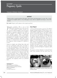
Case Report<br />
A 20-year-old female patient reported to the OPD of<br />
Karpaga Vinayaga Institute of Dental Sciences, with a<br />
chief complaint of painful mass on the gingiva over a<br />
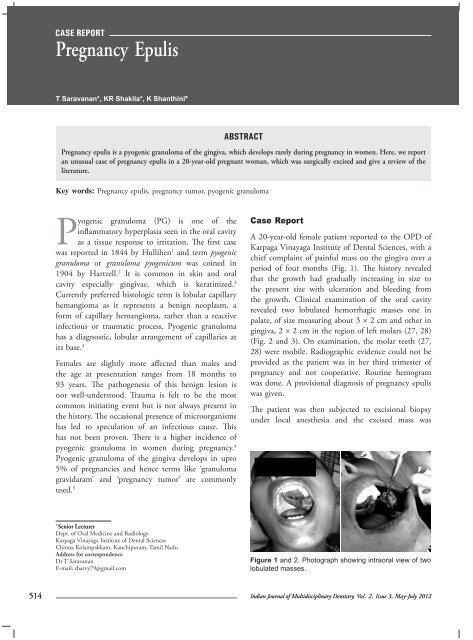
period of four months (Fig. 1). The history revealed<br />
that the growth had gradually increasing in size to<br />
the present size with ulceration and bleeding from<br />
the growth. Clinical examination of the oral cavity<br />
revealed two lobulated hemorrhagic masses one in<br />
palate, of size measuring about 3 × 2 cm and other in<br />
gingiva, 2 × 2 cm in the region of left molars (27, 28)<br />
(Fig. 2 and 3). On examination, the molar teeth (27,<br />
28) were mobile. Radiographic evidence could not be<br />
provided as the patient was in her third trimester of<br />
pregnancy and not cooperative. Routine hemogram<br />
was done. A provisional diagnosis of pregnancy epulis<br />
was given.<br />
The patient was then subjected to excisional biopsy<br />
under local anesthesia and the excised mass was<br />
*Senior Lecturer<br />
Dept. of Oral Medicine and Radiology<br />
Karpaga Vinayaga Institute of Dental Sciences<br />
Chinna Kolampakkam, Kanchipuram, Tamil Nadu<br />
Address for correspondence<br />
Dr T Saravanan<br />
E-mail: sharvy79@gmail.com<br />
Figure 1 and 2. Photograph showing intraoral view of two<br />
lobulated masses.<br />
514<br />
Indian Journal of Multidisciplinary Dentistry, Vol. 2, <strong>Issue</strong> 3, <strong>May</strong>-<strong>Jul</strong>y 2012