Volume 7 Issue 4 - Australasian Society for Ultrasound in Medicine
Volume 7 Issue 4 - Australasian Society for Ultrasound in Medicine
Volume 7 Issue 4 - Australasian Society for Ultrasound in Medicine
You also want an ePaper? Increase the reach of your titles
YUMPU automatically turns print PDFs into web optimized ePapers that Google loves.
ISSN 1441-6891<br />
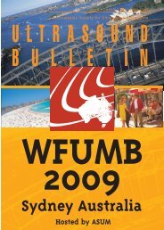
<strong>Volume</strong> 7 Number 4 November 2004<br />
<strong>Australasian</strong> <strong>Society</strong> <strong>for</strong> <strong>Ultrasound</strong> <strong>in</strong> Medic<strong>in</strong>e<br />
ASUM Multidiscipl<strong>in</strong>ary Workshop<br />
Melbourne 16–20 March 2005<br />
DMU Preparation Courses 16–20 March<br />
DDU Preparation Course 16–17 March<br />
O&G Workshop 17–20 March<br />
Vascular Workshop 18–19 March<br />
Musculoskeletal Workshop 19 March<br />
Cardiac Workshop 19 March<br />
Breast and Paediatric Sessions 19 March<br />
Registration brochures <strong>in</strong>cluded with<br />
with this issue<br />
<br />
<br />
<br />
<br />
<br />
<br />
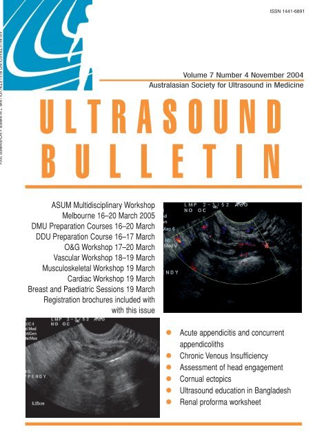
Acute appendicitis and concurrent<br />
appendicoliths<br />
Chronic Venous Insufficiency<br />
Assessment of head engagement<br />
Cornual ectopics<br />
<strong>Ultrasound</strong> education <strong>in</strong> Bangladesh<br />
Renal pro<strong>for</strong>ma worksheet
Our obsession is quality.<br />
Our goal is per<strong>for</strong>mance.<br />
Our promise is value.<br />
Our products are...<br />
Toshiba - The Leader <strong>in</strong> Premium Level<br />
Cl<strong>in</strong>ical Innovation<br />
For more <strong>in</strong><strong>for</strong>mation call 02 9887 8063
President<br />
Dr David Rogers<br />
Immediate Past President<br />
Dr Glen McNally<br />
Honorary Secretary<br />
Mrs Roslyn Savage<br />
Honorary Treasurer<br />
Dr Dave Carpenter<br />
Chief Executive Officer<br />
Dr Carol<strong>in</strong>e Hong<br />
ULTRASOUND BULLETIN<br />
Official publication of<br />
the <strong>Australasian</strong> <strong>Society</strong><br />
<strong>for</strong> <strong>Ultrasound</strong> <strong>in</strong> Medic<strong>in</strong>e<br />
Published quarterly<br />
ISSN 1441-6891<br />
Indexed by the Sociedad Iberoamericana<br />
de In<strong>for</strong>macion Cientifien (SIIC) Databases<br />
Editor<br />
Dr Roger Davies<br />
Women’s and Children’s Hospital, SA<br />
Co-Editor<br />
Mr Keith Henderson<br />
ASUM Education Manager<br />
Assistant Editors<br />
Ms Kaye Griffiths AM<br />
Anzac Research Institute CRGH, NSW<br />
Ms Louise Lee<br />
Gold Coast Hospital, Qld<br />
Editorial contributions<br />
Orig<strong>in</strong>al research, case reports, quiz cases,<br />
short articles, meet<strong>in</strong>g reports and calendar<br />
<strong>in</strong><strong>for</strong>mation are <strong>in</strong>vited and should be<br />
addressed to The Editor and sent to ASUM<br />
at the address below<br />
Published on behalf of ASUM<br />
by M<strong>in</strong>nis Communications<br />
Mr Bill M<strong>in</strong>nis, Director<br />
4/16 Maple Grove<br />
Toorak Melbourne Victoria 3142 Australia<br />
tel +61 3 9824 5241 fax +61 3 9824 5247<br />
email m<strong>in</strong>nis@m<strong>in</strong>niscomms.com.au<br />
Disclaimer<br />
Unless specifically <strong>in</strong>dicated, op<strong>in</strong>ions<br />
expressed should not be taken as those of<br />
the <strong>Australasian</strong> <strong>Society</strong> <strong>for</strong> <strong>Ultrasound</strong> <strong>in</strong><br />
Medic<strong>in</strong>e or of M<strong>in</strong>nis Communications<br />
Membership and general enquiries<br />
should be directed to ASUM at the address<br />
below<br />
AUSTRALASIAN SOCIETY FOR<br />
ULTRASOUND IN MEDICINE<br />
2/181 High Street<br />
Willoughby Sydney NSW 2068 Australia<br />
tel +61 2 9958 7655 fax +61 2 9958 8002<br />
email asum@asum.com.au<br />
website:http //www.asum.com.au<br />
ABN 64 001 679 161<br />
ISO 9001: 2000<br />
Certified<br />
Quality<br />
Management<br />
Systems<br />
ASUM <strong>Ultrasound</strong> Bullet<strong>in</strong> 2004 November 7: 4<br />
Notes from the Editor<br />
Another year is rapidly com<strong>in</strong>g to a<br />
close. Thoughts will soon turn to the<br />
Festive Season, and away from occupation<br />
or education. Be<strong>for</strong>e relax<strong>in</strong>g<br />
over the summer holiday, peruse this<br />
edition of the <strong>Ultrasound</strong> Bullet<strong>in</strong>,<br />
which is replete with articles and items<br />
of uni<strong>for</strong>mly high standard.<br />
First, there is a part one of a superb<br />
summary from the multidiscipl<strong>in</strong>ary<br />
workshop on chronic venous <strong>in</strong>sufficiency<br />
by D Coghlan. After study<strong>in</strong>g this,<br />
readers will be encouraged to attend the<br />
upcom<strong>in</strong>g workshops <strong>in</strong> 2005.<br />
A comprehensive review of acute<br />
appendicitis has been contributed by J<br />
Spurway and B Simmons – their contributions<br />
are always valuable. The<br />
rema<strong>in</strong><strong>in</strong>g articles: on two methods of<br />
assesss<strong>in</strong>g head engagement by P<br />
Dietz and V Lanzarone and two case<br />
studies on corunal ectopics by D Moir<br />
and K McMahon are equally valuable;<br />
all are commended to readers.<br />
Abstracts from the highly successful<br />
ASUM 2004 scientific meet<strong>in</strong>g are<br />
THE EXECUTIVE<br />
President’s message 5<br />
CEO’s desk 7<br />
EDUCATION<br />
Fetal measurements requirements 30<br />
Exam<strong>in</strong>ation dates and fees <strong>for</strong> 2005 32<br />
Book reviews 40<br />
RESEARCH AND TECHNICAL<br />
Acute appendicitis and concurrent<br />
appendicoliths 11<br />
Chronic venous <strong>in</strong>sufficiency 14<br />
Assessment of head engagement 22<br />
Cornual ectopics 23<br />
ASUM ASM Abstracts 50<br />
SONOGRAPHER<br />
OBSERVATIONS<br />
Pro<strong>for</strong>ma renal worksheet 21<br />
worth detailed study. Many new ideas are<br />
first presented at meet<strong>in</strong>gs such as<br />
ASUM 2004, sometimes two years<br />
ahead of <strong>for</strong>mal publication.<br />
Readers will appreciate the wealth<br />
of <strong>in</strong><strong>for</strong>mation and education available<br />
through ASUM and are encouraged to<br />
consider attend<strong>in</strong>g a scientific meet<strong>in</strong>g<br />
at least every two or three years.<br />
Feedback, good and otherwise, regard<strong>in</strong>g<br />
the 2004 meet<strong>in</strong>g is also sought,<br />
through letters to the Editor.<br />
Many readers contributed to The<br />
ASUM Professional Survey on the use<br />
of worksheets as well as the <strong>in</strong>teraction<br />
between sonographer and physician<br />
<strong>in</strong> daily ultrasound practice. The<br />
responses were sometimes unexpected<br />
and always <strong>in</strong>terest<strong>in</strong>g. A prelim<strong>in</strong>ary<br />
analysis is presented <strong>in</strong> this issue.<br />
ASUM editorial staff wish all<br />
readers and members of ASUM a<br />
happy and safe Festive Season, and a<br />
great start to 2005.<br />
Roger Davies<br />
Editor<br />
ASIA LINK<br />
<strong>Ultrasound</strong> education <strong>in</strong> Bangladesh 26<br />
THE SOCIETY<br />
Multidiscipl<strong>in</strong>ary workshop<br />
2005 draft program 2<br />
ASUM Professional Survey 34<br />
ASUM 2004 photo spread 36<br />
New Zealand news 39<br />
ASUM honors ultrasound leaders 40<br />
Giulia Franco Teach<strong>in</strong>g Fellowship<br />
2004 reports 42<br />
ASUM Council appo<strong>in</strong>tments<br />
2004–2005 45<br />
New members 46<br />
Corporate members 47<br />
<strong>Ultrasound</strong> calendar 49<br />
Guidel<strong>in</strong>es <strong>for</strong> authors 64<br />
ASUM <strong>Ultrasound</strong> Bullet<strong>in</strong>: 2004 November 7: 4<br />
1
ASUM multidiscipl<strong>in</strong>ary workshop 16–20 March 2005<br />
2 ASUM <strong>Ultrasound</strong> Bullet<strong>in</strong>: 2004 November 7: 4
www.asum.com.au/open/meet_mdw05_home.htm<br />
ASUM <strong>Ultrasound</strong> Bullet<strong>in</strong> 2004 November 7: 4<br />
3
CARRYING HIGH<br />
PERFORMANCE<br />
ULTRASOUND<br />
INTO THE<br />
FUTURE<br />
<br />
THE BEGINNING<br />
SonoSite began as a division of ATL<br />
(now Philips) <strong>in</strong> 1995 when the U.S.<br />
Department of Defence awarded ATL<br />
a grant to develop a sophisticated and<br />
highly portable ultrasound device.<br />
SonoSite became a separate publiclylisted<br />
company <strong>in</strong> 1998 and has achieved<br />
global leadership <strong>in</strong> the hand-carried,<br />
high-per<strong>for</strong>mance, ultrasound market.<br />
<br />
NOW<br />
SonoSite has established a direct<br />
operation to support the expand<strong>in</strong>g<br />
needs of highly-portable, highper<strong>for</strong>mance,<br />
ultrasound users <strong>in</strong><br />
Australia and New Zealand.<br />
SonoSite Australasia will develop local<br />
tra<strong>in</strong><strong>in</strong>g and educational resources to<br />
assist and supplement the activities of<br />
InSight Oceania – SonoSite’s<br />
<strong>Australasian</strong> distribution partner.<br />
Greg Brand is a 25 year<br />
senior management veteran<br />
of the ultrasound <strong>in</strong>dustry<br />
and is the manag<strong>in</strong>g director<br />
of SonoSite Australasia.<br />
Shelley Thomson is SonoSite’s cl<strong>in</strong>ical<br />
market<strong>in</strong>g manager <strong>for</strong> Australasia.<br />
She has over 14 years experience <strong>in</strong><br />
the ultrasound <strong>in</strong>dustry and, similarly<br />
to Greg, is widely-known through past<br />
positions with ATL and with Philips.<br />
• mobile • durable • reliable • easy to use<br />
SONOSITE AUSTRALASIA PTY LTD<br />
Australia 1300 663 516 New Zealand 0800 888 204 Web www.sonosite.com
THE EXECUTIVE<br />
President’s message<br />
Dr David Rogers<br />
Greet<strong>in</strong>gs.<br />
This is my first <strong>Ultrasound</strong> Bullet<strong>in</strong><br />
message s<strong>in</strong>ce I began my role as<br />
ASUM President dur<strong>in</strong>g the ASM <strong>in</strong><br />
Sydney <strong>in</strong> September; and what a fantastic<br />
meet<strong>in</strong>g it was. The speakers<br />
were of a very high standard and <strong>in</strong>troduced<br />
us to many new advances. The<br />
venue was superb and made great use<br />
of the excellent weather Sydney turned<br />
on <strong>for</strong> the event. Special mention must<br />
be made of the supreme ef<strong>for</strong>t of the<br />
Organis<strong>in</strong>g Committee to produce a top<br />
class event, especially the convenors,<br />
Glenn McNally and Jenny Kidd. Our<br />
thanks to all.<br />
Glenn McNally’s presidency<br />
I take over the role of president from<br />
Glenn McNally who leaves large boots<br />
to fill. Glenn has worked tirelessly <strong>for</strong><br />
ASUM dur<strong>in</strong>g his term and has placed<br />
the <strong>Society</strong> <strong>in</strong> a top position.<br />
Dur<strong>in</strong>g Glenn’s presidency,<br />
ASUM won its bid to host the<br />
WFUMB 2009 Conference. This is a<br />
great honour, giv<strong>in</strong>g the <strong>Society</strong> a<br />
truly <strong>in</strong>ternational position. ASUM<br />
can now be proud to have two councillors<br />
to WFUMB, Glenn McNally and<br />
Stan Barnett as Secretary.<br />
Dur<strong>in</strong>g Glenn’s presidency ASUM<br />
expanded its work and reputation <strong>in</strong> Asia,<br />
with further development of the Asia<br />
L<strong>in</strong>k program. Highlights of this <strong>in</strong>clude<br />
the development of the DMU (Asia)<br />
course at the Vision College <strong>in</strong> Kuala<br />
Lumpur and the recent ASUM meet<strong>in</strong>g<br />
held <strong>in</strong> Kuala Lumpur. This was an<br />
excellent meet<strong>in</strong>g with a high quality<br />
academic program which will enhance<br />
our <strong>Society</strong>’s reputation <strong>in</strong> Malaysia.<br />
Special thanks to speakers who<br />
generously donated their time, especially<br />
Roger Gent, Andrew Ngu,<br />
Simon Meagher, Stan Barnett, Glenn<br />
McNally and from Kuala Lumpur,<br />
Raman Subramanian, Patrick Chia, TP<br />
Baskaran, Teresa Chow and Sulaiman<br />
Tamanang. Further Asian meet<strong>in</strong>gs are<br />
planned <strong>in</strong> Bangkok <strong>in</strong> 12 months time<br />
and liaison with Ch<strong>in</strong>a is planned next<br />
year.<br />
As President, Glenn had oversight<br />
of the sale of the Willoughby office.<br />
which has placed ASUM <strong>in</strong> a good<br />
position to purchase a new, more suitable<br />
premises sometime with<strong>in</strong> the<br />
next two years. With an expand<strong>in</strong>g<br />
role <strong>for</strong> the Secretariat and the promotion<br />
of WFUMB 2009, more appropriate<br />
space is required.<br />
F<strong>in</strong>ally, under Glenn’s direction<br />
the <strong>Society</strong> is <strong>in</strong> a strong f<strong>in</strong>ancial<br />
position. Although this is not the core<br />
aim of ASUM, it enables us to offer<br />
greater service to members. Many<br />
other ultrasound societies do not<br />
employ a secretariat and their activities<br />
are m<strong>in</strong>imal.<br />
Secretariat<br />
ASUM’s great strength is its secretariat.<br />
Under the very capable direction of<br />
Carol<strong>in</strong>e Hong as CEO and Keith<br />
Henderson as Education Manager, the<br />
<strong>Society</strong> is able to be very active. These<br />
days, few people have spare time to<br />
organise activities. The Secretariat is<br />
able to put <strong>in</strong> all the groundwork, leav<strong>in</strong>g<br />
ASUM volunteers to do the work<br />
of education. The recent meet<strong>in</strong>g <strong>in</strong><br />
Kuala Lumpur was a sh<strong>in</strong><strong>in</strong>g example<br />
of this, where the speakers had all the<br />
organisation done <strong>for</strong> them and only<br />
had to turn up and give their lectures.<br />
Additionally, f<strong>in</strong>ancial strength has<br />
enabled us to seed the Research Fund,<br />
which will be an important self-susta<strong>in</strong><strong>in</strong>g<br />
asset. Members’ attention is<br />
drawn to this fund. It would be ideal if<br />
the fund were to be used to sponsor<br />
orig<strong>in</strong>al research to be presented at the<br />
WFUMB 2009 Conference.<br />
New Council<br />
At the AGM, two new Councillors had<br />
their nom<strong>in</strong>ation ratified. We welcome<br />
Prof Ron Benzie and Dr David Davis<br />
Payne to Council. Ron is Professor of<br />
Obstetrics at Nepean Hospital <strong>in</strong> Sydney<br />
and David is a paediatric radiologist <strong>in</strong><br />
my home town of Auckland. Ron will<br />
chair the Standards of Practise<br />
Committee and David will chair the<br />
Education Committee.<br />
Future plans<br />
Dur<strong>in</strong>g my term as President, there are<br />
some objectives that I wish the <strong>Society</strong><br />
to achieve. Clearly, we will need to<br />
source and purchase a new office, a<br />
process we have already begun. The<br />
DMU has ga<strong>in</strong>ed certification <strong>for</strong> the<br />
next five years; to improve the DMU,<br />
I believe we need to develop an onl<strong>in</strong>e<br />
course <strong>for</strong> DMU candidates to assist <strong>in</strong><br />
education and preparation <strong>for</strong> the<br />
exam. This would also satisfy the<br />
requirements of our candidates outside<br />
of ma<strong>in</strong> centres. We also need to complete<br />
the Onl<strong>in</strong>e Handbook to fulfil its<br />
potential as an outstand<strong>in</strong>g resource.<br />
Branch activities<br />
Branch activities will also be a focus.<br />
These seem to be at an all time low<br />
and need attention. ASUM struggles a<br />
little <strong>in</strong> this area as it has a diverse<br />
membership and f<strong>in</strong>ds it difficult to be<br />
all th<strong>in</strong>gs to everyone. ASUM centrally<br />
can only provide support to branches<br />
to facilitate activities. The activities<br />
themselves need to be driven by the<br />
branches. I hope to be able to visit<br />
each Branch over the next year to<br />
establish more effective communication<br />
and to stimulate activity. There is<br />
a need <strong>for</strong> regular educational meet<strong>in</strong>gs<br />
<strong>in</strong> most areas.<br />
I hope the Christmas season treats<br />
everyone well and Santa br<strong>in</strong>gs you all<br />
you wish <strong>for</strong>.<br />
Merry Christmas and Happy New<br />
Year<br />
David Rogers<br />
President ASUM<br />
ASUM <strong>Ultrasound</strong> Bullet<strong>in</strong>: 2004 November 7: 4<br />
5
OBSTETRICAL &<br />
GYNAECOLOGICAL<br />
ULTRASOUND<br />
SONOLOGIST REQUIRED<br />
LOCUM<br />
VASCULAR &<br />
GENERAL<br />
SONOGRAPHER<br />
available from January 2005<br />
mobile 0427 676 212<br />
email ausrover@bigpond.com<br />
A unique opportunity exists <strong>for</strong> a sonologist<br />
with experience <strong>in</strong> this field to jo<strong>in</strong> an established<br />
<strong>in</strong>dependent practice <strong>in</strong> Perth,Western<br />
Australia.<br />
Call Dr John Phillips<br />
Perth <strong>Ultrasound</strong><br />
Mobile 0408 913 053<br />
tel 08 9382 1500<br />
Sonographers<br />
Mercy Radiology NZ<br />
We are seek<strong>in</strong>g experienced<br />
sonographers to jo<strong>in</strong> our dedicated team at<br />
Mercy Radiology, Auckland, New Zealand<br />
We have a large modern practice.<br />
offer<strong>in</strong>g a variety of ultrasound services,<br />
dedicated to provid<strong>in</strong>g excellence <strong>in</strong> service <strong>in</strong><br />
a quality car<strong>in</strong>g environment.<br />
We offer an excellent remuneration<br />
package <strong>for</strong> sonographers seek<strong>in</strong>g a change<br />
of lifestyle and environment.<br />
● Top l<strong>in</strong>e equipment<br />
● Fabulous environment<br />
● Patient focused<br />
Send your CV to:<br />
Radiographic Manager<br />
P O Box 9056, Newmarket<br />
Auckland<br />
New Zealand<br />
tel +64 9 623 5883<br />
Email<br />
sdixon@radiology.co.nz<br />
SonoSite Cl<strong>in</strong>ical Sales Specialist<br />
SonoSite is the world leader <strong>in</strong> high per<strong>for</strong>mance portable ultrasound<br />
technology, and is dedicated to enabl<strong>in</strong>g tra<strong>in</strong>ed cl<strong>in</strong>icians<br />
utilise high-resolution ultrasound imag<strong>in</strong>g at the po<strong>in</strong>t of care.<br />
SonoSite has recently established direct operations <strong>in</strong> Australia and<br />
New Zealand and is look<strong>in</strong>g <strong>for</strong> enthusiastic, self-motivated staff to<br />
jo<strong>in</strong> our grow<strong>in</strong>g team.<br />
As a SonoSite Cl<strong>in</strong>ical Sales Specialist your ma<strong>in</strong> responsibilities<br />
will be to build strong customer relationships, achieve sales targets,<br />
apply market<strong>in</strong>g strategies, develop new bus<strong>in</strong>ess opportunities and<br />
demonstrate SonoSite products and manage your sales territory.<br />
The successful candidate will possess the follow<strong>in</strong>g:<br />
● Drive and Determ<strong>in</strong>ation to succeed<br />
● Results orientated, and have a genu<strong>in</strong>e desire to over achieve<br />
● Exceptional communication skills<br />
● Highly developed plann<strong>in</strong>g, time management and<br />
organisation skills<br />
● Well developed team work skills to enhance the results of the<br />
team as well as your <strong>in</strong>dividual results<br />
Cl<strong>in</strong>ical ultrasound knowledge is essential and previous sales experience<br />
will be viewed favourably. Product and sales tra<strong>in</strong><strong>in</strong>g will be<br />
provided along with a highly competitive compensation package.<br />
Submit your application of <strong>in</strong>terest, together with current<br />
resume to:<br />
Shelley Thomson, Cl<strong>in</strong>ical Market<strong>in</strong>g Manager<br />
SonoSite Australasia Pty Ltd<br />
email shelley.thomson@sonosite.com<br />
or calll Shelley on 1300 663 516 (Aust) or 0800 888 204 (NZ)<br />
Your <strong>in</strong>terest will be treated <strong>in</strong> the strictest of confidence.<br />
6 ASUM <strong>Ultrasound</strong> Bullet<strong>in</strong>: 2004 November 7: 4
THE EXECUTIVE<br />
COUNCIL 2004–2005<br />
EXECUTIVE<br />
President<br />
David Rogers NZ<br />
Medical Councillor<br />
Honorary Secretary<br />
Roslyn Savage Qld<br />
Sonographer Councillor<br />
Honorary Treasurer<br />
Dave Carpenter NSW<br />
Medical Councillor<br />
MEMBERS<br />
Medical Councillors<br />
Roger Davies SA<br />
Glenn McNally NSW<br />
Mathew Andrews Vic<br />
Ron Benzie NSW<br />
Dave Carpenter NSW<br />
David Davies-Payne NZ<br />
Sonographer Councillors<br />
Stephen Bird SA<br />
Margaret Condon Vic<br />
Kaye Griffiths NSW<br />
Jan<strong>in</strong>e Horton WA<br />
Roslyn Savage Qld<br />
ASUM Head Office<br />
Chief Executive Officer<br />
Carol<strong>in</strong>e Hong<br />
Education Manager<br />
Keith Henderson<br />
All correspondence should be<br />
directed to<br />
The Chief Executive Officer<br />
ASUM<br />
2/181 High St<br />
Willoughby<br />
NSW 2068<br />
Australia<br />
asum@asum.com.au<br />
http//www.asum.com.au<br />
CEO’s message<br />
Dr Carol<strong>in</strong>e Hong<br />
ASUM 2004 Annual Scientific<br />
Meet<strong>in</strong>g<br />
Another showcase meet<strong>in</strong>g of ASUM<br />
over four glorious days at Star City<br />
Hotel <strong>in</strong> Sydney from 23rd to 26th<br />
September 2004 f<strong>in</strong>ished with overwhelm<strong>in</strong>g<br />
success. The event attracted<br />
over 600 delegates from Australia,<br />
New Zealand and overseas. The Skills<br />
Day and Nuchal Translucency Day<br />
workshops both were fully subscribed<br />
on the pre-congress day – <strong>in</strong> the midst<br />
of all the flurry of activities happen<strong>in</strong>g<br />
<strong>in</strong> excit<strong>in</strong>g Sydney.<br />
The Annual Scientific Meet<strong>in</strong>g<br />
provided a valuable <strong>for</strong>um <strong>for</strong> professionals<br />
<strong>in</strong> ultrasound <strong>in</strong> medic<strong>in</strong>e from<br />
all over the world to meet and discuss<br />
topics relevant to the various practice<br />
fields.<br />
We were extremely <strong>for</strong>tunate to<br />
have great keynote speakers and delegates<br />
from all parts of Australia, New<br />
Zealand and overseas. The exhibition<br />
halls were packed on most days with<br />
<strong>in</strong>terested delegates enjoy<strong>in</strong>g the new<br />
products, reestablish<strong>in</strong>g old contacts,<br />
mak<strong>in</strong>g new contacts and network<strong>in</strong>g<br />
relevant to the advancement of the<br />
ultrasound profession. The quality of<br />
the speakers was superb.<br />
As expected, the ASUM social<br />
event, the Gala D<strong>in</strong>ner at the Sheraton<br />
on the Park, was sensational, with<br />
danc<strong>in</strong>g until the early hours of the<br />
morn<strong>in</strong>g to the rhythm of the funkiest<br />
music from Almondo Hurley and his<br />
10-piece band.<br />
Special thanks aga<strong>in</strong> go to all the<br />
speakers, exhibitors and sponsors and<br />
to the team beh<strong>in</strong>d the scenes, <strong>in</strong>clud<strong>in</strong>g<br />
the Organis<strong>in</strong>g Committee, ASUM<br />
secretariat staff and ICMS staff.<br />
DMU Exam<strong>in</strong>ers accreditation<br />
A Practical Exam<strong>in</strong>ers Accreditation<br />
Tra<strong>in</strong><strong>in</strong>g Day was conducted on<br />
Thursday 23rd September 2004 at Star<br />
City. Twenty-three people achieved<br />
accreditation as Practical Exam<strong>in</strong>ers<br />
<strong>for</strong> the ASUM DMU follow<strong>in</strong>g the<br />
tra<strong>in</strong><strong>in</strong>g day. This is consistent with<br />
ASUM’s commitment to ma<strong>in</strong>ta<strong>in</strong> a<br />
high standard <strong>in</strong> the DMU exam<strong>in</strong>ation<br />
process, requir<strong>in</strong>g the DMU practical<br />
exam<strong>in</strong>ers be re-accredited once<br />
every three years More <strong>in</strong><strong>for</strong>mation<br />
can be obta<strong>in</strong>ed from James Hamilton<br />
at dmu@asum.com.au.<br />
ASUM awards Life Member and<br />
Honorary Fellows<br />
The Annual General meet<strong>in</strong>g of<br />
ASUM was held on Saturday 25th<br />
September 2004. At this meet<strong>in</strong>g, it<br />
was approved that Dr Jack Jell<strong>in</strong>s be<br />
awarded Life Membership <strong>for</strong> outstand<strong>in</strong>g<br />
contribution to the <strong>Society</strong><br />
and to the profession. It was also<br />
announced that Assoc Prof Albert Lam<br />
and Mrs Jane Fonda were approved by<br />
Council to be awarded Honorary<br />
Fellows of the <strong>Society</strong> (see pages 40,<br />
41).<br />
ASUM Secretariat<br />
Council has commissioned a report to<br />
advise the <strong>Society</strong> on the purchase of<br />
new ASUM Head office premises follow<strong>in</strong>g<br />
from the sale <strong>in</strong> May this year.<br />
The search will cont<strong>in</strong>ue and ASUM has<br />
a leaseback option up to May 2007.<br />
Two new staff, Nancy Leung and<br />
Matthew Byron have jo<strong>in</strong>ed ASUM<br />
recently as adm<strong>in</strong>istrative assistants.<br />
This will provide the necessary support<br />
required to deliver the <strong>in</strong>creas<strong>in</strong>g<br />
DMU, educational and adm<strong>in</strong>istrative<br />
services. Fund<strong>in</strong>g <strong>for</strong> the new staff<br />
members are generated from with<strong>in</strong><br />
the revenue collected from the new<br />
and <strong>in</strong>creased ASUM projects and<br />
services.<br />
ASUM <strong>Ultrasound</strong> Bullet<strong>in</strong>: 2004 November 7: 4<br />
7
THE EXECUTIVE<br />
ASUM Branches<br />
A luncheon meet<strong>in</strong>g of Council with<br />
Branch Chairs was held on Saturday<br />
25th September immediately follow<strong>in</strong>g<br />
the Incom<strong>in</strong>g Council meet<strong>in</strong>g. It<br />
was pleas<strong>in</strong>g <strong>for</strong> the new President and<br />
Council members to meet with<br />
Branches <strong>in</strong><strong>for</strong>mally to discuss branch<br />
matters. Dr Dave Rogers, <strong>in</strong> his role as<br />
the new President, <strong>in</strong>tends to visit<br />
branches to cont<strong>in</strong>ue to enhance communication<br />
with the Council.<br />
Past Presidents’ Meet<strong>in</strong>g<br />
Past Presidents of ASUM and the previous<br />
Ultrasonographers Group met<br />
on 24th September 2004 at a luncheon<br />
held at Star City. It was a great occasion<br />
<strong>for</strong> the current Council members<br />
as well as the CEO of ASUM to hear<br />
the views and stories of many of the<br />
past presidents, who contributed to<br />
beg<strong>in</strong>n<strong>in</strong>gs of the <strong>Society</strong> that have led<br />
to where it is today globally.<br />
ASUM – Denmark l<strong>in</strong>k<br />
A proposal and presentation was made to<br />
Council by Assoc Prof Christian Nolsoe<br />
of the Danish <strong>Society</strong> <strong>for</strong> Diagnostic<br />
<strong>Ultrasound</strong> (DSDU). He proposed the<br />
development of an ASUM–DSDU<br />
l<strong>in</strong>k, with the Danish Royal family as<br />
the patrons of an Australian–Danish<br />
scientific and educational foundation <strong>for</strong><br />
the promotion of medical ultrasound <strong>in</strong><br />
the two countries.<br />
Assoc Prof Nolsoe was also a<br />
speaker at ASUM 2004. Council<br />
agreed to pursue the l<strong>in</strong>kage.<br />
Certa<strong>in</strong>ly, the recent wedd<strong>in</strong>g of His<br />
Royal Highness Crown Pr<strong>in</strong>ce<br />
Frederic of Denmark and Australian<br />
Mary Donaldson has generated a lot of<br />
mutual <strong>in</strong>terest <strong>in</strong> both countries.<br />
WFUMB 2009<br />
Promotional and exploratory ef<strong>for</strong>ts<br />
cont<strong>in</strong>ue <strong>in</strong> this area. Council<br />
approved <strong>for</strong> the WFUMB promotion<br />
team to consist of Dr Stan Barnett<br />
(WFUMB 2009 Convenor), Dr Glenn<br />
McNally (WFUMB 2009 Treasurer),<br />
Dr Dave Rogers (ASUM President)<br />
and Dr Carol<strong>in</strong>e Hong (ASUM CEO).<br />
At the Annual Scientific Meet<strong>in</strong>g,<br />
two WFUMB Councillors, Professor<br />
Michel Claudon from France and<br />
Professor Giovanni Cerri from Brazil<br />
were among the keynote speakers. It<br />
was most <strong>in</strong>terest<strong>in</strong>g to talk with them<br />
and learn about the activities of<br />
WFUMB <strong>in</strong>ternationally.<br />
Professor Cerri will be the<br />
President of WFUMB when the World<br />
Congress comes to Sydney <strong>in</strong> 2009.<br />
Future meet<strong>in</strong>g dates<br />
It is never too early to plan your calendar<br />
to attend the next ASUM meet<strong>in</strong>g.<br />
Register your <strong>in</strong>terest early and<br />
email asum@asum.com.au if you need<br />
any additional <strong>in</strong><strong>for</strong>mation. Also go to<br />
the website: www.asum.com.au <strong>for</strong><br />
regular updates on meet<strong>in</strong>gs and how<br />
to make contact with the organisers.<br />
For your diary<br />
■ ASUM 2005<br />
Multidiscipl<strong>in</strong>ary<br />
Workshop 18–19th March<br />
■<br />
■<br />
2005 Melbourne.<br />
ASUM 2005 NZ Branch<br />
jo<strong>in</strong>t RANZCR NZ meet<strong>in</strong>g<br />
29–31st July 2005<br />
Well<strong>in</strong>gton<br />
ASUM 2005 Annual<br />
Scientific Meet<strong>in</strong>g 29<br />
September to 2nd October<br />
2005 Adelaide<br />
■ ASUM Asia L<strong>in</strong>k Meet<strong>in</strong>g<br />
November 2005 Bangkok<br />
Dr Carol<strong>in</strong>e Hong<br />
Chief Executive Officer<br />
carol<strong>in</strong>ehong@asum.com.au<br />
ASUM Giulia Franco<br />
Teach<strong>in</strong>g Fellowships<br />
2004 and 2005<br />
Sponsored by Toshiba <strong>Ultrasound</strong><br />
The Giulia Franco Teach<strong>in</strong>g Fellowship was established by<br />
ASUM <strong>in</strong> association with Toshiba Medical to provide educational<br />
opportunities <strong>for</strong> sonographers <strong>in</strong> all parts of Australia<br />
and New Zealand.<br />
The fellowships <strong>in</strong>crease the opportunity <strong>for</strong> members outside<br />
the ma<strong>in</strong> centres to have access to quality educational<br />
opportunities.<br />
It is named to commemorate Giulia Franco whose passion<br />
<strong>for</strong> ultrasound education took her to all parts of Australia<br />
and New Zealand, and cont<strong>in</strong>ued as she moved <strong>in</strong>to a bus<strong>in</strong>ess<br />
career with Toshiba. Its first award, <strong>in</strong> 2004, provided<br />
educational programs <strong>in</strong> Western Australia.<br />
Further details are on the ASUM website at<br />
www.asum.com.au<br />
Branches wish<strong>in</strong>g to propose programs <strong>for</strong> the 2005 Teach<strong>in</strong>g<br />
Fellowships should contact:<br />
Keith Henderson<br />
tel +61 2 9958 6200 fax +61 2 9958 8002<br />
email khenderson@asum.com.au<br />
Send nom<strong>in</strong>ations and proposals to:<br />
The Education Manager<br />
ASUM 2/181 High St<br />
Willoughby 2068 NSW<br />
Australia<br />
ASUM Chris Kohlenberg<br />
Teach<strong>in</strong>g Fellowships 2004 and 2005<br />
Sponsored by<br />
GE Medical Systems <strong>Ultrasound</strong><br />
The Chris Kohlenberg Teach<strong>in</strong>g Fellowship was established by<br />
ASUM <strong>in</strong> association with GE Medical Systems <strong>Ultrasound</strong> to<br />
<strong>in</strong>crease the opportunity <strong>for</strong> members outside the ma<strong>in</strong> centres<br />
to have access to quality educational opportunities.<br />
It has been awarded annually s<strong>in</strong>ce 1998, provid<strong>in</strong>g educational<br />
programs <strong>in</strong> provide educational opportunities <strong>for</strong><br />
members <strong>in</strong> New Zealand, Queensland, New South Wales,<br />
Northern Territory, Western Australia, Victoria, South<br />
Australia and Tasmania.<br />
It is named to commemorate Dr Chris Kohlenberg, who died<br />
while travell<strong>in</strong>g to educate sonographers.<br />
Further details are on the ASUM website www.asum.com.au<br />
Branches wish<strong>in</strong>g to propose programs <strong>for</strong> the 2005 Teach<strong>in</strong>g<br />
Fellowships should, <strong>in</strong> the first <strong>in</strong>stance, contact:<br />
Keith Henderson<br />
tel +61 2 9958 6200 fax +61 2 9958 8002<br />
email khenderson@asum.com.au<br />
Send nom<strong>in</strong>ations and proposals to:<br />
The Education Manager<br />
ASUM 2/181 High St<br />
Willoughby 2068 NSW<br />
Australia<br />
8 ASUM <strong>Ultrasound</strong> Bullet<strong>in</strong>: 2004 November 7: 4
An ISO Certified Company<br />
ECLIPSE ®<br />
PROBE COVER<br />
LATEX-FREE<br />
Pre-gelled <strong>in</strong>side with Aquasonic ® 100<br />
<strong>Ultrasound</strong> Transmission Gel<br />
PARKER LABORATORIES, INC. 286 Eldridge Road, Fairfield, NJ 07004<br />
Tel. 973-276-9500 Fax 973-276-9510 E-mail: parker@parkerlabs.com www.parkerlabs.com<br />
U.S.A. and International patents granted
ASUM <strong>Ultrasound</strong> Bullet<strong>in</strong> 2004 August 7: 4: 11–13<br />
DIAGNOSTIC ULTRASOUND<br />
A review of acute appendicitis and<br />
concurrent appendicoliths<br />
Jacquel<strong>in</strong>e Spurway and Bradley Simmons<br />
Introduction<br />
Diseases of the appendix are among the most common surgical<br />
emergencies <strong>in</strong> the western world, yet the appendix is<br />
the smallest and functionally most irrelevant segment of the<br />
gastro<strong>in</strong>test<strong>in</strong>al tract 1 .<br />
The peak <strong>in</strong>cidence of acute appendicitis is <strong>in</strong> late childhood<br />
and adolescence, when there is a 3:2 male-to-female<br />
ratio. This ratio tends to equalise by the fourth decade of<br />
life. In 2001 the mortality rate <strong>in</strong> Australia from acute<br />
appendicitis was 0.70 deaths per million people and <strong>in</strong> New<br />
Zealand <strong>in</strong> 2000 it was 1.77 deaths per million. This is <strong>in</strong><br />
contrast to The Bahamas, which had a mortality rate of 6.72<br />
per million <strong>in</strong> 2000 2 .-<br />
The cl<strong>in</strong>ical presentation of acute appendicitis <strong>in</strong>cludes<br />
generalised abdom<strong>in</strong>al pa<strong>in</strong> that beg<strong>in</strong>s <strong>in</strong> the epigastrium<br />
and later localises to the right lower quadrant, fever, loss of<br />
appetite, nausea, vomit<strong>in</strong>g and diarrhoea.<br />
Physical exam<strong>in</strong>ation may demonstrate rectal tenderness,<br />
abdom<strong>in</strong>al guard<strong>in</strong>g or a mass or tenderness <strong>in</strong> the<br />
right iliac fossa. Laboratory f<strong>in</strong>d<strong>in</strong>gs frequently <strong>in</strong>clude<br />
leukocytosis, bandemia and an abnormal ur<strong>in</strong>alysis 1 .<br />
CT f<strong>in</strong>d<strong>in</strong>gs may <strong>in</strong>clude an enlarged appendix (> 7 mm<br />
diameter), an appendicolith, a thick, enhanc<strong>in</strong>g appendiceal<br />
wall, de<strong>for</strong>mity or thicken<strong>in</strong>g of the apex of the caecum and<br />
strand<strong>in</strong>g <strong>in</strong> the adjacent mesenteric fat. Peritoneal fluid<br />
Figure 1 Mildly dilated appendix (black arrows) with preservation of<br />
the expected multilayered appearance of bowel. Note bl<strong>in</strong>d end<br />
(white arrow)<br />
Correspondence to<br />
JF Spurway & BJ Simmons<br />
Medical Imag<strong>in</strong>g Department<br />
Orange Base Hospital<br />
PO Box 319<br />
Orange NSW 2800<br />
email Jacquel<strong>in</strong>e.Spurway@mwahs.gov.nsw.au<br />
Figure 2 A non-compressible, enlarged appendix (arrows) with loss<br />
of def<strong>in</strong>ition of the bowel wall layers, particularly the echogenic submucosa.<br />
At surgery a per<strong>for</strong>ated appendix was found<br />
may also be seen 3 .<br />
The sonographic detection or exclusion of other diseases<br />
facilitates the cl<strong>in</strong>ical differential diagnosis of right lower<br />
abdom<strong>in</strong>al pa<strong>in</strong> 4 . The reported sensitivity <strong>in</strong> the ultrasound<br />
diagnosis of acute appendicitis is approximately 75–90%,<br />
while specificity and accuracy are greater than 90% 1 .<br />
The appendix<br />
The normal appendix<br />
Ultrasonically the appendix appears as five dist<strong>in</strong>ct alternat<strong>in</strong>g<br />
echogenic and hypoechoic concentric layers, correspond<strong>in</strong>g<br />
to the layers of bowel wall 5 (Figure 1). The average<br />
adult appendix is 8 to 10 cm long 1 with a range of 4 to 25<br />
cm. Compare this to the koala which has an enlarged caecum,<br />
the equivalent of the human appendix, which is 2<br />
metres long and 10 cm <strong>in</strong> diameter 6,7 .<br />
Acute appendicitis<br />
Appendicitis is caused by obstruction of the appendiceal<br />
lumen followed by <strong>in</strong>fection 1 . <strong>Ultrasound</strong> identification of<br />
acute appendicitis is based on the visualisation of a “tubular,<br />
noncompressible, aperistaltic bowel loop that demonstrates<br />
a connection with the caecum and a distal bl<strong>in</strong>d end,<br />
with a diameter greater than 6 mm” 5 (Figure 2). A calcified<br />
appendicolith may be present, which assists <strong>in</strong> differentiat<strong>in</strong>g<br />
a dilated appendix from adjacent bowel loops.<br />
Pericaecal fluid may be present and the pericaecal fat can be<br />
prom<strong>in</strong>ent. Mesenteric adenitis <strong>in</strong> the right lower quadrant is<br />
identifiable by ultrasound <strong>in</strong> 40% of appendicitis 5 . Scann<strong>in</strong>g<br />
should be <strong>in</strong>itiated <strong>in</strong> the region of maximal pa<strong>in</strong> <strong>in</strong>dicated by<br />
the patient to expedite the sonographic evaluation. Graded<br />
compression may assist <strong>in</strong> the localisation of the appendix.<br />
Colour Doppler appearance of appendicitis represents a<br />
ASUM <strong>Ultrasound</strong> Bullet<strong>in</strong> 2004 November 7: 4<br />
11
A review of acute appendicitis and concurrent appendicoliths<br />
Figure 3 Appendicolith with posterior shadow<strong>in</strong>g<br />
time cont<strong>in</strong>uum that varies with the severity of disease.<br />
Initially there may be no detectable <strong>in</strong>crease <strong>in</strong> colour<br />
Doppler flow signal mak<strong>in</strong>g the absence of colour Doppler<br />
flow signal non-diagnostic, as it can be seen <strong>in</strong> both normal<br />
and abnormal appendices. Visualisation of <strong>in</strong>creased colour<br />
Doppler flow <strong>in</strong> the appendiceal wall or a right iliac fossa<br />
mass is supportive of appendicitis. This reflects <strong>in</strong>creas<strong>in</strong>g<br />
hyperperfusion of the appendiceal wall 5 . The addition of<br />
colour Doppler to B-mode imag<strong>in</strong>g results <strong>in</strong> a sensitivity of<br />
87%, specificity of 97%, and accuracy of 93% <strong>in</strong> the diagnosis<br />
of acute appendicitis <strong>in</strong> children 5 .<br />
Differentiat<strong>in</strong>g non-per<strong>for</strong>ated appendicitis from per<strong>for</strong>ated<br />
appendicitis is important <strong>for</strong> operative plann<strong>in</strong>g.<br />
<strong>Ultrasound</strong> appearances <strong>in</strong> cases of per<strong>for</strong>ation <strong>in</strong>clude<br />
thicken<strong>in</strong>g of adjacent bowel wall, atonic bowel loops, <strong>in</strong>terloop<br />
fluid pockets, no tenderness on transducer pressure,<br />
asymmetrical appendiceal wall thicken<strong>in</strong>g, hypoechoic rim<br />
around the appendix and free fluid conta<strong>in</strong><strong>in</strong>g debris.<br />
Non-visualisation of a per<strong>for</strong>ated appendix may be due<br />
to failure to recognise a decompressed, but thickened,<br />
appendix because of surround<strong>in</strong>g thickened, matted loops of<br />
bowel or failure to recognise the decompressed and excessively<br />
necrosed remnants of the per<strong>for</strong>ated appendix. Other<br />
factors contribut<strong>in</strong>g to non-visualisation of an abnormal<br />
appendix are prevention of adequate compression ow<strong>in</strong>g to<br />
reflex abdom<strong>in</strong>al rigidity caused by per<strong>for</strong>ation and atonic<br />
dilation of bowel loops caused by peritonitis 5 .<br />
Sonographic false positive diagnoses are uncommon,<br />
but may <strong>in</strong>clude hydrosalp<strong>in</strong>x, periappendicitis result<strong>in</strong>g<br />
from tubo-ovarian abscess, Crohn’s disease, psoas muscle<br />
fibres, <strong>in</strong>spissated stool mimick<strong>in</strong>g an appendicolith or<br />
resolv<strong>in</strong>g appendicitis 5 .<br />
Sonographic false negative diagnoses may occur <strong>in</strong><br />
patients with a retrocaecal appendicitis, ascites, ileus, and<br />
small bowel obstruction or with a markedly enlarged or gasfilled<br />
appendix 5 .<br />
The ultrasound diagnosis of acute appendicitis can be<br />
limited by operator experience, the presence of overly<strong>in</strong>g<br />
bowel gas, and patient obesity.<br />
Appendicoliths<br />
A calcified appendicolith appears sonographically as a<br />
curved, echogenic structure with posterior shadow<strong>in</strong>g<br />
(Figure 3). Air or <strong>in</strong>spissated faeces with<strong>in</strong> the bowel lumen<br />
12 ASUM <strong>Ultrasound</strong> Bullet<strong>in</strong> 2004 November 7: 4<br />
Figure 4 Trans-abdom<strong>in</strong>al image of RIF show<strong>in</strong>g heterogeneous mass<br />
may produce posterior acoustic shadow<strong>in</strong>g mimick<strong>in</strong>g a calcified<br />
appendicolith 5,8 .<br />
The appendicolith or faecolith is a calcified deposit<br />
with<strong>in</strong> the appendix. It is present <strong>in</strong> approximately 30% of<br />
children with acute appendicitis 8 .<br />
An appendicolith is a mass of <strong>in</strong>spissated faecal material<br />
that <strong>for</strong>ms around a <strong>for</strong>eign body <strong>in</strong> the appendix, grow<strong>in</strong>g<br />
slowly with the deposition of successive lam<strong>in</strong>ae, sometimes<br />
with calcification 9 . The radiopacity of appendicoliths depends<br />
on their m<strong>in</strong>eral content with only 7 to 12% be<strong>in</strong>g visualised<br />
radiographically <strong>in</strong> adults with acute appendicitis 10 .<br />
The first documented report of the detection of appendicolith<br />
by ultrasound was <strong>in</strong> 1983, 11 prior to this a radiographically<br />
demonstrated fecalith was widely considered a<br />
virtually pathognomonic sign of acute appendicitis 12 .<br />
Case study<br />
A thirty-five year-old female presented to the Emergency<br />
Department compla<strong>in</strong><strong>in</strong>g of right-sided pelvic pa<strong>in</strong>, which<br />
had been <strong>in</strong>termittently present <strong>for</strong> two days, but had<br />
<strong>in</strong>creased <strong>in</strong> <strong>in</strong>tensity recently. On physical exam<strong>in</strong>ation she<br />
had rebound tenderness on the right side. Abdom<strong>in</strong>al tenderness<br />
was worse with movement and cough<strong>in</strong>g and the<br />
pa<strong>in</strong> radiated across her abdomen. The patient had a slightly<br />
elevated temperature at 37.4°C and rest<strong>in</strong>g pulse rate of<br />
103 beats per m<strong>in</strong>ute.<br />
The patient was suffer<strong>in</strong>g from nausea, hot and cold<br />
sweats and depressed appetite. Her last menstrual period<br />
was three weeks previously and she believed she was not<br />
pregnant. The patient was alert but <strong>in</strong> obvious distress.<br />
Investigative procedures showed a negative beta-hCG<br />
and an elevated white cell count.<br />
The patient was referred to the Medical Imag<strong>in</strong>g<br />
Department <strong>for</strong> a pelvic ultrasound to <strong>in</strong>vestigate the presence<br />
of appendiceal or ovarian pathology. As the patient was nil by<br />
mouth her bladder was filled by <strong>in</strong>creas<strong>in</strong>g IV fluid <strong>in</strong>put.<br />
The patient revealed that she had been pregnant three times<br />
result<strong>in</strong>g <strong>in</strong> two live births and an uneventful hydatidi<strong>for</strong>m mole<br />
treated by a dilatation and curettage. She had previously had a<br />
left ovarian cyst dra<strong>in</strong>ed and was currently not on hormonal<br />
contraception. On transabdom<strong>in</strong>al imag<strong>in</strong>g the bladder was very<br />
poorly distended. The right ovary and kidneys appeared to be<br />
essentially normal. A 35 x 38 x 32 mm heterogenous mass was<br />
identified <strong>in</strong> the right iliac fossa (Figure 4).
Jaquel<strong>in</strong>e Spurway and Bradley Simmons<br />
Figure 5 Thickened caecum<br />
Figure 6 Enlarged appendix with transverse diameter of 15 mm<br />
Figure 7 Hyperaemic response <strong>in</strong> appendiceal wall<br />
Transvag<strong>in</strong>al ultrasound showed Nabothian cysts <strong>in</strong> the<br />
cervix, normal myometrial echotexture, a normal endometrial<br />
appearance and thickness (9 mm). The left ovary was<br />
unremarkable with a volume of 5.4 cc. The right ovary was<br />
enlarged with a volume 16.5 cc and conta<strong>in</strong>ed a ruptured<br />
follicle consistent with a recent ovulation.<br />
Superolateral to the right ovary abnormal caecum was<br />
identified with thickened, oedematous, hyperaemic walls<br />
(Figure 5).<br />
No peristalsis was noted <strong>in</strong> real time. An abnormal<br />
appendix was identified with a transverse diameter of<br />
15mm. (Figure 6). The walls of the appendix were thickened,<br />
hyperaemic (Figure 7) and tender on direct transducer<br />
pressure. With<strong>in</strong> the appendix at least three hyperechoic,<br />
shadow<strong>in</strong>g foci were identified (Figure 8) consistent with<br />
appendicoliths. Some free fluid was noted around the right<br />
ovary, appendix and with<strong>in</strong> the Pouch of Douglas.<br />
Later the same day the patient underwent a laparoscopic<br />
appendicectomy. Pathological review of the surgical<br />
specimen showed acute suppurative appendicitis with rupture<br />
and local peritonitis.<br />
References<br />
1 Gore, RM, Lev<strong>in</strong>e MS. Textbook of Gastro<strong>in</strong>test<strong>in</strong>al Radiology,<br />
2nd Edition, 2000, WB Saunders, USA, page 1140.<br />
2 http://wwww.nationmaster.com/graph-T/mor_acu_app_cap<br />
Accessed 18 August 2004.<br />
3 http://www.mritutor.org/mriteach/1100/1100.html Accessed 6 July<br />
2004.<br />
4 Z Gastroenterol. 1988 Nov; 26 (11): 708–714. [Sonography <strong>in</strong> the<br />
diagnosis of appendicitis – a prospective study] Blank W, Braun B.<br />
Figure 8 Appendicoliths with posterior shadow<strong>in</strong>g<br />
5 http://lunis.luc.edu/radiology/Appendicitis/fluid.htm Accessed 17<br />
August 2004.<br />
6 http://www.home.swiftsl.com.au/~endangered/koala.htm. Accessed 1<br />
September 2004.<br />
7 http://www.wires.org.au/animals/koala.htm. Accessed 1 September<br />
2004.<br />
8 http://www.amershamhealth.com/medcyclopaedia/medical/<strong>Volume</strong> 5<br />
20VII/AAPPENDICOLITH.ASP Accessed 20 July 2004.<br />
9 Morson, B.C., Systemic pathology – Alimentary tract. 3rd Edition,<br />
1991, Longman Group, Hong Kong, page 294.<br />
10 Fenoglio-Preiser, C.M. et al, Gastro<strong>in</strong>test<strong>in</strong>al pathology – An atlas<br />
and text, 2nd Edition, 1999, Lipp<strong>in</strong>cott-Raven, Philadelphia, page<br />
523.<br />
11 J Can Assoc Radiol. 1983 Mar; 34 (1): 66–67. Ultrasonic demonstra<br />
tion of appendicolith. Forel F, Filiatrault D, Grignon A.<br />
12 Am J Emerg Med. 1996 Jul;14 (4): 394–397. The myth of the<br />
fecalith. Maenza RL, Smith L, Wolfson AB.<br />
ASUM <strong>Ultrasound</strong> Bullet<strong>in</strong> 2004 November 7: 4<br />
13
DIAGNOSTIC ULTRASOUND<br />
ASUM <strong>Ultrasound</strong> Bullet<strong>in</strong> 2004 August 7:4: 14–21<br />
Chronic venous <strong>in</strong>sufficiency<br />
Debbie Coghlan<br />
Camperdown Vascular Laboratory<br />
The aim of this article is to give students a solid understand<strong>in</strong>g<br />
of:<br />
1 Pathophysiology and venous haemodynamics<br />
beh<strong>in</strong>d chronic venous <strong>in</strong>sufficiency (CVI).<br />
2 Detailed anatomic overview of the deep and<br />
superficial ve<strong>in</strong>s of the leg.<br />
3 A practical and useful protocol that has been implemented<br />
at the Camperdown Vascular Laboratory.<br />
Pathophysiology of chronic venous <strong>in</strong>sufficiency<br />
Chronic venous <strong>in</strong>sufficiency is an advanced stage of<br />
venous disease caused either by superficial or deep venous<br />
pathology, <strong>in</strong> which venous return is impaired, usually over<br />
a number of years, by reflux, obstruction or calf muscle<br />
pump failure. This leads to susta<strong>in</strong>ed venous hypertension<br />
and ultimately to cl<strong>in</strong>ical complications <strong>in</strong>clud<strong>in</strong>g oedema,<br />
eczema, lipodermatosclerosis1 and ulceration.<br />
CVI results from dysfunctional valves that reduce<br />
venous return, which thus <strong>in</strong>creases venous pressure.<br />
Because exist<strong>in</strong>g valves are destroyed, venous blood flow is<br />
bi-directional, result<strong>in</strong>g <strong>in</strong> <strong>in</strong>efficient venous outflow. The<br />
net effect of this change is that the weight of the venous<br />
blood column from the right atrium is transmitted along the<br />
full length of the ve<strong>in</strong>s. Very high venous pressure is exerted<br />
at the ankle and the venules become the f<strong>in</strong>al pathway <strong>for</strong><br />
the highest venous pressure.<br />
Primary varicose ve<strong>in</strong>s<br />
One popular explanation <strong>for</strong> the production of <strong>in</strong>competent<br />
venous valves is an <strong>in</strong>herent structural weakness of the ve<strong>in</strong>s<br />
themselves. The most strik<strong>in</strong>g feature of this problem is the<br />
positive family history usually obta<strong>in</strong>ed.<br />
The haemodynamic factors play a significant role <strong>in</strong> produc<strong>in</strong>g<br />
primary varicosities. The most important of these is<br />
the high hydrostatic pressure to which the ve<strong>in</strong>s to the lower<br />
extremities are subjected as a result of the patient stand<strong>in</strong>g.<br />
Other factors <strong>in</strong>clude heavy muscular work such as lift<strong>in</strong>g,<br />
repeated stra<strong>in</strong><strong>in</strong>g at stool, pregnancy, and pelvic tumours,<br />
all of which <strong>in</strong>crease <strong>in</strong>traabdom<strong>in</strong>al tension.<br />
Secondary varicose ve<strong>in</strong>s<br />
The most serious of all venous disorders occur when the deep<br />
venous system is obstructed and the valvular mechanisms are<br />
destroyed or rendered <strong>in</strong>competent. This is thought to occur<br />
when there has been an episode of deep venous thrombosis<br />
that leads to two problems – obstruction of the deep ve<strong>in</strong>s and<br />
subsequent destruction of the venous valves.<br />
When venous obstruction occurs, the venous blood is<br />
<strong>for</strong>ced to follow alternate pathways to reach the heart. One<br />
way is to <strong>for</strong>ce the open<strong>in</strong>g of new pathways (co-laterals),<br />
or to use the superficial system as the alternate pathway.<br />
However, the flow <strong>in</strong> the communicat<strong>in</strong>g (per<strong>for</strong>at<strong>in</strong>g) ve<strong>in</strong>s<br />
is now reversed, becom<strong>in</strong>g deep to superficial.<br />
Aetiology<br />
Chronic venous <strong>in</strong>sufficiency can be divided <strong>in</strong>to primary<br />
and secondary varicose ve<strong>in</strong>s. Both lead to venous hypertension,<br />
which produce associated signs and symptoms<br />
(expla<strong>in</strong>ed later).<br />
Primary varicose ve<strong>in</strong>s are those which only <strong>in</strong>volve the<br />
superficial system. Secondary varicose ve<strong>in</strong>s are usually<br />
attributable to previous DVT, which have caused damage to<br />
the valves of the deep ve<strong>in</strong>s and per<strong>for</strong>ators.<br />
Normal deep<br />
superficial<br />
ve<strong>in</strong>s<br />
Superficial ve<strong>in</strong><br />
<strong>in</strong>competence<br />
Deep ve<strong>in</strong><br />
<strong>in</strong>competence<br />
Comb<strong>in</strong>ed<br />
deep and<br />
superficial ve<strong>in</strong><br />
<strong>in</strong>competence<br />
Lipodermatosclerosis<br />
14 ASUM <strong>Ultrasound</strong> Bullet<strong>in</strong> 2004 November 7: 4<br />
Venous eczema and ulceration
Debbie Coghlan<br />
Venous hypertension<br />
High pressure results <strong>in</strong> the <strong>for</strong>mation of oedema fluid, as<br />
well as extravasation of red blood cells and large prote<strong>in</strong><br />
molecules that leak out from capillaries. Initially, soft pitt<strong>in</strong>g<br />
oedema is present at the ankle, but over a period of time<br />
the sk<strong>in</strong> becomes thickened and acquires a woody feel<strong>in</strong>g.<br />
The haemoglob<strong>in</strong> from these cells is broken down to<br />
haem pigment, which is taken up by tissue macrophages,<br />
hence the brown sk<strong>in</strong> pigment associated with chronic<br />
<strong>in</strong>sufficiency.<br />
Venous hypertension also produces a fibr<strong>in</strong> cuff around<br />
capillaries. This <strong>in</strong>hibits oxygen diffusion to adjacent tissues,<br />
thereby caus<strong>in</strong>g local tissue atrophy and sk<strong>in</strong> ulceration.<br />
These signs are often referred to as lipodermatosclerosis<br />
or post thrombotic syndrome.<br />
Various types of dermatitis, <strong>in</strong>clud<strong>in</strong>g erythema and<br />
scal<strong>in</strong>g, are also associated with this condition. Trivial trauma<br />
may cause sk<strong>in</strong> breakdown and ulceration. The ulcers<br />
are usually shallow but may penetrate to the level of the<br />
deep fascia.<br />
When the valves <strong>in</strong> the ve<strong>in</strong>s of the lower extremities<br />
become <strong>in</strong>competent, there is regurgitation of blood <strong>in</strong> the<br />
superficial venous system, produc<strong>in</strong>g varicosities. Depend<strong>in</strong>g<br />
upon the site of <strong>in</strong>volvement, this reflux takes place either<br />
through the saphenofemoral junction or through communicat<strong>in</strong>g<br />
of per<strong>for</strong>ator vessels. As a result of the marked dissention<br />
of the superficial ve<strong>in</strong>s and the poor support they possess,<br />
these vessels enlarge and become tortuous, <strong>in</strong> the<br />
process of which the valves locally also become <strong>in</strong>competent.<br />
If the per<strong>for</strong>at<strong>in</strong>g ve<strong>in</strong>s valves are <strong>in</strong>competent as well,<br />
<strong>in</strong>creased pressure is transmitted through per<strong>for</strong>at<strong>in</strong>g ve<strong>in</strong>s<br />
to the capillary bed; this <strong>in</strong> turn results <strong>in</strong> exudation of prote<strong>in</strong>-rich<br />
fluid and red blood cells <strong>in</strong>to the subcutaneous tissues.<br />
Degenerated red blood cells and organised prote<strong>in</strong>rich<br />
exudate produce <strong>in</strong>duration and hyperpigmentation.<br />
Work, by Browse, suggests that fibr<strong>in</strong> is deposited<br />
around capillaries as a result of <strong>in</strong>creased capillary permeability.<br />
These deposits prevent diffusion of oxygen to adjacent<br />
cells, caus<strong>in</strong>g limitation <strong>in</strong> heal<strong>in</strong>g capability and or<br />
cutaneous atrophy so that ulceration may result from even<br />
m<strong>in</strong>or <strong>in</strong>juries.<br />
Cl<strong>in</strong>ical consequences of chronic<br />
venous <strong>in</strong>sufficiency<br />
➢ Oedema<br />
➢ Varicose eczema (scaly brown sk<strong>in</strong>, especially<br />
medial lower leg and ankle region)<br />
➢ Lipodermatosclerosis<br />
➢ Venous ulceration<br />
Presentation<br />
●<br />
●<br />
●<br />
●<br />
●<br />
●<br />
●<br />
●<br />
●<br />
●<br />
●<br />
●<br />
●<br />
●<br />
Pa<strong>in</strong><br />
Itch<br />
Swell<strong>in</strong>g (oedema)<br />
Superficial signs of CVI (discussed above)<br />
Primary varicose ve<strong>in</strong>s<br />
Secondary varicose ve<strong>in</strong>s<br />
Recurrent varicose ve<strong>in</strong>s<br />
Recurrent cellulitis<br />
Cosmetic<br />
Pre sclerotherapy<br />
Restless leg syndrome<br />
Ach<strong>in</strong>g limb<br />
Recurrent cellulitis<br />
Klippel-Trenaunay syndrome<br />
Venous haemodynamics<br />
At any given time, about 75% of circulat<strong>in</strong>g blood <strong>in</strong> your<br />
body is mov<strong>in</strong>g through the venous system. So, understand<strong>in</strong>g<br />
the mechanisms by which venous return to the heart is<br />
accomplished, is crucial to understand<strong>in</strong>g the physiology of<br />
the vascular system.<br />
Phasicity<br />
Phasicity, <strong>in</strong> the venous system, refers to the ebb and flow<br />
that occurs <strong>in</strong> normal ve<strong>in</strong>s <strong>in</strong> response to respiration. All<br />
deep ve<strong>in</strong>s normally exhibit phasicity,<br />
The two phases of respiration are <strong>in</strong>spiration and expiration.<br />
The way <strong>in</strong> which the blood moves <strong>in</strong> phase with respiration<br />
differs accord<strong>in</strong>g to the part of the body affected and<br />
the position <strong>in</strong> which the body is placed.<br />
The effects of respiration<br />
Respiration has profound effects on venous pressure and<br />
flow.<br />
Inspiration<br />
The thoracic cavity expands, the diaphragm lowers and the<br />
abdomen becomes smaller.<br />
The volume of the ve<strong>in</strong>s of the thorax <strong>in</strong>creases and the pressure<br />
decreases <strong>in</strong> response to the reduced <strong>in</strong>trathoracic pressure.<br />
In the abdomen, because of the descent of the<br />
diaphragm, the pressure <strong>in</strong>creases. Increased abdom<strong>in</strong>al<br />
pressure decreases pressure gradients between peripheral<br />
ve<strong>in</strong>s <strong>in</strong> the lower extremities and the abdomen, thus reduc<strong>in</strong>g<br />
flow <strong>in</strong> the vessels.<br />
Signs of chronic venous <strong>in</strong>sufficiency<br />
● Pigmentation<br />
● Lipodermatosclerosis<br />
● Eczema<br />
● Ulceration<br />
● Varicose ve<strong>in</strong>s<br />
● Oedema<br />
● Dermatitis (especially eczema)<br />
● Atrophic blanche (white areas on sk<strong>in</strong>)<br />
● Fibrosis (hard contracted sk<strong>in</strong> of the lower leg,<br />
known as ‘bottle neck appearance’)<br />
ASUM <strong>Ultrasound</strong> Bullet<strong>in</strong> 2004 November 7: 4<br />
15
DIAGNOSTIC ULTRASOUND<br />
Expiration<br />
The thoracic cavity decreases, the diaphragm elevates and<br />
the abdomen becomes larger. The volume of the ve<strong>in</strong>s of the<br />
thorax decreases and the pressure <strong>in</strong>creases.<br />
In the abdomen, because of the elevation of the<br />
diaphragm, the pressure decreases. Decreased abdom<strong>in</strong>al<br />
pressure <strong>in</strong>creases pressure gradients between the peripheral<br />
ve<strong>in</strong>s <strong>in</strong> the lower extremities and the abdomen, thus<br />
<strong>in</strong>creas<strong>in</strong>g flow to the vessels.<br />
In a nutshell, when you breathe <strong>in</strong>, flow out of the legs<br />
slows or stops. When you breathe out, flow resumes.<br />
Valsalva manoeuvre<br />
Valsalva, <strong>for</strong>ced expirations or cough<strong>in</strong>g, causes compression<br />
both the <strong>in</strong>tra-thoracic and <strong>in</strong>tra-abdom<strong>in</strong>al ve<strong>in</strong>s.<br />
Flow <strong>in</strong> the vena cava can be markedly reduced as a result.<br />
The effect of posture and exercise on venous pressure at ankle<br />
16 ASUM <strong>Ultrasound</strong> Bullet<strong>in</strong> 2004 November 7: 4<br />
Hydrostatic pressure<br />
Hydrostatic pressure is the pressure exerted by fluid with<strong>in</strong><br />
a closed system. Hydrostatic pressure varies with position.<br />
When sup<strong>in</strong>e, there is virtually no hydrostatic pressure <strong>in</strong><br />
the legs, as they are at the same level as the right atrium,<br />
which has a pressure of zero.<br />
Gravity exerts significant effects on venous return<br />
because of our upright posture. As body posture is changed<br />
from sup<strong>in</strong>e to stand<strong>in</strong>g, gravity acts upon the vascular volume,<br />
so that blood accumulates <strong>in</strong> the lower extremities.<br />
Because venous compliance is high, most of the blood volume<br />
shift occurs <strong>in</strong> the ve<strong>in</strong>s. There<strong>for</strong>e, when stand<strong>in</strong>g,<br />
venous volume and pressure becomes very high <strong>in</strong> the feet<br />
and lower limbs.<br />
When venous valves are work<strong>in</strong>g correctly, every movement<br />
of the leg causes blood to be pumped <strong>in</strong>ward and<br />
upward past a series of valves. Dur<strong>in</strong>g walk<strong>in</strong>g, the normal<br />
pressure <strong>in</strong> the venous system of the lower leg is low.<br />
Immediately after walk<strong>in</strong>g, the early stand<strong>in</strong>g pressure <strong>in</strong><br />
the normal leg rema<strong>in</strong>s low. Arterial <strong>in</strong>flow fills the leg<br />
ve<strong>in</strong>s slowly and the only source of venous pressure is the<br />
hydrostatic pressure of a column of blood as high as the<br />
nearest competent valve.<br />
After prolonged stand<strong>in</strong>g, the ve<strong>in</strong>s are completely filled<br />
and all the venous valves float open. At this time, high<br />
hydrostatic venous pressure results from the unbroken column<br />
of fluid that extends from the head to the foot.<br />
Failed valves cause the column of stand<strong>in</strong>g blood <strong>in</strong> the<br />
ve<strong>in</strong> to rema<strong>in</strong> high even when dur<strong>in</strong>g walk<strong>in</strong>g. The hydrostatic<br />
pressure <strong>in</strong>creased dur<strong>in</strong>g and immediately after<br />
ambulation.<br />
High venous pressure is directly responsible <strong>for</strong> many<br />
aspects of venous <strong>in</strong>sufficiency syndrome, <strong>in</strong>clud<strong>in</strong>g edema,<br />
tissue prote<strong>in</strong> deposition, perivascular fibr<strong>in</strong> cuff<strong>in</strong>g, red cell<br />
extravasation, impaired arterial <strong>in</strong>flow, and other locally<br />
mediated disturbances.<br />
Not all of the sequelae of venous <strong>in</strong>sufficiency are related<br />
to venous hypertension, and not all patients with venous<br />
hypertension develop ulceration. Some patients with venous<br />
ulceration do not have marked venous hypertension.<br />
The venous valves and the muscle pump<br />
Venous valves play a very important role <strong>in</strong> the function of<br />
venous return, especially <strong>in</strong> the lower extremities. They are<br />
irregularly located along the ve<strong>in</strong>s, but are always found at<br />
the junctions of tributaries with ma<strong>in</strong> venous channels or<br />
where two large ve<strong>in</strong>s jo<strong>in</strong>. Venous valves are usually bicuspid<br />
and occasionally tricuspid.<br />
In the leg, some ve<strong>in</strong>s have fewer valves than others. The<br />
deep system has more valves than the superficial.<br />
Venous valves direct flow, they keep the blood mov<strong>in</strong>g<br />
back toward the heart <strong>in</strong> both the deep and the superficial<br />
ve<strong>in</strong>s. In the per<strong>for</strong>at<strong>in</strong>g ve<strong>in</strong>s, the valves direct the flow<br />
from the superficial to the deep ve<strong>in</strong>s, if the venous valves<br />
are function<strong>in</strong>g normally, they prevent reflux.<br />
Venous valves open and close <strong>in</strong> conjunction with the<br />
action of the muscles.<br />
A<br />
B C D<br />
Types of valves: A unicuspid B bicuspid C tricuspid D quaricuspid<br />
VEIN<br />
NUMBER OF VALVES<br />
Posterior tibial<br />
9–19 valves<br />
Anterior tibial<br />
9–11 valves<br />
Peroneal<br />
7 valves<br />
Popliteal<br />
4 valves<br />
Superficial femoral<br />
4 or 5 valves<br />
Common femoral<br />
extremely rare to have<br />
any valves<br />
External iliac 1 and sometimes 2<br />
Common iliac<br />
extremely rare<br />
Long and short saphenous 7–9 between them<br />
Normal ve<strong>in</strong><br />
Varicose ve<strong>in</strong><br />
Incompetent<br />
venous valves<br />
When the deep valves are<br />
<strong>in</strong>competent, blood that is<br />
squeezed upward by muscle<br />
contraction simply flows<br />
back down dur<strong>in</strong>g muscle<br />
relaxation. Additionally,<br />
blood is often <strong>for</strong>ced back<br />
down the ve<strong>in</strong>s toward the<br />
foot by muscle contraction,<br />
this dramatically <strong>in</strong>creases<br />
distal venous pressure and<br />
venous hypertension and<br />
other symptoms may occur.<br />
The condition is exacerbated<br />
if the per<strong>for</strong>ator valves<br />
are <strong>in</strong>sufficient as well.
Debbie Coghlan<br />
The muscle pump<br />
In the extremities, the deep ve<strong>in</strong>s are surrounded by muscles.<br />
As these muscles contract, they squeeze the ve<strong>in</strong>s with<strong>in</strong><br />
them. Muscles do not rema<strong>in</strong> permanently contracted, to<br />
function, they must alternately contract and relax, thus propell<strong>in</strong>g<br />
the blood up the leg.<br />
In the venous system, it is the <strong>in</strong>teraction of the venous<br />
valves and the muscle pump that keep venous blood mov<strong>in</strong>g,<br />
and mov<strong>in</strong>g <strong>in</strong> the right direction. When these two elements<br />
are work<strong>in</strong>g properly, the ve<strong>in</strong>s <strong>in</strong> the lower extremities can<br />
empty so effectively that empty<strong>in</strong>g is actually complete.<br />
If a person is stand<strong>in</strong>g still, they have an ankle venous<br />
pressure of about 90 mm Hg. When that person beg<strong>in</strong>s to<br />
exercise the leg muscles, they activate the calf pump. If the<br />
valves and the pump are function<strong>in</strong>g properly, the ankle<br />
venous pressure will fall to 30 mm Hg or even lower – possibly<br />
even 1 mm Hg.<br />
Dur<strong>in</strong>g muscle contraction<br />
1 The large blood volume resid<strong>in</strong>g <strong>in</strong> the deep calf<br />
ve<strong>in</strong>s and soleal s<strong>in</strong>uses is literally squeezed up and<br />
out of the calf.<br />
2 The proximal deep valves are <strong>for</strong>ced open.<br />
3 The distal valves close to prevent caudal flow.<br />
4 The per<strong>for</strong>ator valves close to prevent flow <strong>in</strong>to the<br />
superficial venous system.<br />
Dur<strong>in</strong>g muscle relaxation:<br />
1 The proximal valves close due to the reflux of<br />
blood back down the ve<strong>in</strong>s.<br />
2 The distal deep and per<strong>for</strong>ator valves open, and<br />
blood flows <strong>in</strong>to the calf deep and muscular ve<strong>in</strong>s.<br />
3 The hydrostatic pressure column has been <strong>in</strong>terrupt<br />
ed and distal <strong>in</strong>tralum<strong>in</strong>al pressure is reduced.<br />
Lipodermatosclerosis1 (LDS) is scleroderma-like harden<strong>in</strong>g<br />
of the legs <strong>in</strong> patients with venous <strong>in</strong>sufficiency. It is<br />
characterised by <strong>in</strong>duration, hyperpigmentation and depression<br />
of the sk<strong>in</strong>. LDS may be either acute or chronic. Most<br />
patients are middle-aged or older women, often with a history<br />
of venous <strong>in</strong>sufficiency. The acute phase of LDS is<br />
characterised by leg pa<strong>in</strong> above the medial malleolus and a<br />
poorly def<strong>in</strong>ed area of erythematous, tender, warm sk<strong>in</strong> with<br />
m<strong>in</strong>imal <strong>in</strong>duration. The better recognised chronic phase of<br />
Normal<br />
LDS is characterised by a more sharply demarcated <strong>in</strong>duration<br />
and depression of the sk<strong>in</strong>, often with hyperpigmentation<br />
or areas of atrophie blanche. The subcutaneous fibrosis<br />
and scarr<strong>in</strong>g may lead to constriction of the mid portion of<br />
the leg, result<strong>in</strong>g <strong>in</strong> a ‘bowl<strong>in</strong>g p<strong>in</strong>’ appearance.<br />
References<br />
1 Gray’s Anatomy.<br />
2 Vascular diagnosis with ultrasound. Hennerici & Neuerburt-<br />
Heusler.<br />
3 Anatomy and Physiology. Mart<strong>in</strong>i.<br />
4 Cl<strong>in</strong>ically oriented anatomy. Moore & Dalley.<br />
5 Cogral Image Library. Coghlan & Ralph.<br />
6 McM<strong>in</strong>n’s Colour Atlas of Human Anatomy. PH Abrahams, RT<br />
Hutch<strong>in</strong>gs, SC Marks.<br />
7 Diagnostic Angiography. Kadir.<br />
8 Abrams Angiography third edition. Herbert I Abrams MD.<br />
9 Peripheral Vascular <strong>Ultrasound</strong> – How, Why and When. Abigail<br />
Thrush, Tim Hartshorne.<br />
10 Introduction to Vascular Ultrasonography. Zwiebel.<br />
11 Grants Atlas of Anatomy. Williams & Wilk<strong>in</strong>s.<br />
12 Anatomy of the Human Body. Henry Gray www.bartleby.com<br />
13 Atlas of Human Anatomy. Frank Netter.<br />
14 Current <strong>Issue</strong>s <strong>in</strong> vascular surgery. Greenhalgh, Jamieson, Nicolaides.<br />
15 Stand<strong>in</strong>g versus sup<strong>in</strong>e positions <strong>in</strong> venous reflux evaluation. Foldes,<br />
Blackburn et al. Journal of Vascular Technology 1991; 15(6):<br />
321–324.<br />
16 Cl<strong>in</strong>ical Doppler <strong>Ultrasound</strong>. Allan, Dubb<strong>in</strong>s, Pozniak & McDicken.<br />
17 Vascular Surgery <strong>Issue</strong>s <strong>in</strong> Current Practice. Greenhalgh, Jamieson &<br />
Nicolaides.<br />
18 Vascular Technology. Rumwell, McPharl<strong>in</strong>.<br />
19 Diagnostic <strong>Ultrasound</strong>. Rumack, Wilson Charboneau.<br />
20 Ultrasonography Exam<strong>in</strong>ation. Odw<strong>in</strong>, Dub<strong>in</strong>sky, Fleischer 2 nd<br />
Edition.<br />
21 Images from www.ctisus.org<br />
22 Anatomy, Physiology & Hemodynamics of the Venous System.<br />
Alexandre Chao. Article from ANZSVS 2001 Vascular Imag<strong>in</strong>g<br />
Symposium.<br />
23 The Swollen Leg. Johnson, Pflug.<br />
24 Vascular Surgical Techniques, an Atlas. Greenhalgh.<br />
25 Stedman’s Medical Dictionary. Stedman.<br />
26 Cardiovascular Physiology Concepts. Lipp<strong>in</strong>cott Williams & Wilk<strong>in</strong>s,<br />
2004.<br />
Abnormal<br />
Contraction<br />
Relaxation<br />
Contraction<br />
Relaxation<br />
Normally, competent valves allow <strong>for</strong> efficient function of the pump<strong>in</strong>g<br />
effect of the calf muscle pump. That is, muscle contraction results only <strong>in</strong><br />
cephalic flow as competent valves close to prevent reflux.<br />
When the valves of the deep venous system become <strong>in</strong>competent blood<br />
will flow both up and down as the muscle pump contracts and relaxes, produc<strong>in</strong>g<br />
distension and <strong>in</strong>creased volume of the deep ve<strong>in</strong>s of the lower<br />
extremity. If the per<strong>for</strong>at<strong>in</strong>g ve<strong>in</strong> valves are <strong>in</strong>competent as well, <strong>in</strong>creased<br />
pressure is transmitted through per<strong>for</strong>at<strong>in</strong>g ve<strong>in</strong>s to the capillary bed. This,<br />
<strong>in</strong> turn leads to venous hypertension.<br />
ASUM <strong>Ultrasound</strong> Bullet<strong>in</strong> 2004 November 7: 4 17
DIAGNOSTIC ULTRASOUND<br />
Scann<strong>in</strong>g protocol <strong>for</strong> chronic venous <strong>in</strong>sufficiency:<br />
1 Purpose<br />
a) To provide anatomical and functional <strong>in</strong><strong>for</strong>mation of<br />
the deep and superficial venous system.<br />
b) To provide a ‘map’ of the lower limb venous system<br />
and to demonstrate valve function and location of<br />
tributaries and per<strong>for</strong>at<strong>in</strong>g ve<strong>in</strong>s.<br />
2 Risk factors<br />
Consistent evidence<br />
a) Age<br />
b) Pregnancy<br />
Inconsistent but strong evidence<br />
a) Family history<br />
b) Female gender<br />
c) Obesity<br />
d) Industrialised populations<br />
e) Stand<strong>in</strong>g vocation<br />
3 Indications<br />
a) Primary varicose ve<strong>in</strong>s<br />
b) Recurrent varicose ve<strong>in</strong>s<br />
c) Previous DVT<br />
d) Pa<strong>in</strong> and tenderness<br />
e) Leg swell<strong>in</strong>g<br />
f) Ach<strong>in</strong>g ‘tired’ limbs<br />
g) Chronic sk<strong>in</strong> changes<br />
h) Ulceration<br />
i) Cl<strong>in</strong>ical venous statis<br />
j) Pre-sclerotherapy assessment<br />
k) Pr-operative leg mark<strong>in</strong>gs<br />
l) Klippel-Trenaunay syndrome<br />
4 Contra<strong>in</strong>dications and limitations<br />
a) Bandages that cannot be removed<br />
b) Extensive exudate<br />
c) Excessive leg tenderness<br />
d) Obese patients<br />
5 Equipment and supplies<br />
a) High resolution duplex system with colour flow<br />
imag<strong>in</strong>g.<br />
b) For the deep ve<strong>in</strong>s a 7-4 MHz transducer, <strong>for</strong> larger l<br />
egs a 3-5Mhz curved l<strong>in</strong>ear array may be needed.<br />
c) For the superficial ve<strong>in</strong>s a 10-12Mhz is best.<br />
d) Exam<strong>in</strong>ation gowns, acoustic coupl<strong>in</strong>g gel, wip<strong>in</strong>g<br />
towels, blankets and pillows, op-site<br />
transducer covers/Gladwrap.<br />
e) Hardcopy capabilities (thermal paper, colour pr<strong>in</strong>ter,<br />
video tape).<br />
6 Patient preparation<br />
a) The patient should be booked <strong>for</strong> an afternoon<br />
appo<strong>in</strong>tment and asked not to wear compression<br />
stock<strong>in</strong>gs <strong>for</strong> at least 12 hours be<strong>for</strong>e.<br />
b) If possible, dur<strong>in</strong>g the morn<strong>in</strong>g of the exam<strong>in</strong>ation,<br />
have the patient walk or stand to produce symptoms;<br />
about 30 m<strong>in</strong>utes prior to the exam<strong>in</strong>ation, the<br />
patient should be rested to prevent<br />
exercise-<strong>in</strong>duced hyperaemic venous flow (ie.<br />
Cont<strong>in</strong>uous venous flow).<br />
c) Reassure the patient by expla<strong>in</strong><strong>in</strong>g the procedure and<br />
allow time <strong>for</strong> questions.<br />
d) A short history is taken to document symptoms. It is<br />
necessary to assess the leg <strong>in</strong> good light be<strong>for</strong>e the<br />
scan is commenced, to visualise the distribution of<br />
any varicose ve<strong>in</strong>s and to document problem areas<br />
such as signs of venous stasis,<br />
e) Below is an example of a table you can use. The leg<br />
diagrams are used to mark any significant cl<strong>in</strong>ical<br />
areas.<br />
f) The patient is asked to undress from the waist down.<br />
Underpants are permitted, but tight undergarments<br />
may have to be removed.<br />
g) A gown is provided <strong>for</strong> patient modesty.<br />
7 Procedure<br />
1 Deep ve<strong>in</strong>s of the lower limb<br />
A) General considerations<br />
➢ Assess with B-Mode, colour and duplex.<br />
➢ B-Mode<br />
●<br />
●<br />
●<br />
Exclude any acute or chronic thrombus.<br />
Exam<strong>in</strong>e the vessel wall to look <strong>for</strong> signs of<br />
residual thrombus such as thicken<strong>in</strong>g,<br />
recanalisation etc.<br />
Look <strong>for</strong> anatomical anomalies such as<br />
duplication, or s<strong>in</strong>gle calf vessel.<br />
HISTORY<br />
Previous surgery<br />
P/H DVT<br />
No. of children<br />
Family history<br />
Other <strong>in</strong>vestigations<br />
INDICATIONS<br />
Leg swell<strong>in</strong>g<br />
Oedema<br />
Spider ve<strong>in</strong>s<br />
Varicose ve<strong>in</strong>s<br />
Ach<strong>in</strong>g legs<br />
Chronic venous changes<br />
Ulcers<br />
18 ASUM <strong>Ultrasound</strong> Bullet<strong>in</strong> 2004 November 7: 4
Debbie Coghlan<br />
● Be aware of chronic conditions such as<br />
cellulitis, lymphoedema, oedema, large<br />
popliteal (Baker’s) cyst or large muscle<br />
tears, haematoma etc.<br />
➢ Colour and duplex<br />
● Inspect the entire length not just junctions.<br />
This can be done with colour flow, however,<br />
duplex will need to be per<strong>for</strong>med <strong>in</strong> order to<br />
measure the time of the reversed flow.<br />
● Competent valves:<br />
❍ Usually hear a ‘thump’ as the valve<br />
closes.<br />
❍ Normal physiological reflux/retro<br />
grade flow can occur, however, this is<br />
usually less than 1 second.<br />
❍ May see some retrograde flow<br />
between competent valves.<br />
● Incompetent valves:<br />
❍ Reflux is usually cont<strong>in</strong>uous, but<br />
must be >1 second and >10cm/sec.<br />
❍ Will sometimes see ‘leak<strong>in</strong>g valves’.<br />
The valves appear to close, however,<br />
some leak<strong>in</strong>g occurs when the tips of<br />
the cusps don’t meet. Reflux will<br />
often be >1 second, however<br />
DIAGNOSTIC ULTRASOUND<br />
➢ Per<strong>for</strong>ator flow goes from superficial to deep.<br />
Incompetence flow will travel from deep to<br />
superficial. Bi-directional flow is considered<br />
<strong>in</strong>competent.<br />
➢ Per<strong>for</strong>ators > 4 mm are documented even if<br />
competent.<br />
➢ Incompetent per<strong>for</strong>ators may cause <strong>in</strong>compe<br />
tence of the associated deep ve<strong>in</strong> between valve<br />
segments.<br />
➢ All per<strong>for</strong>ators and large tributaries are measured<br />
<strong>in</strong> the thigh from the gro<strong>in</strong> down or knee<br />
crease up. In the calf measure from the knee<br />
crease down or sole of foot up. (some measure<br />
from the medial malleolus, however this can be<br />
difficult with ankle swell<strong>in</strong>g).<br />
B) Test procedure<br />
➢ Return the patient to the stand<strong>in</strong>g position and<br />
use a higher resolution probe (<strong>for</strong> most patients<br />
10+Mhz, however a lower resolution probe may<br />
be necessary <strong>in</strong> some patients).<br />
➢ At the gro<strong>in</strong> exam<strong>in</strong>e the SFJ <strong>in</strong> transverse and<br />
B-mode measur<strong>in</strong>g the SFJ at the orig<strong>in</strong> (Figure 4).<br />
➢ Assess this area with colour and duplex test<strong>in</strong>g<br />
<strong>for</strong> reflux. Take particular note of the anatomical<br />
tributaries <strong>in</strong> this region.<br />
➢ Tributaries may course from the pelvis via the<br />
SEPV and render the SFJ <strong>in</strong>competent.<br />
➢ Valves vary <strong>in</strong> this area and may be at the junction<br />
or distal to this. The first valve is often<br />
referred to as the term<strong>in</strong>al valve and the second<br />
the subterm<strong>in</strong>al valve (See Figures 5a & b).<br />
There may be no valve <strong>for</strong> several centimetres.<br />
➢ Follow the LSV the whole length of the thigh.<br />
Note where the tributary junctions are and<br />
follow any varicose tributaries to their term<strong>in</strong>a<br />
tion po<strong>in</strong>t. (This will often be <strong>in</strong> an <strong>in</strong>competent<br />
per<strong>for</strong>ator).<br />
➢ Numerous non described tributaries<br />
may be present.<br />
Figure 4<br />
Figure 5a<br />
Figure 5b<br />
20 ASUM <strong>Ultrasound</strong> Bullet<strong>in</strong> 2004 November 7: 4<br />
➢ Look <strong>for</strong> per<strong>for</strong>ators cours<strong>in</strong>g from the LSV or<br />
tributaries to the deep system and measure these<br />
at the subcutaneous facia.<br />
➢ At this po<strong>in</strong>t check <strong>for</strong> competency. You may<br />
have to augment both proximal and distal to the<br />
per<strong>for</strong>ator.<br />
➢ Sit the patient on the bed and exam<strong>in</strong>e the calf<br />
as you did with the deep system.<br />
➢ As with the thigh superficial ve<strong>in</strong>s, check <strong>for</strong> all<br />
tributaries and per<strong>for</strong>ators.<br />
➢ Per<strong>for</strong>ators <strong>in</strong> the calf are more numerous than<br />
the thigh and more likely to be <strong>in</strong>competent.<br />
➢ Take special note of the tributaries around the<br />
cl<strong>in</strong>ical areas.<br />
➢ The SPJ is varied and may cont<strong>in</strong>ue <strong>in</strong>to the<br />
thigh.<br />
➢ The SSV is exam<strong>in</strong>ed <strong>in</strong> a similar fashion to the<br />
LSV, tak<strong>in</strong>g note of varicose tributaries and<br />
per<strong>for</strong>at<strong>in</strong>g ve<strong>in</strong>s.<br />
➢ Recurrent ve<strong>in</strong>s – carefully assess the gro<strong>in</strong><br />
region <strong>for</strong> neovascularisation jo<strong>in</strong><strong>in</strong>g with a<br />
stump of the LSV.<br />
➢ The popliteal fossa is carefully assessed as it<br />
may develop a recurrent SPJ.<br />
8 Interpretation<br />
Reflux<br />
Reverse flow <strong>in</strong> a ve<strong>in</strong> last<strong>in</strong>g 1 sec with a velocity of<br />
10 cm/sec.<br />
M<strong>in</strong>ior/mild reflux<br />
o Reverse flow <strong>in</strong> a ve<strong>in</strong> last<strong>in</strong>g 1 sec but < 10<br />
cm/sec<br />
o<br />
Reverse flow <strong>in</strong> a ve<strong>in</strong> last<strong>in</strong>g < 1 sec but with a<br />
prolonged valve closure signal<br />
Per<strong>for</strong>ator <strong>in</strong>competence<br />
Deep to superficial flow upon augmentation or bi-directional<br />
flow<br />
9 Documentation<br />
B-mode and/or colour pictures are taken and kept with the<br />
patients records. Below is an example of the worksheet we<br />
use at the Camperdown Vascular Laboratory. We <strong>in</strong>clude the<br />
follow<strong>in</strong>g on this worksheet.<br />
a) Indicate the competent and <strong>in</strong>competent ve<strong>in</strong>s.<br />
b) Size of the SFJ.<br />
c) Size and location of the SPJ.<br />
d) Distribution of varicosities.<br />
e) Areas of previous surgery or ligation.<br />
f) Incompetent per<strong>for</strong>ators measured from an appropriate<br />
anatomical landmark, with diameter<br />
measurements.<br />
g) Areas of acute/chronic thrombus.<br />
h) Incidental f<strong>in</strong>d<strong>in</strong>gs.<br />
10 Report<strong>in</strong>g<br />
a) In urgent cases, a prelim<strong>in</strong>ary report is telephoned<br />
or faxed to the referr<strong>in</strong>g doctor.<br />
b) A detailed worksheet is kept with the patient’s<br />
records and a copy sent to the referr<strong>in</strong>g doctor.<br />
c) A f<strong>in</strong>al report is signed by one of the medical<br />
directors with<strong>in</strong> 24 hours and sent to the referr<strong>in</strong>g<br />
physicians.
Debbie Coghlan<br />
Camperdown Vascular Laboratory<br />
Name:<br />
VENOUS WORKSHEET RIGHT LEG<br />
Date:<br />
SFJ<br />
LSV<br />
(Term<strong>in</strong>al value)<br />
Thigh<br />
mm<br />
mm Calf mm<br />
SPJ<br />
Knee crease cm mm<br />
SSV<br />
mm<br />
COMMENTS<br />
REFLUX – INCOMPETENT PERFORATORS –<br />
COMPETENT<br />
INCOMPETENT<br />
THROMBUS<br />
LIGATION<br />
PERFORATORS – COMPETENT X VENOCUFF<br />
X<br />
XXXXXX<br />
CIV = Common Iliac V SFJ = Saphenofemoral Junction ALTV = Anterio-lateral Thigh V<br />
IIV = Internal Iliac V LSV = Long Saphenous V PAV = posterior Arch V<br />
EIV = External Iliac V SEV = Superficial Epiogastric ve<strong>in</strong> SPJ = Saphenopopliteal Junction<br />
CFN = Common Femoral V SCIV = Superficial Circumflex Iliac V SS = Short Saphenous<br />
SFV = Superficial Femoral V<br />
SEPV = Superficial External Pudendal V<br />
POPV = Popliteal V<br />
PMTV = Postero-Medial Thigh V<br />
ASUM <strong>Ultrasound</strong> Bullet<strong>in</strong> 2004 November 7: 4 21
DIAGNOSTIC ULTRASOUND<br />
ASUM <strong>Ultrasound</strong> Bullet<strong>in</strong> 2004 August 7:4: 22<br />
Intra- and <strong>in</strong>terobserver variability of two<br />
ultrasound methods <strong>for</strong> the assessment of<br />
head engagement<br />
Hans Peter Dietz and Valeria Lanzarone<br />
Figure 1 Dotplot illustrat<strong>in</strong>g <strong>in</strong>terobserver variability of method A of<br />
measur<strong>in</strong>g engagement of the fetal head relative to the symphysis<br />
pubis; r = 0.77, p < 0.001<br />
Introduction<br />
Head engagement <strong>in</strong> late pregnancy can be assessed by<br />
translabial ultrasound and has recently been shown to be<br />
predictive of delivery mode 1 . The orig<strong>in</strong>ally described<br />
method (method A) relied on the <strong>in</strong>feroposterior marg<strong>in</strong> of<br />
the symphysis pubis and a l<strong>in</strong>e parallel to the ma<strong>in</strong> transducer<br />
axis as a reference, <strong>in</strong>troduc<strong>in</strong>g the possibility of<br />
angle error due to variations <strong>in</strong> transducer position. In this<br />
study we tested this and an alternative method (method B)<br />
<strong>for</strong> repeatability.<br />
Methods<br />
In a prospective cl<strong>in</strong>ical study, 30 nulliparous women<br />
between 36 and 40 weeks’ gestation were assessed by<br />
translabial ultrasound, sup<strong>in</strong>e and after bladder empty<strong>in</strong>g,<br />
us<strong>in</strong>g the midsagittal plane. The two assessors (VL and<br />
HPD) were bl<strong>in</strong>ded aga<strong>in</strong>st each other’s f<strong>in</strong>d<strong>in</strong>gs. Both<br />
<strong>in</strong>vestigators obta<strong>in</strong>ed three measurements <strong>for</strong> both methods.<br />
For method A, the l<strong>in</strong>e of reference was taken at the<br />
<strong>in</strong>feroposterior symphyseal marg<strong>in</strong> and extended parallel to<br />
the <strong>in</strong>cident beam, similar to techniques used <strong>in</strong> pelvic floor<br />
imag<strong>in</strong>g 2 . For method B we used as reference a l<strong>in</strong>e at right<br />
angles to the central axis of the symphysis pubis, placed<br />
through the <strong>in</strong>feroposterior symphyseal marg<strong>in</strong>. The central<br />
Address <strong>for</strong> correspondence<br />
Assoc Prof Hans Peter Dietz<br />
Western Cl<strong>in</strong>ical School, Nepean Campus, University of Sydney<br />
Nepean Hospital, Penrith NSW 2750 Australia<br />
email hpdietz@bigpond.com<br />
fax +61 2 4734 3485<br />
Figure 2 Dotplot illustrat<strong>in</strong>g <strong>in</strong>terobserver variability of method B of<br />
measur<strong>in</strong>g engagement of the fetal head relative to the symphysis<br />
pubis; r = 0.86, p < 0.001<br />
axis of the symphysis pubis is generally more easily identifiable<br />
<strong>in</strong> young women as the <strong>in</strong>terpubic disc is echolucent.<br />
Later <strong>in</strong> life <strong>in</strong>creas<strong>in</strong>g calcification renders this structure<br />
more echodense.<br />
Results<br />
Full datasets were obta<strong>in</strong>ed <strong>for</strong> all 30 patients. Intraobserver<br />
variability was low (Pearson’s r = 0.94, ICC 0.93 <strong>for</strong> method<br />
A and r = 0.91, ICC = 0.93 <strong>for</strong> method B). Interobserver<br />
variability was higher, with method B show<strong>in</strong>g superior<br />
agreement (r = 0.86, p < 0.001) over method A (r = 0.77,<br />
p < 0.001). Intraclass correlations were determ<strong>in</strong>ed as 0.62<br />
(good) <strong>for</strong> method A and 0.80 (excellent) <strong>for</strong> method B.<br />
Figures 1 and 2 show dotplots correlat<strong>in</strong>g measurements<br />
obta<strong>in</strong>ed by the two <strong>in</strong>vestigators.<br />
Conclusions<br />
The assessment of fetal head engagement by translabial<br />
ultrasound is best per<strong>for</strong>med by us<strong>in</strong>g the central axis of the<br />
symphysis pubis as a reference. This is feasible due to the<br />
echolucency of the <strong>in</strong>terpubic disc <strong>in</strong> young women.<br />
The method has excellent test-retest reliability and<br />
seems superior to a previously described method utilis<strong>in</strong>g<br />
the <strong>in</strong>feroposterior marg<strong>in</strong> of the symphysis pubis as a po<strong>in</strong>t<br />
of reference.<br />
References<br />
1 Dietz HP and Bennett MJ. Predict<strong>in</strong>g Operative Delivery. ASUM<br />
Bullet<strong>in</strong> 2002; (5): 3–8.<br />
2 Dietz HP, Haylen BT, and Broome J. <strong>Ultrasound</strong> <strong>in</strong> the quantification<br />
of female pelvic organ prolapse. <strong>Ultrasound</strong> <strong>in</strong> Obstetrics &<br />
Gynecology 2001; 18 (5): 511–514.<br />
22<br />
ASUM <strong>Ultrasound</strong> Bullet<strong>in</strong> 2004 November 7: 4
ASUM <strong>Ultrasound</strong> Bullet<strong>in</strong> 2004 August 7: 4: 23–25<br />
CASE STUDIES<br />
Cornual ectopics – two case studies<br />
Deborah Moir and Kerry McMahon<br />
Case 1 Figure 1 Thickened, irregular endometrium suggest<strong>in</strong>g a<br />
decidual reaction, however no <strong>in</strong>trauter<strong>in</strong>e pregnancy is identified<br />
Case 1 Figure 3 Th<strong>in</strong>n<strong>in</strong>g of the myometrial mantle surround<strong>in</strong>g the<br />
<strong>in</strong>terstitial gestation<br />
Introduction<br />
Cornual or <strong>in</strong>terstitial ectopic pregnancies are extremely<br />
rare, account<strong>in</strong>g <strong>for</strong> 1.8 % of all ectopias, and < 0.01% of<br />
pregnancies. If not diagnosed, the condition can be fatal –<br />
caus<strong>in</strong>g uter<strong>in</strong>e rupture. The mortality rate is more than<br />
twice that of other tubal pregnancies. The diagnosis of <strong>in</strong>terstitial<br />
ectopic pregnancy can be difficult. However, there are<br />
specific sonographic features enabl<strong>in</strong>g the diagnosis of this<br />
rare condition. We present two cases present<strong>in</strong>g to our <strong>in</strong>stitution<br />
over the last six months.<br />
Correspondence to<br />
Deborah Moir<br />
Sunnybank Women’s Crntre<br />
Queensland X-Ray Suite 15 The McCullough Centre<br />
McCullough Street<br />
Brisbane Qld 4109 Australia<br />
email debrobmoir@bigpond.com<br />
Deborah Moir DCR DMU<br />
Dr Kerry McMahon FRANZCR<br />
Case 1 Figure 2 The <strong>in</strong>terstitial l<strong>in</strong>e sign<br />
Case 1<br />
A 35-year-old G2P1 woman was referred from her obstetrician<br />
follow<strong>in</strong>g confirmed pregnancy secondary to IVF<br />
treatment. Two fertilised ova had been transferred <strong>in</strong>to the<br />
endometrial cavity. By dates the patient should have been 6<br />
weeks pregnant, however scann<strong>in</strong>g per<strong>for</strong>med by her obstetrician<br />
showed only blood products with<strong>in</strong> the endometrial<br />
cavity. The patient was referred <strong>for</strong> clarification of site of<br />
gestation and viability. Although hav<strong>in</strong>g had a child many<br />
years prior she had suffered <strong>in</strong>fertility <strong>for</strong> the last 5 years.<br />
Previous assessments showed bilateral hydrosalp<strong>in</strong>x and <strong>for</strong><br />
this reason bilateral salp<strong>in</strong>gectomy had been per<strong>for</strong>med.<br />
Scann<strong>in</strong>g was per<strong>for</strong>med on an ATL 5000 ultrasound<br />
mach<strong>in</strong>e (Philips Medical Systems, E<strong>in</strong>dhoven, The<br />
Netherlands) with both transabdom<strong>in</strong>al and endovag<strong>in</strong>al<br />
scann<strong>in</strong>g.<br />
On endovag<strong>in</strong>al scann<strong>in</strong>g, there appeared to be 2 cystic<br />
areas <strong>in</strong> the myometrium with<strong>in</strong> the right cornual section of<br />
the uterus. These areas were felt to most likely represent<br />
early, ectopic gestational sacs. Pregnancy however, at this<br />
stage, appeared to be just < 6 weeks and this <strong>in</strong><strong>for</strong>mation<br />
was discussed with the referr<strong>in</strong>g obstetrician. Given this was<br />
an extremely wanted pregnancy early follow-up was recommended.<br />
Repeat exam<strong>in</strong>ation 3 days later showed that one of the<br />
presumed gestational sacs now conta<strong>in</strong>ed a small, viable<br />
fetal pole, whilst the other sac was now difficult to discern.<br />
The gestation was <strong>in</strong> the right horn of the uterus and considered<br />
to be a right cornual ectopic pregnancy. There was<br />
approximately 4 mm of myometrium surround<strong>in</strong>g the sac.<br />
At this stage, term<strong>in</strong>ation of pregnancy was not an<br />
option <strong>for</strong> the patient, particularly psychologically, and<br />
given she desperately wanted to keep the pregnancy, she<br />
returned on two further occasions <strong>for</strong> ultrasound follow-up.<br />
On the f<strong>in</strong>al occasion, the myometrium surround<strong>in</strong>g the gestational<br />
sac had th<strong>in</strong>ned significantly, and the decidual vessels<br />
almost reached the serosa. The patient was by now 9<br />
ASUM <strong>Ultrasound</strong> Bullet<strong>in</strong> 2004 November 7: 4<br />
23
Cornual ectopics – two case studies<br />
Case 2 Figure 1 Right hydrosalp<strong>in</strong>x.<br />
Case 2 Figure 2 A 3D image reconstructed <strong>in</strong> the coronal plane<br />
demonstrat<strong>in</strong>g the gestation sac <strong>in</strong> the left cornua. The endometrial<br />
cavity is seen to the right of the gestational sac<br />
Case 2 Figure 3 Crown rump length measurement of the viable fetal<br />
pole <strong>in</strong> the left cornua<br />
weeks gestation. After extensive counsell<strong>in</strong>g the pregnancy<br />
was term<strong>in</strong>ated. The patient was admitted to hospital and<br />
<strong>in</strong>trasac/<strong>in</strong>traplacental methotrexate mixed with adrenal<strong>in</strong><br />
was adm<strong>in</strong>istered with no response. Five days follow<strong>in</strong>g her<br />
BhCG was 106,000. For this reason she was further treated<br />
with 100 mg <strong>in</strong>travenous methotrexate stat, followed by 200<br />
mg of <strong>in</strong>travenous <strong>in</strong>fusion over 12 hours. The fetal heart<br />
motion was observed to have ceased and progressive BhCG<br />
levels returned to normal.<br />
Case 2<br />
A G1P0, 26-year-old patient presented <strong>for</strong> a rout<strong>in</strong>e early<br />
pregnancy exam<strong>in</strong>ation. This was her first pregnancy after<br />
several years of <strong>in</strong>fertility. She had a history of bilateral<br />
hydrosalp<strong>in</strong>x. Exam<strong>in</strong>ation was per<strong>for</strong>med on a Voluson<br />
750 Expert (GE Medical Systems, Milwaukee, Wiscons<strong>in</strong>,<br />
USA.) with <strong>in</strong>itial transabdom<strong>in</strong>al scan followed by<br />
endovag<strong>in</strong>al scann<strong>in</strong>g. On transvag<strong>in</strong>al scann<strong>in</strong>g it was evident<br />
that a viable pregnancy was present, however, implanted<br />
<strong>in</strong> the <strong>in</strong> the left cornua of the uterus. The patient was<br />
counselled about the f<strong>in</strong>d<strong>in</strong>gs and was sent back to her general<br />
practitioner. From here she was referred to the local<br />
maternity hospital where medical term<strong>in</strong>ation was recommended.<br />
An <strong>in</strong>itial dose of methotrexate was adm<strong>in</strong>istered.<br />
She elected to stay <strong>in</strong> hospital overnight, and began to experience<br />
pa<strong>in</strong> and haemorrhag<strong>in</strong>g. She was taken to theatre<br />
and was found to have a ruptured cornual ectopic pregnancy.<br />
She had an emergency cornual excision.<br />
Discussion<br />
The diagnosis of <strong>in</strong>terstitial ectopic pregnancy can be difficult.<br />
Eccentric position<strong>in</strong>g of the gestational sac and<br />
th<strong>in</strong>n<strong>in</strong>g of the myometrial mantle have been described<br />
as sonographic features 1 and more recently the <strong>in</strong>terstitial<br />
24 ASUM <strong>Ultrasound</strong> Bullet<strong>in</strong> 2004 November 7: 4<br />
Case 2 Figure 4 Gestational sac ectopically sited <strong>in</strong> the left cornua.<br />
The thickened endometrium is visible to the right of the gestation<br />
sac<br />
l<strong>in</strong>e sign 2 . Ectopic pregnancy is <strong>in</strong>creased <strong>in</strong> patients with<br />
hydrosalp<strong>in</strong>x and, <strong>in</strong>terest<strong>in</strong>gly, both of these patients had a<br />
history of bilateral hydrosalp<strong>in</strong>x. The first patient had<br />
already had bilateral salp<strong>in</strong>gectomy per<strong>for</strong>med and was an<br />
IVF patient. The second patient, although she had already had<br />
a hysterosalpgogram (HSG) show<strong>in</strong>g hydrosalp<strong>in</strong>x, but had not<br />
sought further medical <strong>in</strong>tervention prior to pregnancy or was<br />
unaware of the possible complications to pregnancy.<br />
It has been established that hydrosalp<strong>in</strong>x has a deleterious<br />
effect on the implantation of transferred embryos, <strong>for</strong> a<br />
number of reasons. Distension of the uter<strong>in</strong>e cavity is usually<br />
present, disturb<strong>in</strong>g the contact between the embryo and<br />
endometrial surface 3 . There is also disturbance of the<br />
<strong>in</strong>trauter<strong>in</strong>e secretory products and the endometrial glands<br />
may be dilated. The tubes are often <strong>in</strong>flamed which may<br />
result <strong>in</strong> the release of cytok<strong>in</strong>es <strong>in</strong>to the tubal fluid and<br />
these are believed to <strong>in</strong>hibit embryo implantation. F<strong>in</strong>ally,<br />
the hydrosalp<strong>in</strong>x fluid may cause a mechanical flush<strong>in</strong>g of<br />
embryos transcervically 4 .<br />
It has been suggested that the treatment of patients with<br />
hydrosalp<strong>in</strong>x should be unilateral or bilateral salp<strong>in</strong>gectomy<br />
prior to IVF 4 . Concerns, however, have been raised that<br />
salp<strong>in</strong>gectomy <strong>in</strong> itself may lead to an <strong>in</strong>crease <strong>in</strong> the rate of<br />
ectopic pregnancies and may even have a detrimental affect<br />
on ovarian function by affect<strong>in</strong>g the ovarian blood supply 4 .<br />
A study by Strandell et al 5 concluded that salp<strong>in</strong>gectomy is<br />
the method of choice <strong>for</strong> treatment of <strong>in</strong>fertility <strong>in</strong> patients<br />
with hydrosalp<strong>in</strong>x. Although these trials showed no <strong>in</strong>crease<br />
<strong>in</strong> the rate of ectopias, it has been shown that those patients<br />
undergo<strong>in</strong>g salp<strong>in</strong>gectomies are at a higher risk of cornual<br />
ectopic pregnancies 6 , which is a more serious condition. In<br />
20% of cases that progress beyond 12 weeks of amenorrhoea,<br />
potentially life-threaten<strong>in</strong>g rupture occurs, with a maternal
Deborah Moir and Kerry McMahon<br />
mortality rate of 2.5% 5 .<br />
In patients with IVF assisted pregnancy, an <strong>in</strong>crease <strong>in</strong><br />
cornual ectopic pregnancies may be <strong>for</strong> a number of reasons.<br />
Infertility patients hav<strong>in</strong>g undergone prior salp<strong>in</strong>gectomy<br />
or hav<strong>in</strong>g some <strong>for</strong>m of tubal dysfunction are considered<br />
to be at a higher risk. Misplacement of the embryos <strong>in</strong><br />
the cornua either by catheter misplacement or <strong>in</strong>directly<br />
secondary to retrograde reflux of the embryo have also been<br />
found to be causes. F<strong>in</strong>ally, the number of transferred<br />
embryos can be a predispos<strong>in</strong>g factor 6 .<br />
<strong>Ultrasound</strong> features of <strong>in</strong>terstitial pregnancies<br />
The usual <strong>in</strong>dicators of an ectopic pregnancy obviously play<br />
a role, these are:<br />
● A beta-hCG level of 500–1000 mUI/ml and no gestation<br />
sac seen transvag<strong>in</strong>ally.<br />
● A decidual cast or pseudo gestational sac <strong>in</strong> the<br />
endometrial cavity, which is a fluid collection <strong>in</strong> the<br />
cavity surrounded by a s<strong>in</strong>gle decidual layer.<br />
● A mass outside of the endometrial cavity, which<br />
demonstrates either a live foetus, or a high velocity<br />
(21cm/sec) and a low resistive pattern that is <strong>in</strong>dicative<br />
of peri trophoblastic flow.<br />
Interstitial ectopic pregnancies also have dist<strong>in</strong>ct appearances<br />
to suggest their location, which are:<br />
● The ‘<strong>in</strong>terstitial l<strong>in</strong>e’ sign, which is an echogenic,<br />
l<strong>in</strong>e extend<strong>in</strong>g from the endometrial canal up to the<br />
cornual sac or haemorrhagic mass 2,8 .<br />
●<br />
Th<strong>in</strong>n<strong>in</strong>g of the surround<strong>in</strong>g myometrial mantle surround<strong>in</strong>g<br />
the gestation to less than 5 mm 8 .<br />
The treatment <strong>for</strong> this condition is often surgery, with<br />
conservative cornual resection replac<strong>in</strong>g hysterectomy.<br />
Methotrexate is another <strong>for</strong>m of treatment adm<strong>in</strong>istered systemically<br />
via <strong>in</strong>tramuscular <strong>in</strong>jection, or locally <strong>in</strong>to the gestational<br />
sac under laparoscopic control. Methotrexate is<br />
usually the method of choice if the patient cannot have<br />
anaesthesia, or has multiple adhesions, if the beta-hCG is<br />
low (5–10,000 mUI/mL), the gestation is less than 4 cm <strong>in</strong><br />
diameter, the patient is not <strong>in</strong> pa<strong>in</strong> or the pregnancy is not<br />
visible on ultrasound. Surgery will be the method of choice<br />
if the patient is haemo-dynamically unstable, the BhCG is<br />
over 10,000 mUI/mL or the pregnancy is greater than 4 cm<br />
<strong>in</strong> diameter 9 . Even ruptured cornual ectopias have been<br />
treated with cornual excision successfully.<br />
Pregnancy. Radiology 1993; 189: 83–88.<br />
3 Bloechle M, Schre<strong>in</strong>er TH, Lisse K. Recurrence of hydrosalp<strong>in</strong>ges<br />
after transvag<strong>in</strong>al aspiration of tubal fluid <strong>in</strong> an IVF cycle with development<br />
of a seromata. Human Reproduction Vol 12 1997; 4:<br />
703–705.<br />
4 Johnson NP, Mak W, Sowter MC. Laparoscopic salp<strong>in</strong>gectomy <strong>for</strong><br />
women with hydrosalp<strong>in</strong>ges enhances the success of IVF: a<br />
Cochrane Review. Human Reproduction Vol 17 2002; 3: 543–548.<br />
5 Strandell A., L<strong>in</strong>dhard A, Waldenstrom U, Thorburn J.<br />
Hydrosalp<strong>in</strong>x and IVF outcome: cumulative results after salp<strong>in</strong>gectomy<br />
<strong>in</strong> a randomised controlled trial. Human reproduction 2001; 11:<br />
2403–2410.<br />
6 Chen CD, Chen SU, Chao KH, Wu MY, Ho HN, Yang YS. Cornual<br />
pregnancy after IVF-ET. The Journal of Reproductive Medic<strong>in</strong>e<br />
1998; 43: 393–396.<br />
7 Kucera E, Helbich TH, Klem I, Schurz B, Sliutz G, Leodolter S, Jour<br />
EA. Systemic methotrexate treatment of <strong>in</strong>terstitial pregnancy- magnetic<br />
resonance imag<strong>in</strong>g (MRI) as a valuable tool. Human<br />
Reproduction 1998; 13: 1723–1726.<br />
8 Rumack CM, Wilson SR, Charboneau JW, Diagnostic <strong>Ultrasound</strong><br />
Vol 2 1998; 998–1004.<br />
9 Canis M, Savary D, Pouly JL, Wattiez A, Mage G. Ectopic pregnancy:<br />
criteria to decide between medical and conservative surgical treat<br />
ment. Journal of Gynaecology, Obstetrics and Biological<br />
Reproduction 2003; 32: 54–63.<br />
Conclusion<br />
Cornual or <strong>in</strong>terstitial ectopic pregnancies are quite rare,<br />
account<strong>in</strong>g <strong>for</strong> 3% of all ectopias. It is def<strong>in</strong>ed as implantation<br />
of the trophoblast <strong>in</strong> the <strong>in</strong>terstitial part of the tube. By<br />
appropriate imag<strong>in</strong>g techniques the diagnosis can be made<br />
accurately with avoidance of catastrophic outcomes. Any<br />
patient present<strong>in</strong>g to the Department with a history of<br />
hydrosalp<strong>in</strong>x, unilateral or bilateral salp<strong>in</strong>gectomies, or IVF<br />
treatment with embryo transfer should be considered at an<br />
<strong>in</strong>creased risk <strong>for</strong> this condition and should be exam<strong>in</strong>ed<br />
very carefully <strong>for</strong> any signs of an <strong>in</strong>terstitial pregnancy.<br />
References<br />
1 Fleischer AC, Pennell RG, McKee MS, et al. Ectopic pregnancy features<br />
at transvag<strong>in</strong>al sonography. Radiology 1990; 174: 375–378.<br />
2 Ackerman TE, Levi CS, Dashefsky SM, Holt SC, L<strong>in</strong>dsay DJ.<br />
Interstitial L<strong>in</strong>e: Sonographic f<strong>in</strong>d<strong>in</strong>g <strong>in</strong> Interstitial (Cornual) Ectopic<br />
ASUM <strong>Ultrasound</strong> Bullet<strong>in</strong> 2004 November 7: 4<br />
25
ASIA LINK<br />
ASUM <strong>Ultrasound</strong> Bullet<strong>in</strong> 2004 August 7: 4: 26–28<br />
<strong>Ultrasound</strong> education <strong>in</strong> Bangladesh<br />
Kanu Gopal Bala<br />
Abstract<br />
Bangladesh, a develop<strong>in</strong>g country, is the eighth biggest<br />
country <strong>in</strong> the world. The present population is 140 million,<br />
which is 2% of the world’s population. In 1980 ultrasonography<br />
was first <strong>in</strong>troduced <strong>in</strong> Bangladesh <strong>for</strong> diagnostic<br />
purposes. S<strong>in</strong>ce then, the technique has been used <strong>in</strong>creas<strong>in</strong>gly.<br />
The educational aspect has become very important. In<br />
1990 the Bangladesh <strong>Society</strong> of Ultrasonography, the<br />
national organisation of sonographers <strong>in</strong> Bangladesh, <strong>for</strong>mulated<br />
a two step education program.<br />
Step I of this program is <strong>for</strong> doctors who have little or no<br />
experience of ultrasonography but are <strong>in</strong>terested <strong>in</strong> the field.<br />
It is a compact 12-week program, after complet<strong>in</strong>g this<br />
course physicians are able to deliver basic abdom<strong>in</strong>al and<br />
pelvic ultrasound services efficiently and accurately.<br />
Step II is aimed at doctors who have been <strong>in</strong> active practice<br />
<strong>in</strong> the field of ultrasound <strong>for</strong> a period of at least two<br />
years. This is an advanced course and is run by the<br />
Bangladesh Institute of <strong>Ultrasound</strong> <strong>in</strong> Medic<strong>in</strong>e and<br />
Research under the University of Science and Technology,<br />
Chittagong. This longer course leads to the Diploma <strong>in</strong><br />
Medical <strong>Ultrasound</strong> Diagnosis. In future, Masters and<br />
Doctoral courses may be <strong>in</strong>troduced.<br />
Introduction<br />
Bangladesh, a develop<strong>in</strong>g country, is the 8th biggest country<br />
<strong>in</strong> the world with a present population of 140 million,<br />
2% of the total world population. Population density of is<br />
1850 per square mile. Twenty per cent of the population is<br />
urban while 80% is rural. Thirty-three percent of the population<br />
is literate 1 .<br />
There are 30,000 qualified doctors <strong>in</strong> Bangladesh and<br />
the doctor/patient ratio is 1: 5000. 1 This ratio, however, is<br />
not ma<strong>in</strong>ta<strong>in</strong>ed throughout the whole country and <strong>in</strong> some<br />
remote areas the doctor/patient ratio stands at 1:20,000.<br />
Naturally people depend on easily available non-qualified<br />
doctors and traditional healers. Because of these factors the<br />
health delivery problem and its solution are different from<br />
those <strong>in</strong> developed countries.<br />
Ultrasonography was first <strong>in</strong>troduced <strong>in</strong> Bangladesh <strong>in</strong><br />
1980 as a diagnostic tool. S<strong>in</strong>ce then, the technique has seen<br />
<strong>in</strong>creased use 2 . At present more than 1200 units are <strong>in</strong> use<br />
throughout the country. Such <strong>in</strong>creased use represents a<br />
challenge <strong>for</strong> the people concerned and the educational<br />
Correspondence to<br />
Prof Kanu Gopal Bala<br />
Professor and Director<br />
Bangladesh Institute of <strong>Ultrasound</strong> <strong>in</strong> Medic<strong>in</strong>e and Research<br />
University of Science and Technology<br />
Chittagong 10/26 Eastern Plaza 9h Floor, Hatiipool<br />
Dhaka 1205, Bangladesh<br />
email bala@agni.com<br />
26 ASUM <strong>Ultrasound</strong> Bullet<strong>in</strong> 2004 November 7: 4<br />
Tra<strong>in</strong><strong>in</strong>g session <strong>in</strong> progress<br />
aspect has become very important.<br />
Diagnostic ultrasound is a rapidly develop<strong>in</strong>g imag<strong>in</strong>g<br />
technology, which is widely used <strong>in</strong> both <strong>in</strong>dustrialised and<br />
develop<strong>in</strong>g countries. S<strong>in</strong>ce its <strong>in</strong>troduction <strong>in</strong> 1960s, ultrasound<br />
has found widespread applications <strong>in</strong> anatomical<br />
imag<strong>in</strong>g, blood flow measurement and evaluation of physiology<br />
<strong>in</strong> almost all aspects of medic<strong>in</strong>e 3 .<br />
<strong>Ultrasound</strong> is considered the primary imag<strong>in</strong>g modality<br />
<strong>for</strong> the detection of most gynaecological, hepatic, biliary,<br />
pancreatic, splenic and renal diseases 4 . <strong>Ultrasound</strong> has<br />
established an enviable safety record. There has not yet been<br />
5, 6,7,8<br />
any published report of harmful biological effects due to<br />
diagnostic ultrasound, <strong>in</strong> either patients or operators.<br />
<strong>Ultrasound</strong> education<br />
The use of diagnostic ultrasound by <strong>in</strong>dividuals without<br />
proper tra<strong>in</strong><strong>in</strong>g and experience adds to the likelihood of<br />
unnecessary exam<strong>in</strong>ations and misdiagnosis. The need <strong>for</strong><br />
adequate education and tra<strong>in</strong><strong>in</strong>g <strong>in</strong> ultrasonography exists<br />
both <strong>in</strong> developed countries and <strong>in</strong> develop<strong>in</strong>g countries like<br />
Bangladesh. The challenge of provid<strong>in</strong>g adequate tra<strong>in</strong><strong>in</strong>g<br />
<strong>in</strong> ultrasonography is made more difficult by the diversity of<br />
Bangladesh’s population s<strong>in</strong>ce no s<strong>in</strong>gle specialty has<br />
monopoly on its use 4 . The effective use of an ultrasound<br />
scanner, although less expensive than other imag<strong>in</strong>g equipment<br />
is very dependent on the physicians 9 .<br />
In developed countries the greater need is <strong>for</strong> tra<strong>in</strong><strong>in</strong>g to<br />
ma<strong>in</strong>ta<strong>in</strong> and <strong>in</strong>crease levels of competency, whereas <strong>in</strong><br />
develop<strong>in</strong>g countries both entry-level tra<strong>in</strong><strong>in</strong>g and cont<strong>in</strong>u<strong>in</strong>g<br />
education are hugely important 10,11,12 . In Bangladesh,<br />
doctors from three different types of background use ultrasound.<br />
A) In<strong>for</strong>mal tra<strong>in</strong><strong>in</strong>g and self educated Interested doctors<br />
get tra<strong>in</strong><strong>in</strong>g from any <strong>in</strong>dividual or from any <strong>in</strong>stitution.<br />
No <strong>for</strong>mal pattern of course guides the tra<strong>in</strong>ee, rather they<br />
tra<strong>in</strong> themselves by ma<strong>in</strong>ta<strong>in</strong><strong>in</strong>g a day-to-day practice<br />
schedule. Ma<strong>in</strong>ly they are self-educated. All the senior<br />
sonographers belong to this group. In fact they are the foundation<br />
of ultrasonography <strong>in</strong> Bangladesh and cont<strong>in</strong>ue to
Kanu Gopal Bala<br />
The society ma<strong>in</strong>ta<strong>in</strong>s <strong>in</strong>ternational contact and <strong>in</strong>put<br />
provide the majority of services <strong>in</strong> both the cl<strong>in</strong>ical and educational<br />
sector.<br />
B) Along with other courses Ultrasonography is <strong>in</strong>corporated<br />
partly <strong>in</strong> the course and curriculums of Radiology,<br />
Nuclear Medic<strong>in</strong>e, Internal Medic<strong>in</strong>e, Gynaecology and<br />
Obstetrics etc 12 . In Bangladesh, none of the courses cover<br />
ultrasonography elaborately. Even Postgraduate courses <strong>in</strong><br />
Radiology <strong>in</strong>clude only two months attachment and few lectures<br />
<strong>in</strong> ultrasonography. This does not fulfil the necessary<br />
tra<strong>in</strong><strong>in</strong>g and lecture requirements. There are many doctors<br />
who, after qualify<strong>in</strong>g <strong>in</strong> any other specialty other than ultrasonography,<br />
are develop<strong>in</strong>g competency <strong>in</strong> ultrasonography<br />
at a personal level. In other words they are also a k<strong>in</strong>d of self<br />
educated sonologist.<br />
C) BSU Programs The Bangladesh <strong>Society</strong> of<br />
Ultrasonography has <strong>for</strong>mulated a two step education program.<br />
Step 1 education <strong>in</strong> ultrasonography<br />
This program is <strong>for</strong> doctors who have little or no exposure<br />
to ultrasonography, but have an <strong>in</strong>terest <strong>in</strong> the field. It is a<br />
12-week-long, compact education program compris<strong>in</strong>g of<br />
lectures, supervised practical, video sessions and slide sessions.<br />
A mid-term evaluation and f<strong>in</strong>al evaluation assess the<br />
progress of students. F<strong>in</strong>al evaluation comprises written,<br />
oral, spott<strong>in</strong>g and log book 14 . Thirteen such programs with<br />
an output of about 400 tra<strong>in</strong>ed doctors have been completed.<br />
From the eighth course onwards, the program has been<br />
affiliated with Jefferson <strong>Ultrasound</strong> Education and Research<br />
Institute [JUREII, Philadelphia, USA]. These programs are<br />
meet<strong>in</strong>g the needs <strong>for</strong> tra<strong>in</strong>ed doctors <strong>in</strong> the field of ultrasound<br />
who will deliver basic services to the people with a<br />
m<strong>in</strong>imal outlay.<br />
The objectives of this course are to help a qualified doctor<br />
to start the ultrasound practice and to provide m<strong>in</strong>imum<br />
basic abdom<strong>in</strong>al and pelvic ultrasound services to<br />
Bangladeshis <strong>in</strong> remote areas.<br />
In addition to the <strong>Society</strong>’s own evaluation process by<br />
the, the students sit <strong>for</strong> the J-UREI competency exam<strong>in</strong>ation.<br />
The exam<strong>in</strong>ation consists of 100 bl<strong>in</strong>d-ended questions,<br />
30 of which are on ultrasound images drawn from 70<br />
different aspects of ultrasound, which are taught dur<strong>in</strong>g the<br />
courses. The answer sheets are sent to JUREI <strong>for</strong> evaluation.<br />
Any student secur<strong>in</strong>g 70% will receive a certificate from<br />
JUREI. This is a very effective system <strong>for</strong> a country like<br />
Hands on tra<strong>in</strong><strong>in</strong>g is at a premium<br />
Bangladesh. The students and the country feel that their<br />
level of education has acquired a m<strong>in</strong>imum level of quality.<br />
Step 2 education ultrasonography<br />
In 1996, the Bangladesh <strong>Society</strong> of Ultrasonography established<br />
its educational enterprise Bangladesh Institute of<br />
<strong>Ultrasound</strong> <strong>in</strong> Medic<strong>in</strong>e and Research (BIUMR). The<br />
Institute was <strong>in</strong>corporated <strong>in</strong>to the University of Science<br />
and Technology <strong>in</strong> 1999. A reputable national and <strong>in</strong>ternational<br />
faculty is committed to develop<strong>in</strong>g the next generation<br />
of specialists <strong>in</strong> ultrasonography and imag<strong>in</strong>g.<br />
The Institute runs a 12-month course, which leads to a<br />
Diploma <strong>in</strong> Medical <strong>Ultrasound</strong> Diagnosis (DMUD).<br />
Doctors with at least two years experience <strong>in</strong> diagnostic<br />
ultrasound are eligible to be admitted <strong>in</strong>to the course.<br />
Dur<strong>in</strong>g the course the students are required to attend:<br />
a) Part 1: Lecture series on basics of ultrasound and<br />
alternate imag<strong>in</strong>g modalities.<br />
b) Part H: Lecture Series on Diagnostic <strong>Ultrasound</strong>.<br />
c) Part III: Cl<strong>in</strong>ical sessions <strong>in</strong> latrasonography and <strong>in</strong><br />
other diagnostic imag<strong>in</strong>g modalities.<br />
The candidates must complete 10 exam<strong>in</strong>ations: Written<br />
Papers I & II, Case report<strong>in</strong>g, Oral and practical – Board I<br />
& II, and Dissertation.<br />
The World Federation <strong>for</strong> <strong>Ultrasound</strong> <strong>in</strong> Medic<strong>in</strong>e &<br />
Biology (WFUMB) has declared the Institute a WFUMB<br />
Center of Excellence. This is the only such centre <strong>in</strong> Asia.<br />
Jefferson <strong>Ultrasound</strong> Research and Education Institute<br />
(JUREI), Thomas Jefferson University has approved the<br />
DMUD program and affiliated the Institute.<br />
Conclusion<br />
The Bangladesh <strong>Society</strong> of Ulatrasonography believes:<br />
A) A m<strong>in</strong>imum basic service of diagnostic medical<br />
ultrasonography should be available to the common people<br />
irrespective of their purchas<strong>in</strong>g ability.<br />
B) Medical ultrasonography should develop as an <strong>in</strong>dependent<br />
subject.<br />
References<br />
1 1998: Statistical Yearbook of Bangladesh. Bangladesh Bureau of<br />
Statistics, Govt of the People’s Republic of Bangladesh, Dhaka 1998.<br />
2 Constitution : Memorandum, Articles & Bylaws. The Bangladesh<br />
<strong>Society</strong> of Ultrasonography, Dhaka 1993.<br />
3 Goldberg BB, Kimmelman BA, Medical diagnostic ultrasound: a retrospective<br />
on its 40th anniversary. Rochester, NY, Eastman Koda<br />
1988; 1–149.<br />
ASUM <strong>Ultrasound</strong> Bullet<strong>in</strong> 2004 November 7: 4 27
<strong>Ultrasound</strong> education <strong>in</strong> Bangladesh<br />
4 Tra<strong>in</strong><strong>in</strong>g <strong>in</strong> diagnostic ultrasound : Essentials, pr<strong>in</strong>ciples and standards.<br />
Report of a Who study group. World Health Organization,<br />
Geneva 1998 (Who technical report series 875).<br />
5 Bio-effects considerations <strong>for</strong> safety of diagnostic ultrasound.<br />
American Institute of <strong>Ultrasound</strong> <strong>in</strong> Medic<strong>in</strong>e. Bio-effects<br />
Committee. Journal of <strong>Ultrasound</strong> <strong>in</strong> Medic<strong>in</strong>e, 1988; 7 (Suppl.9).<br />
6 WFUMB Symposium on Safety and Standardization <strong>in</strong> Medical<br />
<strong>Ultrasound</strong>, issues and recommendations regard<strong>in</strong>g thermal mecha<br />
nisms <strong>for</strong> biological effects of ultrasound. Hornbaek, Denmark, 30<br />
August – 1 September 1991. <strong>Ultrasound</strong> <strong>in</strong> medic<strong>in</strong>e and biology,<br />
1992; 18: 731–810.<br />
7 Bio-Effects and safety of diagnostic ultrasound. American Institute of<br />
<strong>Ultrasound</strong> <strong>in</strong> Medic<strong>in</strong>e, Laurel, MD 1993.<br />
8 Medical ultrasound safety. Part I: Bio-effects and biophysics. Part 11<br />
: Prudent use. Part III: Implement<strong>in</strong>g ALARA. American Institute of<br />
<strong>Ultrasound</strong> <strong>in</strong> Medic<strong>in</strong>e, Laurel, MD, 1994.<br />
9 Effective choices <strong>for</strong> diagnostic imag<strong>in</strong>g <strong>in</strong> cl<strong>in</strong>ical practice. Report<br />
of WHO Scientific Group. World Organization, Geneva 1990 (WHO<br />
Technical Report Series. No 732).<br />
10 Basic ultrasound tra<strong>in</strong><strong>in</strong>g <strong>for</strong> general practitioners. Report of a Jo<strong>in</strong>t<br />
Work<strong>in</strong>g Party, Royal College of General Practitioners & Royal<br />
College of Radiologists, London 1993.<br />
11 Educational guidel<strong>in</strong>es <strong>for</strong> diagnostic sonogmphy. American Institute<br />
of <strong>Ultrasound</strong> <strong>in</strong> Medic<strong>in</strong>e, Rokville, Md 1993.<br />
12 Manual vol. III: Institute of Postgraduate Medic<strong>in</strong>e & Research,<br />
Dhaka 1994.<br />
13 Certificate <strong>in</strong> Cl<strong>in</strong>ical Ultrasonography: Prospectus, Course &<br />
Curriculum. The Bangladesh <strong>Society</strong> of LJltrasonography, Dhaka<br />
1999.<br />
14 Course & Curriculum: Diploma <strong>in</strong> Medical <strong>Ultrasound</strong> Diagnosis.<br />
The Bangladesh Institute of <strong>Ultrasound</strong> <strong>in</strong> Medic<strong>in</strong>e & Research,<br />
Dhaka 1996.<br />
15 Prospectus: Diploma <strong>in</strong> Medical <strong>Ultrasound</strong> Diagnosis, The<br />
Bangladesh Institute of <strong>Ultrasound</strong> <strong>in</strong> Medic<strong>in</strong>e & Research, Dhaka<br />
1996.<br />
ASUM Victoria Branch<br />
<strong>Ultrasound</strong> Lecture Series <strong>for</strong> 2005<br />
The 2005 <strong>Ultrasound</strong> Lecture Series will be held on Wednesday even<strong>in</strong>gs from 2nd February to 3rd August 2005<br />
The ASUM Lecture Series has been prepared to assist ultrasound tra<strong>in</strong>ees with the ASUM DMU, <strong>for</strong> registrars<br />
<strong>in</strong> tra<strong>in</strong><strong>in</strong>g and <strong>for</strong> those who would like a broad update <strong>in</strong> a particular area of ultrasound.<br />
Lectures topics cover Physics, Obstetrics / Gynaecology, Paediatrics / Abdomen, Small parts / Musculoskeletal and<br />
Vascular.<br />
Registration can be <strong>for</strong> <strong>in</strong>dividual sections or the whole series. Early registration is recommended.<br />
Wednesday even<strong>in</strong>gs 6.00 pm to 7.30 pm<br />
Radiology Lecture Theatre, 2nd Floor (above Emergency)<br />
The Royal Melbourne Hospital<br />
Cost:<br />
Whole series: ASUM members $250. Non-members $300<br />
1 Section: ASUM members $60. Non-members $75<br />
O&G: ASUM members $80 Non-members $95<br />
Please note the series commences with<br />
Physics 6.00 – 7.30 pm Wednesday 2nd February – 23rd February<br />
Timetables, registration <strong>for</strong>ms and enquiries, contact:<br />
Merilyn Denn<strong>in</strong>g, Department of Radiology<br />
The Royal Melbourne Hospital, RMH Post Office Victoria 3050;<br />
tel 03 9342 8786 or fax 03 9342 8369<br />
Payment with registration <strong>for</strong>m by cheque or money order payable to ASUM Victoria Branch<br />
Convenor: Dr Alexandria Taylor<br />
28 ASUM <strong>Ultrasound</strong> Bullet<strong>in</strong> 2004 November 7: 4
SONOGRAPHER’S OBSERVATIONS<br />
Sonographer’s observations: Renal artery duplex<br />
Pro<strong>for</strong>ma<br />
NAME Image Quality Poor<br />
Good<br />
Excellent<br />
MRN<br />
DMI<br />
Cl<strong>in</strong>ical Indications<br />
Date<br />
DIRECT ASSESSMENT<br />
RIGHT RENAL ARTERY<br />
LEFT RENAL ARTERY<br />
PSV (cms -1 ) EDV (cms -1 ) PSV (cms -1 ) EDV (cms -1 )<br />
Aorta<br />
Orig<strong>in</strong><br />
Prox<br />
Mid<br />
Distal<br />
Orig<strong>in</strong><br />
Prox<br />
Mid<br />
Distal<br />
PSV < 180 cms -1 PSC < 180 cms -1<br />
NORMAL<br />
NORMAL<br />
RAR = PSV Renal Artery = = RAR < 3.5 NORMAL<br />
PSV Aorta at SMA orig<strong>in</strong><br />
INDIRECT ASSESSMENT<br />
RIGHT KIDNEY<br />
ARCUATE ARTERIES<br />
LEFT KIDNEY<br />
ARCUATE ARTERIES<br />
Acceleration Acceleration Resistive Acceleration Acceleration Resistive<br />
Time (msec) Index (cms -2 ) Index Time (msec) Index (cms -2) Index<br />
UPPER<br />
POLE<br />
MID<br />
UPPER<br />
POLE<br />
MID<br />
LOWER<br />
POLE<br />
LOWER<br />
POLE<br />
NORMAL AT < 70 msec AI > 300 cms -2 RI < 0.7 NORMAL AT < 70 msec AI > 300 cms -2 RI < 0.7<br />
VALUES NORMAL NORMAL NORMAL VALUES NORMAL NORMAL NORMAL<br />
Rt Kidney ................ cm<br />
Lt Kidney ................ cm<br />
Aorta ......................................................................................................................................................<br />
Bladder volume ............... cc Post micturition bladder volume ............... cc<br />
Prostate volume ............... cc<br />
Sonographer ........................................................ Date .........................................<br />
ASUM <strong>Ultrasound</strong> Bullet<strong>in</strong> 2004 November 7: 4<br />
29
EDUCATION<br />
ASUM <strong>Ultrasound</strong> Bullet<strong>in</strong> 2004 August 7: 4: 30–31<br />
Fetal measurements requirements<br />
Lynette Hassall<br />
The measurements required <strong>for</strong> a fetus may seem to be a<br />
very basic skill, and common knowledge, but it must be<br />
re<strong>in</strong><strong>for</strong>ced to all students attempt<strong>in</strong>g DMU Practical exams.<br />
It is vital that all ultrasound fetal measurements must be<br />
per<strong>for</strong>med precisely and meticulously every time they are<br />
done. It is only <strong>in</strong> this manner that dat<strong>in</strong>g errors can be m<strong>in</strong>imised.<br />
An over-measurement of fetal abdom<strong>in</strong>al circumference<br />
and head circumference may be the difference between<br />
the measurements cross<strong>in</strong>g a centile l<strong>in</strong>e <strong>in</strong> a later scan or<br />
fetal growth rema<strong>in</strong><strong>in</strong>g with<strong>in</strong> their centile l<strong>in</strong>e.<br />
All images must be optimised with close attention to<br />
depth, focus and TGC, sector width, and zoom functions.<br />
All measurements should be done at least twice.<br />
Abdom<strong>in</strong>al circumference<br />
First, the abdom<strong>in</strong>al circumference must be ROUND. If it is<br />
oval the measurement is greater than it should be. If the<br />
image is oval then unfreeze the image and try aga<strong>in</strong>.<br />
Kidneys must not be <strong>in</strong>cluded <strong>in</strong> the image. If you can<br />
see the kidneys then you are too low <strong>in</strong> the abdomen.<br />
Stomach must be visualised, along with the middle third of<br />
the umbilical ve<strong>in</strong>. The measurement is made at the outside<br />
of the sk<strong>in</strong> edge.<br />
Biparietal diameter and head<br />
circumference<br />
Biparietal diameter and head circumference are measured at<br />
the same level.<br />
You must see cavum septum pellucidum, thalami and<br />
falx cerebri <strong>in</strong> the image. The falx of the bra<strong>in</strong> must be equidistant<br />
from the upper and lower skull, with one side of the<br />
bra<strong>in</strong> a mirror image of the other. The measurement is taken<br />
above the level of the roof of the orbits, and must not<br />
<strong>in</strong>clude any posterior fossa. If the posterior fossa is show<strong>in</strong>g<br />
<strong>in</strong> the image the head circumference is over estimated.<br />
The measurement is per<strong>for</strong>med at the widest part of the<br />
head, from outer table to <strong>in</strong>ner table.(The cavum septum<br />
pellucidum appears as an empty box, the thalami resemble<br />
a butterfly and the falx is a straight white l<strong>in</strong>e).<br />
<strong>Ultrasound</strong> image of correct head<br />
image <strong>for</strong> BPD and HC measurement<br />
Correct outl<strong>in</strong>e<br />
Incorrect outl<strong>in</strong>e<br />
Sp<strong>in</strong>e<br />
Mid 1/3 UV<br />
Stomach<br />
Graphic representation<br />
of correct abdom<strong>in</strong>al<br />
circumference<br />
measurement<br />
Correct head placement <strong>for</strong> BPD<br />
and HC measurement<br />
30 ASUM <strong>Ultrasound</strong> Bullet<strong>in</strong> 2004 November 7: 4<br />
Femur length<br />
The femur must be at 90º to the ultrasound beam, any other<br />
angle can lead to <strong>in</strong>consistencies <strong>in</strong> the measurement.<br />
The proximal femur must be measured; this means the<br />
femur closest to the transducer.<br />
Do not measure the distal femur (the one furthest from the<br />
beam), this is <strong>in</strong>correct. The shaft of the femur must cast a shadow<br />
to prove you are measur<strong>in</strong>g the full length of the bone.<br />
Do not measure the epiphysis.<br />
Do not measure the small side lobe artifact from the end<br />
of the shaft, you can visualise where the bone ends us<strong>in</strong>g the<br />
shadow<strong>in</strong>g from the shaft, an artifact will not cause this<br />
shadow.
Proximal Femur 90° to beam<br />
Shadow from femur shaft<br />
Distal femur<br />
Graphic<br />
representation of<br />
correct femur<br />
visualisation <strong>for</strong><br />
measurement<br />
Humerus length<br />
The same pr<strong>in</strong>ciples of measurement of the femur apply to<br />
measurements of the Humerus. Aga<strong>in</strong>, the humerus must be<br />
at 90º to the beam. The measurement must be of the proximal<br />
humerus, the one closest to the transducer.<br />
Humerus 90° to US beam<br />
<strong>Ultrasound</strong><br />
image of correct<br />
femur length<br />
measurement<br />
Graphic<br />
representation of<br />
humerus length<br />
measurement<br />
<strong>Ultrasound</strong> image<br />
of correct<br />
humeral length<br />
measurement<br />
It is only by adher<strong>in</strong>g very strictly to the measurement<br />
criteria that accurate and reproducible measurements of the<br />
fetus may be obta<strong>in</strong>ed. You cannot use a ‘near enough is<br />
good enough’ technique.<br />
All ultrasound departments must produce comparable<br />
and accurate results. This can only be achieved by each<br />
sonographer ma<strong>in</strong>ta<strong>in</strong><strong>in</strong>g the highest standards. Tutor sonographers<br />
must <strong>in</strong>sist that all students ma<strong>in</strong>ta<strong>in</strong> the same high<br />
standards at all times, and a quality assurance program may<br />
be helpful.<br />
ASUM Policies and Statements; D7 Statement on Normal<br />
<strong>Ultrasound</strong> Fetal Measurements<br />
ASUM<br />
Beres<strong>for</strong>d Buttery<br />
Overseas<br />
Tra<strong>in</strong>eeship<br />
It is with great pride that ASUM and GE have the<br />
opportunity to offer an annual tra<strong>in</strong>eeship <strong>in</strong> the field of<br />
obstetric and gynaecological ultrasound, <strong>in</strong> memory of<br />
Beres<strong>for</strong>d Buttery FRANZCOG, DDU, COGUS who<br />
made an <strong>in</strong>estimable contribution to his profession.<br />
S<strong>in</strong>ce its foundation GE Medical Systems has constantly<br />
been at the <strong>for</strong>efront of research and technical<br />
<strong>in</strong>novation, with GE today be<strong>in</strong>g recognised as a world<br />
leader <strong>in</strong> the supply of diagnostic imag<strong>in</strong>g systems.<br />
The award will cover attendance at an appropriate educational<br />
program at the Thomas Jefferson Research and<br />
Education Institute <strong>in</strong> Philadelphia and will <strong>in</strong>clude tuition<br />
fees, economy airfare and accommodation <strong>for</strong> the duration<br />
of the course (usually four days).<br />
The award will be made to applicants who:<br />
1 Seek to further develop their skills and experience<br />
<strong>in</strong> obstetric and gynaecological ultrasound.<br />
2 Have as a m<strong>in</strong>imum qualification Part 1 of the<br />
DDU or DMU (or equivalent) and have completed<br />
their most recent ultrasound qualification with<strong>in</strong><br />
the last 10 years.<br />
3 Have been a f<strong>in</strong>ancial member of ASUM <strong>for</strong> a<br />
m<strong>in</strong>imum of two years prior to the clos<strong>in</strong>g date<br />
Applications should <strong>in</strong>clude:<br />
● A curriculum vitae<br />
● Details of current and post employment, par<br />
ticularly <strong>in</strong> the field of obstetrics and<br />
gynaecology;<br />
● Testimonials from two referees <strong>in</strong> support of<br />
the application <strong>in</strong>clud<strong>in</strong>g contact address<br />
and telephone number;<br />
● An outl<strong>in</strong>e of professional goals and<br />
objectives;<br />
● An <strong>in</strong>dication of benefit from award of the<br />
Tra<strong>in</strong>eeship.<br />
The successful applicant is asked to provide a<br />
written report on return from the course.<br />
Applications address<strong>in</strong>g the criteria should be<br />
<strong>for</strong>warded by Friday 24 June 2005 to:<br />
GE Beres<strong>for</strong>d Buttery Overseas Tra<strong>in</strong>eeship<br />
c/- ASUM 2/ 181 High Street Willoughby<br />
NSW 2068 Australia<br />
ASUM <strong>Ultrasound</strong> Bullet<strong>in</strong> 2004 November 7: 4<br />
31
EDUCATION<br />
Exam<strong>in</strong>ation dates and fees <strong>for</strong> 2005<br />
DMU dates 2005<br />
DMU Application clos<strong>in</strong>g dates<br />
●<br />
●<br />
●<br />
Student Status Applications<br />
Monday 31st January.<br />
Exemption Applications Part I and Part II<br />
Monday 31st January<br />
Exam<strong>in</strong>ation Applications Part I and Part II<br />
Thursday 31st March<br />
DMU Prep Course<br />
●<br />
Melbourne<br />
Wednesday 16th March – Sunday 20th March 2005<br />
DMU Exam<strong>in</strong>ations:<br />
●<br />
●<br />
●<br />
●<br />
●<br />
●<br />
●<br />
Written<br />
Part I and II Written Exam<strong>in</strong>ations<br />
Saturday 30th July 2005<br />
Oral Exam<strong>in</strong>ation and OSCE*<br />
(Venues to be decided)<br />
Cardiac<br />
Saturday 8th October 2005<br />
General<br />
Saturday 15th October 2005<br />
Obstetric<br />
Saturday 15th October 2005<br />
Vascular<br />
Saturday 8th October 2005<br />
Part I Written Exam<strong>in</strong>ation Re-sit<br />
(on application)<br />
Saturday 5th November 2005<br />
*The DMU Board of Exam<strong>in</strong>ers determ<strong>in</strong>ed the f<strong>in</strong>al locations<br />
<strong>for</strong> the OSCEs after f<strong>in</strong>al candidate numbers, venue<br />
availability and Exam<strong>in</strong>er requirements were known.<br />
Candidates are aga<strong>in</strong> rem<strong>in</strong>ded that while the dates <strong>for</strong><br />
OSCEs are fixed, all modalities are not necessarily exam<strong>in</strong>ed<br />
at every centre.<br />
Practical Exam<strong>in</strong>ations<br />
Practical Exam<strong>in</strong>ation are conducted at the candidate’s cl<strong>in</strong>ical<br />
practice, where possible, by arrangement between the<br />
ASUM DMU Board of Exam<strong>in</strong>ers, the candidate and the<br />
practice managers between April and November.<br />
DMU Practical Exam<strong>in</strong>er Accreditation and<br />
Tra<strong>in</strong><strong>in</strong>g Days<br />
●<br />
●<br />
●<br />
Melbourne<br />
Thursday 17th March (Multidiscipl<strong>in</strong>ary Workshop)<br />
Well<strong>in</strong>gton, New Zealand<br />
Friday 29th July (NZ Branch Meet<strong>in</strong>g and<br />
RANZCR meet<strong>in</strong>g)<br />
Adelaide<br />
Thursday 29th September (Annual Scientific<br />
Meet<strong>in</strong>g)<br />
ULTRASOUND<br />
EDUCATION<br />
Visit the ASUM website: www.asum.com.au<br />
<strong>for</strong> up-to-the m<strong>in</strong>ute <strong>in</strong><strong>for</strong>mation<br />
32 ASUM <strong>Ultrasound</strong> Bullet<strong>in</strong> 2004 November 7: 4<br />
DMU fees Part I 2005<br />
Exemption Application Fees – Part I and Part II<br />
●<br />
●<br />
$A150.00+GST = $AU165.00 (Australia)<br />
$A150.00 (New Zealand and elsewhere)<br />
Deferment Application Fees<br />
●<br />
●<br />
$A150.00+GST = $AU165.00 (Australia).<br />
$A150.00 (New Zealand and elsewhere).<br />
ASUM Member<br />
Anatomy, Physiology and Pathology (APP) only<br />
(Hav<strong>in</strong>g previously been granted an Exemption to PHY)<br />
● $A600.00 + GST* = $AU660.00 (Australia)<br />
● $A600.00 (New Zealand and elsewhere)<br />
Physical Pr<strong>in</strong>ciples of <strong>Ultrasound</strong> and<br />
Instrumentation (PHY) only<br />
(Hav<strong>in</strong>g previously been granted an Exemption to APP)<br />
● $A600.00 + GST* = $AU660.00 (Australia)<br />
● $A600.00 (New Zealand and elsewhere)<br />
APP and PHY<br />
●<br />
●<br />
$A900.00 + GST* = $AU990.00 (Australia)<br />
$A900.00 (New Zealand and elsewhere)<br />
Non-Member<br />
APP only<br />
(Hav<strong>in</strong>g previously been granted an Exemption to PHY)<br />
● $A1200.00 + GST* = $A1320.00 Australia)<br />
● $A1200.00 (New Zealand and elsewhere)<br />
PHY only<br />
(Hav<strong>in</strong>g previously been granted an Exemption to APP)<br />
● $A1200.00 + GST* = $AU1320.00 (Australia)<br />
● $A1200.00 (New Zealand and elsewhere)<br />
APP and PHY<br />
●<br />
●<br />
$A1500.00 + GST* = $AU1650.00 (Australia)<br />
$A1500.00 (New Zealand and elsewhere)<br />
*GST applies to Australian Residents only<br />
DMU fees Part II 2005<br />
Exemption Application Fees – Part I and Part II<br />
●<br />
●<br />
$A150.00 + GST = $AU165.00 (Australia).<br />
$A150.00 (New Zealand and elsewhere).<br />
Deferment Application Fees – Part I and II<br />
●<br />
●<br />
$A150.00 + GST = $AU165.00 (Australia)<br />
$A150.00 (New Zealand and elsewhere)<br />
Written, Practical and OSCE<br />
ASUM Member<br />
●<br />
●<br />
$A1600.00 + GST* = $AU1760.00 (Australia)<br />
$A1600.00 (New Zealand and elsewhere)<br />
Non-Member<br />
●<br />
●<br />
$A2100.00 + GST* = $AU2310.00 (Australia)<br />
$A2100.00 (New Zealand and elsewhere)
Written only<br />
ASUM Member<br />
●<br />
●<br />
$A900.00 + GST* = $A990.00 (Australia)<br />
$A900.00 (New Zealand and elsewhere)<br />
Non-Member<br />
●<br />
●<br />
$A1400.00 + GST* = $A1540.00 (Australia)<br />
$A1400.00 (New Zealand and elsewhere)<br />
Written and Practical<br />
ASUM Member<br />
●<br />
●<br />
$A1500.00 + GST* = $A1650.00 (Australia)<br />
$A1500.00 (New Zealand and elsewhere)<br />
Non-Member<br />
●<br />
●<br />
$A2000.00 + GST* = $A2200.00 (Australia)<br />
$A2000.00 (New Zealand and elsewhere)<br />
Written and OSCE<br />
ASUM Member<br />
●<br />
●<br />
$A1100.00 + GST* = $A1210.00 (Australia)<br />
$A1100.00 (New Zealand and elsewhere)<br />
Non-Member<br />
●<br />
●<br />
$A1600.00 + GST* = $A1760.00 (Australia)<br />
$A1600.00 (New Zealand and elsewhere)<br />
Practical and OSCE<br />
ASUM Member<br />
●<br />
●<br />
$1300.00 + GST* = $A1430.00 (Australia)<br />
$A1300.00 (New Zealand and elsewhere)<br />
Non-Member<br />
●<br />
●<br />
$A1800.00 + GST* = $A1980.00 (Australia)<br />
$A1800.00 (New Zealand and elsewhere)<br />
Practical only<br />
ASUM Member<br />
●<br />
●<br />
$A1100.00 + GST* = $A1210.00 (Australia)<br />
$A1100.00 (New Zealand and elsewhere)<br />
Non-Member<br />
●<br />
●<br />
$A1600.00 + GST* = $A1760.00 (Australia)<br />
$A1600.00 (New Zealand and elsewhere)<br />
OSCE only<br />
ASUM Member<br />
●<br />
●<br />
$A700.00 + GST* = $A770.00 (Australia)<br />
$A700.00 (New Zealand and elsewhere)<br />
Non-Member<br />
●<br />
●<br />
$A1200.00 + GST* = $A1320.00 (Australia)<br />
$A1200.00 (New Zealand & elsewhere)<br />
*GST applies to Australian Residents only<br />
DMU EXAMINATIONS<br />
James Hamilton<br />
email dmu@asum.com.au tel +61 2 9958 7655<br />
will answer your questions about the DMU<br />
DDU dates and fees 2005<br />
Exam<strong>in</strong>ation Fee Part I<br />
ASUM Member<br />
●<br />
●<br />
$A900.00 + GST* = $A990.00 (Australia)<br />
$A900.00 (New Zealand and elsewhere)<br />
Non-Member<br />
●<br />
●<br />
$A1140.00 + GST* = $A1254.00 (Australia)<br />
$A1140.00 (New Zealand and elsewhere)<br />
Exam<strong>in</strong>ation Fee Part II<br />
ASUM Member<br />
$A1600.00 + GST* = $A1760.00 (Australia)<br />
$A1600.00 (New Zealand and elsewhere)<br />
Non-Member<br />
●<br />
●<br />
$A1840 + GST* = $A2024.00 (Australia)<br />
$A1840.00 (New Zealand and elsewhere)<br />
Part II Casebook Fee<br />
●<br />
●<br />
$A300 + GST* = $A330.00 (Australia)<br />
$A300.00 (New Zealand and elsewhere)<br />
Fees quoted above are from July 1st 2002 and may be subject<br />
to change.<br />
In<strong>for</strong>mation perta<strong>in</strong><strong>in</strong>g to the next exam<strong>in</strong>ations<br />
2005 Part I<br />
The Part I Exam<strong>in</strong>ations <strong>for</strong> 2005 will be held on Monday<br />
16th May 2005 with applications clos<strong>in</strong>g on Monday 21st<br />
March 2005.<br />
2005 Part II<br />
Casebooks <strong>for</strong> 2005 Part II DDU Exam<strong>in</strong>ation must be submitted<br />
by Monday 17th January 2005 and accompanied by<br />
the prescribed fee of $A330.00 <strong>for</strong> all participants.<br />
The Written Exam<strong>in</strong>ation <strong>for</strong> Part II will be held on<br />
Monday 16th May 2005 with the clos<strong>in</strong>g date be<strong>in</strong>g<br />
Monday 21st March 2005.<br />
The Oral Exam<strong>in</strong>ation <strong>for</strong> Part II will be held on<br />
Saturday 18th June 2005 <strong>in</strong> Sydney. The Oral Exam <strong>for</strong><br />
Cardiology candidates will be <strong>in</strong> Melbourne on Thursday<br />
16th June 2005.<br />
Results<br />
Exam<strong>in</strong>ation results will be mailed to candidates <strong>in</strong> early<br />
July, follow<strong>in</strong>g the DDU Board of Exam<strong>in</strong>ers meet<strong>in</strong>g.<br />
The ASUM <strong>Ultrasound</strong> Bullet<strong>in</strong> publishes <strong>in</strong><strong>for</strong>mation<br />
relat<strong>in</strong>g to changes <strong>in</strong> fees, exam<strong>in</strong>ation dates, Regulations,<br />
etc. Members are kept up to date with this and other related<br />
<strong>in</strong><strong>for</strong>mation by automatically receiv<strong>in</strong>g the Bullet<strong>in</strong>.<br />
*GST applies to Australian Residents only<br />
DDU EXAMINATIONS<br />
Marie Cawood<br />
email ddu@asum.com.au tel +61 2 9958 7655<br />
will answer your questions about the DMU<br />
ASUM <strong>Ultrasound</strong> Bullet<strong>in</strong> 2004 November 7: 4<br />
33
THE SOCIETY<br />
ASUM Professional Survey<br />
The use of worksheets <strong>in</strong> ultrasound exam<strong>in</strong>ations is a<br />
somewhat controversial area. While worksheets may be an<br />
<strong>in</strong>valuable part of the imag<strong>in</strong>g process as a communication<br />
tool between the sonographer and the report<strong>in</strong>g doctor, the<br />
status of the worksheet as regards any <strong>in</strong>terim or f<strong>in</strong>al report<br />
rema<strong>in</strong>s uncerta<strong>in</strong>. There are vary<strong>in</strong>g op<strong>in</strong>ions as to the<br />
necessity to file and reta<strong>in</strong> the worksheet as part of the<br />
patient’s record.<br />
To <strong>in</strong>vestigate this issue, ASUM has been conduct<strong>in</strong>g<br />
research <strong>in</strong>to the use of sonographer worksheets <strong>in</strong> cl<strong>in</strong>ical<br />
practice. A survey relat<strong>in</strong>g to sonographer worksheets was<br />
circulated to ASUM members. The response was substantial,<br />
with 331 replies received <strong>in</strong> a <strong>for</strong>mat allow<strong>in</strong>g <strong>in</strong>clusion<br />
<strong>in</strong> these prelim<strong>in</strong>ary results. Further results will be published<br />
<strong>in</strong> the next issue of the <strong>Ultrasound</strong> Bullet<strong>in</strong>.<br />
Please consider the responses summarised so far and<br />
submit any commentary to the <strong>Ultrasound</strong> Bullet<strong>in</strong> Editor.<br />
These responses will be reviewed when prepar<strong>in</strong>g an ASUM<br />
position statement on the use of sonographer observations<br />
or worksheets.<br />
Commentary<br />
The survey resulted <strong>in</strong> a broad cross section of respondents<br />
from both sonographer and physician streams, from city and<br />
country practice and from private and public workplaces. A<br />
very large percentage of practices reported immediate availability<br />
of physician support dur<strong>in</strong>g normal hours, at least<br />
part of the time. There were more concerns regard<strong>in</strong>g physician<br />
support <strong>in</strong> non-private city practices expressed by<br />
sonographers. There was a very diverse range of views<br />
expressed regard<strong>in</strong>g support, rang<strong>in</strong>g from:<br />
‘Is always available <strong>for</strong> consultation with patient if<br />
needed. Mutual respect [between] sonographer and<br />
sonologists essential’ and:<br />
‘Majority of places I work have onsite radiologist’<br />
to:<br />
‘Many radiologists with differ<strong>in</strong>g attitudes and<br />
approaches to support<strong>in</strong>g sonographic staff. Some<br />
[are] fabulous, others just want you to tell them what<br />
to say <strong>in</strong> the report’.<br />
Geographic issues were more common <strong>in</strong> non-city<br />
Respondent’s position or occupational classification<br />
Radiologist Obstetrician Nuclear Vascular Cardiologist Sonographer Other<br />
physician Surgeon Physician<br />
59 18 5 4 2 237 6<br />
Comment while the numbers of physicians <strong>in</strong> various subgroups is small, overall there is a broad representation of imag<strong>in</strong>g and cl<strong>in</strong>ical<br />
specialists.There was a very significant response from sonographers on the issues canvassed. Sonographers’qualifications <strong>in</strong>cluded AMS<br />
<strong>in</strong> 19 (of whom 13 also held a DMU), DMU <strong>in</strong> 102, BappSc <strong>in</strong> 18, Other qualification <strong>in</strong> 35 and 78 with no qualification stated.<br />
34 ASUM <strong>Ultrasound</strong> Bullet<strong>in</strong> 2004 November 7: 4<br />
Practice details<br />
Private practice was the pr<strong>in</strong>cipal place of work <strong>in</strong> a city-based practice <strong>in</strong> 168 respondents. A further 52 were <strong>in</strong> non-private city<br />
based practice giv<strong>in</strong>g a total of 220 city-based respondents. 80 respondents were <strong>in</strong> a non-city private practice, while 31 respondents<br />
were non-city based non-private practice.<br />
A total of 111 sonographers were private practice city based, while 71 were non-city private practice based and 34 were city based<br />
non-private practice sonographers with 21 other sonographer respondents. A wide mix of practitioners responded to the survey.<br />
With the exception of one sonographer-based practice, all city private practice sites reported part time or full time radiologist or<br />
sonologist availability dur<strong>in</strong>g normal hours. Some non-private city sites reported limited availability due to recruitment, holiday<br />
cover and related issues. 15 of 92 non-city-based sonographers reported little or no availability of on site radiologist or sonologist,<br />
ma<strong>in</strong>ly due to geographic considerations.<br />
21 (23%) of radiologists and physicians, regarded immediately available physician support of sonographers as desirable while<br />
the rema<strong>in</strong><strong>in</strong>g 72 (77%) deemed physician support of sonographers as essential. 106 (44%) of sonographers, rated physician<br />
support as desirable, with 123 ( 52%) rat<strong>in</strong>g support as essential and 8 (3%) sonographers rat<strong>in</strong>g physician support as irrelevant<br />
or did not respond.<br />
229 (69%) of 331 respondents thought worksheets useful <strong>for</strong> medico-legal reasons. An overwhelm<strong>in</strong>g 315 (95%) thought the<br />
worksheets useful <strong>for</strong> cl<strong>in</strong>ical reasons, with 4 of the rema<strong>in</strong><strong>in</strong>g 18 deem<strong>in</strong>g worksheets useful only <strong>for</strong> medico-legal reasons.<br />
296 (89%) respondents, <strong>in</strong>clud<strong>in</strong>g all but one physician, regarded the sonographer worksheets as unacceptable as a substitute<br />
<strong>for</strong> a <strong>for</strong>mal radiologist/sonologist report to be sent to the referr<strong>in</strong>g cl<strong>in</strong>ician. One radiologist and 34 sonographers disagreed. 12<br />
of these 34 sonographers were not city based.<br />
Respondents were asked whether they would like to see the role of sonographer extended to <strong>in</strong>clude the provision of written<br />
reports to referr<strong>in</strong>g cl<strong>in</strong>icians. 30 respondents <strong>in</strong>clud<strong>in</strong>g 2 radiologists agreed with this proposition. 133 respondents <strong>in</strong>clud<strong>in</strong>g 59<br />
physicians and 75 sonographers disagreed. 28 physicians and 139 sonographers agreed that provision of a written sonographer’s<br />
report would sometimes be appropriate.
practices, with comments rang<strong>in</strong>g from:<br />
‘Sonologist available 5 days per week’ to:<br />
‘Sole sonographer <strong>in</strong> town, radiologist 250 km<br />
away’.<br />
Around 77%of physicians thought direct support of<br />
sonographers was essential, and just over half of sonographers<br />
agreed. Only a very small m<strong>in</strong>ority (3%) of sonographers<br />
believed physician support was irrelevant. A number<br />
of comments suggested that, if anyth<strong>in</strong>g, improved support<br />
would be preferred.<br />
Comments <strong>in</strong>cluded:<br />
‘Fantastic, <strong>in</strong>terested radiologist support <strong>in</strong> our small<br />
group’ to:<br />
‘Radiologist present but normally to[o] busy’.<br />
Worksheets were used <strong>in</strong> at least some types of study<br />
almost universally.<br />
Comments <strong>in</strong>cluded:<br />
‘Display<strong>in</strong>g <strong>in</strong>fo[rmation] <strong>in</strong> a concise systematic<br />
manner. Comprehensively display<strong>in</strong>g data from<br />
films – no omission[s]’ to:<br />
‘An essential discipl<strong>in</strong>e to focus the sonographer on<br />
protocol and come to a cl<strong>in</strong>ical conclusion’.<br />
There was little support (around 10%) <strong>for</strong> the sonographer’s<br />
worksheet or report be<strong>in</strong>g the sole means of advis<strong>in</strong>g<br />
referr<strong>in</strong>g cl<strong>in</strong>icians about the results of the exam<strong>in</strong>ation.<br />
Typical comments ranged from:<br />
‘F<strong>in</strong>al report generated [is] by sonologist – their<br />
responsibility to ensure report is accurate representation<br />
of exam<strong>in</strong>ation’ to:<br />
‘Some radiologists report directly from worksheet there<strong>for</strong>e<br />
is some responsibility. Very frustrat<strong>in</strong>g as songraphers<br />
do all the work but the radiologists take the credit’.<br />
Further results to be reported <strong>in</strong> the next issue will concentrate<br />
on how the worksheet is utilised <strong>in</strong> cl<strong>in</strong>ical practice<br />
Roger Davies<br />
Chairman<br />
ASUM Research and Grants Committee<br />
SONOGRAPHER – CHARGE POSITION<br />
Location: IANZ accredited, hospital-based private company, New Zealand<br />
Sonographer with supervisory experience and excellent cl<strong>in</strong>ical skills required to lead<br />
the provision of ultrasound services with<strong>in</strong> the context of a thriv<strong>in</strong>g private radiology company.<br />
You must possess the developmental skills to grow the service and<br />
to contribute as a member of the operational management team of the company.<br />
email tanya.swetnam@ful<strong>for</strong>dradiology.co.nz<br />
to request a position description<br />
ASUM <strong>Ultrasound</strong> Bullet<strong>in</strong> 2004 November 7: 4<br />
35
THE SOCIETY<br />
Registrations top 600 at ASUM 2004 Sydney<br />
The ASUM 2004 Annual Scientific Meet<strong>in</strong>g, held over four days from 23–26th<br />
September, at Star City <strong>in</strong> Sydney, was an outstand<strong>in</strong>g success. The meet<strong>in</strong>g<br />
attracted over 600 delegates from Australia, New Zealand and overseas.<br />
The well-balanced program comb<strong>in</strong>ed presentations by local and overseas<br />
keynote speakers with opportunities to network and to enjoy social gather<strong>in</strong>gs.<br />
36 ASUM <strong>Ultrasound</strong> Bullet<strong>in</strong> 2004 November 7: 4
ASUM <strong>Ultrasound</strong> Bullet<strong>in</strong>: 2004 November 7: 4<br />
37
MSK <strong>Ultrasound</strong> 2005<br />
Palazzo Versace, Gold Coast<br />
5 - 6 March<br />
Presented by Phoenix Conferenc<strong>in</strong>g<br />
MSK <strong>Ultrasound</strong> 2005<br />
“The Master Class”<br />
You are <strong>in</strong>vited to participate <strong>in</strong> 'MSK <strong>Ultrasound</strong> 2005' be<strong>in</strong>g held at Australia's only 6 star Hotel, Palazzo<br />
Versace on the Gold Coast, from 5-6 March 2005. This exclusive sett<strong>in</strong>g has been specially chosen to create an<br />
<strong>in</strong>timate and constructive learn<strong>in</strong>g environment. The <strong>in</strong>tensive program will be delivered by specialists and<br />
sonographers who are recognised <strong>for</strong> their extensive knowledge and teach<strong>in</strong>g skills. Interactive workshops and<br />
open teach<strong>in</strong>g <strong>for</strong>ums will be presented by the National Faculty, and are <strong>in</strong>cluded <strong>in</strong> the registration fee. The<br />
course is limited to 250 delegates to ensure detailed tutelage.<br />
Invited International Guest Speaker<br />
Jon Jacobson<br />
Cl<strong>in</strong>ical Associate Professor, Michigan University<br />
National Faculty<br />
Frank Burke<br />
David Lisle John Read Mark Bryant<br />
Greg Cowderoy Jenny Noakes Neil Simmons Barry Lennon<br />
James L<strong>in</strong>klater Ronnie Ptasznik Amanda Woodward Peter Murphy<br />
PROVISIONAL PROGRAM<br />
SATURDAY 5th MARCH<br />
SUNDAY 6th MARCH<br />
08:30-09:00 U/S of the Rotator Cuff - Jon Jacobson 08:50-09:20 Ankle & Foot Sonography Pt. 1 - Jon Jacobson<br />
09:00-09:15 Shoulder Injections- Frank Burke 09:20-09:40 Ankle & Foot Sonography Pt. 2 - Jon Jacobson<br />
09:15-09:45 U/S of the Shoulder:Beyond the Rotator Cuff - Jon Jacobson 09:40-10:10 Achilles Tend<strong>in</strong>osis- Ronnie Ptasznik<br />
09:45-10:05 What the Surgeon wants from MSUS of the upper limb 10:10-10:25 The Role of Power Doppler <strong>in</strong> MSK U/S - James L<strong>in</strong>klater<br />
MORNING TEA<br />
10:30-11:00 Elbow U/S with MRI Correlation - Jon Jacobson 11:00-11:20<br />
MORNING TEA<br />
What the Surgeon wants from MSUS of the lower limb<br />
11:00-11:15 Biceps Tendon - Dave Lisle 11:20-11:50 Hip & Thigh U/S with MRI Correlation- Jon Jacobson<br />
11:15-11:45 Wrist and Hand U/S with MRI Correlation - Jon Jacobson 11:50-12:05 <strong>Ultrasound</strong> of the Buttocks Neil Simmons<br />
11:45-12:00 Skiers Thumb and the Stener's Lesion- Greg Cowderoy 12:05-12:20 <strong>Ultrasound</strong> of the Gro<strong>in</strong> - John Read<br />
12:00-13:10 LUNCH END<br />
13:10-14:40 Workshops / Open Teach<strong>in</strong>g Forums<br />
14:40-15:00 TEA<br />
15:00-16:50 Workshops / Open Teach<strong>in</strong>g Forums<br />
For further <strong>in</strong><strong>for</strong>mation, or to receive a registration brochure, contact Event Solutions<br />
Ph 07 3855 3711 Fax 07 3855 2811 Email: shan@eventsolutions.com.au<br />
Visit the Phoenix Conferenc<strong>in</strong>g website <strong>for</strong> workshop details, program updates and registration <strong>in</strong><strong>for</strong>mation<br />
www.phoenixconf.com<br />
38 ASUM <strong>Ultrasound</strong> Bullet<strong>in</strong>: 2004 November 7: 4
THE SOCIETY<br />
New Zealand news<br />
This year’s Annual Conference was<br />
held <strong>in</strong> Christchurch at Rydges Hotel,<br />
which is located <strong>in</strong> the heart of the city<br />
and overlooks the Avon River.<br />
The Conference Committee, led by<br />
Rex de Ryke, organised a wonderful<br />
meet<strong>in</strong>g from beg<strong>in</strong>n<strong>in</strong>g to end. Well<br />
done team.<br />
The Friday night social event was a<br />
cocktail party sponsored by Toshiba.<br />
Siemens sponsored the Saturday night<br />
d<strong>in</strong>ner at the Antarctic Centre.<br />
Everyone seemed to have a great time<br />
and enjoyed the enterta<strong>in</strong>ment so<br />
much it was hard to get them out and<br />
onto the bus <strong>for</strong> the return trip to the<br />
Hotel. Ten po<strong>in</strong>ts <strong>for</strong> the patient bus<br />
driver who even dropped some off at<br />
strategic po<strong>in</strong>ts along the way home.<br />
Thanks also to Philips (Plat<strong>in</strong>um<br />
sponsor) and Kodak. It is through the<br />
generous support and sponsorship of<br />
bus<strong>in</strong>ess that we were able to provide<br />
delegates with a valued packed conference.<br />
The scientific program covered a<br />
wide range of topics. In fact, look<strong>in</strong>g<br />
back over the program there was a little<br />
bit of everyth<strong>in</strong>g.<br />
The speakers were from New<br />
Zealand and Australia and I enjoyed<br />
them all. We had one paper and two<br />
posters by students. I was very<br />
impressed by all three. Well done<br />
Alex, Sumi and Lucy. Good luck <strong>for</strong><br />
your exams.<br />
For the students, there was a mock<br />
OSCE Exam on the Thursday prior to<br />
the Conference.<br />
Prizes <strong>for</strong> this year’s conference<br />
went to:<br />
■ Gerald Duff and Alison<br />
Sommerville Award $400.00:<br />
awarded to Dr David Kerr<br />
<strong>for</strong> his talk on gro<strong>in</strong> and hip<br />
ultrasound.<br />
■ Best Sonographer Paper<br />
$300.00: awarded to Dr Isabel<br />
Wright, a vascular sonograph-er<br />
<strong>in</strong> Christchurch, <strong>for</strong> her talk on<br />
varicose ve<strong>in</strong> ultrasound.<br />
■ Best Student Paper – $300.00:<br />
awarded to Alex Jarkov, <strong>for</strong><br />
his talk on neonatal adrenal<br />
haemorrhage.<br />
■ Best Poster Prize – $300.00:<br />
awarded to Lucy Berwick, <strong>for</strong><br />
her poster. ‘What’s that <strong>in</strong> that<br />
spleen?’<br />
The Best Registrar Paper Award<br />
had no candidate to award it to. Maybe<br />
next year?<br />
The ASUM Council also attended<br />
the Conference and used some of the<br />
time to hold ta Coouncil Meet<strong>in</strong>g. It<br />
was great to have them here and I hope<br />
they had a chance to enjoy some of the<br />
sights Christchurch and the South<br />
Island had to offer.<br />
Next year’s conference will be <strong>in</strong><br />
Well<strong>in</strong>gton as a comb<strong>in</strong>ed meet<strong>in</strong>g<br />
between NZ ASUM and the<br />
RANZCR, from July 29–31st 2005.<br />
The focus will be on abdom<strong>in</strong>al imag<strong>in</strong>g.<br />
For more <strong>in</strong><strong>for</strong>mation visit<br />
www.mianz.co.nz/conference.html<br />
Yvonne Taylor<br />
ASUM New Zealand<br />
ASA 2005 RIVER SOUNDS<br />
The 12th National Conference of the<br />
Australian Sonographers Association<br />
Brisbane Convention & Exhibition Centre<br />
20-22 May 2005<br />
Featur<strong>in</strong>g an <strong>in</strong>novative education program <strong>in</strong>clud<strong>in</strong>g:<br />
Plenary and live scann<strong>in</strong>g workshops<br />
International keynote speaker<br />
Cardiac day, <strong>in</strong>clud<strong>in</strong>g workshops<br />
Dedicated ½ day on breast sonography<br />
together with network<strong>in</strong>g at the three excit<strong>in</strong>g social events<br />
Want a head start on accru<strong>in</strong>g CPD po<strong>in</strong>ts <strong>for</strong> the new triennium -<br />
why not consider proffer<strong>in</strong>g a paper or poster <strong>for</strong> <strong>in</strong>clusion <strong>in</strong> the program?<br />
Five cash prizes of $750 on offer. Abstracts must be submitted onl<strong>in</strong>e by 18 March 2005.<br />
Full details and onl<strong>in</strong>e registration will be available from early December.<br />
www.A-S-A.com.au<br />
For further <strong>in</strong><strong>for</strong>mation please contact:<br />
ASA National Office PO Box 709 Moorabb<strong>in</strong> VIC 3189<br />
Ph: 03 9585 2996 Fax: 03 9585 2331<br />
Email: conference@A-S-A.com.au<br />
ASUM <strong>Ultrasound</strong> Bullet<strong>in</strong>: 2004 November 7: 4<br />
39
THE SOCIETY<br />
ASUM honours ultrasound leaders<br />
Jane Fonda receives her award from<br />
Immediate Past President Glen McNally<br />
Jane Fonda BSc (Applied Science) Med<br />
DMU AMS<br />
Honorary Fellow<br />
Jane Fonda was <strong>in</strong>troduced to sonography<br />
<strong>in</strong> 1976, when she was seconded<br />
by Sr Anne Byrne, Matron of St<br />
Margaret’s Children’s Hospital, to<br />
establish the <strong>Ultrasound</strong> Department.<br />
She operated one of the first commercial<br />
UI Octosons, tra<strong>in</strong>ed with<br />
William Garrett at the Royal Hospital <strong>for</strong><br />
Women, Padd<strong>in</strong>gton, and jo<strong>in</strong>ed <strong>in</strong> the<br />
Ultrasonics Institute’s weekly report<strong>in</strong>g<br />
sessions, where her acumen <strong>in</strong> obstetrics<br />
sonography was recognised.<br />
Throughout these 29 years, Jane<br />
has greatly assisted the development<br />
of sonography, via a wide-rang<strong>in</strong>g<br />
cl<strong>in</strong>ical practice, professional<br />
development and teach<strong>in</strong>g. She was<br />
awarded the Diploma of Medical<br />
Ultrasonography <strong>in</strong> 1980.<br />
As a lecturer, s<strong>in</strong>ce 1993 at the<br />
University of Sydney, Faculty of<br />
Health Sciences, School of Medical<br />
Radiation Sciences, Jane lectures and<br />
coord<strong>in</strong>ates <strong>for</strong> the Graduate Programs<br />
<strong>in</strong> Medical Sonography, <strong>in</strong>clud<strong>in</strong>g<br />
development of work materials, film<br />
libraries <strong>for</strong> cont<strong>in</strong>u<strong>in</strong>g research and<br />
distant learn<strong>in</strong>g, and coord<strong>in</strong>ation of<br />
over 300 supervisors <strong>for</strong> students <strong>in</strong><br />
their cl<strong>in</strong>ical practices. She cont<strong>in</strong>ues<br />
to work part-time <strong>in</strong> cl<strong>in</strong>ical practice<br />
to ma<strong>in</strong>ta<strong>in</strong> her skills. She was<br />
Assistant Course Coord<strong>in</strong>ator of the<br />
UI RHW Course from 1982–1990.<br />
Jane has presented some 20 papers<br />
at scientific meet<strong>in</strong>gs both <strong>in</strong> Australia<br />
and <strong>in</strong>ternationally and has three scientific<br />
publications, with some <strong>in</strong><br />
progress.<br />
Her contributions to the <strong>Society</strong>,<br />
throughout her career, are substantial<br />
and <strong>in</strong>clude: lectur<strong>in</strong>g at the Sydney<br />
Technical College Diagnostic<br />
<strong>Ultrasound</strong> Course, 1982–87, developed<br />
and organised by ASUM; serv<strong>in</strong>g<br />
as Secretary of the DMU Board of<br />
Exam<strong>in</strong>ers from 1982–87; lectur<strong>in</strong>g<br />
<strong>for</strong> the NSW Branch courses and participat<strong>in</strong>g<br />
<strong>in</strong> my Committee organis<strong>in</strong>g<br />
the First World Congress of<br />
Sonographers’ meet<strong>in</strong>g (1985).<br />
In the 1990s, she was a panel<br />
member of the Competency-based<br />
Professional Accreditation Board<br />
(1993–95), which developed guidel<strong>in</strong>es<br />
<strong>for</strong> sonography practice; elected<br />
to the ASUM Council (1997–2001)<br />
and has been Secretary of the NSW<br />
Branch from 1997. Jane has also been<br />
a member of the Annual Scientific<br />
Committee (1989–2001), and has been<br />
a member of the Organis<strong>in</strong>g<br />
Committee and lecturer <strong>for</strong> the DMU<br />
Preparation Course, s<strong>in</strong>ce 1998.<br />
The <strong>Society</strong> is enriched by such<br />
members as Jane Fonda, who gives so<br />
much. She is a deserved recipient of<br />
Honorary Fellowship of ASUM.<br />
Kaye Griffiths<br />
Albert Lam MB BS (HK) MD (Syd)<br />
FRANZCR DDU<br />
Honorary Fellow<br />
After qualify<strong>in</strong>g<br />
as a<br />
Radiologist,<br />
Albert subspecialised<br />
<strong>in</strong> paediatric<br />
radiology<br />
and tra<strong>in</strong>ed<br />
<strong>in</strong> ultrasoundwith<br />
Drs Gary<br />
Albert Lam<br />
Gates, Fred Sample and George Leopold<br />
<strong>in</strong> Cali<strong>for</strong>nia <strong>in</strong> the mid-1970s.<br />
He returned to Australia to establish<br />
the first paediatric ultrasound unit<br />
<strong>in</strong> NSW <strong>in</strong> 1979. He has rema<strong>in</strong>ed as<br />
the head of this pioneer<strong>in</strong>g ultrasound<br />
unit at the, then, Royal Alexandra<br />
Hospital <strong>for</strong> Children, ever s<strong>in</strong>ce.<br />
Albert was appo<strong>in</strong>ted Cl<strong>in</strong>ical<br />
Associate Professor <strong>in</strong> both the<br />
Departments of Paediatrics and<br />
Radiology at the University of Sydney<br />
<strong>in</strong> 1994 and is also concurrently the<br />
Deputy Head of Department of<br />
Radiology at the University.<br />
With over quarter of a century of<br />
association with the Children’s<br />
Hospital, numerous <strong>Australasian</strong> registrars<br />
with<strong>in</strong> Australasia <strong>in</strong> the field of<br />
radiology and paediatrics were tra<strong>in</strong>ed<br />
locally <strong>in</strong> his unit.<br />
In addition, over 30 overseas postgraduate<br />
Fellows from 13 countries<br />
took up tra<strong>in</strong><strong>in</strong>g and returned home to<br />
practice paediatric ultrasound. This<br />
enormously improves the health care<br />
<strong>in</strong> children from almost all countries<br />
of Asia and as far away as Turkey and<br />
Ukra<strong>in</strong>e.<br />
Albert became an ASUM member<br />
<strong>in</strong> 1979 and, s<strong>in</strong>ce, has worked closely<br />
with the <strong>Society</strong>. He convened and lectured<br />
<strong>in</strong> numerous educational and scientific<br />
meet<strong>in</strong>gs <strong>in</strong>clud<strong>in</strong>g: the NSW<br />
Branch from 1985 to the mid 1990s<br />
and the DMU Preparation Courses<br />
through its evolution. He was ASUM’s<br />
NSW Branch Chairman from 1991 to<br />
1994, as well as be<strong>in</strong>g the DDU<br />
Exam<strong>in</strong>er <strong>for</strong> paediatrics.<br />
Albert’s research, education and<br />
exam<strong>in</strong>er l<strong>in</strong>ks throughout Australasia<br />
and also <strong>for</strong> the <strong>Society</strong> saw him<br />
deservedly appo<strong>in</strong>ted as the <strong>in</strong>augural<br />
presenter of the UI/UL Plenary<br />
Lecture at the ASUM Goldcoast<br />
Meet<strong>in</strong>g <strong>in</strong> 2002.<br />
Albert is also the Associate Editor<br />
of the Journal of <strong>Ultrasound</strong><br />
International s<strong>in</strong>ce its <strong>in</strong>auguration<br />
and serves as a reviewer <strong>for</strong><br />
<strong>Australasian</strong> Radiology, Journal of<br />
Paediatric and Child Health, the<br />
ASUM <strong>Ultrasound</strong> Bullet<strong>in</strong> and<br />
Australian and New Zealand Journal<br />
of Phlebology.<br />
He has 71 publications <strong>in</strong> peer review<br />
medical journals, five chapters <strong>in</strong> books<br />
and 90 conference abstracts to date. He<br />
has been an <strong>in</strong>vited lecturer <strong>in</strong> no less<br />
than 32 <strong>in</strong>ternational meet<strong>in</strong>gs and 29<br />
<strong>Australasian</strong> meet<strong>in</strong>gs.<br />
Albert’s elevation by Council is<br />
certa<strong>in</strong>ly deserved.<br />
Kaye Griffiths<br />
40 ASUM <strong>Ultrasound</strong> Bullet<strong>in</strong>: 2004 November 7: 4
Jack Jell<strong>in</strong>s<br />
Jack Jell<strong>in</strong>s BSc BE PhD Hon MD FAIUM<br />
FACPSEM<br />
Life Member<br />
Jack Jell<strong>in</strong>s graduated <strong>in</strong> Science<br />
(BSc) and Eng<strong>in</strong>eer<strong>in</strong>g (BE) from the<br />
University of Sydney and, <strong>in</strong> 1965,<br />
commenced his scientific research <strong>in</strong>to<br />
ultrasonic imag<strong>in</strong>g of the breast at the<br />
Commonwealth Acoustic Laboratories<br />
(CAL), later known as the<br />
Ultrasonics Institute (UI).<br />
Over the next 25 years, Jack, collaborat<strong>in</strong>g<br />
with George Kossoff (CAL-<br />
UI) and Professor Thomas Reeve at<br />
the Royal North Shore Hospital, made<br />
a significant contribution to breast<br />
ultrasound – <strong>in</strong>clud<strong>in</strong>g the identification<br />
of bistable ultrasound anatomy of<br />
the breast <strong>in</strong> the late 1960s; the <strong>in</strong>troduction<br />
of grey scale breast sonography<br />
<strong>in</strong> 1969; the development of ultrasonic<br />
diagnostic criteria <strong>for</strong> breast<br />
pathology <strong>in</strong> the early 1970s; and the<br />
implementation of Doppler techniques<br />
<strong>in</strong> a water bath imag<strong>in</strong>g system <strong>in</strong><br />
1982.<br />
He retired from the<br />
Ultrasonics Institute <strong>in</strong> 1991<br />
but cont<strong>in</strong>ued teach<strong>in</strong>g and<br />
provid<strong>in</strong>g educational sem<strong>in</strong>ars<br />
on breast ultrasound through the<br />
International Breast <strong>Ultrasound</strong><br />
School (IBUS).<br />
Dedicated to research <strong>in</strong> and<br />
advanc<strong>in</strong>g the field of breast ultrasound,<br />
Jack has published more than<br />
75 scientific articles and book chapters,<br />
and has co-edited two books. He<br />
has been <strong>in</strong>vited to numerous national<br />
and <strong>in</strong>ternational meet<strong>in</strong>gs, and has a<br />
strong commitment to the teach<strong>in</strong>g of<br />
high-resolution ultrasound physics<br />
and related topics <strong>in</strong> breast disease<br />
evaluation with ultrasonic imag<strong>in</strong>g<br />
and Doppler techniques.<br />
He was elected to fellowships of<br />
the American Institute of <strong>Ultrasound</strong><br />
<strong>in</strong> Medic<strong>in</strong>e (AIUM) <strong>in</strong> 1985 and the<br />
<strong>Australasian</strong> College of Physical<br />
Scientists and Eng<strong>in</strong>eers <strong>in</strong> Medic<strong>in</strong>e<br />
(ACPSEM) <strong>in</strong> 1986.<br />
At the 1988 meet<strong>in</strong>g of the World<br />
Federation of <strong>Ultrasound</strong> <strong>in</strong> Medic<strong>in</strong>e<br />
and Biology (WFUMB), Jack was presented<br />
with a Pioneer Award <strong>in</strong> recognition<br />
of his many contributions to the<br />
development of breast ultrasound. In<br />
the same year, he obta<strong>in</strong>ed a PhD from<br />
the University of New South Wales <strong>for</strong><br />
his thesis ‘Ultrasonic exam<strong>in</strong>ation of<br />
the breast – imag<strong>in</strong>g and vascularity<br />
assessment’. He jo<strong>in</strong>ed the Editorial<br />
Board of <strong>Ultrasound</strong> <strong>in</strong> Medic<strong>in</strong>e and<br />
Biology <strong>in</strong> 1998,<br />
Jack is committed to promot<strong>in</strong>g<br />
<strong>in</strong>ternational cooperation between<br />
societies and organisations devoted to<br />
breast disease assessment. Follow<strong>in</strong>g<br />
the First International Congress on the<br />
Ultrasonic Exam<strong>in</strong>ation of the Breast,<br />
held <strong>in</strong> Philadelphia <strong>in</strong> 1979, the<br />
International Association <strong>for</strong> Breast<br />
<strong>Ultrasound</strong> (IABU) was <strong>for</strong>med <strong>for</strong><br />
the purpose of dissem<strong>in</strong>at<strong>in</strong>g research<br />
and cl<strong>in</strong>ical results through scientific<br />
meet<strong>in</strong>gs. Jack was its first President<br />
and rema<strong>in</strong>s on its Executive<br />
Committee as Honorary Secretary.<br />
As its found<strong>in</strong>g President, Jack<br />
directs its <strong>in</strong>ternational educational<br />
activity <strong>in</strong>volv<strong>in</strong>g a highly dedicated<br />
faculty of <strong>in</strong>ternational breast disease<br />
experts, and co-authored the ‘IBUS<br />
Guidel<strong>in</strong>es <strong>for</strong> the Ultrasonic<br />
Exam<strong>in</strong>ation of the Breast’ which was<br />
published <strong>in</strong> the European Journal of<br />
<strong>Ultrasound</strong>.<br />
He is a member of the Senological<br />
International <strong>Society</strong> (SIS) Executive<br />
and, <strong>in</strong> 1995, was <strong>in</strong>strumental <strong>in</strong><br />
establish<strong>in</strong>g the steer<strong>in</strong>g committee<br />
that resulted <strong>in</strong> the establishment of<br />
the <strong>Australasian</strong> <strong>Society</strong> <strong>for</strong> Breast<br />
Disease (ASBD) of which he became<br />
Honorary Secretary. In recognition of<br />
his <strong>in</strong>ternational contributions to<br />
breast ultrasound, Jack was awarded a<br />
Doctorate <strong>in</strong> Medic<strong>in</strong>e and Surgery,<br />
Honoris Causa (Hon MD) from the<br />
University of Ferrara <strong>in</strong> Italy <strong>in</strong> 2001.<br />
Jack Jell<strong>in</strong>s was a found<strong>in</strong>g member<br />
to ASUM <strong>in</strong> 1970. He was an ASUM<br />
Councillor from 1979 to 1989, Honorary<br />
Secretary from 1979 until 1986 and<br />
Vice-President from 1989 to1991.<br />
Dur<strong>in</strong>g this tenure, Jack was Secretary-<br />
General <strong>for</strong> ASUM’s first world meet<strong>in</strong>g,<br />
WFUMB ’85, held <strong>in</strong> Sydney.<br />
Jack was <strong>in</strong>strumental <strong>in</strong> develop<strong>in</strong>g<br />
the physical pr<strong>in</strong>ciples curriculum<br />
<strong>for</strong> the Diploma of Diagnostic<br />
<strong>Ultrasound</strong> (DDU) and Diploma of<br />
Medical Ultrasonography (DMU). He<br />
has served on the DDU Board of<br />
Exam<strong>in</strong>ers (1987 to the present) and<br />
the DMU Board of Exam<strong>in</strong>ers<br />
(1990–2002).<br />
Kaye Griffiths<br />
34 years of service<br />
to sonography<br />
The Presidents of ASUM from 1970 to<br />
2004 and the Presidents of the<br />
Ultrasonographers Group from 1978 to<br />
1995 got together <strong>for</strong> this historic<br />
photograph dur<strong>in</strong>g the 2004 ASM.<br />
Back row: David Rogers, Mike Dadd, Peter<br />
Warren, Kaye Griffiths, Dave Carpenter and<br />
Roy Mann<strong>in</strong>g. Mid row: Glenn McNally,<br />
Susie Woodward, Sue Davies, Maureen<br />
Varga. Front row: Beverley Barraclough,<br />
David Rob<strong>in</strong>son, William Garrett, George<br />
Kossoff and Thomas Reeve.<br />
(Photo Carol<strong>in</strong>e Hong)<br />
ASUM <strong>Ultrasound</strong> Bullet<strong>in</strong>: 2004 November 7: 4<br />
41
THE SOCIETY<br />
Toshiba Giulia Franco Teach<strong>in</strong>g Fellowship:<br />
planes, tra<strong>in</strong>s and automobiles<br />
Marilyn Zelesco<br />
Even<strong>in</strong>g function <strong>in</strong> Kalgoorlie L–R Margaret Christie, Julia Mayes, Shaun O’Regan, Vicki<br />
Hooper and Marilyn Zelesco<br />
In early 2004, the WA Branch was<br />
notified that it was the recipient of the<br />
Giulia Franco Teach<strong>in</strong>g Fellowship.<br />
Toshiba has generously funded this<br />
Fellowship <strong>in</strong> honour of Giulia<br />
Franco, a well-known and respected<br />
colleague and past employee.<br />
The WA Branch sought to nom<strong>in</strong>ate<br />
a Teach<strong>in</strong>g Fellow who would be<br />
deserv<strong>in</strong>g of this tribute and would<br />
suit its local needs. Our first choice<br />
was Shaun O’Regan of I-Med,<br />
Tasmania. We were delighted when he<br />
was able to <strong>in</strong>clude us <strong>in</strong> his busy<br />
schedule. Once we had secured his<br />
time, we put together a gruell<strong>in</strong>g program<br />
that covered four different locations<br />
<strong>in</strong> the State.<br />
Shaun O’Regan started his lecture<br />
tour when he arrived <strong>in</strong> Perth on<br />
Saturday, August 14th 2004.<br />
From Perth he travelled by tra<strong>in</strong> to<br />
Bunbury, a coastal community 200 km<br />
south of the capital. Bunbury is a fast<br />
grow<strong>in</strong>g city that serves as the regional<br />
centre <strong>for</strong> many rural towns <strong>in</strong> the<br />
district.<br />
A full day sem<strong>in</strong>ar, convened by<br />
senior sonographer, Kev<strong>in</strong> Jones, was<br />
held on Sunday. It was attended by 10<br />
sonographers and two radiologists –<br />
all were employees of the private<br />
practice Imag<strong>in</strong>g The South. This<br />
practice provides ultrasound and medical<br />
imag<strong>in</strong>g services to various locations<br />
with<strong>in</strong> the State’s southern<br />
region.<br />
Shaun delivered n<strong>in</strong>e lectures to this<br />
group. Light refreshments, provided by<br />
senior sonographers Kev<strong>in</strong> Jones and Ian<br />
Went, followed the sem<strong>in</strong>ar.<br />
The sem<strong>in</strong>ar was an excellent<br />
opportunity <strong>for</strong> the sonographers and<br />
radiologists who attended to ga<strong>in</strong> CPD<br />
<strong>in</strong> their home town.<br />
Shaun travelled back to Perth on<br />
Monday 16th August on the Austral<strong>in</strong>d<br />
tra<strong>in</strong>, arriv<strong>in</strong>g <strong>in</strong> the early even<strong>in</strong>g.<br />
On Tuesday August 17th he travelled<br />
to Kalgoorlie with WA Branch representative<br />
Marilyn Zelesco.<br />
Kalgoorlie Chief MIT Margaret<br />
Christie had organised a personalised<br />
tour, conducted by two guides from the<br />
Goldfields Tourism Bureau, <strong>for</strong> him. The<br />
four-hour tour gave Shaun an <strong>in</strong>sight <strong>in</strong>to<br />
the old and modern history of this<br />
colourful city, as well as an opportunity<br />
to ga<strong>in</strong> a feel <strong>for</strong> the broad cross section<br />
of clientele that requires ultrasound<br />
<strong>in</strong>vestigations <strong>in</strong> this region.<br />
Shaun conducted a sem<strong>in</strong>ar at the<br />
Regional Hospital <strong>in</strong> the afternoon. it<br />
was convened by Margaret Christie<br />
and attended by five sonographers and<br />
a radiologist. One sonographer, Vicki<br />
Hooper, travelled from Merred<strong>in</strong><br />
approximately 300 km west of<br />
Kalgoorlie to attend.<br />
In addition to the topics covered<br />
dur<strong>in</strong>g the day, a practical scann<strong>in</strong>g<br />
session demonstrat<strong>in</strong>g sonography of<br />
the rotator cuff was conducted.<br />
After the sem<strong>in</strong>ar, the participants<br />
moved on to a local restaurant <strong>for</strong><br />
refreshments and a meal. The meal<br />
was an excellent opportunity <strong>for</strong> professional<br />
network<strong>in</strong>g and <strong>in</strong><strong>for</strong>mal discussions<br />
with the teach<strong>in</strong>g fellow.<br />
Shaun O’Regan returned to Perth<br />
on Wednesday 18th August to conduct<br />
an even<strong>in</strong>g Branch meet<strong>in</strong>g. The topic<br />
was ‘Cl<strong>in</strong>ical prediction of acute<br />
DVT’ and was delivered to an audience<br />
of approximately 25. This presentation<br />
outl<strong>in</strong>ed the results of ongo<strong>in</strong>g<br />
cl<strong>in</strong>ical research be<strong>in</strong>g funded by<br />
the Commonwealth Department of<br />
Health and Age<strong>in</strong>g.<br />
WA Branch Education Coord<strong>in</strong>ator<br />
Elvie Haluszkiewicz convened the<br />
meet<strong>in</strong>g.<br />
On Thursday 19th August Shaun<br />
O’Regan conducted a half-day sem<strong>in</strong>ar<br />
at Royal Perth Hospital <strong>for</strong> Part 2<br />
DMU candidates. This four-hour session<br />
was <strong>for</strong>matted to encourage discussion<br />
<strong>in</strong> an <strong>in</strong><strong>for</strong>mal sett<strong>in</strong>g. The<br />
well received sem<strong>in</strong>ar <strong>in</strong>clud<strong>in</strong>g the follow<strong>in</strong>g<br />
topics: abdom<strong>in</strong>al and pelvic<br />
ultrasound, second trimester morphology<br />
scan and a DMU practical exam tuition<br />
on ‘Do’s and don’ts’. Later <strong>in</strong> the<br />
even<strong>in</strong>g, WA Branch representatives<br />
Elvie Haluszkiewicz and Jacqual<strong>in</strong>e<br />
San<strong>for</strong>d escorted Shaun to a meal generously<br />
sponsored by Toshiba and hosted<br />
by Paul Philips.<br />
On Friday 20th August Shaun travelled<br />
to Broome to conduct the f<strong>in</strong>al lecture<br />
series of his tour. WA Federal<br />
Councillor Jan<strong>in</strong>e Horton escorted<br />
Shaun on this stage of his journey.<br />
42 ASUM <strong>Ultrasound</strong> Bullet<strong>in</strong>: 2004 November 7: 4
Senior Broome MIT Debbie Foster convened the sem<strong>in</strong>ar.<br />
Sonographers from Broome, Derby, Port Hedland and Karratha<br />
attended the one-day sem<strong>in</strong>ar on Saturday. As these towns are<br />
all a m<strong>in</strong>imum of two hours travel from Broome (Port Hedland<br />
and Karratha be<strong>in</strong>g between six and eight hours by road),<br />
attend<strong>in</strong>g was a commendable ef<strong>for</strong>t by these rural sonographers<br />
– who were delighted to be able to attend a sem<strong>in</strong>ar <strong>in</strong><br />
their region. These tra<strong>in</strong>ees are amongst the most isolated <strong>in</strong><br />
Australia and worthy recipients of educational grants. The topics<br />
covered at this sem<strong>in</strong>ar were:<br />
In the even<strong>in</strong>g, these north west sonographers were able<br />
to show Shaun some traditional hospitality and used the<br />
<strong>for</strong>um as a relaxed opportunity <strong>for</strong> network<strong>in</strong>g and camaraderie<br />
amongst colleagues work<strong>in</strong>g <strong>in</strong> similar sett<strong>in</strong>gs.<br />
In summary, Shaun delivered a total of 28 talks to more<br />
than 50 sonographers, tra<strong>in</strong>ees and radiologists. All of the<br />
presentations were left on CD <strong>in</strong> Broome so that copies may<br />
be distributed to <strong>in</strong>terested attendees.<br />
The feedback from this tour was overwhelm<strong>in</strong>gly positive,<br />
with lectures that appealed to a broad cross section of<br />
skills and <strong>in</strong>terests. Shaun’s commitment and professionalism<br />
<strong>in</strong> conduct<strong>in</strong>g the tour impressed the WA Branch.<br />
Because of the geographical spread of Western<br />
Australia, the tour it<strong>in</strong>erary was arduous and admittedly one<br />
that the WA Branch was reluctant to pursue. However,<br />
thanks to the tremendous enthusiasm of all of the <strong>in</strong>dividual<br />
convenors and Shaun’s dedication to the provision of education,<br />
we couldn’t refuse any delegates that expressed<br />
<strong>in</strong>terest. As a result, I feel that the WA Branch and Shaun<br />
O’Regan truly fulfilled Toshiba and ASUM’s charter <strong>for</strong> the<br />
Giulia Franco Teach<strong>in</strong>g Fellowship.<br />
In conclusion, the follow<strong>in</strong>g need to be acknowledged<br />
<strong>for</strong> their assistance <strong>in</strong> the tour: Shaun O’Regan, Kev<strong>in</strong><br />
Jones, Margaret Christie, Elvie Haluszkiewicz, Jacqual<strong>in</strong>e<br />
San<strong>for</strong>d, Jan<strong>in</strong>e Horton, Debbie Foster and Judy Vickress.<br />
Caption<br />
Shaun O’Regan and Julia Mayes at the Mt Charlotte lookout <strong>in</strong><br />
Kalgoorlie<br />
Attendees from ‘Imag<strong>in</strong>g the South’ at the South West Sem<strong>in</strong>ar <strong>in</strong><br />
Bunbury<br />
Broome sonographer says ‘thanks ASUM’<br />
Carol<strong>in</strong>e Hong<br />
Chief Executive Officer<br />
Asum<br />
Dear Carol<strong>in</strong>e,<br />
I would like to write on behalf of the sonographers and<br />
tra<strong>in</strong>ees who attended the Guilia Franco Travell<strong>in</strong>g<br />
Fellowship Didactic Professional Update Day recently <strong>in</strong><br />
Broome.<br />
The day was very reward<strong>in</strong>g <strong>for</strong> all who participated. I have<br />
lived <strong>in</strong> Broome <strong>for</strong> 10 years. This is the first occasion that<br />
we have had any Professional Development come to us.<br />
The other sonographers gladly travelled up to n<strong>in</strong>e hours by<br />
vehicle to attend this day and we would all like to extend our<br />
gratitude to ASUM <strong>for</strong> organis<strong>in</strong>g the event.<br />
I would also like to thank Toshiba <strong>for</strong> their sponsorship and<br />
Shaun O’Regan <strong>for</strong> mak<strong>in</strong>g the day so easy to understand<br />
and professionally reward<strong>in</strong>g.<br />
Thanks aga<strong>in</strong><br />
Debbie Foster<br />
Broome Health Services<br />
EDUCATION FOR THE<br />
MEDICAL<br />
PROFESSION SINCE 1985<br />
HOME STUDY COURSES IN ALL<br />
ASPECTS OF DIAGNOSTIC MEDICAL<br />
ULTRASOUND INCLUDING:<br />
BREAST, MUSCULOSKELETAL,<br />
ABDOMEN, OBSTETRICS,<br />
GYNAECOLOGY, ECHOCARDIOGRAPHY,<br />
NEUROSONOLOGY<br />
AND VASCULAR<br />
VISIT OUR WEBSITE:<br />
WWW.BURWIN.COM<br />
ASUM <strong>Ultrasound</strong> Bullet<strong>in</strong> 2004 November 7: 4<br />
43
THE SOCIETY<br />
Toshiba Giulia Franco Teach<strong>in</strong>g Fellowship:<br />
Western Australia sem<strong>in</strong>ars<br />
Shaun O’Regan<br />
I would like to thank ASUM <strong>for</strong> giv<strong>in</strong>g<br />
me the opportunity to undertake the<br />
2004 Giulia Franco Teach<strong>in</strong>g<br />
Fellowship. This <strong>in</strong>volved an eightday<br />
lecture tour throughout Western<br />
Australia, <strong>in</strong>clud<strong>in</strong>g sem<strong>in</strong>ars <strong>in</strong><br />
Bunbury, Kalgoorlie, Perth and<br />
Broome as outl<strong>in</strong>ed <strong>in</strong> the follow<strong>in</strong>g<br />
program.<br />
Sunday 15th August:<br />
Bunbury<br />
An all day sem<strong>in</strong>ar was convened by<br />
Kev<strong>in</strong> Jones and attended by 10 sonographers<br />
and two radiologists.<br />
Presentations covered the follow<strong>in</strong>g<br />
range of topics:<br />
■<br />
■<br />
■<br />
■<br />
■<br />
■<br />
■<br />
■<br />
■<br />
Duplex Doppler of lower limb<br />
arteries<br />
Peripheral arterial wave<strong>for</strong>ms:<br />
what they can tell us<br />
<strong>Ultrasound</strong> of the rotator cuff<br />
Polycystic ovary syndrome<br />
<strong>Ultrasound</strong> of the female<br />
pelvis<br />
Cl<strong>in</strong>ical prediction of acute<br />
lower limb DVT<br />
Sonographic appearances of<br />
recurrent DVT<br />
<strong>Ultrasound</strong> of the eye<br />
Scrotal ultrasound<br />
Tuesday 17th August:<br />
Kalgoorlie<br />
An even<strong>in</strong>g sem<strong>in</strong>ar was convened by<br />
Margaret Christie and attended by four<br />
sonographers and a radiologist.<br />
Requested topics <strong>in</strong>cluded the follow<strong>in</strong>g<br />
lectures:<br />
Wednesday 18th August:<br />
presentation at<br />
Branch Meet<strong>in</strong>g <strong>in</strong> Perth<br />
At the request of the WA State Branch,<br />
a talk on the cl<strong>in</strong>ical prediction of<br />
acute lower limb DVT was delivered<br />
to an audience of 25 sonographers,<br />
tra<strong>in</strong>ees and doctors at an even<strong>in</strong>g<br />
meet<strong>in</strong>g held at Royal Perth Hospital.<br />
This presentation outl<strong>in</strong>ed the results<br />
of ongo<strong>in</strong>g cl<strong>in</strong>ical research be<strong>in</strong>g<br />
funded by the Commonwealth<br />
Department of Health and Ag<strong>in</strong>g.<br />
Thursday 19th August:<br />
DMU tutorials at<br />
Royal Perth Hospital<br />
Three DMU candidates, two tra<strong>in</strong>ees<br />
based at Royal Perth Hospital and a<br />
student sonographer from Karratha,<br />
attended a session from 9.30 am until<br />
1.30 pm. Topics covered <strong>in</strong>cluded<br />
abdom<strong>in</strong>al and pelvic ultrasound, second<br />
trimester morphology scan and<br />
‘Do and don’ts’ <strong>for</strong> the DMU practical<br />
exam.<br />
Saturday 21st August:<br />
Broome<br />
An all-day sem<strong>in</strong>ar was organised by<br />
Debbie Foster and attended by six<br />
tra<strong>in</strong>ees and sonographers, <strong>in</strong>clud<strong>in</strong>g<br />
WA ASUM Councillor, Jan<strong>in</strong>e Horton,<br />
who flew up from Perth to participate.<br />
Others travelled from Port Headland,<br />
Karratha and Derby. The follow<strong>in</strong>g<br />
topics were covered:<br />
■ Upper abdom<strong>in</strong>al ultrasound<br />
■ Cl<strong>in</strong>ical prediction of acute<br />
■ <strong>Ultrasound</strong> of the eye<br />
DVT<br />
■ Female pelvic ultrasound<br />
■ Sonographic appearances of ■ Polycystic ovary syndrome<br />
recurrent DVT<br />
■ Sonographic appearances of<br />
■ <strong>Ultrasound</strong> of the pelvis<br />
recurrent DVT<br />
■ Duplex Doppler of lower limb ■ <strong>Ultrasound</strong> of the rotator cuff<br />
arteries<br />
■ Foetal echocardiography<br />
■ <strong>Ultrasound</strong> of the rotator cuff ■ Markers <strong>for</strong> aneuploidy<br />
In addition to the lectures a practical<br />
workshop on shoulder ultrasound<br />
■ <strong>Ultrasound</strong> of the paediatric<br />
was undertaken.<br />
hip (DDH)<br />
44 ASUM <strong>Ultrasound</strong> Bullet<strong>in</strong>: 2004 November 7: 4<br />
Summary<br />
In all, a total of 28 talks were presented<br />
to over 50 sonographers, doctors<br />
and students. All presentations were<br />
recorded on CD and left with Debbie<br />
Foster <strong>in</strong> Broome to distribute to participants<br />
throughout the State.<br />
From my perspective, the sem<strong>in</strong>ars<br />
were very productive and, I believe,<br />
well received and appreciated by the<br />
participants.<br />
I particularly enjoyed the lively<br />
discussion that typified these sessions<br />
and I ga<strong>in</strong>ed a lot of satisfaction from<br />
this direct <strong>in</strong>teraction with my colleagues<br />
<strong>in</strong> WA.<br />
I was truly impressed with the<br />
enthusiasm and desire to learn demonstrated<br />
by the participants, some of<br />
who travelled <strong>for</strong> many hours from<br />
remote areas of the State to attend my<br />
talks. Such enthusiasm and professional<br />
commitment augurs well <strong>for</strong> the<br />
future of sonography <strong>in</strong> this country.<br />
I would like to thank the WA State<br />
Branch and the Education Committee<br />
of ASUM <strong>for</strong> the opportunity to serve<br />
as this years Giulia Franco Teach<strong>in</strong>g<br />
Fellow.<br />
I also thank Toshiba <strong>for</strong> their generous<br />
sponsorship of this Fellowship,<br />
which enabled me to travel as extensively<br />
as I did. Without this f<strong>in</strong>ancial<br />
assistance, many of the sonographers<br />
and tra<strong>in</strong>ees work<strong>in</strong>g <strong>in</strong> remote areas<br />
of WA would have missed this all too<br />
rare professional development opportunity.<br />
I would also like to thank Judy<br />
Vickress <strong>for</strong> coord<strong>in</strong>at<strong>in</strong>g my travel<br />
arrangements and Marilyn Zelesco,<br />
Elvie Haluszkiewicz, Kev<strong>in</strong> Jones,<br />
Margaret Christie and Debbie Foster<br />
<strong>for</strong> organis<strong>in</strong>g the various sem<strong>in</strong>ars<br />
and <strong>for</strong> the tremendous hospitality<br />
extended to me dur<strong>in</strong>g my time <strong>in</strong><br />
Western Australia.<br />
Shaun O’Regan<br />
Giulia Franco<br />
Teach<strong>in</strong>g Fellow 2004
ASUM Council appo<strong>in</strong>tments <strong>for</strong> 2004–2005<br />
Council is made up of seven medical/<br />
scientific councillors and five<br />
sonographer councillors.<br />
Medical/Scientific Councillors<br />
Dr David Rogers President<br />
Dr Glenn McNally Immediate Past President<br />
Dr Matthew Andrews<br />
Prof Ron Benzie<br />
Dr Dave Carpenter<br />
Dr Roger Davies<br />
Dr David Davies-Payne<br />
Sonographer Councillors<br />
Mr Stephen Bird<br />
Mrs Margaret Condon<br />
Ms Kaye Griffiths AM<br />
Ms Jan<strong>in</strong>e Horton<br />
Mrs Roslyn Savage<br />
President<br />
Dr David Rogers<br />
Immediate Past President<br />
Dr Glenn McNally<br />
Honorary Secretary<br />
Mrs Roslyn Savage<br />
Honorary Treasurer<br />
Dr Dave Carpenter<br />
DDU Board of Exam<strong>in</strong>ers 2004–2005<br />
Chair<br />
Dr Chris Wriedt<br />
President<br />
Dr David Rogers<br />
Honorary Secretary<br />
Mrs Roslyn Savage<br />
Committee Secretary<br />
Dr Barry Chatterton<br />
Committee Members<br />
Dr Dave Carpenter<br />
Dr John Crozier<br />
Dr Jack Federman<br />
Dr Jack Jell<strong>in</strong>s<br />
Dr Simon Meagher<br />
Dr Rob Robertson<br />
Dr Susie Woodward<br />
DMU Board of Exam<strong>in</strong>ers 2004–2005<br />
Chair and Honorary Secretary<br />
Mrs Roslyn Savage<br />
President<br />
Dr David Rogers<br />
Committee Secretary<br />
Dr Lucia Pemble<br />
Committee Members<br />
Mr Mike Dadd<br />
Mr Roger Gent<br />
Dr Denise Ladwig<br />
Mrs Louise Morris<br />
Mr Mart<strong>in</strong> Necas<br />
Mrs Rebecca Neish<br />
Mrs Naomi Rasmussen<br />
Mr Christopher Sykes<br />
Mrs Cather<strong>in</strong>e West<br />
DDU Development and Medical Affairs<br />
Committee 2004–2005<br />
Chair<br />
Matthew Andrews<br />
Committee Members<br />
Prof Ron Benzie<br />
Dr Roger Davies<br />
Dr Glenn McNally<br />
Dr David Davies-Payne<br />
Dr David Rogers<br />
DMU Advisory and Sonographer Affairs<br />
Committee 2004–2005<br />
Chair<br />
Ms Jan<strong>in</strong>e Horton<br />
Committee Members<br />
Mr Stephen Bird<br />
Mrs Ros Savage<br />
Ms Kaye Griffiths<br />
Mrs Margaret Condon<br />
Education and ASM Committee 2004–2005<br />
Chair<br />
Dr David Davies-Payne<br />
Committee Members<br />
Prof Ron Benzie<br />
Research and Grants Committee<br />
2004–2005<br />
Chair<br />
Dr Roger Davies<br />
Committee Members<br />
Dr Dave Carpenter<br />
Ms Jan<strong>in</strong>e Horton<br />
Safety and Standards Committee<br />
2004–2005<br />
Chair<br />
Dr Stan Barnett<br />
Committee Members<br />
Mr Stephen Bird<br />
Dr Dave Carpenter<br />
Dr Roger Davies<br />
Marsh Edwards<br />
George Kossoff<br />
Standards of Practice Committee<br />
2004–2005<br />
Chair<br />
Prof Ron Benzie<br />
Committee Members<br />
Dr Glenn McNally<br />
Asia L<strong>in</strong>k Committee 2004–2005<br />
Chair<br />
Dr Glenn McNally (Immediate Past President)<br />
Dr Carol<strong>in</strong>e Hong ASUM CEO<br />
WFUMB Promotion Team<br />
WFUMB 2009 Convenor<br />
Dr Stan Barnett<br />
WFUMB 2009 Treasurer<br />
Dr Glenn McNally<br />
ASUM President<br />
Dr Dave Rogers<br />
ASUM CEO<br />
Dr Carol<strong>in</strong>e Hong<br />
Liaison Experts 2004–2005<br />
Cardiac liaison Dr Gary Sholler<br />
Vascular liaison Dr John Crozier<br />
Awards announced at the Annual Scientific Meet<strong>in</strong>g, 25 September 2004<br />
CONGRATULATIONS<br />
Tania Griffiths 2004 Sonographers Research Presentation Award<br />
(sponsored by Philips Medical)<br />
Sofie Piessens 2004 Best Research Presentation Award<br />
(sponsored by Siemens <strong>Ultrasound</strong>)<br />
Kathryn Busche 2004 Best Cl<strong>in</strong>ical Presentation Award<br />
(sponsored by Siemens <strong>Ultrasound</strong>)<br />
Vanessa P<strong>in</strong>cham Frances Miceli 2004 Best Poster Award<br />
(sponsored by ASUM)<br />
ASUM <strong>Ultrasound</strong> Bullet<strong>in</strong>: 2004 November 7: 4<br />
45
THE SOCIETY<br />
New members June – September 2004<br />
June 2004<br />
Full members<br />
Emma Alexander Qld<br />
Jill Bryant NSW<br />
Michael Carr NSW<br />
Thomas Daly NSW<br />
David Davies-Payne NZ<br />
Anthony Daynes Qld<br />
David Fauchon NSW<br />
Bernadette Gourley NZ<br />
Gael Harrison NZ<br />
Leah Kallos SA<br />
Murty Mantha Qld<br />
Glenda McLean Vic<br />
Raj Nagarajan Vic<br />
Kerri Owen SA<br />
Averlea Robertson NZ<br />
Trevor Stewart NSW<br />
Associate members<br />
Daniel Colomb<strong>in</strong>i WA<br />
Imo Inyang Vic<br />
Josie Macfarlane NZ<br />
Kimberly McConchie Vic<br />
Direshni Naidu WA<br />
Hien Quach Vic<br />
Shameen Ramlall NZ<br />
Dail Redwood SA<br />
Tra<strong>in</strong>ee members<br />
Honey Lai Vic<br />
Correspond<strong>in</strong>g members<br />
Gregory Antonio HK<br />
Wanda Taylor USA<br />
July 2004<br />
Full members<br />
Kim Bailey Vic<br />
Julie Beaumont NZ<br />
Claire Br<strong>in</strong>dley NZ<br />
Tony Calder-Mason NSW<br />
Ben Castle NZ<br />
Carolyn Challenger NZ<br />
Cornelia Deak<strong>in</strong>s NZ<br />
Cather<strong>in</strong>e Duncan NSW<br />
Jill Ellis NZ<br />
Amanda Evans NT<br />
Donna Ferry NZ<br />
G<strong>in</strong>ny Frame NZ<br />
Sue Goldsmith NZ<br />
Joanne Hall NZ<br />
Sandra Hellewell NZ<br />
Peter Henderson NZ<br />
Azile Hooper NZ<br />
Michelle JohnstoneNZ<br />
Maree Loye NZ<br />
Philippa Maurer NZ<br />
Tanya McCahon Tas<br />
Beverley McFarlane Vic<br />
Betty McLeod NZ<br />
Aynsley Moore NZ<br />
Julian Nguyen Vic<br />
Diane Oppawsky Vic<br />
Elizabeth Ouzas NSW<br />
Gal<strong>in</strong>a Palachevskaia NSW<br />
Julia Parij NSW<br />
Anthony Parmiter SA<br />
Rita Richter NSW<br />
Pamela Spence NZ<br />
Lisa Stenberg NSW<br />
Kaye Thompson NZ<br />
Susan Topp<strong>in</strong> NZ<br />
Timothy Valley NSW<br />
Margaret Van Der L<strong>in</strong>de SA<br />
Tracey Ward NZ<br />
Karen Wark NSW<br />
Jeanne White WA<br />
Associate members<br />
Jodie Bam<strong>for</strong>d Vic<br />
Jody Bartsch NSW<br />
Relda Beere NZ<br />
Deidre Coppen WA<br />
Gillian Evans SA<br />
Omaya Hammad NSW<br />
James Harley Vic<br />
Shaista Hussa<strong>in</strong>i NSW<br />
Farhana Jaikaran NZ<br />
Mary-Leigh Neale NSW<br />
Da Thao Tran NSW<br />
Tra<strong>in</strong>ee members<br />
Sanjay Patel NSW<br />
Correspond<strong>in</strong>g members<br />
Tatiana Perova Russia<br />
August 2004<br />
Full members<br />
Bonita Anderson Qld<br />
Michael Gray NSW<br />
Lisa Hicks ACT<br />
Vicki Hooper WA<br />
Cara Kirsten SA<br />
Carol Schultze NSW<br />
Associate members<br />
Mark Jackson Vic<br />
Tra<strong>in</strong>ee members<br />
Chandima Perera ACT<br />
September 2004<br />
Full members<br />
Lynda Brooks NZ<br />
Maria Busu NSW<br />
L<strong>in</strong>da Hamilton NZ<br />
Gail Pask<strong>in</strong> NZ<br />
Rida Saleeb NSW<br />
Associate members<br />
Susan Gibb Qld<br />
Priscilla Serratore NSW<br />
2005 DMU and DDU Preparation Courses<br />
The DMU and DDU preparation courses will be run <strong>in</strong> conjunction<br />
with the Multidiscipl<strong>in</strong>ary workshop <strong>in</strong> Melbourne<br />
DDU course dates: 16–17th March 2005<br />
DMU course dates: 16–20th March 2005<br />
www.asum.com.au/open/meet_mdw05_home.htm<br />
46 ASUM <strong>Ultrasound</strong> Bullet<strong>in</strong>: 2004 November 7: 4
Corporate Members of ASUM 2004<br />
Agfa-Gevaert Ltd<br />
Scopix, Matrix Images, Digital Memories<br />
David Chambers<br />
tel +61 3 9264 7711<br />
email david.chambers1@agfa.com<br />
Aloka/ SoÏnoSite<br />
InSight<br />
John Walstab<br />
tel +61 1800 228 118<br />
email jwalstab@<strong>in</strong>sight.com.au<br />
Australian Medical Couches<br />
Couch Manufacturer<br />
Marcus Egli<br />
tel +61 3 9589 3242<br />
email megli@bigpond.net.au<br />
Bambach Saddle Seat Pty Ltd<br />
Sue Johnston<br />
tel +61 2 9939 8325<br />
email sjohnston@bambach.com.au<br />
Bristol-Myers Squibb Medical Imag<strong>in</strong>g<br />
<strong>Ultrasound</strong> Contrast and Nuclear Imag<strong>in</strong>g Agents<br />
Wayne Melville<br />
tel +61 2 9701 9108 mob 0409 985 011<br />
email wayne.melville@bms.com<br />
Central Data Networks Pty Ltd<br />
CDN. Af<strong>for</strong>dable PACS and Medical Imag<strong>in</strong>g Networks<br />
Robert Zanier<br />
tel 1300 722 632 mob 0407 069 307<br />
email <strong>in</strong>fo@cdn.com.au<br />
Elsevier Australia<br />
Health Science Publisher<br />
Effie Papas<br />
tel +61 2 9517 8953<br />
email e.papas@elsevier.com<br />
Excelray Australia Pty Ltd<br />
Medical Imag<strong>in</strong>g Solutions<br />
David George<br />
tel +61 2 9888 1000<br />
email excelray@excelray.com.au<br />
GE Healthcare<br />
Tsui Lan<br />
tel +61 2 9846 4850<br />
email tsui_m<strong>in</strong>.lian@med.ge.com<br />
Mayne Health<br />
Comprehensive Health<br />
Darryl Lambert<br />
tel +61 0412 547 021<br />
email darryl.lambert@maynegroup.com<br />
Medf<strong>in</strong> Aust Pty Ltd<br />
Leas<strong>in</strong>g F<strong>in</strong>ance <strong>for</strong> Medical Practitioners<br />
Michael Fazzolari<br />
tel +61 2 9462 2200<br />
email michael_fazzolari@medf<strong>in</strong>.com.au<br />
Meditron Pty Ltd<br />
Acoustic Imag<strong>in</strong>g, Dornier, Kontron<br />
Michael Fehrman<br />
tel +61 3 9879 6200<br />
michaelf@dornier.meditron.com.au<br />
Pen<strong>in</strong>sular Vascular Diagnostics<br />
Vascular <strong>Ultrasound</strong> Education<br />
Claire Johnston<br />
tel +61 3 9781 5001<br />
email pvdvic@austarmetro.com.au<br />
Philips Medical Systems Australasia Pty Ltd<br />
Incorporat<strong>in</strong>g <strong>for</strong>merly ATL, HP, Agilent<br />
Liz Jani<br />
tel +61 2 9947 0165<br />
email liz.jani@philips.com<br />
Queensland X-Ray<br />
Radiology<br />
Lynne Salmon +61 7 3343 9466<br />
lsalmon@qldxray.com.au<br />
Rentworks Ltd<br />
Medical Leas<strong>in</strong>g Equipment<br />
Don Hardman<br />
tel +61 2 9937 1074<br />
email don.hardman@rentworks.com<br />
Scher<strong>in</strong>g Pty Ltd<br />
Ethical Pharmaceuticals<br />
John Peace<br />
tel +61 2 9317 8666<br />
email jpeace@scher<strong>in</strong>g.com.au<br />
Siemens <strong>Ultrasound</strong><br />
Nick Kapsumallis<br />
tel +61 2 9491 5863<br />
email nick.kapsimallis@siemens.com<br />
Sonosite Australasia Pty Ltd<br />
Portable <strong>Ultrasound</strong><br />
Greg Brand 1300 663 516<br />
greg.brand@sonosite.com<br />
Toshiba (Aust) Pty Ltd Medical Division<br />
David Rigby<br />
tel +61 2 9887 8026<br />
email drigby@toshiba-tap.com<br />
PRACTICAL ULTRASOUND TRAINING<br />
WITH THE AIU<br />
The staff of the AIU would like to offer best wishes<br />
<strong>for</strong> Christmas and a happy New Year to all of<br />
the ultrasound community.<br />
The Australian Institute of <strong>Ultrasound</strong> offers a range<br />
of proven, ‘hands on’, <strong>in</strong>tensive workshops <strong>for</strong> the<br />
ultrasound practitioner.<br />
Com<strong>in</strong>g up next year are more opportunities to jo<strong>in</strong><br />
us and learn, refresh, have fun.<br />
New features <strong>for</strong> next year:<br />
● Extended classroom facilities<br />
● Extended scann<strong>in</strong>g room facilities<br />
● New full-time tutorial staff<br />
● New programs<br />
Programs <strong>for</strong> January & February fill<strong>in</strong>g fast, call or<br />
email now, or register on-l<strong>in</strong>e<br />
To f<strong>in</strong>d out more contact:<br />
on-l<strong>in</strong>e www.aiu.edu.au<br />
email: tony@aiu.edu.au<br />
tel 07 5526 6655<br />
fax 07 5526 6041<br />
ASUM <strong>Ultrasound</strong> Bullet<strong>in</strong>: 2004 November 7: 4<br />
47
EDUCATION<br />
Book reviews<br />
<strong>Ultrasound</strong> Diagnosis of Fetal<br />
Anomalies<br />
Author/Editor M Entezami, M Albig et<br />
al.<br />
Publisher Thieme<br />
Year 2004<br />
Approx Cost $A 302.50<br />
ISBN 3-13-1318619<br />
This 371-page textbook describ<strong>in</strong>g the<br />
ultrasound diagnosis of fetal abnormalities<br />
is very well written and presented<br />
and conta<strong>in</strong>s 488 illustrations.<br />
The illustrations are beautiful, colourful<br />
and appropriate. Between them the<br />
co-authors have had a wealth of experience<br />
<strong>in</strong> the subject matter. The book<br />
is divided <strong>in</strong>to five sections consist<strong>in</strong>g<br />
of 18 chapters. The layout of each of<br />
the chapters makes it very easy to read<br />
and, with the illustrations, it is an ideal<br />
textbook <strong>for</strong> reference <strong>in</strong> the diagnosis<br />
of fetal abnormality.<br />
The first chapter on ultrasound<br />
screen<strong>in</strong>g details what to look <strong>for</strong> <strong>in</strong><br />
the first, second and third trimester of<br />
pregnancy dur<strong>in</strong>g rout<strong>in</strong>e ultrasound<br />
screen<strong>in</strong>g.<br />
The second section on ‘Systematic<br />
Scann<strong>in</strong>g of Fetal Abnormalities’<br />
describes the anomalies seen <strong>in</strong> different<br />
parts of the fetus accompanied by<br />
beautiful illustrations. The chapters<br />
<strong>in</strong>clude anomalies of the central nervous<br />
system, face and neck, thorax, the<br />
heart, the abdomen, ur<strong>in</strong>ary tract and<br />
skeletal anomalies.<br />
The third section on<br />
‘Chromosomal Disorders and their<br />
Soft Markers’, consists of two chapters.<br />
The first, entitled ‘Chromosomal<br />
Disorders’ describes the features of<br />
common Trisomys. The second chapter<br />
titled ‘Soft Markers of<br />
Chromosome’ describes the various<br />
soft markers and their cl<strong>in</strong>ical relevance.<br />
The fourth section of the book<br />
titled ‘Selected Syndromes and<br />
Association’ gives detailed abnormal<br />
f<strong>in</strong>d<strong>in</strong>gs <strong>in</strong> 45 cl<strong>in</strong>ical syndromes. The<br />
syndromes described are well illustrated<br />
with post mortem pictures to match<br />
the ultrasound f<strong>in</strong>d<strong>in</strong>gs.<br />
The last section of the book, titled<br />
‘Other causes of Fetal Diseases<br />
Anomalies’ consists of seven chapters<br />
48 ASUM <strong>Ultrasound</strong> Bullet<strong>in</strong> 2004 November 7: 4<br />
describ<strong>in</strong>g various cl<strong>in</strong>ical conditions,<br />
which could affect fetal outcome.<br />
These <strong>in</strong>cludes fetal hydrops,<br />
<strong>in</strong>trauter<strong>in</strong>e <strong>in</strong>fection, diseases of placenta,<br />
cord and amniotic fluid, multiple<br />
pregnancy, growth disturbance,<br />
diabetes and drugs.<br />
This is a very well written and<br />
illustrated textbook. It is an <strong>in</strong>valuable<br />
reference <strong>for</strong> cl<strong>in</strong>icians <strong>in</strong>volved <strong>in</strong><br />
prenatal diagnosis, counsell<strong>in</strong>g and<br />
management of patients <strong>in</strong> the diagnosis<br />
of fetal abnormalities and try<strong>in</strong>g to<br />
group together abnormalities <strong>in</strong>to syndromes.<br />
Dr Andrew Ngu<br />
Breast <strong>Ultrasound</strong><br />
Author/Ed A Thomas Stavros<br />
Publisher Lipp<strong>in</strong>cott, Williams &<br />
Wilk<strong>in</strong>s<br />
Year 2004<br />
Pages 1013<br />
Approx cost $A203.00<br />
ISBN 0-397-51624-X<br />
Thomas Stavros is Assistant Cl<strong>in</strong>ical<br />
Professor of the University of<br />
Colorado School of Medic<strong>in</strong>e <strong>in</strong><br />
Denver and a well recognised and<br />
much published author on breast<br />
imag<strong>in</strong>g.<br />
In view of the rapidly advanc<strong>in</strong>g<br />
technological changes <strong>in</strong> ultrasound<br />
this beautifully bound, superb book,<br />
has taken close to ten years to complete.<br />
Tom Stavros has written and reedited<br />
the book on numerous occasions<br />
and the s<strong>in</strong>gle author approach<br />
ensures uni<strong>for</strong>m organisation of the<br />
text. The chapter on ultrasound-guided<br />
<strong>in</strong>terventional procedures has been<br />
written by his partner and ‘co-conspirator’<br />
Steve Parker.<br />
The first few chapters of the book<br />
deal with equipment, technique and<br />
anatomy and then much of the book<br />
describes the pathological and sonographic<br />
appearances of various breast<br />
conditions.<br />
The book has been written with a<br />
strong emphasis on how gross<br />
histopathologic morphology of both<br />
benign and malignant processes alters<br />
the sonographic anatomy. This enables<br />
the reader to ga<strong>in</strong> a greater understand<strong>in</strong>g<br />
of the reasons beh<strong>in</strong>d the<br />
sonographic image and, hence, to better<br />
characterise solid breast nodules.<br />
Throughout the text the author also<br />
refers to mammographic f<strong>in</strong>d<strong>in</strong>gs and<br />
suggests various sonographic and<br />
mammographic algorithms with management<br />
rules to aid with problem<br />
solv<strong>in</strong>g.<br />
Chapters on the male breast, the<br />
iatrogenically altered breast and<br />
Doppler evaluation of the breast are<br />
also <strong>in</strong>cluded. At the end of each chapter<br />
a useful and thorough reference list<br />
is provided.<br />
The book is designed to be a reference<br />
text. Thus, each chapter is<br />
designed to stand alone which can lead<br />
to some <strong>in</strong><strong>for</strong>mation be<strong>in</strong>g presented<br />
more than once, but this organisation,<br />
along with thorough cross-referenc<strong>in</strong>g<br />
aids to the readability of the text.<br />
This large and comprehensive text<br />
on breast sonography is fully illustrated<br />
with the most up to date images and<br />
provides an excellent reference <strong>for</strong><br />
radiologists, breast surgeons and sonographers<br />
per<strong>for</strong>m<strong>in</strong>g breast ultrasound,<br />
an excellent addition to any radiological<br />
library.<br />
Dr Prue Neerhut<br />
ASUM<br />
EXAMINATIONS<br />
Marie Cawood<br />
email<br />
ddu@asum.com.au<br />
tel +61 2 9958 7655<br />
will answer all of<br />
your questions<br />
about the DDU<br />
James Hamilton<br />
email<br />
dmu@asum.com.au<br />
tel +61 2 9958 7655<br />
will answer all of<br />
your questions<br />
about the DMU
Calendar of ultrasound events<br />
ULTRASOUND EVENTS<br />
2005<br />
Mon 31 Jan 2005<br />
Applications due <strong>for</strong> ASAR Student Status<br />
Contact: James Hamilton DMU Coord<strong>in</strong>ator<br />
Ph: +61 2 9958 0317<br />
Fax: +61 2 9958 8002<br />
Email: dmu@asum.com.au<br />
Mon 31 Jan 2005<br />
DMU Exemption Applications due.<br />
Contact: James Hamilton DMU Coord<strong>in</strong>ator<br />
Ph: +61 2 9958 0317<br />
Fax: +61 2 9958 8002<br />
Email: dmu@asum.com.au<br />
Wed 2 Feb – 3 Aug<br />
2005 ASUM Victoria Branch Meet<strong>in</strong>g<br />
<strong>Ultrasound</strong> Lecture Series<br />
Venue: Radiology Lecture Theatre<br />
2nd Floor, The Royal Melbourne Hospital<br />
Time: Every Wednesday Even<strong>in</strong>g<br />
6.00 – 7.30 pm<br />
Contact: Merilyn Denn<strong>in</strong>g<br />
Ph: (03) 9342 8786<br />
Fax: (03) 9342 8369<br />
Wed 16 Mar 2005<br />
5 days DMU Prep Course<br />
Contact: James Hamilton DMU Coord<strong>in</strong>ator<br />
Ph: +61 2 9958 0317<br />
Fax: +61 2 9958 8002<br />
Email: dmu@asum.com.au<br />
Wed 16 Mar 2005<br />
2 days DDU Technical Sem<strong>in</strong>ar<br />
Contact: DDU Coord<strong>in</strong>ator<br />
Ph: +61 2 9958 7655<br />
Fax: +61 2 9958 8002<br />
Email: ddu@asum.com.au<br />
Thu 17 Mar 2005<br />
Nuchal Translucency Course<br />
Contact: ASUM<br />
2/181 High Street, Willoughby, NSW, 2068<br />
Ph: +61 2 9958 7655<br />
Fax: +61 2 9958 8002<br />
Email: education@asum.com.au<br />
Fri 18 Mar 2005<br />
2 days ASUM Multidiscipl<strong>in</strong>ary Workshop<br />
<strong>in</strong>volv<strong>in</strong>g <strong>in</strong>teractive programs <strong>in</strong> Obstetric,<br />
Gynaecological, Musculoskeletal, Vascular,<br />
Cardiac, Small Parts and Breast <strong>Ultrasound</strong><br />
Contact: ASUM<br />
2/181 High Street, Willoughby, NSW, 2068<br />
Ph: +61 2 9958 7655<br />
Fax: +61 2 9958 8002<br />
Email: education@asum.com.au<br />
Fri 18 Mar 2005<br />
3 days ASUM Obstetric and Gynaecological<br />
<strong>Ultrasound</strong> Symposium held <strong>in</strong> conjunction<br />
with the ASUM Multidiscipl<strong>in</strong>ary Workshop<br />
Contact: ASUM<br />
2/181 High Street, Willoughby, NSW, 2068<br />
Ph: +61 2 9958 7655<br />
Fax: +61 2 9958 8002<br />
Email: education@asum.com.au<br />
Fri 18 Mar 2005<br />
2 days ASUM Vascular Symposium held <strong>in</strong><br />
conjunction with the ASUM<br />
Multidiscipl<strong>in</strong>ary Workshop<br />
Contact: ASUM<br />
2/181 High Street, Willoughby, NSW, 2068.<br />
Ph: +61 2 9958 7655<br />
Fax: +61 2 9958 8002<br />
Email: education@asum.com.au<br />
Thu 31 Mar 2005 DMU Exam<strong>in</strong>ation<br />
Applications<br />
Contact: James Hamilton, DMU Coord<strong>in</strong>ator<br />
Ph: +61 2 9958 0317<br />
Fax: +61 2 9958 8002<br />
Email: dmu@asum.com.au<br />
Sun 19 Jun 2005<br />
3 days 2005 AIUM Annual Convention<br />
Venue: Walt Disney World Swan and Dolph<strong>in</strong>,<br />
Orlando, FL USA<br />
Contact: Brenda K<strong>in</strong>ney<br />
AIUM<br />
Ph: 1-301-498-4100<br />
Email: bk<strong>in</strong>ney@aium.org<br />
Website: www.aium.org<br />
Fri 29 Jul 2005<br />
3 days ASUM NZ Jo<strong>in</strong>t Meet<strong>in</strong>g with<br />
RANZCR<br />
Location: Well<strong>in</strong>gton NZ<br />
Sat 30 Jul 2005<br />
DMU Part I and Part II Written Exam<strong>in</strong>ations<br />
Contact: James Hamilton, DMU Coord<strong>in</strong>ator<br />
Ph: +61 2 9958 0317<br />
Fax: +61 2 9958 8002<br />
Email: dmu@asum.com.au<br />
Aug – Nov DMU Part II Practical<br />
Exam<strong>in</strong>ations<br />
Contact: James Hamilton, DMU Coord<strong>in</strong>ator<br />
Ph: +61 2 9958 0317<br />
Fax: +61 2 9958 8002,<br />
Email: dmu@asum.com.au<br />
Thu 29 Sep 2005<br />
4 days ASUM 2005 35th Annual Scientific<br />
Meet<strong>in</strong>g of the <strong>Australasian</strong> <strong>Society</strong> <strong>for</strong><br />
<strong>Ultrasound</strong> <strong>in</strong> Medic<strong>in</strong>e<br />
Venue: Adelaide Convention Centre, Adelaide.<br />
Contact: ASUM<br />
2/181 High Street, Willoughby, NSW, 2068<br />
Ph: +61 2 9958 7655<br />
Fax: +61 2 9958 8002<br />
Email: asum@asum.com.au<br />
Sat 8 Oct 2005<br />
DMU OSCE Cardiac and Vascular<br />
Exam<strong>in</strong>ations<br />
Contact: James Hamilton, DMU Coord<strong>in</strong>ator<br />
Ph: +61 2 9958 0317<br />
Fax: +61 2 9958 8002<br />
Email: dmu@asum.com.au<br />
Sat 15 Oct 2005<br />
DMU OSCE General + Obstetrics<br />
Exam<strong>in</strong>ations<br />
Contact: James Hamilton, DMU Coord<strong>in</strong>ator<br />
Ph: +61 2 9958 0317<br />
Fax: +61 2 9958 8002<br />
Email: dmu@asum.com.au<br />
Thu 10 Nov 2005<br />
2 Days ASUM Thailand<br />
Location: Bangkok<br />
Contact: ASUM<br />
2/181 High Street, Willoughby, NSW, 2068<br />
Ph: +61 2 9958 7655;<br />
Fax: +61 2 9958 8002<br />
Email: asum@asum.com.au<br />
2006<br />
Sat 29 Jul 2006<br />
DMU Part I and Part II Written Exam<strong>in</strong>ations<br />
– Provisional<br />
Thu 14 Sep 2006<br />
ASUM 2006 Melbourne<br />
2007<br />
Sat 28 Jul 2007<br />
DMU Part I and Part II Written Exam<strong>in</strong>ations<br />
– Provisional<br />
2008<br />
Sat 26 Jul 2008<br />
DMU Part I and Part II Written Exam<strong>in</strong>ations<br />
– Provisional<br />
2009<br />
Thu 5th Sep 2009<br />
4 days ASUM hosts: WFUMB 2009 World<br />
Congress <strong>in</strong> Sydney, Australia.<br />
ASUM relies on <strong>in</strong><strong>for</strong>mation<br />
supplied by organisers <strong>for</strong> non-<br />
ASUM events <strong>in</strong>cluded <strong>in</strong> the<br />
calendar. No responsibility is taken<br />
<strong>for</strong> <strong>in</strong>correct <strong>in</strong><strong>for</strong>mation and<br />
Members are advised to contact<br />
event organisers’ direct.<br />
ASUM <strong>Ultrasound</strong> Bullet<strong>in</strong>: 2004 November 7: 4<br />
49
ASM ABSTRACTS<br />
ASUM Ulrasound Bullet<strong>in</strong> November 7: 4: 50–63<br />
Abstracts 34th Annual Scientific Meet<strong>in</strong>g<br />
2004 Sydney, New South Wales<br />
First trimester fetal mal<strong>for</strong>mation<br />
Simon Meagher, University of Melbourne, Vic<br />
First trimester ultrasound is a valuable tool <strong>in</strong> early pregnancy<br />
dat<strong>in</strong>g. More recently high resolution transvag<strong>in</strong>al ultrasound<br />
has been employed to assess the nuchal translucency risk estimation<br />
of many aneuploidies between 11–14 weeks of gestation.<br />
Transvag<strong>in</strong>al ultrasound provides the opportunity to asses<br />
embryonic and early fetal structure and has proven to be a valuable<br />
tool <strong>in</strong> the detection of many structural abnormalities.<br />
Many abnormalities previously detected as late as 18–20 weeks<br />
may now be visualised at transvag<strong>in</strong>al ultrasound at early as<br />
9–10 weeks gestation ie. 7–8 weeks follow<strong>in</strong>g conception.<br />
While many first anomalies have sonographic features similar<br />
to those described <strong>in</strong> the second and third trimester of pregnancy<br />
others have characteristic sonographic features conf<strong>in</strong>ed<br />
to the first trimester. There are many practical and psychological<br />
benefits to the early diagnosis of fetal mal<strong>for</strong>mation. Earlier<br />
prenatal diagnosis may provide the opportunity <strong>for</strong> early prenatal<br />
medical and <strong>in</strong>terventional therapies which may <strong>in</strong> the<br />
future lead to improved fetal outcome. The appropriate <strong>in</strong>terpretation<br />
of first trimester ultrasound however requires an <strong>in</strong><br />
depth knowledge of sonoembryology and the pathophysiology<br />
of fetal mal<strong>for</strong>mation. In contrast to the mid trimester exam<strong>in</strong>ation<br />
the diagnosis <strong>in</strong> the first trimester frequently requires<br />
more than one exam<strong>in</strong>ation. For example the diagnosis of a<br />
small omphalocele may be suspected at 9–10 weeks gestation<br />
but may only be confirmed at a repeat exam<strong>in</strong>ation after 12<br />
weeks gestation when the physiological return of bowel to the<br />
abdom<strong>in</strong>al cavity should have occurred. Thus <strong>in</strong> the first<br />
trimester there is a learn<strong>in</strong>g curve <strong>in</strong> the sonographic screen<strong>in</strong>g<br />
<strong>for</strong> selected fetal structural abnormalities. Several publications<br />
have assessed the sensitivity of transvag<strong>in</strong>al ultrasound <strong>in</strong> the<br />
first trimester <strong>in</strong> detect<strong>in</strong>g a variety of structural anomalies.<br />
From this data it is evident that the accuracy of fetal diagnosis<br />
<strong>in</strong>creases over time and that on average the learn<strong>in</strong>g curve is<br />
2–3 years. This lecture will present 100 video clips of fetal<br />
mal<strong>for</strong>mation <strong>in</strong> an ef<strong>for</strong>t reduce the learn<strong>in</strong>g curve associated<br />
with first trimester fetal mal<strong>for</strong>mation diagnosis.<br />
Variations <strong>in</strong> the quality of ultrasoundexam<strong>in</strong>ations <strong>in</strong> an<br />
unregulated market<br />
Paula S Woletz, American Institute of <strong>Ultrasound</strong> <strong>in</strong> Medic<strong>in</strong>e, United<br />
States<br />
In the early 1990s, the US National Institutes of Health <strong>in</strong>itiated<br />
a multi-center cl<strong>in</strong>ical trial to determ<strong>in</strong>e whether rout<strong>in</strong>e<br />
ultrasound exam<strong>in</strong>ations dur<strong>in</strong>g pregnancy would lead to<br />
improved obstetric/neonatal outcomes. Not only did the f<strong>in</strong>d<strong>in</strong>gs<br />
dispute the assumption that rout<strong>in</strong>e antepartum ultrasound<br />
would result <strong>in</strong> improved outcomes, they uncovered vastly discrepant<br />
qualities <strong>in</strong> the per<strong>for</strong>mance and <strong>in</strong>terpretation of these<br />
sonograms. Voluntary accreditation of ultrasound practices was<br />
developed as a way of “rais<strong>in</strong>g the bar” and promot<strong>in</strong>g quality<br />
assurance <strong>in</strong> an unregulated medical arena. The components of<br />
an adequate ultrasound exam<strong>in</strong>ation and examples of deficiencies<br />
will be presented.<br />
Recent technical advances <strong>in</strong> ultrasound<br />
Michel Claudon, Hôpital d’Enfants – CHU Nancy, France<br />
Significant advances have been recently <strong>in</strong>troduced <strong>in</strong>to various<br />
fields of technology, tak<strong>in</strong>g advantage from the use of new<br />
50 ASUM <strong>Ultrasound</strong> Bullet<strong>in</strong> 2004 November 7: 4<br />
piezoelectric materials and the large diffusion of broadband<br />
transducers. Various types of modulation may be applied to the<br />
pulse characteristics, us<strong>in</strong>g s<strong>in</strong>gle pulse, multipulse or multil<strong>in</strong>e<br />
techniques, result<strong>in</strong>g <strong>in</strong> improved spatial resolution and better<br />
penetration. Non-l<strong>in</strong>ear imag<strong>in</strong>g uses the harmonics component,<br />
which is generated by tissues or by contrast agents.<br />
Different modalities can be used to separate harmonics from<br />
fundamental bands from the received signal. New Doppler<br />
modes have been developed, <strong>in</strong>clud<strong>in</strong>g large band modes,<br />
while greyscale flow imag<strong>in</strong>g allows the simultaneous imag<strong>in</strong>g<br />
of blood flow and tissues. Compound<strong>in</strong>g techniques improve<br />
the contrast resolution of tissues and reduce artefacts. 3D techniques<br />
are now currently available, real-time 4D imag<strong>in</strong>g has<br />
been recently <strong>in</strong>troduced. Elastographic imag<strong>in</strong>g is still under<br />
evaluation, but promis<strong>in</strong>g cl<strong>in</strong>ical results have been shown.<br />
Portable units have been <strong>in</strong>troduced, giv<strong>in</strong>g compromise<br />
between good technical per<strong>for</strong>mances and reasonable cost and<br />
allow<strong>in</strong>g ultrasound to be used <strong>in</strong> ICU, bedside situation and<br />
outpatients. Recent release of the DICOM specification has<br />
made the full <strong>in</strong>tegration of ultrasound to the PACS systems<br />
easier. All these advances make the contribution and the potential<br />
of ultrasound <strong>in</strong> patient management still grow<strong>in</strong>g.<br />
Aneuploidy detection: an evidence based approach<br />
Harris J F<strong>in</strong>berg, Phoenix Per<strong>in</strong>atal Associates, United States<br />
Screen<strong>in</strong>g <strong>for</strong> aneuploidy has evolved from recognis<strong>in</strong>g risk based<br />
on maternal age, to identify<strong>in</strong>g added risk based on serum analytes<br />
and/or sonographic marker f<strong>in</strong>d<strong>in</strong>gs, and then to us<strong>in</strong>g favourable<br />
maternal serum screen<strong>in</strong>g and normalcy on sonograms to reduce a<br />
pregnant woman’s a priori high risk status.<br />
Current research supports a further evolution toward universal<br />
screen<strong>in</strong>g of all pregnant women. Each woman, regardless<br />
of age, can get her own unique risk assessment <strong>for</strong> trisomies<br />
21 and 18 and sp<strong>in</strong>a bifida <strong>in</strong> her current pregnancy.<br />
This is based on a comb<strong>in</strong>ation of sonographic observations<br />
used to mathematically adjust the woman’s <strong>in</strong>dividualised risk<br />
based on an evaluation of serum analytes <strong>in</strong> second and progressively<br />
more so <strong>in</strong> first trimester.<br />
A normal second trimester detailed genetic sonogram may<br />
reduce the serum screen Down syndrome risk by 60–70% and<br />
the risk of Trisomy 18 by a factor of at least ten times lower.<br />
Comb<strong>in</strong>ed second trimester screen<strong>in</strong>g may detect 80% or more<br />
Trisomy 21 cases at 5% false positive.<br />
Recent emphasis has been on first trimester sonographic<br />
screen<strong>in</strong>g by the Dorsal Nuchal Translucency (DNT) together<br />
with blood analytes, free beta-hCG and pregnancy-associated<br />
plasma prote<strong>in</strong> A (PAPP-A). This screen<strong>in</strong>g strategy may have<br />
similar sensitivity and specificity weeks earlier.<br />
Comb<strong>in</strong><strong>in</strong>g first and second trimester screen<strong>in</strong>g may potentially<br />
exceed 90% sensitivity but with a 3–4 week delay between <strong>in</strong>itiation<br />
of the screen<strong>in</strong>g and the report of the risk level.<br />
Cl<strong>in</strong>ical value of 3-D and 4-D ultrasound<br />
<strong>in</strong> prenatal diagnosis<br />
Ronald J Benzie, Nepean Hospital, University of Sydney, NSW<br />
This presentation will focus on the question ‘Do we need to<br />
<strong>in</strong>vest <strong>in</strong> 3-D/4-D ultrasound – is it a luxury or a necessity <strong>for</strong><br />
prenatal diagnosis?’. The advantages of this technique will be<br />
discussed as well as the disadvantages <strong>in</strong>clud<strong>in</strong>g the use of 3-D<br />
ultrasound <strong>for</strong> enterta<strong>in</strong>ment.
ASM Abstracts<br />
A literature review will be presented of the fetal anomalies<br />
<strong>in</strong> which 3D/4D ultrasound has been shown to be of value.<br />
Experience <strong>in</strong> our own centre with volume acquisition <strong>in</strong><br />
pathology of the lungs, adrenals, kidneys and sp<strong>in</strong>e will be discussed.<br />
Emphasis will be placed on the use of 3D and 4D ultrasound<br />
<strong>in</strong> counsell<strong>in</strong>g of patients and preoperative usefulness <strong>for</strong><br />
paediatric surgeons. F<strong>in</strong>ally its potential as a research tool will<br />
be demonstrated.<br />
Prenatal imag<strong>in</strong><strong>in</strong>g and postnatal imag<strong>in</strong>g<br />
Rita L Teele, National Women’s Hospital and Starship Children’s<br />
Hospital, New Zealand<br />
This case-based presentation emphasises the l<strong>in</strong>kage between<br />
prenatal diagnosis of an abnormality and postnatal imag<strong>in</strong>g.<br />
Although exquisite anatomic detail may be revealed on prenatal<br />
ultrasonography of the fetus, the specific diagnosis of an<br />
abnormality may be elusive. Considerations concern<strong>in</strong>g prenatal<br />
diagnosis <strong>in</strong>clude the follow<strong>in</strong>g:<br />
1 One scan represents a snapshot <strong>in</strong> time. A sequence of<br />
scans may provide a more complete picture of a prolem.<br />
2 Normal development may masquerade as abnormality,<br />
particularly <strong>in</strong> the first trimester.<br />
3 There is an overlap between ‘normal’ and ‘abnormal’.<br />
For example, echogenic bowel as a solitary f<strong>in</strong>d<strong>in</strong>g may<br />
or may not be important.<br />
4 Anatomy does not equal physiology. For example, a<br />
measurement of the renal pelvis does not predict renal<br />
function.<br />
5 Size of an abnormality does not always correlate with<br />
outcome. For example, a large ovarian cyst may pro--<br />
duce fewer symptoms <strong>in</strong> the neonate compared to a<br />
small duplication cyst of bowel.<br />
6 Fetal circulation is different from the neonatal. Certa<strong>in</strong><br />
cardiac abnormalities may be undiagnosed <strong>in</strong> utero.<br />
7 A label tends to stick. Beware of us<strong>in</strong>g diagnostic labels<br />
when there are uncerta<strong>in</strong>ties.<br />
8 Abnormalities may be acquired after scans have been<br />
normal; specifically, <strong>in</strong>fection and ischaemia may affect<br />
the fetus <strong>in</strong> the third trimester.<br />
9 A Fetal Medic<strong>in</strong>e Panel is a <strong>for</strong>um both <strong>for</strong> discussion of<br />
abnormalities identified on prenatal scann<strong>in</strong>g and <strong>for</strong> rec--<br />
ommendations concern<strong>in</strong>g obstetric and/or paediatric<br />
follow up.<br />
10 Abnormalities identified prenatally must be followed<br />
up postnatally.<br />
The chang<strong>in</strong>g role of non-<strong>in</strong>vasive vascular<br />
studies and how they impact on decision mak<strong>in</strong>g<br />
Philip J Walker, University of Queensland, Qld<br />
The <strong>in</strong>vestigation and management of vascular diseases underwent<br />
a revolution when duplex ultrasound (DUS) became<br />
widely available. This signalled the end of the era of rout<strong>in</strong>e<br />
angiographic assessment. Duplex ultrasound could be used to<br />
evaluate and plan <strong>in</strong>tervention <strong>in</strong> many arterial beds. Up to 90%<br />
of carotid surgery could be planned after DUS alone. With<br />
DUS, PAD could be evaluated and management planned <strong>for</strong><br />
many, with angiography used only selectively. AAA was diagnosed<br />
with DUS and <strong>in</strong> most cases surgery was undertaken<br />
after axial CT. Duplex ultrasound usurped venography as the<br />
first l<strong>in</strong>e imag<strong>in</strong>g modality <strong>for</strong> diagnosis of both venous thrombosis<br />
and <strong>in</strong>competence. As a process however, DUS has been<br />
h<strong>in</strong>dered by its operator dependence and the time consum<strong>in</strong>g<br />
nature of many of the studies.<br />
The advent of non-<strong>in</strong>vasive helical CT and MR angiography<br />
has provided a competitive option to DUS and catheter<br />
angiography <strong>for</strong> the evaluation of vascular diseases <strong>in</strong> many<br />
<strong>in</strong>stances. These imag<strong>in</strong>g modalities are not encumbered with<br />
the potential morbidity and cost of angiography. Compared to<br />
DUS they are more expensive modalities but can often be per<strong>for</strong>med<br />
<strong>in</strong> a shorter time and they are not as operator dependent.<br />
In some areas that are difficult to study with DUS (eg.<br />
pelvis, abdomen, renal arteries, chest, <strong>in</strong>tracranial circulation)<br />
the advantages of these modalities are obvious.<br />
The maturation of these new imag<strong>in</strong>g modalities <strong>for</strong> vascular<br />
applications has occurred at a time when there is a grow<strong>in</strong>g<br />
trend towards endovascular therapeutic techniques (eg. aortic<br />
stent-graft<strong>in</strong>g) where additional <strong>in</strong><strong>for</strong>mation is required <strong>for</strong><br />
plann<strong>in</strong>g and follow-up, than is required <strong>for</strong> conventional open<br />
surgical procedures. Such <strong>in</strong><strong>for</strong>mation is not always available<br />
from DUS. There is a need <strong>for</strong> comparative studies of the accuracy,<br />
advantages and disadvantages, and costs, of the various<br />
imag<strong>in</strong>g modalities <strong>for</strong> different <strong>in</strong>dications. Despite the challenge<br />
from the newer imag<strong>in</strong>g modalities, properly per<strong>for</strong>med<br />
DUS will rema<strong>in</strong> an important, cost effective modality <strong>for</strong> the<br />
evaluation of patients with vascular diseases.<br />
This presentation will discuss the current trends <strong>in</strong> vascular<br />
imag<strong>in</strong>g, and the strengths and weaknesses of the various imag<strong>in</strong>g<br />
modalities <strong>for</strong> <strong>in</strong>dividual vascular applications.<br />
Imag<strong>in</strong>g the carotid artery after open<br />
and endovascular procedures<br />
Kathleen A Carter, Vascular & Transplant Specialists & Eastern<br />
Virg<strong>in</strong>ia Medical School, United States<br />
Duplex ultrasound has been shown to be a very accurate way of<br />
identify<strong>in</strong>g and follow<strong>in</strong>g disease <strong>in</strong> the carotid arteries. It is<br />
also useful <strong>in</strong> follow<strong>in</strong>g patients after both open and endovascular<br />
<strong>in</strong>terventions. Follow<strong>in</strong>g standard surgical endarterectomy,<br />
it may be used to identify restenosis from <strong>in</strong>timal hyperplasia,<br />
recurrent atherosclerotic disease, identify technical<br />
defects at the time of surgical repair or later and identify proximal<br />
or distal clamp <strong>in</strong>juries.<br />
The use of carotid stents has been grow<strong>in</strong>g <strong>in</strong> popularity, particularly<br />
s<strong>in</strong>ce the <strong>in</strong>troduction of protection devices. The use of<br />
a stent is designed to restore the lumen by compress<strong>in</strong>g the<br />
plaque and push<strong>in</strong>g out the wall of the ICA. The stent is often<br />
placed across the orifice of the external carotid artery and may<br />
<strong>in</strong>volve the common carotid artery as well. Standard velocity criteria<br />
validated with angiography <strong>for</strong> native diseased arteries may<br />
overestimate disease <strong>in</strong> stented arteries. The vessel compliance is<br />
decreased and higher velocities are often seen <strong>in</strong> stented segments.<br />
Early results of the CREST trial shows that PSV < 150<br />
cm/sec correlates with normal lumen (0–19%stenosis) with a<br />
sensitivity of 75% and a specificity of 91%.<br />
Carotid stent<strong>in</strong>g is a still evolv<strong>in</strong>g endovascular procedure<br />
and because its efficacy is yet to be determ<strong>in</strong>ed, long term follow-up<br />
will be necessary to evaluate its results. Duplex<br />
<strong>Ultrasound</strong> is an ideal modality <strong>for</strong> monitor<strong>in</strong>g patients follow<strong>in</strong>g<br />
stent placement and may also have a role <strong>in</strong> pre-procedure<br />
plann<strong>in</strong>g and patient/lesion selection.<br />
The importance of graft surveillance <strong>in</strong> lower extremity bypass<br />
Bernard M Bourke, Gos<strong>for</strong>d District Hospital, NSW<br />
In a landmark study Szilagyi 1 , us<strong>in</strong>g angiography, has demonstrated<br />
that significant structural defects will occur <strong>in</strong> almost<br />
one third of <strong>in</strong>tra<strong>in</strong>gu<strong>in</strong>al ve<strong>in</strong> bypasses (IVB) at a mean follow-up<br />
of 5 years (the majority with<strong>in</strong> 6–9 months). Stenoses<br />
<strong>in</strong> ve<strong>in</strong> grafts are associated with a spectrum of morphologic<br />
changes and the natural history of the lesion appears to be<br />
l<strong>in</strong>ked to a hyperplastic process occurr<strong>in</strong>g <strong>in</strong> the ve<strong>in</strong> wall as<br />
well as thrombotic events on the lumen surface.<br />
Typically these lesions develop without symptoms and,<br />
when haemodynamically significant, graft failure will occur<br />
unless a lesion is repaired. Up to 15% of grafts could fail dur<strong>in</strong>g<br />
a surveillance <strong>in</strong>terval on the basis of simple ankle brachial<br />
<strong>in</strong>dices (ABI) measurements and a positive ABI result will<br />
<strong>in</strong>dicate a graft <strong>in</strong> jeopardy less than one third of the time.<br />
Duplex scann<strong>in</strong>g is a valuable tool <strong>for</strong> post-operative sur-<br />
ASUM <strong>Ultrasound</strong> Bullet<strong>in</strong> 2004 November 7: 4<br />
51
ASM Abstracts<br />
veillance both to identify the “fail<strong>in</strong>g bypass” and to characterise<br />
the natural history of stenosis <strong>in</strong> ve<strong>in</strong> grafts. Rout<strong>in</strong>e surveillance<br />
has demonstrated that 12–37% of IVB develop a progressive<br />
stenotic lesion that warrants correction.<br />
Angiographic stenosis of greater than 70% is the best predictor<br />
of subsequent graft failure and such angiographic stenosis<br />
correlates with a PSV ratio of 3.0 (sensitivity 80%, specificity<br />
84%). Duplex surveillance has also <strong>in</strong>dicated that sites of<br />
flow disturbance can resolve with time. Approximately one<br />
half progress and require correction, one third regress and the<br />
rema<strong>in</strong>der can rema<strong>in</strong> dormant.<br />
Controversy exists as to the criteria used to <strong>in</strong>dicate a graft<br />
is fail<strong>in</strong>g, the frequency of surveillance, the additional value of<br />
ABI, the use of angiography and the method of correction of<br />
identified abnormalities 2 .<br />
1 Biologic Fate of Autogenous Ve<strong>in</strong> Implants as Arterial<br />
Substitutes. Szilagyi DE et al. Ann Surg. 1973: 178:<br />
232–244.<br />
2 Duplex Scan Surveillance of Infra-Ingu<strong>in</strong>al Bypass<br />
Grafts. Bourke BM. Editorial Aust N.Z. J. Surg (1998)<br />
68, 249–250.<br />
The fast scan – where has it been? Where is it go<strong>in</strong>g?<br />
John P McGahan, University of Cali<strong>for</strong>nia, Davis Medical Center,<br />
United States<br />
Objectives<br />
● Review ultrasound f<strong>in</strong>d<strong>in</strong>gs of traumatic <strong>in</strong>juries to the<br />
abdomen<br />
● Familiarise audience with limitations of the trauma ultra<br />
sound<br />
● Demonstrate methods of per<strong>for</strong>mance of the trauma ultra<br />
sound<br />
<strong>Ultrasound</strong> technique<br />
The focus of the ultrasound exam<strong>in</strong>ation <strong>in</strong> patients with blunt<br />
abdom<strong>in</strong>al trauma is detection of free <strong>in</strong>traperitoneal fluid. As<br />
such a comprehensive exam<strong>in</strong>ation <strong>in</strong>cludes real-time imag<strong>in</strong>g<br />
of the RUQ, LUQ, the paracolic gutters, and the mid-epigastrium.<br />
The seventh view is a subxiphoid view of the theart to<br />
check <strong>for</strong> pericardial effusions.<br />
<strong>Ultrasound</strong> f<strong>in</strong>d<strong>in</strong>gs<br />
Free fluid is usually hypoechoic <strong>in</strong> appearance. However, with<br />
more active hemorrhage, it may become more echogenic with<br />
swirl<strong>in</strong>g debris noted with<strong>in</strong> the fluid.<br />
<strong>Ultrasound</strong> f<strong>in</strong>d<strong>in</strong>gs – organ <strong>in</strong>jury<br />
Injuries to the liver, spleen, and kidney area are difficult to<br />
identify with sonography. Liver lesions are often isoechoic<br />
with the rest of the liver. In one series, m<strong>in</strong>or liver lesions were<br />
echogenic and became isoechoic or cystic appear<strong>in</strong>g with heal<strong>in</strong>g.<br />
Often, renal <strong>in</strong>juries are best detected as a disorganised<br />
appearance to the particular portion of the kidney that is<br />
<strong>in</strong>jured.<br />
Future<br />
In the future ultrasound contrast agents may be useful to detect<br />
solid organ <strong>in</strong>juries. This use of contrast agents will be<br />
reviewed.<br />
References:<br />
1 McGahan JP, Rose J, Coates TL, Wisner DH, Newberry<br />
P. Use of ultrasonography <strong>in</strong> the patient with acute<br />
abdom<strong>in</strong>al trauma. J <strong>Ultrasound</strong> Med 1997; 16:<br />
653–662.<br />
2 Richards JR, Schleper NH, Woo BD, Bohnen PA,<br />
McGahan JP. Sonographic assessment of blunt abdomi<br />
nal trauma: a 4-year prospective study. J Cl<strong>in</strong><br />
<strong>Ultrasound</strong> 2002; 30: 59–67.<br />
52 ASUM <strong>Ultrasound</strong> Bullet<strong>in</strong> 2004 November 7: 4<br />
<strong>Ultrasound</strong> evaluation of acute right lower<br />
quadrant abdom<strong>in</strong>al pa<strong>in</strong><br />
Giovanni G Cerri, Hospital das Cl<strong>in</strong>icas – University of Sao Paulo,<br />
Brazil<br />
The great number of possible pathologies br<strong>in</strong>g<strong>in</strong>g a patient<br />
with acute right lower quadrant (RLQ) abdom<strong>in</strong>al pa<strong>in</strong> to an<br />
emergency department can make this a challeng<strong>in</strong>g cl<strong>in</strong>ical<br />
scenario. Acute appendicitis leads the list of common causes to<br />
be considered, but numerous other entities, both surgical and<br />
non-surgical, may present <strong>in</strong> such a manner. In typical sett<strong>in</strong>gs,<br />
such as <strong>in</strong> young males present<strong>in</strong>g with classic appendicitis, it<br />
is generally accepted that further diagnostic procedures are<br />
unnecessary. However, daily experience has shown that these<br />
account <strong>for</strong> a m<strong>in</strong>ority of cases, and that imag<strong>in</strong>g studies have<br />
had much to contribute, by improv<strong>in</strong>g diagnostic accuracy and<br />
lower<strong>in</strong>g the number of unnecessary surgical operations.<br />
Sonography has advantages that make it a uniquely fit imag<strong>in</strong>g<br />
tool <strong>for</strong> the evaluation of these patients <strong>in</strong> the emergency<br />
department, be<strong>in</strong>g <strong>in</strong>nocuous, readily available, fast and lowcost.<br />
The <strong>in</strong>nate ability of US to address gynaecological and<br />
obstetric pathologies comb<strong>in</strong>ed with its advances <strong>in</strong> the assessment<br />
of gastro<strong>in</strong>test<strong>in</strong>al abnormalities enables it to screen efficiently<br />
most pathologic conditions that will br<strong>in</strong>g these patients<br />
to the emergency room. Unusual causes of RLQ pa<strong>in</strong> such as<br />
ur<strong>in</strong>ary and vascular disorders can be also efficiently diagnosed.<br />
Physicians need to be familiarised with the correct<br />
exam<strong>in</strong><strong>in</strong>g technique and US appearances of these disease<br />
processes, <strong>in</strong> order to modify positively the outcome of these<br />
acutely ill patients.<br />
<strong>Ultrasound</strong> contrast agents <strong>in</strong> rout<strong>in</strong>e cl<strong>in</strong>ical practice<br />
Jane A Bates, BMUS, United K<strong>in</strong>gdom<br />
Dur<strong>in</strong>g the last decade, microbubble contrast agents have predom<strong>in</strong>antly<br />
been the preserve of research projects, tend<strong>in</strong>g to<br />
be conf<strong>in</strong>ed to specialist and research centres.<br />
Can, and should, contrast agent scann<strong>in</strong>g now be translated<br />
<strong>in</strong>to rout<strong>in</strong>e practice, to be utilised by busy sonographers dur<strong>in</strong>g<br />
busy sessions? The answer is undoubtedly <strong>in</strong> the affirmative.<br />
Contrast agents can be a quick, well-tolerated and accurate<br />
adjunct to many areas of ultrasound diagnosis, provid<strong>in</strong>g<br />
<strong>in</strong>creased diagnostic confidence, obviat<strong>in</strong>g the need <strong>for</strong> further<br />
(often <strong>in</strong>vasive) imag<strong>in</strong>g and offer<strong>in</strong>g much-needed and immediate<br />
reassurance to patients.<br />
Lesion characterisation, especially <strong>in</strong> the liver, is particularly<br />
successful us<strong>in</strong>g contrast agents, and requires little overall tra<strong>in</strong><strong>in</strong>g<br />
<strong>for</strong> the experienced sonographer. Other areas <strong>in</strong>clude renal lesions,<br />
vascular leaks and follow-up of disease follow<strong>in</strong>g surgical treatment.<br />
Several case studies will be presented to illustrate the<br />
advantages of us<strong>in</strong>g contrast <strong>in</strong> rout<strong>in</strong>e practice.<br />
Echocardiography; who, why and what. An overview of echo<br />
practice<br />
Rob A Phillips, University of Queensland, Qld<br />
S<strong>in</strong>ce the first cl<strong>in</strong>ical use of echocardiography, nearly 30 years<br />
ago, it has become a ubiquitous and accepted tool <strong>for</strong> evaluation<br />
of many cardiovascular diseases. Over this time the usefulness<br />
and applications of echocardiography have been proven by<br />
research and academic publications from specialised centres of<br />
excellence. However, these centres of excellence may have specific<br />
experience and expertise <strong>in</strong> the cl<strong>in</strong>ical area of publication,<br />
which is not reproduced <strong>in</strong> the echo community at large. Clearly<br />
the benefits to the community of echocardiography depend on<br />
high practice standards across a broad range of applications <strong>in</strong> a<br />
variety of cl<strong>in</strong>ical sett<strong>in</strong>gs by a number of practitioners. While<br />
echocardiography is recognised as a user dependent modality,<br />
the use of universal standards of practice and protocols as to who<br />
should per<strong>for</strong>m an exam<strong>in</strong>ation, why they should per<strong>for</strong>m an
ASM Abstracts<br />
exam<strong>in</strong>ation, and what exam<strong>in</strong>ation should be per<strong>for</strong>med, may<br />
contribute significantly to improved practice.<br />
There are currently pressures <strong>for</strong> echocardiography to be<br />
practiced more widely <strong>in</strong> multiple cl<strong>in</strong>ical environments.<br />
However atta<strong>in</strong><strong>in</strong>g the operator skills to deliver effective and reliable<br />
diagnostic results <strong>in</strong> multiple cl<strong>in</strong>ical discipl<strong>in</strong>es may take<br />
time and considerable tra<strong>in</strong><strong>in</strong>g be<strong>for</strong>e the results <strong>in</strong> published literature<br />
are achievable. Additionally the concept of the limited<br />
exam<strong>in</strong>ation by operators of limited experience has also achieved<br />
some cl<strong>in</strong>ical acceptance. However, it is important that the results<br />
associated with published best practice be achieved to fulfil our<br />
statutory responsibilities to patients and justify community economic<br />
support.<br />
This presentation reviews the <strong>in</strong>dications and applications<br />
of echocardiography and presents support<strong>in</strong>g evidence, and<br />
discusses the potential <strong>for</strong> greater standardisation of echo practice<br />
goaled toward delivery of proven, equitable and cost-effective<br />
care.<br />
Quality issues <strong>in</strong> ultrasound<br />
Matthew W Andrews, RANZCR, Vic<br />
In address<strong>in</strong>g quality issues <strong>in</strong> the provision of a diagnostic and<br />
<strong>in</strong>terventional ultrasound service, each component of the exam<strong>in</strong>ation<br />
must be addressed, as the overall quality of the study<br />
will only be as high as the weakest component.<br />
An ultrasound exam<strong>in</strong>ation can be seen as the sum of the<br />
follow<strong>in</strong>g components:<br />
● Patient<br />
● Referral<br />
● Equipment: Mach<strong>in</strong>e<br />
● Sonographer<br />
● Sonologist<br />
● Report<br />
● Payer<br />
This presentation will address each of these factors, identify<strong>in</strong>g,<br />
with perspective, the major issues, which determ<strong>in</strong>e the<br />
quality of each step.<br />
Role extension <strong>for</strong> sonographers<br />
Robyn Tantau, Advanced Medical Imag<strong>in</strong>g, NSW<br />
Sonographers are currently <strong>for</strong>ced to move <strong>in</strong>to applications,<br />
sales, education or management to have a career path with<strong>in</strong><br />
the profession.<br />
The Australian Sonographers’ Association (ASA), <strong>in</strong><br />
response to lobby<strong>in</strong>g from its membership is look<strong>in</strong>g <strong>in</strong>to ways<br />
by which an extension of the sonographer’s current role could<br />
be <strong>in</strong>stituted.<br />
There are already other professions <strong>in</strong> the medical field where<br />
this has happened. Nurse practitioners are the most well known.<br />
Research is be<strong>in</strong>g commissioned to look at the viability of<br />
such an extension of role <strong>for</strong> sonographers and to propose a<br />
model <strong>for</strong> this if it is thought to be viable.<br />
There are many potential benefits to the bus<strong>in</strong>ess of ultrasound<br />
with the creation of a sonographer practitioner role. An<br />
attractive career path will be <strong>in</strong> place that will help reta<strong>in</strong> sonographers<br />
<strong>in</strong> the field and help address the current shortage of<br />
suitably qualified sonographers.<br />
Not all sonographers will chose to follow this path and only<br />
those sonographers who are suitably tra<strong>in</strong>ed would be able to<br />
extend to this role. Sonographer practitioner is the next logical<br />
step <strong>for</strong> the development of the profession and all stakeholders<br />
will benefit from its <strong>in</strong>stigation.<br />
My presentation will look at some of the issues fac<strong>in</strong>g the<br />
profession, how role extension may come about and some of<br />
the potential problems that need to be resolved.<br />
Results/recommendations from AIUM’s compact/hand-held<br />
ultrasound <strong>for</strong>um<br />
Paula S Woletz, American Institute of <strong>Ultrasound</strong> <strong>in</strong> Medic<strong>in</strong>e, United<br />
States<br />
In April 2004, the American Institute of <strong>Ultrasound</strong> <strong>in</strong><br />
Medic<strong>in</strong>e held a multispecialty <strong>for</strong>um to study the impact of<br />
low-cost, high-resolution lightweight ultrasound mach<strong>in</strong>e technology,<br />
which has created new opportunities, applications, and<br />
challenges <strong>in</strong> diagnostic medical sonography. Key figures <strong>in</strong><br />
the medical field addressed issues central to the use of compact<br />
ultrasound devices, <strong>in</strong>clud<strong>in</strong>g education, research, quality<br />
assurance, reimbursement, po<strong>in</strong>t-of-care ultrasound versus<br />
referred ultrasound procedures, and the utilisation of these<br />
devices <strong>in</strong> the future. Forum participants predicted <strong>in</strong>creas<strong>in</strong>g<br />
use <strong>in</strong> emergency medic<strong>in</strong>e, focused subspecialty applications,<br />
screen<strong>in</strong>g exam<strong>in</strong>ations, more comprehensive diagnostic studies,<br />
procedure guidance, and ultimately as an adjunct to rout<strong>in</strong>e<br />
physical exams. These topics and related issues <strong>in</strong> medical education,<br />
reimbursement, and evolv<strong>in</strong>g technology will be discussed.<br />
Quality issues <strong>in</strong> the UK<br />
Jane A Bates, BMUS, United K<strong>in</strong>gdom<br />
A shortage of radiologists dur<strong>in</strong>g the last two decades has driven<br />
role extension <strong>for</strong> sonographer practitioners. This has resulted<br />
not only <strong>in</strong> a high level of sonographer expertise and development,<br />
but also <strong>in</strong> the sonographer practitioner as a specialist<br />
member of the multidiscipl<strong>in</strong>ary team.<br />
Demand <strong>for</strong> ultrasound has, however, outstripped capacity.<br />
There is now a sonographer shortage, and more cl<strong>in</strong>icians are<br />
demand<strong>in</strong>g ultrasound ‘on tap’ <strong>in</strong> the sett<strong>in</strong>g of a one-stop cl<strong>in</strong>ic<br />
or surgery.<br />
This has generated quality issues. Tra<strong>in</strong><strong>in</strong>g more sonographers<br />
is not possible due to the already compromised staff<strong>in</strong>g<br />
levels <strong>in</strong> over-pressed departments. Some cl<strong>in</strong>icians are choos<strong>in</strong>g<br />
to provide ultrasound themselves, without proper tra<strong>in</strong><strong>in</strong>g<br />
or audit.<br />
<strong>Ultrasound</strong> outside the sett<strong>in</strong>g of the conventional hospital<br />
radiology department has huge implications <strong>for</strong> the patient.<br />
Reluctant practitioners must now explore how to provide tra<strong>in</strong><strong>in</strong>g<br />
<strong>for</strong> a first l<strong>in</strong>e ultrasound scan which can achieve an acceptable<br />
contribution to patient management while meet<strong>in</strong>g proper<br />
standards <strong>for</strong> accuracy, audit and patient care.<br />
Education and QI: members’ perspective of the value of CPD <strong>in</strong><br />
improv<strong>in</strong>g their practices<br />
Keith Henderson, <strong>Australasian</strong> <strong>Society</strong> <strong>for</strong> <strong>Ultrasound</strong> <strong>in</strong> Medic<strong>in</strong>e,<br />
NSW<br />
ASUM members have used ASUM’s MOSIPP program as a means<br />
to record their CPD s<strong>in</strong>ce the program’s <strong>in</strong>ception <strong>in</strong> 1997. In this<br />
time a considerable body of data has developed. This presentation<br />
will draw on these data to demonstrate trends <strong>in</strong> members’ cont<strong>in</strong>u<strong>in</strong>g<br />
professional development CPD practices and reveal <strong>in</strong>dicators<br />
of the relative values that they place on their learn<strong>in</strong>g experiences.<br />
Individual and group learn<strong>in</strong>g curves <strong>for</strong> ultrasound fetal biometry<br />
measurements<br />
Sarath L Weeras<strong>in</strong>ghe, FMHS, Anthony D Revel, FMHS, Hisham<br />
Mirghani, FMHS, Fikri Abu-Zidan, FMHS, UAE University, United Arab<br />
Emirates<br />
Objectives<br />
To use cumulative sum analysis (CUSUM) <strong>in</strong> the estimation of<br />
the number of exam<strong>in</strong>ations a tra<strong>in</strong>ee primary health care doctor<br />
requires to achieve an acceptable level of accuracy <strong>in</strong> fetal<br />
measurements.<br />
ASUM <strong>Ultrasound</strong> Bullet<strong>in</strong> 2004 November 7: 4<br />
53
ASM Abstracts<br />
Design<br />
A prospective observational study.<br />
Methods<br />
Three primary health care doctors, who had had no <strong>for</strong>mal<br />
tra<strong>in</strong><strong>in</strong>g <strong>in</strong> ultrasound attended a two week course <strong>in</strong> ultrasound<br />
measurements of fetal biparietal diameter (BPD), head circumference<br />
(HC), abdom<strong>in</strong>al circumference (AC) and femur length<br />
(FL). Follow<strong>in</strong>g this each tra<strong>in</strong>ee measured the fetal BPD, HC,<br />
AC, FL accord<strong>in</strong>g to established criteria on 100 consecutive<br />
antenatal patients, (300 patients / 1200 measurements). The<br />
supervisor repeated the measurements. If the tra<strong>in</strong>ee measurement<br />
was with<strong>in</strong> ±5% or 10% of that of the supervisor, the<br />
measurement was considered a success and if <strong>in</strong> excess a failure.<br />
The CUSUM <strong>for</strong> each of the tra<strong>in</strong>ee measurements were<br />
calculated at set failure rates of 5% and 10% as: (Sn) =<br />
(Xi–X0), where Xi = 0 <strong>for</strong> success and Xi = 1 <strong>for</strong> failure, and<br />
X0 = a predeterm<strong>in</strong>ed acceptable failure rate. The CUSUM values<br />
were plotted aga<strong>in</strong>st the number of exam<strong>in</strong>ations to produce<br />
CUSUM graphs. The po<strong>in</strong>t at which the curve either<br />
plateaus or acquires a consistent negative slope <strong>in</strong>dicates the<br />
number of exam<strong>in</strong>ations required to achieve competency <strong>in</strong><br />
per<strong>for</strong>m<strong>in</strong>g a particular measurement.<br />
Results<br />
For the whole group the m<strong>in</strong>imum number of exam<strong>in</strong>ations<br />
required to achieve competency at 5%/10% were; BPD 65/10,<br />
HC 65/15, AC 80/45, FL 85/85. Individual learn<strong>in</strong>g curves will<br />
be presented.<br />
Conclusions<br />
To achieve competency, fewer exam<strong>in</strong>ations are required <strong>for</strong><br />
BPD/HC compared to AC/FL. CUSUM curves identify the<br />
po<strong>in</strong>t of competency and quantifies the duration of tra<strong>in</strong><strong>in</strong>g <strong>for</strong><br />
each tra<strong>in</strong>ee and <strong>for</strong> each measurement, which helps to <strong>in</strong>dividualise<br />
a tra<strong>in</strong><strong>in</strong>g program.<br />
Imag<strong>in</strong>g the Bush – ACRRM’s <strong>in</strong>novative<br />
educational resources <strong>for</strong> rural and remote doctors <strong>in</strong> obstetric<br />
ultrasound<br />
Dan L Manahan, Australian College of Rural and Remote Medic<strong>in</strong>e and<br />
Queensland Health, Qld, Roz M Glazebrook, Australian College of Rural<br />
and Remote Medic<strong>in</strong>e, Qld, Alan B Chater, Australian College of Rural and<br />
Remote Medic<strong>in</strong>e and Private General Practitioner, Qld, Sue Davies,<br />
Australian Institute of <strong>Ultrasound</strong>, Qld, Gary Pritchard, Brisbane<br />
<strong>Ultrasound</strong> <strong>for</strong> Women, Qld, Rae Roberts, K<strong>in</strong>g Edward Memorial Hospital,<br />
WA, Brendan Steele, Private Rural Obstetrician and Gynaecologist, Qld<br />
Over the last two years the Australian College of Rural and<br />
Remote Medic<strong>in</strong>e (ACRRM) developed an obstetric ultrasound<br />
professional development program <strong>for</strong> rural and remote<br />
Australian doctors with fund<strong>in</strong>g provided by the<br />
Commonwealth Department of Health and Age<strong>in</strong>g. The program<br />
was based on the results of research on the educational<br />
needs of the target group. The College ran n<strong>in</strong>e basic obstetric<br />
ultrasound two-day workshops which <strong>in</strong>volved 141 rural and<br />
remote doctors hav<strong>in</strong>g ‘hands on’ scann<strong>in</strong>g of pregnant models<br />
us<strong>in</strong>g lower end ultrasound mach<strong>in</strong>es. As part of the program a<br />
number of endur<strong>in</strong>g obstetric ultrasound educational resources<br />
were developed <strong>in</strong>clud<strong>in</strong>g a basic obstetric ultrasound manual,<br />
two videos of national obstetric ultrasound satellite broadcasts,<br />
a DVD and a CD ROM.<br />
This paper will describe the development of these resources<br />
and demonstrate them. The CD ROM conta<strong>in</strong>s an ultrasound<br />
simulation section where doctors can manipulate the controls<br />
on an ultrasound mach<strong>in</strong>e to improve the image of the scan.<br />
They can also mouse over the controls to learn what each knob<br />
does (knobology). The CD ROM conta<strong>in</strong>s video clips of types<br />
of transducers, f<strong>in</strong>d<strong>in</strong>g the cervix and vag<strong>in</strong>a on ultrasound,<br />
first trimester scann<strong>in</strong>g, biometry <strong>in</strong> second trimester, look<strong>in</strong>g<br />
<strong>for</strong> the placenta, artefacts, measur<strong>in</strong>g the amniotic fluid <strong>in</strong>dex,<br />
the abnormal obstetric ultrasound and an amniotic band. There<br />
54 ASUM <strong>Ultrasound</strong> Bullet<strong>in</strong> 2004 November 7: 4<br />
are also 17 obstetric cases presented, <strong>in</strong>clud<strong>in</strong>g present<strong>in</strong>g history,<br />
scans with hotspots, true and false and short question and<br />
answer sections<br />
<strong>Ultrasound</strong> weight estimation and diagnosis<br />
of macrosomia: a mathematical model<br />
Max Mongelli, Nepean Hospital Penrith NSW, Australia and Ron<br />
Benzie, Nepean Hospital, University of Sydney, Australia<br />
Objective<br />
To assess the frequency of the diagnosis of macrosomia <strong>in</strong> relation<br />
to differ<strong>in</strong>g weight estimation <strong>for</strong>mulas <strong>in</strong> unselected pregnancies.<br />
Methods<br />
Computer model<strong>in</strong>g techniques were employed. Computer<br />
software generated fetal biometry measurements accord<strong>in</strong>g to<br />
published UK standards, from 37 to 40 weeks. For each set of<br />
measurements, an estimated fetal weight was obta<strong>in</strong>ed by 11<br />
ultrasound weight <strong>for</strong>mulas. The diagnosis of macrosomia was<br />
made if the weight estimate was greater than 4500g. Cohorts of<br />
5000 pregnancies <strong>for</strong> each week of gestation were studied.<br />
Results<br />
The frequency of diagnosis of macrosomia <strong>in</strong>creased progressively<br />
with advanc<strong>in</strong>g gestational age. The type of weight estimation<br />
<strong>for</strong>mula had a large <strong>in</strong>fluence on the frequency of diagnosis<br />
of macrosomia. It ranged from 0 with Campbell’s <strong>for</strong>mula,<br />
to 10–15% with Shepard’s or Birnholz’s <strong>for</strong>mula. The<br />
Hadlock group of <strong>for</strong>mulae yielded frequencies of 0.7% to 1%.<br />
Conclusions<br />
Intervention rates <strong>for</strong> suspected fetal macrosomia may be related<br />
to tim<strong>in</strong>g and the type of weight estimation <strong>for</strong>mula <strong>in</strong> use.<br />
<strong>Ultrasound</strong> detection of non-palpable Implanon<br />
Sofie G Piessens and Amanda Sampson, Royal Women’s Hospital,<br />
Melbourne, Vic<br />
Objective<br />
Two studies have suggested that ultrasound and MRI can be<br />
used to localise non-palpable Implanon <strong>in</strong> phantoms but so far<br />
only case reports have been published about the management<br />
of actual patients with non-palpable Implanon. The aim of our<br />
study was to determ<strong>in</strong>e whether ultrasound is a reliable method<br />
to localise a non-palpable Implanon <strong>in</strong> vivo and to observe<br />
whether ultrasound localisation helps to reduce unsuccessful<br />
attempts to remove the implant.<br />
Methods<br />
We prospectively studied all patients referred to the ultrasound<br />
department of the Royal Women’s Hospital <strong>in</strong> Melbourne <strong>for</strong><br />
localisation of their non-palpable Implanon between December<br />
2001 and April 2004. All patients were followed up to ascerta<strong>in</strong><br />
the accuracy of the ultrasound f<strong>in</strong>d<strong>in</strong>gs. If the rod was located,<br />
follow-up cont<strong>in</strong>ued until the Implanon was surgically<br />
removed. If the rod could not be located, an etonogestrel determ<strong>in</strong>ation<br />
was per<strong>for</strong>med which, if negative, confirmed the<br />
absence of the rod.<br />
Results<br />
Thirty-four patients were referred <strong>for</strong> ultrasound localisation of<br />
non-palpable Implanon. Twenty-five Implanons were seen and<br />
n<strong>in</strong>e could not be seen with ultrasound. So far we have confirmed<br />
follow up <strong>in</strong> 30/34 patients. Where the Implanon was<br />
localised, it was proven present <strong>in</strong> 21/22 cases. Where the<br />
Implanon was not seen, the implant was proven absent <strong>in</strong> 6/7<br />
cases. Eleven of the 34 patients underwent one or more unsuccessful<br />
attempts to remove the implant be<strong>for</strong>e referral to the<br />
ultrasound department. Only one patient had an unsuccessful<br />
attempt after ultrasound mark<strong>in</strong>g.
ASM Abstracts<br />
Conclusion<br />
In experienced hands, ultrasound is an effective tool to localise<br />
non-palpable implanon and to assist the surgeon <strong>in</strong> successfully<br />
remov<strong>in</strong>g the implant. The positive predictive value is excellent<br />
at 96%. With a negative predictive value of 85%, MRI and<br />
etonogestrel determ<strong>in</strong>ation is required <strong>in</strong> those cases where<br />
ultrasound does not detect the implant to confirm or refute the<br />
absence of the rod.<br />
The work practice of sonographers per<strong>for</strong>m<strong>in</strong>g<br />
nuchal translucency measurements<br />
Tania L Griffiths, Monash University, Vic and Euan M Wallace, Monash<br />
University, Vic<br />
Background<br />
Nuchal translucency (NT) has become a widely used marker<br />
<strong>for</strong> fetal aneuploidy screen<strong>in</strong>g. The method of NT measurement<br />
is critical to optimum per<strong>for</strong>mance of NT as a marker.<br />
While it might be assumed by the profession and referr<strong>in</strong>g<br />
physicians that sonographers will per<strong>for</strong>m the NT measurement<br />
<strong>in</strong> a consistent and uni<strong>for</strong>m manner that reflects best practice,<br />
<strong>for</strong> a variety of reasons, this may not always be so. The<br />
aim of this study was to survey current NT measurement practice<br />
<strong>in</strong> Australian sonographers.<br />
Methods<br />
An on-l<strong>in</strong>e survey was developed and posted on the ASA website.<br />
Registrants <strong>for</strong> the ASA 2004 meet<strong>in</strong>g were specifically<br />
directed to the survey. The survey explored various aspects<br />
of current practice.<br />
Results<br />
Two hundred and five sonographers have completed the survey.<br />
A full analysis of the data has not been completed at the time<br />
of abstract submission. However, of the respondents to date,<br />
86% regularly undertake NT measurements as part of their<br />
work. It is clear that, <strong>for</strong> these respondents, there are differences<br />
<strong>in</strong> various technical aspects of NT measurement. 64% of<br />
respondents measured NT us<strong>in</strong>g an image size of 100% of fetal<br />
CRL, 30% us<strong>in</strong>g 200% of CRL. Image enhancement (compound<br />
imag<strong>in</strong>g, harmonics, X-res or similar) is not used by<br />
38%, sometimes used by 43% and always used by 18% of<br />
sonographers <strong>in</strong> the acquisition of NT measurement. Also,<br />
approximately 57% of sonographers regularly undertak<strong>in</strong>g NT<br />
measurements have completed a <strong>for</strong>mal NT course.<br />
Conclusions<br />
There are widespread differences <strong>in</strong> the approach to NT measurement.<br />
It is likely that these differences will have an impact<br />
upon NT-derived Down syndrome risks and there<strong>for</strong>e upon<br />
cl<strong>in</strong>ical decisions aris<strong>in</strong>g from the exam<strong>in</strong>ation. Validation of<br />
the techniques <strong>for</strong> NT measurement is required along with the<br />
<strong>in</strong>troduction of uni<strong>for</strong>m, evidence based guidel<strong>in</strong>es and standardised<br />
technique. The ASUM should consider the adoption<br />
of a standard technique <strong>for</strong> the measurement of NT.<br />
Accurate ultrasound estimation of gestational<br />
age <strong>in</strong> late pregnancy<br />
Max Mongelli, Nepean Hospital Penrith, NSW, Yux<strong>in</strong> Ng, National<br />
University of S<strong>in</strong>gapore, S<strong>in</strong>gapore and Stephen Chew, National<br />
University of S<strong>in</strong>gapore, S<strong>in</strong>gapore<br />
Objective<br />
To derive an accurate <strong>for</strong>mula <strong>for</strong> ultrasound estimation of gestational<br />
age <strong>in</strong> late pregnancy.<br />
Methods<br />
A database of 149 s<strong>in</strong>gleton pregnancies conceived by artificial<br />
reproductive techniques was studied. A total of 210 third<br />
trimester ultrasound fetal biometry measurements were available.<br />
Biometry variables <strong>in</strong>cluded the HC, FAC and the FL.<br />
The dataset was equally divided <strong>in</strong>to a derivation sample and a<br />
target sample. To derive the equations of best fit, regression<br />
analysis was used, with true gestational age as dependent variable<br />
and fetal biometry measurements as <strong>in</strong>dependent variables.<br />
The <strong>for</strong>mulas were tested on the target set and the gestational<br />
age estimates were compared with the actual gestational<br />
age. Cl<strong>in</strong>ical per<strong>for</strong>mance was estimated <strong>in</strong> terms of absolute<br />
errors and the 95% CI.<br />
Results<br />
The gestational ages ranged from 25 to 40 weeks, with a mean<br />
of 33 weeks. The best per<strong>for</strong>m<strong>in</strong>g derived <strong>for</strong>mula was a comb<strong>in</strong>ation<br />
of HC and FL. This had a mean absolute error of 5.6<br />
days, and a 95% confidence limit of -14 to +15 days. In contrast,<br />
us<strong>in</strong>g the HC only gave a mean absolute error of 7.8 days<br />
and a 95% confidence limit of -22 to +18 days.<br />
Conclusions<br />
<strong>Ultrasound</strong> estimation of gestational age <strong>in</strong> late pregnancy is<br />
better than <strong>in</strong>dicated by older publications. It is more accurately<br />
estimated by comb<strong>in</strong><strong>in</strong>g the HC with the FL than by just<br />
us<strong>in</strong>g s<strong>in</strong>gle measurements only.<br />
Ultrasonography <strong>in</strong> the neonatal nursery<br />
Rita L Teele, National Women’s Hospital & Starship Children’s<br />
Hospital, New Zealand<br />
With the <strong>in</strong>troduction of portable ultrasound equipment 25<br />
years ago, the cl<strong>in</strong>ical practice of neonatology changed <strong>for</strong>ever.<br />
With<strong>in</strong> the secure conf<strong>in</strong>es of the neonatal nursery, the<br />
neonate can be scanned – literally and figuratively – from top<br />
to toe.<br />
This presentation will highlight some of the more unusual<br />
applications of neonatal ultrasonography, <strong>in</strong> addition to not<strong>in</strong>g<br />
the ongo<strong>in</strong>g role of neurosonography, particularly of the premature<br />
<strong>in</strong>fant.<br />
Considerations <strong>for</strong> neonatal scann<strong>in</strong>g are as follows:<br />
● Appropriate equipment with high frequency transduc<br />
ers, <strong>in</strong>clud<strong>in</strong>g l<strong>in</strong>ear<br />
● Appropriate handl<strong>in</strong>g of neonates<br />
● Cl<strong>in</strong>ically targeted exam<strong>in</strong>ation; frequent consultation;<br />
rout<strong>in</strong>e conferences<br />
Integration of ultrasonography with other imag<strong>in</strong>g <strong>in</strong>dications<br />
<strong>for</strong> neonatal scann<strong>in</strong>g <strong>in</strong>clude the follow<strong>in</strong>g:<br />
● Postnatal scann<strong>in</strong>g of abnormality discovered on prenatal<br />
scans<br />
● Suspected <strong>in</strong>tracranial abnormality<br />
● Neck mass<br />
● Echocardiography <strong>for</strong> suspected congenital heart dis<br />
ease or patent ductus arteriosus of prematurity<br />
● Chest mass<br />
● Intra-abdom<strong>in</strong>al mass<br />
● Some cases of gastro-enteric abnormality<br />
● Position/complications related to catheters<br />
● Suspected renal disease<br />
● Ambiguous genitalia<br />
● Suspected sp<strong>in</strong>al abnormality<br />
● Abnormality of hips and/or other jo<strong>in</strong>ts<br />
● Soft tissue mass<br />
● Scrotal mass<br />
Paediatric ocular ultrasound<br />
Amanda J Crow, The Children’s Hospital at Westmead, NSW<br />
<strong>Ultrasound</strong> of the paediatric eye is useful to aid diagnosis <strong>in</strong><br />
both congenital and acquired conditions. High resolution<br />
equipment is readily available, with no special equipment<br />
required <strong>for</strong> the adequate visualisation of the eye. Knowledge<br />
of technique of ocular ultrasound and the various pathological<br />
states encountered may help avoid imag<strong>in</strong>g which either<br />
requires anaesthetic and/or ioniz<strong>in</strong>g radiation.<br />
The shallow depth of the eye and low attenuation of the<br />
aqueous humor allow <strong>for</strong> the use of a very high frequency<br />
transducer. A l<strong>in</strong>ear array of at least 10MHz is required.<br />
ASUM <strong>Ultrasound</strong> Bullet<strong>in</strong> 2004 November 7: 4<br />
55
ASM Abstracts<br />
Normal warmed gel may be used except <strong>in</strong> the case of a recent<br />
penetrat<strong>in</strong>g eye <strong>in</strong>jury. Patients are scanned <strong>in</strong> the sup<strong>in</strong>e position<br />
with their head cradled by a pillow to discourage movement.<br />
Scann<strong>in</strong>g is per<strong>for</strong>med through the closed lid.<br />
In general no sedation or anaesthetic is required <strong>for</strong> ocular<br />
ultrasounds. Congenital anomalies are usually detected <strong>in</strong><br />
young <strong>in</strong>fants, feed<strong>in</strong>g the baby prior to the exam<strong>in</strong>ation will<br />
encourage a more settled state.<br />
Imag<strong>in</strong>g of the eye is required when there is obstruction of<br />
the view obta<strong>in</strong>ed with an ophthalmoscope, either by lens opacity<br />
or by solid matter with<strong>in</strong> the aqueous humor.<br />
Pathology falls <strong>in</strong>to two categories, congenital and<br />
acquired.<br />
Congenital anomalies <strong>in</strong>clude:<br />
● Congenital cataracts<br />
● Microphthalmia<br />
● PHPV (persistent hyperplastic primary vitreous)<br />
● Coats’ disease<br />
Acquired conditions <strong>in</strong>clude:<br />
● Infection<br />
● Trauma<br />
● Tumour<br />
Progression of haemorrhage lead<strong>in</strong>g to contraction and<br />
traction ret<strong>in</strong>al detachment can be monitored by serial ultrasounds.<br />
An awake patient will mean there is movement of the eye,<br />
this can be of diagnostic value to visualise movement of fibr<strong>in</strong>ous<br />
bands and to allow complete visualisation of different areas<br />
of the eye.<br />
The judicious use of colour and spectral Doppler is essential<br />
to confidently diagnose PHPV, ret<strong>in</strong>al detachment and to<br />
differentiate between a mass and vitreous haemorrhage.<br />
Contrast study <strong>for</strong> hepatic sonography<br />
Michel Claudon, Hôpital d’Enfants – CHU Nancy, France<br />
The development of ultrasound contrast agents (UCA), which<br />
per<strong>for</strong>m as blood pool tracers, have overcome the limitations of<br />
conventional B-Mode and colour or power Doppler US and<br />
enable the display of parenchymal microvasculature. The<br />
assessment of microbubbles usually requires contrast specific<br />
imag<strong>in</strong>g modes. Contrast specific US modes are generally<br />
based on the cancellation and/or separation of l<strong>in</strong>ear US signals<br />
from tissue and utilisation of the nonl<strong>in</strong>ear response from<br />
microbubbles.<br />
Dependent on contrast agent and US-mode, the dynamic<br />
lesion enhancement pattern is visualised dur<strong>in</strong>g <strong>in</strong>termittent or<br />
cont<strong>in</strong>uous <strong>in</strong>sonation.<br />
An <strong>in</strong>herent advantage of second generation UCA is the<br />
possibility to assess the contrast enhancement patterns <strong>in</strong> real<br />
time. Enhancement patterns are described dur<strong>in</strong>g subsequent<br />
vascular phases (eg. arterial, portal-venous and late phase <strong>for</strong><br />
liver lesions), similar to contrast enhanced computer tomography<br />
(CECT) and/or contrast enhanced magnetic resonance<br />
imag<strong>in</strong>g (CEMRI).<br />
In adults, based on characteristic enhancement patterns<br />
throughout the vascular phases, contrast-enhanced ultrasound<br />
(CEUS) of the liver permits clear improvements <strong>in</strong> the characterisation<br />
and detection of FLL when compared to un-enhanced<br />
US, with close diagnostic agreement with other well established<br />
radiological imag<strong>in</strong>g methods such as CECT or CEMRI.<br />
In Europe UCA have not been approved <strong>in</strong> children, except<br />
<strong>for</strong> the diagnosis of vesico-ureteric reflux. We report our <strong>in</strong>itial<br />
experience on the use of a second generation UCA <strong>in</strong> the evaluation<br />
of focal liver lesions <strong>in</strong> children, <strong>in</strong>clud<strong>in</strong>g benign<br />
lesions such as adenomas and focal nodular hyperplasia, malignant<br />
lesions and the follow-up of chronic hepatopathies.<br />
56 ASUM <strong>Ultrasound</strong> Bullet<strong>in</strong> 2004 November 7: 4<br />
The RANZCR/NATA accreditation program<br />
Lawrence Lau, The Royal Australian and New Zealand College of<br />
Radiologists, NSW<br />
In the mid 1990s, The Royal Australian and New Zealand<br />
College of Radiologists considered the implications and development<br />
of practice accreditation.<br />
The Accreditation Guidel<strong>in</strong>es and Quality Committee was<br />
<strong>for</strong>med <strong>in</strong> 1997 to lead the development of an accreditation<br />
program. It was agreed that the program would be implemented<br />
<strong>in</strong> stages, with Stage 1 (Registration stage) <strong>in</strong>troduced <strong>in</strong><br />
1999 and Stage 2 (Registration stage) available <strong>in</strong> 2001. Stage<br />
3 (Accreditation stage) was launched <strong>in</strong> 2003 and is known as<br />
the RANZCR/NATA Accreditation Program.<br />
This paper will cover the features related to the different<br />
stages of the program development, <strong>in</strong>clud<strong>in</strong>g the sett<strong>in</strong>g and<br />
updat<strong>in</strong>g of accreditation standards and assessment processes.<br />
The collaboration with the stakeholders will be highlighted.<br />
The role of the Medical Imag<strong>in</strong>g Accreditation Advisory<br />
Committee (MIAAC) will be discussed. The range of issues<br />
relat<strong>in</strong>g to accreditation, ie. accreditation requirements, assessment<br />
and application processes and the role of assessors will be<br />
presented. The paper will conclude with the benefits of accreditation,<br />
the challenges ahead and the supports available.<br />
AIUM accreditation and quality improvement<br />
f<strong>in</strong>d<strong>in</strong>gs from a case control study<br />
Alfred Abuhamad, Eastern Virg<strong>in</strong>ia Medical School, United States,<br />
Beryl Benacerraf, Harvard Medical School, United States, Paula S<br />
Woletz, American Institute of <strong>Ultrasound</strong> <strong>in</strong> Medic<strong>in</strong>e, United States<br />
and Bonnie Burke, Eastern Virg<strong>in</strong>ia Medical School, United States<br />
The most critical element of the AIUM ultrasound practice<br />
accreditation is the submission of actual case studies. The<br />
AIUM and other organisations have developed collaborative<br />
ultrasound exam<strong>in</strong>ation per<strong>for</strong>mance guidel<strong>in</strong>es that serve as<br />
the standards by which case study submissions are graded.<br />
Once accredited, practices must apply <strong>for</strong> re-accreditation<br />
every three years.<br />
Case study scores from a random sample of re-accreditation<br />
applications were compared to each practice’s scores from<br />
its <strong>in</strong>itial application. The re-accreditation case study scores<br />
were also compared to scores earned by <strong>in</strong>itial applications that<br />
were received <strong>in</strong> the same time period as the re-accreditation<br />
applications. The re-accreditation scores were significantly<br />
higher than the scores earned by the same practices on their <strong>in</strong>itial<br />
application. Re-accreditation scores were also significantly<br />
higher than the scores of <strong>in</strong>itial applications that were submitted<br />
at the same time as the re-accreditation applications.<br />
Conclusion<br />
Practices that seek and receive AIUM ultrasound accreditation<br />
were able to improve the scores of case studies and compliance<br />
with published m<strong>in</strong>imum standards and guidel<strong>in</strong>es when reevaluated<br />
three years after the <strong>in</strong>itial application scores. This<br />
improvement should translate <strong>in</strong>to an enhancement of the quality<br />
of ultrasound practice. (Abuhamad A, Benacerraf B, Woletz<br />
P, Burke B. The accreditation of ultrasound practices: impact<br />
on compliance with m<strong>in</strong>imum per<strong>for</strong>mance guidel<strong>in</strong>es Journal<br />
of <strong>Ultrasound</strong> <strong>in</strong> Medic<strong>in</strong>e 2004; <strong>in</strong> press)<br />
ICAVL – the effect of an accreditation body<br />
<strong>in</strong> improv<strong>in</strong>g practice standards<br />
Kathleen A Carter, Vascular and Transplant Specialists, United States<br />
Committed to balanc<strong>in</strong>g the chang<strong>in</strong>g needs of both the vascular<br />
community and the general public, the ICAVL was created<br />
<strong>in</strong> 1990 by unit<strong>in</strong>g physicians, technologists and sonographers<br />
from 10 sponsor<strong>in</strong>g organisations. Collaborat<strong>in</strong>g together,<br />
those physicians, technologists and sonographers composed<br />
the body of work known as The Standards, an extensive docu-
ASM Abstracts<br />
ment def<strong>in</strong><strong>in</strong>g the m<strong>in</strong>imal requirements <strong>for</strong> vascular laboratories<br />
to provide high quality care. Laboratories use The<br />
Standards as both a guidel<strong>in</strong>e and the foundation to create and<br />
achieve realistic quality care goals.<br />
The purpose of the Intersocietal Commission <strong>for</strong> the<br />
Accreditation of Vascular Laboratories (ICAVL) is to provide a<br />
mechanism <strong>for</strong> accreditation of facilities that per<strong>for</strong>m comprehensive<br />
test<strong>in</strong>g <strong>for</strong> vascular disease with non-<strong>in</strong>vasive test<strong>in</strong>g<br />
modalities. Through the accreditation process, laboratories<br />
assess every aspect of daily operation and its impact on the<br />
quality of health care provided to patients. While complet<strong>in</strong>g<br />
the accreditation application, laboratories often identify and<br />
correct potential problems, revis<strong>in</strong>g protocols and validat<strong>in</strong>g<br />
quality assurance programs. Because accreditation is renewed<br />
every three years, a long-term commitment to quality and selfassessment<br />
is developed and ma<strong>in</strong>ta<strong>in</strong>ed. Laboratories may use<br />
ICAVL accreditation as the foundation to create and achieve<br />
realistic quality care goals. The ICAVL has accredited more<br />
than 1600 vascular laboratories through a well-established<br />
<strong>in</strong>tersocietal accreditation program. In the majority of states<br />
with<strong>in</strong> the US, Medicare reimbursement <strong>for</strong> vascular laboratories<br />
h<strong>in</strong>ges on either ICAVL laboratory accreditation or technologist<br />
certification.<br />
ICAVL Mission Statement<br />
The ICAVL is dedicated to promot<strong>in</strong>g high quality non-<strong>in</strong>vasive<br />
vascular diagnostic test<strong>in</strong>g <strong>in</strong> the delivery of health care by<br />
provid<strong>in</strong>g a peer review process of laboratory accreditation.<br />
www.icavl.org<br />
<strong>Ultrasound</strong> and haematological assessment of devices <strong>for</strong> deep<br />
ve<strong>in</strong> thrombosis prophylaxis<br />
John P Woodcock, Rhys J Morris, John C Gidd<strong>in</strong>gs, Heather M Ralis,<br />
Gwyneth M Jenn<strong>in</strong>gs and Delyth A Davies, Cardiff University, United<br />
K<strong>in</strong>gdom<br />
Purpose<br />
Intermittent pneumatic compression is an accepted physical<br />
method of DVT (deep ve<strong>in</strong> thrombosis) prophylaxis, and is<br />
thought to act by prevention of venous stasis and by effect<strong>in</strong>g fibr<strong>in</strong>olysis.<br />
Some systems <strong>in</strong>flate rapidly to produce high venous blood<br />
flow velocities, though this has not been shown to be beneficial<br />
<strong>in</strong> prevention of DVT. The objective of this research was to<br />
determ<strong>in</strong>e whether there was a positive l<strong>in</strong>k between more<br />
rapid velocities, and changes <strong>in</strong> fibr<strong>in</strong>olysis.<br />
Methods<br />
A rapidly <strong>in</strong>flat<strong>in</strong>g <strong>in</strong>termittent calf compression system, and a<br />
more gently <strong>in</strong>flat<strong>in</strong>g equivalent were applied to 20 healthy male<br />
volunteers <strong>for</strong> two hours each. Blood flow velocity was measured<br />
<strong>in</strong> the femoral ve<strong>in</strong> with Doppler ultrasound. Venous blood<br />
samples were taken <strong>for</strong> analysis of blood coagulation and fibr<strong>in</strong>olytic<br />
potential.<br />
Results<br />
The rapid system produced significantly higher venous peak<br />
velocities and augmentations at the beg<strong>in</strong>n<strong>in</strong>g, middle and end<br />
of the two hours, as was expected. TFPI (tissue factor pathway<br />
<strong>in</strong>hibitor) and tPA (tissue plasm<strong>in</strong>ogen activator) levels were<br />
significantly <strong>in</strong>creased after the compression period <strong>for</strong> both<br />
pumps, and PAI-1 (plasm<strong>in</strong>ogen activator <strong>in</strong>hibitor 1) reduced,<br />
though the tPA change was probably caused by the period of<br />
rest. A D-Dimer test <strong>for</strong> global fibr<strong>in</strong>olysis showed significant<br />
<strong>in</strong>creases <strong>for</strong> the gentler system, but not <strong>for</strong> the rapid system.<br />
Conclusions<br />
Differences <strong>in</strong> the <strong>in</strong>flation rates of <strong>in</strong>termittent compression<br />
systems clearly affect venous blood flow velocities, but while<br />
this data confirms that both types suppress procoagulant activation,<br />
rapid <strong>in</strong>flation clearly produces no extra benefit <strong>in</strong><br />
<strong>in</strong>creas<strong>in</strong>g global fibr<strong>in</strong>olysis, and may be less effective. Rapid<br />
flow <strong>in</strong> ve<strong>in</strong>s dur<strong>in</strong>g compression is not necessarily beneficial<br />
there<strong>for</strong>e, and simple prevention of stasis may be all that is<br />
needed.<br />
<strong>Ultrasound</strong> detection of arterial neovascularisation <strong>in</strong><br />
recanalis<strong>in</strong>g venous thrombus: a new diagnostic sign<br />
Kathryn J Busch, Camperdown Vascular Laboratory, NSW, Geoffrey I<br />
White, Royal Pr<strong>in</strong>ce Alfred Hospital, NSW, Alison E Burnett, Vascular<br />
Laboratory, Royal Pr<strong>in</strong>ce Alfred Hospital, NSW, Debbie A Coghlan,<br />
Camperdown Vascular Laboratory, NSW and John P Harris, University<br />
of Sydney, NSW<br />
It is known that recanalisation of venous thrombus can be readily<br />
observed us<strong>in</strong>g colour duplex ultrasound (CDU). As ultrasound<br />
capabilities advance however, it is now possible to readily<br />
image a phase of the recanalisation process that has rarely<br />
been described <strong>in</strong> humans. We recently detected arterial neovascularisation<br />
(ANV) <strong>in</strong> 85% of 20 consecutive patients present<strong>in</strong>g<br />
with lower extremity venous thrombosis.<br />
The purpose of this presentation is to describe the ultrasound<br />
features of ANV and to demonstrate images document<strong>in</strong>g<br />
the phenomenon.<br />
Us<strong>in</strong>g a Philips HDI 5000 ultrasound system, a standard<br />
venous CDU imag<strong>in</strong>g protocol was implemented with particular<br />
attention to flow <strong>in</strong> and around the thrombus. Arterial neovascularisation<br />
was observed as small vessels that occur <strong>in</strong> the<br />
tissue surround<strong>in</strong>g a thombosed ve<strong>in</strong> or predom<strong>in</strong>antly on the<br />
vessel wall with small ‘buds’ enter<strong>in</strong>g the thrombus which:<br />
● In more prom<strong>in</strong>ent cases appear as serpig<strong>in</strong>ous small<br />
arterial channels with<strong>in</strong> the thrombus<br />
● Occur when thrombus is predom<strong>in</strong>ately homogenous<br />
and occlusive or virtually occlusive<br />
● Exhibit a low resistive signature similar to a renal artery<br />
● May arise from ma<strong>in</strong> the arterial tree eg. geniculate<br />
artery<br />
● May be paired with a ve<strong>in</strong><br />
● Range <strong>in</strong> diameter from 0.3 mm–3.0 mm (average<br />
diameter approx 1.0 mm)<br />
● Are not detected when thrombus is retracted and organised<br />
<strong>in</strong> appearance<br />
The presence of ANV can be identified with duplex ultrasound<br />
and may be more common than previously realised.<br />
There may be a new role <strong>for</strong> CDU <strong>in</strong> document<strong>in</strong>g this phenomenon<br />
although the cl<strong>in</strong>ical significance has yet to be determ<strong>in</strong>ed.<br />
The determ<strong>in</strong>ation of fetal head engagement by cl<strong>in</strong>ical<br />
exam<strong>in</strong>ation and translabial ultrasound<br />
Valeria Lanzarone and Hans Peter Dietz, RPAH Sydney, NSW<br />
Objective<br />
While engagement of the fetal head is universally used <strong>in</strong> cl<strong>in</strong>ical<br />
obstetric practice, to date no attempts have been made to<br />
def<strong>in</strong>e engagement with modern imag<strong>in</strong>g techniques. A recent<br />
pilot study suggested that head engagement can be determ<strong>in</strong>ed<br />
by translabial ultrasound and is associated with delivery mode.<br />
In this paper the authors tested parameters of head engagement<br />
<strong>for</strong> their predictive value.<br />
Methods<br />
Two hundred and two nulliparous women <strong>in</strong> their first ongo<strong>in</strong>g<br />
pregnancy were seen between 36 and 40 weeks’ gestation <strong>in</strong> a<br />
prospective observational study. The assessment <strong>in</strong>cluded an<br />
<strong>in</strong>terview, abdom<strong>in</strong>al palpation and (<strong>in</strong> a subset of 154 women)<br />
vag<strong>in</strong>al exam<strong>in</strong>ation <strong>for</strong> a Bishop Score, as well as translabial<br />
ultrasound. Antenatal and delivery <strong>in</strong><strong>for</strong>mation was obta<strong>in</strong>ed<br />
from patient notes and the <strong>in</strong>stitutional obstetric database.<br />
Head engagement was determ<strong>in</strong>ed <strong>in</strong> the midsagittal translabial<br />
plane, sup<strong>in</strong>e and after void<strong>in</strong>g, with a l<strong>in</strong>e vertical to the<br />
central axis of the symphysis pubis used as plane of reference.<br />
ASUM <strong>Ultrasound</strong> Bullet<strong>in</strong> 2004 November 7: 4<br />
57
ASM Abstracts<br />
Results<br />
Of 202 women, one was excluded from analysis as she was<br />
found to have a previously undiagnosed breech presentation. The<br />
average age was 30, the average gestational age was 38 weeks.<br />
Head engagement was determ<strong>in</strong>ed abdom<strong>in</strong>ally <strong>in</strong> 200 women,<br />
vag<strong>in</strong>ally <strong>in</strong> 154 and sonographically <strong>in</strong> 201 women.<br />
The three methods were strongly <strong>in</strong>tercorrelated (all p < 0.001).<br />
All methods were associated with delivery mode, with the sonographic<br />
method show<strong>in</strong>g by far the strongest association (p <<br />
0.001 with both normal vag<strong>in</strong>al delivery and vag<strong>in</strong>al delivery.<br />
Conclusions<br />
Head engagement <strong>in</strong> the late third trimester is a predictor of<br />
vag<strong>in</strong>al delivery <strong>in</strong> nulliparous women. It can be assessed by<br />
translabial ultrasound, and the imag<strong>in</strong>g assessment clearly is<br />
more predictive than abdom<strong>in</strong>al or vag<strong>in</strong>al palpation of the<br />
fetal head. Used <strong>in</strong> conjunction with other parameters, it may<br />
allow construction of a predictive model <strong>for</strong> use <strong>in</strong> a cl<strong>in</strong>ical<br />
<strong>in</strong>tervention trial.<br />
Posterior compartment descent on 2-D and 3-D pelvic<br />
floor ultrasound<br />
Hans Peter Dietz, NSW, Anneke Steensma, RPAH Sydney, NSW.<br />
Objectives<br />
Rectocele is regarded as the archetypal traumatic pelvic floor<br />
lesion, due to trauma susta<strong>in</strong>ed dur<strong>in</strong>g crown<strong>in</strong>g of the fetal<br />
head. There is little <strong>in</strong><strong>for</strong>mation on prevalence and aetiology of<br />
posterior compartment descent which may encompass per<strong>in</strong>eal<br />
hypermobility, isolated enterocele or a ‘true’ rectocele due to a<br />
defect of the rectovag<strong>in</strong>al septum. Our objective was to determ<strong>in</strong>e<br />
the prevalence of true rectocele <strong>in</strong> a urogynaecological<br />
population.<br />
Methods<br />
Two hundred and seven women were cl<strong>in</strong>ically evaluated <strong>for</strong><br />
prolapse and exam<strong>in</strong>ed by translabial ultrasound, sup<strong>in</strong>e and<br />
after void<strong>in</strong>g, us<strong>in</strong>g 3D capable equipment (Kretz Voluson 730<br />
and Medison SA 8000) with 7–4 MHz volume transducer.<br />
Downwards displacement of rectocele or rectal ampulla was<br />
used to quantify posterior compartment prolapse. A rectovag<strong>in</strong>al<br />
septal defect was seen as a sharp discont<strong>in</strong>uity <strong>in</strong> the ventral<br />
anorectal muscularis.<br />
Results<br />
Cl<strong>in</strong>ically, a rectocele was diagnosed <strong>in</strong> 112 cases.<br />
Rectovag<strong>in</strong>al septal defects were observed sonographically <strong>in</strong><br />
78 (39%) women. There was a highly significant relationship<br />
between ultrasound and cl<strong>in</strong>ical grad<strong>in</strong>g (P < 0.001). Of 112<br />
cl<strong>in</strong>ical rectoceles, 63 (56%) showed a fascial defect, <strong>in</strong> eight<br />
(7%) there was per<strong>in</strong>eal hypermobility without fascial defect,<br />
and <strong>in</strong> three (3%) an isolated enterocele. In 38 (34%), no sonographic<br />
abnormality was detected. Neither position of the<br />
ampulla nor presence, width or depth of defects correlated with<br />
vag<strong>in</strong>al parity. In contrast, age showed a weak association with<br />
rectal descent (r = -0.212, P = 0.003), the presence of fascial<br />
defects (P = 0.002) and their depth (P = 0.02).<br />
Conclusions<br />
Rectovag<strong>in</strong>al septal defects are readily identified on translabial<br />
ultrasound as a herniation of rectal wall and contents <strong>in</strong>to the<br />
vag<strong>in</strong>a. Such defects are common and associated with age, not<br />
parity. The aetiology of defects of the rectovag<strong>in</strong>al septum may<br />
have to be re-exam<strong>in</strong>ed.<br />
<strong>Volume</strong> contrast imag<strong>in</strong>g (VCI): cl<strong>in</strong>ical<br />
applications <strong>in</strong> obstetrics and gynecology<br />
Teresa M Clapham and Gary R Pritchard, Brisbane <strong>Ultrasound</strong> <strong>for</strong><br />
Women, Qld<br />
<strong>Volume</strong> Contrast Imag<strong>in</strong>g (VCI) is a relatively new technique<br />
<strong>in</strong> B-mode imag<strong>in</strong>g obta<strong>in</strong>ed by acquir<strong>in</strong>g a small, real time 3D<br />
58 ASUM <strong>Ultrasound</strong> Bullet<strong>in</strong> 2004 November 7: 4<br />
volume acquisition. It promotes contrast enhancement and<br />
effectively reduces the signal to noise ratio with<strong>in</strong> an image.<br />
In our cl<strong>in</strong>ical department, we have discovered the vast<br />
benefits of this technology dur<strong>in</strong>g the day to day scann<strong>in</strong>g of<br />
obstetrics and gynaecological cases.<br />
It is a useful tool <strong>in</strong> imag<strong>in</strong>g and often optimises B-mode<br />
images <strong>in</strong> situations where images are compromised by<br />
<strong>in</strong>creased signal to noise ratio and/or speckle artefact, such as<br />
maternal body habitus and fetal soft tissue structures such as<br />
kidneys, diaphragm and bra<strong>in</strong> etc. This presentation aims to<br />
describe the VCI technology and give various pictorial and 2D<br />
comparative representations.<br />
The ‘why’ factor <strong>in</strong> sonographic evaluation of the fetal bra<strong>in</strong><br />
and face<br />
Jane S Fonda, University of Sydney, NSW<br />
Sonographic evaluation of the fetal bra<strong>in</strong> and face is part of the<br />
18–20 week morphology scan. The study<strong>in</strong>g of fetal anatomy<br />
is an important component along with the monitor<strong>in</strong>g of<br />
growth. Normal bra<strong>in</strong> patterns need to be thoroughly understood<br />
and observed dur<strong>in</strong>g real time evaluation. Sonographic<br />
evaluation is operator dependent and limitations may occur due<br />
to maternal obesity or fetal position. Transvag<strong>in</strong>al evaluation<br />
may be helpful, however with the limitations of depth or even<br />
patient compliance, reevaluation of the fetus at 22 weeks may<br />
be necessary.<br />
There are no guarantees, with all the antenatal diagnostic<br />
procedures, that a fetus will be normal <strong>in</strong> every aspect. When a<br />
fetus has a structural abnormality the cl<strong>in</strong>ical detection may<br />
depend on its size. Discrim<strong>in</strong>ation between normal and abnormal<br />
may not be as difficult as differential diagnostic features<br />
that dist<strong>in</strong>guish one abnormality from another.<br />
The person who exam<strong>in</strong>es the patient sonographically can<br />
impact management decisions. Education comb<strong>in</strong>ed with cont<strong>in</strong>u<strong>in</strong>g<br />
quality assurance is the most practical means of reduc<strong>in</strong>g<br />
the variability among sonographers.<br />
An embryologic approach to sp<strong>in</strong>al ultrasonography<br />
Rita L Teele, National Women’s Hospital & Starship Children’s<br />
Hospital, New Zealand<br />
Defects <strong>in</strong> gastrulation/notochord development (7–17 days post<br />
ovulation):<br />
● Split cord mal<strong>for</strong>mations: diastematomyelia/<br />
diplomyelia<br />
● Segmentation anomalies of vertebral bodies<br />
Defects <strong>in</strong> primary neurulation (18–27 days post ovulation):<br />
● Rachischisis<br />
● Anencephaly<br />
● Encephalocele<br />
● Sp<strong>in</strong>a bifida aperta (open defect)<br />
● Men<strong>in</strong>gomyelocele (closed defect)<br />
Defects <strong>in</strong> ectodermal dysjunction (27–28 days post<br />
ovulation):<br />
● Dermal s<strong>in</strong>us<br />
Defects <strong>in</strong> secondary neurulation and rretrogressive differentiation<br />
(28–56 days post ovulation):<br />
● Lipomyelomen<strong>in</strong>gocele<br />
● Tethered cord<br />
● Rudimentary tail<br />
● Caudal agenesis<br />
● Myelocystocele associated with cloacal mal<strong>for</strong>mations<br />
● Sacrococcygeal teratoma<br />
Sp<strong>in</strong>a bifida and Arnold Chiari II mal<strong>for</strong>mation:<br />
Arnold Chiari II mal<strong>for</strong>mation likely is secondary to decompression<br />
of the rhombencephalic vesicle when CSF leaks from<br />
a distal defect. Decompression results <strong>in</strong> <strong>in</strong>adequate stimulation<br />
of mesenchyme of posterior fossa. The develop<strong>in</strong>g<br />
<strong>in</strong>fratentorial bra<strong>in</strong> is compressed and displaced. The normal
ASM Abstracts<br />
flow of CSF is <strong>in</strong>terrupted and ventriculomegaly results.<br />
(McLone and Knepper, 1989)<br />
Diastematomyelia<br />
Diastematomyelia may be related to duplication of the notochord<br />
and subsequent <strong>in</strong>duction of two neural tubes. Vertebral anomalies<br />
are common. Comb<strong>in</strong>ation of diastematomyelia with other neural<br />
anomalies, eg. lipomyelomen<strong>in</strong>gocele or myelomen<strong>in</strong>gocele is<br />
common.<br />
Sacrococcygeal teratoma<br />
The pluripotential cells <strong>in</strong> the caudal cell mass fail to complete<br />
their retrogressive differentiation and develop <strong>in</strong>to a teratoma<br />
related to the coccyx.<br />
Indications <strong>for</strong> sp<strong>in</strong>al ultrasonography of the neonate are as<br />
follows:<br />
● Sacral agenesis (shallow <strong>in</strong>tergluteal cleft and flat<br />
glutei)<br />
● Vertebral anomalies, especially sacral<br />
● Sacrococcygeal cleft or dimple that is deep and/or atyp<br />
ical<br />
● A mass, dermal abnormality or hairy patch over sp<strong>in</strong>e<br />
● Abnormal neurologic exam<strong>in</strong>ation of legs<br />
● Abnormal rectum/anus<br />
● Sacrococcygeal teratoma<br />
● Parasp<strong>in</strong>al mass <strong>in</strong> chest or abdomen<br />
Doppler and the paediatric kidney<br />
Michel Claudon, Hôpital d’Enfants – CHU Nancy, France<br />
Renal Doppler imag<strong>in</strong>g has been successfully per<strong>for</strong>med, as<br />
the blood flow <strong>in</strong> a normal kidney is important, result<strong>in</strong>g <strong>in</strong> a<br />
high Doppler signal level.<br />
Several Doppler techniques can be used, <strong>in</strong>clud<strong>in</strong>g color,<br />
amplitude and duplex modes. A precise knowledge of their<br />
respective benefits and limitations allow <strong>for</strong> the appropriate<br />
choice, a better optimisation of technical sett<strong>in</strong>gs <strong>in</strong> a given<br />
patient, and understand<strong>in</strong>g of normal vascular anatomy and<br />
hemodynamics.<br />
From a practical po<strong>in</strong>t of view, the wish of a good sensisitivity<br />
to low blood flow is often compromised by motion artefacts. Other<br />
modalities, such as Dynaflow and non-Doppler techniques, have<br />
been recently <strong>in</strong>troduced, lead<strong>in</strong>g to a better spatial resolution.<br />
The actual trend to use higher transmission frequencies further<br />
improves the spatial resolution, and <strong>in</strong>creases the sensitivity<br />
to flow. New software packages been <strong>in</strong>troduced <strong>in</strong> the<br />
attempt to reduce motion artifacts.<br />
Cl<strong>in</strong>ical applications of Doppler <strong>in</strong>clude the detection and<br />
follow-up of renal ve<strong>in</strong> or artery thrombosis, the diagnosis of<br />
acute pyelonephritis, the evaluation of vascularity of renal<br />
tumors and pseudotumors, the detection of cross<strong>in</strong>g vessels <strong>in</strong><br />
UPJ syndromes, the follow-up of patients after renal transplantation.<br />
Ureteral jets may be an adjunct <strong>for</strong> the diagnosis of obstruction<br />
of the upper ur<strong>in</strong>ary tract <strong>in</strong> some cases. Doppler has been<br />
used <strong>for</strong> the detection of vesico-ureteral reflux, but now contrast-enhanced<br />
US appears more sensitive and reliable.<br />
Intussusception with a leadpo<strong>in</strong>t <strong>in</strong> children<br />
Albert H Lam, University of Sydney, Children’s Hospital at Westmead,<br />
NSW<br />
Objective<br />
To review and correlate the diagnostic sonographic features of<br />
small bowel, ileo-colic <strong>in</strong>tussusception secondary to a leadpo<strong>in</strong>t<br />
with surgical and pathological f<strong>in</strong>d<strong>in</strong>gs.<br />
Materials and methods: Review of surgical and pathological<br />
f<strong>in</strong>d<strong>in</strong>gs with sonographic studies of proven cases of <strong>in</strong>tussusception<br />
secondary to a leadpo<strong>in</strong>t <strong>in</strong> the Royal Alexandra<br />
Hospital <strong>for</strong> Children, Sydney from 1983 to 2003 were undertaken<br />
retrospectively.<br />
Seventeen cases were collected which <strong>in</strong>cludes six cases of<br />
<strong>in</strong>verted Meckel’s diverticulum, four cases of lymphosarcoma,<br />
three cases of duplication cyst and four cases of small bowel<br />
polyps, two associated with Peutz-Jeghers syndrome, and two<br />
with <strong>in</strong>flammatory myofibroblastic tumour.<br />
Four cases of Henoch-Schole<strong>in</strong> purpura and four cases of<br />
cystic fibrosis were excluded, as there were no def<strong>in</strong>ite lead<br />
po<strong>in</strong>ts identified and these were treated conservatively.<br />
Results<br />
The common leadpo<strong>in</strong>t po<strong>in</strong>ts are <strong>in</strong>verted Meckel’s diverticulum,<br />
<strong>in</strong>test<strong>in</strong>al duplication cyst, lymphosarcoma, and small<br />
bowel polyps. Preoperatively sonography correctly diagnose<br />
all cases and the imag<strong>in</strong>g features correlate well with the surgical<br />
and pathological f<strong>in</strong>d<strong>in</strong>gs. In our series, barium or air<br />
enema reduction was only per<strong>for</strong>med <strong>in</strong> cases with early ileocolic<br />
<strong>in</strong>tussusception as to partly reduce the <strong>in</strong>tussusception<br />
be<strong>for</strong>e surgery.<br />
Conclusion<br />
Sonography correctly depicts the lead po<strong>in</strong>ts of secondary<br />
<strong>in</strong>tussusception pre-operatively and guides subsequent management.<br />
A high <strong>in</strong>dex of suspicion and careful search <strong>for</strong> a<br />
leadpo<strong>in</strong>t is <strong>in</strong>dicated <strong>in</strong> cases of atypical age of presentation,<br />
recurrent <strong>in</strong>tussusceptions, cystic fibrosis and cl<strong>in</strong>ical signs of<br />
Peutz-Jegher syndrome.<br />
References<br />
1 <strong>Ultrasound</strong> of <strong>in</strong>tussusception with lead po<strong>in</strong>ts<br />
A Lam, K Firman,Australas Radiol 1991; 35: 343–345.<br />
2 The impact of imag<strong>in</strong>g <strong>in</strong> the management of <strong>in</strong>tussuscep<br />
tion ow<strong>in</strong>g to pathologic lead po<strong>in</strong>ts <strong>in</strong> children. O<br />
Navarro, F Dugougeat et al Pediatr Radiol 2000; 30:<br />
594–603.<br />
3 Lead<strong>in</strong>g po<strong>in</strong>ts <strong>in</strong> childhood <strong>in</strong>tussusception<br />
SH E<strong>in</strong>, J Pediatr Surg 1976; 11.<br />
An approach to the diagnosis of skeletal dysplasia <strong>in</strong> the fetus<br />
Rita L Teele, National Women’s Hospital & Starship Children’s<br />
Hospital, New Zealand<br />
The identification and specific diagnosis of skeletal dysplasia<br />
<strong>in</strong> the fetus is one of the most difficult tasks <strong>for</strong> the sonographer<br />
and sonologist. The rarity of skeletal dysplasia is such that<br />
an obstetric sonographer, work<strong>in</strong>g full time, might encounter<br />
one case every two years. Many dysplasias do not become<br />
manifest until the third trimester; there<strong>for</strong>e, an 18–20 week<br />
scan is often unreveal<strong>in</strong>g.<br />
Although a family history of skeletal dysplasia guides prenatal<br />
<strong>in</strong>vestigation, most fetuses with skeletal dysplasia are the<br />
result of spontaneous chromosomal mutation.<br />
When skeletal dysplasia is suspected, by virtue of short or<br />
abnormally shaped limbs, the follow<strong>in</strong>g protocol is offered as a<br />
guide:<br />
● Length of all long bones: femur, humerus, ulna, radius,<br />
tibia and fibula<br />
● Sagittal and coronal views of femora and humeri<br />
● Transverse view of the clavicles<br />
● Sagittal views of the scalpulae<br />
● Ribs: length and shape<br />
● Thoracic circumference/abdom<strong>in</strong>al circumference<br />
● Axial view of the feet with measurement of foot lengths<br />
● Number of digits of hands and feet, if possible<br />
● Sagittal, transverse, and coronal views of the sp<strong>in</strong>e<br />
● Profile and coronal views of the face with 3D view if<br />
available<br />
● Skull: shape, softness, sutures<br />
After skeletal dysplasia is diagnosed:<br />
● In lethal skeletal dysplasia, (skeletal anomalies <strong>in</strong><br />
association with pulmonary hypoplasia) thoracic cir<br />
cumference is usually equal to, or less than one half the<br />
ASUM <strong>Ultrasound</strong> Bullet<strong>in</strong> 2004 November 7: 4<br />
59
ASM Abstracts<br />
abdom<strong>in</strong>al circumference<br />
● Severe <strong>in</strong>trauter<strong>in</strong>e growth retardation may mimic<br />
skeletal dysplasia<br />
● Aneuploidy is frequently associated with skeletal<br />
dysplsia<br />
● Some teratogens, and diabetes mellitus, are associated<br />
with skeletal dysplasia<br />
● Discussion of f<strong>in</strong>d<strong>in</strong>gs with parents should follow<br />
directly after the scans, but avoid specific diagnostic<br />
labels unless certa<strong>in</strong><br />
● Consultation with other services, specifically obstetrics,<br />
paediatrics, genetics should be scheduled. Consider<br />
other imag<strong>in</strong>g such as radiography, MRI<br />
● ‘Common’ diseases happen commonly. Most skeletal<br />
dysplasias diagnosed prenatally fall <strong>in</strong> to the four<br />
groups of thanatophoric dysplasia, osteogenesis imper<br />
fecta, achondroplasia and achondrogenesis<br />
Simple obstetric measurements<br />
John P McGahan, University of Cali<strong>for</strong>nia, Davis Medical Center,<br />
United States<br />
Objectives:<br />
● To familiarise the audience with different measurements<br />
used throughout pregnancy<br />
● To present a simple approach to common obstetrical<br />
measurements<br />
● To understand the basis of obstetric ultrasound measure<br />
ments<br />
Several measurements are utilised <strong>in</strong> the first trimester of pregnancy.<br />
1 mm<br />
These values can <strong>in</strong>clude the follow<strong>in</strong>g:<br />
Mean sac diameter and gestational sac approximately 1<br />
mm/day.<br />
4 mm<br />
With a 4 mm embryo, embryonic cardiac activity can almost<br />
always be identified us<strong>in</strong>g endovag<strong>in</strong>al technique with modern<br />
probes. It is rare with a 4 mm embryo that there will be a lack<br />
of cardiac activity. If there is any doubt a follow up scan should<br />
be obta<strong>in</strong>ed <strong>in</strong> a few days.<br />
8 mm<br />
When the mean sac diameter is 8 mm us<strong>in</strong>g endovag<strong>in</strong>al scann<strong>in</strong>g,<br />
a yolk sac is almost always identified. The yolk sac is<br />
always identified be<strong>for</strong>e the embryo.<br />
16 mm<br />
A mean sac diameter of 16 mm is the discrim<strong>in</strong>atory level <strong>for</strong> reliably<br />
identify<strong>in</strong>g an embryo. Certa<strong>in</strong>ly, it is imperative that whenever<br />
there is a doubt that a repeat ultrasound is obta<strong>in</strong>ed <strong>in</strong> 2–3 days.<br />
Several measurements are used <strong>for</strong> the second trimester of<br />
pregnancy.<br />
10 mm – atrial width<br />
In exam<strong>in</strong><strong>in</strong>g the atria width, the orig<strong>in</strong>al study and a follow up<br />
study have determ<strong>in</strong>ed that an upper limit of normal of the atrial<br />
width is 10 mm.<br />
10 mm – cisterna magna<br />
The anterior-posterior measurement <strong>for</strong> a cisterna magna is<br />
between 1 mm and 10 mm.<br />
Double jeopardy: troubles with tw<strong>in</strong>s – part 1: decision cascade:<br />
what <strong>for</strong>m of tw<strong>in</strong>n<strong>in</strong>g event has occurred?<br />
Harris J F<strong>in</strong>berg, Phoenix Per<strong>in</strong>atal Associates, United States<br />
All tw<strong>in</strong> pregnancies are high risk relative to s<strong>in</strong>gleton gestations.<br />
There is substantially greater risk of morbidity and mortality<br />
<strong>in</strong> monochorionic as compared to dichorionic tw<strong>in</strong>n<strong>in</strong>g,<br />
and even greater risk <strong>in</strong> monochorionic-monoamniotic tw<strong>in</strong>s. It<br />
is, there<strong>for</strong>e, <strong>in</strong>dicated that the <strong>for</strong>m of tw<strong>in</strong>n<strong>in</strong>g should be<br />
60 ASUM <strong>Ultrasound</strong> Bullet<strong>in</strong> 2004 November 7: 4<br />
determ<strong>in</strong>ed on the <strong>in</strong>itial sonogram dur<strong>in</strong>g which tw<strong>in</strong>s are<br />
recognised. This permits appropriate levels of frequency and<br />
<strong>in</strong>tensity of monitor<strong>in</strong>g the growth and well be<strong>in</strong>g of the tw<strong>in</strong>s.<br />
This is most easily and accurately done on scans up to 10<br />
weeks gestation, but reasonable and reliable criteria can be<br />
applied throughout the second trimester and beyond.<br />
Diagnostic methods <strong>for</strong> early and later gestational stages will<br />
be presented.<br />
Monochorionic tw<strong>in</strong>s, identical tw<strong>in</strong>s shar<strong>in</strong>g one placenta,<br />
are uniquely at risk of develop<strong>in</strong>g several life threaten<strong>in</strong>g conditions<br />
that are specifically attributable to the presence of vascular<br />
anastamoses with<strong>in</strong> the placenta. These l<strong>in</strong>k the blood circulations<br />
of the two tw<strong>in</strong>s, and they may be artery-artery,<br />
artery-ve<strong>in</strong>, and/or ve<strong>in</strong>-ve<strong>in</strong>. Depend<strong>in</strong>g on their type and<br />
number, these anatamoses provide a pathophysiologic explanation<br />
<strong>for</strong>:<br />
1 Tw<strong>in</strong>-tw<strong>in</strong> transfusion syndrome<br />
2 The misnamed tw<strong>in</strong> embolisation syndrome, <strong>in</strong> which<br />
the rema<strong>in</strong><strong>in</strong>g live tw<strong>in</strong> develops bra<strong>in</strong> and other organ<br />
<strong>in</strong>farctions follow<strong>in</strong>g the death of the co-tw<strong>in</strong>, and<br />
3 The condition of acardius or tw<strong>in</strong> reversed arterial per<br />
fusion sequence<br />
Monoamniotic tw<strong>in</strong>s have all the risks of a shared placental<br />
circulation, and the additional significant risks of cord entanglement<br />
and knott<strong>in</strong>g, as well. Conjo<strong>in</strong>ed tw<strong>in</strong>s, with only rare<br />
survivors, are always monoamniotic.<br />
Part 1: Decision cascade: what <strong>for</strong>m of tw<strong>in</strong>n<strong>in</strong>g event has<br />
occurred?<br />
Part 2: Monochorionic tw<strong>in</strong>s: diagnosis, problems, and<br />
pathophysiology.<br />
Sonography of the foot and ankle<br />
Neil Simmons, WA<br />
Rather than give an exhaustive (and exhaust<strong>in</strong>g) overview of<br />
the entire foot and ankle region, I should like to focus on several<br />
areas on which sonography has a part to play <strong>in</strong> the cl<strong>in</strong>ical<br />
management of the patient.<br />
These <strong>in</strong>clude:<br />
1 Assessment of the pa<strong>in</strong>ful <strong>for</strong>efoot<br />
2 Stag<strong>in</strong>g of tibialis posterior tendon abnormalities<br />
3 Neural entrapments on the dorsal aspect of the foot<br />
4 The role of sonography <strong>in</strong> assist<strong>in</strong>g orthopaedic<br />
surgeons <strong>in</strong> patient management<br />
Reference will be made to particular cases demonstrat<strong>in</strong>g<br />
the relevant po<strong>in</strong>ts. The dynamics of the tibialis posterior tendon<br />
and the consequences of its failure will be discussed.<br />
<strong>Ultrasound</strong> of the foot and ankle<br />
Rob McGregor, ACT<br />
Exam<strong>in</strong>ation of the foot and ankle with ultrasound is a wellestablished<br />
diagnostic technique but <strong>in</strong> common with some<br />
other musculoskeletal studies the role of ultrasound may need<br />
redef<strong>in</strong><strong>in</strong>g as new techniques such as MRI are developed and<br />
implemented.<br />
This paper will outl<strong>in</strong>e the common current role of ultrasound<br />
exam<strong>in</strong>ation of the foot and ankle along with suggested<br />
mechanisms to enhance the tra<strong>in</strong><strong>in</strong>g of new practitioners. In<br />
addition I will discuss potential applications to expand the use<br />
of ultrasound <strong>in</strong> this area.<br />
Emerg<strong>in</strong>g issues <strong>in</strong> venous disease<br />
Philip J Walker, University of Queensland, Qld<br />
Venous duplex ultrasound (DUS) is the ma<strong>in</strong>stay of <strong>in</strong>vestigation<br />
<strong>for</strong> venous thrombosis (VT) and venous <strong>in</strong>sufficiency.<br />
For VT, DUS is used <strong>for</strong> screen<strong>in</strong>g, <strong>for</strong> diagnosis, and <strong>for</strong><br />
follow-up to document extent of residual VT and <strong>in</strong>competence,<br />
and to stratify <strong>in</strong>dividuals <strong>for</strong> their risk of DVT recurrence.<br />
Use of cl<strong>in</strong>ical probability scores and D-dimer may limit<br />
unnecessary DUS <strong>in</strong> some patient groups. The extent of the
ASM Abstracts<br />
DUS <strong>for</strong> DVT is debated. Should both deep and superficial<br />
ve<strong>in</strong>s be studied? Should only the symptomatic limb be studied<br />
(ie. unilateral or bilateral)? Should the calf ve<strong>in</strong>s and iliocaval<br />
segment be studied? Should DUS be conf<strong>in</strong>ed to the thigh, or<br />
<strong>in</strong>volve sampl<strong>in</strong>g of the femoral and popliteal ve<strong>in</strong> segments<br />
only? Age<strong>in</strong>g of VT and differentiation between recurrent VT<br />
or post phlebitic symptoms rema<strong>in</strong>s problematic, hence it is<br />
mandatory to carefully document the anatomic extent of VT at<br />
all times.<br />
For venous <strong>in</strong>sufficiency, DUS can accurately document<br />
the anatomy and sites of <strong>in</strong>competence. There is argument as to<br />
whether it is required <strong>for</strong> all primary LSV varicose ve<strong>in</strong>s, but<br />
most cl<strong>in</strong>icians would deem it compulsory <strong>for</strong> suspected SSV<br />
<strong>in</strong>competence and <strong>for</strong> congenital, complicated, secondary and<br />
recurrent venous disease.<br />
Ovarian and pelvic ve<strong>in</strong> <strong>in</strong>competence can be evaluated<br />
with DUS. The role of functional tests (such as the various<br />
plethysmographic tests) to grade ‘severity’ of venous disease is<br />
controversial. Guidel<strong>in</strong>es are emerg<strong>in</strong>g <strong>for</strong> the assessment of<br />
ve<strong>in</strong>s and valves that may predict the success of restorative procedures<br />
(valve repair or replacement), as opposed to the need<br />
<strong>for</strong> ablative (ve<strong>in</strong> stripp<strong>in</strong>g) procedures. Duplex ultrasound<br />
(DUS) can be helpful <strong>in</strong> diagnos<strong>in</strong>g venous compression syndromes<br />
(thoracic outlet compression, iliac ve<strong>in</strong> compression,<br />
popliteal ve<strong>in</strong> entrapment) although additional imag<strong>in</strong>g is usually<br />
required. Duplex ultrasound is <strong>in</strong>creas<strong>in</strong>gly used <strong>for</strong><br />
<strong>in</strong>traprocedural guidance dur<strong>in</strong>g valve repair or replacement procedures<br />
and some of the newer ‘m<strong>in</strong>imally <strong>in</strong>vasive’ venous closure<br />
techniques (echosclerotherapy, endovenous radiofrequency<br />
or laser closure) and can be valuable <strong>in</strong> determ<strong>in</strong><strong>in</strong>g success<br />
post-<strong>in</strong>tervention. This presentation will discuss the expand<strong>in</strong>g<br />
role of venous duplex <strong>in</strong> the management of venous diseases.<br />
Impact of new technology on vascular imag<strong>in</strong>g<br />
Andrew Csillag, NSW<br />
Computer tomography angiography (CTA) and magnetic resonance<br />
angiography (MRA) have had a profound effect on vascular<br />
imag<strong>in</strong>g. There seems little doubt that all purely diagnostic<br />
angiography will be done with CTA and MRA <strong>in</strong> the not too distant<br />
future.<br />
Catheter angiography (DSA) is already decl<strong>in</strong><strong>in</strong>g and will<br />
cont<strong>in</strong>ue to do so, but is still required <strong>for</strong> vascular <strong>in</strong>terventional<br />
procedures and assessment of small vessel disease. There<br />
will be a decl<strong>in</strong>e <strong>in</strong> some Doppler ultrasound studies, especially<br />
renal, mesenteric and peripheral arterial studies.<br />
However the new technology is not without its problems.<br />
Cost, availability, potential <strong>for</strong> over <strong>in</strong>vestigation, cl<strong>in</strong>ical validation<br />
and the decl<strong>in</strong>e <strong>in</strong> numbers of tra<strong>in</strong>ed angiographers and<br />
angiography units still required <strong>for</strong> vascular <strong>in</strong>tervention will<br />
all have to be addressed.<br />
While vascular imag<strong>in</strong>g with the new technologies is<br />
already excellent, the next five years will see enormous<br />
advances.<br />
Color Doppler evaluation of vascular emergencies<br />
Giovanni G Cerri, Hospital das Cl<strong>in</strong>icas University of Sao Paulo, Brazil<br />
Vascular processes account <strong>for</strong> a great amount of consultations<br />
<strong>in</strong> a radiology emergency department. They may be divided,<br />
accord<strong>in</strong>g to etiology, <strong>in</strong>to traumatic and non-traumatic urgencies.<br />
Ma<strong>in</strong> pathologies <strong>in</strong> the first group are arterial rupture and<br />
dissection, pseudoaneurysms, and arteriovenous fistulas. The<br />
latter comprises thrombophlebitis, deep venous thrombosis,<br />
embolism, arterial dissections and aneurysm-related complications,<br />
ma<strong>in</strong>ly rupture. Despite the importance and high accuracy<br />
of a well-per<strong>for</strong>med, thorough physical exam<strong>in</strong>ation, imag<strong>in</strong>g<br />
studies are strong adjuncts <strong>in</strong> the confirmation of suspected<br />
entities as well as <strong>in</strong> the exclusion of potential differentials,<br />
when such a gamut of pathologies is considered. The vital role<br />
of real time B-mode and Doppler sonography can not be<br />
overemphasised <strong>in</strong> the management of these patients, contribut<strong>in</strong>g<br />
to diagnosis and therapeutic plann<strong>in</strong>g, be it cl<strong>in</strong>ical<br />
or surgical. Arteriography and venography are still considered<br />
gold standard exam<strong>in</strong>ations, but their <strong>in</strong>vasive character, high<br />
cost and restricted availability are serious limitations to place<br />
them as first-l<strong>in</strong>e diagnostic studies. Vascular <strong>in</strong>sults are potentially<br />
serious, life-threaten<strong>in</strong>g conditions that are quite often presented<br />
to the imag<strong>in</strong>g professional. Meticulous technique and deep<br />
comprehension of vascular anatomy as well as of flow dynamics<br />
are essential requisites <strong>for</strong> the per<strong>for</strong>mance of high yield, accurate<br />
sonographic exam<strong>in</strong>ations.<br />
Renal artery duplex ultrasound<br />
Kathleen A Carter, Vascular & Transplant Specialists and Eastern<br />
Virg<strong>in</strong>ia Medical School, United States<br />
Overview In<strong>for</strong>mation<br />
Advances <strong>in</strong> ultrasound technology <strong>in</strong>clud<strong>in</strong>g the availability of<br />
improved image process<strong>in</strong>g and improvement <strong>in</strong> Doppler<br />
colour flow imag<strong>in</strong>g have <strong>in</strong>creased the application and use of<br />
duplex ultrasound <strong>for</strong> the evaluation of the renal arteries. This<br />
application still requires a considerable amount of skill and<br />
experience on the part of the exam<strong>in</strong>er and the <strong>in</strong>terpreter.<br />
Renal arteries are difficult to evaluate <strong>in</strong> their entirety due<br />
to their depth, overly<strong>in</strong>g bowel gas, movement with respiration,<br />
vessel size and the <strong>in</strong>cidence of multiple renal arteries. Despite<br />
these limitations, the technique has proven to be accurate and<br />
reliable <strong>in</strong> experienced hands. The overall accuracy <strong>in</strong> identify<strong>in</strong>g<br />
haemodynamically significant renal artery disease has been<br />
reported to be 80% to 96%. A known limitation of this technique<br />
is the <strong>in</strong>ability to consistently identify multiple renal<br />
arteries, which occur <strong>in</strong> approximately 20% of the population.<br />
The renovascular duplex evaluation can be very technically<br />
demand<strong>in</strong>g. The learn<strong>in</strong>g curve <strong>for</strong> the exam<strong>in</strong>ation is longer<br />
than <strong>for</strong> many other vascular duplex studies. Success will rely<br />
on the skill and experience of the exam<strong>in</strong>er. There are, however,<br />
some key po<strong>in</strong>ts that will assist <strong>in</strong> <strong>in</strong>sur<strong>in</strong>g the best possible<br />
study is obta<strong>in</strong>ed. These will be addressed <strong>in</strong> regard to: patient<br />
preparation, equipment, exam<strong>in</strong>ation time, knowledge<br />
(anatomic landmarks and variations as well as normal and<br />
abnormal haemodynamics <strong>in</strong> the aorta and kidneys; understand<strong>in</strong>g<br />
of renal pathophysiology), technique and criteria.<br />
What you should know when exam<strong>in</strong><strong>in</strong>g the fetal heart<br />
John P McGahan, University of Cali<strong>for</strong>nia, Davis Medical Center,<br />
United States<br />
Objectives:<br />
● Understand the sonographic anatomical features of the<br />
four-chamber view of the heart<br />
● To recognise major fetal cardiac mal<strong>for</strong>mation<br />
● Understand how to per<strong>for</strong>m a four-chamber view of the<br />
heart and views of the outflow tracts<br />
When per<strong>for</strong>m<strong>in</strong>g a rout<strong>in</strong>e four-chamber view of the heart,<br />
various questions should be asked. These <strong>in</strong>clude:<br />
1 Is the heart <strong>in</strong> its normal position?<br />
2 Is the heart normal <strong>in</strong> size <strong>in</strong> comparison to the fetal<br />
thorax?<br />
3 Is the axis of the <strong>in</strong>terventricular septum normal?<br />
4 Are the ventricular chambers approximately equal <strong>in</strong><br />
size?<br />
5 Is there a septal defect?<br />
6 Is there any abnormality of the endocardium,<br />
myocardium, or pericardium?<br />
Anomalies missed by the four-chamber view<br />
Several anomalies may be difficult to diagnose with a fourchamber<br />
view of the heart. These <strong>in</strong>clude tetralogy of Fallot,<br />
transposition of the great arteries, truncus arteriosus and VSDs.<br />
There<strong>for</strong>e, <strong>in</strong> addition to the four-chamber view of the heart it<br />
may be important to assess the relationship of the aorta and the<br />
ASUM <strong>Ultrasound</strong> Bullet<strong>in</strong> 2004 November 7: 4<br />
61
ASM Abstracts<br />
pulmonary arteries to the ventricular cavity. This may be per<strong>for</strong>med<br />
easily by obta<strong>in</strong><strong>in</strong>g views of the outflow tracts.<br />
Outflow tracts<br />
To help better identify the relationship of the aorta to the left<br />
ventricle, a more sophisticated exam<strong>in</strong>ation of the fetal heart<br />
can <strong>in</strong>clude aortic or pulmonary or artery outflow tracts. These<br />
views will be reviewed.<br />
Double jeopardy: troubles with tw<strong>in</strong>s – part 2: monochorionic<br />
tw<strong>in</strong>s: diagnosis, problems and pathophysiology<br />
Harris F<strong>in</strong>berg, Phoenix Per<strong>in</strong>atal Associates, United States<br />
All tw<strong>in</strong> pregnancies are high risk relative to s<strong>in</strong>gleton gestations.<br />
There is substantially greater risk of morbidity and<br />
mortality <strong>in</strong> monochorionic as compared to dichorionic tw<strong>in</strong>n<strong>in</strong>g,<br />
and even greater risk <strong>in</strong> monochorionic-monoamniotic<br />
tw<strong>in</strong>s. It is, there<strong>for</strong>e, <strong>in</strong>dicated that the <strong>for</strong>m of tw<strong>in</strong>n<strong>in</strong>g<br />
should be determ<strong>in</strong>ed on the <strong>in</strong>itial sonogram dur<strong>in</strong>g which<br />
tw<strong>in</strong>s are recognised. This permits appropriate levels of frequency<br />
and <strong>in</strong>tensity of monitor<strong>in</strong>g the growth and well be<strong>in</strong>g<br />
of the tw<strong>in</strong>s.<br />
This is most easily and accurately done on scans up to 10<br />
weeks gestation, but reasonable and reliable criteria can be<br />
applied throughout second trimester and beyond. Diagnostic<br />
methods <strong>for</strong> early and later gestational stages will be presented.<br />
Monochorionic tw<strong>in</strong>s, identical tw<strong>in</strong>s shar<strong>in</strong>g one placenta,<br />
are uniquely at risk of develop<strong>in</strong>g several life threaten<strong>in</strong>g conditions<br />
that are specifically attributable to the presence of vascular<br />
anastamoses with<strong>in</strong> the placenta. These l<strong>in</strong>k the blood circulations<br />
of the two tw<strong>in</strong>s, and they may be artery-artery,<br />
artery-ve<strong>in</strong>, and/or ve<strong>in</strong>-ve<strong>in</strong>. Depend<strong>in</strong>g on their type and<br />
number, these anatamoses provide a pathophysiologic explanation<br />
<strong>for</strong>:<br />
1 Tw<strong>in</strong>-tw<strong>in</strong> transfusion syndrome<br />
2 The misnamed tw<strong>in</strong> embolisation syndrome, <strong>in</strong> which<br />
the rema<strong>in</strong><strong>in</strong>g live tw<strong>in</strong> develops bra<strong>in</strong> and other organ<br />
<strong>in</strong>farctions follow<strong>in</strong>g the death of the co-tw<strong>in</strong><br />
3 The condition of acardius or tw<strong>in</strong> reversed arterial<br />
perfusion sequence<br />
Monoamniotic tw<strong>in</strong>s have all the risks of a shared placental<br />
circulation, and the additional significant risks of cord entanglement<br />
and knott<strong>in</strong>g, as well.<br />
Conjo<strong>in</strong>ed tw<strong>in</strong>s, with only rare survivors, are always<br />
monoamniotic.<br />
Part 1: Decision cascade: what <strong>for</strong>m of tw<strong>in</strong>n<strong>in</strong>g event has<br />
occurred?<br />
Part 2: Monochorionic tw<strong>in</strong>s: diagnosis, problems, and<br />
pathophysiology<br />
Ouch! Now what do I do?<br />
Val Gregory, Royal Pr<strong>in</strong>ce Alfred Hospital, Sydney, NSW<br />
Ouch! Now what do I do? This is an all to familiar phrase heard<br />
by those who provide education on safer scann<strong>in</strong>g <strong>for</strong> sonographers.<br />
Under Occupational Health, Safety and Rehabilitation legislation<br />
employers have a legal obligation to ensure a safe<br />
workplace without risk to the employee’s health, safety and<br />
welfare. The employee has a legal obligation to report all workrelated<br />
<strong>in</strong>juries to their employer. The professional bodies the<br />
<strong>Australasian</strong> <strong>Society</strong> of <strong>Ultrasound</strong> <strong>in</strong> Medic<strong>in</strong>e and the<br />
Australian Sonographers Association have published a policy<br />
document titled ‘Guidel<strong>in</strong>es <strong>for</strong> Reduc<strong>in</strong>g Injuries to<br />
Sonographer/Sonologist’. But how do we make the<br />
Sonographer work safely?<br />
Published literature reports the high <strong>in</strong>cidence of work<br />
related <strong>in</strong>juries <strong>in</strong> our profession, their causes and prevention.<br />
However, sonographers still scan unsafely, with poor posture,<br />
lack of work breaks, poorly adjusted equipment and unreasonable<br />
workloads.<br />
62 ASUM <strong>Ultrasound</strong> Bullet<strong>in</strong> 2004 November 7: 4<br />
Why do sonographers so often th<strong>in</strong>k that they will not succumb<br />
to the problems that have occurred <strong>in</strong> over 80% of their<br />
colleagues?<br />
Why don’t sonographers take time to adjust the work<br />
processes and environment to suit them?<br />
Why do sonographers cont<strong>in</strong>ue to work while <strong>in</strong>jured?<br />
Why are they reluctant to report their work-related <strong>in</strong>juries?<br />
In this presentation I will discuss these po<strong>in</strong>ts as well as<br />
how to encourage Sonographers to look after themselves and<br />
prevent serious <strong>in</strong>juries occurr<strong>in</strong>g <strong>in</strong> the first place.<br />
Detect<strong>in</strong>g fetal Down syndrome <strong>in</strong> a general pregnant<br />
population us<strong>in</strong>g mid-trimester sonographic markers<br />
Philip J Schluter, University of Queensland, Qld and Gary Pritchard,<br />
Brisbane <strong>Ultrasound</strong> <strong>for</strong> Women, Qld<br />
Objective<br />
To report adjusted effect sizes of mid-trimester sonographic<br />
markers associated with Down's syndrome <strong>in</strong> a general pregnant<br />
population.<br />
Study design<br />
A large prospective cohort study, conducted between March<br />
1993 and December 2002 <strong>in</strong> south-east Queensland, on women<br />
first scanned between 15 to 22 weeks gestation and hav<strong>in</strong>g<br />
nuchal sk<strong>in</strong>fold, echogenic <strong>in</strong>tracardiac focus, renal pelvic dilation,<br />
echogenic bowel, biparietal diameter, short femur, short<br />
humerus, and aneuploid anomaly prospectively recorded.<br />
Results<br />
Data was available <strong>for</strong> 73 Down's affected and 16,891 unaffected<br />
pregnancies. Strong col<strong>in</strong>earity existed between short<br />
humerus and femur lengths, necessitat<strong>in</strong>g the removal of femur<br />
length <strong>in</strong> pursuant multivariate models. In the f<strong>in</strong>al most parsimonious<br />
statistical model, all <strong>in</strong>cluded sonographic variables<br />
were statistically significant, as were the <strong>in</strong>teractions between<br />
gestational age and nuchal sk<strong>in</strong>fold, and short humerus length<br />
and aneuploid anomaly. Compared to a reference women aged<br />
20 years at 16 weeks gestation who has no sonographic <strong>in</strong>dications,<br />
adjusted relative risk estimate (and 95% confidence<br />
<strong>in</strong>tervals) were: 1.1 (95% CI: 1.1, 1.2) <strong>for</strong> each <strong>in</strong>creas<strong>in</strong>g year<br />
of age, 0.6 (95% CIL 0.5, 0.8) <strong>for</strong> each <strong>in</strong>creas<strong>in</strong>g week of gestation,<br />
8.2 (95% CI: 2.8, 23.8) <strong>for</strong> nuchal sk<strong>in</strong>fold <strong>in</strong>dication,<br />
10.0 (95% CI: 5.4, 18.5) <strong>for</strong> short humerus <strong>in</strong>dication, 5.0<br />
(95% CI: 2.2, 11.1) <strong>for</strong> echogenic bowel <strong>in</strong>dication, 7.2 (95%<br />
CI: 3.9, 13.2) <strong>for</strong> echogenic <strong>in</strong>tracardiac focus <strong>in</strong>dication, 3.6<br />
(95% CI: 1.6, 8.2) <strong>for</strong> renal pelvic dilation <strong>in</strong>dication, 87.6<br />
(95% CI: 30.7, 250.1) <strong>for</strong> aneuploid anomaly <strong>in</strong>dication, 1.6<br />
(95% CI: 1.1, 2.4) <strong>for</strong> the gestational age and nuchal sk<strong>in</strong>fold<br />
<strong>in</strong>teraction, and 0.2 (95% CI: 0.0, 0.9) <strong>for</strong> the short humerus<br />
length and aneuploid anomaly <strong>in</strong>teraction. Adjusted relative<br />
risk estimates were substantially different from their crude estimates.<br />
Conclusions<br />
Due to dependencies between ultrasonic markers, the use of<br />
multivariate models is fundamentally important <strong>in</strong> accurately<br />
quantify<strong>in</strong>g prenatal Down's syndrome risk.<br />
Def<strong>in</strong><strong>in</strong>g the risk of unplanned operative delivery<br />
Hans Peter Dietz, RPAH Sydney, NSW and Valeria Lanzarone, RPAH<br />
Sydney, NSW<br />
Objective<br />
Emergency operative delivery is often a traumatic experience<br />
<strong>for</strong> the mother and associated with excess morbidity, resource<br />
use and medicolegal risk. Prediction would there<strong>for</strong>e hold considerable<br />
promise. In this study we created a predictive model<br />
from known and newly described predictors of vag<strong>in</strong>al delivery<br />
and tested this model <strong>for</strong> its predictive value.<br />
Methods<br />
Two hundred and two nulliparous women were seen between
ASM Abstracts<br />
36 and 40 weeks gestation <strong>in</strong> a prospective observational study.<br />
A subset of 126 women had an assessment <strong>in</strong>clud<strong>in</strong>g an <strong>in</strong>terview,<br />
vag<strong>in</strong>al exam<strong>in</strong>ation <strong>for</strong> a Bishop Score, as well as<br />
translabial ultrasound. The latter was per<strong>for</strong>med sup<strong>in</strong>e and<br />
after void<strong>in</strong>g to determ<strong>in</strong>e cervical length, pelvic organ descent<br />
on Valsalva and head engagement.<br />
Results<br />
Maternal age, current body mass <strong>in</strong>dex, Bishop score and a history<br />
of C/S <strong>in</strong> mother or sisters were predictive of vag<strong>in</strong>al<br />
delivery on univariate analysis, as were the follow<strong>in</strong>g measurements<br />
obta<strong>in</strong>ed on translabial ultrasound: position of the bladder<br />
neck on Valsalva, cervical length and head engagement relative<br />
to the symphysis pubis. Head engagement and age were<br />
the strongest s<strong>in</strong>gle predictors. The best regression model <strong>for</strong><br />
predict<strong>in</strong>g vag<strong>in</strong>al delivery conta<strong>in</strong>ed all of the above except<br />
cervical length on US and yielded a corrected Nagelkerke's R2<br />
of 26.8%. The likelihood of caesarean section <strong>in</strong> the deciles of<br />
predictive scor<strong>in</strong>g was as follows (from the lowest decile to the<br />
highest): 8%, 0%, 0%, 8%, 0%, 16%, 42%, 33%, 42%, 85%.<br />
Conclusions<br />
Prediction of delivery mode <strong>in</strong> nulliparous women is feasible<br />
and moderately powerful. A comb<strong>in</strong>ation of cl<strong>in</strong>ical and ultrasound<br />
parameters yielded a predictive model that may become<br />
useful as an entry criterion <strong>for</strong> <strong>in</strong>tervention trials. Intervention<br />
may become possible both at the low-risk (eg. select<strong>in</strong>g women<br />
<strong>for</strong> low-tech birth<strong>in</strong>g units) and the high-risk end of the spectrum<br />
– eg. offer<strong>in</strong>g elective caesarean section to women with a<br />
high likelihood of emergency operative delivery.<br />
The placenta is more than an edge! – conf<strong>in</strong>ed placental<br />
mosaicism<br />
Teresa M Clapham, Brisbane <strong>Ultrasound</strong> <strong>for</strong> Women, Qld and Gary R<br />
Pritchard, Brisbane <strong>Ultrasound</strong> <strong>for</strong> Women, Qld<br />
An <strong>in</strong>dividual has mosaicism if they are composed of two or<br />
more cell l<strong>in</strong>es of different chromosomal constitution, but<br />
essentially derived from the same zygote. (K<strong>in</strong>g, RC and<br />
Stansfield, W, 1990)<br />
Placental mosaicism denotes tissue-specific chromosomal<br />
mosaicism affect<strong>in</strong>g the placenta. When this is conf<strong>in</strong>ed to the<br />
placenta, it is known as 'Conf<strong>in</strong>ed Placental Mosaicism' (CPM)<br />
Many cases have been reported (<strong>in</strong> the literature) of conf<strong>in</strong>ement<br />
of trisomies with<strong>in</strong> the placenta, with a phenotypically<br />
normal fetus. Association between 'unexpla<strong>in</strong>ed' <strong>in</strong>trauter<strong>in</strong>e<br />
growth restriction (IUGR) with CPM has also been reported.<br />
Placental mosaicism would be suspected if there were evidence<br />
of placental cysts, <strong>in</strong>creased thickness and echogenicity<br />
of the placenta and the presence of gender discrepancies<br />
between CVS sampl<strong>in</strong>g and ultrasonic evaluation.<br />
Careful assessment of the placenta is required sonographically<br />
and cytologically. The diagnosis of CPM is most commonly<br />
made when (after the diagnosis of chromosomal<br />
mosaicism <strong>in</strong> a CVS sample), the second prenatal test<strong>in</strong>g<br />
(either by amniocentesis or fetal blood analysis) shows a normal<br />
diploid karyotype. (Kalousek, D.K., and Vekemans M.,<br />
1996)<br />
Extensive sonographic evaluation of the fetus is also<br />
required to ensure no associated anomalies and/or IUGR is evident.<br />
After the birth, the placenta should be sent <strong>for</strong> cytogenic<br />
analysis to confirm the placental mosaicism.<br />
Our department has had several cases, where placental<br />
mosaicism has been detected. This has effectively altered the<br />
management <strong>for</strong> Trisomy 21 and Nuchal Translucency risks<br />
assessments and the f<strong>in</strong>al outcome <strong>for</strong> the parents.<br />
This presentation will discuss placental mosaicism, and<br />
five associated cases studies.<br />
References<br />
1 Kalousek, D and Vekemans, M. Conf<strong>in</strong>ed Placental<br />
Mosaicism. J Med Genetics 1996; 33: 529–533.<br />
2 K<strong>in</strong>g R and Stansfield W. Dictionary of Genetics. (3rd<br />
Ed.) New York: Ox<strong>for</strong>d University Press, 1990: p252.<br />
Agenesis of the corpus callosum <strong>in</strong> the fetus<br />
Elizabeth V Graham, Monash Medical Center, Clayton Campus, Vic<br />
<strong>Ultrasound</strong> f<strong>in</strong>d<strong>in</strong>gs of Agenesis of the Corpus Callosum<br />
(ACC) <strong>in</strong>clude a number of <strong>in</strong>tracranial abnormalities. These<br />
will be highlighted <strong>in</strong> this presentation.<br />
A rout<strong>in</strong>e obstetric ultrasound should <strong>in</strong>clude three standard<br />
planes of <strong>in</strong>tracranial views that will help exclude the<br />
presence of ACC.<br />
Discussion po<strong>in</strong>ts <strong>in</strong>clude:<br />
● The gestation at which the Corpus Callosum is seen<br />
● The appearance of the "true" Cavum Septum<br />
Pellucidum<br />
● The lateral ventricular configuration<br />
● The significance of ventriculomegaly<br />
● Identification of the pericallosal artery<br />
● Other <strong>in</strong>tracranial and extracranial abnormalities that<br />
may be associated with ACC.<br />
I will present a case series of eight fetuses with ACC identified<br />
antenatally that were confirmed with postnatal ultrasound<br />
or at post mortem. Our experience with ACC has allowed us to<br />
develop a Diagnostic Pathway to identify ACC <strong>in</strong> obstetric<br />
ultrasound exam<strong>in</strong>ation.<br />
ASUM <strong>Ultrasound</strong> Bullet<strong>in</strong> 2004 November 7: 4<br />
63
GUIDELINES<br />
Guidel<strong>in</strong>es <strong>for</strong> authors<br />
Authors are <strong>in</strong>vited to submit papers<br />
<strong>for</strong> publication <strong>in</strong> the categories<br />
described below. F<strong>in</strong>al responsibility<br />
<strong>for</strong> accept<strong>in</strong>g material lies with the<br />
Editor, and the right is reserved to<br />
<strong>in</strong>troduce changes necessary to ensure<br />
con<strong>for</strong>mity with the editorial standards<br />
of the <strong>Ultrasound</strong> Bullet<strong>in</strong>.<br />
Orig<strong>in</strong>al research<br />
Manuscripts will be subject to expert<br />
referee prior to acceptance <strong>for</strong> publication.<br />
Manuscripts will be accepted on<br />
the understand<strong>in</strong>g that they are contributed<br />
solely to the <strong>Ultrasound</strong><br />
Bullet<strong>in</strong>.<br />
Quiz cases<br />
A case study presented as a quiz,<br />
<strong>in</strong>volv<strong>in</strong>g no more than three or four<br />
images and a paragraph briefly summaris<strong>in</strong>g<br />
the cl<strong>in</strong>ical history as it was<br />
known at the time. It will pose two or<br />
three questions, and a short explanation.<br />
Case reports<br />
Case reports are more substantial presentations<br />
resembl<strong>in</strong>g short scientific<br />
papers which illustrate new <strong>in</strong><strong>for</strong>mation,<br />
or a new or important aspect of<br />
established knowledge.<br />
Review articles<br />
Review articles are orig<strong>in</strong>al papers, or<br />
articles review<strong>in</strong>g significant areas <strong>in</strong><br />
ultrasound and will normally be illustrated<br />
with relevant images and l<strong>in</strong>e<br />
draw<strong>in</strong>gs. Unless specifically commissioned<br />
by the Editor, articles will be<br />
subject to expert referee prior to<br />
acceptance <strong>for</strong> publication.<br />
Forum articles<br />
Members are <strong>in</strong>vited to contribute<br />
short articles express<strong>in</strong>g their observations,<br />
op<strong>in</strong>ions and ideas. Forum articles<br />
should not normally exceed 1000<br />
words <strong>in</strong> length. They will not be refereed<br />
but will be subject to editorial<br />
approval.<br />
Calendar items<br />
Organisers of meet<strong>in</strong>gs and educational<br />
events relevant to medical ultrasound<br />
are <strong>in</strong>vited to submit details <strong>for</strong><br />
publication <strong>in</strong> the <strong>Ultrasound</strong> Bullet<strong>in</strong>.<br />
Each list<strong>in</strong>g must conta<strong>in</strong>: activity<br />
title, dates, venue, organis<strong>in</strong>g body<br />
and contact details <strong>in</strong>clud<strong>in</strong>g name,<br />
address, telephone and facsimile numbers<br />
(where available) and email<br />
address (where available). Notices will<br />
not usually be accepted <strong>for</strong> courses run<br />
by commercial organisations.<br />
Corporate news<br />
Corporate members are <strong>in</strong>vited to publish<br />
news about the company, <strong>in</strong>clud<strong>in</strong>g<br />
structural changes, staff movements<br />
and product developments.<br />
Each corporate member may submit<br />
one article of about 200 words annually.<br />
Logos, illustrations and tables cannot<br />
be published <strong>in</strong> this section.<br />
Format<br />
Manuscripts should be submitted <strong>in</strong><br />
triplicate <strong>in</strong> pr<strong>in</strong>t and on PC <strong>for</strong>matted<br />
diskette as MS Word documents.<br />
Images must be suppled separtely and<br />
not embedded. Powerpo<strong>in</strong>t presentations<br />
are not accepted.<br />
• Font size: maximum 12 pt, m<strong>in</strong>imum<br />
10 pt<br />
• Double spac<strong>in</strong>g <strong>for</strong> all pages<br />
• Each manuscript should have the<br />
follow<strong>in</strong>g:<br />
Title page, abstract, text, references,<br />
tables, legends <strong>for</strong> illustrations.<br />
• Title page should <strong>in</strong>clude the:<br />
Title of manuscript, the full<br />
names of the authors listed <strong>in</strong> order<br />
of their contribution to the work,<br />
the department or practice from<br />
which the work orig<strong>in</strong>ated, and<br />
their position.<br />
Correspond<strong>in</strong>g author’s name,<br />
contact address, contact telephone<br />
number and facsimile number<br />
(where available) <strong>for</strong> correspondence.<br />
• Abbreviations may be used after<br />
be<strong>in</strong>g first written <strong>in</strong> full with<br />
abbreviation <strong>in</strong> parentheses.<br />
• References should be cited us<strong>in</strong>g<br />
the Vancouver style, numbered<br />
accord<strong>in</strong>g to the sequence of citation<br />
<strong>in</strong> the text, and listed <strong>in</strong><br />
numerical order <strong>in</strong> the bibliography.<br />
Examples of Vancouver style:<br />
1 In-text citation Superscript. If at<br />
the end of of a sentence the number(s)<br />
should be placed after the<br />
full stop or comma.<br />
2 Journal article Britten J, Gold<strong>in</strong>g<br />
RH, Cooperberg PL. Sludge balls<br />
to gall stones. J <strong>Ultrasound</strong> Med<br />
1984; 3: 81–84.<br />
3 Book: Strunk W Jr, White EB.<br />
The elements of style (3rd ed.).<br />
New York: Macmillan, 1979.<br />
4. Book section Kriegshauser JS,<br />
Carroll BA. The ur<strong>in</strong>ary tract. In:<br />
Rumack CM, Wilson SR,<br />
Charboneau JW, eds. Diagnostic<br />
<strong>Ultrasound</strong>. St Louis, 1991:<br />
209–260.<br />
Abstract<br />
Manuscripts <strong>for</strong> feature articles and<br />
orig<strong>in</strong>al research must <strong>in</strong>clude an<br />
abstract not exceed<strong>in</strong>g 200 words,<br />
which describes the scope, major f<strong>in</strong>d<strong>in</strong>gs<br />
and pr<strong>in</strong>cipal conclusions. The<br />
abstract should be mean<strong>in</strong>gful without<br />
reference to the ma<strong>in</strong> text.<br />
Images<br />
Images may be submitted as hard copy<br />
(<strong>in</strong> triplicate) or <strong>in</strong> digital <strong>for</strong>mat.<br />
Images sent must have all personal<br />
and hospital or practice identifiers<br />
removed. Do not embed images <strong>in</strong><br />
text. Separate images are required <strong>for</strong><br />
publication purposes.<br />
A figure legend must be provided<br />
<strong>for</strong> each image. Hard copy images<br />
should be presented as glossy pr<strong>in</strong>t or<br />
orig<strong>in</strong>al film. Any labell<strong>in</strong>g should be<br />
entered on the front of the glossy pr<strong>in</strong>t<br />
us<strong>in</strong>g removable labels. Send one copy<br />
of illustrations without labell<strong>in</strong>g as<br />
this can be added electronically prior<br />
to publication. On the back of the pr<strong>in</strong>t<br />
<strong>in</strong>clude the author’s name, figure number<br />
and a directional arrow <strong>in</strong>dicat<strong>in</strong>g<br />
the top of the pr<strong>in</strong>t.<br />
Digitised graphics should be supplied<br />
as JPG or TIFF files on PC <strong>for</strong>matted<br />
3.5” diskette or CD, which<br />
must be clearly labelled with the<br />
author’s name and the names of the<br />
image files.<br />
Copyright<br />
Authors are required to provide assurance<br />
that they own all property rights<br />
to submitted manuscripts, and to transfer<br />
to ASUM the right to freely reproduce<br />
and distribute the manuscript.<br />
64 ASUM <strong>Ultrasound</strong> Bullet<strong>in</strong>: 2004 November 7: 4
ASUM ULTRASOUND BULLETIN VOLUME 7 NUMBER 4 NOVEMBER 2004


