Characterization of the distinct orthotopic bone-forming activity of 14 ...
Characterization of the distinct orthotopic bone-forming activity of 14 ...
Characterization of the distinct orthotopic bone-forming activity of 14 ...

Create successful ePaper yourself
Turn your PDF publications into a flip-book with our unique Google optimized e-Paper software.
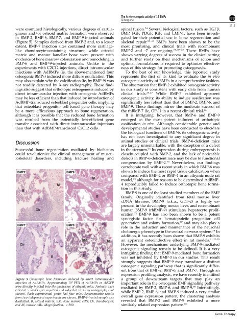
were examined histologically, various degrees <strong>of</strong> cartilaginous<br />
and/or osteoid matrix formation were observed<br />
in BMP-2, BMP-6, BMP-7, and BMP-9-injected animals<br />
(Figure 5). Samples derived from BMP-2 and, to a lesser<br />
extent, BMP-7 injection sites contained more cartilagelike<br />
chondrocyte-containing structure, while osteoid<br />
matrix and mature lamellar <strong>bone</strong> were present with<br />
evidence <strong>of</strong> <strong>bone</strong> marrow colonization and remodeling in<br />
BMP-6 and BMP-9-injected animals. Unlike in <strong>the</strong><br />
experiments with C2C12 injections, direct intramuscular<br />
injections with AdBMPs (ie, <strong>the</strong> above-mentioned four<br />
osteogenic BMPs) induced more diffuse ossification. This<br />
may also explain why <strong>the</strong> calcification (ie, by BMP-9) was<br />
not readily detected by X-ray radiography. These findings<br />
also suggest that <strong>orthotopic</strong> osteogenesis induced by<br />
direct intramuscular injection with osteogenic AdBMPs<br />
may be less efficient than that induced by introduction <strong>of</strong><br />
AdBMP-transduced osteoblast progenitor cells, implying<br />
that osteoblast progenitor cell-based gene <strong>the</strong>rapy may<br />
be a more efficacious approach to <strong>bone</strong> regeneration,<br />
although it is possible that <strong>the</strong> reduced <strong>bone</strong> formation<br />
was resulted from <strong>the</strong> potentially less-efficient gene<br />
transfer associated with direct intramuscular injections<br />
than that with AdBMP-transduced C2C12 cells.<br />
Discussion<br />
Successful <strong>bone</strong> regeneration mediated by bi<strong>of</strong>actors<br />
could revolutionize <strong>the</strong> clinical management <strong>of</strong> musculoskeletal<br />
disorders, including fracture healing and<br />
Figure 5 Orthotopic <strong>bone</strong> formation induced by direct intramuscular<br />
injection <strong>of</strong> AdBMPs. Approximately 10 9 PFU <strong>of</strong> AdBMPs or AdGFP<br />
were directly injected into <strong>the</strong> quadriceps <strong>of</strong> athymic mice. Animals were<br />
killed at 5 weeks after injection and subjected to X-ray radiography (not<br />
shown). Each experimental group had four mice. Representative results<br />
from two independent experiments are shown. BMP-6-treated sample was<br />
decalcified. B, osteoid matrix; BM, <strong>bone</strong> marrow cells; Ch, chondrocytes;<br />
and M, muscle cells. Magnification, 200.<br />
The in vivo osteogenic <strong>activity</strong> <strong>of</strong> <strong>14</strong> BMPs<br />
Q Kang et al<br />
spinal fusion. 54 Several biological factors, such as TGFb,<br />
BMP, FGF, PDGF, IGF, and LMP-1, have been investigated<br />
for <strong>the</strong>ir potential use in <strong>bone</strong> regeneration and<br />
skeletal repair. 55–69 BMPs have been shown to be <strong>the</strong><br />
most promising, and clinical trials with recombinant<br />
BMP-2 and -7 are ongoing. 54,70–73 These BMPs have<br />
shown varying degrees <strong>of</strong> success in <strong>the</strong> clinical setting<br />
and fur<strong>the</strong>r study on <strong>the</strong>ir mechanisms <strong>of</strong> action and<br />
optimal formulations is required to optimize effectiveness<br />
<strong>of</strong> this strategy for promoting osteogenesis.<br />
To <strong>the</strong> best <strong>of</strong> our knowledge, this reported study<br />
represents <strong>the</strong> first <strong>of</strong> its kind to evaluate <strong>the</strong> in vivo<br />
osteogenic <strong>activity</strong> <strong>of</strong> BMPs in a comprehensive fashion.<br />
The observation that BMP-2 exhibited osteogenic <strong>activity</strong><br />
in our study is consistent with early data from human<br />
clinical trials. 25,27 While BMP-7 exhibited apparent<br />
osteogenic <strong>activity</strong>, its ability to induce ossification was<br />
significantly less robust than that <strong>of</strong> BMP-2, BMP-6, and<br />
BMP-9. These findings mirror <strong>the</strong> moderate success <strong>of</strong><br />
<strong>the</strong> rhBMP-7 (ie, OP-1) in a recent clinical trial. 26<br />
It is intriguing, however, that BMP-6 and BMP-9<br />
emerged as <strong>the</strong> most potent inducers <strong>of</strong> <strong>orthotopic</strong><br />
ossification in vivo. Although considerable genetic and<br />
developmental studies have been conducted to elucidate<br />
<strong>the</strong> biological functions <strong>of</strong> BMP-6, its osteogenic <strong>activity</strong><br />
has not been investigated to any significant degree in<br />
animal studies or clinical trials. BMP-6-deficient mice<br />
are largely unremarkable, with <strong>the</strong> exception <strong>of</strong> a defect<br />
in <strong>the</strong> sternum. 74 Its expression during embryogenesis is<br />
closely coupled with BMP-2, and <strong>the</strong> lack <strong>of</strong> noticeable<br />
defects in BMP-6-deficient mice may be due to functional<br />
compensation by BMP-2. 74 Never<strong>the</strong>less, our findings<br />
corroborate well with a recent study in which BMP-6 was<br />
shown to induce <strong>the</strong> most rapid tissue calcification when<br />
compared with BMP-2 or BMP-4 in an athymic nude rat<br />
model, 75 although for reasons to be determined AdBMP-<br />
4 reproducibly failed to induce <strong>orthotopic</strong> <strong>bone</strong> formation<br />
in this study.<br />
BMP-9 is one <strong>of</strong> <strong>the</strong> least studied members <strong>of</strong> <strong>the</strong> BMP<br />
family. Originally identified from fetal mouse liver<br />
cDNA libraries, BMP-9 (a.k.a., GDF-2) is highly expressed<br />
in <strong>the</strong> developing mouse liver, and recombinant<br />
human BMP-9 (rhBMP-9) stimulates hepatocyte proliferation.<br />
76 BMP-9 has also been shown to be a potent<br />
synergistic factor for hematopoietic progenitor cell<br />
generation and colony formation, 77 and may also play a<br />
role in <strong>the</strong> induction and maintenance <strong>of</strong> <strong>the</strong> neuronal<br />
cholinergic phenotype in <strong>the</strong> central nervous system. 78 In<br />
addition, it has recently been shown that BMP-9 exhibits<br />
an apparent osteoinductive effect in rat models. 21,23,79<br />
However, <strong>the</strong> mechanisms underlying BMP-9-mediated<br />
osteogenic signaling remain to be defined. It is a very<br />
intriguing finding that BMP-9-mediated <strong>bone</strong> formation<br />
was not inhibited by BMP-3 in our studies. This result<br />
strongly suggests that BMP-9 may transduce a <strong>distinct</strong><br />
osteogenic signaling pathway that is significantly different<br />
from that <strong>of</strong> BMP-2, BMP-6, and BMP-7. Through an<br />
expression pr<strong>of</strong>iling analysis, we have recently identified<br />
a group <strong>of</strong> downstream targets that may play an<br />
important role in <strong>the</strong> osteogenic BMP signaling pathway<br />
mediated by BMP-2, BMP-6, and BMP-9. 80 Interestingly,<br />
while BMP-2, BMP-6, and BMP-9 induced a very similar<br />
overall gene expression pattern, <strong>the</strong> clustering analysis<br />
revealed that BMP-2 and BMP-9 exhibited a more<br />
similarly related expression pattern. 80<br />
1317<br />
Gene Therapy