Characterization of the distinct orthotopic bone-forming activity of 14 ...
Characterization of the distinct orthotopic bone-forming activity of 14 ...
Characterization of the distinct orthotopic bone-forming activity of 14 ...

Create successful ePaper yourself
Turn your PDF publications into a flip-book with our unique Google optimized e-Paper software.
Gene Therapy (2004) 11, 1312–1320<br />
& 2004 Nature Publishing Group All rights reserved 0969-7128/04 $30.00<br />
www.nature.com/gt<br />
RESEARCH ARTICLE<br />
<strong>Characterization</strong> <strong>of</strong> <strong>the</strong> <strong>distinct</strong> <strong>orthotopic</strong><br />
<strong>bone</strong>-<strong>forming</strong> <strong>activity</strong> <strong>of</strong> <strong>14</strong> BMPs using recombinant<br />
adenovirus-mediated gene delivery<br />
Q Kang 1,2,5 , MH Sun 1,5 , H Cheng 1,5 , Y Peng 1,3 , AG Montag 1,4 , AT Deyrup 1,4,6 , W Jiang 1 , HH Luu 1 ,<br />
J Luo 1 , JP Szatkowski 1 , P Vanichakarn 1 , JY Park 1 ,YLi 1 , RC Haydon 1 and T-C He 1,3<br />
1<br />
Molecular Oncology Laboratory, Department <strong>of</strong> Surgery, The University <strong>of</strong> Chicago Medical Center, Chicago, IL, USA; 2 The Children’s<br />
Hospital <strong>of</strong> Chongqing University <strong>of</strong> Medical Sciences, Chongqing, China; 3 Committee on Genetics, The University <strong>of</strong> Chicago, Chicago,<br />
IL, USA; and 4 Department <strong>of</strong> Pathology, The University <strong>of</strong> Chicago, Chicago, IL, USA<br />
Efficacious <strong>bone</strong> regeneration could revolutionize <strong>the</strong> clinical<br />
management <strong>of</strong> <strong>bone</strong> and musculoskeletal disorders. Although<br />
several <strong>bone</strong> morphogenetic proteins (BMPs) (mostly BMP-2<br />
and BMP-7) have been shown to induce <strong>bone</strong> formation, it is<br />
unclear whe<strong>the</strong>r <strong>the</strong> currently used BMPs represent <strong>the</strong> most<br />
osteogenic ones. Until recently, comprehensive analysis <strong>of</strong><br />
osteogenic <strong>activity</strong> <strong>of</strong> all BMPs has been hampered by <strong>the</strong> fact<br />
that recombinant proteins are ei<strong>the</strong>r not biologically active or<br />
not available for all BMPs. In this study, we used recombinant<br />
adenoviruses expressing <strong>the</strong> <strong>14</strong> types <strong>of</strong> BMPs (AdBMPs),<br />
and demonstrated that, in addition to currently used BMP-2<br />
and BMP-7, BMP-6 and BMP-9 effectively induced <strong>orthotopic</strong><br />
ossification when ei<strong>the</strong>r AdBMP-transduced osteoblast<br />
progenitors or <strong>the</strong> viral vectors were injected into <strong>the</strong><br />
quadriceps <strong>of</strong> athymic mice. Radiographic and histological<br />
evaluation demonstrated that BMP-6 and BMP-9 induced <strong>the</strong><br />
most robust and mature ossification at multiple time points.<br />
BMP-3, a negative regulator <strong>of</strong> <strong>bone</strong> formation, was shown to<br />
effectively inhibit <strong>orthotopic</strong> ossification induced by BMP-2,<br />
BMP-6, and BMP-7. However, BMP-3 exerted no inhibitory<br />
effect on BMP-9-induced <strong>bone</strong> formation, suggesting that<br />
BMP-9 may transduce osteogenic signaling differently. Our<br />
findings suggest that BMP-6 and BMP-9 may represent more<br />
effective osteogenic factors for <strong>bone</strong> regeneration.<br />
Gene Therapy (2004) 11, 1312–1320. doi:10.1038/<br />
sj.gt.3302298; Published online 22 July 2004<br />
Keywords: <strong>bone</strong> formation; <strong>bone</strong> morphogenetic protein; <strong>bone</strong> regeneration; osteogenesis; osteoblast differentiation;<br />
recombinant adenovirus<br />
Introduction<br />
Bone regeneration is critical to <strong>the</strong> effective management<br />
<strong>of</strong> many <strong>bone</strong> and musculoskeletal disorders, such as<br />
fracture healing, spinal fusion, and osteoporosis, which<br />
are responsible for a large portion <strong>of</strong> healthcare<br />
expenditure in <strong>the</strong> developed countries—approximately<br />
$<strong>14</strong> billion is spent annually on treating osteoporotic<br />
fractures in <strong>the</strong> US alone. 1 Bone is a highly mineralized<br />
tissue and is one <strong>of</strong> <strong>the</strong> few organs that retains <strong>the</strong><br />
potential for regeneration in adult life. Bone also undergoes<br />
continuous remodeling throughout life. 2,3 Three<br />
major types <strong>of</strong> cells are present in <strong>bone</strong> tissues: <strong>bone</strong><strong>forming</strong><br />
osteoblasts, <strong>bone</strong>-resorbing osteoclasts, and<br />
chondrocytes. Osteoblasts are derived from <strong>the</strong> mesenchymal<br />
stem cells, which also serve as precursor cells for<br />
myocytes, adipocytes, and chondrocytes. It has been<br />
known for almost half a century that demineralized <strong>bone</strong><br />
Correspondence: Dr T-C He, Molecular Oncology Laboratory, Department<br />
<strong>of</strong> Surgery, The University <strong>of</strong> Chicago Medical Center, 5841 South<br />
Maryland Avenue, MC3079, Chicago, IL 60637, USA<br />
5<br />
QK, MHS, and HC Contributed equally to this work<br />
6<br />
Current address: Department <strong>of</strong> Pathology, Emory University, Atlanta,<br />
GA, USA.<br />
Received 18 August 2003; accepted 7 February 2004; published<br />
online 22 July 2004<br />
can induce de novo <strong>bone</strong> formation. 4 The molecular<br />
identity <strong>of</strong> <strong>the</strong> <strong>bone</strong>-<strong>forming</strong> factors in demineralized<br />
<strong>bone</strong> was subsequently revealed to be <strong>bone</strong> morphogenetic<br />
proteins (BMPs). 5 BMPs belong to <strong>the</strong> TGFb<br />
superfamily, and play an important role in embryonic<br />
development and <strong>bone</strong> formation. 6,7 At least 15 types <strong>of</strong><br />
BMPs have been identified in humans. BMP signal<br />
transduction begins via interaction with <strong>the</strong> heterodimeric<br />
complex <strong>of</strong> two transmembrane serine/threonine<br />
kinase receptors, BMPR type I and BMPR type II. 8,9 The<br />
activated receptor kinases phosphorylate <strong>the</strong> transcription<br />
factors Smads 1, 5, and/or 8. The phosphorylated<br />
Smads <strong>the</strong>n form a heterodimeric complex with Smad 4<br />
in <strong>the</strong> nucleus and activate <strong>the</strong> expression <strong>of</strong> target genes<br />
in concert with o<strong>the</strong>r coactivators. 10–12<br />
Although <strong>the</strong> molecular mechanisms underlying<br />
osteoblast differentiation remain to be defined, BMPs<br />
play an important role in regulating osteoblast differentiation<br />
and subsequent <strong>bone</strong> formation. Traditionally,<br />
various <strong>bone</strong> grafts have been used to promote osteogenesis<br />
in <strong>bone</strong> and musculoskeletal disorders. The identification<br />
<strong>of</strong> BMPs has generated great interest due to <strong>the</strong>ir<br />
potential use in <strong>bone</strong> regeneration. 3 Several recombinant<br />
forms <strong>of</strong> BMPs, mostly rhBMP-2 and rhBMP-7 (a.k.a.,<br />
OP-1), have been shown to induce <strong>bone</strong> formation<br />
in vivo, 13–24 and both rhBMP-2 and rhBMP-7 have been
tested in clinical trials. 25–27 In addition to <strong>the</strong> direct<br />
application <strong>of</strong> recombinant BMP proteins, numerous<br />
reports have confirmed <strong>the</strong> ability <strong>of</strong> adenoviral or<br />
retroviral vector-mediated gene transfer <strong>of</strong> several BMPs<br />
to induce <strong>bone</strong> formation in animal models. 18,20,21,23,28–44<br />
Although a plethora <strong>of</strong> studies have demonstrated <strong>the</strong><br />
ability <strong>of</strong> several BMPs, mostly BMP-2 and BMP-7, to<br />
promote osteogenesis, it is unclear whe<strong>the</strong>r or not BMPs<br />
o<strong>the</strong>r than those currently being tested in clinical trials<br />
are more potent stimulators <strong>of</strong> new <strong>bone</strong> formation.<br />
Thus, it is important to conduct a comprehensive<br />
comparative analysis <strong>of</strong> <strong>the</strong> in vivo osteogenic <strong>activity</strong><br />
<strong>of</strong> all BMPs. This line <strong>of</strong> investigation has been<br />
hampered by <strong>the</strong> fact that recombinant proteins are<br />
ei<strong>the</strong>r not biologically active or not available for all<br />
BMPs. We have recently constructed a panel <strong>of</strong> recombinant<br />
adenoviral vectors that express <strong>the</strong> <strong>14</strong> types <strong>of</strong><br />
human BMPs (BMP-2–BMP-15). 45 Recombinant adenoviral<br />
vectors are ideal for this line <strong>of</strong> investigation for<br />
following reasons. 46–51 First, adenoviral vectors can<br />
transduce osteoblast progenitor cells with high efficiency.<br />
Second, <strong>the</strong> biologically active BMPs are continuously<br />
produced inside mammalian cells. Third,<br />
BMP-mediated <strong>bone</strong> osteoblast differentiation does not<br />
require long-term expression. Fourth, adenoviral vectors<br />
can be used in both in vitro and in vivo studies. In this<br />
study, we sought to carry out a comprehensive analysis<br />
<strong>of</strong> <strong>the</strong> <strong>distinct</strong> in vivo <strong>bone</strong>-<strong>forming</strong> <strong>activity</strong> <strong>of</strong> <strong>the</strong> <strong>14</strong><br />
types <strong>of</strong> human BMPs. Using an <strong>orthotopic</strong> ossification<br />
animal model, we demonstrate that BMP-2, BMP-6, and<br />
BMP-9 (BMP-7 to a lesser extent) are <strong>the</strong> most potent<br />
osteoinductive BMPs. Our findings strongly suggest<br />
that, in addition to BMP-2 and BMP-7 that are currently<br />
used in clinical trails, BMP-6 and BMP-9 could represent<br />
The in vivo osteogenic <strong>activity</strong> <strong>of</strong> <strong>14</strong> BMPs<br />
Q Kang et al<br />
equally, if not more effective osteogenic factors for <strong>bone</strong><br />
regeneration in a clinical setting.<br />
Results<br />
Distinct ability to induce an osteogenic marker, alkaline<br />
phosphatase (ALP), by <strong>14</strong> BMPs in <strong>the</strong> osteoblast<br />
progenitor C2C12 cells in vitro<br />
In order to comprehensively analyze <strong>the</strong> <strong>distinct</strong> osteogenic<br />
<strong>activity</strong> <strong>of</strong> BMPs, we have recently constructed a<br />
panel <strong>of</strong> recombinant adenoviral vectors that express <strong>the</strong><br />
<strong>14</strong> types <strong>of</strong> human BMPs, designated as AdBMPs. 45 As<br />
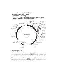
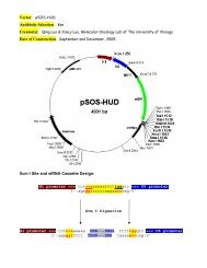
shown in Figure 1a, <strong>the</strong> level <strong>of</strong> transgene expression <strong>of</strong><br />
<strong>the</strong> AdBMPs were in general comparable (ie, o2-fold<br />
difference among different BMPs), as demonstrated by<br />
RT-PCR analysis. These PCR products should be specifically<br />
derived from <strong>the</strong> adenoviral vector-mediated<br />
expression (ra<strong>the</strong>r than from <strong>the</strong> endogenous genes), as<br />
<strong>the</strong> 3 0 -end primer was derived from <strong>the</strong> SV40 poly-A<br />
cassette. Using <strong>the</strong>se adenoviral vectors, we have<br />
recently demonstrated that BMPs displayed a <strong>distinct</strong><br />
ability to induce osteoblast differentiation <strong>of</strong> mesenchymal<br />
progenitor cells in vitro. 45 In this study, we sought to<br />
determine <strong>the</strong> relative in vivo osteogenic <strong>activity</strong> <strong>of</strong> <strong>the</strong> <strong>14</strong><br />
types <strong>of</strong> BMPs. We first tested <strong>the</strong> ability <strong>of</strong> individual<br />
AdBMPs to induce <strong>the</strong> earlier osteogenic marker alkaline<br />
phosphatase in <strong>the</strong> C2C12 line, which is myoblastic and<br />
can be trans-differentiated into osteoblast progenitors<br />
upon BMP stimulation. As shown in Figure 1b, ALP<br />
<strong>activity</strong> was remarkably induced by five <strong>of</strong> <strong>the</strong> <strong>14</strong> BMPs<br />
at four days after AdBMP infections. Among <strong>the</strong><br />
five osteogenic BMPs, BMP-2, BMP-6, and BMP-9<br />
induced <strong>the</strong> ALP <strong>activity</strong> to a much greater extent<br />
1313<br />
Figure 1 Induction <strong>of</strong> alkaline phosphatase <strong>activity</strong> by <strong>the</strong> <strong>14</strong> types <strong>of</strong> human BMPs in C2C12 cells. (a) Relative level <strong>of</strong> AdBMP-mediated transgene<br />
expression. C2C12 cells were infected with AdBMPs or AdGFP for 40 h. Total RNA was isolated and converted into cDNA products by reverse<br />
transcription, which were used for RT-PCR reactions using BMP-specific primers (5 0 -end) and a 3 0 -end primer derived from SV40 polyA. Resultant<br />
products ranged from 500 to 600 bps (indicated by arrows). ‘+’, PCR products from +RT reactions <strong>of</strong> <strong>the</strong> original cDNA syn<strong>the</strong>sis; ‘ ’, PCR products from<br />
RT reactions <strong>of</strong> <strong>the</strong> original cDNA syn<strong>the</strong>sis; ‘M’, 1-Kb Plus ladder from Invitrogen; ‘G’, AdGFP; ‘2–15’, AdBMP-2 to AdBMP-15. (b) Subconfluent<br />
C2C12 cells were infected with AdBMPs and <strong>the</strong> control AdGFP. At 4 days after infection, cells were lysed for colorimetric assays <strong>of</strong> alkaline phosphatase<br />
<strong>activity</strong> using p-nitrophenyl phosphate as a substrate. Representative results from at least three independent experiments are shown. See Materials and<br />
methods for details.<br />
Gene Therapy
13<strong>14</strong><br />
The in vivo osteogenic <strong>activity</strong> <strong>of</strong> <strong>14</strong> BMPs<br />
Q Kang et al<br />
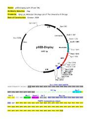
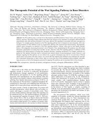
Figure 2 Orthotopic <strong>bone</strong> formation induced by AdBMP-transduced C2C12 in athymic nude mice. Exponentially growing C2C12 cells were infected with<br />
AdBMPs or <strong>the</strong> control AdGFP for 15 h. Approximately 5 10 6 <strong>of</strong> <strong>the</strong> infected cells were injected into <strong>the</strong> right quadriceps <strong>of</strong> athymic nude mice (a triangle<br />
indicated in <strong>the</strong> GFP group as an example). At 3 weeks (a) and 5 weeks (b) after injections, mice were killed and subjected to X-ray radiography. Each<br />
experimental group contained four mice, and representative radiographies from three batches <strong>of</strong> experiments were shown.<br />
(approx. 169-, 215-, and 273-fold over <strong>the</strong> GFP control,<br />
respectively), while BMP-4 and BMP-7 increased ALP<br />
<strong>activity</strong> by 44- and 73-fold, respectively. These findings<br />
are consistent with our previous studies. 45 It should be<br />
pointed out that several BMPs (eg, BMP-10, and BMP-13)<br />
also induced a 2–3-fold increase <strong>of</strong> ALP <strong>activity</strong> over <strong>the</strong><br />
basal level under <strong>the</strong> same assay conditions.<br />
Orthotopic <strong>bone</strong> formation effectively induced<br />
by several but not all BMPs in athymic mice<br />
We next sought to test <strong>the</strong> in vivo osteogenic effect <strong>of</strong> <strong>the</strong><br />
<strong>14</strong> BMPs. In order to effectively assess <strong>the</strong> osteogenic<br />
ability <strong>of</strong> <strong>the</strong> BMPs, we used an <strong>orthotopic</strong> ossification<br />
animal model, in which C2C12 cells were first transduced<br />
with AdBMPs and subsequently implanted into<br />
<strong>the</strong> quadriceps muscles <strong>of</strong> athymic nude mice by<br />
intramuscular injection. Orthotopic ossification was<br />
assessed by X-ray radiography and histological evaluation<br />
at 3 and 5 weeks after injections. As illustrated in<br />
Figure 2, ossification was readily detected on X-ray<br />
radiographies from <strong>the</strong> animals injected with AdBMP-2,<br />
6, 7, and 9-transduced C2C12 cells at 3 weeks (Figure 2a)<br />
and 5 weeks (Figure 2b). For BMP-6 and BMP-9,<br />
histologic examination at both time points (Figure 3a<br />
and b) revealed robust and highly mineralized woven<br />
<strong>bone</strong> with scattered osteoblast-rimming and occasional<br />
osteoclasts. Osteoid was also present. In addition, BMP-6<br />
showed <strong>bone</strong> marrow elements with a range <strong>of</strong> hematopoietic<br />
cells. At <strong>the</strong> 3-week time point, BMP-2 was<br />
characterized by well-calcified foci without <strong>bone</strong> formation;<br />
however, at <strong>the</strong> 5-week time point, <strong>the</strong>se foci had<br />
developed into mature <strong>bone</strong>. BMP-7, on <strong>the</strong> o<strong>the</strong>r hand,<br />
was shown to induce much weaker ossification. Interestingly,<br />
we failed to detect any signs <strong>of</strong> ossification in<br />
<strong>the</strong> animals injected with AdBMP-4-transduced C2C12<br />
cells, which is surprising because we have demonstrated<br />
that BMP-4 is capable <strong>of</strong> inducing ALP <strong>activity</strong> in vitro<br />
(Figure 1b). 45 These results were reproducible in two<br />
additional rounds <strong>of</strong> animal studies using different<br />
batches <strong>of</strong> AdBMP-4 preparations, which were consistently<br />
shown to induce ALP <strong>activity</strong> in C2C12 cells<br />
in vitro (data not shown). Fur<strong>the</strong>r, our RT-PCR analysis<br />
demonstrated that <strong>the</strong> expression level <strong>of</strong> BMP-4 was<br />
comparable with that <strong>of</strong> o<strong>the</strong>r BMPs, especially BMP-2,<br />
BMP-6, and BMP-9 under <strong>the</strong> same assay condition<br />
(Figure 1a). Currently, we are still searching for any<br />
satisfactory explanations for this discrepancy between<br />
BMP-4’s in vitro versus in vivo osteogenic <strong>activity</strong>. Never<strong>the</strong>less,<br />
all <strong>of</strong> <strong>the</strong> above findings were reproducible in<br />
three batches <strong>of</strong> independent experiments. The remaining<br />
samples had no evidence <strong>of</strong> ossification at 5 weeks.<br />
The injection site in <strong>the</strong>se sections demonstrated exuberant<br />
granulation tissue and reparative changes.<br />
Gene Therapy
The in vivo osteogenic <strong>activity</strong> <strong>of</strong> <strong>14</strong> BMPs<br />
Q Kang et al<br />
1315<br />
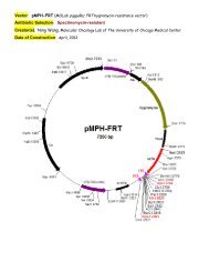
Figure 3 Histological evaluation <strong>of</strong> AdBMP-induced <strong>orthotopic</strong> ossification. (a) H & E staining <strong>of</strong> <strong>the</strong> AdBMP-transduced C2C12 injection sites at 3<br />
weeks. (b) H & E staining <strong>of</strong> <strong>the</strong> AdBMP-transduced C2C12 injection sites at 5 weeks. (c). Masson’s Trichrome staining <strong>of</strong> <strong>the</strong> AdBMP-transduced C2C12<br />
injection sites at 3 weeks. Muscle fibers and cytoplasm were stained red and collage <strong>of</strong> osteoid matrix were stained blue. B, osteoid matrix (indicated by<br />
arrows); C, injected C2C12 cells; and M, muscle cells. Magnification, 200.<br />
Histology <strong>of</strong> BMP-induced <strong>bone</strong> formation<br />
We next examined <strong>the</strong> histology <strong>of</strong> <strong>the</strong> recovered<br />
injection sites. Overall, <strong>the</strong> histology correlated well with<br />
<strong>the</strong> findings from X-ray radiography. At <strong>the</strong> 3-week time<br />
point, BMP-2, BMP-6, BMP-7, and BMP-9 demonstrated<br />
varying degrees <strong>of</strong> ossification. BMP-2 and BMP-7 were<br />
<strong>the</strong> least developed with small foci <strong>of</strong> woven <strong>bone</strong><br />
(Figure 3a). Both BMP-6 and BMP-9, however, had<br />
multiple foci <strong>of</strong> immature woven trabecular <strong>bone</strong>. In<br />
addition, BMP-9 demonstrated focal cartilaginous differentiation.<br />
The <strong>bone</strong> in BMP-6 and BMP-9-treated animals<br />
formed a shell-like rim around a proliferating mass <strong>of</strong><br />
Gene Therapy
1316<br />
The in vivo osteogenic <strong>activity</strong> <strong>of</strong> <strong>14</strong> BMPs<br />
Q Kang et al<br />
spindle-shaped C2C12 cells. The 5-week samples demonstrated<br />
increased maturation with more mature osteoid<br />
matrix and trabecular <strong>bone</strong>-like structures with accentuation<br />
<strong>of</strong> <strong>the</strong> shell-like rim, especially in BMP-6 and<br />
BMP-9-treated animals. There was less extensive ossification<br />
in <strong>the</strong> BMP-2 sections. Bone marrow elements<br />
were present in <strong>the</strong> BMP-6 sections and chondrocytes<br />
and cartilaginous matrix were increased in <strong>the</strong> BMP-9<br />
sections (Figure 3b). Interestingly, <strong>the</strong> injected C2C12<br />
cells formed a desmoid-like cell mass in nonosteogenic<br />
BMP injections and <strong>the</strong> GFP control. Even in <strong>the</strong> animals<br />
injected with BMP-2, BMP-6, BMP-7, and BMP-9-transduced<br />
C2C12 cells, such cell mass was still visible, and<br />
multiple ossification centers were observed at <strong>the</strong><br />
periphery <strong>of</strong> <strong>the</strong> cell mass. BMP-2-, BMP-6-, BMP-7-,<br />
and BMP-9-induced osteogenesis was fur<strong>the</strong>r confirmed<br />
by Masson’s Trichrome staining (Figure 3c).<br />
Antagonistic effect <strong>of</strong> BMP-3 on BMP-induced<br />
<strong>bone</strong> formation<br />
We sought to investigate how <strong>the</strong> osteogenic BMPs were<br />
affected by BMP-3, a known negative regulator <strong>of</strong> <strong>bone</strong><br />
formation, as BMP-3 knockout animals exhibited an<br />
increase in <strong>bone</strong> density. 52 When C2C12 cells transduced<br />
by AdBMP-3 and one <strong>of</strong> <strong>the</strong> four osteogenic AdBMPs<br />
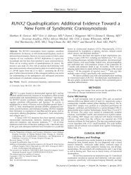
were coinjected intramuscularly for 3 weeks, BMP-2 and<br />
BMP-6-induced ossification was completely blocked by<br />
BMP-3, and most <strong>of</strong> <strong>the</strong> BMP-7-induced ossification was<br />
inhibited by BMP-3 (Figure 4a). However, BMP-3<br />
coinjection did not exert any effect on BMP-9-induced<br />
calcification (Figure 4a), strongly suggesting that BMP-9<br />
may exert its osteogenic <strong>activity</strong> via a <strong>distinct</strong> signaling<br />
mechanism. Similar results were obtained for <strong>the</strong> 5-week<br />
groups (data not shown). The histological findings were<br />
consistent with those from X-ray radiographic results<br />
(Figure 4b). The three samples without ossification<br />
demonstrated C2C12 cell proliferation with entrapped<br />
skeletal muscle while <strong>the</strong> BMP-3+BMP-9 sections had<br />
multiple foci <strong>of</strong> woven trabecular <strong>bone</strong> similar to BMP-9<br />
injection alone.<br />
Bone formation induced by direct intramuscular<br />
injection <strong>of</strong> AdBMPs<br />
Recent studies suggest that skeletal muscles may harbor<br />
pluripotent mesenchymal stem cells, including osteoblast<br />
progenitors. 34,44,53 We next tested <strong>the</strong> osteoinductive<br />
<strong>activity</strong> <strong>of</strong> <strong>the</strong> <strong>14</strong> BMPs via direct intramuscular injection<br />
<strong>of</strong> AdBMPs. At <strong>the</strong> 3 and 5-week time points, we did not<br />
observe apparent ossification on X-ray radiography (data<br />
not shown). However, when <strong>the</strong> 5-week injection sites<br />
Figure 4 BMP-3-mediated inhibition <strong>of</strong> <strong>bone</strong> formation induced by BMP-2, BMP-6, and BMP-7, but not by BMP-9. (a) Osteogenic AdBMPs (ie, BMP-2,<br />
-6, -7, and -9)-transduced C2C12 cells were ei<strong>the</strong>r injected alone (top row) or coinjected with AdBMP-3 (bottom row) into <strong>the</strong> right quadriceps <strong>of</strong> athymic<br />
mice. Animals were killed at 3 weeks and subjected to X-ray radiography. Ossification sites were indicated by arrows. Each experimental group contained<br />
four mice, and representative radiographies from three batches <strong>of</strong> experiments are shown. (b) Histological evaluation <strong>of</strong> <strong>the</strong> BMP-3 coinjection sites. B,<br />
osteoid matrix; C, injected C2C12 cells; and M, muscle cells. Magnification, 150.<br />
Gene Therapy
were examined histologically, various degrees <strong>of</strong> cartilaginous<br />
and/or osteoid matrix formation were observed<br />
in BMP-2, BMP-6, BMP-7, and BMP-9-injected animals<br />
(Figure 5). Samples derived from BMP-2 and, to a lesser<br />
extent, BMP-7 injection sites contained more cartilagelike<br />
chondrocyte-containing structure, while osteoid<br />
matrix and mature lamellar <strong>bone</strong> were present with<br />
evidence <strong>of</strong> <strong>bone</strong> marrow colonization and remodeling in<br />
BMP-6 and BMP-9-injected animals. Unlike in <strong>the</strong><br />
experiments with C2C12 injections, direct intramuscular<br />
injections with AdBMPs (ie, <strong>the</strong> above-mentioned four<br />
osteogenic BMPs) induced more diffuse ossification. This<br />
may also explain why <strong>the</strong> calcification (ie, by BMP-9) was<br />
not readily detected by X-ray radiography. These findings<br />
also suggest that <strong>orthotopic</strong> osteogenesis induced by<br />
direct intramuscular injection with osteogenic AdBMPs<br />
may be less efficient than that induced by introduction <strong>of</strong><br />
AdBMP-transduced osteoblast progenitor cells, implying<br />
that osteoblast progenitor cell-based gene <strong>the</strong>rapy may<br />
be a more efficacious approach to <strong>bone</strong> regeneration,<br />
although it is possible that <strong>the</strong> reduced <strong>bone</strong> formation<br />
was resulted from <strong>the</strong> potentially less-efficient gene<br />
transfer associated with direct intramuscular injections<br />
than that with AdBMP-transduced C2C12 cells.<br />
Discussion<br />
Successful <strong>bone</strong> regeneration mediated by bi<strong>of</strong>actors<br />
could revolutionize <strong>the</strong> clinical management <strong>of</strong> musculoskeletal<br />
disorders, including fracture healing and<br />
Figure 5 Orthotopic <strong>bone</strong> formation induced by direct intramuscular<br />
injection <strong>of</strong> AdBMPs. Approximately 10 9 PFU <strong>of</strong> AdBMPs or AdGFP<br />
were directly injected into <strong>the</strong> quadriceps <strong>of</strong> athymic mice. Animals were<br />
killed at 5 weeks after injection and subjected to X-ray radiography (not<br />
shown). Each experimental group had four mice. Representative results<br />
from two independent experiments are shown. BMP-6-treated sample was<br />
decalcified. B, osteoid matrix; BM, <strong>bone</strong> marrow cells; Ch, chondrocytes;<br />
and M, muscle cells. Magnification, 200.<br />
The in vivo osteogenic <strong>activity</strong> <strong>of</strong> <strong>14</strong> BMPs<br />
Q Kang et al<br />
spinal fusion. 54 Several biological factors, such as TGFb,<br />
BMP, FGF, PDGF, IGF, and LMP-1, have been investigated<br />
for <strong>the</strong>ir potential use in <strong>bone</strong> regeneration and<br />
skeletal repair. 55–69 BMPs have been shown to be <strong>the</strong><br />
most promising, and clinical trials with recombinant<br />
BMP-2 and -7 are ongoing. 54,70–73 These BMPs have<br />
shown varying degrees <strong>of</strong> success in <strong>the</strong> clinical setting<br />
and fur<strong>the</strong>r study on <strong>the</strong>ir mechanisms <strong>of</strong> action and<br />
optimal formulations is required to optimize effectiveness<br />
<strong>of</strong> this strategy for promoting osteogenesis.<br />
To <strong>the</strong> best <strong>of</strong> our knowledge, this reported study<br />
represents <strong>the</strong> first <strong>of</strong> its kind to evaluate <strong>the</strong> in vivo<br />
osteogenic <strong>activity</strong> <strong>of</strong> BMPs in a comprehensive fashion.<br />
The observation that BMP-2 exhibited osteogenic <strong>activity</strong><br />
in our study is consistent with early data from human<br />
clinical trials. 25,27 While BMP-7 exhibited apparent<br />
osteogenic <strong>activity</strong>, its ability to induce ossification was<br />
significantly less robust than that <strong>of</strong> BMP-2, BMP-6, and<br />
BMP-9. These findings mirror <strong>the</strong> moderate success <strong>of</strong><br />
<strong>the</strong> rhBMP-7 (ie, OP-1) in a recent clinical trial. 26<br />
It is intriguing, however, that BMP-6 and BMP-9<br />
emerged as <strong>the</strong> most potent inducers <strong>of</strong> <strong>orthotopic</strong><br />
ossification in vivo. Although considerable genetic and<br />
developmental studies have been conducted to elucidate<br />
<strong>the</strong> biological functions <strong>of</strong> BMP-6, its osteogenic <strong>activity</strong><br />
has not been investigated to any significant degree in<br />
animal studies or clinical trials. BMP-6-deficient mice<br />
are largely unremarkable, with <strong>the</strong> exception <strong>of</strong> a defect<br />
in <strong>the</strong> sternum. 74 Its expression during embryogenesis is<br />
closely coupled with BMP-2, and <strong>the</strong> lack <strong>of</strong> noticeable<br />
defects in BMP-6-deficient mice may be due to functional<br />
compensation by BMP-2. 74 Never<strong>the</strong>less, our findings<br />
corroborate well with a recent study in which BMP-6 was<br />
shown to induce <strong>the</strong> most rapid tissue calcification when<br />
compared with BMP-2 or BMP-4 in an athymic nude rat<br />
model, 75 although for reasons to be determined AdBMP-<br />
4 reproducibly failed to induce <strong>orthotopic</strong> <strong>bone</strong> formation<br />
in this study.<br />
BMP-9 is one <strong>of</strong> <strong>the</strong> least studied members <strong>of</strong> <strong>the</strong> BMP<br />
family. Originally identified from fetal mouse liver<br />
cDNA libraries, BMP-9 (a.k.a., GDF-2) is highly expressed<br />
in <strong>the</strong> developing mouse liver, and recombinant<br />
human BMP-9 (rhBMP-9) stimulates hepatocyte proliferation.<br />
76 BMP-9 has also been shown to be a potent<br />
synergistic factor for hematopoietic progenitor cell<br />
generation and colony formation, 77 and may also play a<br />
role in <strong>the</strong> induction and maintenance <strong>of</strong> <strong>the</strong> neuronal<br />
cholinergic phenotype in <strong>the</strong> central nervous system. 78 In<br />
addition, it has recently been shown that BMP-9 exhibits<br />
an apparent osteoinductive effect in rat models. 21,23,79<br />
However, <strong>the</strong> mechanisms underlying BMP-9-mediated<br />
osteogenic signaling remain to be defined. It is a very<br />
intriguing finding that BMP-9-mediated <strong>bone</strong> formation<br />
was not inhibited by BMP-3 in our studies. This result<br />
strongly suggests that BMP-9 may transduce a <strong>distinct</strong><br />
osteogenic signaling pathway that is significantly different<br />
from that <strong>of</strong> BMP-2, BMP-6, and BMP-7. Through an<br />
expression pr<strong>of</strong>iling analysis, we have recently identified<br />
a group <strong>of</strong> downstream targets that may play an<br />
important role in <strong>the</strong> osteogenic BMP signaling pathway<br />
mediated by BMP-2, BMP-6, and BMP-9. 80 Interestingly,<br />
while BMP-2, BMP-6, and BMP-9 induced a very similar<br />
overall gene expression pattern, <strong>the</strong> clustering analysis<br />
revealed that BMP-2 and BMP-9 exhibited a more<br />
similarly related expression pattern. 80<br />
1317<br />
Gene Therapy
1318<br />
In conclusion, we have demonstrated <strong>the</strong> relative<br />
osteogenic ability <strong>of</strong> <strong>14</strong> BMPs and identified BMP-6 and<br />
BMP-9 (in addition to <strong>the</strong> currently used BMP-2 and<br />
BMP-7) as <strong>the</strong> most potent BMPs to induce <strong>orthotopic</strong><br />
<strong>bone</strong> formation in vivo. Our results also suggest that <strong>the</strong><br />
stem/progenitor cell-based ex vivo gene <strong>the</strong>rapy may<br />
represent a more effective approach to <strong>bone</strong> regeneration.<br />
Future studies will focus on elucidating <strong>the</strong> major<br />
signaling differences among BMPs so that maximal<br />
synergy in <strong>bone</strong> formation can be achieved by combining<br />
BMPs that act through overlapping or converging<br />
signaling pathways. This line <strong>of</strong> investigation would<br />
help to elucidate <strong>the</strong> molecular mechanisms underlying<br />
<strong>bone</strong> formation and lead to <strong>the</strong> development <strong>of</strong> more<br />
efficacious approaches towards <strong>bone</strong> regeneration.<br />
Materials and methods<br />
The in vivo osteogenic <strong>activity</strong> <strong>of</strong> <strong>14</strong> BMPs<br />
Q Kang et al<br />
Cell culture and chemicals<br />
HEK 293 and C2C12 cell lines were obtained from <strong>the</strong><br />
ATCC (Manassas, VA, USA), and were maintained in<br />
complete DMEM supplemented with 10% fetal calf<br />
serum (FCS, Mediatech, Herndon, VA, USA), 100 U <strong>of</strong><br />
penicillin, and 100 mg <strong>of</strong> streptomycin at 371C in5%CO 2 .<br />
Unless indicated o<strong>the</strong>rwise, all chemicals were purchased<br />
from Sigma-Aldrich (St Louis, MO, USA).<br />
Recombinant adenoviral vectors expressing BMPs<br />
The cDNA clones for human BMP-2, -3 (a.k.a., osteogenin),<br />
-4, -5, -6, -8 (a.k.a., OP-2), -9 (a.k.a., GDF-2), -10,<br />
-12 (a.k.a., GDF-7 or CDMP-3), and -13 (a.k.a., GDF6 or<br />
CDMP2) were kindly provided by <strong>the</strong> Genetics Institute<br />
(Cambridge, MA, USA). The coding sequences for BMP-<br />
7 (a.k.a., OP-1), -11 (a.k.a., GDF-11), -<strong>14</strong> (a.k.a., GDF-5 or<br />
CDMP-1), and -15 (a.k.a., GDF-9) were PCR amplified<br />
from a human osteosarcoma cDNA library. The coding<br />
regions <strong>of</strong> <strong>the</strong> above BMPs were subcloned into pAd-<br />
Track-CMV, resulting in pAdTrack-BMPs; recombinant<br />
adenoviruses expressing BMPs (ie, AdBMPs) were<br />
subsequently generated as previously described. 45,81 For<br />
a control, we used an analogous adenovirus expressing<br />
only GFP (ie, AdGFP), as previously described. 81 Details<br />
on vector constructions are available upon request.<br />
RNA purification and reverse transcriptase-PCR<br />
analysis<br />
The RT-PCR analysis was carried out as previously<br />
described. 80 Specifically, C2C12 cells were seeded in<br />
25 cm 2 cell culture flasks, and infected with an optimal<br />
and compatible titer <strong>of</strong> AdBMPs or AdGFP. At 40 h after<br />
infection, total RNA was isolated using RNAgent Total<br />
RNA Isolation kit (Promega, Madison, WI, USA)<br />
according to <strong>the</strong> manufacturer’s instructions. Total<br />
RNA (10 mg) was used to generate cDNA templates for<br />
reverse transcriptase-PCR. The first-strand cDNA syn<strong>the</strong>sis<br />
was performed using a hexamer (Promega,<br />
Madison, WI, USA) and Superscript II reverse transcriptase<br />
(Invitrogen). The first-strand cDNA products were<br />
fur<strong>the</strong>r diluted 50-fold and used as PCR templates.<br />
Expression level was determined by touchdown PCR<br />
analysis using respective pairs <strong>of</strong> oligonucleotides to<br />
amplify <strong>the</strong> 3 0 -end <strong>of</strong> each BMP gene and <strong>the</strong> 5 0 -end <strong>of</strong><br />
SV40 poly A region. Touchdown PCR was performed by<br />
using <strong>the</strong> following program: 941C 2 min for one cycle,<br />
12 cycles at 921C 20 s, 681C 30 s, and 701C 45 s with<br />
a decrease <strong>of</strong> one degree per cycle, and 35 cycles at<br />
921C 20 s, 551C 30 s, and 701C 45 s. The amplified<br />
products (ranging from 500–600 bps) were resolved on<br />
1% agarose gels, and visualized under UV light after<br />
ethidium bromide staining.<br />
Determination <strong>of</strong> alkaline phosphatase <strong>activity</strong><br />
Exponentially growing C2C12 cells were seeded in 48-<br />
well cell culture plates, and infected with AdBMPs or<br />
AdGFP (multiplicities <strong>of</strong> infection, or MOIs ¼ 50–200).<br />
The induction <strong>of</strong> alkaline phosphatase <strong>activity</strong> was<br />
assessed at 4 days after infection. Alkaline phosphatase<br />
<strong>activity</strong> was determined by using p-nitrophenyl phosphate<br />
(Sigma-Aldrich) as a substrate. Absorbance at<br />
405 nm was recorded at 1, 2, and 3 min after <strong>the</strong> cell<br />
lysate was mixed with <strong>the</strong> substrate. Each assay<br />
condition was carried out in triplicate and normalized<br />
with <strong>the</strong> concentrations <strong>of</strong> total cellular proteins. Enzyme<br />
<strong>activity</strong> was expressed as nanomoles <strong>of</strong> p-nitrophenol<br />
produced per minute per mg <strong>of</strong> total cellular proteins.<br />
Orthotopic <strong>bone</strong> formation in athymic nude mice<br />
The use <strong>of</strong> animals was approved by <strong>the</strong> Institutional<br />
Animal Care and Use Committee. Young athymic nude<br />
mice (male, 5–6 months, Frederick Cancer Research<br />
Center) were used in this study. Each experimental<br />
group had four animals. For <strong>the</strong> injection with adenovirus-transduced<br />
C2C12 cells, subconfluent C2C12 cells<br />
were infected with AdBMPs or AdGFP at preoptimized<br />
titers (MOIs B 50–100). At 15 h after infection, cells were<br />
collected and resuspended in PBS at an approximate<br />
density <strong>of</strong> 1 10 8 cells/ml. In total, 50 ml <strong>of</strong> <strong>the</strong> cell<br />
suspension (approx. 5 10 6 cells) was used for <strong>the</strong><br />
intramuscular injection <strong>of</strong> right quadriceps. For cell<br />
mixing experiments, approximately 2.5 10 6 cells <strong>of</strong><br />
AdBMP-3- and AdBMP-infected cells were combined<br />
prior to intramuscular injections. For direct intramuscular<br />
injections, approximately 10 9 PFU <strong>of</strong> AdBMPs or<br />
AdGFP were briefly dialyzed against PBS to remove<br />
CsCl, and suspended in a final volume <strong>of</strong> 50 ml for direct<br />
injections into <strong>the</strong> right quadriceps. Injected animals<br />
resumed activities immediately without any restraints on<br />
food and drink. At 3 and 5 weeks after injections,<br />
animals were killed and subjected to X-ray radiography.<br />
The injected sites were harvested for histological<br />
evaluation. Representative results from three independent<br />
batches <strong>of</strong> experiments are shown.<br />
H & E staining and Masson’s Trichrome staining<br />
After X-ray radiography, <strong>the</strong> injected thighs were<br />
recovered, fixed in 10% formalin overnight, and embedded<br />
in paraffin. Serial sections at 12 mm <strong>of</strong> <strong>the</strong><br />
embedded specimens were carried out, and mounted<br />
onto treated slides. The sections were stained with<br />
hematoxylin and eosin (H & E) and Masson’s Trichrome.<br />
Some samples were subjected to calcification prior to H<br />
& E staining.<br />
Note added in pro<strong>of</strong><br />
While this report was under review, Li LZ et al<br />
coincidently analyzed <strong>the</strong> osteogenic <strong>activity</strong> <strong>of</strong> BMP-2,<br />
BMP-4, BMP-6, BMP-7, and BMP-9 (Gene Therapy 2003;<br />
Gene Therapy
10:1735–1743). Their findings also suggest that BMP-9 is<br />
one <strong>of</strong> <strong>the</strong> most osteogenic BMPs in rat models <strong>of</strong> <strong>bone</strong><br />
formation.<br />
Acknowledgements<br />
We thank Dr Mark Bolander <strong>of</strong> Mayo Clinic for valuable<br />
discussion and critical review <strong>of</strong> <strong>the</strong> manuscript. We<br />
thank <strong>the</strong> Genetics Institute <strong>of</strong> Cambridge, MA, for<br />
providing human BMP cDNAs. We apologize to <strong>the</strong><br />
researchers whose original work was not cited due to<br />
space constraints. The work was supported in part by<br />
research grants from <strong>the</strong> Aircast Foundation, <strong>the</strong> Brinson<br />
Foundation, North American Spine Society, and <strong>the</strong><br />
Orthopaedic Research and Education Foundation. Q<br />
Kang was a recipient <strong>of</strong> an International Postdoctoral<br />
Fellowship from <strong>the</strong> National Institutes <strong>of</strong> Health (F05<br />
AT0020<strong>14</strong>-01).<br />
References<br />
1 DeWitt N. Bone and cartilage. Nature 2003; 423: 315.<br />
2 Olsen BR, Reginato AM, Wang W. Bone development. Annu Rev<br />
Cell Dev Biol 2000; 16: 191–220.<br />
3 Reddi AH. Role <strong>of</strong> morphogenetic proteins in skeletal tissue<br />
engineering and regeneration. Nat Biotechnol 1998; 16: 247–252.<br />
4 Urist MR. Bone: formation by autoinduction. Science 1965; 150:<br />
893–899.<br />
5 Wozney JM et al. Novel regulators <strong>of</strong> <strong>bone</strong> formation: molecular<br />
clones and activities. Science 1988; 242: 1528–1534.<br />
6 Hogan BL. Bone morphogenetic proteins: multifunctional<br />
regulators <strong>of</strong> vertebrate development. Genes Dev 1996; 10:<br />
1580–1594.<br />
7 Zou H et al. BMP signaling and vertebrate limb development.<br />
Cold Spring Harb Symp Quant Biol 1997; 62: 269–272.<br />
8 Massague J, Weis-Garcia F. Serine/threonine kinase receptors:<br />
mediators <strong>of</strong> trans<strong>forming</strong> growth factor beta family signals.<br />
Cancer Surv 1996; 27: 41–64.<br />
9 Yamashita H, Ten Dijke P, Heldin CH, Miyazono K. Bone<br />
morphogenetic protein receptors. Bone 1996; 19: 569–574.<br />
10 Heldin CH, Miyazono K, ten Dijke P. TGF-beta signalling from<br />
cell membrane to nucleus through SMAD proteins. Nature 1997;<br />
390: 465–471.<br />
11 Massague J. TGF-beta signal transduction. Annu Rev Biochem<br />
1998; 67: 753–791.<br />
12 Wrana JL. Regulation <strong>of</strong> Smad <strong>activity</strong>. Cell 2000; 100: 189–192.<br />
13 Heckman JD, Boyan BD, Aufdemorte TB, Abbott JT. The use <strong>of</strong><br />
<strong>bone</strong> morphogenetic protein in <strong>the</strong> treatment <strong>of</strong> non-union in a<br />
canine model. J Bone Joint Surg Am 1991; 73: 750–764.<br />
<strong>14</strong> Gerhart TN et al. Healing segmental femoral defects in sheep<br />
using recombinant human <strong>bone</strong> morphogenetic protein. Clin<br />
Orthop 1993; 349: 317–326.<br />
15 Lee SC et al. Healing <strong>of</strong> large segmental defects in rat femurs is<br />
aided by RhBMP-2 in PLGA matrix. J Biomed Mater Res 1994; 28:<br />
1<strong>14</strong>9–1156.<br />
16 Bostrom MP, Camacho NP. Potential role <strong>of</strong> <strong>bone</strong> morphogenetic<br />
proteins in fracture healing. Clin Orthop 1998; 355 (Suppl):<br />
S274–S282.<br />
17 Cheng SL et al. In vitro and in vivo induction <strong>of</strong> <strong>bone</strong> formation<br />
using a recombinant adenoviral vector carrying <strong>the</strong> human<br />
BMP-2 gene. Calcif Tissue Int 2001; 68: 87–94.<br />
18 Krebsbach PH, Gu K, Franceschi RT, Ru<strong>the</strong>rford RB. Gene<br />
<strong>the</strong>rapy-directed osteogenesis: BMP-7-transduced human<br />
fibroblasts form <strong>bone</strong> in vivo (in process citation). Hum Gene<br />
Ther 2000; 11: 1201–1210.<br />
The in vivo osteogenic <strong>activity</strong> <strong>of</strong> <strong>14</strong> BMPs<br />
Q Kang et al<br />
19 Partridge K et al. Adenoviral BMP-2 gene transfer in<br />
mesenchymal stem cells: in vitro and in vivo <strong>bone</strong> formation on<br />
biodegradable polymer scaffolds. Biochem Biophys Res Commun<br />
2002; 292: <strong>14</strong>4–152.<br />
20 Riew KD et al. Induction <strong>of</strong> <strong>bone</strong> formation using a recombinant<br />
adenoviral vector carrying <strong>the</strong> human BMP-2 gene in a rabbit<br />
spinal fusion model. Calcif Tissue Int 1998; 63: 357–360.<br />
21 Varady P et al. Morphologic analysis <strong>of</strong> BMP-9 gene <strong>the</strong>rapyinduced<br />
osteogenesis. Hum Gene Ther 2001; 12: 697–710.<br />
22 Varady P et al. CT and radionuclide study <strong>of</strong> BMP-2 gene<br />
<strong>the</strong>rapy-induced <strong>bone</strong> formation. Acad Radiol 2002; 9: 632–637.<br />
23 Helm GA et al. Use <strong>of</strong> <strong>bone</strong> morphogenetic protein-9 gene<br />
<strong>the</strong>rapy to induce spinal arthrodesis in <strong>the</strong> rodent. J Neurosurg<br />
2000; 92: 191–196.<br />
24 Sandhu HS, Khan SN, Suh DY, Boden SD. Demineralized<br />
<strong>bone</strong> matrix, <strong>bone</strong> morphogenetic proteins, and animal models<br />
<strong>of</strong> spine fusion: an overview. Eur Spine J 2001; 10 (Suppl. 2):<br />
S122–S131.<br />
25 Boden SD, Zdeblick TA, Sandhu HS, Heim SE. The use <strong>of</strong><br />
rhBMP-2 in interbody fusion cages. Definitive evidence <strong>of</strong><br />
osteoinduction in humans: a preliminary report. Spine 2000; 25:<br />
376–381.<br />
26 Friedlaender GE. OP-1 clinical studies. J Bone Joint Surg Am 2001;<br />
83-A: S160–S161.<br />
27 Valentin-Opran A et al. Clinical evaluation <strong>of</strong> recombinant<br />
human <strong>bone</strong> morphogenetic protein-2. Clin Orthop 2002; 395:<br />
110–120.<br />
28 Whang K et al. Ectopic <strong>bone</strong> formation via rhBMP-2 delivery<br />
from porous bioabsorbable polymer scaffolds. J Biomed Mater Res<br />
1998; 42: 491–499.<br />
29 Alden TD et al. Percutaneous spinal fusion using <strong>bone</strong><br />
morphogenetic protein-2 gene <strong>the</strong>rapy. J Neurosurg 1999; 90:<br />
109–1<strong>14</strong>.<br />
30 Oyama M et al. Retrovirally transduced <strong>bone</strong> marrow stromal<br />
cells isolated from a mouse model <strong>of</strong> human osteogenesis<br />
imperfecta (oim) persist in <strong>bone</strong> and retain <strong>the</strong> ability to form<br />
cartilage and <strong>bone</strong> after extended passaging. Gene Therapy 1999;<br />
6: 321–329.<br />
31 Alden TD et al. In vivo endochondral <strong>bone</strong> formation using a<br />
<strong>bone</strong> morphogenetic protein 2 adenoviral vector. Hum Gene Ther<br />
1999; 10: 2245–2253.<br />
32 Baltzer AW et al. Genetic enhancement <strong>of</strong> fracture repair: healing<br />
<strong>of</strong> an experimental segmental defect by adenoviral transfer <strong>of</strong><br />
<strong>the</strong> BMP-2 gene. Gene Therapy 2000; 7: 734–739.<br />
33 Baltzer AW et al. Potential role <strong>of</strong> direct adenoviral gene transfer<br />
in enhancing fracture repair. Clin Orthop 2000; 379 (Suppl):<br />
S120–S125.<br />
34 Bosch P et al. The efficiency <strong>of</strong> muscle-derived cell-mediated<br />
<strong>bone</strong> formation. Cell Transplant 2000; 9: 463–470.<br />
35 Breitbart AS et al. Gene-enhanced tissue engineering:<br />
applications for <strong>bone</strong> healing using cultured periosteal cells<br />
transduced retrovirally with <strong>the</strong> BMP-7 gene. Ann Plast Surg<br />
1999; 42: 488–495.<br />
36 Franceschi RT, Wang D, Krebsbach PH, Ru<strong>the</strong>rford RB. Gene<br />
<strong>the</strong>rapy for <strong>bone</strong> formation: in vitro and in vivo osteogenic<br />
<strong>activity</strong> <strong>of</strong> an adenovirus expressing BMP7. J Cell Biochem 2000;<br />
78: 476–486.<br />
37 Gazit D et al. Engineered pluripotent mesenchymal cells<br />
integrate and differentiate in regenerating <strong>bone</strong>: a novel cellmediated<br />
gene <strong>the</strong>rapy. J Gene Med 1999; 1: 121–133.<br />
38 Lieberman JR et al. Regional gene <strong>the</strong>rapy with a BMP-2-producing<br />
murine stromal cell line induces heterotopic and <strong>orthotopic</strong> <strong>bone</strong><br />
formation in rodents. J Orthop Res 1998; 16: 330–339.<br />
39 Lou J, Xu F, Merkel K, Manske P. Gene <strong>the</strong>rapy: adenovirusmediated<br />
human <strong>bone</strong> morphogenetic protein-2 gene transfer<br />
induces mesenchymal progenitor cell proliferation and<br />
differentiation in vitro and <strong>bone</strong> formation in vivo. J Orthop Res<br />
1999; 17: 43–50.<br />
1319<br />
Gene Therapy
1320<br />
The in vivo osteogenic <strong>activity</strong> <strong>of</strong> <strong>14</strong> BMPs<br />
Q Kang et al<br />
40 Mason JM et al. Expression <strong>of</strong> human <strong>bone</strong> morphogenic protein<br />
7 in primary rabbit periosteal cells: potential utility in<br />
gene <strong>the</strong>rapy for osteochondral repair. Gene Therapy 1998; 5:<br />
1098–1104.<br />
41 Musgrave DS et al. Adenovirus-mediated direct gene <strong>the</strong>rapy<br />
with <strong>bone</strong> morphogenetic protein-2 produces <strong>bone</strong>. Bone 1999;<br />
24: 541–547.<br />
42 Okubo Y et al. Osteoinduction by <strong>bone</strong> morphogenetic protein-2<br />
via adenoviral vector under transient immunosuppression.<br />
Biochem Biophys Res Commun 2000; 267: 382–387.<br />
43 Ripamonti U et al. Bone induction by BMPs/OPs and related<br />
family members in primates. J Bone Joint Surg Am 2001; 83-A:<br />
S116–S127.<br />
44 Lee JY et al. Effect <strong>of</strong> <strong>bone</strong> morphogenetic protein-2-expressing<br />
muscle-derived cells on healing <strong>of</strong> critical-sized <strong>bone</strong> defects in<br />
mice. J Bone Joint Surg Am 2001; 83-A: 1032–1039.<br />
45 Cheng H et al. Osteogenic <strong>activity</strong> <strong>of</strong> <strong>the</strong> <strong>14</strong> types <strong>of</strong> human <strong>bone</strong><br />
morphogenetic proteins (BMPs). J Bone Joint Surgery Am 2003;<br />
85-A: 1544–1552.<br />
46 Robbins PD, Tahara H, Ghivizzani SC. Viral vectors for gene<br />
<strong>the</strong>rapy. Trends Biotechnol 1998; 16: 35–40.<br />
47 Kay MA, Glorioso JC, Naldini L. Viral vectors for gene <strong>the</strong>rapy:<br />
<strong>the</strong> art <strong>of</strong> turning infectious agents into vehicles <strong>of</strong> <strong>the</strong>rapeutics.<br />
Nat Med 2001; 7: 33–40.<br />
48 Alemany R, Balague C, Curiel DT. Replicative adenoviruses for<br />
cancer <strong>the</strong>rapy. Nat Biotechnol 2000; 18: 723–727.<br />
49 Breyer B et al. Adenoviral vector-mediated gene transfer for<br />
human gene <strong>the</strong>rapy. Curr Gene Ther 2001; 1: <strong>14</strong>9–162.<br />
50 Thomas CE, Ehrhardt A, Kay MA. Progress and problems with<br />
<strong>the</strong> use <strong>of</strong> viral vectors for gene <strong>the</strong>rapy. Nat Rev Genet 2003; 4:<br />
346–358.<br />
51 Sun MH et al. Bone morphogenetic proteins and <strong>bone</strong><br />
regeneration: from biology to clinical applications. Adv<br />
Osteoporotic Fract Manag 2003; 2: 70–78.<br />
52 Bahamonde ME, Lyons KM. BMP3: to be or not to be a BMP.<br />
J Bone Joint Surg Am 2001; 83-A: S56–S62.<br />
53 Lee JY et al. Enhancement <strong>of</strong> <strong>bone</strong> healing based on ex vivo gene<br />
<strong>the</strong>rapy using human muscle-derived cells expressing <strong>bone</strong><br />
morphogenetic protein 2. Hum Gene Ther 2002; 13: 1201–1211.<br />
54 Lieberman JR, Daluiski A, Einhorn TA. The role <strong>of</strong> growth<br />
factors in <strong>the</strong> repair <strong>of</strong> <strong>bone</strong>. Biology and clinical applications.<br />
J Bone Joint Surg Am 2002; 84-A: 1032–1044.<br />
55 Noda M, Camilliere JJ. In vivo stimulation <strong>of</strong> <strong>bone</strong> formation<br />
by trans<strong>forming</strong> growth factor-beta. Endocrinology 1989; 124:<br />
2991–2994.<br />
56 Joyce ME, Terek RM, Jingushi S, Bolander ME. Role <strong>of</strong><br />
trans<strong>forming</strong> growth factor-beta in fracture repair. Ann N Y<br />
Acad Sci 1990; 593: 107–123.<br />
57 Lind M et al. Trans<strong>forming</strong> growth factor-beta enhances fracture<br />
healing in rabbit tibiae. Acta Orthop Scand 1993; 64: 553–556.<br />
58 Nielsen HM, Andreassen TT, Ledet T, Oxlund H. Local injection<br />
<strong>of</strong> TGF-beta increases <strong>the</strong> strength <strong>of</strong> tibial fractures in <strong>the</strong> rat.<br />
Acta Orthop Scand 1994; 65: 37–41.<br />
59 Critchlow MA, Bland YS, Ashhurst DE. The effect <strong>of</strong> exogenous<br />
trans<strong>forming</strong> growth factor-beta 2 on healing fractures in <strong>the</strong><br />
rabbit. Bone 1995; 16: 521–527.<br />
60 Sumner DR et al. Enhancement <strong>of</strong> <strong>bone</strong> ingrowth by<br />
trans<strong>forming</strong> growth factor-beta. J Bone Joint Surg Am 1995; 77:<br />
1135–1<strong>14</strong>7.<br />
61 Canalis E, Centrella M, McCarthy T. Effects <strong>of</strong> basic fibroblast<br />
growth factor on <strong>bone</strong> formation in vitro. J Clin Invest 1988; 81:<br />
1572–1577.<br />
62 Kato T et al. Single local injection <strong>of</strong> recombinant fibroblast<br />
growth factor-2 stimulates healing <strong>of</strong> segmental <strong>bone</strong> defects in<br />
rabbits. J Orthop Res 1998; 16: 654–659.<br />
63 Nakamura T et al. Recombinant human basic fibroblast growth<br />
factor accelerates fracture healing by enhancing callus<br />
remodeling in experimental dog tibial fracture. J Bone Miner<br />
Res 1998; 13: 942–949.<br />
64 Radomsky ML, Thompson AY, Spiro RC, Poser JW. Potential role<br />
<strong>of</strong> fibroblast growth factor in enhancement <strong>of</strong> fracture healing.<br />
Clin Orthop 1998; 355 (Suppl): S283–S293.<br />
65 Radomsky ML et al. Novel formulation <strong>of</strong> fibroblast growth<br />
factor-2 in a hyaluronan gel accelerates fracture healing in<br />
nonhuman primates. J Orthop Res 1999; 17: 607–6<strong>14</strong>.<br />
66 Nash TJ et al. Effect <strong>of</strong> platelet-derived growth factor on tibial<br />
osteotomies in rabbits. Bone 1994; 15: 203–208.<br />
67 Thaller SR, Dart A, Tesluk H. The effects <strong>of</strong> insulin-like growth<br />
factor-1 on critical-size calvarial defects in Sprague–Dawley rats.<br />
Ann Plast Surg 1993; 31: 429–433.<br />
68 Trippel SB. Potential role <strong>of</strong> insulinlike growth factors in fracture<br />
healing. Clin Orthop 1998; 355 (Suppl): S301–S313.<br />
69 Boden SD et al. Lumbar spine fusion by local gene <strong>the</strong>rapy with<br />
a cDNA encoding a novel osteoinductive protein (LMP-1). Spine<br />
1998; 23: 2486–2492.<br />
70 Linkhart TA, Mohan S, Baylink DJ. Growth factors for<br />
<strong>bone</strong> growth and repair: IGF, TGF beta and BMP. Bone 1996;<br />
19: 1S–12S.<br />
71 Azari K et al. Therapeutic potential <strong>of</strong> <strong>bone</strong> morphogenetic<br />
proteins. Expert Opin Investig Drugs 2001; 10: 1677–1686.<br />
72 Alden TD et al. Bone morphogenetic protein gene <strong>the</strong>rapy. Spine<br />
2002; 27: S87–S93.<br />
73 Yoon ST, Boden SD. Osteoinductive molecules in orthopaedics:<br />
basic science and preclinical studies. Clin Orthop 2002; 395:<br />
33–43.<br />
74 Solloway MJ et al. Mice lacking Bmp6 function. Dev Genet 1998;<br />
22: 321–339.<br />
75 Jane J et al. Ectopic osteogenesis using adenoviral <strong>bone</strong><br />
morphogenetic protein (BMP)-4 and BMP-6 gene transfer. Mol<br />
Ther 2002; 6: 464.<br />
76 Song JJ et al. Bone morphogenetic protein-9 binds to liver<br />
cells and stimulates proliferation. Endocrinology 1995; 136:<br />
4293–4297.<br />
77 Ploemacher RE et al. Bone morphogenetic protein 9 is a potent<br />
synergistic factor for murine hemopoietic progenitor cell<br />
generation and colony formation in serum-free cultures.<br />
Leukemia 1999; 13: 428–437.<br />
78 Lopez-Coviella I et al. Induction and maintenance <strong>of</strong> <strong>the</strong><br />
neuronal cholinergic phenotype in <strong>the</strong> central nervous system<br />
by BMP-9. Science 2000; 289: 313–316.<br />
79 Dumont RJ et al. Ex vivo <strong>bone</strong> morphogenetic protein-9 gene<br />
<strong>the</strong>rapy using human mesenchymal stem cells induces spinal<br />
fusion in rodents. Neurosurgery 2002; 51: 1239–1245.<br />
80 Peng Y et al. Transcriptional characterization <strong>of</strong> <strong>bone</strong><br />
morphogenetic proteins (BMPs)-mediated osteogenic signaling.<br />
J Cell Biochem 2003; 90: 1<strong>14</strong>9–1165.<br />
81 He TC et al. A simplified system for generating recombinant<br />
adenoviruses. Proc Natl Acad Sci USA 1998; 95: 2509–25<strong>14</strong>.<br />
Gene Therapy