

Achilles Peritendinitis - The Podiatry Institute
Achilles Peritendinitis - The Podiatry Institute
Achilles Peritendinitis - The Podiatry Institute

You also want an ePaper? Increase the reach of your titles
YUMPU automatically turns print PDFs into web optimized ePapers that Google loves.
ACH ILLES<br />
PERITEN DIN ITIS<br />
Alan S.<br />
Banks, D.P.M.<br />
<strong>Achilles</strong> tendon pathology may be conveniently divided<br />
into either acute or chronic manifestations, although<br />
the two are probably more related than is currently<br />
appreciated. Chronic inflammation about the<br />
achilles is a source of pain and disability for an increasing<br />
number of patients. In many instances symptoms may<br />
prove recalcitrant to a mu ltitude of conservative modalities<br />
and eventually require surgical intervention. Today,<br />
there are a number of papers which discuss the chronic<br />
forms of achilles pathology. However, very little has<br />
been published in podiatric literature. <strong>The</strong> purpose of<br />
this presentation is to discuss this timely subject and to<br />
enhance the awareness of those within the profession<br />
regarding the condition.<br />
52"t"<br />
Anterior<br />
*@'<br />
Posterior<br />
352<br />
ANATOMY<br />
<strong>The</strong> achilles tendon does not possess a synovial sheath,<br />
but is surrounded by a distinct connective tissue envelope<br />
known as the paratenon or peritendonous material.<br />
<strong>The</strong> peritendonous structures are highly vascular, and as<br />
such, are susceptible to inflammatory processes. Tendon<br />
derives its blood supply from four different sources: From<br />
muscle proximally, from osseous tissue distally, from<br />
peritendonous structures, and mesotendon. Anatomic<br />
studies show that very little blood is supplied to the<br />
achilles via muscle or osseous anastomoses. <strong>The</strong>refore,<br />
the majority of the vascular elements are provided by the<br />
peritendonous structures. Also of interest is a relatively<br />
dysvascular area of the tendon approximately 2-6 cm<br />
proximal to its insertion. This was described by Lagergren<br />
and Lindholm in their study on the vascular supply<br />
of the achilles tendon.l<br />
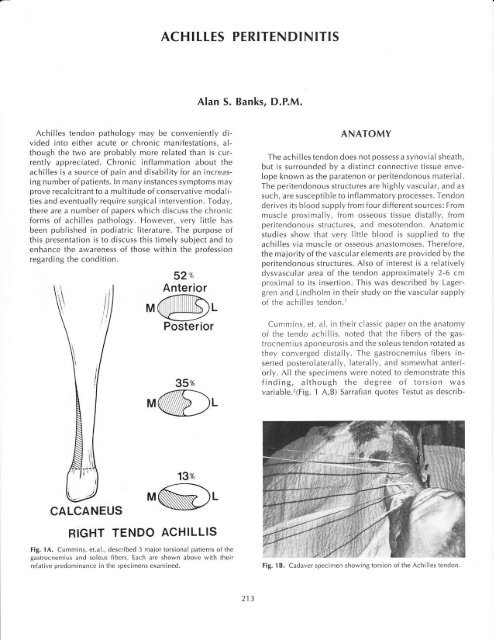
Cummins, et. al. in their classic paper on the anatomy<br />
of the tendo achillis, noted that the fibers of the gastrocnemius<br />
aponeurosis and the soleus tendon rotated as<br />
they converged distally. <strong>The</strong> gastrocnemius fibers inserted<br />
posterolaterally, Iaterally, and somewhat anteriorly.<br />
All the specimens were noted to demonstrate this<br />
finding, although the degree of torsion was<br />
variable.2(Fig. 1 A,B) Sarrafian quotes Testut as describ-<br />
13%<br />
CALCANEUS<br />
RIGHT TENDO ACHILLIS<br />
Fig. 1A. Cummins, et.al., described 3 major torsional patterns of the<br />
gastrocnemius and soleus fibers. Each are shown above with their<br />
relative predominance in the specimens examined.<br />
Fig. 1B. Cadaver specimen showing torsion of the <strong>Achilles</strong> tendon.<br />
213
ing the torsion of the fibers as being greatest 2 to 5 cm<br />
from the tendon insertion.3 This correlates precisely with<br />
the area of minimal vascularity for the achilles. More<br />
than likely this occurience is not consequential.<br />
NOMENCLATU RE<br />
Puddu et.al. felt that the generic term "tendonitis" was<br />
inappropriate for this condition.4 Based upon anatomic<br />
and pathologic findings they suggested three different<br />
forms of chronic achilles pathology: (a) lnflammation<br />
which involves only the surrounding tissues without affecting<br />
the tendon itself was termed peritendinitis. (b)<br />
<strong>Peritendinitis</strong> with tendonosis was preferred whenever a<br />
concomitant degenerative process within the achilles<br />
itself was observed. (c) Tendonosis denoted a pure degenerative<br />
process within the tendon which was asymptomatic<br />
since the surrounding tissues were not inflamed.<br />
Poor flexibility of the achilles (equinus)was also noted<br />
to be responsible for a number of cases.s Patients with<br />
equinus are constantly applying stress to the triceps, especially<br />
when the shortage of ankle dorsiflexion is only<br />
partially compensated in other anatomic areas. Pathologic<br />
pronation is evident in those who are able to fully<br />
compensate at times creating the scenario described<br />
above. Patients with gastrocnemius equinus who possess<br />
normal soleal flexibility would theoretically be more<br />
susceptible to inflammatory processes due to the different<br />
degrees of tension within the tendon itself.<br />
SOURCES OT INFLAMMATION<br />
As indicated above, it is the peritendonous structures<br />
which are capable of eliciting an inflammatory response.<br />
Thus direct trauma to these tissues may serve as the<br />
stimulus for chronic pathology. In our experience this<br />
has most commonly been seen following blunt injury.<br />
Hemorrhage involving the peritendonous structures may<br />
result in the formation of an organized hematoma and<br />
resultant fibrosis, each having the potential to impair<br />
normal, smooth function. Chronic inflammation may<br />
result if early resolution of this problem is not<br />
attained.(Fig. 2A,B) Fortunately, most cases will resolve<br />
with vigorous physical therapy measures, but the progress<br />
may be slow.<br />
Biomechanical deformities within the foot and Ieg<br />
have also been implicated as contributi ng factors in the<br />
development of achilles peritendinitis. CIement, et.al.,<br />
felt that patients who pronated excessively subjected the<br />
tendon to rapid opposing components of rotation. As the<br />
heel is undergoing an excessive range of motion, the<br />
knee is also extending. <strong>The</strong>refore, the leg and foot are<br />
subjected to simultaneous contradictory medial and<br />
lateral rotation. This was purported to blanche the vessels<br />
within the peritendonous areas and lead to inflammation.s<br />
Those patients who undergo excessive pronation<br />
from a supinated position, or those with a high<br />
midtarsal or subtalar joint axis are primarily susceptible.<br />
A patient with the latter condition will have a higher<br />
proportion of transverse plane motion in the foot for the<br />
same degree of overall joint mobility. <strong>The</strong>refore, torsional<br />
forces are accentuated even further upon the<br />
tendinous structures.<br />
Fig. 2A. This patient sustained an acute blu nt traumatic episode to the<br />
posterior aspect of the mid-leg. Several months Iater peritendinitis<br />
remains despite conservative measures.<br />
Fig. 28. <strong>The</strong> swelling surrounding the achilles can be appreciated<br />
214
Vascular studies of the achilles tendon have demonstrated<br />
an area of relatively poor perfusion 2-6 cm from<br />
the insertion of the tendon.l As discussed above, this has<br />
been considered to be a cause of chronic inflammation<br />
and also acute rupture. However, as rotational stress is<br />
suggested as a primary factor in developing inflammation,<br />
the degree of anatomic torsion present within the<br />
tendon may also be a factor to consider. A tendon which<br />
already has a high degree of rotation of the gastrocnemius<br />
fibers is less likely to withstand the mechanical<br />
demands of excess pronation or equinus.<br />
Osseous deformity adjacent to the achilles may also<br />
serve to irritate the peritendonous tissues and result in<br />
inflammation. lmpingement from the posterosuperior<br />
aspect of the calcaneus (Haglund's deformity) is one<br />
such condition.6 Hypertrophic spurring at the insertion<br />
of the tendo achillis may incite inflammation due to<br />
mechanical irritation. Those patients with recalcitrant<br />
and extensive symptoms associated with insertional<br />
spurring may have actually avulsed bone. <strong>The</strong> persistent<br />
irritation due to the loose body is another factor to<br />
consider.<br />
Arthritic diseases are also known to manifest at times<br />
with peritendinitis. More recently reports have noted an<br />
association with the HLA-827 antigen.T Although the<br />
achilles does not have a true synovial sheath, arthritic<br />
processes should be considered in those patients with recalcitrant<br />
symptoms or in those where no other obvious<br />
causes can be ascertained.<br />
and rest. Orthotic control of inappropriate rearfoot<br />
pronation has also proven effective in those with under-<br />
Iying mechanical problems. Steroid injections for any of<br />
these three affectations of the achilles are contraindicated.<br />
Patients suffering from prolonged peritendinitis with<br />
tendonosis or isolated tendonosis are at risk for rupture<br />
of the achilles. Numerous studies have shown that degenerative<br />
changes of some form are almost always<br />
present at the time of surgical repair. <strong>The</strong>refore, most<br />
clinicians seem to limit conservative care to approximately<br />
a one year duration.<br />
Those cases which are recalcitrant may require surgical<br />
intervention. lf the primary problem is an osseous<br />
deformity, then resection of the spurring or prominence<br />
may prove adequate. Patients without osseous involvement<br />
may be helped by surgical exploration of the<br />
tendon and surrounding tissues. <strong>The</strong> literature supports<br />
this form of intervention, and most agree upon the intraoperative<br />
approach. If the paratendonous tissues are<br />
fibrotic, inflamed, or adherent, they should be resected.<br />
Any degenerative areas of the tendon should similarly be<br />
excised.12,13,''(Fig. 3) Some authors advocate longitudinal<br />
incisions within the tendon to search for occult areas<br />
of pathology. This also has the purported benefit of<br />
encouraging new vascular ingrowth for the tendon.a MRI<br />
scans may prove to be of benefit in identifying specific<br />
areas of pathology prior to surgery.<br />
Two other conditions may be seen in conjunction with<br />
peritendinitis with tendonosis. <strong>The</strong> first is acute tendon<br />
rupture. Just as tibialis posterior tendon ruptures are seen<br />
following inflammatory processes, the same occurs with<br />
the tendo achillis. Several authors have noted evidence<br />
of degeneration at the time of surgical repair for acute<br />
rupture.a,B,e Chronic tendonosis without pain may also be<br />
a problem leading to eventual rupture.a<br />
Dystrophic calcification within the body of the achilles<br />
tendon may also be a direct result of chronic inflammation<br />
and/or degeneration.l0,1l This should not be overlooked<br />
when a patient presents with radiographic evidence<br />
of this finding.<br />
IREATMENT<br />
Fortunately most patients with symptomatic achilles<br />
pathology will respond to aggressive conservative means<br />
if implemented early. This consists of various forms of<br />
physical therapy (ice massage, ultrasound, stretching/<br />
strengthening exercises), casting, NSAlDs, heel raises,<br />
Fig. 3. lntraoperative photo ofa patient treated conservatively for over<br />
a year for chronic peritendinitis. Adherence of the paratenon is appreciated<br />
distally, as well as degeneration of the tendon representing tendonos<br />
i s.<br />
215
Postoperative care generally consists of a below knee<br />
cast for approximately four weeks. At two weeks the cast<br />
is bivalved and the patient allowed to undergo gentle<br />
Iimited passive range of motion twice daily. At four<br />
weeks weight bearing is instituted in a guarded manner,<br />
at times with crutch assist and a slight heel raise. Physical<br />
therapy may be used to help stretch and strengthen the<br />
achilles. Activities progressively advance from walking,<br />
to cycling, and finally running.<br />
Cenerally the results following surgery have been<br />
reported as being very successful. One probably is curious<br />
as to whether or not the excision of a primary<br />
vascular supply such as the peritendon is advantageous.<br />
However, to date no later sequela have been published.<br />
As mentioned above, it is felt that the vascular ingrowth<br />
will later occur from surrounding tissues.<br />
References<br />
12.<br />
13.<br />
14.<br />
Lagergren C, Lindholm A: Vascular distribution on<br />
the achilles tendon - an angiographic and microangiographic<br />
study. Acta Chir Scand, 1 1 6:491 -<br />
495,1958.<br />
Cummins EJ, Anson Bl, Carr BW, Wright RR: <strong>The</strong><br />
structure of the calcaneal tendon (of achilles) in<br />
relation to orthopedic surgery. Surgery Gynecology<br />
Obstetrics, B3:107 , 1946.<br />
Sarrafian SK: Anatomy of the Foot and Ankle, )B<br />
Lippincott Co., Philadelphia, 1983, p.249.<br />
Puddu G, lppolito E, Postacchini F: A classification<br />
of achilles tendon disease. Am J Sports Med, 4:1 45-<br />
150,1976.<br />
Clement DB, Taunton JE, Smart CW:<strong>Achilles</strong> tendinitis<br />
and peritendinitis: etiology and treatment.<br />
Am J Sports Med, 12:179-184, 1984.<br />
Schepsis AA, Leach RE: Surgical management of<br />
achilles tendinitis. Am J Sports Med, 15:308-315,<br />
1987.<br />
Olivieri I, Gemignani C, Cherardi S, Crassi L,<br />
Ciompi ML: lsolated HLA-827 associated achilles<br />
tendinitis. Ann Rheumatic Dis, 46:626-627, 1987.<br />
Fox lM, Blaxina Me, Jobe FW, Kerlan RK, Carter VS,<br />
Shields CL, Carlson CJ: Degeneration and rupture<br />
of the achilles tendon. Clin Orthop,lOT:221-224,<br />
1975.<br />
)ozsa L, Kvist M, Balint BJ, Reffy A, Jarvined M,<br />
Lehto M, Barzo M: <strong>The</strong> role of recreational sport activity<br />
in achilles tendon rupture. Am J Sports Med,<br />
17:338-343, 1989.<br />
Fiamengo SA, Warren RF, MarshallJL, Vigorita VT,<br />
Hersh A: Posterior heel pain associated with a calcaneal<br />
step and achilles tendon calcification. C/in<br />
O.rthop, 167 :203-211 , 1982.<br />
Downey MS, Kalish SK: Surgical excision and repair<br />
of calcifications of the tendo achillis. Surgery<br />
of the Foot and Leg, 1986, McGlamry ED, (ed.),<br />
Pod iatry lnstitute Publish ing Co., Tucker, CA,<br />
'l<br />
986, pp.1 34-139.<br />
Leach RE, James S, Wasilewski S: <strong>Achilles</strong> tendinitis.<br />
Am J Sports Med,9:93-98, 1981.<br />
Clancy WG, Neidhart D, Brand RL: <strong>Achilles</strong> tendonitis<br />
in runners: A report of five cases. Am J<br />
Sports Med, 4:46-57, 1976.<br />
Gould N, Korson R: Stenosing tenosynovitis of the<br />
pseudosheath of the tendo achilles. Foot Ankle,<br />
1:179-187 , 1980.<br />
2.<br />
-).<br />
4.<br />
5.<br />
6.<br />
7.<br />
o.<br />
9.<br />
.10.<br />
].l.<br />
216


