

Congenital Overlapping Fifth Toe Deformity - The Podiatry Institute
Congenital Overlapping Fifth Toe Deformity - The Podiatry Institute
Congenital Overlapping Fifth Toe Deformity - The Podiatry Institute

You also want an ePaper? Increase the reach of your titles
YUMPU automatically turns print PDFs into web optimized ePapers that Google loves.
CHAPTER 33<br />
CONGENITAL OVERTAPPING FIFTFI TOE<br />
DEFORMITY: Review and Presentation of a<br />
Consolidated Surgical Approach<br />
Michael S. Douney, D.P.M.<br />
<strong>The</strong> congenital overlapping fifth toe deformiry, also<br />
known as congenital digitus minimus varus or<br />
congenital digitus quinti valr'ts) is a complex and<br />
challenging condition which requires equally<br />
complex and challenging treatment. <strong>The</strong>re are three<br />
primary components to the overlapping fifth toe<br />
which make it a tripTane deformity. Adduction in the<br />
transverse plane, dorsiflexion in the sagittal plane,<br />
and varus rotation (i.e., exlernal rotation) in the<br />
frontal plane ali occur at the fifth metatarsophalangeal<br />
joint. Rarely, contracture will also occur<br />
within the fifth toe itself. Due to the toe's posirion,<br />
the deformity has shortening or contracture of the<br />
medial collateral ligament, the medial aspect of the<br />
metatarsophalangeal joint, the exlensor digitor"um<br />
Iongus tendon slip, and the skin of the dorsomedial<br />
aspect of the fourth interdigital web space. Virh<br />
time, osseous adaption of the proximal phalanx or<br />
fifth metatarsal head can also develop. Due to its<br />
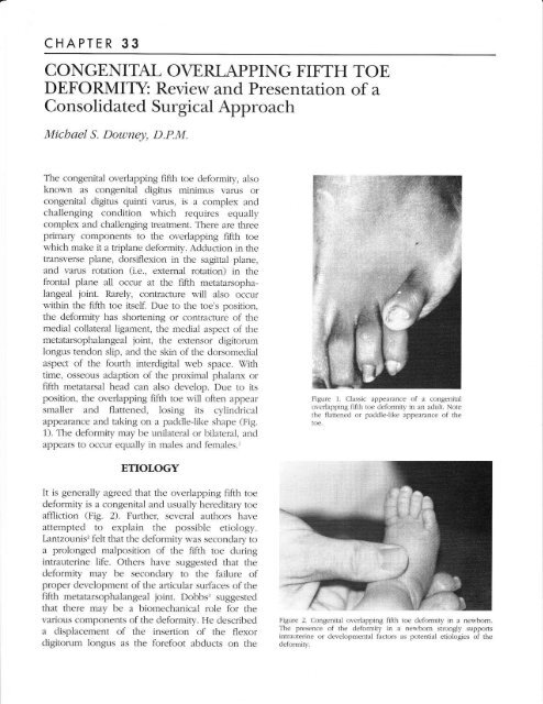
position, the overlapping fifth toe will often appear<br />
smaller and flattened, losing its cylindrical<br />
appearance and taking on a paddleJike shape (Fig.<br />
1). <strong>The</strong> deformity may be unilateral or bilateral, and<br />
appears to occur equaily in males and females.'<br />
Figure 1. Classic appearance of a congenital<br />
overlapping fiftl'r toe deformity in an adult. Note<br />
the flattened or paddleJike appearance of the<br />
toe.<br />
ETIOLOGY<br />
It is generally agreed that the overlapping fifth toe<br />
deformity is a congenital and usually hereditary toe<br />
affliction (Fig. 2). Further, several authors have<br />
attempted to explain the possible etiology.<br />
Lantzounis'zfelt that the deformity was secondary to<br />
a prolonged malposition of the fifth toe during<br />
intrauterine life. Others have suggested that the<br />
deformity may be secondary to the failure of<br />
proper development of the articular sr-rrfaces of the<br />
fifth metatarsophalangeal joint. Dobbs3 suggested<br />
that there may be a biomechanical role for the<br />
various components of the deformity. He described<br />
a displacement of the insertion of the flexor<br />
digitorum longus as the forefoot abducts on the<br />
Figure 2. <strong>Congenital</strong> overlapping fifth toe deformiry in a newbom.<br />
<strong>The</strong> presence of the deformity in a newborn strongly supports<br />
intreuterine or developmental factors as potential etiologies of the<br />
cleformity.
I98 CHAPTER 33<br />
rearfoot, especially in a pronated foot. This<br />
displacement leads to an abnormal medial force at<br />
the insertion of the long flexor into the distal<br />
phalanx. According to Dobbs, this leads to varus<br />
rotation of the fifth toe and adduction of the<br />
intermediate and distal phalanges. Further, as the<br />
flexor tendon displaces, a stable plantarflexory force<br />
is lost allowing dorsiflexory contracture at the fifth<br />
metatarsophalangeal joint. This biomechanical<br />
rationale may explain the deformity in some adults,<br />
or the progression of the deformity in some<br />
individuals, but does not appear to be the only<br />
etiology. As already noted, the deformity is often<br />
present at birth and clearly has developed before any<br />
weight-bearing force has been applied to the foot.<br />
CLIMCAL PRESENTATION<br />
<strong>The</strong> congenital overlapping fifth toe deformily is<br />
usually asymptomatic in infancy and early childhood,<br />
but typically becomes symptomatic as the<br />
child ages and approaches adulthood. In some<br />
instances, the deformity does not become painful<br />
until later in life. About 500/o of overlapping fifth toe<br />
deformities remain asymptomatic throughout the<br />
patient's life.<br />
Of those who develop symptoms, most will<br />
complain of dorsal digital irritation to the fifth toe,<br />
or of a painful heloma durum on the top of the toe.<br />
Conventional shoes often aggravate the toe. Less<br />
frequently, a heloma molle in the fourth web space<br />
or an onychoclal.us (i.e., a corn or callous aiong<br />
the nail groove) will be the offending complaint.<br />
Often, even when the deformity is asymptomatic,<br />
the patient or their parents may be concerned over<br />
the cosmetic appearance of the toe and the potential<br />
for future problems.<br />
Careful evaluation of the overlapping fifth toe<br />
deformity will allow the clinician to determine its<br />
flexibility and the status of the fifth metatarsophalangeal<br />
joint. <strong>The</strong> tautness of the extensor<br />
digitorum longus tendon slip to the fifth toe, and<br />
the dorsal-medial skin can be assessed by<br />
plantarflexing the fifth metatarsophalangeal joint.<br />
Palpation of the fifth metatarsophalangeal joint will<br />
generally allow an estimation as to the amount of<br />
joint subluxation or dislocation. Radiographs will<br />
confirm the status of the fifth metatarsophalangeal<br />
joint and reveal any osseous adaption of the fifth<br />
ray components. Although not mentioned in the<br />
literature, the author has found that more<br />
aggressive treatment for the deformiry is necessary<br />
if the deformity is: (1) more rigid in nature; (2)<br />
associated with an irreducible or dislocated fifth<br />
metatarsophalangeal joint; or (3) associated with<br />
adaptiv e osseous changes.<br />
C ONS ERVATTVE TREATMENT<br />
Most authors feel that consetwative treatment is<br />
fruitless in the treatment of the congenital overlapping<br />
fifth toe deformity. In very mild cases, or in<br />
the very young, stretching or strapping of the toe<br />
in abduction and plantarflexion can be attempted.<br />
Jordan and Casellia advocated a simple adhesive<br />
splint, which they applied at as young an age as<br />
possible (but not younger than 4 or 5 months of<br />
the digit is not long enough until then).<br />
^ge1 ^s<br />
<strong>The</strong>y stated that "Alignment of the deformed digits<br />
may be expected within 4 to 6 weeks depending<br />
upon the age of the patient and the severity of the<br />
deformity. Complete correction is then achieved by<br />
retaining the properly aligned digit for an<br />
additional 2 to B weeks in its corrected attitude."4 In<br />
the older palient, the only reliable non-surgical<br />
treatment available is accommodation. <strong>The</strong> use of<br />
padding, digital shields or molds, and extra-depth<br />
shoes or sandals may satisfy and allevtate digital<br />
irritation in some patients. Vhen consetwative<br />
treatment fails or when symptoms warrant, surgical<br />
intervention is indicated.<br />
SURGICAL TREATMENT<br />
Numerous procedures have been proposed in the<br />
literature for the correction of the congenital overlapping<br />
fifth toe deformity. <strong>The</strong> oldest surgery for<br />
the deformity is amputation. However, foliowing<br />
simple amputation of the fifth toe, it became<br />
recognized that painful callouses developed<br />
around the head of the remaining fifth metatarsal.5<br />
Other deformities, such as hammerloes, lesser<br />
digital drift, irritation to the fourth toe, or lesser<br />
metatarsalgia have also frequently resulted following<br />
amputation' (Fig. 3).<br />
In the 7940s, owing to the stigma associated<br />
with an amputation, and in some areas stimulated<br />
by the need for an alternative surgery in Civil<br />
Service applicants (who could not obtain Civil<br />
Serwice employment with either an overlapping<br />
fifth toe deformity or an amputated fifth toe), other<br />
procedures were developed. Most of these
CHAPTER 33<br />
I99<br />
Figure J. Patient who underwent amputation of<br />
a congenital overJapping fifth toe as a teenager.<br />
Note d-rat the patient now has lateral drifting of<br />
her remaining lesser digits, a corn on the fourth<br />
toe, and a bunion deformity. <strong>The</strong> patient also<br />
complained of metatarsalgia under the fouth<br />
metatafsal.<br />
procedures emphasize soft tissue correction, but<br />
some also include osseous procedures. <strong>The</strong><br />
objective of each technique is to reduce the three<br />
primary components of the deformity (i.e., the<br />
dorsiflexion, adduction, and l.arus rotation present<br />
at the fifth metatarsophalangeal joint).<br />
In 7940, Lantzounis'? described his proposed<br />
procedure. Through a dorsal longitudinal incision<br />
centered over the fifth metatarsophalangeal joint,<br />
he isolated the extensor digitorum longus tendon<br />
slip to the fifth toe, and sectioned it at the distal<br />
end of the incision. He then longitudinally incised<br />
the periosteum over the distal end of the fifth<br />
metatarsal, the joint capsule, and the periosteum<br />
over the base of the proximal phalanx of the fifth<br />
toe. Lantzounis then created what he termed a<br />
"periosteocapsular flap" by elevating the incised<br />
periosteum and capsule dorsally, laterally, and<br />
medially. A drill hole was then created at the distal<br />
end of the fifth fietatarsal and the proximal end of<br />
the severed tendon slip was threaded through the<br />
drill hole and sutured back onto itself. A horizontal<br />
mattress suture was then placed into the "periosteocapsular<br />
fl,ap" plantarly and tied with the toe held<br />
in a corrected position. Thus, simply stated, the<br />
procedure was a modified Jones' suspension of the<br />
extensor digitorum longus tendon, a dorsal<br />
capsulotomy, and a planlar capsulorrhaphy.<br />
Lantzounis reported his results in 25 of the<br />
procedures in 79 patients. <strong>The</strong> ages of his patients<br />
ranged from 2 to 25 years old with an average of<br />
12 years old. One patient who had undergone<br />
correction for bllateral deformities was lost to<br />
follow-up, leaving 23 operations that were<br />
reviewed from B months to 6 years postoperatively.<br />
Lantzounis found excellent results in 16 cases<br />
(-700/o), good results in 4 cases (170/o) and poor<br />
results in 3 cases (730/o).<br />
In 1942, Lapidus6 described another unique<br />
procedure for the correction of the congenital overlapping<br />
fifth toe deformity. He approached the<br />
deformity through a hockey-stick incision made<br />
along the dorsomedial border of the fifth toe,<br />
starting at the distal interphalangeal joint and<br />
running proximally to the fourth web space, and<br />
then curying laterally over the dorsum of the fifth<br />
metatarsophalangeal joint and continuing<br />
dorsolaterally to the lateral aspect of the fifth<br />
metatarsal head. Next, a separate transverse<br />
incision was made over the extensor digitorum<br />
longus tendon at the midshaft of the fifth<br />
metatarsal. <strong>The</strong> extensor tendon slip to the fifth toe<br />
was identified and transected. <strong>The</strong> tendon was then<br />
gently pulled distally into the wound over the fifth<br />
toe. After the extensor tendon was harwested, a<br />
capsulotomy of the fifth metatarsophalangeal joint<br />
was performed. Next, an oblique subcutaneous<br />
channel was created from the distal interphalangeal<br />
joint of the fifth toe dorsomedially under the<br />
phalanges to the plantar-lateral aspect of the fifth<br />
metatarsophalangeal joint. <strong>The</strong> previouslyharvested<br />
extensor tendon was then rerouted<br />
through the channel created, and sutured under<br />
tension with the toe held in the corrected position,<br />
to the conjoined tendon of the abductor digiti<br />
quinti and flexor digitorum brevis to the fifth toe.<br />
Lapidus stated that his results had been "quite<br />
satisfactory" in a "few cases," but did not give more<br />
specific details regarding the efficacy of his<br />
espoused procedure.<br />
Whereas Lantzounis and Lapidus primarily<br />
dealt with the extensor tendon and joint capsule, in<br />
7943, Goodwin and Swisher5 suggested release of<br />
the dorsal skin contracture as well. <strong>The</strong> authors<br />
advocated a Y-shaped incision over the dorsum of<br />
the fifth metatarsophalangeal joint with the stem of<br />
the Y extending proximally. <strong>The</strong> branches of the Y
200 CHAPTER 33<br />
were situated diagonally distal-ward about halfway<br />
around the fifth toe. <strong>The</strong> incision was then carried<br />
deep, and a Z-plasty lengthening of the extensor<br />
digitorum longus tendon, and a dorsal or dorsomedial<br />
capsulotomy of the fifth metatarsophalangeal<br />
joint was performed. <strong>The</strong> toe was then held in the<br />
comected position and closure was accomplished.<br />
<strong>The</strong> proximal arm of the Y is progressively closed<br />
distalward, allowing the triangular flap of skin to slip<br />
distally as far as is necessary to allow closure of the<br />
skin without tension. In this sense, the authors'<br />
incision is closed in typical V-Y skin plasty fashion.<br />
Postoperatively, the authors stressed the need for<br />
maintenance of the corrected position for up to 6<br />
weeks, with either a plaster-of-Paris cast or an<br />
adhesive strapping. In reporling their results, the<br />
authors were brief in stating that their "technique has<br />
been highly successful in 100 percent of a series of<br />
about twenty cases, including males and females,<br />
children and adults, and patients in both private and<br />
institutional practice."<br />
Similar to Goodwin and Swisher, StammT and<br />
\7i1son8 described a V-Y skin plasty for part of the<br />
corection of the congenital overlapping fifth toe<br />
deformity. <strong>The</strong>y created a V-shaped skin incision<br />
over the fourth web space, with the base oriented<br />
distal-lateral and the apex proximal-medial. <strong>The</strong><br />
skin flap was then elevated, and a tenotomy of the<br />
extensor tendon and capsulotomy of the fifth<br />
metatarsophalangeal joint performed. Ciosure was<br />
then accomplished with the toe held in a corrected<br />
position and the incision closed in V-Y skin plasty<br />
fashion. Like Goodwin and Swisher, Vilson<br />
emphasized the need for postoperative<br />
maintenance of the correction with a plaster-of-<br />
Paris cast for several weeks. Vilson felt the<br />
procedure was best suited for patients under the<br />
age of 30, aod reported his results in 7 cases<br />
followed over a 2-year period. He described "very<br />
satisfactory" functional results in all of the cases,<br />
with one slight recuffence due to an error in<br />
operative technique. Stamm did not give afiy<br />
results in his description of the procedure.<br />
Many years later, in 7990, Patone reported on<br />
the long-term results of Stamm's and Wilson's<br />
approach. He noted that advocates of the V-Y<br />
plasty felt that the procedure was a successful,<br />
technically straight-forward approach. Conversely,<br />
he noted that detractors of the operation insisted<br />
there was a high recurrence rate. Paton reviewed<br />
20 V-Y plasty procedures in 75 children with a<br />
mean age of 9 years,2 months. Early retrospective<br />
assessment of his cases demonstrated good results<br />
in 74 patients (700/o), acceptable results in J patients<br />
(.750/o), and poor results in J patients (15o/o). Later<br />
follow-up at an ^verage<br />
of 2 years, 1 month postoperative<br />
revealed a marked deterioration in the<br />
repofted success of the procedure. At the second<br />
follow-up, there were good results in 6 patients<br />
(30o/o), acceptable results in 2 patients (10%o), and<br />
poor results in 12 patients (6Oolo). In light of the<br />
poor iong-term results, Paton recommended that<br />
the V-Y plasty approach for the correction of a<br />
congenital overlapping fifth toe deformity be<br />
abandoned.<br />
In one of the most unique approaches,<br />
Cockin'o reported on an operation he attributed to<br />
Mr. R. \fieeden Butler. He related that Butler had<br />
performed his operation since the early 1950s.<br />
Cockin described Butler's procedure as a double<br />
racquet incision with a circumferential incision<br />
around the toe (i.e., the racquet head) and a dorsal<br />
and plantar handle. <strong>The</strong> plantar handle was made<br />
slightly longer, and was inclined laterally to allow<br />
improved position of the toe at closure. <strong>The</strong> skin<br />
flaps created were then raised with careful<br />
preservation of the neurovascular bundles. An<br />
extensor tenotomy and dorsal capsulotomy of the<br />
fifth metatarsophalangeal joint were then<br />
performed. Next, the toe was swung downward<br />
and laterally into a corrected position, and closure<br />
accomplished. After closure, the resultant dorsal<br />
incision would be longer than the plantar incision.<br />
Cockin reported on 70 cases in 55 patients, ranging<br />
from 5 months to 45 years of age. Follow-up<br />
evaluation was done from 1 to 10 years postoperative.<br />
Cockin reported 54 cases (9tolo) as good<br />
results (i.e, satisfactory to both surgeon and patient<br />
with full correction of the deformity), 4 cases (60/o)<br />
as fair results (i.e., satisfactory to the patient with<br />
an element of the deformity left uncorrected), and<br />
2 cases (30/o) as failures (i.e., recurrence of the<br />
deformity). Cockin reported 2 cases of wound<br />
infection, 3 cases of delayed healing, but no cases<br />
of long-term neurovascular compromise.<br />
In 7985, Black et al." repofied on their<br />
experience with the Butler procedure. <strong>The</strong>y<br />
evaluated 36 procedures in 30 patients. <strong>The</strong>ir<br />
patients ranged from 3 to 18 years of age and were<br />
assessed at an avetage of 2 years,4 months postoperative.<br />
<strong>The</strong>y reported excellent results (i.e.,<br />
satisfactory to both patient and surgeon with full
CI]APTER 33 201<br />
correction of the deformity) in 28 cases (78%o),<br />
good results (i.e., satisfactory to the patient in terms<br />
of pain relief and footwear, but did not show full<br />
correction and had excessive scar formation) in 6<br />
cases (77V4, and failure (i.e., recurrence of the<br />
deformity with pain and an unacceptable scar) in 2<br />
cases (5olo). <strong>The</strong>se surgeons reported one<br />
significant dysvascular episode immediately postoperative,<br />
which ultimately resolved with no<br />
permanent damage to the toe.<br />
Alternatively, surgical syndactylization (i.e.,<br />
webbing of the fourth and fifth toes together) has<br />
been championed by several authors.'''6 In infancy<br />
and early childhood, syndactylization of the fourth<br />
and fifth toes alone is used to correct the deformity.<br />
As the deformity becomes more fixed later in life,<br />
the syndactylization is accompanied by either a<br />
pafiial or complete proximal phalangectomy. A<br />
dorsal fifth metatarsophalangeal joint capsulotomy<br />
and tenotomy of the extensor tendon are typically<br />
a part of the procedure. In 1950, McFarland'2 was<br />
the first to discuss this surgical approach. He<br />
described his early results as "promising," but provided<br />
no further follow-up data. In 7954, Scrase't<br />
reported on a series of syndactylization procedures<br />
combined with excision of the base of the proximal<br />
phalanx of the fifth toe. In his 42 cases, he reported<br />
39 O3'yo) good results and 3 (70/o) fair results. Scrase<br />
did not provide his criteria for what he termed a<br />
"good result" or a "fait result."<br />
In 7965, Leonard and Rising'5 discussed their<br />
success in B cases of syndactylization performed on<br />
6 patients with a congenital overlapping fifth toe<br />
deformity. <strong>The</strong>y stated that "All have had satisfactory<br />
results except one in which the deformity was not<br />
completely corected at the time of surgery." <strong>The</strong>y<br />
noted follow-up for as long as 5 years with no<br />
recurrence of the deformiry reported. Recognizing<br />
the results of McFarland, Tachdjian'6 has espoused<br />
surgical syndactylization as his procedure of choice<br />
for congenital overlapping fifth toe deformities.<br />
Conversely, Giannestras" condemned syndactylization<br />
for the overlapping fifth toe deformrty as he felt<br />
"it produces a deformlty in order to correct a<br />
deformity." Giannestras felt that the Lantzounis<br />
procedure or Butler procedure were better options<br />
which "have withstood the test of time both from a<br />
functional and cosmetic standpoint."<br />
In 7954, Ruiz-Mora'B described yet another<br />
approach to the overlapping fifth toe deformity.<br />
His procedure consisted of a total proximal<br />
phalangectomy performed through a plantar<br />
longitudinal skin ellipse. Closure of the plantar skin<br />
ellipse aids in the prevention of recurrent<br />
deformity. Janecki and \X/ilde'e reported on their<br />
experiences with the Ruiz-Mora procedure. <strong>The</strong>y<br />
performed the procedure on 28 patients, but 5<br />
were lost to follow-up. Subsequently, they<br />
followed 31, procedures in 22 patients for an<br />
at,etage of 3.5 years postoperative. <strong>The</strong>ir patients<br />
were older, averaging 48 years of age at operation.<br />
<strong>The</strong>y reported that all of the patients had complete<br />
relief of their symptoms and correction of the<br />
deformity with a good cosmetic appearance of the<br />
shoftened fifth toe. However, Janecki and \7i1de<br />
noted two significant problems postoperatively in<br />
their patients. First, in 10 of their cases (320/o) a<br />
hammefioe deformity of the ad1acent fourth toe<br />
with a painful corn developed due to the excessive<br />
shortening of the fifth toe caused by the Ruiz-Mora<br />
technique (Fig. 4). None of these patients had a<br />
pre-existing hammefioe deformity of the fourth toe.<br />
Second, in 7 of their series (230/0) a painful<br />
prominence of the fifth metatarsal head or a tailor's<br />
bunion developed. Five of these 7 patients had<br />
both a bunionette and a painful corn on the fourth<br />
toe. Based upon their findings, Janecki and Wilde<br />
recommended less bone resection and suggested<br />
excision of only the head and neck of the proximal<br />
phalanx. <strong>The</strong>y did not report any follow-up on<br />
their proposed modification.<br />
In 7954, Thompson'o discussed his modification<br />
of the Ruiz-Mora procedure. He added a dorsal<br />
Z-plasty skin approach to the other components of<br />
the Ruiz-Mora procedure (i.e., the proximal<br />
Figure ,1. Postoperative appearance approximately 2 yearc after aRuiz-<br />
Mora procedure for a congenital overlapping fifth toe defomity. Note<br />
the significant shotening of the fifth toe. <strong>The</strong> patient presented complaining<br />
of digital irritation to her other lesser toes (toes 2,3,4).
202 CHAPTER 33<br />
phalangectomy and plantar skin ellipse). He felt<br />
that this helped maintain the correction, but did not<br />
give any specific results.<br />
Kaplan'?l also offered a new procedure in 1954<br />
which was very similar to that espoused by Janecki<br />
and Vilde in the 1970s. Through a dorsal<br />
longitudinal incision, Kaplan performed an extensor<br />
tendon transfer by sectioning the extensor tendon at<br />
the proximal interphalangeal joint, and reinserting it<br />
into the base of the proximal phalanx. Nex1, he<br />
performed a capsulotomy of the dorsal, medial, and<br />
lateral aspects of the fifth metatarsophalangeal joint.<br />
<strong>The</strong> head of the proximal phalanx was then excised.<br />
Plantarly, two semi-elliptical incisions were made at<br />
the web of the fifth toe, and the resulting wedge of<br />
skin excised. Closure was then accomplished.<br />
Kaplan reported performing the procedure in 59<br />
cases with no failures.<br />
A few other procedures have been described<br />
more recently but have not become overly popular.<br />
in 1977, dissatisfied with earlier approaches,<br />
Anderson" accurately noted that a combined<br />
approach was necessary to coffect the overlapping<br />
fifth toe deformity. He combined resection of the<br />
fifth metatarsal head and/or the head of the proximal<br />
phalanx with a dorsal V-Y skin plasty, dorsal<br />
fifth metatarsophalangeal joint capsulotomy,<br />
Z-plasty lengthening of the extensor tendon, and<br />
plantar skin ellipse. No results were given by<br />
Anderson in his papet. In 7978, Rosner et a1.23<br />
described a procedure consisting of an abductory<br />
wedge osteotomy of the proximal phaianx, a<br />
dorsal and medial fifth metatarsophalangeal joint<br />
capsulotomy, and an extensor tenotomy. <strong>The</strong>ir<br />
wedge osteotomy was made approximately 5 mm<br />
distal to the fifth metatarsophalangeal,oint with the<br />
base lateral and the apex medial. <strong>The</strong> osteotomy<br />
was fixated with a Kirschner wire (i.e., K-wire).<br />
Rosner et al. discussed performing the procedure in<br />
only one patient, and provided no long-term results.<br />
Although numerous approaches have been<br />
endorsed for the congenital overlapping fifth toe<br />
deformity, none has been met with universal, nor<br />
even overwhelming, acceptance. This is most likely<br />
due to the recalcitrant nature of the deformity, and<br />
the significant potential for recurrence following<br />
a{ry surgical approach, short of amputation.<br />
Further, those procedures that limit the likelihood<br />
of recurrence (e.g., amputation or the Ruiz-Mora<br />
procedure) tend to create new problems or<br />
deformities (Figs. 3, 4). Most of the procedures<br />
described are effective for the mild deformiLy, and<br />
even work in some cases of more severe deformity.<br />
Unfortunately, many of the procedures fail to<br />
correct at least one of the features of the deformity<br />
and therefore recurrence occurs.<br />
In order to surgically correct a congenital<br />
overlapping fifth toe deformity, all aspects of the<br />
deformity must be corrected. Dorsally and<br />
medially, the contracted skin, extensor tendon, and<br />
fifth metatarsophalangeal joint capsule must be<br />
released or lengthened. Plantarly and Taterally,<br />
redundant soft tissues such as the skin and fifth<br />
metatarsophalangeal joint capsule must be tightened.<br />
Finally, any osseous deformity must be<br />
addressed. Drawing upon the historical approaches<br />
for the deformity, the author proposes a<br />
"consolid,ated. surgical approacb " for the correction<br />
of the overlapping fifth toe deformity.<br />
CONSOLIDATED SURGICAI APPROACH<br />
<strong>The</strong> consolidated surgical approacb attempts to<br />
address all of the components of the overlapping<br />
fifth toe deformity. Generally, less aggressive<br />
correction will be required for the more flexible<br />
deformity, or deformity present in the younger<br />
patient, while more aggressive correction will be<br />
necessary for the rigid deformity or deformity<br />
present in the adult. <strong>The</strong> components of the deformily<br />
are as follows:<br />
1. Dorsal or dorsomedial skin contracture;<br />
2. Extensor digitorum longus tendon contracture;<br />
3, Dorsal or dorsomedial fifth metatarsophalangeal<br />
joint capsule contracture and plantar plate<br />
adherence;<br />
4. Osseous adaption of the fifth metatarsal head,<br />
proximal phalanx, or other bony structures;<br />
5. Plantar or plantar-lateral fifth metatarsophalangeal<br />
joint capsule redundancy;<br />
6. Plantar or plantar-lateral skin redundancy.<br />
<strong>The</strong> first component of the deformity to be<br />
addressed is the dorsal or dorsomedial skin<br />
contracture. A dorsal or dorsomedial Z-plasty skin<br />
approach is utilized. <strong>The</strong> central arm of the Z-plasty<br />
is placed directly in line with the skin contracture.<br />
If the contracture is dorsal, the central arm is<br />
placed longitudinally over the fifth metatarsophalangeal<br />
joint. If the contracture is dorsomedial, the<br />
central arm is placed diagonally from proximalmedial<br />
to distal-lateral over the fourth<br />
intermetatarsal space. <strong>The</strong> arms of the Z-plasty are
CHAPTER 33 203<br />
then made with each arm forming a 60 degree<br />
angle with the central arm of the "Z" (Fig. 5).<br />
After the Z-plasty has been planned, the<br />
incision can be carried distally onto the fifth toe by<br />
longitudinally extending the central arm of the "Z."<br />
In cases of severe deformity and skin contracture,<br />
a double Z-plasry incision may be necessary to<br />
achieve enough lengthening of the dorsally<br />
contracted skin (Fig. 5-A). Dissection is then caried<br />
deep through the subcutaneous tissues, carefully<br />
preserwing the subcutaneous tissues and vascular<br />
supply of the flaps created by the Z-plasty (Fig.<br />
68). At the time of closure, the flaps of the "2" are<br />
transposed and sutured in standard Z-plasty<br />
fashion, and the distal, longitudinal extension onto<br />
the toe is closed.<br />
<strong>The</strong> next component of the deformity to be<br />
addressed is the extensor digitorum longus tendon<br />
to the fifth toe. If the deformity is mild and/or<br />
flexible, an open Z-plasty lengthening of the<br />
tendon is performed. In such cases, after full<br />
correction is obtained, the tendon is reapproximated<br />
in a lengthened position. If the deformity is<br />
moderate to severe, and/or fixed in nature, the<br />
tendon is transected at the proximal interphalangeal<br />
joint and later transferred to the fifth metatarsal neck<br />
creating a modified Jones' tenosuspension. In all<br />
cases, a complete release of the exlensor hood<br />
apparatus is performed (Figs. 6C,6D).<br />
<strong>The</strong> third component of the deformity is the<br />
fifth metatarsophalangeal joint contracture. Once<br />
the extensor tendon has been reflected, the joint<br />
can be directly addressed. A capsuiotomy of the<br />
dorsal, medial, and occasionally the lateral aspects<br />
of the ioint capsule is performed (Figs. 6E, 6F).<br />
Release of the planlar plate is then accomplished<br />
by passing a McGlamry elevator or other blunt<br />
instrument into the capsulotomy incision, and<br />
befween the fifth metatarsal head and the plantar<br />
plate (Figs. 6G, 6H). <strong>The</strong> capsule is not closed<br />
dorsally or medially.<br />
After a complete soft tissue release has been<br />
achieved dorsally and medially, the osseous<br />
structures and adaption can be more readily<br />
assessed. If osseous deformity or adaption has<br />
occurred al the proximal interphalangeal joint,<br />
resection of the proximal phalangeal head is<br />
accomplished (Figs. 6I, 6D. This is commonly<br />
needed in adults, or in more rigid deformities.<br />
Rarely, in very severe deformities or following<br />
recurrence, a partial or total fifth metatarsal head<br />
resection or implantation of the fifth metatarsophalangeal<br />
joint is considered, to aid in reduction of the<br />
fifth metatarsophalangeal joint deformity (Fig. 5K).<br />
<strong>The</strong> fifth component to be addressed is the<br />
plantar or plantar-lateral fifth metatarsophalangeal<br />
joint capsular redundancy. In milder or more<br />
flexible deformities, no planlar capsulorrhaphy is<br />
performed. In moderate to severe deformities, a<br />
plantar capsulorrhaphy similar to that described by<br />
Lantzounis'Z is executed.<br />
<strong>The</strong> sixth and final component to be dealt<br />
with is the plantar skin redundancy. An ovoidshaped<br />
or diamond-shaped skin ellipse is removed<br />
from the plantar web area of the fifth toe (Fig. 6L).<br />
This skin wedge is designed to remove the<br />
redundant plantar skin and to aid in the<br />
maintenance of the correction. <strong>The</strong> skin ellipse can<br />
be angled slightly more laterally al its proximal<br />
end, to further ard in achieving the desired position<br />
of correction. Careful dissection of the skin wedge<br />
is necessary. Only skin should be removed, and the<br />
deeper subcutaneous tissues containing the<br />
neurovascular elements should not be violated<br />
(Fig. 6M). <strong>The</strong> plantar skin plasty is then closed<br />
with non-absorbable suture (Fig. 5N).<br />
Once full correction has been achieved,<br />
closure is accomplished. In more long-standing or<br />
rigid deformities, the extensor tendon is transferred<br />
to the neck of the fifth metatarsal. To accomplish<br />
this, a small bone trephine (,i.e., 2 mm or 3 mm<br />
diameter trephine) is used to create a channel from<br />
lateral to medial through the fifth metatarsal (Figs.<br />
60, 6P). <strong>The</strong>n, as described by Lantzounis,' the<br />
extensor tendon is routed through the trephine<br />
hole and sutured back upon itself (Figs. 6Q, 5R).<br />
Otherwise, the extensor tendon is repaired in its<br />
iengthened position. If desired, a K-wire may be<br />
insefted to aid in the maintenance of the corrected<br />
position. <strong>The</strong> author typically utilizes either a<br />
0.045" or 0.062" K-wire which is driven from the<br />
end of the toe across the fifth metatarsophalangeal<br />
joint and into the fifth metatarsal with the toe held<br />
in its corrected position (Fig. 55). Finally, skin<br />
closure is accomplished and closure of the Z-plasty<br />
completed (Fig. 6T).<br />
Postoperatively, the foot is placed into a<br />
sterile dressing which is continued, except for<br />
intermittent wound inspections, for approximately<br />
2 to 3 weeks, until suture removal. If a K-wire has<br />
been utilized, it is typically maintained for 4 to 6<br />
weeks. If a K-wire has not been used, the dressing
204 CI]APTER 33<br />
must be carefully applied to maintain the corrected<br />
position. <strong>The</strong> patient is allowed to ambulate on the<br />
surgical foot, in a surgical shoe with paclding<br />
extending from the heel to the digital suicus.<br />
Padding a shoe in this fashion eliminates mosr of<br />
the dorsiflexory propulsive forces at the fifth<br />
metatarsophalangeal joint. Alternatively, a short leg<br />
cast can be utilized. After dressing removal, a<br />
digital splint is dispensed to help maintain the<br />
correction for several more weeks.<br />
<strong>The</strong> consolidated. surgical approach draws<br />
upon many historical surgical procedures and<br />
combines many espoused approaches to create a<br />
comprehensive surgical technique which<br />
endeavors to completely correct the congenital<br />
overlapping fifth toe deformity. <strong>The</strong> author has<br />
fotrnd the consolidatecl surgical approacb to result<br />
in predictable and effective correction of the<br />
congenital overlapping fifth toe deformity,<br />
regardless of the severity of the deformity (Figs. 6U,<br />
6V). f'ull correction at the time of surgery, and<br />
maintenance of the correction during the<br />
immediate postoperative period with a K-wire and<br />
digital splinting are the key points to prevent<br />
recurrence of the deformitv.<br />
Figure 5. Z-plasq, skin incision oriented to coffect dorsomeclial skin<br />
contracture,<br />
Figure 6A. Consolidatecl surgical approach for correction of severe<br />
congenital overlapping fifth toe deformity in a young adult. Dorsal<br />
clouble Z-plasty skin incision is planned.<br />
Figure 68. Skin incisions are made, careftrlly preseruing the bloocl<br />
supply to the Z-plasty skin flaps.<br />
Figure 6C. Extensor digitorum longus tendon is identified and<br />
sectionecl at the level of the PIPJ.
CHAPTER 33 205<br />
Figure 6D, Extensor hood recession is performed, and the tendon is<br />
dissectecl free proximally to the fifth metatarsal head.<br />
Figure 6E. Dorsal and dorsomedial capsulotomy are performed<br />
Figure 6F. <strong>The</strong> fifth metatarsophalangeal joint is exposecl<br />
Figure 6G. A McGlamry elevator is used to release the plantar plate of<br />
the fifth metatarsophalangeal joint,<br />
Figure 6H. A McGlamry elevator being passed between the fifth<br />
metatarsal head and plantar plate to accomplish the release.<br />
Figure 6I. Contracture at the PIPJ is noted, with adaption of the<br />
proximal phalangeal heac1.
206 CHAPTER 33<br />
Figure 6J. Resection of the proximal phalangeal head to correcr PIPJ<br />
contracture.<br />
Figr-rre 6K. Inspection of the fifth metatarsal head demonstrates no<br />
aclaptive defomity in this case.<br />
Figure 6L. <strong>The</strong> redundant plantar skin is ellipsecl in cliamond fasl'rion<br />
Figure 64,1, Reclundant skin only is excised avoicling clamage to<br />
neurovascular structllres.<br />
Figure 6N. Closure is accon-rplished with non-absorbable sutures.<br />
Sutures are all typicalll, placed in position before an1, are tied to allorv<br />
easier visualization of suture placement.<br />
Figure 60, A sn.rall bone trephine is usecl to create a lateral to rnedial<br />
hole in the fifth metatarsal neck.
CHAPTER 33 207<br />
Figure 6P. Hole created in fifth metatarsal neck for n-rodifiecl Jones<br />
tenosuspension.<br />
Figure 6Q. Previously h:rrr,'ested extensor cligitorum longus tendon is<br />
routed through the trephrne ho1e.<br />
Figure 6R. <strong>The</strong> ertensor tendon is sutured upon ltself<br />
Figure 65. A 0.062" (1.6 mm) K-u,ire is inserted temporarily to aid in<br />
m:rintaining alignment of the coffectecl position.<br />
Figure 6T. Immediate postoperative appeal:ance after closure of all<br />
wounds.<br />
Figure 6Lr. Prcoperativc appeamnce of deformitl,
208 CI]APTER 33<br />
REFERENCES<br />
Figure 6V. Postoperative appearance of correction at 6 weeks.<br />
SUMMARY<br />
<strong>The</strong> most common form of overlapping toe<br />
deformity is the congenital overlapping fifth toe<br />
deformity. More often than not, the deformity does<br />
not result in symptomatology until adolescence or<br />
adulthood. At this time, conservative measures are<br />
rarely successful in treating the condition. Many<br />
sophisticated surgical procedures have been<br />
advanced for the correction of the overlapping fifth<br />
toe deformity. Today, amputation and surgical<br />
syndactylization are less attractive approaches to<br />
both the patient and surgeon. Many other proposed<br />
procedures have been found to result in a high rate<br />
of recurrence of the deformity. Combining the<br />
"best" ideas from many different espoused<br />
procedures, the author has presented a<br />
consolidated surgical approach for the correction<br />
of the congenital overlapping fifth toe deformity.<br />
This approach has been described in detail and<br />
addresses each of the known elements of the<br />
deformity. Long-term results utilizing the<br />
consolidated surgical approach have been very<br />
good. Statistical analysis remains to confirm the<br />
clinical obseruations.<br />
3<br />
4<br />
5.<br />
6<br />
7.<br />
8<br />
t)<br />
10<br />
11<br />
12<br />
13.<br />
1,4.<br />
1.5.<br />
1,6.<br />
77.<br />
18.<br />
1.9.<br />
20<br />
21.<br />
22<br />
23<br />
Downey MS, Rubin L: Common Pediatric Digital Deformities, In<br />
DeValentine SJ, ed.: Foot and Ankle Disorders in Cbildren. New<br />
York, N.Y.: Churchill Livingstone; 7992: 407-437.<br />
Lantzounis LA: <strong>Congenital</strong> subluxation of the fifth toe and its<br />
correction by a periosteocapsuloplasty and tendon transplantatiot1.<br />
J Bone Joint Surg 22:747-L50, L940.<br />
Dobbs BM: Afihroplasty of the fifth digit. Clinics in Podiatric<br />
Med and Surg 3:29-39, 1986.<br />
Jordan RP, Caselli MA: <strong>Overlapping</strong> deformity of the digits in the<br />
pediatric patient: a conseffative approach to treaffient. J Am<br />
Podiatr Assoc 68,503-505, 1978.<br />
Goodwin FC, Swisher FM: <strong>The</strong> treatment of congenital hyperextension<br />
of the fifth toe. J Bone Joint Surg 25:193-196, 1943.<br />
Lapidus PW: Transplantation of the extensor tendon for<br />
correction of the overlapping fifth toe. J Bone Joint Surg<br />
24:555-559, 1942.<br />
Stamm TT: Minor surgery of the foot-elevated fifth toe. In Cariing<br />
ER, Ross JP, eds.: Britisb Surgical Practice, Vol. 4. London,<br />
England: Butterwofth; 1948: 161-1,62.<br />
Yr'ilson JN: V-Y correction for varus deformity of the fifth toe. Br<br />
J Surg 41:L33-135, 1953.<br />
Paton RSr: V-Y plasty for correction of varus fifth toe. J Pediatric<br />
Orlbop 10;218-249, 1990.<br />
Cockin J: Butler's operation for an over-riding flfth loe. J Bone<br />
Joint Surg 508;78-81, 1968.<br />
Black GB, Grogan DP, Bobechko \WP: Butler arthroplasty for<br />
coffection of the adducted fifth toe: a retfospective study<br />
of 36 operations berween 1968 and 1982. I Pediatric Ortbop<br />
5:139-441, 1985.<br />
McFarland B: <strong>Congenital</strong> defomities of the spine and limbs. In<br />
Platt H, ed.: Modern Trends in Ofibopaedics New York, N.Y.:<br />
Paul B. Hoeber 1950: 107-137.<br />
Scrase WH: <strong>The</strong> treatment of dorsal adduction deformities of the<br />
fifth toe. J Bone Joint Surg 368t146, 1954.<br />
Kelikian H, Clalton L, Loseff H: Surgical syndactylia of the toes.<br />
Clin Ofibop I9:208-2J0. 1q61,<br />
Leonard MH, Rising EE: Syndactylization to maintain correction<br />
of overlapping 5th toe. Clin Ofibop 43:241-243, 1965.<br />
Tachdjian MO: Tbe Cbilcl's FootPhiladelphia, PA.: \flB Saunders;<br />
1985: 335-350.<br />
Giannestras NJ: Other problems of the forepart of the foot. In<br />
Giannestras NJ, ed.: Foot Disorderc: Medical and Surgical<br />
ManagementPhiladelphia, PA: Lea & Febiger; 1973: 410-443.<br />
Ruiz-Mora J: Plastic corection of overiding fifth toe. Vol. 6,<br />
Ortbopedic Letters Club 1954.<br />
Janecki CJ, \7i1de AH: Results of phalangectomy of the fifth toe<br />
for hammertoe: the Ruiz-Mora procedure. J Bone Joint SutE<br />
58A:1005-1007, 1976.<br />
Thompson TC: Surgical treatment of disorders of the fore part of<br />
the foot. J Bone Joint Surg 46A:L117-L1.28, 1964.<br />
Kaplan EG: A new approach to the surgical correction of overlapping<br />
toes. J Foot Surg 3:24-25, L964.<br />
Anderson BV: Combination surgery for overlapping fifd-r digit. /<br />
Am Podidtty Assoc 6l:L37-1.38, 1971..<br />
Rosner M, Knudsen FIA, Sharon SM; <strong>Overlapping</strong> fifth toe: a new<br />
surgical approach. J Foot Surg 17:67-69, 1.978.<br />
ADDITIONAL REFERENCES<br />
Bouchard JL; <strong>Congenital</strong> defomities of the forefoot. In McGlamry ED,<br />
Banks AS, Downey MS, eds.: Comprebensiue Textbook of Foot<br />
Surgery, Vol.2, 2nd ed.. Baltimore, MD.;STilliams & rWilkins; 1992:<br />
11.91.-1.231.<br />
Coughlin MJ, Mann RA; Lesser toe deformities. In Mann RA, Coughlin<br />
MJ, eds: Surgery of the Foot and Ankle 6th ed. St. Louis, MO:<br />
Mosby; 1993: 341-41.1.


