

IN.STEP FASCIOTOMY - The Podiatry Institute
IN.STEP FASCIOTOMY - The Podiatry Institute
IN.STEP FASCIOTOMY - The Podiatry Institute

You also want an ePaper? Increase the reach of your titles
YUMPU automatically turns print PDFs into web optimized ePapers that Google loves.
CHAPTER 31 157<br />
inflated. \fith the hallux held in forced clorsiflexion,<br />
the medial and lateral margins of the fascia are<br />
identified. A 1.5 to 2.0 cm transverse incision is<br />
centered over the fascial band just distal to the calcaneal<br />
fat pad. <strong>The</strong> incision is directly deepened by<br />
sharp dissection until the fascia is identified. A selfretaining<br />
retractor is inserted. <strong>The</strong> margins of the<br />
fascia are clearly visualized, and with the hallux<br />
held in dorsiflexion, the fascia is transected. <strong>The</strong><br />
lateral band of the fascia is not identified, and no<br />
attempt at transection is made. Once severed, the<br />
fascia should immediately separate. If separation<br />
does not occur, the field is carefully palpated and<br />
any remaining deep or marginal fibers are cut. In<br />
the rare instance where separation does not occllr,<br />
a small section of the fascia can be removed. <strong>The</strong><br />
wound is closed with horizontal mattress retention<br />
4-0 nylon sutures, and the skin is closed with several<br />
simple 4-0 nylon sutures. No buried<br />
absorbable sutures are employed. A dry dressing is<br />
applied (Figs. 1-4). <strong>The</strong> patient is instructed to limit<br />
weight bearing for the first 24 hours and keep the<br />
extremity elevated and iced. <strong>The</strong> next day the<br />
patient can ambulate to tolerance. On the third<br />
postoperative day, the patient removes the bandage<br />
and applies a sterile cloth bandage. <strong>The</strong><br />
patient is allowed to get the wound wet, but soaking<br />
is discouraged. <strong>The</strong> sutures are relnoved on the<br />
10th postoperative day.<br />
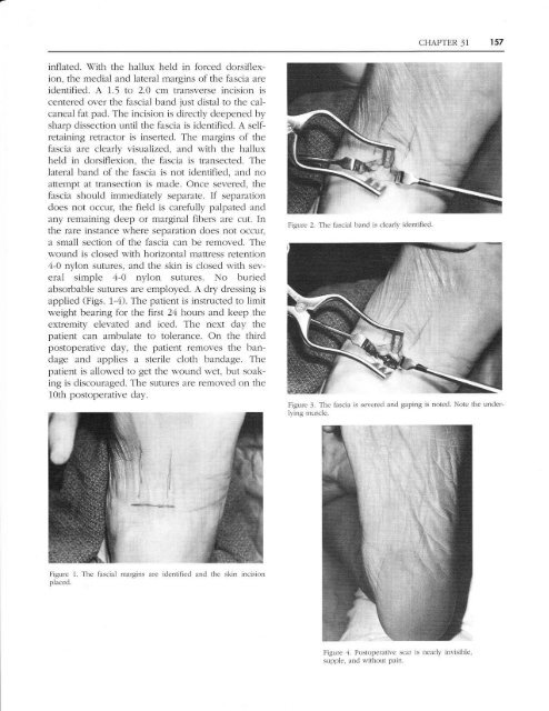
Figure 2. <strong>The</strong> fascial band is clearly identified<br />
Figure J. <strong>The</strong> fascia is sevcrecl and gaping is noted. Note the underlying<br />
rnuscle.<br />
Figure 1. <strong>The</strong> fiascial margins<br />
placed.<br />
are identified and the skin incision<br />
Figure ,1. Postoperative scal is nearly invisible.<br />
supple, and s,-ithollt pain,


