IN.STEP FASCIOTOMY - The Podiatry Institute
IN.STEP FASCIOTOMY - The Podiatry Institute
IN.STEP FASCIOTOMY - The Podiatry Institute
Create successful ePaper yourself
Turn your PDF publications into a flip-book with our unique Google optimized e-Paper software.
CHAPTER 3I<br />
<strong>IN</strong>.<strong>STEP</strong> <strong>FASCIOTOMY</strong><br />
Jelfrey Boberg, DP.M.<br />
It has been estimated that approximately l5o/o of<br />
patients presenting to the podiatrist's office complain<br />
of inferior heel pain. <strong>The</strong> vast majority of<br />
these cases are a mechanically induced periostitisfasciitis<br />
of the calcaneus with or without the<br />
presence of a spur. <strong>The</strong>se symptoms are variously<br />
referred to as heel spurs, plantar fasciitis, or more<br />
accurately, heel spur syndrome. <strong>The</strong> diagnosis is<br />
verified not by any specific clinical test or radiograph,<br />
but by a complexus of symptoms and<br />
clinical findings. <strong>The</strong> patient typically has pain<br />
upon rising in the morning, and after periods of<br />
rest. Following periods of prolonged ambulation,<br />
the pain may become constant and unremitting. On<br />
inspection, no edema or erythema is present. <strong>The</strong><br />
plantar medial aspect of the calcaneus is quite tender<br />
to palpation, and frequently the tenderness<br />
extends distally to include the proximal half of the<br />
plantar fascia. Pain can often be reproduced by<br />
dorsiflexing the digits, which places tension on the<br />
plantar fascia. Heel pain may present in either a<br />
ca\.Lls or valgus foot, but in the author's experience<br />
the flexible forefoot valgus foot-type is the most<br />
common to develop chronic heel pain.<br />
<strong>The</strong>se signs and symptoms are consistent with<br />
inflammation of the plantar fascia and perifascial<br />
structures. Clinical experience has shown that the<br />
presence or absence of a radiographically-defined<br />
spur does not correlate with a patient's symptoms.<br />
One study of randomly selected radiographs<br />
showed only 390/o of those with heel spurs as having<br />
a history of heel pain. It is now commonly<br />
accepted that tension on the plantar fascia by elongation<br />
of the medial arch, and dorsiflexion of the<br />
digits (windlass effect), are the pathomechanics<br />
associated with plantar f'asciitis.<br />
Treatment is designed to reduce the inflammation<br />
and eliminate its etiology. <strong>The</strong> majority of<br />
patients respond to various modalities including<br />
cofiicosteroids (both injectable and oral), NSAIDs,<br />
physical therapy, tapings, night splints, and<br />
orthotics, either individually or more commonly in<br />
combination. Unfortunately, a small subset of<br />
patients do not respond to conselvative care and<br />
require surgical interyention. Multiple surgeries<br />
throtrgh a variety of incisional approaches have<br />
been described. <strong>The</strong>se can be divided into two<br />
classes of procedures: osseous and soft tissue.<br />
Various types of osseous procedures have<br />
been described from osteotomy, drilling for decompression,<br />
excision of the tuberosity to simple<br />
exostectomy. For many years, denuding the calcaneus<br />
of its myofascial attachments and simple spur<br />
removal was the procedure of choice. Due to a<br />
prolonged recovery period and an estimated 50%<br />
success rate, this method was discounted by many<br />
surgeons, forcing patients to alter their lifestyles or<br />
continue to suffer with heel pain.<br />
Soft tissue surgery is based on the belief that<br />
the calcaneal spur is a reactive process and not a<br />
causative one. Medial calcaneal neurectomy and<br />
plantar neurolysis are directed at a relatively questionable,<br />
small group of patients with neurogenic<br />
pain. Plantar fasciotomy/fasciectomy compromise<br />
the largest group of soft tissue procedures. If<br />
tension on the pTantar fascia is the etiology of heel<br />
pain, then surgical release of the fascia should<br />
prove successful. Plantar fasciotomy for the relief<br />
of heel pain was described over half a century ago.<br />
However, it wasn't until Barrett and Day in 7991<br />
reported on the technique of endoscopic fasciotomy<br />
that the profession has seen an explosion<br />
in the number of soft tissue procedures being<br />
performed for heel pain. <strong>The</strong> early results, while<br />
encouraging, should cause us to pause and reflect.<br />
Barrett and Day report a complication rale<br />
involving 12 of 65 patients, most of these apparently<br />
minor. Kinley, et al. reported serious<br />
complications in 11 of 66 patients, and an additional<br />
15 patients with minor complications. <strong>The</strong><br />
author has been performing a fasciotomy for the<br />
last ,1 years and will describe the technique and<br />
postoperative results.<br />
TECHNIQUE<br />
Following the administration of a local anesthetic<br />
nen.e block, a pneumatic ankle tourniquet is
CHAPTER 31 157<br />
inflated. \fith the hallux held in forced clorsiflexion,<br />
the medial and lateral margins of the fascia are<br />
identified. A 1.5 to 2.0 cm transverse incision is<br />
centered over the fascial band just distal to the calcaneal<br />
fat pad. <strong>The</strong> incision is directly deepened by<br />
sharp dissection until the fascia is identified. A selfretaining<br />
retractor is inserted. <strong>The</strong> margins of the<br />
fascia are clearly visualized, and with the hallux<br />
held in dorsiflexion, the fascia is transected. <strong>The</strong><br />
lateral band of the fascia is not identified, and no<br />
attempt at transection is made. Once severed, the<br />
fascia should immediately separate. If separation<br />
does not occur, the field is carefully palpated and<br />
any remaining deep or marginal fibers are cut. In<br />
the rare instance where separation does not occllr,<br />
a small section of the fascia can be removed. <strong>The</strong><br />
wound is closed with horizontal mattress retention<br />
4-0 nylon sutures, and the skin is closed with several<br />
simple 4-0 nylon sutures. No buried<br />
absorbable sutures are employed. A dry dressing is<br />
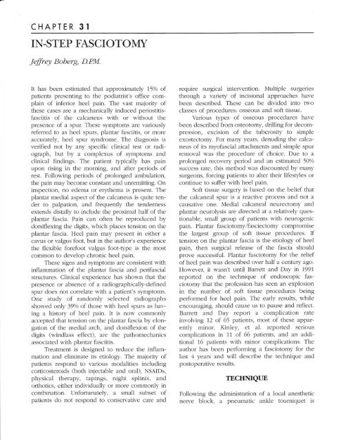
applied (Figs. 1-4). <strong>The</strong> patient is instructed to limit<br />
weight bearing for the first 24 hours and keep the<br />
extremity elevated and iced. <strong>The</strong> next day the<br />
patient can ambulate to tolerance. On the third<br />
postoperative day, the patient removes the bandage<br />
and applies a sterile cloth bandage. <strong>The</strong><br />
patient is allowed to get the wound wet, but soaking<br />
is discouraged. <strong>The</strong> sutures are relnoved on the<br />
10th postoperative day.<br />
Figure 2. <strong>The</strong> fascial band is clearly identified<br />
Figure J. <strong>The</strong> fascia is sevcrecl and gaping is noted. Note the underlying<br />
rnuscle.<br />
Figure 1. <strong>The</strong> fiascial margins<br />
placed.<br />
are identified and the skin incision<br />
Figure ,1. Postoperative scal is nearly invisible.<br />
supple, and s,-ithollt pain,
I58 CHAPTER 31<br />
RESULTS<br />
At the time of this writing, thirty-seven questionnaires<br />
were mailed. <strong>The</strong>se patients were at least 3<br />
months postoperative. <strong>The</strong> longest postoperative<br />
patient was 3.5 years. Fifteen responses have been<br />
received to date. All patients with the exception of<br />
one, had pain existing for over one year prior to<br />
surgery. \7ith pain rated on a scale of 0-5 (O-no<br />
pain, 5-severe pain) the average respondent had a<br />
pain level of 4.65 prior to surgery. Postoperative<br />
heel pain was 1.59. Postoperative foot pain was<br />
2.0, with most patients experiencing either medial<br />
arch or lateral pain. <strong>The</strong> average time to full recovery<br />
with a minimum of pain was 3.4 weeks.<br />
Recurring heel pain or pain which develops in<br />
other parts of the foot tends to develop early in the<br />
postoperative period.<br />
Closer review of the postoperative results<br />
shows two distinct categories of results. <strong>The</strong> majority<br />
of patients are very satisfied with their surgery<br />
and would recommend it to a family member or<br />
friend. A small subset (three patients) showed<br />
absolutely no improvement. <strong>The</strong>se patients were all<br />
extremely obese, and two had previous operations<br />
(heel spur resection, tarsal tunnel release).<br />
Plantar fasciotomy for the relief of chronic<br />
heel pain is very effective in most individuals.<br />
However, no procedure is w'ithout risk. Until guidelines<br />
delineate which patients will not respond to<br />
conserwative care and will be helped by surgery, all<br />
forms of conservative care should be exhausted<br />
prior to surgical intervention.<br />
DISCUSSION<br />
<strong>The</strong> fasciotomy technique described appears<br />
to be comparable to results previously reported for<br />
the endoscopic technique. <strong>The</strong> instep fasciotomy is<br />
simple to perform, requires minimal time to<br />
become proficient, does not require any special<br />
instrumentation, and can easily be performed in<br />
the office in 10 to 15 minutes.<br />
<strong>The</strong> location of the skin incision is important<br />
to the success of this procedure. Initially, the<br />
author chose a percutaneous incision in the medial<br />
arch, where the bowstringing of the fascia is most<br />
evident. Several of these patients developed a<br />
thickening of the subcutaneous tissue which was<br />
quite uncomfortable to walk on, although their<br />
heel pain was relieved. <strong>The</strong> ideal incision location<br />
appears to be just distal to the heel pad. This incision<br />
is in a non-weight-bearing part of the foot, and<br />
has enough overlying subcutaneous tissue so the<br />
fascial scar is not apparent.


