April 2007 - Division of Biology and Medicine - Brown University
April 2007 - Division of Biology and Medicine - Brown University
April 2007 - Division of Biology and Medicine - Brown University

You also want an ePaper? Increase the reach of your titles
YUMPU automatically turns print PDFs into web optimized ePapers that Google loves.
Images In <strong>Medicine</strong><br />
Malignant Gastric Ulcer on Multidetector Row CT<br />
<strong>and</strong> Endoscopy<br />
Brian D. Midkiff, MD, MPH, Philip A. McAndrew, MD, <strong>and</strong> William W. Mayo-Smith, MD<br />
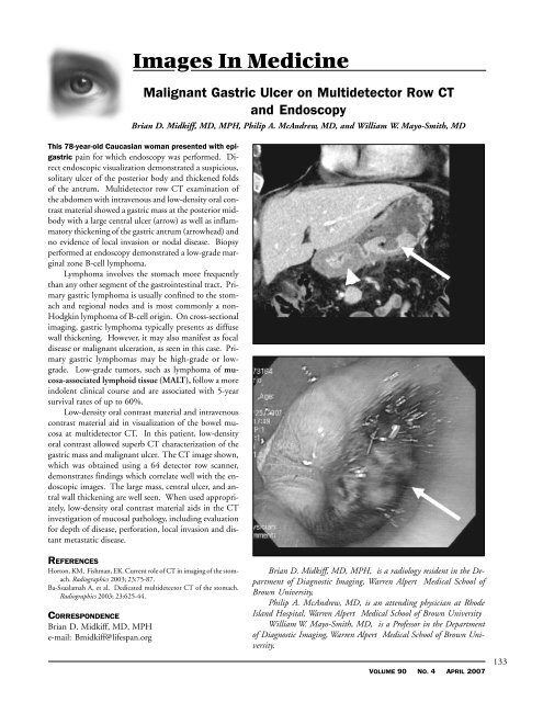
This 78-year-old Caucasian woman presented with epigastric<br />
pain for which endoscopy was performed. Direct<br />
endoscopic visualization demonstrated a suspicious,<br />
solitary ulcer <strong>of</strong> the posterior body <strong>and</strong> thickened folds<br />
<strong>of</strong> the antrum. Multidetector row CT examination <strong>of</strong><br />
the abdomen with intravenous <strong>and</strong> low-density oral contrast<br />
material showed a gastric mass at the posterior midbody<br />
with a large central ulcer (arrow) as well as inflammatory<br />
thickening <strong>of</strong> the gastric antrum (arrowhead) <strong>and</strong><br />
no evidence <strong>of</strong> local invasion or nodal disease. Biopsy<br />
performed at endoscopy demonstrated a low-grade marginal<br />
zone B-cell lymphoma.<br />
Lymphoma involves the stomach more frequently<br />
than any other segment <strong>of</strong> the gastrointestinal tract. Primary<br />
gastric lymphoma is usually confined to the stomach<br />
<strong>and</strong> regional nodes <strong>and</strong> is most commonly a non-<br />
Hodgkin lymphoma <strong>of</strong> B-cell origin. On cross-sectional<br />
imaging, gastric lymphoma typically presents as diffuse<br />
wall thickening. However, it may also manifest as focal<br />
disease or malignant ulceration, as seen in this case. Primary<br />
gastric lymphomas may be high-grade or lowgrade.<br />
Low-grade tumors, such as lymphoma <strong>of</strong> mucosa-associated<br />
lymphoid tissue (MALT), follow a more<br />
indolent clinical course <strong>and</strong> are associated with 5-year<br />
survival rates <strong>of</strong> up to 60%.<br />
Low-density oral contrast material <strong>and</strong> intravenous<br />
contrast material aid in visualization <strong>of</strong> the bowel mucosa<br />
at multidetector CT. In this patient, low-density<br />
oral contrast allowed superb CT characterization <strong>of</strong> the<br />
gastric mass <strong>and</strong> malignant ulcer. The CT image shown,<br />
which was obtained using a 64 detector row scanner,<br />
demonstrates findings which correlate well with the endoscopic<br />
images. The large mass, central ulcer, <strong>and</strong> antral<br />
wall thickening are well seen. When used appropriately,<br />
low-density oral contrast material aids in the CT<br />
investigation <strong>of</strong> mucosal pathology, including evaluation<br />
for depth <strong>of</strong> disease, perforation, local invasion <strong>and</strong> distant<br />
metastatic disease.<br />
REFERENCES<br />
Horton, KM, Fishman, EK. Current role <strong>of</strong> CT in imaging <strong>of</strong> the stomach.<br />
Radiographics 2003; 23:75-87.<br />
Ba-Ssaalamah A, et al. Dedicated multidetector CT <strong>of</strong> the stomach.<br />
Radiographics 2003; 23:625-44.<br />
CORRESPONDENCE<br />
Brian D. Midkiff, MD, MPH<br />
e-mail: Bmidkiff@lifespan.org<br />
Brian D. Midkiff, MD, MPH, is a radiology resident in the Department<br />
<strong>of</strong> Diagnostic Imaging, Warren Alpert Medical School <strong>of</strong><br />
<strong>Brown</strong> <strong>University</strong>,<br />
Philip A. McAndrew, MD, is an attending physician at Rhode<br />
Isl<strong>and</strong> Hospital, Warren Alpert Medical School <strong>of</strong> <strong>Brown</strong> <strong>University</strong><br />
William W. Mayo-Smith, MD, is a Pr<strong>of</strong>essor in the Department<br />
<strong>of</strong> Diagnostic Imaging, Warren Alpert Medical School <strong>of</strong> <strong>Brown</strong> <strong>University</strong>.<br />
VOLUME 90 NO. 4 APRIL <strong>2007</strong><br />
133