April 2007 - Division of Biology and Medicine - Brown University
April 2007 - Division of Biology and Medicine - Brown University
April 2007 - Division of Biology and Medicine - Brown University

You also want an ePaper? Increase the reach of your titles
YUMPU automatically turns print PDFs into web optimized ePapers that Google loves.
Orthopedics<br />
Volume 90 No. 4 <strong>April</strong> <strong>2007</strong>
UNDER THE JOINT<br />
EDITORIAL SPONSORSHIP OF:<br />
The Warren Alpert Medical School <strong>of</strong><br />
<strong>Brown</strong> <strong>University</strong><br />
Eli Y. Adashi, MD, Dean <strong>of</strong> <strong>Medicine</strong><br />
& Biological Science<br />
Rhode Isl<strong>and</strong> Department <strong>of</strong> Health<br />
David R. Gifford, MD, MPH, Director<br />
Quality Partners <strong>of</strong> Rhode Isl<strong>and</strong><br />
Richard W. Besdine, MD, Chief<br />
Medical Officer<br />
Rhode Isl<strong>and</strong> Medical Society<br />
Barry W. Wall, MD, President<br />
EDITORIAL STAFF<br />
Joseph H. Friedman, MD<br />
Editor-in-Chief<br />
Joan M. Retsinas, PhD<br />
Managing Editor<br />
Stanley M. Aronson, MD, MPH<br />
Editor Emeritus<br />
EDITORIAL BOARD<br />
Stanley M. Aronson, MD, MPH<br />
Jay S. Buechner, PhD<br />
John J. Cronan, MD<br />
James P. Crowley, MD<br />
Edward R. Feller, MD<br />
John P. Fulton, PhD<br />
Peter A. Hollmann, MD<br />
Sharon L. Marable, MD, MPH<br />
Anthony E. Mega, MD<br />
Marguerite A. Neill, MD<br />
Frank J. Schaberg, Jr., MD<br />
Lawrence W. Vernaglia, JD, MPH<br />
Newell E. Warde, PhD<br />
OFFICERS<br />
Barry W. Wall, MD<br />
President<br />
K. Nicholas Tsiongas, MD, MPH<br />
President-Elect<br />
Diane R. Siedlecki, MD<br />
Vice President<br />
Margaret A. Sun, MD<br />
Secretary<br />
Mark S. Ridlen, MD<br />
Treasurer<br />
Kathleen Fitzgerald, MD<br />
Immediate Past President<br />
DISTRICT & COUNTY PRESIDENTS<br />
Ge<strong>of</strong>frey R. Hamilton, MD<br />
Bristol County Medical Society<br />
Herbert J. Brennan, DO<br />
Kent County Medical Society<br />
Rafael E. Padilla, MD<br />
Pawtucket Medical Association<br />
Patrick J. Sweeney, MD, MPH, PhD<br />
Providence Medical Association<br />
Nitin S. Damle, MD<br />
Washington County Medical Society<br />
Jacques L. Bonnet-Eymard, MD<br />
Woonsocket District Medical Society<br />
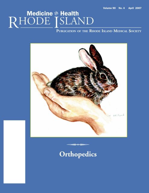
Cover: “Rabbit In H<strong>and</strong>,” watercolor,<br />
2006, by Nancy Whitcomb, a Providence-based<br />
painter, illustrator <strong>and</strong> art<br />
teacher. She exhibits locally at the galleries<br />
<strong>of</strong> the Providence Art Club, <strong>and</strong><br />
can be reached by e-mail at<br />
nsw125@cox.net.<br />
<strong>Medicine</strong> Health<br />
VOLUME 90 NO. 4 <strong>April</strong> <strong>2007</strong><br />
R HODE I SLAND<br />
PUBLICATION OF THE RHODE ISLAND MEDICAL SOCIETY<br />
COMMENTARIES<br />
106 The Journal <strong>of</strong> Goldberg Research [<strong>April</strong> Fool]<br />
Joseph H. Friedman, MD<br />
107 Jefferson Consults a British Physician<br />
Stanley M. Aronson, MD<br />
CONTRIBUTIONS<br />
SPECIAL ISSUE: ORTHOPEDICS<br />
Guest Editor: Michael G. Ehrlich, MD<br />
108 Introduction: Orthopedics<br />
Michael G. Ehrlich, MD<br />
108 Pediatric Upper Extremity Conditions: Traumatic <strong>and</strong> Congenital<br />
Julia A. Katarincic, MD<br />
111 Adolescents <strong>and</strong> Upper Extremity Sports Injuries: The Throwing Arm<br />
Robert M. Shalvoy, MD<br />
115 Idiopathic Scoliosis In Children: An Update<br />
Craig P. Eberson, MD<br />
122 Pediatric Femur Fractures: Treatment In the Year <strong>2007</strong><br />
Patricia Solga, MD<br />
COLUMNS<br />
127 GERIATRICS FOR THE PRACTICING PHYSICIAN – Transitional Care<br />
John Kleckley, MD<br />
129 HEALTH BY NUMBERS – Arthritis <strong>and</strong> Associated Health Conditions <strong>and</strong><br />
Risks Among Rhode Isl<strong>and</strong> Adults in 2005<br />
Jana Hesser, PhD, Yongwen Jiang, PhD, <strong>and</strong> Patricia Rajotte<br />
132 POINT OF VIEW – Retiree-Volunteers <strong>and</strong> the Abbreviation Study<br />
Joyce Binyon <strong>and</strong> Maureen McGarry<br />
133 IMAGES IN MEDICINE – Malignant Gastric Ulcer on Multidetector<br />
Row CT <strong>and</strong> Endoscopy<br />
Brian D. Midkiff, MD, MPH, Philip A. McAndrew, MD, <strong>and</strong><br />
William W. Mayo-Smith, MD<br />
134 PHYSICIAN’S LEXICON – A Well-Nourished Vocabulary<br />
Stanley M. Aronson, MD<br />
134 Vital Statistics<br />
136 <strong>April</strong> Heritage<br />
<strong>Medicine</strong> <strong>and</strong> Health/Rhode Isl<strong>and</strong> (USPS 464-820), a monthly publication, is owned <strong>and</strong> published by the Rhode Isl<strong>and</strong> Medical Society, 235<br />
Promenade St., Suite 500, Providence, RI 02908, Phone: (401) 331-3207. Single copies $5.00, individual subscriptions $50.00 per year, <strong>and</strong> $100<br />
per year for institutional subscriptions. Published articles represent opinions <strong>of</strong> the authors <strong>and</strong> do not necessarily reflect the <strong>of</strong>ficial policy <strong>of</strong> the Rhode Isl<strong>and</strong><br />
Medical Society, unless clearly specified. Advertisements do not imply sponsorship or endorsement by the Rhode Isl<strong>and</strong> Medical Society. Periodicals postage<br />
paid at Providence, Rhode Isl<strong>and</strong>. ISSN 1086-5462. POSTMASTER: Send address changes to <strong>Medicine</strong> <strong>and</strong> Health/Rhode Isl<strong>and</strong>, 235 Promenade St.,<br />
Suite 500, Providence, RI 02908. Classified Information: RI Medical Journal Marketing Department, P.O. Box 91055, Johnston, RI 02919,<br />
phone: (401) 383-4711, fax: (401) 383-4477, e-mail: rimj@cox.net. Production/Layout Design: John Teehan, e-mail: jdteehan@sff.net.<br />
VOLUME 90 NO. 4 APRIL <strong>2007</strong><br />
105
Commentaries<br />
106<br />
The Journal <strong>of</strong> Goldberg Research [<strong>April</strong> Fool]<br />
For those readers who have never scrolled<br />
through the incredible list <strong>of</strong> journals<br />
available on-line at <strong>Brown</strong>, there are wonders<br />
to behold. There are over 10,000<br />
journals, I think, probably a lot more. You<br />
can find annals, archives, journals, <strong>and</strong><br />
transactions <strong>of</strong> almost every topic under<br />
the sun.<br />
Every once in a while I stumble on a<br />
new journal that makes me take an instant<br />
detour from the search path I had intended.<br />
My favorite journal, which I truly<br />
invite the reader with access to the <strong>Brown</strong><br />
library system to look up, is The Journal <strong>of</strong><br />
Near-Death Studies, a publication <strong>of</strong> the<br />
International Association <strong>of</strong> Near-Death<br />
Experiences. Unfortunately, in 2003, this<br />
journal passed beyond near-death <strong>and</strong> is<br />
either hibernating or fully dead. The topics<br />
covered in the journal are, in fact fascinating,<br />
such as multi-dimensionality <strong>of</strong> the<br />
near death experience, but readers who<br />
may have experienced near-death experiences<br />
will probably not find this amusing.<br />
The very next journal in the <strong>Brown</strong><br />
list, which is alphabetically arranged, is the<br />
Journal <strong>of</strong> Negative Results in Biomedicine.<br />
This journal is based on a terrific idea.<br />
Imagine the possibilities <strong>of</strong> publishing all<br />
those results that no other journal wants<br />
because they’re negative. Of course, there<br />
are <strong>of</strong>ten good reasons to publish negative<br />
studies. They <strong>of</strong>ten contradict current<br />
treatment paradigms, based more on<br />
“common sense” or “st<strong>and</strong>ard practice”<br />
than on evidence; however, a whole journal<br />
<strong>of</strong> negative studies must be playing to<br />
the grinch in us. Who wants to read only<br />
about things that fail? When your promotions<br />
committee reviews your CV won’t<br />
you worry that they will think this is a joke,<br />
like being published in the Journal <strong>of</strong><br />
Irreproducible Results, another journal, really<br />
dead before its time?<br />
I thought the idea <strong>of</strong> a Journal <strong>of</strong> Negative<br />
Results to be a great idea that should<br />
have universal application. Why just a journal<br />
<strong>of</strong> negative results in biomedicine?<br />
What is so special about medicine? Why<br />
MEDICINE & HEALTH/RHODE ISLAND<br />
<br />
not bioengineering, biophysics, metaphysics,<br />
“real” physics, organic chemistry, whatever?<br />
The Journal <strong>of</strong> Airplanes that Don’t<br />
Fly? If there are positive studies to be published,<br />
there must be negative studies that<br />
need to see the light <strong>of</strong> day as well. I have<br />
formed a committee to study the possibility<br />
<strong>of</strong> a franchise for Journals <strong>of</strong> Negative<br />
Results. If successful we can publish a journal<br />
devoted to the “best” or most exciting<br />
negative results which might be published<br />
annually in The Best Negative Results, or<br />
The Journal <strong>of</strong> the Best Studies <strong>of</strong> Things that<br />
Don’t Work.<br />
But what about results that are sort<br />
<strong>of</strong> in the middle? The Journal <strong>of</strong> Nearly-<br />
Effective Therapies would be perfect for<br />
that. Imagine if you had a treatment that<br />
worked a little better than placebo, say,<br />
something like the cholinesterase inhibitors<br />
in Alzheimer’s disease, or riluzole for<br />
amyotrophic lateral sclerosis. Instead <strong>of</strong><br />
getting trumpeted in the New Engl<strong>and</strong><br />
Bugle, where statistical significance trumps<br />
clinical significance, they could be published<br />
in the journal where they belong,<br />
where all results are nearly-effective. My<br />
critics have charged that this journal really<br />
should be called The Journal <strong>of</strong> Nearly<br />
Effective Therapies that Cost a Lot <strong>of</strong> Money,<br />
<strong>and</strong> I can hardly disagree. Why not spend<br />
a lot for something that is significant? And<br />
the placebo effect <strong>of</strong> spending a lot cannot<br />
be overemphasized.<br />
Of course, one can also make an argument<br />
for a journal to cover insignificant<br />
results. This ambiguous title, Journal<br />
<strong>of</strong> Insignificant Results, could include<br />
results that were statistically insignificant,<br />
with a “trend” towards significance, or it<br />
can include statistically significant, clinically<br />
insignificant results, thus including<br />
results that are either clinically or statistically<br />
insignificant. I had tried to establish,<br />
Power, The Journal <strong>of</strong> Small Clinical<br />
Trials, but no one was interested 10 years<br />
ago. Now its time may be drawing near<br />
as “Designer Journals” become more<br />
popular.<br />
For those who become more emotionally<br />
involved, manuscripts can be published<br />
in the Journal <strong>of</strong> Disappointing Results.<br />
But the worst journal <strong>of</strong> all is the Journal<br />
<strong>of</strong> Boring Results, which has had difficulties<br />
getting peer reviewers.<br />
Probably the most interesting journal<br />
is the Journal <strong>of</strong> Goldberg Research because<br />
the results are always so unpredictable. I<br />
learned about Goldberg research when I<br />
was a resident. I rotated with a neuro-ophthalmologist<br />
who began discussing the<br />
problems <strong>of</strong> having a bad fellow or resident.<br />
These take up a lot <strong>of</strong> time without<br />
any reward for the attending physician,<br />
so they are sent <strong>of</strong>f to do “Goldberg research.”<br />
Of course the fellow <strong>and</strong> I, hoping<br />
not to be invited to join the Goldberg<br />
research working group, did not know<br />
what this was, <strong>and</strong> asked. The attending<br />
explained, “You tell the fellow to go to the<br />
chart room <strong>and</strong> review the charts <strong>of</strong> everyone<br />
ever admitted to the hospital<br />
named Goldberg <strong>and</strong> write down why<br />
they are admitted.” In a New York City<br />
hospital there are a lot <strong>of</strong> Goldberg charts<br />
to be perused.<br />
Personally I am a bit gladdened that<br />
the Journal <strong>of</strong> Near-Death Studies has fulfilled<br />
its destiny. There are way too many<br />
journals in the world <strong>and</strong> they cost far too<br />
much. Neurology, the journal <strong>of</strong> the American<br />
Academy <strong>of</strong> Neurology, has gone from<br />
monthly to biweekly to weekly in the last<br />
decade. It is too much. At first as the journals<br />
multiply they are thin, but they soon<br />
thicken, <strong>and</strong> the amount <strong>of</strong> material to be<br />
ignored increases too quickly. Journals<br />
summarizing the other journals now exist<br />
<strong>and</strong> soon there will be journals summarizing<br />
the journals summarizing the actual<br />
journals, the “source” documents. As we<br />
get to know more <strong>and</strong> more about less <strong>and</strong><br />
less, I wonder about how many <strong>of</strong> these<br />
articles should have been published in the<br />
Journal <strong>of</strong> Goldberg Research.<br />
– JOSEPH H. FRIEDMAN,MD
Jefferson Consults a British Physician<br />
A review <strong>of</strong> the st<strong>and</strong>ard American medical school curriculum<br />
reveals little that might prepare future physicians for political<br />
leadership; <strong>and</strong> certainly nothing to educate them in the rudiments<br />
<strong>of</strong> political philosophy. The mission <strong>of</strong> the four years <strong>of</strong><br />
medical schooling in this nation is simply to prepare c<strong>and</strong>idates<br />
for a competent <strong>and</strong> compassionate practice <strong>of</strong> medicine<br />
with little attention invested in the technical intricacies <strong>of</strong> public<br />
<strong>of</strong>fice or public policy.<br />
Accordingly, American physicians have rarely entered the<br />
domain <strong>of</strong> politics [the recently retired senator from Tennessee,<br />
William H. Frist, MD, is a notable exception.] History<br />
texts, however, do recall those rare physicians who had forsaken<br />
their caring ministry to take on new roles <strong>of</strong> political<br />
leadership. Sun Yat-Sen, for example, relinquished his medical<br />
practice in Hong Kong to assume the first presidency <strong>of</strong> China<br />
in the early 20th Century. Georges Clemenceau went from<br />
the general practice <strong>of</strong> medicine to polemic journalism <strong>and</strong><br />
then, finally, to the premiership <strong>of</strong> France, leading his embattled<br />
nation during the hazardous years <strong>of</strong> the first World War. And<br />
<strong>of</strong> course a physician from Buenos Aires, Ernesto “Che”<br />
Guevaro , played an important role in the Cuban revolution<br />
<strong>of</strong> the 1950s. But these are rare exceptions.<br />
The major players in the political leadership <strong>of</strong> this nation<br />
have largely been lawyers <strong>and</strong> career military personnel with<br />
an assortment <strong>of</strong> diverse pr<strong>of</strong>essions but no physicians. Of our<br />
43 presidents, 24 had been lawyers, six had primary careers in<br />
the military, three were farmers or ranchers [T. Roosevelt, L. B.<br />
Johnson <strong>and</strong> J. Carter], four were in business [A. Johnson, H.<br />
Truman, G. H. W. Bush <strong>and</strong> G. W. Bush], two were in teaching<br />
[W. Harding <strong>and</strong> W. Wilson], two were writers or editors<br />
[W. Harding <strong>and</strong> J. Kennedy], one was an engineer [H. Hoover]<br />
<strong>and</strong> one was an actor [R. Reagan]. Not a single physician in<br />
this group <strong>of</strong> presidents although one, William Henry Harrison,<br />
had briefly attended medical school.<br />
Yet some practitioners <strong>of</strong> medicine, in a quiet way, exerted<br />
a marginal influence in molding the political agenda <strong>of</strong> this<br />
nation during its formative days. Fifty-six men signed the Declaration<br />
<strong>of</strong> Independence. And in doing so pledged their lives,<br />
their fortunes <strong>and</strong> their sacred honor in behalf <strong>of</strong> a statement<br />
asserting their freedom from British rule. Of these 56 Americans,<br />
five were physicians: Josiah Bartlett <strong>of</strong> New Hampshire,<br />
Lyman Hall <strong>of</strong> Georgia, Benjamin Rush <strong>of</strong> Pennsylvania, Matthew<br />
Thornton <strong>of</strong> New Hampshire <strong>and</strong> Oliver Wolcott <strong>of</strong><br />
Connecticut.<br />
These five physician-patriots signed this precious document<br />
<strong>and</strong> then some served as military physicians <strong>and</strong> later as<br />
judges <strong>and</strong> legislators. Their signatures on the Declaration attested<br />
to their agreement with its declared premises, but the<br />
inspired words <strong>of</strong> this immortal document were assembled solely<br />
by a lawyer <strong>and</strong> scholar from Virginia, Thomas Jefferson.<br />
From what source, either within himself or from the writings<br />
<strong>of</strong> others, did Jefferson distill such emboldened thoughts<br />
as his assertion that certain truths are self-evident ? And that<br />
men, having been created equal, are accordingly endowed with<br />
<br />
certain unalienable rights such as life, liberty <strong>and</strong> the pursuit<br />
<strong>of</strong> happiness? Jefferson’s home in Monticello contained an extensive<br />
library; <strong>and</strong> prominent in this collection <strong>of</strong> scholarly<br />
texts were the writings <strong>of</strong> an English philosopher <strong>and</strong> physician<br />
named John Locke. Jefferson as well as some <strong>of</strong> his colleagues,<br />
readily acknowledged the enormous influence exerted<br />
by Locke upon the principles motivating the separation <strong>of</strong> the<br />
colonies from their mother country.<br />
Who was Locke ? Engl<strong>and</strong>’s most eminent philosopher<br />
<strong>and</strong> political theorist was born in 1632 in Somersetshire. His<br />
father was a modest l<strong>and</strong>owner, a village attorney, a devout<br />
Puritan <strong>and</strong> a fervent adherent <strong>of</strong> the anti-royalist Parliamentary<br />
party. Young Locke was educated at home by his father<br />
until age 14. Those were years <strong>of</strong> great social turmoil <strong>and</strong> Locke’s<br />
father left home periodically to join the forces <strong>of</strong> Cromwell in<br />
fighting the troops <strong>of</strong> King Charles. In 1652 young Locke<br />
entered Christ Church College, Oxford. The college in those<br />
civil war days was firmly on the side <strong>of</strong> those who advocated<br />
religious tolerance.<br />
Locke prospered at Oxford, finding it an academic community<br />
that challenged his inquisitive mind. After receiving<br />
his baccalaureate degree, he stayed on to study both medicine<br />
<strong>and</strong> philosophy, enamored with the writings <strong>of</strong> Descartes <strong>and</strong><br />
Hobbes. He was moved, too, by the new spirit <strong>of</strong> experimentalism<br />
<strong>and</strong> even joined the Royal Society, Engl<strong>and</strong>’s renowned<br />
association <strong>of</strong> physical <strong>and</strong> biological scientists. For the next<br />
few years Locke practiced medicine but was attracted by the<br />
political <strong>and</strong> intellectual circle surrounding the Earl <strong>of</strong><br />
Shaftsbury. Locke transferred to London to enter the household<br />
<strong>of</strong> Shaftsbury as physician <strong>and</strong> confidential secretary.<br />
During the restoration years immediately after 1660,<br />
Locke’s status in Engl<strong>and</strong> became perilous, <strong>and</strong> he fled to<br />
France, living there for a number <strong>of</strong> years. Then back to Engl<strong>and</strong><br />
<strong>and</strong> then fleeing again, this time to the tolerant environment<br />
<strong>of</strong> Holl<strong>and</strong>, where the influence <strong>of</strong> Erasmus, Grotius,<br />
Descartes <strong>and</strong> Spinoza prevailed.<br />
During these years <strong>of</strong> political turmoil <strong>and</strong> flight Locke<br />
composed his enduring essays on the nature <strong>of</strong> civil government<br />
<strong>and</strong> the merit <strong>of</strong> political, civil <strong>and</strong> religious tolerance.<br />
And it was in Locke’s Second Treatise on Government that he<br />
set down the many memorable phrases that stirred Jefferson’s<br />
ideological soul. These were thrilling concepts such as the view<br />
that the state exists solely to preserve the rights <strong>of</strong> its citizens,<br />
Or further, that certain rights were not gifts to be granted arbitrarily<br />
by those in temporal authority but, rather, unalienable<br />
rights which included the rights <strong>of</strong> life, liberty, property<br />
<strong>and</strong> the pursuit <strong>of</strong> a tranquil existence [a phrase that Jefferson<br />
had amended to a pursuit <strong>of</strong> happiness.]<br />
Somewhere, mingling congenially with our founding fathers,<br />
was the amiable ghost <strong>of</strong> a sometime physician named<br />
John Locke.<br />
– STANLEY M. ARONSON, MD<br />
VOLUME 90 NO. 4 APRIL <strong>2007</strong><br />
107
This issue focuses on some <strong>of</strong> the many<br />
musculoskeletal issues that the primary care<br />
physician confronts on a regular basis. Some<br />
estimates have suggested that as many as half<br />
<strong>of</strong> the cases seen in a generalist’s <strong>of</strong>fice are<br />
related to the musculoskeletal system. Patients<br />
<strong>of</strong>ten ask their primary care physicians, for<br />
advice, especially since many <strong>of</strong> these problems<br />
occur in children. These questions may<br />
relate to timing <strong>of</strong> surgery, the need for it or<br />
the newer techniques now available.<br />
For example, Julia Katarincic, in her<br />
essay on congenital problems in children,<br />
points out that surgery on syndactaly should<br />
generally be deferred until two years <strong>of</strong> age.<br />
Otherwise, the web between the fingers<br />
tends to recur. On the other h<strong>and</strong>, if there<br />
is a big discrepancy between lengths <strong>of</strong> adjacent<br />
fingers, then the longer one will<br />
bend, so they should be done earlier. These<br />
pearls should remain on your shelf as a great<br />
reference.<br />
Similarly, an obstetrician should be able<br />
to tell parents <strong>of</strong> a child with brachial birth<br />
palsy that most will recover. The pediatrician<br />
or primary care physician should know<br />
that if biceps function fails to develop by six<br />
months, microsurgical repair is indicated.<br />
These <strong>and</strong> other points in the article should<br />
Introduction: Orthopedics<br />
<br />
Michael G. Ehrlich, MD<br />
be in the knowledge base <strong>of</strong> the primary<br />
care provider.<br />
Rob Shalvoy addresses the very important<br />
issue <strong>of</strong> over-abuse injuries in adolescents<br />
engaged in sports. He explains how<br />
little league pitchers crush the lateral joint<br />
surfaces <strong>of</strong> the elbow, <strong>and</strong> tear the ligaments<br />
on the medial. He goes on to explain how<br />
the injuries can be avoided by certain limitations<br />
on the numbers <strong>and</strong> types <strong>of</strong><br />
pitches. Similar problems affect swimmers<br />
<strong>and</strong> other athletes, sometimes who are being<br />
egged on by their parents. The lessons<br />
in his article should be shared by physicians<br />
with the local trainers <strong>and</strong> coaches.<br />
Craig Eberson is primarily interested<br />
in scoliosis <strong>and</strong> other deformities <strong>of</strong> the<br />
spine. Knowing when to refer these patients<br />
is <strong>of</strong> vital importance, since minor<br />
curves are ubiquitous in the population, <strong>and</strong><br />
thous<strong>and</strong>s <strong>of</strong> children are being identified<br />
by school screening programs. Most <strong>of</strong><br />
these children don’t need to go to a musculoskeletal<br />
specialist, <strong>and</strong> it saves society<br />
money, parents time, <strong>and</strong> children, needless<br />
x-rays if the primary care physician is<br />
properly involved. He also has interesting<br />
perspectives on the new treatments for<br />
scoliosis, including the use <strong>of</strong> thorascopic<br />
surgery, nighttime bracing, <strong>and</strong> the use <strong>of</strong><br />
pedicle screws.<br />
Finally Patricia Solga has contributed a<br />
very interesting article on the current management<br />
<strong>of</strong> femur fractures in children. Of<br />
all the developments in pediatric orthopaedics,<br />
this area has undergone the most<br />
radical changes in the last decade. She discusses<br />
the new role <strong>of</strong> treating infant femur<br />
fractures with Pavlik harnesses, the orthosis<br />
used primarily for hip dislocations, <strong>and</strong> the<br />
new reliance on internal fixation as opposed<br />
to traction <strong>and</strong> spica casts. This has great<br />
implications for the parents now taking care<br />
<strong>of</strong> these children, <strong>and</strong> provides much relief<br />
compared to the old ways.<br />
Therefore, we hope that you find this<br />
issue challenging, interesting <strong>and</strong> useful.<br />
Michael G. Ehrlich, MD, is Vincent<br />
Zecchino Pr<strong>of</strong>essor <strong>and</strong> Chairman, Department<br />
<strong>of</strong> Orthopaedics, Warren Alpert Medical<br />
School <strong>of</strong> <strong>Brown</strong> <strong>University</strong>, <strong>and</strong> Surgeonin-Chief,<br />
Orthopaedics, Rhode Isl<strong>and</strong> <strong>and</strong><br />
Miriam Hospitals.<br />
CORRESPONDENCE:<br />
Michael G. Ehrlich, MD<br />
e-mail: Mehrlich@lifespan.org<br />
108<br />
Children may present with a variety <strong>of</strong><br />
upper extremity conditions. The cause<br />
can be traumatic, congenital or developmental.<br />
1,2 The first physician involved in<br />
the diagnosis <strong>of</strong> congenital or developmental<br />
processes is the pediatrician or<br />
family practitioner. Parents with injured<br />
children <strong>of</strong>ten seek emergency treatment,<br />
with follow-up by the child’s primary<br />
physician.<br />
EMBRYOLOGY<br />
The upper extremity limb bud first<br />
appears on the dorsal ventral embryo at<br />
about 4 weeks’ gestation; development is<br />
complete by 8 weeks. The development<br />
MEDICINE & HEALTH/RHODE ISLAND<br />
Pediatric Upper Extremity Conditions:<br />
Traumatic <strong>and</strong> Congenital<br />
Julia A. Katarincic, MD<br />
<br />
<strong>of</strong> the arm is about 2 weeks ahead <strong>of</strong> leg<br />
development. The limb bud first appears<br />
as a paddle on the dorsal side <strong>of</strong> the embryo.<br />
This is important to remember because<br />
<strong>of</strong> the proximity early in gestation<br />
to the chest wall. Certain early insults<br />
such as brachsyndactyly (short/ absent<br />
fingers <strong>and</strong> metacarpals) may be associated<br />
with Pol<strong>and</strong>’s syndrome (absent pectoralis<br />
muscle). The proximal to distal<br />
growth <strong>of</strong> the limb bud is controlled by<br />
the sonic hedgehog gene. Any insult to<br />
this gene can lead to a congential amputation.<br />
Radial to ulnar limb growth is<br />
controlled by the wnt gene. Problems<br />
related to this gene include radial or ulnar<br />
dysplasia. 3,4 The h<strong>and</strong> paddle is complete<br />
at 6 weeks. Apoptosis, or<br />
preprogrammed cell death, will cause the<br />
tissue <strong>of</strong> the finger clefts to regress leading<br />
to a five fingered h<strong>and</strong>. 5 A disruption<br />
in the process can lead to syndactytly<br />
(webbed fingers).<br />
SYNDACTYLY<br />
Apoptosis occurs in the h<strong>and</strong> paddle<br />
at about 6 weeks, leading to a five-fingered<br />
h<strong>and</strong>. Any disruption in this<br />
preprogrammed cell death will lead to<br />
syndactyly or webbed fingers. Syndactyly<br />
is classified as complete or incomplete <strong>and</strong><br />
complex or simple. Complete syndactyly
is when the fingers are joined together<br />
all the way to the tip. Incomplete syndactyly<br />
means the webbing stops before<br />
the end <strong>of</strong> the fingers. In a simple syndactyly,<br />
bone structure is normal. In a<br />
patient with complex syndactyly, there<br />
are <strong>of</strong>ten more bones than normal with<br />
anomalous attachments <strong>of</strong> muscles. Treatment<br />
for these children is separation to<br />
improve h<strong>and</strong> span <strong>and</strong> help function.<br />
Typically 18-24 months <strong>of</strong> age is the appropriate<br />
age for the division. Doing the<br />
surgery at a younger age increases the<br />
chance <strong>of</strong> web creep, a recurrence <strong>of</strong> the<br />
syndactyly at the base <strong>of</strong> the fingers, the<br />
most common complication <strong>of</strong> the surgery.<br />
The exception to this timing is a<br />
child with a border digit syndactyly with<br />
a length discrepancy. If one finger is tethered<br />
by its neighbor, earlier division (6<br />
to 12 months <strong>of</strong> age) is recommended to<br />
limit grown deformities. Full thickness<br />
skin grafts, usually from the groin are always<br />
required to cover the divided fingers.<br />
POLYDACTYLY<br />
Polydactyly is any condition with<br />
more than five fingers on a h<strong>and</strong>. In<br />
white <strong>and</strong> Asian children a duplicate<br />
thumb is common, while in black children<br />
the little finger is <strong>of</strong>ten duplicated.<br />
If the opposite is found with no family<br />
history, an associated syndrome should be<br />
ruled out. If there is no known or suspected<br />
bleeding disorder, these can be<br />
tied <strong>of</strong>f in the nursery. The downside is<br />
that the adult will have a nubbin on the<br />
h<strong>and</strong> at the point <strong>of</strong> ligation. There are<br />
also reports <strong>of</strong> neuromas developing at<br />
the point <strong>of</strong> ligation. If the polydactylous<br />
digit is large, it should be removed in the<br />
operating room after 6 months <strong>of</strong> age.<br />
Joint reconstruction may be required. It<br />
is also important to tell the parents, especially<br />
when the thumb is involved, that<br />
the digits aren’t “duplicate” because one<br />
tends to be smaller, <strong>and</strong> this may affect<br />
reconstruction <strong>and</strong> function.<br />
AMPUTATIONS<br />
A vascular insult in the apical ectodermal<br />
ridge will lead to a congenital upper<br />
limb amputation. The most common<br />
is a congenital below elbow amputation.<br />
Traditional teaching is to fit these children<br />
with a passive prosthesis at about 6<br />
months <strong>of</strong> age. Most children with a normal<br />
contra-lateral extremity will not wear<br />
a prosthesis because <strong>of</strong> the lack <strong>of</strong> sensation.<br />
Some <strong>of</strong> these patients will use a<br />
prosthesis for a specific function or a cosmetic<br />
prosthesis as teenagers <strong>and</strong> adults<br />
in social situations.<br />
A recent study has shown that children<br />
with congental upper limb amputations<br />
do not wear their prostheses <strong>and</strong><br />
have higher self esteem than teenagers<br />
without an upper extremity anomaly.<br />
Also in this group is intercalary failure<br />
<strong>of</strong> formation. Some type <strong>of</strong> insult occurs,<br />
typically to the metacarpal area, leaving<br />
a short h<strong>and</strong> with small digits. These<br />
digits or nubbins will have a nail, which<br />
differentiates this diagnosis from constriction<br />
b<strong>and</strong>s. There is no evidence <strong>of</strong><br />
Streeter’s dysplasia or amniotic b<strong>and</strong><br />
noted at delivery to differentiate the diagnosis.<br />
Unfortunately, there are limited<br />
reconstruction options in this group.<br />
BENT FINGERS<br />
Trigger Thumb <strong>and</strong> Finger<br />
There are three common reasons for<br />
a flexed thumb in a young child: spasticity,<br />
a hypoplastic EPL (extensor pollicus<br />
longus) <strong>and</strong> a trigger thumb.<br />
If a toddler cannot fully extend the<br />
thumb, it is most likely a trigger thumb.<br />
Also included in the differential should<br />
be a hypoplastic thumb extensor tendon<br />
or spasticity. Typically, a caregiver notices<br />
the child cannot give a “thumb’s up”. The<br />
family may associate it with trauma but<br />
the timing is coincidental. A large nodule,<br />
a Noda’s nodule, may be palpable at<br />
the base <strong>of</strong> the thumb along the FPL<br />
(flexor pollicus longus tendon). The nodule<br />
causes the FPL to get stuck on the<br />
proximal side <strong>of</strong> the A1 pulley. As the<br />
child or parent tries to extend the thumb,<br />
the friction causes an increase in the size<br />
<strong>of</strong> the nodule <strong>and</strong> the cycle continues.<br />
Surgical release <strong>of</strong> the A1 pulley is required.<br />
Release is recommended by age<br />
4 to avoid any permanent flexion deformity<br />
<strong>of</strong> the thumb. One released, the<br />
trigger should not recur.<br />
Trigger fingers in children are a different<br />
process. In a toddler, surgery may<br />
involve release <strong>of</strong> the A1 <strong>and</strong> A3 pulleys<br />
<strong>and</strong> possibly a resection <strong>of</strong> a portion <strong>of</strong><br />
the flexor digitorum superficialis tendon.<br />
If a pre-adolescent or adolescent presents<br />
with a trigger finger, the clinician should<br />
rule out rheumatoid arthritis.<br />
CAMPTODACTYLY<br />
Camptodactyly means bent finger.<br />
It is inherited as autosomal dominant. The<br />
little finger PIP (proximal interphalangeal<br />
joint) joint is most commonly<br />
involved, followed by the ring finger. The<br />
parents should be examined because <strong>of</strong>ten<br />
they don’t realize they have the condition.<br />
Surgery is rarely indicated because<br />
a functional deficit is uncommon. Extension<br />
splinting or casting for the involved<br />
joint is appropriate if the child is symptomatic<br />
or if the deformity is progressive.<br />
CRUSH INJURIES<br />
Fingertip crush injuries are one <strong>of</strong><br />
the most common reasons for visits to the<br />
emergency room. Typically, the finger is<br />
caught in a door jam or car door. The<br />
nail bed may be injured with an underlying<br />
tuft fracture or the tip may be<br />
avulsed. If there is a subungal hematoma<br />
<strong>of</strong> 50% or more, it may be reasonable to<br />
remove the nail <strong>and</strong> repair the nail bed.<br />
If this is not done at the time <strong>of</strong> the injury,<br />
a late repair is probably not indicated<br />
unless there is some sign <strong>of</strong> infection.<br />
In children with open growth plates,<br />
one has to be suspicious <strong>of</strong> a Seymour<br />
fracture. 6 There is displacement <strong>of</strong> a<br />
growth plate fracture <strong>of</strong> the distal phalynx<br />
<strong>and</strong> the germinal matrix (base <strong>of</strong> the nail<br />
bed) becomes interposed into the fracture<br />
site. In this case, the fracture will be<br />
irreducible because <strong>of</strong> the interposed s<strong>of</strong>t<br />
tissue. These need to be treated<br />
emergently like an open fracture to avoid<br />
the development <strong>of</strong> osteomyelitis.<br />
Children may also avulse the entire<br />
tip <strong>of</strong> their finger, commonly from getting<br />
caught in a recliner or an exercise<br />
bike. If the injury is distal to the nail, microsurgical<br />
replantation is not possible.<br />
It may be possible in children younger<br />
than age 8 to replace the tip as a composite<br />
graft <strong>and</strong> obtain reasonable<br />
cosmesis <strong>and</strong> good function. If the tip is<br />
not available to be put back on, these will<br />
typically heal well by secondary intention.<br />
TENDON LACERATIONS<br />
Children not uncommonly will sustain<br />
a laceration to the underside <strong>of</strong> their<br />
fingers with a scissors or knife. Or a child<br />
may reach into the garbage <strong>and</strong> be cut<br />
with a piece <strong>of</strong> glass or the lid <strong>of</strong> a can.<br />
Another type <strong>of</strong> tendon injury is a jersey<br />
VOLUME 90 NO. 4 APRIL <strong>2007</strong><br />
109
finger where the FDP (Flexor digitorum<br />
pr<strong>of</strong>undus) tendon is avulsed from the<br />
insertion at the base <strong>of</strong> the distal phalynx.<br />
The tendon may retract to the palm <strong>of</strong><br />
along the finger. These need to be repaired<br />
in the first 10 days.<br />
These children are typically hard to<br />
examine. An Xray will rule out an injury<br />
down to bone or an avulsion fracture in<br />
the case <strong>of</strong> an FDP avulsion. The individual<br />
FDS (Flexor digitorum superficialis) <strong>and</strong><br />
FDP tendons should be examined. If this<br />
is not possible, the resting position <strong>of</strong> the<br />
h<strong>and</strong> should be evaluated or tenodesis<br />
evaluated. Hold the h<strong>and</strong> <strong>and</strong> move the<br />
wrist up <strong>and</strong> down. When the wrist is<br />
flexed, the fingers should all extend <strong>and</strong><br />
when the wrist is extended, the fingers are<br />
all flexed. If there is any asymmetry, a tendon<br />
laceration should be suspected. Nerve<br />
injuries are even more difficult to assess. A<br />
child older than 8 may be able to tell you<br />
that a finger is numb. With a nerve laceration<br />
the fingers will lose their normal<br />
sweat pattern <strong>and</strong> feel cool to exam. If<br />
there is a tendon laceration, repair should<br />
be undertaken by 2 or 3 weeks. Delays<br />
may make repair impossible or necessitate<br />
the use <strong>of</strong> a nerve graft. Post-operative care<br />
for children with a tendon injury is 4 weeks<br />
in a cast followed by therapy if the patient<br />
will cooperate.<br />
BRACHIAL PLEXUS LESION<br />
Brachial plexus injuries from work<br />
occur in about .5% <strong>of</strong> live births. Often<br />
these newborns have a pale, immobile<br />
“marble like” arm. Differential includes<br />
a clavicle fracture (a good prognostic sign<br />
since the plexus is decompressed as the<br />
baby is delivered), proximal humerus<br />
fracture or septic joint. The most common<br />
injury, occurring 76% <strong>of</strong> the time,<br />
is an Erb’s Palsy, involving the upper<br />
trunk, or C5, C6 <strong>and</strong> possibly the C7<br />
nerve root. A total Brachial Plexus palsy<br />
occurs 23 % <strong>of</strong> the time. A Klumpke’s<br />
palsy, or isolated lower trunk lesion, is very<br />
rare. 7 The majority (56%) <strong>of</strong> these injuries<br />
are related to a shoulder dystocia. Babies<br />
delivered by cesarian section may sustain<br />
a lower plexus injury because their arms<br />
are abducted over their heads at the end<br />
<strong>of</strong> delivery. Other risk fractures include<br />
maternal diabetes, breech presentation,<br />
maternal obesity <strong>and</strong> or an instrumented<br />
delivery. There is also a 10% risk <strong>of</strong> having<br />
a child with a brachial plexus lesion<br />
in a subsequent pregnancy. 8<br />
About 90% <strong>of</strong> these children will<br />
have a full functional recovery. Children<br />
who do not recover biceps function by 5<br />
months typically require microsurgical<br />
nerve reconstruction to improve the<br />
chance <strong>of</strong> a functional recovery. Lack <strong>of</strong><br />
wrist extension in children with no biceps<br />
function is a poor prognostic sign.<br />
Infants who do not show signs <strong>of</strong> recovery<br />
by 3 months (total plexus lesion)<br />
or 5 months (upper trunk lesion) will require<br />
operative treatment. This includes<br />
first exploration <strong>of</strong> the plexus. If there is<br />
a neuroma in continuity or a rupture <strong>of</strong><br />
the plexus, the lesion may be graftable<br />
using a cable <strong>of</strong> sural nerve graft from<br />
the leg. The deficit from harvesting the<br />
sural nerve is a tiny numb patch on the<br />
lateral border <strong>of</strong> the foot.<br />
If the nerve root was avulsed from<br />
the spinal cord, the lesion is irreparable.<br />
A nerve will have to be borrowed from<br />
other muscles, without causing another<br />
deficit, to power the extremity. Sources<br />
<strong>of</strong> this “extraplexal neurotization” in children<br />
include a branch <strong>of</strong> the spinal accessory<br />
nerve to the trapezius or a single<br />
fascicle <strong>of</strong> the ulnar nerve to the FCU<br />
(Flexor carpi ulnaris). In adults the<br />
phrenic nerve or intracostal nerves are<br />
possible sources but this should be<br />
avoided in children.<br />
In these cases, recovery is noticed at<br />
4 to 6 months post operatively. Recovery<br />
will continue for approximately 3 years.<br />
Parents should be told that the affected<br />
limb will be 8-10% shorter <strong>and</strong> smaller.<br />
Even children who recover reasonable<br />
h<strong>and</strong> <strong>and</strong> elbow function may have<br />
a shoulder internal rotation contracture.<br />
These children should work to maintain<br />
shoulder external rotation, through<br />
therapy or overhead activities such as volleyball<br />
or swimming. In children who develop<br />
a shoulder contracture, a tendon<br />
transfer or a humeral osteotomy may be<br />
required to improve function.<br />
SUMMARY<br />
There are a variety <strong>of</strong> upper extremity<br />
conditions both traumatic <strong>and</strong> congenital<br />
that the physician will encounter.<br />
Some require emergent treatment; some<br />
should be observed. It is important to<br />
make the correct diagnosis <strong>and</strong> refer in a<br />
timely fashion.<br />
REFERENCES<br />
1. Gallant G, Bora W. Congenital deformities <strong>of</strong> the<br />
upper extremities. JAAOS 1996;4:161-71.<br />
2. Purvis J, Burke R. Recreational injuries in children.<br />
JAAOS 2001; 9:365-74.<br />
3. Jaffurs D, Evans C. The Human Genome Project.<br />
JAAOS 1998; 6: 1-14.<br />
4. Rodriguez-Esteban C, Schwabe JWB, et al. Radical<br />
fringe positions the apical ectodermal ridge at<br />
the dorsalventral boundry <strong>of</strong> the vertebral limb.<br />
Nature 1997; 386: 360-6.<br />
5. Chen Y, Zhao X. Shaping limps by apoptosis. J<br />
Exp Zool 1998; 282: 691-702.<br />
6. Seymour N. Juxta epiphyseal fractures <strong>of</strong> the terminal<br />
phalynx <strong>of</strong> the finger. JBJS Br 1966; 48:<br />
347-9.<br />
7. Benson L, Ezaki MB, et al. Brachial plexus birth<br />
palsy. Ortho Trans 1996; 20:311.<br />
8. Al-Quattan MM, Al-Kharfy TM. Obstetrical brachial<br />
plexus injuries in subsequent deliveries. Ann<br />
Plastic Surg 1996;37:545-8.<br />
Julia A. Katarincic, MD, is Assistant<br />
Pr<strong>of</strong>essor <strong>of</strong> Orthopedics, Warren Alpert<br />
Medical School <strong>of</strong> <strong>Brown</strong> <strong>University</strong>.<br />
110<br />
MEDICINE & HEALTH/RHODE ISLAND<br />
CORRESPONDENCE<br />
Julia A. Katarincic, MD<br />
2 Dudley Street, Suite 200<br />
Providence, RI 02905<br />
e-mail: Jkatarincic@lifespan.org
Adolescents <strong>and</strong> Upper Extremity Sports Injuries:<br />
The Throwing Arm<br />
Adolescents, who comprise the largest<br />
group <strong>of</strong> competitive athletes in the<br />
United States, are also the most active,<br />
usually participating daily while in season.<br />
As a result, adolescents as a group<br />
are responsible for the most athletic injuries.<br />
Not all <strong>of</strong> adolescents’ sports injuries<br />
are the result <strong>of</strong> frequent exposure to<br />
sports. The many pronounced <strong>and</strong> rapidly<br />
occurring changes that occur in the<br />
adolescent body can create vulnerabilities<br />
that result in injury. This article will<br />
highlight some common athletic injuries.<br />
The large ongoing injury surveillance<br />
studies <strong>of</strong> the National Athletic<br />
Trainers Association (NATA) <strong>and</strong> the<br />
Athletic Health Care System (AHCS)<br />
revealed that 70 to 80% <strong>of</strong> reportable<br />
injuries in all sports are relatively minor,<br />
causing the athlete to miss fewer than 7<br />
days <strong>of</strong> participation. Four to eight percent<br />
<strong>of</strong> injuries cause a time loss <strong>of</strong> greater<br />
than 3 weeks. More than 60% <strong>of</strong> all high<br />
school athletic injuries occur during practice,<br />
reflecting the greater amount <strong>of</strong> time<br />
spent at practice compared to games. 1<br />
In the adolescent, injuries to the<br />
upper extremity are potentially serious<br />
because <strong>of</strong> the tremendous growth potential<br />
<strong>and</strong> intricate articulations <strong>of</strong> the<br />
shoulder <strong>and</strong> elbow. They are common<br />
in all sports but <strong>of</strong> particular concern in<br />
baseball, swimming, gymnastics <strong>and</strong> tennis.<br />
Of these, baseball is by far the greatest<br />
contributor to injury in the upper<br />
extremity both in frequency <strong>and</strong> severity.<br />
Despite numerous studies <strong>and</strong> growing<br />
recommendations on baseball pitching,<br />
an alarming upward trend in musculoskeletal<br />
injuries has been reported in<br />
younger <strong>and</strong> less mature patients.<br />
UPPER EXTREMITY INJURIES<br />
There are two categories <strong>of</strong> injuries<br />
to consider in the upper extremity. The<br />
first is direct injury secondary to falls on<br />
an outstretch h<strong>and</strong> or direct contact.<br />
These injuries are more common in<br />
younger athletes <strong>and</strong> those who have yet<br />
to develop the basic skills <strong>of</strong> their sport.<br />
The second category is overuse injuries<br />
<br />
Robert M. Shalvoy, MD<br />
from repetitive motions (as in tennis,<br />
swimming <strong>and</strong> baseball pitching/throwing).<br />
Direct Injuries<br />
Direct injuries to the upper extremity<br />
are secondary to a fall or direct contact<br />
or collision. These injuries are not<br />
unique to the adolescent: there’s a higher<br />
rate in younger children. The rate <strong>of</strong> injury<br />
from routine recreation or “play”<br />
equals that <strong>of</strong> organized sport. These injuries<br />
can be minimized by taking a “safety<br />
first” approach to sports <strong>and</strong> instituting<br />
guidelines <strong>of</strong> fair play <strong>and</strong> safe playing<br />
conditions.<br />
Overuse Injuries<br />
The adolescent athlete is particularly<br />
vulnerable to overuse injuries in the upper<br />
extremity. During adolescence, rapid<br />
increases in muscle mass <strong>and</strong> strength<br />
result in increased forces <strong>and</strong> stresses with<br />
athletic activity. Bone growth occurs at<br />
the physis. Rapid growth means a widening<br />
<strong>and</strong> relative s<strong>of</strong>tening or weakening<br />
<strong>of</strong> the physis which translates to an<br />
increased vulnerability to injury.<br />
Swimming entails long hours <strong>of</strong> practice<br />
<strong>and</strong> large volume <strong>of</strong> work. Injuries<br />
occur from a resultant poorly balanced<br />
muscle development in the shoulder girdle<br />
from a highly repetitive activity with little<br />
variation. This can lead to contracture or<br />
limited mobility in the shoulder joint <strong>and</strong><br />
detrimental joint mechanics. The result is<br />
tendonitis in the rotator cuff <strong>and</strong> occult<br />
shoulder instabilities.<br />
The swimmer who complains <strong>of</strong><br />
shoulder pain usually has visible muscle<br />
imbalances characterized by a protracted<br />
or “shoulders forward” posture. Treatment<br />
rarely includes surgical intervention,<br />
but frequently the swimmer must<br />
stop swimming <strong>and</strong> undertake a course<br />
<strong>of</strong> physical therapy to correct muscle imbalances<br />
across the shoulder, regain control<br />
<strong>of</strong> the scapula, <strong>and</strong> stabilize the shoulder.<br />
When the athlete returns to the<br />
pool, s/he must continue a maintenance<br />
exercise program to prevent recurrence.<br />
A therapist well versed in shoulder mechanics<br />
<strong>and</strong> the dem<strong>and</strong>s <strong>of</strong> swimming is<br />
crucial. Similarly, tennis is known for<br />
repetitive stress in the racquet arm. Overh<strong>and</strong><br />
strokes <strong>and</strong> serves provide particular<br />
stress on the shoulder joint <strong>and</strong> rotator<br />
cuff complex while ground strokes<br />
tend to concentrate stress on the elbow<br />
<strong>and</strong> forearm. Again, overuse tennis injuries<br />
rarely result in surgical treatment<br />
in the adolescent athlete, but repetitive<br />
play in the developing extremity frequently<br />
results in tendonitis in the elbow,<br />
shoulder or both. Rotator cuff tendonitis<br />
with contracture is common as is medial<br />
<strong>and</strong> lateral epicondylitis—“tennis elbow”—in<br />
the flexor <strong>and</strong> extensor musculature<br />
<strong>of</strong> the forearm where it inserts<br />
at the elbow. Less common, but consistent<br />
with the vulnerability <strong>of</strong> the adolescent<br />
athlete, is apophysitis in the physis<br />
<strong>of</strong> the proximal humerus or the<br />
epicondyles <strong>of</strong> the immature elbow, both<br />
a form <strong>of</strong> stress fracture.<br />
The athlete is more likely to present<br />
with diminished range <strong>of</strong> motion in the<br />
shoulder than in the elbow <strong>and</strong> x-ray with<br />
physical examination is usually sufficient<br />
to make a diagnosis. Like swimming, a<br />
reduced volume <strong>of</strong> play <strong>and</strong> a formal<br />
physical therapy program with a specialty-trained<br />
therapist can correct most<br />
overuse injuries. Maintenance exercise<br />
is necessary to prevent recurrence <strong>and</strong><br />
should include a core strengthening program<br />
that includes the hips <strong>and</strong> abdominal<br />
muscles.<br />
Throwing Injuries<br />
Baseball pitching can lead to injury<br />
in the throwing arm. The repetitive stress<br />
<strong>of</strong> pitching can cause permanent change<br />
in the growth plate, articular cartilage,<br />
ligament <strong>and</strong> tendons <strong>of</strong> the shoulder <strong>and</strong><br />
elbow. Any discussions on throwing injuries<br />
requires a working underst<strong>and</strong>ing<br />
<strong>of</strong> the physis <strong>and</strong> ossification centers <strong>of</strong><br />
the shoulder <strong>and</strong> elbow as well as a basic<br />
underst<strong>and</strong>ing <strong>of</strong> the biomechanics <strong>of</strong><br />
pitching. Fast pitch s<strong>of</strong>tball or “windmill”<br />
pitching has more recently been studied<br />
VOLUME 90 NO. 4 APRIL <strong>2007</strong><br />
111
Table 1. Swimmer’s shoulder<br />
Limit swimming<br />
Stretch pectoralis<br />
Stabilize scapula<br />
Strengthen rotator cuff<br />
112<br />
although upper extremity injury is much<br />
less frequently reported in either male or<br />
female athletes.<br />
Anatomy<br />
The primary growth plate in the<br />
shoulder is in the proximal humerus.<br />
Eighty percent <strong>of</strong> the overall growth <strong>of</strong><br />
the humerus occurs in this proximal<br />
physis. Substantial growth can still remain<br />
in boys until skeletal age 16.<br />
The elbow is a complex joint composed<br />
<strong>of</strong> three individual joints within a<br />
common articular area. The elbow contains<br />
no less than six secondary ossification<br />
centers forming <strong>and</strong> developing over<br />
varying periods during development.<br />
This creates a highly vulnerable area during<br />
adolescence because the onset <strong>of</strong> ossification<br />
can be as late as thirteen years<br />
<strong>of</strong> age in certain areas <strong>of</strong> the elbow <strong>and</strong><br />
the time for actual fusion <strong>of</strong> the physis<br />
can be as late as sixteen or seventeen years<br />
<strong>of</strong> age. In particular, the medial epicondyle<br />
begins to form at about five or<br />
six years <strong>of</strong> age as a small concavity on<br />
the medial aspect <strong>of</strong> the ossification border.<br />
During the fusion process, just before<br />
completion <strong>of</strong> growth, the capitulum,<br />
lateral epicondyle, <strong>and</strong> trochlea<br />
fused to form one epiphyseal center.<br />
Metaphyseal bone separates the medial<br />
epicondyle from this center. The medial<br />
epicondyle is the last to fuse to the metaphysis<br />
<strong>and</strong> may not be fused until the late<br />
teens. (Figures 1 or 2). 2<br />
Pitching Biomechanics<br />
Baseball pitching is one <strong>of</strong> the most<br />
dem<strong>and</strong>ing motions on the human body<br />
in sports. A pitcher generates high levels<br />
<strong>of</strong> energy in the arm <strong>and</strong> body to accelerate<br />
the baseball. Upon release, energy<br />
is needed to decelerate the pitching arm.<br />
Forces <strong>and</strong> torques generated in the<br />
shoulder <strong>and</strong> elbow joints to accelerate<br />
<strong>and</strong> decelerate the arm are dangerously<br />
MEDICINE & HEALTH/RHODE ISLAND<br />
Figs 1 <strong>and</strong> 2. Normal elbow 15 yo pitcher.<br />
near the physiological limits <strong>of</strong> the joints<br />
<strong>and</strong> ligaments. Cumulative affects <strong>of</strong> frequent<br />
pitching can lead to system overload<br />
<strong>and</strong> overuse injury.<br />
A pitch can be divided into six<br />
phases. (Table 3). This continuous fluid<br />
motion takes place in about 0.14 seconds<br />
between the front foot’s placement on the<br />
ground <strong>and</strong> the ultimate release <strong>of</strong> the ball.<br />
During the cocking phase, high<br />
shoulder forces <strong>and</strong> torques are generated.<br />
To balance the rapidly rotating torso <strong>of</strong> the<br />
pitcher, a peak compressive force between<br />
550 <strong>and</strong> 570 N is generated at the shoulder.<br />
High forces <strong>and</strong> torques are also generated<br />
at the elbow during the cocking<br />
phase, specifically a varus torque is generated<br />
to withst<strong>and</strong> valgus stress equaling<br />
about 50 to 75Nm. During the acceleration<br />
phase, the elbow extends reaching an<br />
angular velocity <strong>of</strong> 2100 to 2400 degrees<br />
per second. During deceleration, large<br />
eccentric loads are needed at both the elbow<br />
<strong>and</strong> shoulder in order to decelerate<br />
the arm. Large forces <strong>and</strong> torques are<br />
again generated. A maximum compressive<br />
force that approximates the pitcher’s<br />
body weight are needed at both the elbow<br />
(800 to 1000 N) <strong>and</strong> shoulder (1000<br />
to 1200 N) to prevent distraction <strong>of</strong> the<br />
joint. These are the greatest forces generated<br />
during the pitching motion. 3<br />
The forces generated by pitching vary<br />
according to the types <strong>of</strong> pitches. A<br />
fastball <strong>and</strong> a change-up generate the<br />
lowest amount <strong>of</strong> stress to the throwing<br />
arm, whereas a curve ball generates<br />
a greater amount <strong>of</strong> medial force<br />
on the elbow as well as varus torque.<br />
Split finger fast balls or “splitters” as<br />
well as screwballs generate the largest<br />
stress on the throwing arm, frequently<br />
exceeding physiologic limits.<br />
Comparing the biomechanics <strong>of</strong><br />
adult pitchers with adolescent pitchers<br />
show that when forces were expressed<br />
as percentages <strong>of</strong> body weight,<br />
joint forces <strong>and</strong> torques were slightly greater<br />
for the adult pitcher, but the adolescent<br />
pitcher produced greater elbow varus torque<br />
than the skeletally matured adult. 3,4<br />
Elbow Injuries Pathomechanics<br />
The valgus torque applied by the<br />
forearm to the elbow can lead to medial<br />
elbow injuries. This includes tendonitis<br />
or medial epicondylitis, avulsions fractures<br />
involving the growth plate <strong>and</strong><br />
medial collateral ligament injuries. While<br />
throwing a fastball, adult pitchers average<br />
64Nm <strong>of</strong> torque with the elbow<br />
flexed. With the elbow flexed <strong>and</strong> a valgus<br />
load applied to the elbow, the ulnar<br />
collateral ligament (UCL) generates<br />
greater than 50% <strong>of</strong> the torque required<br />
to resist this force. Similarly, the UCL<br />
starts to fail at about 32Nm which is half<br />
<strong>of</strong> the torque produced when throwing<br />
a fastball. Clearly the load on the UCL<br />
during pitching appears to be near the<br />
maximum capacity <strong>of</strong> this ligament. It is<br />
generally accepted that in the skeletally<br />
immature elbow, the strength <strong>of</strong> the<br />
physis is less than the strength <strong>of</strong> the ligamentous<br />
structures. In the growing elbow,<br />
the load <strong>of</strong> stress affects not only the<br />
UCL but the medial epicondyle <strong>of</strong> the<br />
humerus. The growth center at the medial<br />
epicondyle is one <strong>of</strong> the last to fuse<br />
<strong>and</strong> is one <strong>of</strong> the most vulnerable structures<br />
in the pitcher’s elbow. Apophysitis,<br />
widening <strong>of</strong> the physis <strong>and</strong> displaced<br />
fractures <strong>of</strong> the epicondyle are commonly<br />
seen. (Figure 3)<br />
Fig 3. pathologic widening <strong>of</strong> medial apophysis
Fig. 4. Surgical fixation 3 month. Fig. 5. Surgical fixation 6 month. Fig 6. Healed hardware removed.<br />
Valgus torque on the elbow can also<br />
lead to compression injuries on the lateral<br />
side <strong>of</strong> the elbow. This creates compression<br />
between the radial head <strong>and</strong> the<br />
capitelum <strong>of</strong> the humerus. This results<br />
in avascular necrosis, osteochondritis<br />
dissecans <strong>and</strong> intra articular osteochondral<br />
fractures. 3,5<br />
Presentation<br />
Throwing injuries to the elbow will<br />
present as pain medially, laterally, or<br />
within the joint. Symptoms may have a<br />
sudden onset or, more likely, an insidious<br />
onset. Symptoms usually begin while<br />
throwing <strong>and</strong> resolve when play is over.<br />
Over time symptoms will become more<br />
consistent <strong>and</strong> will persist after play.<br />
On examination the contralateral<br />
elbow should be used for comparison.<br />
The injured elbow may lack extension.<br />
Tenderness can usually be isolated to the<br />
involved area either over the epicondyle,<br />
across the joint in the area <strong>of</strong> the ligament,<br />
or distally in the musculature <strong>of</strong><br />
the medial elbow. Pain maybe identified<br />
Table 2. Tennis Program<br />
Reduce volume <strong>of</strong> play<br />
Stretch rotator cuff<br />
Forearm musculature<br />
Stabilize scapula<br />
Strengthen rotator cuff<br />
Forearm<br />
Core strengthening<br />
Table 3. Phases <strong>of</strong> Pitching<br />
Windup<br />
Stride<br />
Arm cocking<br />
Arm acceleration<br />
Arm deceleration<br />
Follow through<br />
laterally or posteriorly in injuries involving<br />
these areas. Pain with pronation <strong>and</strong><br />
supranation usually indicates injury in the<br />
radiocapitelar joint. Stability examination<br />
is more likely to elicit complaints <strong>of</strong><br />
pain <strong>and</strong> very subtle evidence <strong>of</strong> instability<br />
than gross instability.<br />
Imaging<br />
The radiographic exam <strong>of</strong> the elbow<br />
should include a true AP, lateral, <strong>and</strong> radial<br />
head view <strong>of</strong> the elbow. Comparison<br />
views <strong>of</strong> the contralateral elbow are useful<br />
in determining whether nuances in<br />
the skeletal anatomy represent pathology<br />
or a particular stage <strong>of</strong> skeletal maturity.<br />
MRI evaluation <strong>of</strong> the elbow can demonstrate<br />
injury to the medial epicondyle,<br />
UCL <strong>and</strong> radiocapitellar joint. Avascular<br />
necrosis <strong>and</strong> osteochondritis dissecans<br />
<strong>of</strong> the capitellum are best demonstrated<br />
in this manner.<br />
Treatment<br />
Any pain in the elbow while throwing<br />
is abnormal. An adolescent pitcher<br />
should not throw through the pain, but<br />
should rest, with anti-inflammatory modalities<br />
including regular icing <strong>and</strong> nonsteroidal<br />
anti-inflammatory medication.<br />
Physical therapy should be recommended<br />
for the adolescent thrower who lacks extension.<br />
A six to twelve week period<br />
<strong>of</strong> rest is typically required<br />
to resolve most symptoms. Physical<br />
damage with widening <strong>of</strong> the<br />
physis seen on x-ray or frank avulsion<br />
fractures may take longer <strong>and</strong><br />
should show radiographic evidence<br />
<strong>of</strong> resolve prior to considering<br />
a return to throwing. Failure<br />
to heal is an indication for<br />
surgical repair. (Figures 4,5,6).<br />
A physical therapist should<br />
supervise a return to throwing,<br />
with a formal return-to-throw<br />
program. Pitching coaches, personal<br />
trainers, <strong>and</strong> baseball clinics<br />
typically lack the expertise to<br />
advance the pitcher safely.<br />
Pitching coaches <strong>and</strong> baseball<br />
specialists, however, can assess<br />
the pitchers’ mechanics <strong>and</strong><br />
identify errors that can contribute<br />
to the development <strong>of</strong> injuries.<br />
SHOULDER INJURIES<br />
There is a large distraction force<br />
across the shoulder joint when throwing<br />
a baseball. Any force that shifts the humeral<br />
head during distraction will place<br />
the glenoid labrum at risk for injury. The<br />
labrum becomes stuck between the humeral<br />
head <strong>and</strong> glenoid rim resulting in<br />
fraying <strong>and</strong> tearing. Because <strong>of</strong> the large<br />
magnitudes <strong>and</strong> rapid changes in force,<br />
the stability <strong>of</strong> the joint is threatened.<br />
Capsular laxity <strong>and</strong> muscle fatigue add<br />
to the problem.<br />
The rotator cuff functions to resist<br />
distraction across the shoulder joint. To<br />
this end, it serves to decelerate the throwing<br />
arm, not accelerate the arm during<br />
delivery as commonly thought. An eccentric<br />
load is placed on the rotator cuff<br />
tendons as the shoulder joint is being distracted.<br />
This is one <strong>of</strong> the hardest loads<br />
to resist. Eccentric loads are known for<br />
causing pain <strong>and</strong> reflex tightness in<br />
muscles <strong>and</strong> tendons. As a result, the<br />
pitcher’s rotator cuff becomes excessively<br />
tight causing decreased internal rotation<br />
<strong>of</strong> the shoulder joint. This repetitive tensile<br />
stress in a tight shoulder leads to rotator<br />
cuff tears. In the adolescent<br />
thrower, the proximal humeral epiphysis<br />
shares the stress. This results in the physis<br />
becoming widened or fractured. Eventually<br />
the epiphysis externally rotates,<br />
Fig 7. Pathologic widening prox humeral apophysis<br />
VOLUME 90 NO. 4 APRIL <strong>2007</strong><br />
113
114<br />
causing a permanent change in version<br />
or alignment <strong>of</strong> the shoulder. To complicate<br />
matters, any changes in the shoulder<br />
are passed down the kinetic chain to<br />
the elbow, further increasing the risk <strong>of</strong><br />
injury in the adolescent thrower.<br />
Presentation<br />
The adolescent pitcher with a shoulder<br />
injury complains <strong>of</strong> pain, typically in<br />
the lateral shoulder but not uncommonly<br />
in the elbow only. The evaluation <strong>of</strong> elbow<br />
pain always requires a thorough examination<br />
<strong>of</strong> the shoulder. Again, an<br />
examination <strong>of</strong> the contralateral shoulder<br />
is required to get an adequate base<br />
line for the throwing shoulder. Evaluation<br />
<strong>of</strong> the scapula may show some asymmetry<br />
or some compensatory muscle dysfunction.<br />
Palpation <strong>of</strong> the shoulder may<br />
reveal tenderness in the subacromial area<br />
around the underlying rotator cuff or<br />
may be slightly more laterally <strong>and</strong> distally<br />
in the region <strong>of</strong> the proximal humeral<br />
physis. Range <strong>of</strong> motion examination in<br />
both the sitting <strong>and</strong> supine positions may<br />
show a discrepancy in motion. Contracture<br />
<strong>of</strong> the rotator cuff will show limitations<br />
in internal rotator which may or may<br />
not be painful. Excessive external rotation<br />
may be seen if the proximal epiphysis<br />
has been shifted or rotated. Stability<br />
examination may reveal apprehension or<br />
frank instability.<br />
Imaging<br />
An AP <strong>and</strong> axillary radiograph <strong>of</strong> the<br />
shoulder is the minimum requirement for<br />
evaluation <strong>of</strong> the shoulder. Additional views<br />
Table 4. Pitch Count Limits<br />
Age Pitches Pitches Season<br />
per game per week<br />
9-10 50 75 1000<br />
11-12 75 100 1000<br />
13-14 75 125 1000<br />
Table 5. Pitch type vs.<br />
age permitted<br />
fastball<br />
change-up<br />
curve ball<br />
knuckle ball<br />
slider<br />
forkball<br />
split finger<br />
8 years<br />
10 years<br />
14 years<br />
15 years<br />
16 years<br />
16 years<br />
18 years<br />
MEDICINE & HEALTH/RHODE ISLAND<br />
including an AP radiograph in internal <strong>and</strong><br />
external rotation or a subacromial outlet<br />
view can be helpful. Images <strong>of</strong> the contralateral<br />
shoulder can prove helpful in<br />
comparing <strong>and</strong> evaluating nuances in the<br />
symptomatic shoulder. The widening <strong>of</strong> the<br />
proximal humerus epiphysis is best identified<br />
radiographically <strong>and</strong> confirmed with<br />
comparison radiographs <strong>of</strong> the other shoulder.<br />
(Figure 7) An MRI study <strong>of</strong> the shoulder<br />
will reveal injuries to the rotator cuff<br />
<strong>and</strong> glenoid labrum.<br />
Treatment<br />
Some shoulder tightness or discomfort<br />
can be expected in a pitcher. Pain that lasts<br />
beyond the period <strong>of</strong> play or becomes constant<br />
is not normal. A period <strong>of</strong> rest is required<br />
to resolve these symptoms. Anti-inflammatory<br />
modalities including ice <strong>and</strong><br />
non-steroidal anti-inflammatory medications<br />
are indicated. A qualified physical<br />
therapist is required to restore flexibility <strong>and</strong><br />
strength to the rotator cuff complex. Once<br />
corrected, a maintenance exercise program<br />
is required to address future stress to the<br />
shoulder. Partial <strong>and</strong> full thickness tears to<br />
the rotator cuff require surgical treatment.<br />
A six to twelve week period <strong>of</strong> rest is prescribed<br />
for injuries to the proximal humeral<br />
physis or rotator cuff. Labral injuries <strong>and</strong><br />
instability are treated with a stabilization exercise<br />
program supervised by a physical<br />
therapist. Surgery is reserved for a failure<br />
to improve or persistent pain complaints.<br />
Recommendations for Pitching<br />
Given the high stress <strong>of</strong> the pitching<br />
motion <strong>and</strong> the vulnerability <strong>of</strong> the adolescent<br />
elbow, limitations on the numbers<br />
<strong>of</strong> pitches, the frequency <strong>of</strong> pitching, <strong>and</strong><br />
the types <strong>of</strong> pitches thrown should be observed.<br />
Traditionally, limits have been set<br />
only as to the number <strong>of</strong> innings a pitcher<br />
can throw in a given week. Given the variability<br />
<strong>of</strong> pitches thrown in an inning, this<br />
is not adequate. Presently, we are moving<br />
towards recommendations <strong>of</strong> pitch counts<br />
for adolescent pitchers as outlined in table<br />
4. Types <strong>of</strong> pitches should be closely monitored.<br />
(Table 5) 4<br />
Adolescent pitchers should be limited<br />
to fast balls <strong>and</strong> change ups to age<br />
14. At that time, curve balls <strong>and</strong> breaking<br />
pitches can be introduced. Sliders<br />
<strong>and</strong> split finger fast balls should be prohibited<br />
until age 16 when the medial<br />
epicondyle is fused.<br />
In addition, adolescent pitchers<br />
should not pitch for multiple teams during<br />
the same season or play for more than<br />
nine months in the calendar year. 6<br />
Fast pitch s<strong>of</strong>tball<br />
Fast pitch s<strong>of</strong>tball or windmill pitching<br />
has a much lower incidence <strong>of</strong> injury<br />
despite pitchers throwing more frequently<br />
with little or not rest between<br />
outings. Injury studies suggest that<br />
throwing injuries are not even the most<br />
commonly occurring injury among s<strong>of</strong>tball<br />
players. Biomechanical studies, however,<br />
show similarities in joint distraction<br />
forces seen at the shoulder joint in windmill<br />
pitchers as well as similarities between<br />
elite adult pitchers <strong>and</strong> adolescent pitchers.<br />
Nevertheless, this has not been reflected<br />
in the number or severity <strong>of</strong> injuries<br />
reported in fast pitch s<strong>of</strong>tball. Few<br />
recommendations limit the type or number<br />
<strong>of</strong> pitches thrown or the frequency<br />
<strong>of</strong> play. Adolescents should be aware that<br />
while experiencing new levels <strong>of</strong> size <strong>and</strong><br />
strength they are still growing <strong>and</strong> particularly<br />
vulnerable to the stresses <strong>of</strong><br />
sports activity.<br />
REFERENCES<br />
1. Bijur PE, Trumble A, et al. Sports <strong>and</strong> recreation<br />
injuries in US children <strong>and</strong> adolescents. Arch Ped<br />
Adolesc Med 1995;149:1009-116.<br />
2. Morrey BF, An K. Articular <strong>and</strong> ligamentous contributions<br />
to the stability <strong>of</strong> the elbow joint. Am J<br />
Sports Med 1983; 11:315-9.<br />
3. Fleisig GS, Dillman CJ, et al. Kinetics <strong>of</strong> baseball<br />
pitching with implications about injury mechanisms.<br />
Am J Sports Med 1995;23:233.<br />
4. Lyman S, Fleisig GS, et al. Effect <strong>of</strong> pitch type,<br />
pitch count, <strong>and</strong> pitching mechanics on risk <strong>of</strong><br />
elbow <strong>and</strong> shoulder pain in youth baseball pitchers.<br />
Am J Sports Med 2002;30:463-8.<br />
5. DaSilva MF, Williams JS, et al. Pediatric throwing<br />
injuries about the elbow. Am J Orthop<br />
1998;27:90-6.<br />
6. USA Baseball Medical <strong>and</strong> Safety Advisory Committee<br />
Position Statement on Youth Baseball Injuries.<br />
May 2004. www.asmi.org.<br />
Robert M. Shalvoy, MD, is Clinical<br />
Assistant Pr<strong>of</strong>essor, Warren Alpert Medical<br />
School <strong>of</strong> <strong>Brown</strong> <strong>University</strong>.<br />
CORRESPONDENCE<br />
Robert M. Shalvoy, MD<br />
<strong>University</strong> Orthopedics<br />
New Engl<strong>and</strong> Center for Athletes<br />
1287 North Main St.<br />
Providence, RI 02904<br />
Phone: (401) 277-0790<br />
e-mail: Rshalvoy@lifespan.org
Idiopathic Scoliosis In Children: An Update<br />
The diagnosis <strong>of</strong> scoliosis in a growing<br />
child is <strong>of</strong>ten a cause for great concern<br />
for the patient’s family <strong>and</strong> primary physician.<br />
Treatment in the past <strong>of</strong>ten involved<br />
lengthy hospital stays, prolonged<br />
bed rest, <strong>and</strong> postoperative casting. Even<br />
patients treated non-operatively suffered<br />
the stigma <strong>of</strong> the Milwaukee brace, a<br />
metal framed structure extending from<br />
the hips to the chin, worn 23 hours daily.<br />
Modern insight into the potential causes<br />
<strong>of</strong> scoliosis, the evaluation <strong>of</strong> a child with<br />
spinal deformity, <strong>and</strong> advanced operative<br />
<strong>and</strong> non-operative treatments allow successful<br />
treatment <strong>of</strong> patients <strong>of</strong> all ages<br />
with scoliosis.<br />
<br />
Craig P. Eberson, MD<br />
BACKGROUND<br />
Scoliosis is defined as a lateral curvature<br />
<strong>of</strong> the spine exceeding 10 degrees as<br />
measured on a radiograph using the<br />
Cobb method. (Figure 1) While some<br />
patients may present with pain, the majority<br />
<strong>of</strong> patients with scoliosis are asymptomatic.<br />
1 Patients <strong>and</strong> their families may<br />
notice an asymmetry <strong>of</strong> the waist, a rib<br />
hump, or a difference in breast size due<br />
to spinal rotation associated with the<br />
curve. Most patients are diagnosed<br />
through a screening program, either by<br />
their primary care provider or through a<br />
school program.<br />
The cause <strong>of</strong> scoliosis remains the<br />
subject <strong>of</strong> intense investigation.<br />
While the cause <strong>of</strong> idiopathic<br />
scoliosis is unknown,<br />
recent investigations have<br />
pointed to a genetic link, as<br />
well as other etiologic factors.<br />
2,3,4,5<br />
Although a single<br />
“scoliosis gene” has not been<br />
found, studies <strong>of</strong> families<br />
with a high incidence <strong>of</strong><br />
scoliosis have revealed several<br />
potential c<strong>and</strong>idates. 4 As in<br />
all disorders, the diagnosis <strong>of</strong><br />
“idiopathic” scoliosis presumes<br />
the exclusion <strong>of</strong> known<br />
causes. A thorough investigation<br />
into possible etiologies<br />
must be undertaken prior to treatment.<br />
(Table 1)<br />
HISTORY AND PHYSICAL EXAM<br />
Evaluation <strong>of</strong> the patient with scoliosis<br />
includes a thorough history <strong>and</strong> physical<br />
examination. A history <strong>of</strong> pain, numbness,<br />
bladder dysfunction, headache, weakness<br />
or dysesthesia should be sought. A family<br />
history <strong>of</strong> scoliosis, connective tissue disorder,<br />
or neurologic disorder is also relevant.<br />
Physical exam includes a neurologic<br />
examination, including reflexes. The abdominal<br />
reflex is performed by stoking<br />
each quadrant <strong>of</strong> skin above <strong>and</strong> below<br />
the umbilicus. The umbilicus should move<br />
toward the stimulus symmetrically. Asymmetric<br />
reflex may indicate the presence <strong>of</strong><br />
a neurological problem either in the spinal<br />
cord or the brain. Testing for ligamentous<br />
laxity (i.e. Marfan Syndrome), a skin<br />
exam to look for lesions associated with<br />
systemic disease (i.e. Neur<strong>of</strong>ibromatosissee<br />
Figure 2) as well as an assessment <strong>of</strong><br />
the spine is performed. The location <strong>of</strong><br />
the curve, degree <strong>of</strong> rotational prominence,<br />
<strong>and</strong> spinal balance (head centered<br />
over the pelvis) are noted. The Adams<br />
forward bending test is the most commonly<br />
used screening examination for<br />
scoliosis. In this test, the rotational component<br />
<strong>of</strong> the curvature is visualized by<br />
having the patient bend forward <strong>and</strong> view-<br />
Table 1. Potential Etiology <strong>of</strong> nonidiopathic scoliosis<br />
Neurologic<br />
Tethered spinal cord<br />
Syringomyelia<br />
Cerebral palsy<br />
Muscular dystrophy<br />
Freidrich’s Ataxia<br />
Other neuromuscular disorder<br />
Connective tissue disorder<br />
Marfan’s syndrome<br />
Ehlers Danlos syndrome<br />
Figure 1. Cobb Angle. Lines are drawn parallel to the<br />
endplates <strong>of</strong> the most tilted vertebrae above <strong>and</strong> below<br />
the apex <strong>of</strong> each curve. The angle formed by the<br />
intersection <strong>of</strong> the lines drawn perpendicular to the<br />
endplate lines is the Cobb angle (*). Modern digital<br />
radiographs allow rapid, easy calculation <strong>of</strong> these lines<br />
Syndromic<br />
Beal’s syndrome<br />
Rett syndrome<br />
Other<br />
Post-sternotomy<br />
Thoracogenic (post thoracic surgery)<br />
VOLUME 90 NO. 4 APRIL <strong>2007</strong><br />
115
116<br />
Figure 2. A. Axillary freckling associated with<br />
neur<strong>of</strong>ibromatosis. B. Café-au-laite spots seen in<br />
Neur<strong>of</strong>ibromatosis.<br />
Figure 3. The Risser sign. Prior to the appearance<br />
<strong>of</strong> a “cap” on the iliac wing, the patient is referred to<br />
as Risser Zero. Upon closure <strong>of</strong> the apophysis, the<br />
patient is Risser V. As the apophysis ossifies, it<br />
moves from lateral to medial, with progression into<br />
each quadrant signifying the next Risser stage. The<br />
arrow on the ilium points to the apophysis, roughly<br />
Risser II in this patient. Risser IV signifies the end<br />
<strong>of</strong> spinal growth in girls, with boys <strong>of</strong>ten growing<br />
through Risser V.<br />
ing the spine at eye level. In idiopathic<br />
scoliosis, there is a rotational deformity as<br />
the spine twists in the direction <strong>of</strong> the convexity<br />
<strong>of</strong> the curve. For a right thoracic<br />
curve, this means that the patient will have<br />
a prominence <strong>of</strong> the ribcage on the right<br />
side. For a left lumbar curve, the<br />
paraspinal muscles on the left will be<br />
prominent. The rotation can be quantified<br />
using a scoliometer; if the maximal<br />
rotational deformity measures less than 7<br />
degrees, the patient is unlikely to have a<br />
curve with a Cobb angle greater than 20<br />
degrees, <strong>and</strong> thus may not need to be referred<br />
to a specialist. Dropping an imaginary<br />
line from the base <strong>of</strong> the neck to the<br />
floor will identify an out <strong>of</strong> balance spine<br />
should the line not pass through the center<br />
<strong>of</strong> the sacrum. The patient should be<br />
checked for a limb-length discrepancy, as<br />
this may cause an apparent rotational deformity.<br />
This is most easily done by comparing<br />
the heights <strong>of</strong> the iliac crests in a<br />
st<strong>and</strong>ing patient.<br />
RADIOGRAPHIC EVALUATION<br />
St<strong>and</strong>ing posteroanterior <strong>and</strong> lateral<br />
views <strong>of</strong> the entire spire are the initial radiographs<br />
ordered. The iliac crests are<br />
MEDICINE & HEALTH/RHODE ISLAND<br />
included in the film, in order to<br />
assess the Risser sign, a marker <strong>of</strong><br />
skeletal maturity. (Figure 3) Films<br />
are scrutinized for evidence <strong>of</strong><br />
congenital spinal malformation,<br />
abnormalities in the frontal or<br />
sagittal planes, spondylolysis, or<br />
spinal lesion. Non idiopathic<br />
curves <strong>of</strong>ten will not have a significant<br />
rotational component<br />
(i.e. osteoid osteoma-see Figure<br />
4). The magnitude <strong>and</strong> direction<br />
<strong>of</strong> each curve is noted.<br />
Indications for magnetic<br />
resonance imaging (MRI) in<br />
scoliosis are based on the presence<br />
or absence <strong>of</strong> certain “red<br />
flags.” These include: young age<br />
(younger than 10), left-sided thoracic<br />
curves, rapid progression <strong>of</strong><br />
a curve, large curve at presentation<br />
without prior history <strong>of</strong><br />
scoliosis, abnormal neurologic examination,<br />
unilateral acquired<br />
foot deformity such as a cavus<br />
foot, atypical pain, <strong>and</strong> others.<br />
The MRI is <strong>of</strong> the entire spine<br />
<strong>and</strong> brainstem, in order to identify<br />
spinal dysraphism, a tethered<br />
spinal cord or lipoma <strong>of</strong> the cord, syrinx,<br />
or Chiari malformation.<br />
CLASSIFICATION<br />
Scoliosis can be classified<br />
by etiology, age <strong>of</strong> onset,<br />
<strong>and</strong> curve location. By<br />
definition, idiopathic scoliosis<br />
has no known etiology.<br />
Idiopathic scoliosis is further<br />
subdivided, somewhat arbitrarily,<br />
by age; infantile (0-<br />
3 years), juvenile (3-10<br />
years) <strong>and</strong> adolescent (older<br />
than 10 years). Finally, the<br />
location <strong>of</strong> the curve is based<br />
upon the apical vertebrae:<br />
curves are described as cervical,<br />
cervicothoracic, thoracic,<br />
thoracolumbar, <strong>and</strong><br />
lumbar. Curves can be further<br />
classified using accepted<br />
systems, such as the<br />
King or Lenke systems. 6,7,8<br />
NATURAL HISTORY<br />
Scoliosis <strong>of</strong> large magnitude<br />
(greater than 100<br />
degrees) has been shown to<br />
cause restrictive lung disease. 9 Curves<br />
greater than 60 degrees can cause<br />
changes detectable on pulmonary function<br />
testing. Treatment is aimed at preventing<br />
curves from reaching this magnitude.<br />
The two factors that determine<br />
curve progression are the amount <strong>of</strong><br />
growth remaining <strong>and</strong> the size <strong>of</strong> the<br />
curve. At maturity, curves less than 30<br />
degrees are unlikely to progress, while<br />
curves greater than 50 degrees tend to<br />
progress roughly 1 degree per year after<br />
the end <strong>of</strong> skeletal growth. 10 Prior to<br />
maturity, curve progression is related to<br />
the amount <strong>of</strong> growth remaining. Remaining<br />
growth can be determined by<br />
using the Risser sign, Greulich <strong>and</strong> Pyle<br />
atlas, menstrual status, <strong>and</strong> chronologic<br />
age. The classic study <strong>of</strong> curve progression,<br />
which used the Risser sign as a<br />
marker <strong>of</strong> maturity, showed that immature<br />
patients (Risser 0 to 1) with curves<br />
between 20 <strong>and</strong> 29 degrees had a 68%<br />
chance <strong>of</strong> progression, where as more<br />
mature patients (Risser 2 to 4) had only<br />
a 23% probability. For smaller curves (5<br />
to 19 degrees), immature patients had a<br />
22% probability <strong>of</strong> progression, while<br />
mature patients showed only a 1.9% likelihood<br />
<strong>of</strong> progression. 11 These factors can<br />
Figure 4. Osteoid osteoma. This patient presented with<br />
an acute onset <strong>of</strong> scoliosis <strong>and</strong> pain at night which<br />
responded to non-steroidal anti-inflammatories, a classic<br />
history for this benign tumor. A. Plain Radiographs<br />
show non-rotational curve centered at T12. B. Bone scan<br />
showing focal uptake on the left side <strong>of</strong> T12. CT-guided<br />
biopsy confirmed the lesion as an osteoma, which was<br />
treated with radi<strong>of</strong>requency ablation.
Figure 5. Young girl in a scoliosis cast. After molding<br />
the cast to control the deformity, the cast is overwrapped<br />
in fiberglass to strengthen it. A hole is cut in the<br />
abdominal portion to allow breathing <strong>and</strong> eating. In this<br />
case, an arm was included to prevent the child from<br />
removing the cast, although this is not typical.<br />
be used to design a treatment regimen,<br />
which is based on remaining growth <strong>and</strong><br />
curve magnitude.<br />
NON-OPERATIVE TREATMENT OF<br />
SCOLIOSIS<br />
The initial management <strong>of</strong> scoliosis<br />
is nonoperative. Despite significant interest,<br />
no well controlled studies demonstrate<br />
successful treatment <strong>of</strong> scoliosis using<br />
electrical stimulation, chiropractic<br />
manipulation, physical therapy, or any <strong>of</strong><br />
the multiple regimens that have been<br />
suggested. These treatments are best reserved<br />
as an adjunct to other more traditional<br />
treatment <strong>and</strong> should not delay<br />
orthopaedic referral. Brace treatment is<br />
usually initiated in curves greater than<br />
25-30 degrees in children with substantial<br />
growth remaining. Bracing is not<br />
designed to correct the curvature, merely<br />
to prevent progression. The success rates<br />
for avoiding surgery using bracing vary<br />
with the age <strong>of</strong> the patient <strong>and</strong> the<br />
magnitude <strong>of</strong> the curve at the initiation<br />
<strong>of</strong> bracing. While traditional methods<br />
require the child to wear the brace 20 or<br />
more hours per day, evidence exists that<br />
suggests nighttime bracing is equally effective<br />
for certain types <strong>of</strong> curves. 12 The<br />
Providence brace is an example <strong>of</strong> one<br />
such orthosis, first used at Hasbro<br />
Children’s Hospital. The brace allows the<br />
effective treatment <strong>of</strong> moderate curves,<br />
particularly in the thoracolumbar region.<br />
By requiring only nighttime use, the<br />
stigma <strong>of</strong> brace wear at school is avoided<br />
<strong>and</strong> compliance is improved.<br />
Another form <strong>of</strong> non-operative<br />
treatment is casting.<br />
(Figure 5) Although it had<br />
fallen somewhat out <strong>of</strong> favor,<br />
there has been a resurgence <strong>of</strong><br />
interest in casting, particularly<br />
in infantile scoliosis, where surgery<br />
is <strong>of</strong>ten delayed for years<br />
to allow lung <strong>and</strong> spinal growth.<br />
The patient is placed on a specialized<br />
casting table <strong>and</strong> a corrective<br />
cast applied. The cast is<br />
changed at regular intervals. If<br />
the curve is completely corrected,<br />
the patient is followed<br />
closely. Other patients are<br />
transitioned into a brace to help<br />
maintain correction. 13<br />
SURGICAL TREATMENT<br />
Treatment <strong>of</strong> curves greater than 50<br />
degrees in mature patients <strong>and</strong> greater<br />
than 45 degrees in immature patients<br />
usually consists <strong>of</strong> surgery. For the purpose<br />
<strong>of</strong> this discussion, surgical treatments<br />
will be divided into early onset scoliosis<br />
(less than 10 years old) <strong>and</strong> adolescent<br />
idiopathic scoliosis (AIS). The growing<br />
spine presents challenges not seen in AIS,<br />
<strong>and</strong> these will be addressed separately.<br />
ADOLESCENT IDIOPATHIC SCOLIOSIS<br />
In the past, surgical treatment was performed<br />
with a Harrington rod, a technology<br />
no longer used. Patients routinely had<br />
to wear a body cast following surgery, <strong>and</strong><br />
the inability to control the saggital plane resulted<br />
in “flatback syndrome” <strong>and</strong> pain.<br />
Modern techniques address the spine in<br />
three dimensions (frontal, sagittal, <strong>and</strong> axial<br />
rotation), thus providing a balanced spine<br />
in all planes. In addition, modern hardware<br />
obtains excellent bone purchase, allowing<br />
patients early mobility without casting or<br />
bracing. 14 (Figure 6) Lumbar <strong>and</strong> thoracolumbar<br />
curves are usually addressed<br />
through an anterior approach, while thoracic<br />
<strong>and</strong> double curves are usually addressed<br />
posteriorly. For large, rigid curves, occasionally<br />
the spine is “released” anteriorly (a<br />
procedure to cut the ligaments <strong>and</strong> remove<br />
the discs from the front <strong>of</strong> the spine) <strong>and</strong><br />
then fused posteriorly as well. The anterior<br />
procedure in the past was done through a<br />
Figure 6. A. preoperative film <strong>of</strong> a young girl with a double major curvature. B. Postop<br />
film demonstrating balanced fixation <strong>of</strong> both curves. This represents st<strong>and</strong>ard “hybrid”<br />
instrumentation <strong>of</strong> scoliosis with pedicle screws below the apex <strong>of</strong> the thoracic curve <strong>and</strong><br />
hooks above.<br />
VOLUME 90 NO. 4 APRIL <strong>2007</strong><br />
117
118<br />
Figure 7. This 10 year-old girl presented with a rapidly progressive scoliosis. Preoperative<br />
evaluation revealed a tethered spinal cord. Following release <strong>of</strong> the tethered cord, she<br />
underwent surgery. A. Preoperative film showing a 79 degree main thoracic scoliosis <strong>and</strong> a 48<br />
degree upper thoracic curve. Note the open triradiate cartilage (arrows), indicating significant<br />
concern <strong>of</strong> “crankshafting”. B. Postoperative film. The patient underwent thoracoscopic<br />
anterior fusion in the prone position, followed immediately by posterior spinal fusion.<br />
thoracotomy, a procedure with significant<br />
postoperative morbidity. Newer minimally<br />
invasive approaches allow the procedure to<br />
be done thoracoscopically, through 3 or 4<br />
half-inch incisions, thus avoiding the postoperative<br />
pain <strong>and</strong> respiratory effects <strong>of</strong> a<br />
thoracotomy. (Figure 7) 15 In all <strong>of</strong> these procedures,<br />
the spine is corrected, <strong>and</strong> bone<br />
graft (autologous or allograft) is used to fuse<br />
the instrumented segment.<br />
The most recent addition to the armamentarium<br />
<strong>of</strong> the scoliosis surgeon is the<br />
thoracic pedicle screw. 16,17 Research into<br />
the morphology <strong>of</strong> the thoracic pedicles in<br />
scoliosis has led to techniques to safely instrument<br />
the spine in this manner. Pedicle<br />
screws extend through the pedicle into the<br />
vertebral body, thus obtaining purchase in<br />
all three columns <strong>of</strong> the spine. This allows<br />
control <strong>of</strong> spinal rotation, a feat not achievable<br />
using st<strong>and</strong>ard hook techniques. In<br />
addition, by combining this technique with<br />
posterior osteotomy, an anterior procedure<br />
can be avoided in many patients with large<br />
curves, due to the significant correction obtained.<br />
18,19,20 While the expense <strong>of</strong> this technique<br />
may limit its use, in the h<strong>and</strong>s <strong>of</strong> an<br />
experienced spine surgeon thoracic pedicle<br />
screws <strong>of</strong>fer an excellent option for treating<br />
the most challenging cases. (Figure 8)<br />
MEDICINE & HEALTH/RHODE ISLAND<br />
EARLY ONSET SCOLIOSIS<br />
The young child with scoliosis who<br />
fails non-operative treatment cannot undergo<br />
st<strong>and</strong>ard treatments due to factors<br />
related to lung development <strong>and</strong> spinal<br />
growth. The alveolar tree <strong>of</strong> the growing<br />
child continues to divide until 8 years<br />
<strong>of</strong> age. Spinal fusion before this age will<br />
impair chest growth <strong>and</strong> prevent normal<br />
pulmonary development. 13 Specific techniques,<br />
such as “growing rods” allow control<br />
<strong>of</strong> the curvature until lung maturity<br />
allows more definitive treatment. (Figure<br />
9) Specially designed hardware is implanted<br />
<strong>and</strong> the spine is distracted to improve<br />
the curve, but no fusion is performed<br />
except at the hook sites. The rods are then<br />
lengthened every six months as an outpatient<br />
procedure, until a fusion can be safely<br />
performed. This technique was popularized<br />
years ago, but was largely ab<strong>and</strong>oned<br />
due to complications <strong>of</strong> the technique.<br />
Newer implants <strong>and</strong> research dedicated<br />
to finding the optimal fixation <strong>and</strong> lengthening<br />
regimens have led to a resurgence<br />
in the use <strong>of</strong> this technique. 21,22,23<br />
In patients older than age 8, with substantial<br />
growth remaining, a st<strong>and</strong>ard pos-<br />
Figure 8. A. preop radiograph <strong>of</strong> a 16 year old patient born with a congenital<br />
longitudinal deficiency <strong>of</strong> both upper extremities <strong>and</strong> severe, rigid scoliosis measuring<br />
greater than 110 degrees. B. After thoracoscopic release <strong>and</strong> fusion <strong>of</strong> seven apical discs,<br />
the patient underwent posterior spinal fusion with pedicle screws. Postoperative Cobb<br />
angle is roughly 30 degrees. Note in increase in trunk height, increase in volume <strong>of</strong> the<br />
right lung, <strong>and</strong> centering <strong>of</strong> the thorax over the pelvis, compared to the preoperative<br />
radiographs. The direction <strong>of</strong> the screws, pointed laterally on the right at the apex,<br />
highlights the severe rotation seen in some cases.
terior fusion will fail to control the curve;<br />
<strong>and</strong> continued spinal growth from anterior<br />
growth centers will cause the deformity<br />
to progress despite a solid fusion posteriorly.<br />
This is true for patients previously<br />
treated with growth rods, as well as for<br />
many juvenile patients. This phenomenon,<br />
known as “crankshaft” because <strong>of</strong> the way<br />
the anterior spine grows <strong>and</strong> rotates<br />
around the posterior fusion, is avoided by<br />
performing an anterior fusion on any patient<br />
with open triradiate cartilage who is<br />
undergoing a posterior spinal fusion.<br />
While this approach was <strong>of</strong>ten avoided in<br />
the past due to the morbidity <strong>of</strong> a thoracotomy,<br />
newer thoracoscopic techniques<br />
allow the minimization <strong>of</strong> pulmonary side<br />
effects <strong>and</strong> postoperative pain while<br />
achieving the same goals. (Figure 7)<br />
Several areas <strong>of</strong> research involve<br />
modulation <strong>of</strong> growth in the young patient<br />
while avoiding fusion. Betz <strong>and</strong> colleagues<br />
have shown early success with stapling<br />
<strong>of</strong> vertebral growth centers<br />
thoracoscopically to control curve progression<br />
by using the growth <strong>of</strong> the concave<br />
spine to maintain or improve spinal alignment.<br />
24 Other authors have attempted<br />
similar technique using a flexible tether. 25<br />
Biologic epiphyseodesis <strong>of</strong> the spine is also<br />
under investigation. 26 All these techniques<br />
seek to modulate the growth <strong>of</strong> the spine<br />
in order to avoid spinal fusion <strong>and</strong> promote<br />
greater spinal flexibility. 27,28,30<br />
CONCLUSION<br />
Spinal deformity in the growing child<br />
represents an area <strong>of</strong> significant concern.<br />
A thorough evaluation includes a history<br />
<strong>and</strong> physical exam as well as radiographs.<br />
Patients with small curves may be followed<br />
without treatment. Patients with larger<br />
curves <strong>and</strong> significant growth remaining<br />
require treatment to prevent curve progression,<br />
either in the form <strong>of</strong> bracing,<br />
casting or surgery. While newer surgical<br />
techniques allow successful treatment <strong>of</strong><br />
patients who fail conservative treatment,<br />
current research is directed toward growth<br />
modulation <strong>and</strong> determination <strong>of</strong> etiologic<br />
factors. An in-depth underst<strong>and</strong>ing <strong>of</strong> the<br />
causes <strong>of</strong> scoliosis will facilitate the formulation<br />
<strong>of</strong> treatment plans that maximize<br />
spinal flexibility <strong>and</strong> function.<br />
REFERENCES<br />
1. Ramirez N, Johnston CE 2nd, et al. J Bone Joint Surg<br />
Am 1997;79:364-8.<br />
2. Burwell RG, Dangerfield PH, et al. Stud Health Technol<br />
Inform 2006;123:385-90.<br />
3. Burwell RG, Freeman BJ, et al. Stud Health Technol<br />
Inform. 2006;123:72-9.<br />
4. Alden KJ, Marosy B,. et al. Spine 2006;31:1815-9.<br />
5. Shapirov RN, Zaidman AM, et al. Mol Biol (Mosk)<br />
2006;40:554-7.<br />
6. King HA, Moe JH, et al. J Bone Joint Surg Am<br />
1983;65:1302-13.<br />
7. Lenke LG, Betz RR, et al. J Bone Joint Surg Am<br />
2001;83:1169-81.<br />
8. Richards BS, Sucato DJ, et al. Spine 2003;28:1148-56.<br />
9. Weinstein SL, Zavala DC, Ponseti IV. J Bone Joint<br />
Surg Am 1981;63:702-12.<br />
10. Weinstein SL, Ponseti IV. J Bone Joint Surg Am<br />
1983;65:447-55.<br />
11. Lonstein JE, Carlson JM. J Bone Joint Surg Am<br />
1984;66:1061-71.<br />
12. Yrjonen T, Ylikoski M, et al. Eur Spine J<br />
2006;15:1139-43.<br />
13. Gillingham BL, Fan RA, Akbarnia BA. J Am Acad<br />
Orthop Surg 2006;14:101-12.<br />
14. Skaggs DL, Bassett GS. Am Fam Physician<br />
1996;53:2327-35.<br />
15. Vedantam R, Lenke LG, et al. Spine 2000;25:82-90.<br />
16. Smorgick Y, Millgram MA, et al. J Spinal Disord Tech<br />
2005;18:522-6.<br />
17. Kuklo TR, Lenke LG, et al. Spine 2005;30:222-6.<br />
18. Dobbs MB, Lenke LG, et al. Spine 2006;31:2386-91.<br />
19. Betz RR, Harms J, et al. Spine 1999;24:225-39.<br />
20. Luhmann SJ, Lenke LG, et al. Spine 2005;30:2061-7.<br />
21. Thompson GH, Akbarnia BA, et al. Spine<br />
2005;30:2039-44.<br />
22. Akbarnia BA, Marks DS, et al. Spine 2005 Sep<br />
1;30(17 Suppl):S46-57.<br />
23. Blakemore LC, Scoles PV, et al. Spine 2001;26:2044-8.<br />
24. Betz RR, D’Andrea LP, et al. Clin Orthop Relat Res<br />
2005;434:55-60.<br />
25. Braun JT, H<strong>of</strong>fman M, et al. Spine 2006;31:1314-20.<br />
26. Lee MC, Bier AD, et al. The feasibility <strong>of</strong> Biologic<br />
Epiphyseodesis. Procedings <strong>of</strong> the Pediatric Orthopaedic<br />
Society <strong>of</strong> North America Annual Meeting,<br />
2006.<br />
27. Braun JT, Hines JL, et al. Spine 2006;31:1776-82.<br />
28. Lowe TG, Wilson L, et al. Spine 2005;30(17<br />
Suppl):S69-74.<br />
29. Braun JT, Akyuz E, Ogilvie JW. Spine 2005;30(17<br />
Suppl):S35-45.<br />
30. Wall EJ, Bylski-Austrow DI, et al. Spine 2005;<br />
15;30:1148-53.<br />
Craig P. Eberson, MD, is Assistant<br />
Pr<strong>of</strong>essor <strong>of</strong> Orthopaedic Surgery, Warren<br />
Alpert Medical School <strong>of</strong> <strong>Brown</strong> <strong>University</strong>,<br />
<strong>and</strong> Director, Pediatric Spine Service,<br />
Hasbro Children’s Hospital.<br />
Figure 9. Infantile scoliosis. This four year old patient failed to respond to brace/cast<br />
treatment. A. Preoperative film showing severe scoliosis. B. Postoperative film after<br />
subcutaneously growth rod placement. There is substantial improvement in Cobb angle,<br />
thoracic height, <strong>and</strong> spinal balance. Fusion is performed only at the top <strong>and</strong> bottom<br />
anchor sites, theoretically allowing the rods to be lengthened every six months as an<br />
outpatient procedure, until final fusion at an older age.<br />
CORRESPONDENCE<br />
Craig P. Eberson, MD<br />
2 Dudley St., #200<br />
Providence, RI 02905<br />
Phone: (401) 457-1550<br />
e-mail: CEberson@lifespan.org<br />
VOLUME 90 NO. 4 APRIL <strong>2007</strong><br />
119
120<br />
MEDICINE & HEALTH/RHODE ISLAND
VOLUME 90 NO. 4 APRIL <strong>2007</strong><br />
121
Pediatric Femur Fractures: Treatment In the Year <strong>2007</strong><br />
As the longest <strong>and</strong> strongest bone in the<br />
human body, the femur is designed to<br />
provide the ability to ambulate without<br />
fracture for many decades. The evolution<br />
<strong>of</strong> Homo sapiens m<strong>and</strong>ated physical<br />
qualities that allowed for bipedal gait,<br />
<strong>and</strong> children require fourteen to sixteen<br />
years to achieve this skeleton. Therefore,<br />
the orthopedic surgeon is obliged to restore<br />
not only anatomic qualities, but also<br />
functional qualities following femoral<br />
fractures.<br />
The proper ratio <strong>and</strong> alignment <strong>of</strong><br />
both the upper <strong>and</strong> lower bones <strong>of</strong> the<br />
leg are necessary to achieve suitable mechanics<br />
for walking. While these requirements<br />
are achieved throughout normal<br />
growth, if an injured femur is left to grow<br />
abnormally, these qualities are compromised.<br />
Fractures in different parts <strong>of</strong> the<br />
femur can affect growth <strong>and</strong> development<br />
in pr<strong>of</strong>ound ways. Mechanisms <strong>of</strong><br />
injury <strong>and</strong> the fracture patterns that follow<br />
dictate orthopaedic treatment. This<br />
treatment must take into consideration<br />
the complex matter <strong>of</strong> the potential for<br />
skeletal growth unique to children, which<br />
belies the commonly held view that the<br />
ability to treat their femoral fractures requires<br />
little underst<strong>and</strong>ing <strong>and</strong> minimal<br />
experience. Treatment by a pediatric specialist<br />
is <strong>of</strong>ten needed to restore adequate<br />
function <strong>and</strong> growth potential in a safe<br />
<strong>and</strong> cost effective manner. 1 In addition,<br />
family needs <strong>and</strong> the psychological requirements<br />
<strong>of</strong> both the child <strong>and</strong> family<br />
<br />
Patricia Solga, MD<br />
must be met. It is not enough to render<br />
care to the child’s bone but ignore the<br />
whole child <strong>and</strong> his family. How well <strong>and</strong><br />
how quickly these goals are met require<br />
meticulous care. All <strong>of</strong> these factors must<br />
be built into the treatment plan <strong>and</strong> execution<br />
by the competent physician.<br />
A brief review <strong>of</strong> the normal growth<br />
<strong>and</strong> development <strong>of</strong> this all-important<br />
bone is given here: At four to six weeks<br />
<strong>of</strong> embryologic development the limb<br />
buds appear, first the upper then lower<br />
extremities. From proximal to distal, that<br />
is from the hip to the toes, the limb develops<br />
in an ordered fashion. Some mesenchymal<br />
tissue differentiates into primitive<br />
cartilage, which is then converted to<br />
bone by the concurrent development <strong>of</strong><br />
the blood vessels that invade the cartilage<br />
<strong>and</strong> stimulate the production <strong>of</strong> bone.<br />
Joints are formed by programmed cell<br />
death, thereby separating the femur from<br />
the acetabulum <strong>and</strong> the femur from the<br />
tibia <strong>and</strong> fibula.<br />
The entire bony femur is enveloped<br />
in thick vascular s<strong>of</strong>t tissue called the periosteum.<br />
Its counterpart in certain cartilaginous<br />
portions <strong>of</strong> the long bone is<br />
the perichondrium. The thickness <strong>of</strong><br />
these s<strong>of</strong>t tissues diminishes as the child<br />
matures <strong>and</strong> is found as a thin <strong>and</strong> much<br />
less well vascularized structure in the<br />
adult bone. The child’s periosteum provides<br />
for both stability <strong>and</strong> rapid healing<br />
<strong>of</strong> fractures.<br />
In describing the femur geographically,<br />
the epiphysis is the most proximal<br />
<strong>and</strong> distal portion <strong>of</strong> the femur. It is covered<br />
by articular cartilage <strong>and</strong> it grows<br />
wider in unison with the acetabulum<br />
proximally <strong>and</strong> distally with the tibia, thus<br />
developing the hip <strong>and</strong> knee joints. The<br />
epiphyseal portions <strong>of</strong> the bone rest upon<br />
the metaphyseal bone which is a well vascularized<br />
area both strong <strong>and</strong> elastic, allowing<br />
the weight <strong>of</strong> the body to be distributed<br />
under the epiphysis. It tapers<br />
gradually to the next anatomic portion<br />
<strong>of</strong> a long bone, the diaphysis which has a<br />
thick cortex that gives it enormous<br />
strength to resist the application <strong>of</strong> strong<br />
forces.<br />
The thinnest <strong>and</strong> most vulnerable<br />
part <strong>of</strong> the long bones is the physis. The<br />
physis is a very small area <strong>of</strong> the long<br />
bone which separates the epiphysis from<br />
the metaphysis. This specialized area is<br />
comprised <strong>of</strong> unique cartilage that, in<br />
an orderly fashion, provides for growth<br />
at either end <strong>of</strong> the long bone. While<br />
injury to any portion <strong>of</strong> the child’s bone<br />
can have dire consequences, the physis<br />
is the most vulnerable area in this regard.<br />
Femoral shaft fractures are far less<br />
common than fractures <strong>of</strong> the clavicle,<br />
wrist, or tibia, however, from the hip to<br />
the knee any femur fracture can result in<br />
a shortened or deformed limb. The severity<br />
<strong>of</strong> the fracture, the age <strong>of</strong> the child,<br />
122<br />
Figure 1a & 1b: AP/lateral infant femur fracture amenable to Pavlik harness treatment.<br />
MEDICINE & HEALTH/RHODE ISLAND<br />
Figure 1c: Infant treated in Pavlik harness.
Figure 2a & 2b: AP/lateral toddler femur fracture amenable to immediate spica cast treatment.<br />
Figure 2c & 2d: AP/lateral toddler femur fracture treated in spica cast healed <strong>and</strong> remodeling.<br />
<strong>and</strong> the location <strong>of</strong> the injury are the three<br />
factors which determine appropriate<br />
treatment. Beginning with birth <strong>and</strong><br />
excluding conditions such as osteogenesis<br />
imperfectus that can cause intrauterine<br />
fractures, femoral shaft fractures can<br />
be caused by traumatic delivery, although<br />
they are exceedingly rare. Historically,<br />
tongue blades were applied to<br />
the limb with circumferential wraps to<br />
provide struts to hold the leg still <strong>and</strong><br />
aligned. This treatment has disappeared<br />
from modern methods. A safer <strong>and</strong> more<br />
effective treatment involves the use <strong>of</strong> the<br />
Pavlik harness, which is a splint that is<br />
widely used to treat development dysplasia<br />
<strong>of</strong> the hip. The harness has the advantage<br />
<strong>of</strong> providing comfort for the newborn<br />
<strong>and</strong> ease <strong>of</strong> care allowing rapid healing<br />
while keeping the femur in acceptable<br />
position. It allows easy access to the<br />
baby’s body <strong>and</strong>, therefore, hygiene is simplified.<br />
In addition, medical care such<br />
as vital sign monitoring is easily achieved.<br />
(Figures 1 a,b,c)<br />
Aside from the neonatal period, the<br />
Pavlik harness can also be used to treat<br />
femoral shaft fractures in babies. This<br />
technique is especially useful for treating<br />
a baby because is confers the advantage<br />
<strong>of</strong> being able to evaluate the cause <strong>and</strong><br />
nature <strong>of</strong> the fracture. For example, the<br />
harness can be removed when the baby<br />
is undressed in order to assess possible<br />
child abuse or to facilitate emergency<br />
treatment. However, the Pavlik cannot<br />
be used in an older baby because <strong>of</strong> the<br />
baby’s size <strong>and</strong> strength <strong>and</strong> the relatively<br />
longer time to healing <strong>of</strong> the fracture<br />
Children between the ages <strong>of</strong> approximately<br />
six months to two to three<br />
years can usually be treated with a spica<br />
or body cast. While the use <strong>of</strong> a body<br />
cast can horrify the family <strong>and</strong> mystify an<br />
inexperienced medical community, the<br />
cast is a safe <strong>and</strong> efficient treatment when<br />
properly applied in the right circumstances.<br />
Generally, the principles <strong>of</strong> cast<br />
treatment dictate that the joint above <strong>and</strong><br />
below the injure bone be immobilized.<br />
It is more challenging to immobilize the<br />
humerus <strong>and</strong> the femur, the bones closest<br />
to the torso. Derived from the Greek<br />
word for wheat, a proper spica cast is<br />
applied with the torso attached to the<br />
lower extremity in a criss-crossed fashion<br />
that mimics the shape <strong>of</strong> a shaft <strong>of</strong> wheat.<br />
Fiberglass is rolled over an impervious<br />
pantaloon that is covered with cotton<br />
material while the child is held in proper<br />
position. The pantaloon provides a moisture<br />
barrier to prevent body secretions<br />
from soaking through the cotton <strong>and</strong><br />
pooling under the fiberglass. 2 The cast is<br />
trimmed to expose the perineum to permit<br />
hygiene.<br />
In the simplest case, a non-displaced<br />
fracture that is not unduly shortened or<br />
angulated is best treated with a spica cast<br />
applied with the child sedated or anesthetized.<br />
3 (Figures 2 a,b,c,d & 3a,b)<br />
Whether the cast is applied in the<br />
emergency room or the operating room,<br />
discharge <strong>of</strong> the child requires post-casting<br />
instructions for care <strong>of</strong> the child in<br />
the cast, appropriate visiting nurse visits,<br />
<strong>and</strong> follow-up visits. These are require-<br />
Figure 3: AP/lateral child non-displaced femur fracture in spica cast.<br />
VOLUME 90 NO. 4 APRIL <strong>2007</strong><br />
123
Figure 4a & 4b: AP/lateral child open femur fracture from gunshot wound with sciatic nerve injury treated<br />
with skeletal traction <strong>and</strong> spica cast. Figure 4 c lateral in spica cast after 3 weeks <strong>of</strong> skeletal traction.<br />
ments to ensure a well-healed fracture<br />
without complications. The child must<br />
be closely supervised <strong>and</strong> proper perineal<br />
care must be provided. The child must<br />
be positioned with the head <strong>and</strong> chest<br />
above the cast to prevent aspiration. This<br />
is especially true when a child has an undeveloped<br />
gag reflex, which may be the<br />
case in a child with neurological impairment.<br />
Under the right circumstances, a<br />
spica cast is a safe <strong>and</strong> reliable method <strong>of</strong><br />
treatment <strong>of</strong> femoral shaft fractures <strong>and</strong><br />
is well tolerated by the child <strong>and</strong> <strong>of</strong>ten<br />
permits return to day care or school. Pain<br />
diminishes predictably, minimizing the<br />
need for analgesics; <strong>and</strong> most children<br />
<strong>and</strong> families adapt to a short-term change<br />
in lifestyle. However, when child care is<br />
compromised by finances or when day<br />
care or school cannot accommodate the<br />
child, the family can experience enormous<br />
strain. A spica cast in the wrong<br />
circumstances can cause potentially devastating<br />
complications. Thus, it is important<br />
for primary care physicians <strong>and</strong><br />
residents to underst<strong>and</strong> spica cast treatment<br />
<strong>of</strong> femur fractures. 4<br />
When a child is beyond the appropriate<br />
age for immediate casting, or in<br />
special circumstances, skeletal traction is<br />
indicated. In the case <strong>of</strong> a severely displaced<br />
fracture, skeletal traction which<br />
involves (Figures 4 a,b,c) placement <strong>of</strong> a<br />
threaded pin below the fracture, with<br />
weights attached to keep the bone out to<br />
length <strong>and</strong> in a reduced position is necessary.<br />
For many decades, skeletal traction<br />
was the st<strong>and</strong>ard treatment <strong>of</strong> any<br />
femoral shaft fractures in children from<br />
five or six <strong>and</strong> through the teenage years.<br />
After four to six weeks <strong>of</strong> skeletal traction<br />
a spica cast was applied. Traction<br />
can be a definitive form <strong>of</strong> treatment or<br />
preparation for surgical treatment. After<br />
the pin is applied the limb is then either<br />
positioned with the hip <strong>and</strong> knee in<br />
extension or with the hip <strong>and</strong><br />
knee at ninety degrees to one<br />
another. Improper placement<br />
<strong>of</strong> the pin in either the femur<br />
or tibia carries significant morbidity<br />
if the growth plate is injured.<br />
After the age <strong>of</strong> about four<br />
or five, use <strong>of</strong> a spica cast for<br />
primary treatment <strong>of</strong> femur<br />
fractures has few advantages<br />
<strong>and</strong> has fortunately been supplanted<br />
by surgical management.<br />
Over the past two decades,<br />
surgical treatment has<br />
emerged as the st<strong>and</strong>ard <strong>of</strong> care.<br />
As is the case in any new treatment,<br />
including orthopaedic surgical<br />
treatment, experimental use <strong>of</strong> a new<br />
technique with new devices precedes<br />
common clinical usage. Once approved<br />
in adults, these techniques including devices<br />
<strong>and</strong> instruments are sometimes<br />
found to be useful in treating fractures<br />
in the immature skeleton with modifications<br />
to address the physiologic characteristics<br />
<strong>of</strong> children. Pilot studies substantiating<br />
safety <strong>and</strong> efficacy allow these new<br />
methods to be applied to children’s fractures<br />
<strong>and</strong>, in particular, to fractures <strong>of</strong> the<br />
femur.<br />
Principles <strong>of</strong> fracture management<br />
in children share common points with<br />
adult fracture management: return to<br />
normal anatomy <strong>and</strong> function. Child<br />
<strong>and</strong> adult fractures should be treated<br />
rapidly, with minimal morbidity to the<br />
patient <strong>and</strong> predictably good results.<br />
There has been substantial progress in<br />
surgically treating common femoral<br />
124<br />
Figure 5a & 5b: AP/lateral teenage femur fracture for rigid intramedullary nail treatment. Figure 5c: AP post operative. The proximal arrow<br />
points to trochanteric entry site to avoid avascular necrosis, distal arrow points to growth plate. Figure 5d: Lateral post operative. The arrow<br />
points to the posterior portion <strong>of</strong> the greater trochanter. Note the insertion site is anterior, thus avoiding the femoral neck vessels<br />
MEDICINE & HEALTH/RHODE ISLAND
Figure 6a & 6b: AP/lat adolescent femur fracture amenable to titanium elastic nails. Figure 6c & 6d AP/lat post operative.<br />
shaft fractures in children older than<br />
four or five, which in many circumstances<br />
has eliminated the need for traction<br />
<strong>and</strong> casting.<br />
Over the last two decades, external<br />
fixators, with pins placed above <strong>and</strong> below<br />
the fracture <strong>and</strong> held in the correct<br />
position during the healing, was first applied<br />
to adults now to children. It remains<br />
one <strong>of</strong> the three st<strong>and</strong>ard surgical<br />
treatments for children. 5, 6, 7, 8 This treatment<br />
provides for rapid stabilization <strong>of</strong><br />
femur fractures in the setting <strong>of</strong> multisystem<br />
trauma. Open fractures <strong>and</strong>/or<br />
derangement <strong>of</strong> s<strong>of</strong>t tissues are relative<br />
indications for external fixators. This<br />
treatment can be used safely in children<br />
generally older than four or five, by which<br />
time the cortex is thick enough to achieve<br />
adequate purchase by the pins. While<br />
alignment <strong>and</strong> stability can be quickly<br />
achieved by external fixators, the risk <strong>of</strong><br />
complications is not negligible or infrequent.<br />
As in any treatment <strong>of</strong> a femur<br />
fracture, the caregivers require education<br />
<strong>and</strong> support during treatment. In the<br />
right social setting, this treatment allows<br />
the child to return home <strong>and</strong> <strong>of</strong>ten to<br />
school immediately.<br />
Similar to external fixation, open reduction<br />
<strong>and</strong> internal fixation with plates<br />
<strong>and</strong> screws can provide rapid <strong>and</strong> satisfactory<br />
treatment <strong>of</strong> femoral fractures. 9,10<br />
Similar risks as with external fixator treatment<br />
are seen. Hardware removal <strong>of</strong><br />
plates <strong>and</strong> screws requires more complicated<br />
surgery than that <strong>of</strong> removing several<br />
external pins. Thus, this type <strong>of</strong> treatment<br />
is reserved for very select cases. 11<br />
Harkening back to the review <strong>of</strong><br />
bone development, by the age <strong>of</strong> twelve<br />
to thirteen the femur is more like an adult<br />
bone in its shape <strong>and</strong> strength. It supports<br />
a much larger frame as puberty<br />
advances. We recognize physiological<br />
differences in children <strong>of</strong> different ages<br />
but, in general, late adolescents <strong>and</strong> teenagers<br />
are <strong>of</strong>ten c<strong>and</strong>idates for rigid intramedullary<br />
nail treatment. 12,13 Use <strong>of</strong><br />
this device has many advantages, including<br />
quick placement with few complications.<br />
These nails provide rigid stability<br />
allowing for early weight bearing without<br />
risk <strong>of</strong> loss <strong>of</strong> alignment or shortening.<br />
Young people, as with adults, <strong>of</strong>ten<br />
can return to their premorbid state<br />
quickly. Pediatric modifications to rigid<br />
nailing reduced the risk <strong>of</strong> compromising<br />
blood supply to the femoral head<br />
leading to the serious consequence <strong>of</strong><br />
avascular necrosis. 14,15,16,17 Circulation to<br />
the femoral neck <strong>and</strong> head is vulnerable<br />
to inappropriate placement <strong>of</strong> the insertion<br />
point necessitating pediatric implants<br />
<strong>and</strong> equipment as well as specific surgical<br />
techniques. (Figures 5 a,b,c,d)<br />
The challenge to provide rapid <strong>and</strong><br />
safe treatment for children between the<br />
ages <strong>of</strong> four or five <strong>and</strong> eleven or twelve<br />
has been met by the invention <strong>and</strong> implementation<br />
<strong>of</strong> titanium elastic nails. 18,19 This<br />
treatment has revolutionized the treatment<br />
<strong>of</strong> femur fractures in this population <strong>and</strong><br />
carries with it enormous advantages for the<br />
well being <strong>of</strong> the child <strong>and</strong> family. The<br />
shape <strong>and</strong> quality <strong>of</strong> the femur in this age<br />
range allows for straight forward treatment<br />
with these nails that poses only minimal<br />
morbidity to the patient. Very briefly<br />
stated, the fracture is lined up <strong>and</strong> small<br />
entry holes are made either at the level <strong>of</strong><br />
the upper metaphysis or the lower metaphysis<br />
<strong>of</strong> the femur. Meticulous attention<br />
to the relationship <strong>of</strong> these entry holes to<br />
the growth plate is required to prevent<br />
damage. With proper choice <strong>of</strong> entry, first<br />
one nail then the second nail are bent into<br />
a slight curve <strong>and</strong> driven across the fracture<br />
ends. The thick strong perisoteum<br />
<strong>of</strong> the femur allows rapid healing even<br />
though fixation is not rigid. The shape <strong>of</strong><br />
the femur <strong>and</strong> the proper contouring <strong>of</strong><br />
the nails stabilize the femur in good alignment.<br />
(Figures 6 a,b,c,d) In special circumstances,<br />
a spica cast is applied for two or<br />
three weeks in order to help maintain the<br />
alignment when a fracture is deemed potentially<br />
unstable or if the child is too<br />
young to be kept at quiet activities during<br />
VOLUME 90 NO. 4 APRIL <strong>2007</strong><br />
125
the early stages <strong>of</strong> healing. Even unusual<br />
pathologic fractures through a bone cyst<br />
or non ossifying fibroma can <strong>of</strong>ten be<br />
treated with elastic nails. In general, most<br />
children are discharged from the hospital<br />
within a day or two <strong>and</strong>, depending on<br />
the fracture pattern <strong>and</strong> age <strong>of</strong> the patient,<br />
weight bearing can begin in days to weeks.<br />
The femur heals at a rate similar to one<br />
treated in a cast, but the benefits are substantial.<br />
Minimal rehabilitation is needed<br />
<strong>and</strong> the child is returned to his pre-morbid<br />
state quickly <strong>and</strong> with minimal disruption<br />
to family life. 20 While removal <strong>of</strong> nails<br />
was always considered m<strong>and</strong>atory after one<br />
or more years, new studies suggest that<br />
removal <strong>of</strong> the nails is entirely elective.<br />
Even though differences exist in the<br />
age <strong>of</strong> the child, the mechanism <strong>of</strong> injury<br />
<strong>and</strong> the fracture patterns, the use <strong>of</strong><br />
elastic nails has revolutionized the treatment<br />
<strong>of</strong> femoral fractures in children.<br />
In summary, while brace <strong>and</strong> cast<br />
treatment are still used to manage femoral<br />
shaft fractures in young children, older<br />
children are best managed by appropriate<br />
surgical intervention. The most important<br />
change in treatment has been the safe <strong>and</strong><br />
reliable use <strong>of</strong> titanium elastic nails.<br />
REFERENCES<br />
1. Smith JT, et al. J Pediatr Orthop 1999; 19: 553-5.<br />
2. Haley, C.A., et al. Am J Orthop 2006; 35: 137-40.<br />
3. Blasier, RD. J Pediatr Orthop 2004; 24: 742-6.<br />
4. Hughes BF, Sponseller PD, Thompson JD. J<br />
Pediatr Orthop 1995; 15: 457-60.<br />
5. Blasier RD, Aronson J, Tursky EA. J Pediatr<br />
Orthop 1997; 17: 342-6.<br />
6. Davis TJ, Topping RE, Blanco JS. Clin Orthop<br />
Relat Res 1995: 191-8.<br />
7. Nork SE, H<strong>of</strong>finger SA. J Orthop Trauma 1998.<br />
12: 563-8.<br />
8. Sabharwal S. J Orthop Trauma 2005; 19:563-9.<br />
9. Kanlic EM, et al.. Clin Orthop Relat Res 2004:<br />
244-51.<br />
10. Kregor PJ, et al. J Bone Joint Surg Am 1993; 75:<br />
1774-80.<br />
11. Reeves RB, Ballard RI, Hughes JL. J Pediatr<br />
Orthop 1990; 10: 592-5.<br />
12. Herndon WA., et al. J Pediatr Orthop 1989; 9:<br />
29-32.<br />
13. Kirby RM, Winquist RA, Hansen ST Jr. J Pediatr<br />
Orthop 1981; 1:193-7.<br />
14. Beebe KS, Sabharwal S, Behrens F. Am J Orthop<br />
2006. 35: 172-4.<br />
15. Crock HV. Clin Orthop Relat Res 1980: 17-27.<br />
16. Letts M. et al. Complications <strong>of</strong> rigid intramedullary<br />
rodding <strong>of</strong> femoral shaft fractures in children. J<br />
Trauma, 2002. 52: 504-16.<br />
17. O’Malley DE, Mazur JM, Cummings RJ. J<br />
Pediatr Orthop 1995; 15: 21-3.<br />
18. Crist BD, et al., Using flexible nails to improve<br />
stabilization <strong>of</strong> proximal femur fractures in adolescents.<br />
Am J Orthop 2006; 35: 37-41.<br />
19. Gregory P, Sullivan JA, Herndon WA. Orthopedics<br />
1995; 18: 645-9.<br />
20. Greisberg J, et al. Orthopedics 2002; 25: 1067-<br />
70; discussion 1070.<br />
Patricia M. Solga, M., is Assistant<br />
Clinical Pr<strong>of</strong>essor, Department <strong>of</strong> Orthopaedic<br />
Surgery, The Warren Alpert Medical<br />
School <strong>of</strong> <strong>Brown</strong> <strong>University</strong>.<br />
CORRESPONDENCE<br />
Patricia Solga, MD<br />
<strong>University</strong> Orthopedics<br />
2 Dudley St, #200<br />
Providence, RI 02905<br />
Phone: (401) 457-1545<br />
e-mail: Patricia_Solga@brown.edu<br />
126<br />
MEDICINE & HEALTH/RHODE ISLAND
THE WARREN ALPERT MEDICAL SCHOOL<br />
OF BROWN UNIVERSITY<br />
<strong>Division</strong> <strong>of</strong> Geriatrics<br />
Department <strong>of</strong> <strong>Medicine</strong><br />
GERIATRICS FOR THE<br />
PRACTICING PHYSICIAN<br />
An 88-year-old woman with a history <strong>of</strong> diabetes mellitus,<br />
congestive heart failure, hypertension, a recently repaired hip fracture,<br />
chronic kidney disease, <strong>and</strong> non-adherence to prescribed<br />
medical care plans was admitted to the hospital with hypotension,<br />
shortness <strong>of</strong> breath, weight gain, <strong>and</strong> decreased urine output. The<br />
patient was found to be in acute renal failure superimposed on<br />
chronic renal insufficiency. Dialysis was recommended; however,<br />
the patient <strong>and</strong> family refused, <strong>and</strong> the patient was made comfort<br />
measures only (CMO) for hospice <strong>and</strong> expired. Chart review <strong>of</strong><br />
hospital discharge summaries showed 11 hospital admissions over<br />
the previous two-<strong>and</strong>-one-half years. There were five admissions<br />
for congestive heart failure, three for hypoglycemia <strong>and</strong> one for<br />
hyperglycemia; the patient was hypertensive on 8 admissions, suggesting<br />
non-adherence to her medications. Her serum creatinine<br />
was 0.7 on the first admission. Four discharges were to two different<br />
skilled nursing facilities. Ten physicians oversaw her care, including<br />
six attendings, four outpatient primary care physicians,<br />
<strong>and</strong> two physicians in the nursing homes. There were at least 15<br />
changes in the doses <strong>of</strong> antihypertensive medications <strong>and</strong> <strong>of</strong> insulin.<br />
That review does not include 1) the large number <strong>of</strong> other<br />
medication changes; 2) the numbers <strong>of</strong> residents, interns, nurses,<br />
<strong>and</strong> therapists who also participated in her care;; or 3) review <strong>of</strong><br />
charts from outpatient <strong>of</strong>fices <strong>and</strong> nursing homes. This patient’s<br />
course is not atypical. In an increasingly complex , disjointed health<br />
care system, patients <strong>and</strong> practitioners navigate through many transitions,<br />
which in turn give rise to many opportunities for errors.<br />
The American Geriatrics Society Health Care Systems Committee<br />
in 2003 defined transitional care as a set <strong>of</strong> actions designed<br />
to ensure the coordination <strong>and</strong> continuity <strong>of</strong> health care as<br />
patients transfer between different locations or different levels <strong>of</strong><br />
care within the same location. These locations include hospitals,<br />
sub-acute <strong>and</strong> post-acute nursing facilities, the patient’s home,<br />
primary <strong>and</strong> specialty care <strong>of</strong>fices, assisted living facilities, longterm<br />
care facilities, <strong>and</strong> any other location or setting involved in a<br />
patient’s care. Ideally, transitional care is based on a comprehensive<br />
plan involving doctors, nurses, <strong>and</strong> therapists who are trained<br />
in chronic care. They should have up-to-date information about<br />
the patient’s goals, preferences, <strong>and</strong> clinical status. The care plan<br />
should include logistical arrangements, patient <strong>and</strong> family education,<br />
<strong>and</strong> coordination among health care pr<strong>of</strong>essionals.<br />
Transitional care is highly dependent on the “h<strong>and</strong><strong>of</strong>f,” which<br />
is the communication <strong>of</strong> essential information <strong>and</strong> the plan <strong>of</strong> care<br />
from one setting to another. Many problems occur in this transfer<br />
<strong>of</strong> information. Practice settings <strong>of</strong>ten operate as “silos” with no<br />
knowledge <strong>of</strong> the problems, medications, services provided, or<br />
patient preferences from the previous setting. Care plans, names<br />
<strong>of</strong> providers, follow-up appointments, laboratory <strong>and</strong> diagnostic<br />
Transitional Care<br />
John Kleckley, MD<br />
Quality Partners <strong>of</strong> RI<br />
EDITED BY ANA C. TUYA, MD<br />
tests, <strong>and</strong> medications are not accurately transferred. Often the<br />
patient <strong>and</strong>/or the caregiver are the only common thread from<br />
one setting to another. Also, there is little financial incentive to<br />
improve care transitions. Medicare reimbursement is directed to<br />
the setting in which care is delivered, rather than to outcomes <strong>of</strong><br />
an individual episode <strong>of</strong> care. Likewise, quality assurance efforts<br />
are directed toward individual settings rather than the transitions<br />
surrounding a single episode. Few quality indicators assess system<br />
or clinician performance.<br />
At many points along the path <strong>of</strong> a transition, breakdowns,<br />
with negative consequences, can occur. Patients <strong>and</strong> caregivers<br />
are <strong>of</strong>ten not told important components <strong>of</strong> the care plan, or what<br />
to expect at the next setting, or what clinical improvement or deterioration<br />
to expect. Medication errors are the most frequently<br />
documented problems in transitions. Patients are seen by multiple<br />
physicians from different specialties, <strong>and</strong> prescribed many<br />
medications. Often physicians are unaware that other doctors are<br />
prescribing medications. Many medications are stopped during a<br />
hospitalization, <strong>and</strong> the patient is not told whether or not to resume<br />
the medications at discharge. Medications begun for acute<br />
problems during hospitalization are <strong>of</strong>ten needlessly continued.<br />
Follow-up appointments are not communicated to the patient, or<br />
transportation is not arranged to insure the patient makes it to the<br />
appointment. Follow-up testing is not communicated to the patient<br />
or the next care provider. Advance directives that were painstakingly<br />
outlined during a prolonged hospitalization or nursing<br />
home stay are not passed along to the next setting.<br />
Barriers to effective care transitions can be organized into three<br />
levels: the delivery system, clinician, <strong>and</strong> patient. The healthcare<br />
delivery system is fragmented into independent institutions, <strong>of</strong>ten<br />
without formal relationships. There is little attention or incentive<br />
to underst<strong>and</strong> or follow the other’s care plans. Each institution<br />
has its own medication formulary, negotiated to reduce their own<br />
costs, not those <strong>of</strong> the patients. There is no unified medical record<br />
system, <strong>and</strong> the receiving location <strong>of</strong>ten is not provided with adequate<br />
information from the delivery location. The Health Insurance<br />
Portability <strong>and</strong> Accountability Act (HIPAA), designed to<br />
protect patient privacy by placing limitations on the sharing <strong>of</strong><br />
information, <strong>of</strong>ten makes the transfer <strong>of</strong> needed vital information<br />
more difficult.<br />
Clinicians now rarely provide care across multiple settings,<br />
more <strong>of</strong>ten relying on institution-based physicians, such as<br />
hospitalists. Changes in reimbursement have made it difficult for<br />
primary care physicians to follow their patients outside <strong>of</strong> the <strong>of</strong>fice.<br />
Financial incentives induce acute care hospitals to transfer<br />
patients very quickly to skilled nursing facilities for rehabilitation<br />
<strong>and</strong> recuperation. These transfers create opportunities for mistakes<br />
when information is incomplete. Case managers <strong>and</strong> social<br />
VOLUME 90 NO. 4 APRIL <strong>2007</strong><br />
127
workers are assigned to specific care settings, rather than providing<br />
longitudinal oversight across multiple locations. As a result, a<br />
patient can have more than one case manager per episode <strong>of</strong> care,<br />
who may represent an institution’s interests rather than the patient’s.<br />
Patients <strong>and</strong> caregivers rarely underst<strong>and</strong> the health care process.<br />
They <strong>of</strong>ten will not take an interest, ask questions, or complain<br />
until there is a problem. Some <strong>of</strong> the blame lies with the<br />
former paternalistic culture <strong>of</strong> medicine that led patients to feel<br />
comfortable <strong>and</strong> safe deferring all to the doctor who could manage<br />
all, <strong>and</strong> knew all. This was more feasible when a physician<br />
could realistically follow a patient through all settings <strong>of</strong> care. This<br />
is no longer the norm, yet many patients have unrealistic expectations<br />
about their condition or plan <strong>of</strong> care; conversely some patients<br />
have no expectations. Also, many patients feel uncomfortable<br />
questioning doctors about the cause <strong>of</strong> hospitalization (e.g.,<br />
why did the patient have the fall that led to the hip fracture).<br />
Perhaps the most difficult situation, <strong>and</strong> the one responsible for<br />
the most errors, is when patients have an incomplete underst<strong>and</strong>ing<br />
<strong>of</strong> the health care process. This can lead to innumerable errors<br />
at any point along the transition process because patients don’t ask<br />
questions since they don’t see that they need to.<br />
The American Geriatrics Society Health Care Systems Committee<br />
outlined five interventions to achieve more effective transitional<br />
care. First, clinical pr<strong>of</strong>essionals must prepare patients <strong>and</strong><br />
their caregivers to receive care in the next setting <strong>and</strong> involve them<br />
in decisions related to the formulation <strong>and</strong> execution <strong>of</strong> the transitional<br />
care plan. Patients must know what to expect in the next<br />
care setting, be able to provide input into the care plan <strong>and</strong> know<br />
how to manage their condition. They should ask appropriate questions<br />
about their condition, <strong>and</strong> how to contact their providers<br />
with questions. Second, bi-directional communication among clinical<br />
pr<strong>of</strong>essionals is essential. A common plan <strong>of</strong> care that includes<br />
an updated problem list, medications, allergies, advanced directives,<br />
contact information, <strong>and</strong> baseline physical <strong>and</strong> cognitive function<br />
must be provided verbally, electronically, or through a paper<br />
record.<br />
Third, policies should be developed that promote high-quality<br />
transitional care. Policymakers need to realize the importance<br />
<strong>of</strong> outcomes <strong>of</strong> care in transitions, <strong>and</strong> implement quality-improvement<br />
strategies that hold both “sender” <strong>and</strong> “receiver” accountable<br />
for successes <strong>and</strong> failures. Education in transitional care should<br />
be provided to all pr<strong>of</strong>essionals involved in the transfer <strong>of</strong> patients<br />
across settings. Fourth, core competencies in the collaboration <strong>of</strong><br />
care must be established <strong>and</strong> monitored by institutions, licensing<br />
boards, <strong>and</strong> certification boards. Finally, research should be conducted<br />
to improve the process <strong>of</strong> transitional care. Patients <strong>and</strong><br />
caregivers must be empowered to underst<strong>and</strong> their own needs<br />
<strong>and</strong> manage them. Research is needed to develop ways to improve<br />
health pr<strong>of</strong>essionals’ effectiveness in transitional care, <strong>and</strong><br />
these systems must be tested.<br />
This area <strong>of</strong> “Transitional Care” is emerging as a central focus<br />
in geriatrics. Clinicians, researchers, <strong>and</strong> health policy analysts are<br />
starting to focus on ways to link a disjointed system open to errors.<br />
In a subsequent special issue <strong>of</strong> <strong>Medicine</strong> & Health/Rhode Isl<strong>and</strong><br />
focused on Nursing Homes (July <strong>2007</strong>), we will discuss this research,<br />
with proposed improvements.<br />
REFERENCES<br />
1. Coleman EA, Boult C. The American Geriatrics Society Health Care Systems<br />
Committee. Improving the quality <strong>of</strong> transitional care for persons with complex<br />
care needs. J Am Geriatr Soc 2003;51:556-7.<br />
2. Coleman EA. Falling through the cracks. J Am Geriatr Soc 2003;51:549-55.<br />
John Kleckley, MD, is a Geriatric <strong>Medicine</strong> Fellow in the <strong>Division</strong><br />
<strong>of</strong> Geriatrics, Warren Alpert Medical School <strong>of</strong> <strong>Brown</strong> <strong>University</strong>.<br />
CORRESPONDENCE<br />
John Kleckley, MD<br />
593 Eddy Street APC 424<br />
Providence RI 02903<br />
Email: jkleckley@lifespan.org<br />
8SOW-RI-GERIATRICS -04<strong>2007</strong><br />
THE ANALYSES UPON WHICH THIS PUBLICATION IS BASED were<br />
performed under Contract Number 500-02-RI02, funded by<br />
the Centers for Medicare & Medicaid Services, an agency <strong>of</strong><br />
the U.S. Department <strong>of</strong> Health <strong>and</strong> Human Services. The content<br />
<strong>of</strong> this publication does not necessarily reflect the views<br />
or policies <strong>of</strong> the Department <strong>of</strong> Health <strong>and</strong> Human Services,<br />
nor does mention <strong>of</strong> trade names, commercial products, or<br />
organizations imply endorsement by the U.S. Government.<br />
The author assumes full responsibility for the accuracy <strong>and</strong><br />
completeness <strong>of</strong> the ideas presented.<br />
128<br />
MEDICINE & HEALTH/RHODE ISLAND
RHODE ISLAND DEPARTMENT OF HEALTH • DAVID GIFFORD, MD, MPH, DIRECTOR OF HEALTH<br />
EDITED BY JAY S. BUECHNER, PHD<br />
Arthritis <strong>and</strong> Associated Health Conditions <strong>and</strong> Risks<br />
Among Rhode Isl<strong>and</strong> Adults In 2005<br />
Jana Hesser, PhD, Yongwen Jiang, PhD, <strong>and</strong> Patricia Rajotte<br />
Arthritis, which comprises over 100 different diseases <strong>and</strong><br />
conditions, is the nation’s leading cause <strong>of</strong> disability. Though<br />
arthritis becomes more common with age, it affects people <strong>of</strong><br />
all ages. About 1 <strong>of</strong> every 5 US adults has arthritis, an estimated<br />
46 million people. 1 For 2003, national direct costs (medical<br />
expenditures) attributable to arthritis <strong>and</strong> other rheumatic<br />
conditions were estimated to be $81 billion; for Rhode Isl<strong>and</strong><br />
these costs were estimated to be $327 million. 2 The number <strong>of</strong><br />
people with arthritis, <strong>and</strong> associated costs, will grow as the population<br />
ages. 2 This report presents survey data on arthritis <strong>and</strong><br />
associated health factors among Rhode Isl<strong>and</strong> adults.<br />
METHODS<br />
Arthritis rates were calculated using self-reported data from<br />
Rhode Isl<strong>and</strong>’s 2005 Behavioral Risk Factor Surveillance System<br />
(BRFSS), a telephone survey administered in all 50 states<br />
<strong>and</strong> 4 US territories with funding <strong>and</strong> specifications from the<br />
Centers for Disease Control <strong>and</strong> Prevention (CDC). 3 The<br />
BRFSS monitors the adult population for access to health care,<br />
certain health conditions, <strong>and</strong> behaviors that contribute to the<br />
leading causes <strong>of</strong> disease <strong>and</strong> death in the US. It is the main<br />
source <strong>of</strong> state arthritis data.<br />
From January through December 2005, the Rhode Isl<strong>and</strong><br />
BRFSS conducted r<strong>and</strong>om-digit dialed telephone interviews<br />
with 3,976 adults ages 18 <strong>and</strong> older. Survey data are weighted<br />
to be representative <strong>of</strong> the age, sex <strong>and</strong> race composition <strong>of</strong><br />
Rhode Isl<strong>and</strong>’s adult population. A person was identified as<br />
having doctor-diagnosed arthritis if s/he responded “yes” to the<br />
st<strong>and</strong>ard BRFSS screening question for arthritis:<br />
“Have you EVER been told by a doctor or<br />
other health pr<strong>of</strong>essional that you have some<br />
form <strong>of</strong> arthritis, rheumatoid arthritis, gout,<br />
lupus, or fibromyalgia?”<br />
Data on the prevalence <strong>of</strong> arthritis among<br />
different demographic groups were calculated<br />
for persons ages 18 <strong>and</strong> older. Because the large<br />
majority <strong>of</strong> Rhode Isl<strong>and</strong> adults with arthritis<br />
are ages 45 <strong>and</strong> older <strong>and</strong> higher rates <strong>of</strong> arthritis<br />
are associated with middle <strong>and</strong> older age,<br />
the analysis <strong>of</strong> data on health status, health risks,<br />
health conditions, <strong>and</strong> arthritis management<br />
was limited to adults ages 45 <strong>and</strong> older (reducing<br />
sample size to 2,464), an age criterion used<br />
in other studies based on BRFSS data. 4,5 “Error”<br />
bars on the charts represent the 95% confidence<br />
limits around the estimates, meaning<br />
there is a 5% chance the true value is NOT included<br />
within the span <strong>of</strong> the error bar.<br />
RESULTS<br />
In 2005, 28% <strong>of</strong> Rhode Isl<strong>and</strong> adults 18 <strong>and</strong> older, about<br />
234,000 people, had doctor-diagnosed arthritis; women (32%)<br />
reported arthritis more <strong>of</strong>ten than men (24%) (Figure 1). The<br />
prevalence <strong>of</strong> arthritis increased markedly with age. In 2005,<br />
61% <strong>of</strong> Rhode Isl<strong>and</strong> adults aged 65 or older (approximately<br />
91,500 adults) had arthritis, compared with 4% <strong>of</strong> those ages<br />
18 – 24. (Figure 1) Although Rhode Isl<strong>and</strong>’s arthritis rates<br />
for age groups under 65 were comparable to national median<br />
rates, the state’s rate for those 65 <strong>and</strong> older (61%) was significantly<br />
higher than the national median <strong>of</strong> 56%. 3<br />
A significantly higher proportion (31%) <strong>of</strong> White non-<br />
Hispanics than either Hispanics (8%) or Other non-Hispanics<br />
(17%) reported doctor-diagnosed arthritis, a difference which<br />
could be due in part to the higher proportion <strong>of</strong> older adults<br />
in the White non-Hispanic population. (Figure 1) However,<br />
even among adults ages 45 <strong>and</strong> up the significant difference<br />
between non-Hispanic whites <strong>and</strong> Hispanics persisted. Arthritis<br />
rates did not differ significantly by education or income.<br />
Among those ages 45 <strong>and</strong> older, a greater proportion <strong>of</strong><br />
persons with than without doctor-diagnosed arthritis reported<br />
fair or poor general health (27% vs. 11%), 14 or more days <strong>of</strong><br />
poor physical health in the prior month (21% vs. 9%), <strong>and</strong> 14<br />
or more days <strong>of</strong> poor mental health in the prior month (13%<br />
vs. 7%). (Figure 2) They also more <strong>of</strong>ten reported any kind <strong>of</strong><br />
activity limitations because <strong>of</strong> physical, mental or emotional<br />
problems (34% vs. 12%), as well as 14 or more days <strong>of</strong> activity<br />
limitation in the prior month (11% vs. 4%). Adults with doc-<br />
Figure 1. Demographic Characteristics <strong>of</strong> Rhode Isl<strong>and</strong> Adults Ages 18 <strong>and</strong><br />
Older With <strong>and</strong> Without Doctor-Diagnosed Arthritis<br />
VOLUME 90 NO. 4 APRIL <strong>2007</strong><br />
129
Figure 2. Health Status <strong>and</strong> Health Risks Among Rhode Isl<strong>and</strong> Adults Ages 45<br />
<strong>and</strong> Older With <strong>and</strong> Without Doctor-Diagnosed Arthritis<br />
tor-diagnosed arthritis more <strong>of</strong>ten reported having<br />
a health problem requiring the special equipment<br />
(16% vs. 5%) <strong>and</strong> experiencing joint symptoms<br />
<strong>of</strong> pain, aching or stiffness in the prior thirty<br />
days (68% vs. 26%).<br />
Older adults with arthritis more <strong>of</strong>ten reported<br />
being sedentary (38%) than those without arthritis<br />
(25%), <strong>and</strong> a higher percentage were overweight<br />
(70% vs. 60%) or obese (28% vs. 19%). (Figure 2)<br />
A significantly higher proportion <strong>of</strong> people with arthritis<br />
than without reported having diabetes (15%<br />
vs. 8%), asthma (12% vs. 7%), high blood pressure<br />
(50% vs. 34%), high cholesterol (51% vs. 40%),<br />
coronary heart disease (12% vs. 4%), <strong>and</strong> osteoporosis<br />
(17% vs. 6%). (Figure 3) These differences persisted<br />
even when controlling for age.<br />
People ages 45 <strong>and</strong> older with doctor-diagnosed<br />
arthritis or with joint symptoms <strong>of</strong> pain, aching<br />
or stiffness lasting more than 3 months were<br />
asked several questions about management <strong>of</strong> their<br />
symptoms. (Figure 3) Ninety-two percent (92%)<br />
<strong>of</strong> persons with doctor-diagnosed arthritis had seen<br />
a doctor about their joint symptoms, compared<br />
with 54% <strong>of</strong> people who reported persistent symptoms<br />
but not doctor-diagnosed arthritis. Thirtythree<br />
percent (33%) <strong>of</strong> people with arthritis had<br />
been advised by a doctor to lose weight; <strong>and</strong> 59%<br />
had been advised to exercise, compared with 18%<br />
<strong>and</strong> 39% respectively <strong>of</strong> persons with persistent<br />
joint symptoms but not doctor-diagnosed arthritis.<br />
Only 12% <strong>of</strong> people with arthritis had ever taken<br />
a class on symptom-management compared with<br />
6% <strong>of</strong> others with joint symptoms. A quarter (25%)<br />
<strong>of</strong> people with arthritis compared with 11% <strong>of</strong><br />
those with persistent joint symptoms said they could<br />
do only some or hardly any <strong>of</strong> the things they like<br />
to do. (Figure 3)<br />
130<br />
Figure 3. Health Conditions <strong>and</strong> Arthritis Management Among Rhode Isl<strong>and</strong><br />
Adults Ages 45 <strong>and</strong> Older With <strong>and</strong> Without Doctor-Diagnosed Arthritis<br />
MEDICINE & HEALTH/RHODE ISLAND<br />
DISCUSSION<br />
Adults with doctor-diagnosed arthritis were<br />
more at risk for multiple indicators <strong>of</strong> poor health<br />
<strong>and</strong> impaired quality <strong>of</strong> life than other adults, even<br />
when those other adults included people with persistent<br />
joint symptoms <strong>of</strong> pain, aching <strong>and</strong> stiffness.<br />
It is likely that some percentage <strong>of</strong> people with joint<br />
symptoms, even without a doctor diagnosis <strong>of</strong> arthritis,<br />
have arthritis, though perhaps not as severely as<br />
those who have been diagnosed. It is also possible<br />
that people with symptoms are not diagnosed because<br />
<strong>of</strong> limited access to health care. Continued<br />
analysis <strong>of</strong> the BRFSS data on arthritis will examine<br />
those adults who do not have doctor- diagnosed arthritis<br />
but do have persistent joint symptoms.<br />
The CDC has funded the Rhode Isl<strong>and</strong> Arthritis<br />
Action Program (RIAAP) since 1999. The<br />
RIAAP works in partnership with the Arthritis<br />
Foundation, the <strong>University</strong> <strong>of</strong> Rhode Isl<strong>and</strong> Physical<br />
Therapy Program, the Department <strong>of</strong> Human
Services, the Department <strong>of</strong> Elderly Affairs, <strong>and</strong> the Department<br />
<strong>of</strong> Health’s Disabilities <strong>and</strong> Health Program. In collaboration<br />
with its multiple partners, the Rhode Isl<strong>and</strong> Arthritis Action<br />
Plan 2001 – 2005 6 was created to identify <strong>and</strong> coordinate<br />
strategies to reduce the burden <strong>of</strong> arthritis in the state.<br />
This spring the Arthritis Planning Council expects to update<br />
the state plan <strong>and</strong> develop action steps. The RIAAP works to<br />
promote self-management education, weight management,<br />
physical activity, <strong>and</strong> proper use <strong>of</strong> arthritis medications.<br />
REFERENCES<br />
1. Prevalence <strong>of</strong> Doctor-Diagnosed Arthritis <strong>and</strong> Arthritis-Attributable Activity<br />
Limitation — United States, 2003—2005. MMWR 2006; 55: 1089 –90.<br />
2. National <strong>and</strong> State Medical Expenditures <strong>and</strong> Lost Earnings Attributable to<br />
Arthritis <strong>and</strong> Other Rheumatic Conditions – United States, 2003. MMWR<br />
<strong>2007</strong>; 56 :4-7.<br />
3. Centers for Disease Control <strong>and</strong> Prevention (CDC). Behavioral Risk Factor<br />
Surveillance System Survey. http://www.cdc.gov/brfss<br />
4. Strine TW, Hootman JM, et al. Frequent mental distress status among adults<br />
with arthritis ages 45 years <strong>and</strong> older, 2001. Arthritis & Rheumatism (Arthritis<br />
Care & Research) 2004; 51: 533–7.<br />
5. Okoro CA, Hootman JM, et al. Disability, arthritis, <strong>and</strong> body weight among<br />
adults 45 years <strong>and</strong> older. Obesity Res 2004; 12: 854-61.<br />
6. Rhode Isl<strong>and</strong> Arthritis Action Plan 2001- 2005. Rhode Isl<strong>and</strong> Department <strong>of</strong><br />
Health, Providence, RI.<br />
ACKNOWLEDGEMENT<br />
Data Source: Rhode Isl<strong>and</strong> Behavioral Risk Factor Surveillance<br />
System, 2005, Center for Health Data <strong>and</strong> Analysis,<br />
Rhode Isl<strong>and</strong> Department <strong>of</strong> Health, <strong>and</strong> supported in part<br />
by the National Center for Chronic Disease Prevention <strong>and</strong><br />
Health Promotion, Centers for Disease Control <strong>and</strong> Prevention<br />
Cooperative Agreements U58/CCU100589 <strong>and</strong> U58/<br />
CCU122791.<br />
Jana Hesser, PhD, is Program Manager, Health Surveys <strong>and</strong><br />
BRFSS Project Director, Center for Health Data <strong>and</strong> Analysis,<br />
Rhode Isl<strong>and</strong> Department <strong>of</strong> Health, <strong>and</strong> Clinical Assistant Pr<strong>of</strong>essor,<br />
Department <strong>of</strong> Community Health, The Warren Alpert<br />
Medical School <strong>of</strong> <strong>Brown</strong> <strong>University</strong>.<br />
Yongwen Jiang, PhD, is a Public Health Epidemiologist,<br />
Center for Health Data <strong>and</strong> Analysis, Rhode Isl<strong>and</strong> Department<br />
<strong>of</strong> Health, <strong>and</strong> Clinical Assistant Pr<strong>of</strong>essor, Department <strong>of</strong> Community<br />
Health, The Warren Alpert Medical School <strong>of</strong> <strong>Brown</strong><br />
<strong>University</strong>.<br />
Patricia Rajotte is Program Manager, Arthritis Program,<br />
<strong>Division</strong> <strong>of</strong> Community Health <strong>and</strong> Equity, Rhode Isl<strong>and</strong> Department<br />
<strong>of</strong> Health.<br />
VOLUME 90 NO. 4 APRIL <strong>2007</strong><br />
131
Point <strong>of</strong> View<br />
Retiree-Volunteers <strong>and</strong> the Abbreviations Study<br />
A few years ago, a friend, who was Director <strong>of</strong> Volunteers<br />
at The Miriam Hospital, kept insisting she could find<br />
something interesting <strong>and</strong> challenging for us to do there.<br />
We are long time members <strong>of</strong> the AARP, a nurse who<br />
became a homemaker when her children arrived <strong>and</strong><br />
never went back to nursing, <strong>and</strong> a retired kindergarten/<br />
reading teacher. Neither <strong>of</strong> us could imagine there could<br />
be anything challenging that we could do. We volunteered<br />
as a team, <strong>and</strong> were soon involved with many unusual<br />
<strong>and</strong> interesting projects.<br />
One <strong>of</strong> the projects took us to the <strong>of</strong>fice <strong>of</strong><br />
Quality Management, where we met the Director <strong>and</strong><br />
worked with her staff. As that project was winding down,<br />
she <strong>and</strong> the medical director <strong>of</strong> Quality Management<br />
explained a new study being introduced at The Miriam<br />
to raise the hospital’s awareness <strong>of</strong> JCAHO St<strong>and</strong>ards.<br />
The study is referred to as the Abbreviations<br />
Study. Its purpose is to eliminate use <strong>of</strong> unsafe abbreviations<br />
that physicians <strong>and</strong> nurses have been using for years,<br />
because <strong>of</strong> serious errors that can occur when read incorrectly.<br />
We were given the list <strong>of</strong> abbreviations <strong>and</strong><br />
shown how to go through patient medical records, looking<br />
for any <strong>of</strong> these abbreviations. On special summary<br />
sheets, made to make the job easier, we list the patient’s<br />
number, date <strong>of</strong> discharge, the type <strong>of</strong> inappropriate<br />
abbreviation used, number <strong>of</strong> times used, <strong>and</strong> by whom.<br />
The person using an unsafe abbreviation is notified by<br />
the hospital records department. We now know that<br />
physician’s signatures are a special type <strong>of</strong> hieroglyphic,<br />
so it is difficult to read them. A woman in the records<br />
department is terrific at figuring them out.<br />
The data are entered into a computer program that<br />
permits automated aggregation <strong>and</strong> reporting. The results<br />
are analyzed <strong>and</strong> acted on by the Quality Management<br />
<strong>and</strong> Medical Records staff.<br />
When our study began in mid 2005, only about a<br />
quarter <strong>of</strong> the charts reviewed were totally free <strong>of</strong> the<br />
prohibited abbreviations. However, after a year <strong>and</strong> a<br />
half <strong>of</strong> feedback <strong>and</strong> directed communications, nearly<br />
80% <strong>of</strong> the medical records are free <strong>of</strong> inappropriate<br />
entries. In addition to finding fewer charts with inappropriate<br />
abbreviations, when found, there was a 45%<br />
per chart reduction in the use <strong>of</strong> the most frequently<br />
found unsafe abbreviation, (QD).<br />
When we began going to the Medical Records department,<br />
the staff were pleasant but seemed skeptical.<br />
Now they have come to realize that we are there to help<br />
make a difference. They have named us “The Tuesday<br />
Ladies” <strong>and</strong> we look forward to seeing <strong>and</strong> working in<br />
their department each week.<br />
In a very small way we feel we have made a contribution<br />
to The Miriam Hospital <strong>and</strong> the safety <strong>of</strong> its patients.<br />
And in a very large way we know that “AARP oldies”<br />
can make significant contributions through volunteering.<br />
– JOYCE BINYON<br />
– MAUREEN MCGARRY<br />
132<br />
MEDICINE & HEALTH/RHODE ISLAND
Images In <strong>Medicine</strong><br />
Malignant Gastric Ulcer on Multidetector Row CT<br />
<strong>and</strong> Endoscopy<br />
Brian D. Midkiff, MD, MPH, Philip A. McAndrew, MD, <strong>and</strong> William W. Mayo-Smith, MD<br />
This 78-year-old Caucasian woman presented with epigastric<br />
pain for which endoscopy was performed. Direct<br />
endoscopic visualization demonstrated a suspicious,<br />
solitary ulcer <strong>of</strong> the posterior body <strong>and</strong> thickened folds<br />
<strong>of</strong> the antrum. Multidetector row CT examination <strong>of</strong><br />
the abdomen with intravenous <strong>and</strong> low-density oral contrast<br />
material showed a gastric mass at the posterior midbody<br />
with a large central ulcer (arrow) as well as inflammatory<br />
thickening <strong>of</strong> the gastric antrum (arrowhead) <strong>and</strong><br />
no evidence <strong>of</strong> local invasion or nodal disease. Biopsy<br />
performed at endoscopy demonstrated a low-grade marginal<br />
zone B-cell lymphoma.<br />
Lymphoma involves the stomach more frequently<br />
than any other segment <strong>of</strong> the gastrointestinal tract. Primary<br />
gastric lymphoma is usually confined to the stomach<br />
<strong>and</strong> regional nodes <strong>and</strong> is most commonly a non-<br />
Hodgkin lymphoma <strong>of</strong> B-cell origin. On cross-sectional<br />
imaging, gastric lymphoma typically presents as diffuse<br />
wall thickening. However, it may also manifest as focal<br />
disease or malignant ulceration, as seen in this case. Primary<br />
gastric lymphomas may be high-grade or lowgrade.<br />
Low-grade tumors, such as lymphoma <strong>of</strong> mucosa-associated<br />
lymphoid tissue (MALT), follow a more<br />
indolent clinical course <strong>and</strong> are associated with 5-year<br />
survival rates <strong>of</strong> up to 60%.<br />
Low-density oral contrast material <strong>and</strong> intravenous<br />
contrast material aid in visualization <strong>of</strong> the bowel mucosa<br />
at multidetector CT. In this patient, low-density<br />
oral contrast allowed superb CT characterization <strong>of</strong> the<br />
gastric mass <strong>and</strong> malignant ulcer. The CT image shown,<br />
which was obtained using a 64 detector row scanner,<br />
demonstrates findings which correlate well with the endoscopic<br />
images. The large mass, central ulcer, <strong>and</strong> antral<br />
wall thickening are well seen. When used appropriately,<br />
low-density oral contrast material aids in the CT<br />
investigation <strong>of</strong> mucosal pathology, including evaluation<br />
for depth <strong>of</strong> disease, perforation, local invasion <strong>and</strong> distant<br />
metastatic disease.<br />
REFERENCES<br />
Horton, KM, Fishman, EK. Current role <strong>of</strong> CT in imaging <strong>of</strong> the stomach.<br />
Radiographics 2003; 23:75-87.<br />
Ba-Ssaalamah A, et al. Dedicated multidetector CT <strong>of</strong> the stomach.<br />
Radiographics 2003; 23:625-44.<br />
CORRESPONDENCE<br />
Brian D. Midkiff, MD, MPH<br />
e-mail: Bmidkiff@lifespan.org<br />
Brian D. Midkiff, MD, MPH, is a radiology resident in the Department<br />
<strong>of</strong> Diagnostic Imaging, Warren Alpert Medical School <strong>of</strong><br />
<strong>Brown</strong> <strong>University</strong>,<br />
Philip A. McAndrew, MD, is an attending physician at Rhode<br />
Isl<strong>and</strong> Hospital, Warren Alpert Medical School <strong>of</strong> <strong>Brown</strong> <strong>University</strong><br />
William W. Mayo-Smith, MD, is a Pr<strong>of</strong>essor in the Department<br />
<strong>of</strong> Diagnostic Imaging, Warren Alpert Medical School <strong>of</strong> <strong>Brown</strong> <strong>University</strong>.<br />
VOLUME 90 NO. 4 APRIL <strong>2007</strong><br />
133
If recent health surveys are accurate,<br />
obesity has become one <strong>of</strong> America’s leading<br />
contributors to morbidity. Esthetic<br />
st<strong>and</strong>ards <strong>of</strong> adiposity may change over<br />
the years [from “pleasingly plump” to<br />
“athletically lean”] but the statistics showing<br />
a significant relationship between excessive<br />
body weight <strong>and</strong> subsequent longevity<br />
are incontestable. A number <strong>of</strong><br />
English words have accumulated to define<br />
the clinical phenomenon <strong>of</strong><br />
overweightness.<br />
Adipose is derived from the Latin,<br />
adiposus meaning filled with oil which<br />
in turn is an <strong>of</strong>fspring <strong>of</strong> a Greek verb,<br />
aliphein, meaning to anoint, <strong>and</strong> from<br />
which the English words aliphatic, lipid,<br />
lipidosis, lipemia <strong>and</strong> lipectomy are derived.<br />
Obesity is a direct descendant <strong>of</strong> the<br />
Latin, obesus, which in turn is from the<br />
Physician’s Lexicon<br />
A Well-Nourished Vocabulary<br />
<br />
Latin ob-edere [the ob- prefix generally<br />
means toward <strong>and</strong> the edere root means<br />
to eat <strong>and</strong> gives rise to such English words<br />
as edible but not structurally similar<br />
words such as edifice or edify.]<br />
Corpulent is from the Latin,<br />
corpulentia meaning grossness <strong>of</strong> flesh<br />
<strong>and</strong> its root, corpus, is the basis <strong>of</strong> English<br />
words such as corporation, corpuscle,<br />
corps <strong>and</strong> corpse, but not corporal,<br />
the non-commissioned rank, which<br />
descends from the Italian, caporale, meaning<br />
a chief [cf. capo ].<br />
Steatosis is from a Greek word<br />
meaning tallow <strong>and</strong> gives rise to steatorrhea<br />
[fatty stools], steatoma [a sebaceous<br />
tumor], steatitis <strong>and</strong> steatopygia [excessively<br />
fatty buttocks; the –pygia root<br />
meaning rump <strong>and</strong> appears in the word,<br />
pygidium, defining the posterior body<br />
part <strong>of</strong> insects.]<br />
Gross, meaning thick, comes from<br />
the Latin, grossus. The thick Roman coin<br />
called dinarius grossus evolved over the<br />
centuries to the current German coin<br />
called groschen.<br />
Many <strong>of</strong> the vernacular words describing<br />
obesity, terms such as fat, stout,<br />
plump, bulky, chubby, portly, overweight<br />
<strong>and</strong> pudgy, are derived from older north<br />
European Teutonic sources. Finally, the<br />
nourished part <strong>of</strong> the term, well-nourished,<br />
comes from the Latin, nutrire,<br />
meaning to nurse.<br />
– STANLEY M. ARONSON, MD<br />
RHODE ISLAND DEPARTMENT OF HEALTH<br />
DAVID GIFFORD, MD, MPH<br />
DIRECTOR OF HEALTH<br />
VITAL STATISTICS<br />
EDITED BY COLLEEN FONTANA, STATE REGISTRAR<br />
Rhode Isl<strong>and</strong> Monthly<br />
Vital Statistics Report<br />
Provisional Occurrence<br />
Data from the<br />
<strong>Division</strong> <strong>of</strong> Vital Records<br />
Underlying<br />
Cause <strong>of</strong> Death<br />
Diseases <strong>of</strong> the Heart<br />
Malignant Neoplasms<br />
Cerebrovascular Diseases<br />
Injuries (Accidents/Suicide/Homicde)<br />
COPD<br />
<strong>April</strong><br />
2006<br />
Number (a)<br />
247<br />
183<br />
44<br />
41<br />
68<br />
Reporting Period<br />
12 Months Ending with <strong>April</strong> 2006<br />
Number (a) Rates (b) YPLL (c)<br />
2,744 256.5 4211.0<br />
2,287 213.8 6,319.0 **<br />
453 42.3 615.0<br />
445 41.6 6,757.5<br />
488 45.6 377.5<br />
Vital Events<br />
Live Births<br />
Deaths<br />
Infant Deaths<br />
Neonatal Deaths<br />
Marriages<br />
Divorces<br />
Induced Terminations<br />
Spontaneous Fetal Deaths<br />
Under 20 weeks gestation<br />
20+ weeks gestation<br />
October<br />
2006<br />
Reporting Period<br />
12 Months Ending with<br />
October 2006<br />
Number Number Rates<br />
1,295 13,247 12.4*<br />
824 9,935 9.3*<br />
(10) (88) 6.6#<br />
(9) (71) 5.4#<br />
752 6,989 6.5*<br />
272 3,117 2.9*<br />
338 4,750 358.6#<br />
61 882 66.6#<br />
(56) (819) 61.8#<br />
(5) (63) 4.8#<br />
(a) Cause <strong>of</strong> death statistics were derived from<br />
the underlying cause <strong>of</strong> death reported by<br />
physicians on death certificates.<br />
(b) Rates per 100,000 estimated population <strong>of</strong><br />
1,069,725<br />
(c) Years <strong>of</strong> Potential Life Lost (YPLL)<br />
Note: Totals represent vital events which occurred in Rhode<br />
Isl<strong>and</strong> for the reporting periods listed above. Monthly provisional<br />
totals should be analyzed with caution because the<br />
numbers may be small <strong>and</strong> subject to seasonal variation.<br />
* Rates per 1,000 estimated population<br />
# Rates per 1,000 live births<br />
** Excludes 1 death <strong>of</strong> unknown age<br />
134<br />
MEDICINE & HEALTH/RHODE ISLAND
advertisement<br />
VOLUME 90 NO. 4 APRIL <strong>2007</strong><br />
135
The Official Organ <strong>of</strong> the Rhode Isl<strong>and</strong> Medical Society<br />
Issued Monthly under the direction <strong>of</strong> the Publications Committee<br />
VOLUME 1 PER YEAR $2.00<br />
NUMBER 1 PROVIDENCE, R.I., JANUARY, 1917 SINGLE COPY, 25 CENTS<br />
<br />
NINETY YEARS AGO, MARCH 1917<br />
Francis I. McConnor, LLM, in “Industrial Accident Compensation<br />
Legislation: The Medical Phase,” bemoaned the lateness<br />
<strong>of</strong> this legislation. “Why were we more than a quarter <strong>of</strong> a<br />
century behind…Europe in…providing accident insurance for<br />
the working people…who are unable to look after themselves?”<br />
In 1884, Bismarck had introduced the first compulsory legislation;<br />
Austria, Norway, Great Britain; Belgium, Medico, Quebec,<br />
Switzerl<strong>and</strong>, Peru, Roumania followed. The Rhode Isl<strong>and</strong><br />
bill advocated: “An injured workman should be allowed<br />
during the period <strong>of</strong> disability an amount equal to 66 <strong>and</strong> 2/<br />
3% <strong>of</strong> his weekly wages, with a maximum <strong>of</strong> $15 <strong>and</strong> a minimum<br />
<strong>of</strong> $5.”<br />
William L. Harris, MD, in “Decadence in Medical Economics,”<br />
responded to critics who charged that the workmen’s<br />
compensation legislation primarily bolstered physicians’ c<strong>of</strong>fers.<br />
Dr. Harris retorted: “Our idealist cannot dream himself<br />
into food <strong>and</strong> clothes <strong>and</strong> housing, unless the substance <strong>of</strong> his<br />
dreams has expression in the proper management <strong>of</strong> the business<br />
side <strong>of</strong> his pr<strong>of</strong>ession.”<br />
J.C. Rutherford, MD, reported on “A Case <strong>of</strong> Gangrenous<br />
Arthritis <strong>of</strong> the Knee <strong>of</strong> Diabetic Origin.” A 52 year-old<br />
man presented with a carbuncle on his head. He had had glycosuria<br />
for 10 years, had lost his right eye in an accident in<br />
1878, had an external urethrotomy in 1892, was circumcised<br />
in 1907. After the carbuncle was lanced <strong>and</strong> the diseased tissue<br />
removed, the wound healed, after 100 days. The patient<br />
complained <strong>of</strong> tenderness in his left knee. “The pain was so<br />
severe <strong>and</strong> so constant that only by taking large doses <strong>of</strong> morphia,<br />
frequently repeated, could he get any relief at all.” The<br />
patient was referred to Dr. Joslin, who “advocated starvation<br />
for 3 days, followed by a strict diabetic diet.” Dr. Harris rejected<br />
that advice, “first, as the patient was sugar-free I did not<br />
consider such treatment necessary, <strong>and</strong> second, I felt that starvation<br />
would, in his greatly weakened condition, be fatal.” As<br />
for amputation, Dr. Joslin said no, that the patient would succumb<br />
to shock. Eventually, the mid-thigh amputation was performed.<br />
There was no odor or pus in the knee, <strong>and</strong> the nearby<br />
blood vessels were healthy. The patient improved, was fitted to<br />
a prosthesis, gained weight, <strong>and</strong> returned to work. One year<br />
later, the patient felt so well that he stopped his diabetic diet.<br />
FIFTY YEARS AGO, MARCH 1957<br />
Philip Cooper, MD, Harold L. Stein, MD, Goldwyn F.<br />
Moore, BS, <strong>and</strong> Harold W. Harrower, MD, discussed “Effect<br />
<strong>of</strong> Cigarette Smoking on Excretion <strong>of</strong> Uropepsin <strong>and</strong> Concentration<br />
<strong>of</strong> Plasma Pepsinogen” [research supported by part by<br />
a grant from the Tobacco Research Committee). The study<br />
followed 20 patients, all smokers, at the Providence Veterans’<br />
Hospital for 13 days. On day 1 patients stopped smoking; for<br />
days 6-13, patients smoked at their usual rates, usual br<strong>and</strong>s,<br />
smoking 3/4 <strong>of</strong> the length <strong>of</strong> each cigarette, <strong>and</strong> recording the<br />
number <strong>of</strong> cigarettes. Researchers did not find a statistically<br />
significant difference <strong>of</strong> uropepsin between the non-smoking<br />
<strong>and</strong> smoking peiords, <strong>and</strong> found a small but statistically significant<br />
difference in the decline in pepsinogen.<br />
A.S. Savastano, MD, <strong>and</strong> Donald F. Lakin, MD, in “Whiplash<br />
Injuries,” noted: “The prognosis is generally good, but a<br />
certain number <strong>of</strong> cases become malingerers or exaggerate their<br />
symptoms until their medicolegal claim is satisfied.”<br />
An Editorial, “Treatment <strong>of</strong> Lung Cancer,” cited reprints<br />
from two British medical statisticians on the mortality among<br />
British doctors. “From their massive studies they have concluded<br />
unequivocally that among physicians the ‘rising mortality from<br />
lung cancer in smokers compared with non-smokers <strong>and</strong> in<br />
heavy smokers compared with lighter smokers has been a feature<br />
<strong>of</strong> each stage <strong>of</strong> life.’”<br />
TWENTY FIVE YEARS AGO, MARCH 1982<br />
Charles E. Millard, MD, in “President’s Corner,” contributed<br />
“You Should Be A Member <strong>of</strong> a Hospital Board.” He<br />
cautioned: “If the physician does not assume a decision-making<br />
role, then…his influence on the hospital will amount to<br />
nothing.”<br />
Herbert Raktansky, MD, Chairman, Impaired Physician<br />
Committee, contributed “Physician – Heal Thyself.” The<br />
American Medical Association estimated that 7-10% <strong>of</strong> physicians<br />
were impaired. The author predicted “excellent prognosis<br />
if properly treated.”<br />
A. John Eliot, MD, in “Ossifying Fibroma <strong>of</strong> the Peripheral<br />
Skeleton,” discussed this rare brain tumor in a 18 year-old girl.<br />
Figure 1 in the article entitled “Biohybrid Limbs: New<br />
Materials <strong>and</strong> New Properties,” (p.4) <strong>of</strong> the January <strong>2007</strong><br />
issue was not credited. The scanning electron micrograph<br />
images were courtesy <strong>of</strong> J. Dennis Bobyn, PhD, Jo Miller<br />
Orthopaedic Research Laboratory, Montreal General Hospital,<br />
McGill <strong>University</strong>.<br />
Sumeet Kumar, MD, <strong>Division</strong> <strong>of</strong> Hematology <strong>and</strong> Oncology,<br />
Caritas St. Elizabeth Medical Center, Tufts <strong>University</strong><br />
School <strong>of</strong> <strong>Medicine</strong>, was a co-author <strong>of</strong> “Immunosuppression<br />
Strategies in Kidney Transplantation,” <strong>2007</strong>;90(3):80-<br />
3. <strong>and</strong> <strong>of</strong> “Considerations for the Inpatient Care <strong>of</strong> Solid<br />
Organ Recipients,” <strong>2007</strong>; 90 (3):84-8.<br />
136<br />
MEDICINE & HEALTH/RHODE ISLAND
integrity<br />
whatdrivesyou?<br />
Whatever it is that sustains you through the daily challenges<br />
<strong>of</strong> your pr<strong>of</strong>ession, know that you have an ally in NORCAL.<br />
(800) 652-1051 ● www.norcalmutual.com<br />
Call RIMS Insurance Brokerage Corporation at (401) 272-1050 to purchase NORCAL coverage.