

Journal of Hematology - Supplements - Haematologica
Journal of Hematology - Supplements - Haematologica
Journal of Hematology - Supplements - Haematologica

Create successful ePaper yourself
Turn your PDF publications into a flip-book with our unique Google optimized e-Paper software.
20<br />
Incubation <strong>of</strong> CD34 + cells with growth factors<br />
For the assessment <strong>of</strong> the progression through<br />
the cell cycle, 1x10 5 /mL CD34+ cells (purity<br />
> 95%) were incubated in Iscove medium containing<br />
5x10 -5 mol/L β-mercaptoethanol, 10<br />
mg/mL human insulin, 200 mg/mL iron-saturated<br />
human transferrin, 20 mg/mL <strong>of</strong> deionized<br />
bovine serum albumin in the presence <strong>of</strong> 100<br />
ng/mL SCF, 20 ng/mL IL-3 and 20 ng/mL G-CSF.<br />
After 6, 12 and 24 hours aliquots <strong>of</strong> cells were<br />
analyzed for their DNA content as described<br />
below.<br />
Immunocytochemical detection <strong>of</strong> statin<br />
Statin was detected on cytocentrifuge preparations<br />
<strong>of</strong> freshly harvested CD34 + cells by an<br />
immuno alkaline-phosphatase method (Streptavidin-biotin<br />
complex, LSAB2 kit, Dakopatts).<br />
Briefly, cells were fixed in 70% ethanol at -20°C<br />
for 20 min and rehydrated in PBS. After permeabilization<br />
<strong>of</strong> the cells with PBS/Tween/BSA<br />
solution for 10 min, slides were incubated in a<br />
moist chamber at room temperature with the<br />
anti-statin monoclonal antibody S-44 (kindly<br />
provided by Dr. E. Wang), diluted 1:200 for 12<br />
hours. After washing with PBS, they were incubated<br />
with a biotinylated anti-mouse rabbit Ig<br />
and then with the phosphatase alkaline/streptavidine<br />
complex. After washing, slides were<br />
stained with the following medium: naphthol-<br />
AS-BI phosphate (50 mg), dimethylformamide<br />
(0.6 mL), Tris HCl pH 8.2 0.05 mmol/L (100<br />
mL), levamisole 1 mol/L, sodium nitrite 4% (0.5<br />
mL) and New Fuchsin 5% (0.2 mL) for 15 min.<br />
Slides were finally washed and counterstained<br />
with Mayer’s Hemalum for 5 min.<br />
Immun<strong>of</strong>luorescent detection <strong>of</strong> statin, propidium<br />
iodide DNA staining and flow cytometry<br />
For flow cytometric analysis, CD34 + cells were<br />
first fixed in 70% cold ethanol at 4°C for at least<br />
30 minutes and then rehydrated in PBS, treated<br />
for 20 minutes with 5% normal goat serum in<br />
PBS and permeabilized with PBS/Tween/BSA<br />
solution for 10 minutes at room temperature in<br />
a moist chamber with 1:200 dilution in PBS <strong>of</strong><br />
the anti-statin monoclonal antibody, S-44. 19,20<br />
Cells were then washed for 10 min in PBS and<br />
incubated with a 1:50 dilution <strong>of</strong> a phycoerythrin<br />
(PE)-conjugated goat anti-mouse IgG (Sigma<br />
Chemical) in PBS/Tween/BSA solution. For<br />
DNA staining, a previously described single step<br />
procedure on a separate sample <strong>of</strong> ethanol-fixed<br />
cells was employed. 21 Flow cytometric determinations,<br />
for both statin-positivity and DNA content,<br />
were made with a Becton Dickinson FAC-<br />
Star flow cytometer, under the conditions previously<br />
described. 22<br />
Statistical methods<br />
Results are expressed as mean ± standard deviation<br />
(SD). Student’s t-test for paired data was<br />
used to test the probability <strong>of</strong> significant differences<br />
between samples; all data were analyzed<br />
using the statistical package Statview 4.02<br />
(BrainPower Inc., Calabasas, CA, USA) run on<br />
a iMac personal computer (Apple Computer<br />
Inc, Cupertino, CA, USA).<br />
Results<br />
Cell cycle status <strong>of</strong> CFC<br />
After incubation for 24 hours in liquid culture<br />
in the presence <strong>of</strong> FBS with or without 10 -6 M<br />
Ara-C CB-derived mononuclear cells (n=7) were<br />
plated in methylcellulose and the number <strong>of</strong><br />
CFC assessed. As shown in Table 1, the proportion<br />
<strong>of</strong> CFC killed by Ara-C (corresponding to<br />
the proportion <strong>of</strong> CFC in the S-phase <strong>of</strong> the cell<br />
cycle) was < 20%, suggesting that the great<br />
majority <strong>of</strong> CB-derived CFCs did not enter the S-<br />
phase within the 24- hour incubation. This was<br />
also true when the single subtypes <strong>of</strong> hematopoietic<br />
progenitors (CFU-GM and BFU-E) were<br />
considered. In order to rule out the possibility<br />
that the entry <strong>of</strong> hematopoietic progenitor cells<br />
into the S-phase could be inhibited by the presence<br />
<strong>of</strong> significant amount <strong>of</strong> TGFβ1 contained<br />
in FBS, 23, 24 we also performed a 24-hour Ara-C<br />
incubation in serum-free culture. The killing <strong>of</strong><br />
CFC by Ara-C was not statistically different<br />
between serum-free cultures and FBS-containing<br />
cultures (data not shown), indicating that<br />
no effect on the cell cycle was exerted by TGFβ1<br />
or by other similar inhibitory components present<br />
in the FBS.<br />
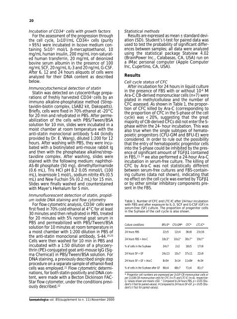
Table 1. Number <strong>of</strong> CFC and LTC-IC after 24-hour incubation<br />
with FBS and after exposure to IL-3, SCF and G-CSF (GF) in<br />
serum-free (SF) culture. The proportion <strong>of</strong> progenitor cells<br />
in the S-phase <strong>of</strong> the cell cycle is also shown.<br />
Culture conditions BFU-E* CFU-GM* CFC* LTC-IC*<br />
24 hours FBS 22±5 12±4 36±8 23±16<br />
24 hours FBS + Ara-C 18±3^ 10±2^ 30±7^ 19±7^<br />
% <strong>of</strong> cells in the S-phase 24±7 2±2 18±5 17±9<br />
24 hours SF + GF 24±13 10±7 37±11 22±9<br />
24 hours SF + GF + Ara-C 8±5# 3±1# 11±8# 4±3#<br />
% <strong>of</strong> cells in the S-phase after GF 66±4 68±7 71±4 81±7<br />
* Progenitor cell numbers are expressed per 2x10 4 CB mononuclear cells or<br />
per 1x10⁶ CB mononuclear cells for CFC (n=7) and LTC-IC (n=4), respectively.<br />
Values shown are means ±SD. ^ Compared to 24 hours FBS, p > 0.05 (Student’s<br />
t-Test for paired values). # Compared to 24 hours SF+GF, p < 0.05 (Student’s<br />
t-Test for paired values).<br />
haematologica vol. 85(supplement to n. 11):November 2000


