

Journal of Hematology - Supplements - Haematologica
Journal of Hematology - Supplements - Haematologica
Journal of Hematology - Supplements - Haematologica

You also want an ePaper? Increase the reach of your titles
YUMPU automatically turns print PDFs into web optimized ePapers that Google loves.
22<br />
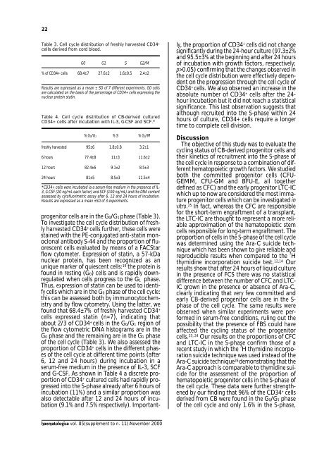
Table 3. Cell cycle distribution <strong>of</strong> freshly harvested CD34 +<br />
cells derived from cord blood.<br />
G0 G1 S G2/M<br />
% <strong>of</strong> CD34+ cells 68.4±7 27.6±2 1.6±0.5 2.4±2<br />
Results are expressed as a mean ± SD <strong>of</strong> 7 different experiments. G0 cells<br />
are calculated on the basis <strong>of</strong> the percentage <strong>of</strong> CD34+ cells expressing the<br />
nuclear protein statin.<br />
Table 4. Cell cycle distribution <strong>of</strong> CB-derived cultured<br />
CD34+ cells after incubation with IL-3, G-CSF and SCF.*<br />
% G0/G1 % S % G2/M<br />
freshly harvested 95±6 1.8±0.8 3.2±1<br />
6 hours 77.4±8 11±3 11.6±2<br />
12 hours 82.4±6 9.1±2 8.5±3<br />
24 hours 81±5 8.5±3 11.5±4<br />
*CD34+ cells were incubated ia a serum-free medium in the presence <strong>of</strong> IL-<br />
3, G-CSF (20 ng/mL each factor) and SCF (100 ng/mL) and the DNA content<br />
assessed by cyt<strong>of</strong>luorimetric assay after 6, 12 and 24 hours <strong>of</strong> incubation.<br />
Results are expressed as a mean ±SD <strong>of</strong> 3 experiments.<br />
progenitor cells are in the G0/G1 phase (Table 3).<br />
To investigate the cell cycle distribution <strong>of</strong> freshly<br />
harvested CD34 + cells further, these cells were<br />
stained with the PE-conjugated anti-statin monoclonal<br />
antibody S-44 and the proportion <strong>of</strong> fluorescent<br />
cells evaluated by means <strong>of</strong> a FACStar<br />
flow cytometer. Expression <strong>of</strong> statin, a 57-kDa<br />
nuclear protein, has been recognized as an<br />
unique marker <strong>of</strong> quiescent cells: 19 the protein is<br />
found in resting (G0) cells and is rapidly downregulated<br />
when cells progress to the G1 phase.<br />
Thus, expression <strong>of</strong> statin can be used to identify<br />
cells which are in the G0 phase <strong>of</strong> the cell cycle:<br />
this can be assessed both by immunocytochemistry<br />
and by flow cytometry. Using the latter, we<br />
found that 68.4±7% <strong>of</strong> freshly harvested CD34 +<br />
cells expressed statin (n=7), indicating that<br />
about 2/3 <strong>of</strong> CD34 + cells in the G0/G1 region <strong>of</strong><br />
the flow cytometric DNA histograms are in the<br />
G0 phase and the remaining are in the G1 phase<br />
<strong>of</strong> the cell cycle (Table 3). We also assessed the<br />
proportion <strong>of</strong> CD34 + cells in the different phases<br />
<strong>of</strong> the cell cycle at different time points (after<br />
6, 12 and 24 hours) during incubation in a<br />
serum-free medium in the presence <strong>of</strong> IL-3, SCF<br />
and G-CSF. As shown in Table 4 a discrete proportion<br />
<strong>of</strong> CD34 + cultured cells had rapidly progressed<br />
into the S-phase already after 6 hours <strong>of</strong><br />
incubation (11%) and a similar proportion was<br />
also detectable after 12 and 24 hours <strong>of</strong> incubation<br />
(9.1% and 7.5% respectively). Importantly,<br />
the proportion <strong>of</strong> CD34 + cells did not change<br />
significantly during the 24-hour culture (97.3±2%<br />
and 95.5±3% at the beginning and after 24 hours<br />
<strong>of</strong> incubation with growth factors, respectively;<br />
p>0.05) confirming that the changes observed in<br />
the cell cycle distribution were effectively dependent<br />
on the progression through the cell cycle <strong>of</strong><br />
CD34 + cells. We also observed an increase in the<br />
absolute number <strong>of</strong> CD34 + cells after the 24-<br />
hour incubation but it did not reach a statistical<br />
significance. This last observation suggests that<br />
although recruited into the S-phase within 24<br />
hours <strong>of</strong> culture, CD34+ cells require a longer<br />
time to complete cell division.<br />
Discussion<br />
The objective <strong>of</strong> this study was to evaluate the<br />
cycling status <strong>of</strong> CB-derived progenitor cells and<br />
their kinetics <strong>of</strong> recruitment into the S-phase <strong>of</strong><br />
the cell cycle in response to a combination <strong>of</strong> different<br />
hematopoietic growth factors. We studied<br />
both the committed progenitor cells (CFU-<br />
GEMM, CFU-GM and BFU-E, all together<br />
defined as CFC) and the early progenitor LTC-IC<br />
which up to now are considered the most immature<br />
progenitor cells which can be investigated in<br />
vitro. 25 In fact, whereas the CFC are responsible<br />
for the short-term engraftment <strong>of</strong> a transplant,<br />
the LTC-IC are thought to represent a more reliable<br />
approximation <strong>of</strong> the hematopoietic stem<br />
cells responsible for long-term engraftment. The<br />
proportion <strong>of</strong> cells in the S-phase <strong>of</strong> the cell cycle<br />
was determined using the Ara-C suicide technique<br />
which has been shown to give reliable and<br />
reproducible results when compared to the 3 H<br />
thymidine incorporation suicide test. 12,14 Our<br />
results show that after 24 hours <strong>of</strong> liquid culture<br />
in the presence <strong>of</strong> FCS there was no statistical<br />
difference between the number <strong>of</strong> CFC and LTC-<br />
IC grown in the presence or absence <strong>of</strong> Ara-C,<br />
clearly indicating that very few committed and<br />
early CB-derived progenitor cells are in the S-<br />
phase <strong>of</strong> the cell cycle. The same results were<br />
observed when similar experiments were performed<br />
in serum-free conditions, ruling out the<br />
possibility that the presence <strong>of</strong> FBS could have<br />
affected the cycling status <strong>of</strong> the progenitor<br />
cells. 23, 24 Our results on the proportions <strong>of</strong> CFC<br />
and LTC-IC in the S-phase confirm those <strong>of</strong> a<br />
recent study in which the 3 H thymidine incorporation<br />
suicide technique was used instead <strong>of</strong> the<br />
Ara-C suicide technique 26 demonstrating that the<br />
Ara-C approach is comparable to thymidine suicide<br />
for the assessment <strong>of</strong> the proportion <strong>of</strong><br />
hematopoietic progenitor cells in the S-phase <strong>of</strong><br />
the cell cycle. These data were further strengthened<br />
by our finding that 96% <strong>of</strong> the CD34 + cells<br />
derived from CB were found in the G0/G1 phase<br />
<strong>of</strong> the cell cycle and only 1.6% in the S-phase,<br />
haematologica vol. 85(supplement to n. 11):November 2000


