Gorter and Grendel
Gorter and Grendel
Gorter and Grendel
Create successful ePaper yourself
Turn your PDF publications into a flip-book with our unique Google optimized e-Paper software.
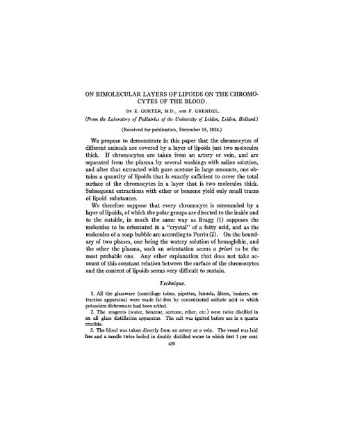
ON BIMOLECULAR LAYERS OF LIPOIDS ON THE CHROMO-<br />
CYTES OF THE BLOOD.<br />
BY E. GORTER, M.D., AND F. GRENDEL.<br />
(From the Laboratory of Pediatrics of the University of Leiden, Leiden, Holl<strong>and</strong>.)<br />
(Received for publication, December 15, 1924.)<br />
We propose to demonstrate in this paper that the chromocytes of<br />
different animals are covered by a layer of lipoids just two molecules<br />
thick. If chromocytes are taken from an artery or vein, <strong>and</strong> are<br />
separated from the plasma by several washings with saline solution,<br />
<strong>and</strong> after that extracted with pure acetone in large amounts, one obtains<br />
a quantity of lipoids that is exactly sufficient to cover the total<br />
surface of the chromocytes in a layer that is two molecules thick.<br />
Subsequent extractions with ether or benzene yield only small traces<br />
of lipoid substances.<br />
We therefore suppose that every chromocyte is surrounded by a<br />
layer of lipoids, of which the polar groups are directed to the inside <strong>and</strong><br />
to the outside, in much the same way as Bragg (1) supposes the<br />
molecules to be orientated in a "crystal" of a fatty acid, <strong>and</strong> as the<br />
molecules of a soap bubble are accordingto Perrin (2). On the boundary<br />
of two phases, one being the watery solution of hemoglobin, <strong>and</strong><br />
the other the plasma, such an orientation seems a priori to be the<br />
most probable one. Any other explanation that does not take account<br />
of this constant relation between the surface of the chromocytes<br />
<strong>and</strong> the content of lipoids seems very difficult to sustain.<br />
Technique.<br />
1. All the glassware (centrifuge tubes, pipettes, funnels, filters, beakers, extraction<br />
apparatus) were made fat-free by concentrated sulfuric acid to which<br />
potassium dichromate had been added.<br />
2. The reagents (water, benzene, acetone, ether, etc.) were twice distilled in<br />
an all glass distillation apparatus. The salt was ignited before use in a quartz<br />
crucible.<br />
3. The blood was taken directly from an artery or a vein. The vessel was laid<br />
free <strong>and</strong> a needle twice boiled in doubly distilled water to which first per cent<br />
439
440 BIMOLECULAR LAYERS OF LIPOIDS ON CHROMOCYTES<br />
soda, <strong>and</strong> then 0.5 per cent potassium oxalate had been added, was introduced<br />
into it. The first stream of blood was discarded to avoid the possibility of error<br />
from contamination with the fat of the subcutaneous tissue. The next portion<br />
was then permitted to flow into a small stoppered weighing bottle, containing<br />
0.5 per cent potassium oxalate. In the case of the goat <strong>and</strong> the sheep the jugular<br />
vein was directly punctured through the skin but in this case the stream of blood<br />
was permitted to flow for some time, so as to wash the needle clean of all contaminating<br />
fatty substances before a measured quantity was received in our glass<br />
vessel. In human subjects the same procedure of puncturing the vein through the<br />
skin was followed.<br />
4. After mixing, 10 cc. (or in later experiments 1 cc.) of blood were pipetted into<br />
a centrifuge tube of 60 cc. <strong>and</strong> three or four times washed with 50 cc. salt solution<br />
(0.9 per cent) in the usual way.<br />
5. The extraction was performed with acetone during 48 or 72 hours. Large<br />
quantities were used.<br />
After several extractions, the acetone was filtered into a glass beaker <strong>and</strong> the<br />
liquid evaporated on a water bath. This procedure was the most difficult part<br />
of the operation because loss was very liable to occur at this time. The residue<br />
was finally taken up in benzene <strong>and</strong> filtered into a measuring flask of 50 cc.,<br />
when 10 cc. of the blood had been used, or in a tube marked at 2.5 or 5 cc., when<br />
0.5 or 1 cc. had been taken. Just before each determination the liquid was made<br />
up to the mark with benzene.<br />
Determination 'of the Surface Occupied by the Lipoids Spread Out in a<br />
Monomolecular Layer on Water.<br />
Langmuir (3) has demonstrated that fats <strong>and</strong> fatty acids spread<br />
in a monomolecular layer when they have been dissolved in benzene<br />
<strong>and</strong> a few drops of the solution are placed on a large surface of water.<br />
Adam (4) has slightly modified the apparatus originally described by<br />
Langmuir. We have made use of Adam's modification. The benzene<br />
solution was delivered out of a calibrated 0.1 cc. pipette.<br />
Now, it has been shown that the molecules of a fatty substance<br />
spreading on a water surface do not exert any pressure in a direction<br />
parallel to the surface before the condition is arrived at that they<br />
form precisely a monomolecular film, in which latter they come to<br />
be arranged in a vertical position. In the Langmuir-Adam apparatus<br />
the water surface chosen is so large that sufficient room is provided<br />
to the molecules so that they are not in close contact with each other.<br />
By the displacement of a strip of copper on which a balance is mounted<br />
one is able to determine the precise moment at which the molecules
E. GORTER AND F. GRENDEL<br />
441<br />
begin to exert a pressure in a horizontal plane, <strong>and</strong> by placing different<br />
weights on the pan of the balance, it is possible to compensate <strong>and</strong><br />
to measure this pressure. The reduction of the size of the surface<br />
is obtained by moving a glass strip covered with a thin layer of paraffin<br />
oil over the edges of the copper tray, which are covered as well<br />
with paraffin oil. As soon as the molecules are in close contact in a<br />
layer exactly one molecule thick, the balance moves out of the equilibrium<br />
position. By placing small weights on the balance one is<br />
able to compress the layer without much further reduction of the size<br />
of the surface, till suddenly by increasing the weight the layer is disturbed<br />
<strong>and</strong> equilibrium of the balance is no longer obtained. The<br />
dimensions of the surface are measured with a ruler.<br />
We always began with the determination of the surface contamination.<br />
By placing 50 mg. in the pan of the balance <strong>and</strong> moving the<br />
glass strip from a distance of about 30 cm. we were able to determine<br />
that it hardly ever exceeded 0.5 cm. at room temperature.<br />
From a pipette 0.1 cc. of the benzene solution of the lipoids of the<br />
chromocytes was blown onto the surface of the water in the tray <strong>and</strong><br />
by moving the glass strip the point was noted at which the balance<br />
began to move, 50 mg. being the weight in the pan. The pressure<br />
exerted on each cm. of the layer was 2 dynes per 50 mg. weight in the<br />
pan.<br />
Determination of the Number <strong>and</strong> the Dimensions of the Chromocytes.<br />
The number of chromocytes was determined by filling the mrlangeur as soon<br />
as possible from the weighing bottle containing the blood, <strong>and</strong> by counting in the<br />
counting chamber of BUrker the cells in 80 small squares, each measuring 1/4,000<br />
c.mm. The surface of the chromocytes was evaluated from blood smears on<br />
slides, coloured by Pappenheim's panoptical dye. With the aid of a drawing<br />
prism of Zeiss 40 to 50 chromocytes were drawn on millimeter paper. By taking<br />
account of the magnifying power of the microscope one was able to measure the<br />
dimensions of the cells in a horizontal <strong>and</strong> a vertical direction.<br />
The surface of the cells was derived from these numbers by making use of Knoll's<br />
(5) formula that in chromocytes having the form of a disc (a form that is taken by<br />
all chromocytes that are spread on glass) the surface is 2D 2 (D being the diameter).<br />
The total surface of the chromocytes from 1 to 10 cc. blood was easily obtained<br />
by multiplying the number of cells by their surface.
442 BIMOLECULAR LAYERS OF LIPOIDS ON CHROMOCYTES<br />
SUMMARY OF RESULTS.<br />
We have examined the blood of man <strong>and</strong> of the rabbit, dog, guinea<br />
pig, sheep, <strong>and</strong> goat. There exists a great difference in the size of<br />
the red blood cells of these animals, but the total surfaces of the chro-<br />
Animal.<br />
Amount of<br />
blood used<br />
for the<br />
analysis.<br />
TABLE I.<br />
'No. of<br />
chromocytes<br />
per c.mm.<br />
Surface of<br />
one chromocyte.<br />
Total<br />
surface of<br />
the chromocytes<br />
(a).<br />
---- -<br />
Surface<br />
occupied<br />
by all the<br />
hlioids of<br />
te chromocytes<br />
(b).<br />
Factor a:b.<br />
Dog A<br />
gm.<br />
40<br />
10<br />
8,000,000<br />
6,890,000<br />
sq. A<br />
98<br />
90<br />
sq. m.<br />
31.3<br />
6.2<br />
sq.m.<br />
62<br />
12.2<br />
2<br />
2<br />
Sheep 1<br />
10<br />
9<br />
9,900,000<br />
9,900,000<br />
29.8<br />
29.8<br />
2.95<br />
2.65<br />
6.2<br />
5.8<br />
2.1<br />
2.2<br />
Rabbit A<br />
10<br />
10<br />
0.5<br />
5,900,000<br />
5,900,000<br />
5,900,000<br />
92.5<br />
92.5<br />
92.5<br />
5.46<br />
5.46<br />
0.27<br />
9.9<br />
8.8<br />
0.54<br />
1.8<br />
1.6<br />
2<br />
B<br />
1<br />
10<br />
10<br />
6,600,000<br />
6,600,000<br />
6,600,000<br />
74.4<br />
74.4<br />
74.4<br />
0.49<br />
4.9<br />
4.9<br />
0.96<br />
9.8<br />
9.8<br />
2<br />
2<br />
2<br />
Guinea Pig A<br />
1<br />
1<br />
5,850,000<br />
5,850,000<br />
89.8<br />
89.8<br />
0.52<br />
0.52<br />
1.02<br />
0.97<br />
2<br />
1.9<br />
Goat 1<br />
1<br />
1<br />
10<br />
10<br />
1<br />
16,500,000<br />
16,500,000<br />
19,300,000<br />
19,300,000<br />
19,300,000<br />
20.1<br />
20.1<br />
17.8<br />
17.8<br />
17.8<br />
0.33<br />
0.33<br />
3.34<br />
3.34<br />
0.33<br />
0.66<br />
0.69<br />
6.1<br />
6.8<br />
0.63<br />
2<br />
2.1<br />
1.8<br />
2<br />
1.9<br />
Man.<br />
1<br />
1<br />
4,740,000<br />
4,740,000<br />
99.4<br />
99.4<br />
0.47<br />
0.47<br />
0.92<br />
0.89<br />
--- --<br />
2<br />
1.9<br />
mocytes from 0.1 cc. blood do not show a similarly great divergence,<br />
because animals having very small cells (goat <strong>and</strong> sheep) have much<br />
greater quantities of these cells in their blood than animals with blood<br />
cells of larger dimensions (dog <strong>and</strong> rabbit).
E. GORTER AND F. GRENDEL 443<br />
We give all the results of our experiments, omitting only those in<br />
which we were unable to avoid losses in the procedure of evaporation<br />
of the acetone.<br />
It is clear that all our results fit in well with the supposition that the<br />
chromocytes are covered by a layer of fatty substances that is two<br />
molecules thick.<br />
BIBLIOGRAPHY.<br />
1. Bragg, W. H., <strong>and</strong> Bragg, W. L., X rays <strong>and</strong> crystal structure, New York,<br />
4th edition, revised <strong>and</strong> enlarged, 1924.<br />
2. Perrin, J., Ann. phys., 1918, x, series 9, 160.<br />
3. Langmuir, I., J. Am. Chem. Soc., 1917, xxxix, 1848.<br />
4. Adam, N. K., Proc. Roy. Soc. London, Series A, 1921, xcix, 336; 1922, ci,<br />
452, 516.<br />
5. Knoll, W., Arch. ges. Physiol., 1923, cxcviii, 367.


