Pollen and Stigma Structure and Function: The Role of Diversity in ...
Pollen and Stigma Structure and Function: The Role of Diversity in ...
Pollen and Stigma Structure and Function: The Role of Diversity in ...

Create successful ePaper yourself
Turn your PDF publications into a flip-book with our unique Google optimized e-Paper software.
<strong>Pollen</strong> <strong>and</strong> <strong>Stigma</strong> <strong>Structure</strong> <strong>and</strong> <strong>Function</strong><br />
S85<br />
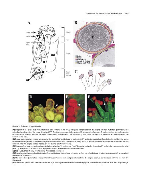
Figure 1. Poll<strong>in</strong>ation <strong>in</strong> Arabidopsis.<br />
(A) Diagram <strong>of</strong> one <strong>of</strong> the two ovary chambers after removal <strong>of</strong> the ovary wall (OW). <strong>Pollen</strong> l<strong>and</strong>s on the stigma, where it hydrates, germ<strong>in</strong>ates, <strong>and</strong><br />
extends a tube that enters the transmitt<strong>in</strong>g tract (TT). <strong>The</strong> tube emerges onto the septum (S), grows up the funiculus (f), <strong>and</strong> enters the micropyle open<strong>in</strong>g<br />
<strong>of</strong> the ovule (O), where it fertilizes the egg <strong>and</strong> central cell. <strong>The</strong> position <strong>of</strong> the transmitt<strong>in</strong>g tract <strong>in</strong>side the septum is shown <strong>in</strong> the cross-section at the<br />
bottom <strong>of</strong> the pistil.<br />
(B) Transmission electron micrograph show<strong>in</strong>g the po<strong>in</strong>t <strong>of</strong> contact between a pollen gra<strong>in</strong> (P) <strong>and</strong> a stigma papillus (S), colorized to highlight the pollen<br />
coat (p<strong>in</strong>k), <strong>in</strong>t<strong>in</strong>e (peach), ex<strong>in</strong>e (green), stigma cell wall (yellow), <strong>and</strong> stigma cuticle (blue). A foot <strong>of</strong> lipid-rich material (arrows) collects between the two<br />
surfaces. <strong>The</strong> th<strong>in</strong> stigma pellicle that covers the cuticle is not dist<strong>in</strong>ct here.<br />
(C) Diagram <strong>of</strong> early events on the stigma, <strong>in</strong>clud<strong>in</strong>g adhesion (1), pollen coat ‘‘foot’’ formation <strong>and</strong> pollen hydration (2), pollen tube emergence from the<br />
gra<strong>in</strong> (3), <strong>and</strong> pollen tube <strong>in</strong>vasion <strong>of</strong> the papillae cell wall <strong>and</strong> extension toward the style (4).<br />
(D) to (F) Sequence <strong>of</strong> early events dur<strong>in</strong>g Arabidopsis poll<strong>in</strong>ation.<br />
(D) <strong>The</strong> pollen coat has mobilized to the site <strong>of</strong> contact between the pollen <strong>and</strong> the stigma, form<strong>in</strong>g a foot between the two surfaces (arrow), as visualized<br />
with the lipid dye FM1-43.<br />
(E) <strong>The</strong> pollen tube (arrow) has emerged from the gra<strong>in</strong>’s ex<strong>in</strong>e wall <strong>and</strong> projects itself <strong>in</strong>to the stigma papillus, as visualized with the cell wall dye<br />
Congo red.<br />
(F) <strong>Pollen</strong> tubes (arrow) w<strong>in</strong>d their way toward the style, mov<strong>in</strong>g between the cell walls <strong>of</strong> the papillae, where they are protected from the Congo red dye.