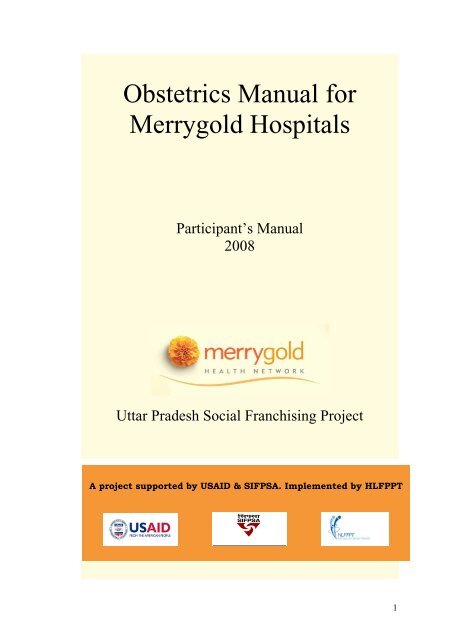
Obstetrics Manual for Merrygold Hospitals - State Innovations in ...
Obstetrics Manual for Merrygold Hospitals - State Innovations in ...
Obstetrics Manual for Merrygold Hospitals - State Innovations in ...

You also want an ePaper? Increase the reach of your titles
YUMPU automatically turns print PDFs into web optimized ePapers that Google loves.
<strong>Obstetrics</strong> <strong>Manual</strong> <strong>for</strong><br />
<strong>Merrygold</strong> <strong>Hospitals</strong><br />
Participant’s <strong>Manual</strong><br />
2008<br />
Uttar Pradesh Social Franchis<strong>in</strong>g Project<br />
A project supported by USAID & SIFPSA. Implemented by HLFPPT<br />
1
Preface<br />
HLFPPT is an organization committed to work with various partners pioneer<strong>in</strong>g<br />
<strong>in</strong>novations <strong>for</strong> better<strong>in</strong>g health outcomes <strong>for</strong> the poor. <strong>Merrygold</strong> Health Network is<br />
one of such <strong>in</strong>novations <strong>in</strong> the field of Social Franchis<strong>in</strong>g.<br />
<strong>Merrygold</strong> Health Network, aims towards achiev<strong>in</strong>g an objective of improv<strong>in</strong>g<br />
Maternal and Child Health through <strong>in</strong>creased access to low cost – high quality<br />
healthcare services, <strong>for</strong> rural and urban work<strong>in</strong>g poor <strong>in</strong> Uttar Pradesh. In U.P. Social<br />
Franchis<strong>in</strong>g Project (supported by USAID and SIFPSA), HLFPPT as an<br />
implement<strong>in</strong>g agency, will be establish<strong>in</strong>g 70 fully franchised <strong>Merrygold</strong> <strong>Hospitals</strong> at<br />
district level, 700 partially franchised Merrysilver Cl<strong>in</strong>ics at block level and will be<br />
work<strong>in</strong>g with more than 10,000 Tarang partners (ASHAs, Chemists, Fare price shop<br />
owners, Tarang health committee members, Op<strong>in</strong>ion leaders, Anganwadi workers,<br />
Depot holders) and AYUSH practitioners at the village level by 2010. Two model<br />
hospitals are already established <strong>in</strong> Kanpur and Agra focus<strong>in</strong>g on maternal and child<br />
health care.<br />
In our endeavour to make this a successful model, it was felt that tra<strong>in</strong><strong>in</strong>g <strong>for</strong> doctors,<br />
nurses and other team members will be a key component to improve the quality of<br />
service delivery and equip the staff with appropriate knowledge and skills.<br />
“<strong>Obstetrics</strong> <strong>Manual</strong> <strong>for</strong> <strong>Merrygold</strong> <strong>Hospitals</strong> – 2008”, was designed under the<br />
guidance and expertise of Prof. Alokendu Chatterjee (Vice President, National Board<br />
of Exam<strong>in</strong>ation, Past President FOGSI), Dr. Joydev Mukherjee (Prof., Department of<br />
Gynecology and <strong>Obstetrics</strong>, R.G. Kar Medical College, Kolkata) and Dr. Partho<br />
Mukherjee (Associate Professor, IPGME & R & SSKM Hospital, Kolkata) to meet<br />
the above objectives. It has been pre-tested with <strong>Merrygold</strong> L0 hospital staff at<br />
Kanpur and Agra. The <strong>in</strong>puts and feedbacks from the hospital staff and comments of<br />
review committee members from SIFPSA and ITAP, has given this manual the<br />
present shape.<br />
I am sure that this manual, when used by hospitals and cl<strong>in</strong>ics <strong>in</strong> the Social<br />
Franchis<strong>in</strong>g Project will as an enabl<strong>in</strong>g tool towards excellent service delivery.<br />
HLFPPT<br />
2
Acknowledgement<br />
<strong>Manual</strong> on Obstetric conditions can prove as a guidel<strong>in</strong>e <strong>for</strong> manag<strong>in</strong>g cases dur<strong>in</strong>g<br />
antenatal, <strong>in</strong>tra-natal and post-partum period or when obstetric emergencies occur<br />
dur<strong>in</strong>g pregnancy. I present “<strong>Obstetrics</strong> <strong>Manual</strong> <strong>for</strong> <strong>Merrygold</strong> <strong>Hospitals</strong> – 2008”,<br />
<strong>for</strong> better and more harmonized obstetrical and medical care. This manual is the result<br />
of s<strong>in</strong>cere <strong>in</strong>tent, aspirations and hard work of all those who are an <strong>in</strong>tegral part of the<br />
network.<br />
I am grateful to Mr. G. Manoj, (CEO, HLFPPT) who has shown faith <strong>in</strong> my entire<br />
team to undertake the task of prepar<strong>in</strong>g this manual.<br />
My s<strong>in</strong>cere thanks to Mr. Rajeev Kapoor I.A.S. (Executive Director - SIFPSA &<br />
Mission Director - NRHM), Mr. S. Krishnaswamy (General Manager Private Sector -<br />
SIFPSA), Dr. M. K. S<strong>in</strong>ha (General Manager Public Sector – SIFPSA), Ms. Savita<br />
Chauhan (Dy. General Manager Private Sector - SIFPSA), Dr. Lovleen Johari (Senior<br />
Reproductive Health Advisor, USAID) and Ms. Shuvi Sharma (Manager - Social<br />
Market<strong>in</strong>g & Franchis<strong>in</strong>g, ITAP) <strong>for</strong> their support and encouragement <strong>for</strong> develop<strong>in</strong>g<br />
this manual.<br />
I thank Dr. Br<strong>in</strong>da Frey, Dr. Vandana Naidu, Dr. Amrita Kansal and Dr. Vibha Bansal<br />
from HLFPPT <strong>for</strong> develop<strong>in</strong>g and design<strong>in</strong>g this manual. I also thank Ms. Divya<br />
Babbar <strong>for</strong> provid<strong>in</strong>g secretarial assistance.<br />
My s<strong>in</strong>cere thanks to Prof. Alokendu Chatterjee, Dr. Joydev Mukherjee, Dr. Patho<br />
Mukherjee, <strong>for</strong> their constant support and guidance <strong>in</strong> the development of this manual.<br />
I express deep appreciation and thanks to Dr. V<strong>in</strong>ita Das, Dr. Shikha Srivastava, Dr.<br />
R<strong>in</strong>ku Srivastava, Dr. Jyoti Vajpayee <strong>for</strong> review<strong>in</strong>g this manual and provid<strong>in</strong>g their<br />
valuable comments.<br />
This manual has been pre tested by UPSF tra<strong>in</strong><strong>in</strong>g team at both the L0 hospitals at<br />
Kanpur and Agra. Ef<strong>for</strong>ts made by Mr. Alok Tabelabux, Mr. B. K. Mishra from<br />
HLFPPT, <strong>in</strong> organiz<strong>in</strong>g tra<strong>in</strong><strong>in</strong>gs and active <strong>in</strong>volvement of entire <strong>Merrygold</strong> hospital<br />
staff <strong>in</strong> tra<strong>in</strong><strong>in</strong>g was commendable.<br />
Special mention needs to be made of Mr. Sharad Agarwal, Dr. Sanjeev Yadav, Dr.<br />
Br<strong>in</strong>da Frey, Mr. Rajeev Shukla, Mr. Gajendra Verma,, Ms. Preeti Dwivedi and entire<br />
U.P. Social Franchis<strong>in</strong>g team <strong>for</strong> their ef<strong>for</strong>ts, valuable time and support <strong>for</strong> arrang<strong>in</strong>g<br />
and organiz<strong>in</strong>g tra<strong>in</strong><strong>in</strong>g program based on this manual.<br />
Dr. Vasanthi Krishnan<br />
Head, Technical Services Division<br />
HLFPPT<br />
3
Design and Development Team –<br />
S.N. Names<br />
Designation & Organization<br />
1. Dr. Vasanthi Krishnan Head, TSD, HLFPPT<br />
2. Dr. Br<strong>in</strong>da Frey Head, Cl<strong>in</strong>ical services - <strong>Merrygold</strong> Health<br />
Network, HLFPPT<br />
3. Dr. Vandana Naidu Program Manager, HLFPPT<br />
4. Dr. Vibha Bansal Program Manager – Tra<strong>in</strong><strong>in</strong>g and Quality<br />
Assurance, HLFPPT<br />
Review Team –<br />
S.N. Names<br />
Designation & Organization<br />
1. Prof. Alokendu Chatterjee Vice President, National Board of<br />
Exam<strong>in</strong>ation, Past President FOGSI<br />
2. Dr. Joydev Mukherjee Prof., Department of Gynecology and<br />
<strong>Obstetrics</strong>, R.G. Kar Medical College,<br />
Kolkata<br />
3. Dr. Patho Mukherjee Associate Professor, IPGME & R & SSKM<br />
Hospital, Kolkata<br />
4. Dr. V<strong>in</strong>ita Das HOD, Queen Mary’s Hospital, KGMU,<br />
Lucknow<br />
5. Dr. Shikha Srivastava Asst. Director health, UPHSDP, Lucknow<br />
6. Dr. R<strong>in</strong>ku Srivastava APC-PS, SIFPSA, Lucknow<br />
7. Dr. Jyoti Vajpayee Country Director, EngenderHealth<br />
4
Abbreviations<br />
AIDS<br />
ANC<br />
ANM<br />
APH<br />
ASHA<br />
AWW<br />
BP<br />
CPD<br />
CS<br />
CVS<br />
EDD<br />
EFM<br />
FHR<br />
FHS<br />
HIV<br />
HLFPPT<br />
IFA<br />
IUD<br />
LAM<br />
LMP<br />
MTP<br />
PHC<br />
PIH<br />
PPH<br />
PPNDT<br />
P/V<br />
RR<br />
PROM<br />
TT<br />
VBAC<br />
Acquired Immuno Deficiency Syndrome<br />
Ante Natal Care<br />
Auxiliary Nurse Midwife<br />
Ante Partum Hemorrhage<br />
Accredited Social Health Worker<br />
Angan Wadi Worker<br />
Blood Pressure<br />
Cephalo - pelvic Disproportion<br />
Cesarean Section<br />
Cerebro-Vascular Accidents<br />
Expected Date of Delivery<br />
Electronic Fetal Monitor<strong>in</strong>g<br />
Foetal Heart Rate<br />
Foetal Heart Sound<br />
Human Immunodeficiency Virus<br />
H<strong>in</strong>dustan Latex Family Plann<strong>in</strong>g Promotion Trust<br />
Iron and Folic Acid<br />
Intra Uter<strong>in</strong>e Death<br />
Lactional Amenorrhoea Method<br />
Last Menstrual period<br />
Medical Term<strong>in</strong>ation of Pregnancy<br />
Primary Health Center<br />
Pregnancy Induced Hypertension<br />
Post Partum Hemorrhage<br />
Preconception Pre-Natal Diagnostic Techniques<br />
Per vag<strong>in</strong>um<br />
Respiratory rate<br />
Premature Rupture of Membranes<br />
Tetanus Toxoid<br />
Vag<strong>in</strong>al Birth after Caesarean<br />
5
Index<br />
No. Contents Page<br />
About the <strong>Manual</strong><br />
Module 1 Care & Management Dur<strong>in</strong>g Antenatal Period<br />
Unit 1.1 Introduction<br />
Unit 1.2 Diagnosis of Pregnancy<br />
Unit 1.3 Cl<strong>in</strong>ical Assessment<br />
Unit 1.4 Advice dur<strong>in</strong>g ante- natal visit<br />
Unit 1.5 Symptoms, signs, probable diagnosis of common ailments.<br />
Unit 1.6 Protocols <strong>for</strong> Antenatal care<br />
Module 2 Complications Dur<strong>in</strong>g Pregnancy<br />
Unit 2.1 Hyperemesis Gravidarum<br />
Unit 2.2 Anaemia <strong>in</strong> Pregnancy<br />
Unit 2.3 Vag<strong>in</strong>al Bleed<strong>in</strong>g <strong>in</strong> Early Pregnancy<br />
Unit 2.4 Ante Partum Hemorrhage<br />
Unit 2.5 Hypertensive Disorders <strong>in</strong> Pregnancy<br />
Unit 2.6 Premature Rupture of Membranes (PROM)<br />
Module 3 Care & Management of Labor & Complications<br />
Unit 3.1 Normal Labor and Use of Partograph<br />
Unit 3.2 Unsatisfactory Progress of Labor / Obstructed Labor<br />
Unit 3.3 Fetal Distress<br />
Unit 3.4 Cord Prolapse<br />
Unit 3.5 Caesarean Section<br />
Unit 3.6 Vag<strong>in</strong>al Birth after Caesarean section<br />
Module 4 Care & Management of Third Stage Of Labor<br />
Unit 4.1 Active Management of Third Stage of Labour<br />
Unit 4.2 Post Partum Hemorrhage<br />
Unit 4.3 Post Partum Care<br />
Unit 4.4 Puerperal Sepsis<br />
Annexure<br />
References<br />
6
List of Figures<br />
No. Figure Page<br />
1 Descend of Fetal head<br />
2 Sample modified WHO Partograph<br />
3 Partograph <strong>for</strong> Exercise 1<br />
4 Partograph <strong>for</strong> Exercise 3<br />
5 Partograph <strong>for</strong> Exercise 5<br />
6 Cont<strong>in</strong>uous Electronic Fetal Monitor<strong>in</strong>g (EFM)<br />
7 Expos<strong>in</strong>g Per<strong>in</strong>eal tear<br />
8 Repair of Per<strong>in</strong>eal Tear (A)<br />
9 Repair of Per<strong>in</strong>eal Tear (B)<br />
10 Repair<strong>in</strong>g the sk<strong>in</strong><br />
List of Tables<br />
No. Tables Page<br />
1 Common ailments dur<strong>in</strong>g Pregnancy<br />
2 Symptoms and Signs <strong>for</strong> the Early Diagnosis of Bleed<strong>in</strong>g <strong>in</strong><br />
Early Pregnancy<br />
3 Protocols on Management of Bleed<strong>in</strong>g <strong>in</strong> Early Pregnancy<br />
4 Differential Diagnosis Of Antepartum Heamorrhage<br />
5 Protocols on rapid <strong>in</strong>itial assessment<br />
6 Differential diagnosis of Hypertensive Disorders of pregnancy<br />
7 Regimens of MgSO4 <strong>for</strong> the management of sever Preeclampsia<br />
& Eclampsia<br />
8 Management of Meconium sta<strong>in</strong>ed liquor<br />
9 Def<strong>in</strong>ition of Normal, Suspicious and Pathological FHR traces<br />
10 Classification of FHR trace features<br />
7
About the <strong>Manual</strong><br />
This <strong>Manual</strong> consists of four modules perta<strong>in</strong><strong>in</strong>g to essential and emergency obstetrics<br />
and newborn care. This manual has been created with an objective of mak<strong>in</strong>g<br />
obstetricians aware regard<strong>in</strong>g most recent developments. The differential diagnosis of<br />
important obstetrical complications like Ante partum hemorrhage has been put <strong>in</strong>to<br />
tabular <strong>for</strong>m, which makes it simpler and easier to follow.<br />
Similarly case studies on different complications will be helpful <strong>in</strong> adopt<strong>in</strong>g practical<br />
approach <strong>in</strong> management of cases.<br />
8
MODULE 1<br />
Care & Management<br />
Dur<strong>in</strong>g<br />
Antenatal Period<br />
Unit 1.1<br />
Unit 1.2<br />
Unit 1.3<br />
Unit 1.4<br />
Unit 1.5<br />
Unit 1.6<br />
Introduction<br />
Cl<strong>in</strong>ical assessment (History tak<strong>in</strong>g,<br />
physical exam<strong>in</strong>ation and rout<strong>in</strong>e<br />
<strong>in</strong>vestigation).<br />
Advice dur<strong>in</strong>g ante- natal visit.<br />
Symptoms, signs, probable diagnosis of<br />
common ailments.<br />
Protocols <strong>for</strong> Antenatal care.<br />
Protocol on Birth Preparedness and<br />
Complication Read<strong>in</strong>ess.<br />
9
About this Module<br />
In this module we will discuss about the care and management of a women dur<strong>in</strong>g her<br />
antenatal period. It will also cover def<strong>in</strong>ed protocols related to antenatal registration,<br />
care, <strong>in</strong>vestigations and management, advice, and danger signs dur<strong>in</strong>g antenatal<br />
period.<br />
Unit 1.1<br />
Introduction<br />
Learn<strong>in</strong>g Objectives<br />
• To diagnose pregnancy<br />
• To know about the appropriate tim<strong>in</strong>g, number and importance of ANC.<br />
Pregnancy is not a disease but every pregnancy is at risk. Ensure that Ante-Natal care<br />
is used as an opportunity to detect and treat exist<strong>in</strong>g problems. Effective Ante-Natal<br />
care can provide a healthy mother and a healthy baby as an outcome. However, you<br />
must realize that even with the most effective screen<strong>in</strong>g tools available, one can not<br />
predict which woman will develop complications related to pregnancy and also when.<br />
• Recognize that “ Every pregnancy is at risk”<br />
• Ensure that Antenatal care is used as an opportunity to detect and treat<br />
exist<strong>in</strong>g problems.<br />
• Make sure that services are available to manage obstetric emergencies when<br />
they occur.<br />
• Prepare pregnant women and their families <strong>for</strong> the eventuality of an<br />
emergency.<br />
1.1.1 Diagnosis of Pregnancy<br />
Symptoms: The woman may come to you with the follow<strong>in</strong>g compla<strong>in</strong>ts:<br />
• Cessation of Menstruation<br />
• Nausea with or without vomit<strong>in</strong>g<br />
• Increased frequency of ur<strong>in</strong>e<br />
• Fatigue<br />
• Perception of foetal movements (after four months)<br />
Signs: on exam<strong>in</strong>ation you may f<strong>in</strong>d<br />
• Breast enlargement<br />
• Changes <strong>in</strong> the sk<strong>in</strong> colour of areola<br />
• Enlargement of abdomen<br />
• Discoloration of vag<strong>in</strong>al mucosa<br />
• Soften<strong>in</strong>g of cervix and uterus<br />
• Uter<strong>in</strong>e enlargement<br />
• Internal and external ballottement<br />
• Ability to discern fetal parts<br />
10
You can confirm the pregnancy by<br />
• Detection of HCG <strong>in</strong> ur<strong>in</strong>e with<strong>in</strong> 3 months of amenorrhea<br />
• Detection of FHS at 20 weeks by auscultation<br />
• Perception of foetal movements on abdom<strong>in</strong>al exam<strong>in</strong>ation<br />
* In case of doubt of congenital anomalies, an USG may be considered –refer cl<strong>in</strong>ical<br />
protocol 1.<br />
To ensure a normal pregnancy with the delivery of a healthy baby from a healthy<br />
mother and <strong>for</strong> screen<strong>in</strong>g the complications dur<strong>in</strong>g pregnancy there are few po<strong>in</strong>ts to<br />
be kept <strong>in</strong> m<strong>in</strong>d, which are:<br />
‣ Early registration: All pregnant women should be registered early, at least<br />
be<strong>for</strong>e 12 weeks.<br />
‣ Antenatal card: Antenatal card should be duly filled <strong>in</strong> by nurse and doctor<br />
<strong>for</strong> every woman registered, and handed over to the pregnant woman.<br />
‣ Number and tim<strong>in</strong>g of visits:.<br />
Ensure that every woman makes at least 3 Antenatal visits apart from<br />
registration. In cases of pregnancy without complications, these visits should<br />
be sufficient. Ideally, the registration should be done as soon as the pregnancy<br />
is suspected and first visit should be scheduled between 4 th and 6 th month<br />
(around 26 weeks). The second visit should be planned <strong>in</strong> the 8 th month (32<br />
weeks) and the third visit <strong>in</strong> the 9 th month (36 weeks).<br />
Source : (MOH&FW manual “Guidel<strong>in</strong>es <strong>for</strong> Antenatal Care and skilled attendance at birth by ANMs<br />
and LHVs”)<br />
11
Unit 1.2 Cl<strong>in</strong>ical assessment (History tak<strong>in</strong>g, physical<br />
exam<strong>in</strong>ation and rout<strong>in</strong>e <strong>in</strong>vestigation)<br />
Learn<strong>in</strong>g Objectives<br />
• Know the important po<strong>in</strong>ts to be asked while collect<strong>in</strong>g history to a pregnant<br />
woman.<br />
• Understand and be able to per<strong>for</strong>m physical exam<strong>in</strong>ation on a pregnant<br />
woman.<br />
• Understand the rout<strong>in</strong>e <strong>in</strong>vestigations to be done dur<strong>in</strong>g antenatal period.<br />
1.2.1 History Tak<strong>in</strong>g<br />
‣ Date of Last Menstrual Period:<br />
• Ask <strong>for</strong> date of 1 st day of the last menstrual period (LMP)<br />
• Calculate the Expected Date of Delivery (EDD) = LMP + 9months and 7<br />
days. This calculation is based on the assumption that the menstrual cycle<br />
was regular and it was a 28-30 days’ cycle. If the cycle varies, EDD will<br />
vary accord<strong>in</strong>gly<br />
• Calculate Period of Gestation <strong>in</strong> weeks: Period of gestation is to be<br />
expressed <strong>in</strong> terms of completed weeks. A fraction of more than 3 days is<br />
to be considered as completed week.<br />
‣ Age of the woman<br />
‣ Duration of marriage<br />
‣ Order of the pregnancy<br />
‣ Gravida (GPAL- Gravida, Para, Abortion, Liv<strong>in</strong>g children)<br />
‣ Last child birth/ Last abortion<br />
‣ History of present pregnancy<br />
‣ Compla<strong>in</strong>ts dur<strong>in</strong>g present pregnancy (to be asked dur<strong>in</strong>g each visit):<br />
• Excessive vomit<strong>in</strong>g<br />
• fever<br />
• Palpitations, easy fatigability and breathlessness at rest<br />
• Puff<strong>in</strong>ess of the face<br />
• Headache or blurr<strong>in</strong>g of vision<br />
• Pass<strong>in</strong>g smaller amounts of ur<strong>in</strong>e<br />
• Vag<strong>in</strong>al bleed<strong>in</strong>g or Leak<strong>in</strong>g of watery fluid per vag<strong>in</strong>am(P/V)<br />
• Pa<strong>in</strong> abdomen at any stage of pregnancy<br />
• Decreased or absent foetal movements<br />
‣ History of problems dur<strong>in</strong>g the previous pregnancy/ delivery (Obstetric<br />
History)-<br />
• Abortion(s) or Premature birth(s),<br />
• Tw<strong>in</strong>s or multiple pregnancies<br />
• Stillbirths(s) or neonatal loss<br />
• Hypertensive disorder of pregnancies(if don’t know, ask <strong>for</strong> a history of<br />
convulsion <strong>in</strong> previous pregnancies)<br />
• Duration of labor<br />
12
• Malpresentation, such as breech delivery<br />
• Ante partum hemorrhage<br />
• Postpartum hemorrhage<br />
• Assisted delivery (vacuum extraction)<br />
• Delivery by caesarean section<br />
• Birth weight of previous baby<br />
• Any surgery on the reproductive tract<br />
• Iso-immunization (Rh-ve) <strong>in</strong> the previous pregnancy<br />
• History of drug <strong>in</strong>take or allergies.<br />
• Any treatment taken or taken drugs <strong>for</strong> <strong>in</strong>fertility.<br />
‣ History of any systemic/ Medical illness (es)-<br />
Relevant history of past medical illnesses e.g. Hypertension, Diabetes, Heart<br />
disease, Tuberculosis, Renal disease, Epilepsy, Asthma, Malaria, Thyroid or<br />
any disease <strong>for</strong> which she has been advised to take treatment, is to be elicited.<br />
‣ Family History:<br />
• systemic illness<br />
• Tw<strong>in</strong>s<br />
• Congenitally mal<strong>for</strong>med baby<br />
• Thalassaemia<br />
‣ Personal History:<br />
• Contraceptive practices prior to pregnancy<br />
• Smok<strong>in</strong>g, alcohol, tobacco or any other substance abuse<br />
• Bowel, bladder, appetite<br />
1.2.2 Physical Exam<strong>in</strong>ation<br />
‣ General Exam<strong>in</strong>ation<br />
• Height<br />
• Weight<br />
• Blood Pressure<br />
• Pallor<br />
• Respiratory rate (RR)<br />
• Pedal edema<br />
• Generalized edema or puff<strong>in</strong>ess of the face<br />
• Breast Exam<strong>in</strong>ation to detect <strong>in</strong>verted nipples, crust<strong>in</strong>g or soreness of<br />
nipples and Lumps.<br />
‣ Systemic Exam<strong>in</strong>ation: to rule out heart disease, respiratory disease, neck glands<br />
etc.<br />
‣ Abdom<strong>in</strong>al Exam<strong>in</strong>ation: Abdom<strong>in</strong>al exam<strong>in</strong>ation is done to monitor the progress<br />
of pregnancy and foetal growth and to check the foetal lie and presentation and<br />
any scar.<br />
‣ Arrangement <strong>for</strong> HIV counsel<strong>in</strong>g<br />
‣ HIV counsel<strong>in</strong>g to precede HIV test<strong>in</strong>g<br />
1.2.3 Rout<strong>in</strong>e <strong>in</strong>vestigations<br />
• Haemoglob<strong>in</strong> (Hb) Estimation<br />
13
• ABO group<strong>in</strong>g & Rh typ<strong>in</strong>g<br />
• Ur<strong>in</strong>e test<strong>in</strong>g <strong>for</strong> Album<strong>in</strong>, Sugar and pus cells<br />
• Blood sugar screen<strong>in</strong>g should be done<br />
• Estimation of fast<strong>in</strong>g and post prandial ( 2 hours after meals) blood glucose<br />
(PPBG), glucose tolerance test when <strong>in</strong>dicated<br />
• VDRL (husband and wife)<br />
• HIV screen<strong>in</strong>g should be done only after counsel<strong>in</strong>g<br />
• Hepatitis B and C antigen<br />
Repetition of <strong>in</strong>vestigations:<br />
• Haemoglob<strong>in</strong> (Hb) Estimation.<br />
• Ur<strong>in</strong>e <strong>for</strong> Album<strong>in</strong> and Sugar and pus cells<br />
14
Unit 1.3<br />
Advice dur<strong>in</strong>g Ante- Natal Visit<br />
Learn<strong>in</strong>g Objectives<br />
• Become aware of the advices to be given to a pregnant woman.<br />
1.3.1 Important advises<br />
• To br<strong>in</strong>g ANC card at each visit.<br />
• Iron & folic acid (IFA) supplementation.<br />
• Injection Tetanus Toxoid (Inj. TT) adm<strong>in</strong>istration ( three doses –first at<br />
16weeks, second at 20 weeks ,third dose sixth month after the second dose)<br />
• Pregnant woman may cont<strong>in</strong>ue her household chores throughout pregnancy if<br />
not tired.<br />
• Hard and strenuous work should be avoided, especially <strong>in</strong> Ist and III trimester.<br />
• Pregnant woman should sleep <strong>for</strong> about 8 hours at night and take 2 hours of<br />
rest dur<strong>in</strong>g the daytime.<br />
• Should take daily bath but be careful aga<strong>in</strong>st slipp<strong>in</strong>g <strong>in</strong> the bath<strong>in</strong>g area due<br />
to imbalance.<br />
• Clean, loose com<strong>for</strong>table, preferably cotton clothes to be worn.<br />
• Retracted nipples to be corrected manually as soon as it is detected to avoid<br />
problems dur<strong>in</strong>g breast-feed<strong>in</strong>g and apply some cream to make them soft.<br />
• Avoid sup<strong>in</strong>e position especially dur<strong>in</strong>g late pregnancy.<br />
• Coitus to be avoided dur<strong>in</strong>g the first trimester and dur<strong>in</strong>g the last 6 weeks.<br />
• Travel by vehicles hav<strong>in</strong>g jerks to be avoided.<br />
• Pregnant women and her attendant to be told about the symptoms and signs of<br />
complications like pa<strong>in</strong> <strong>in</strong> abdomen, watery discharge per vag<strong>in</strong>um, bleed<strong>in</strong>g<br />
per vag<strong>in</strong>um, severe and persistent headache with blurr<strong>in</strong>g of vision, vomit<strong>in</strong>g,<br />
swell<strong>in</strong>g all over the body or feet, high grade fever, dim<strong>in</strong>ished foetal<br />
movements, undue breathlessness, palpitations, decreased ur<strong>in</strong>e output. Tell<br />
them to br<strong>in</strong>g the pregnant woman to the hospital whenever any of these<br />
occur.<br />
Dietary advice dur<strong>in</strong>g pregnancy<br />
The woman should be advised to eat more than her normal diet throughout her<br />
pregnancy. Remember, a pregnant woman needs about 300 extra kcal per day<br />
compared to her usual diet. The woman's food <strong>in</strong>take should be especially<br />
rich <strong>in</strong> prote<strong>in</strong>s, iron, vitam<strong>in</strong> A and other essential micronutrients.<br />
15
Unit 1.4<br />
Ailments<br />
Symptoms, Signs, Probable Diagnosis of Common<br />
Learn<strong>in</strong>g Objectives<br />
• To know about the symptoms, signs and management of common ailments<br />
dur<strong>in</strong>g pregnancy.<br />
1.4.1 Common Ailments dur<strong>in</strong>g pregnancy<br />
The table given below gives a clear picture about the common ailments dur<strong>in</strong>g<br />
pregnancy, their signs and symptoms and recommended actions to be taken.<br />
Table 1: Common ailments dur<strong>in</strong>g Pregnancy<br />
Symptoms Signs/<strong>in</strong>vestigations Most probable diagnosis Action(s) to be taken<br />
Vomit<strong>in</strong>g dur<strong>in</strong>g<br />
the first<br />
trimester<br />
May be physiological Advise the woman to eat<br />
small frequent meals; chew<br />
be<strong>for</strong>e swallow<strong>in</strong>g; avoid<br />
greasy food; eat lots of<br />
green vegetables and dr<strong>in</strong>k<br />
plenty of fluids. If vomit<strong>in</strong>g<br />
is excessive <strong>in</strong> the morn<strong>in</strong>g,<br />
ask her to eat dry foods<br />
such as biscuits or toast<br />
after wak<strong>in</strong>g up <strong>in</strong> the<br />
morn<strong>in</strong>g.<br />
Excessive<br />
vomit<strong>in</strong>g,<br />
especially after<br />
the first<br />
trimester<br />
The woman may be<br />
dehydrated<br />
Hyperemesis gravidarum<br />
Refer to protocol on<br />
Hyperemesis Gravidarum.<br />
Palpitations,<br />
easy fatigability,<br />
breathlessness at<br />
rest<br />
Swell<strong>in</strong>g of one<br />
or both feet or<br />
tighten<strong>in</strong>g of<br />
r<strong>in</strong>gs on f<strong>in</strong>gers<br />
and toes. BP<br />
normal.<br />
Puff<strong>in</strong>ess of the<br />
face, generalized<br />
body oedema<br />
Conjunctival and/or<br />
palmer pallor present<br />
Hb level 140/90 mmHg.<br />
Prote<strong>in</strong>uria absent<br />
Severe anaemia<br />
Hypertensive disorder of<br />
pregnancy<br />
Refer to protocol on<br />
Anemia dur<strong>in</strong>g pregnancy<br />
Rest with feet up<br />
Manage as per cl<strong>in</strong>ical<br />
protocol given <strong>for</strong> PIH<br />
16
- BP >140/90 mm Hg<br />
Prote<strong>in</strong>uria present<br />
- Pre-Eclampsia - Advise her on the danger<br />
signs of imm<strong>in</strong>ent<br />
eclampsia and eclampsia<br />
Heartburn and<br />
nausea<br />
Reflux<br />
- Advise the woman to<br />
avoid spicy and rich foods.<br />
- Ask her to take cold milk<br />
dur<strong>in</strong>g attacks.<br />
- If severe, antacids may be<br />
prescribed.<br />
Increased<br />
frequency of<br />
ur<strong>in</strong>ation up to<br />
10 - 12 weeks of<br />
pregnancy. No<br />
burn<strong>in</strong>g dur<strong>in</strong>g<br />
micturition.<br />
- Increased<br />
frequency of<br />
ur<strong>in</strong>ation after<br />
12 weeks, or<br />
persistent<br />
symptoms, or<br />
burn<strong>in</strong>g on<br />
- Tenderness may be<br />
present at the sides of<br />
the abdomen and<br />
back<br />
- Body temperature<br />
may be raised<br />
May be physiological due<br />
to pressure of the gravid<br />
uterus on the ur<strong>in</strong>ary<br />
bladder<br />
Ur<strong>in</strong>ary tract <strong>in</strong>fection<br />
(UTI)<br />
Reassure her that it will be<br />
relieved on its own.<br />
Refer to protocol on<br />
Ur<strong>in</strong>ary tract <strong>in</strong>fection<br />
ur<strong>in</strong>ation<br />
Constipation Physiological Advise the woman to take<br />
more fluids, leafy<br />
vegetables and a fiber-rich<br />
Diet.<br />
If not relieved, give her<br />
Isabgol, 2 tablespoonfuls to<br />
be taken at bedtime, with<br />
water or with milk.<br />
Do NOT prescribe strong<br />
laxatives as they may start<br />
uter<strong>in</strong>e contractions<br />
Bleed<strong>in</strong>g P/V,<br />
be<strong>for</strong>e 20 weeks<br />
of gestation<br />
Check the pulse and<br />
BP to assess <strong>for</strong><br />
shock<br />
Threatened abortion /<br />
spontaneous abortion /<br />
hydatidi<strong>for</strong>m mole /<br />
ectopic pregnancy<br />
Refer to protocol on<br />
Bleed<strong>in</strong>g <strong>in</strong> Early<br />
pregnancy<br />
- Ask <strong>for</strong> history of<br />
violence<br />
-Spontaneous abortion<br />
due to violence<br />
Put her <strong>in</strong> touch with local<br />
support groups<br />
17
Bleed<strong>in</strong>g P/V,<br />
after 20 weeks<br />
of gestation<br />
Check the pulse and<br />
BP to assess <strong>for</strong><br />
shock<br />
Ante partum<br />
haemorrhage<br />
Do NOT carry out a vag<strong>in</strong>al<br />
exam<strong>in</strong>ation under any<br />
circumstances.<br />
Refer to the next<br />
level/specialist<br />
Fever<br />
Blood peripheral<br />
smear is positive <strong>for</strong><br />
malaria parasite.<br />
Malaria<br />
Manage accord<strong>in</strong>g to the<br />
NAMP guidel<strong>in</strong>es <strong>for</strong><br />
malaria <strong>in</strong> pregnancy.<br />
Body temperature is<br />
raised<br />
Site of <strong>in</strong>fection<br />
somewhere, <strong>in</strong>clud<strong>in</strong>g<br />
possible sepsis<br />
Refer to next level<br />
Decreased or<br />
absent foetal<br />
movements<br />
(NOTE: foetal<br />
movements are<br />
felt only after<br />
about 4 months<br />
of gestation.<br />
FHS heard, and<br />
with<strong>in</strong> the normal<br />
range of 120-160/<br />
m<strong>in</strong>ute<br />
Baby is normal<br />
Fetal kick chart <strong>for</strong> 6 hours.<br />
If less than 4, refer to<br />
higher center where CTG<br />
available. if >4 , call the<br />
woman next day <strong>for</strong><br />
reassessment. If the count is<br />
>4,then<br />
Reassure the woman<br />
FHS heard, but the<br />
rate is160/ m<strong>in</strong>ute<br />
Foetal distress Repeat FHS after 15<br />
M<strong>in</strong>utes.<br />
If the FHS is still out of the<br />
normal range, refer.<br />
FHS not heard Intrauter<strong>in</strong>e foetal death In<strong>for</strong>m the woman and her<br />
family that the baby might<br />
not be well. Refer to the<br />
next level<br />
Vag<strong>in</strong>al<br />
discharge, with<br />
or without<br />
abdom<strong>in</strong>al pa<strong>in</strong><br />
(not blood<br />
sta<strong>in</strong>ed or<br />
bleed<strong>in</strong>g)<br />
Leak<strong>in</strong>g of<br />
watery fluids<br />
P/V<br />
Wet pads/cloths<br />
RTI/STI<br />
Premature rupture of<br />
Membrane.<br />
Advise the woman<br />
regard<strong>in</strong>g vag<strong>in</strong>al hygiene,<br />
i.e. clean<strong>in</strong>g the external<br />
genitalia with soap and<br />
Water.<br />
Look if the woman is <strong>in</strong><br />
labor / refer<br />
18
Unit1.5<br />
Protocols <strong>for</strong> Antenatal care<br />
Learn<strong>in</strong>g Objectives<br />
• Understand and appreciate the protocols <strong>for</strong> Antenatal care <strong>in</strong> L0 and L1<br />
hospitals of <strong>Merrygold</strong> health Network.<br />
1.5.1 Registration at Reception<br />
• Name, age, address, order of pregnancy (GPAL - Gravida, Para, Abortion,<br />
Live birth), LMP (Last Menstrual Period). Classification criteria (annex - 1)<br />
<strong>for</strong>m to be filled up<br />
• Patient goes to the nurs<strong>in</strong>g station. The Nurse checks height, BP, weight,<br />
ur<strong>in</strong>e sugar/ prote<strong>in</strong>. Advises client <strong>for</strong> Hb %, Blood Group, PPBG/ 50 gm<br />
glucose challenge test.<br />
1.5.2 Patient goes to Medical Officer's Room<br />
• The Medical officer assesses the Patient's status and makes a decision<br />
between: M<strong>in</strong>imum four visits, if the classification criteria card does not<br />
<strong>in</strong>clude any ‘Yes’ answer. Even with 1 ‘YES’ answer, WHO focussed<br />
Antenatal care of four visits will not apply.<br />
• In<strong>for</strong>mation about rout<strong>in</strong>e USG to exclude congenial abnormalities should be<br />
given to all women. It should be done between 18 weeks to 20 weeks<br />
gestational age. Any woman refus<strong>in</strong>g to have an USG is responsible <strong>for</strong> the<br />
consequences.<br />
• If low ly<strong>in</strong>g placenta is seen at 18-20 weeks, repeat USG at 34-36 weeks.<br />
Transvag<strong>in</strong>al sonography is recommended <strong>in</strong> early pregnancy. In case of low<br />
ly<strong>in</strong>g placenta, Trans abdom<strong>in</strong>al scans are preferred<br />
1.5.3 Management<br />
1.5.3.1 Dur<strong>in</strong>g First Trimester of Pregnancy:<br />
• Diagnos<strong>in</strong>g pregnancy by ur<strong>in</strong>e pregnancy test<br />
• If test is positive, note the uter<strong>in</strong>e size (PV exam<strong>in</strong>ation)<br />
• Folic Acid (5 mg) supplementation only<br />
• If she wants MTP then refer to MTP Cl<strong>in</strong>ic<br />
Management as per 'Basic Component of the new WHO Antenatal Care<br />
model (Annex - 1)<br />
General Antenatal Advice<br />
• Nutrition support (Extra 300 kcal per day compared to her usual diet,<br />
especially rich <strong>in</strong> prote<strong>in</strong>s, iron, vitam<strong>in</strong> A and other essential<br />
micronutrient is recommended).<br />
• Iron supplementation<br />
• Folic Acid supplementation<br />
19
• Advice to complete all 2 doses of TT course<br />
• Rout<strong>in</strong>e <strong>in</strong>vestigations<br />
- Haemoglob<strong>in</strong> (Hb) Estimation<br />
- ABO group<strong>in</strong>g & Rh typ<strong>in</strong>g<br />
- Ur<strong>in</strong>e test<strong>in</strong>g <strong>for</strong> Album<strong>in</strong>, Sugar and pus cells<br />
- Blood sugar screen<strong>in</strong>g should be done<br />
- Estimation of fast<strong>in</strong>g and post prandial ( 2 hours after meals) blood<br />
glucose<br />
- (PPBG), glucose tolerance test when <strong>in</strong>dicated<br />
- VDRL (husband and wife)<br />
- HIV screen<strong>in</strong>g should be done only after counsel<strong>in</strong>g<br />
- Hepatitis B and C antigen<br />
• Advice about sexual <strong>in</strong>tercourse, work and exercise<br />
• Birth preparedness and complication read<strong>in</strong>ess *<br />
• In<strong>for</strong>m date of next visit<br />
• Counsell<strong>in</strong>g aga<strong>in</strong>st abuse of alcohol and tobacco<br />
• Use of / cont<strong>in</strong>ue use of condoms to prevent STI.<br />
• Restrict<strong>in</strong>g the use of other medic<strong>in</strong>es without Doctor's advice<br />
• Danger signs that are to be noted by the woman and appropriately reported<br />
to the care centre immediately:<br />
a. Any bleed<strong>in</strong>g per vag<strong>in</strong>um any time<br />
b. Any discharge of water per vag<strong>in</strong>um<br />
c. Severe cont<strong>in</strong>uous headache<br />
d. Disturbance of vision<br />
e. Convulsions<br />
f. High fever and prolonged Malaise<br />
g. Unusual abdom<strong>in</strong>al pa<strong>in</strong><br />
h. Difficulty <strong>in</strong> breath<strong>in</strong>g<br />
• In case of any deviation from normalcy then refer to other protocols as<br />
appropriate<br />
20
Unit 1.6 Protocol on Birth Preparedness & Complication<br />
read<strong>in</strong>ess<br />
All pregnant women and accompany<strong>in</strong>g relative (husband, parent or <strong>in</strong>-laws)<br />
should be well <strong>in</strong><strong>for</strong>med about:<br />
• The Expected Date of Delivery (EDD)<br />
• The various danger signs dur<strong>in</strong>g Antenatal, natal and post natal period. In<br />
case of any of the danger signs, they should report to the hospital at once.<br />
• The JSY scheme and the Voucher scheme of the Government.<br />
• The total cost of a normal delivery and a caesarean section. Tell the family<br />
to keep aside a small sav<strong>in</strong>gs that will come <strong>in</strong> handy <strong>in</strong> any emergency<br />
Counsel all pregnant women and their families on the follow<strong>in</strong>g:<br />
• Reach<strong>in</strong>g a decision regard<strong>in</strong>g conduct<strong>in</strong>g the delivery by a Skilled Birth<br />
Attendant <strong>in</strong> an <strong>in</strong>stitution and not by any unskilled one at home.<br />
• Identify<strong>in</strong>g a person who will be able to arrange transport when the woman<br />
goes <strong>in</strong>to labor.<br />
• Make prior arrangements <strong>for</strong> support at home, <strong>in</strong> case they have older<br />
children who need to be looked after <strong>for</strong> the period that the woman is <strong>in</strong><br />
hospital.<br />
• To keep ready two sets of soft cloth<strong>in</strong>g (washed, sun-dried, and neatly<br />
packed) <strong>for</strong> themselves and the baby. Clean sanitary pads would be<br />
required dur<strong>in</strong>g her post natal period<br />
• Care of the breast and exclusive breast feed<strong>in</strong>g<br />
• Family plann<strong>in</strong>g methods.<br />
21
MODULE 2<br />
Complications<br />
Dur<strong>in</strong>g Pregnancy<br />
Unit 2.1<br />
Unit 2.2<br />
Unit 2.3<br />
Hyperemesis Gravidarum<br />
Anaemia <strong>in</strong> Pregnancy<br />
Vag<strong>in</strong>al Bleed<strong>in</strong>g <strong>in</strong> Early Pregnancy<br />
Unit 2.4 Ante Partum Hemorrhage<br />
Unit 2.5 Rapid Initial Assessment and Management of<br />
Shock<br />
Unit 2.6 Hypertensive Disorders <strong>in</strong> Pregnancy<br />
Unit 2.7 Premature Pre - Labor Rupture of<br />
Membranes (PPROM)<br />
Unit 2.8 Preterm Labor<br />
Unit 2.9 Management of Breech Presentation<br />
22
About this Module<br />
In this module, we will be discuss<strong>in</strong>g the different complications that can occur dur<strong>in</strong>g<br />
the period of pregnancy. The participants will have an opportunity to work <strong>in</strong> groups on<br />
case studies, detail out the l<strong>in</strong>e of management as well as understand standard protocols<br />
that are laid down <strong>for</strong> each of the conditions.<br />
Unit 2.1<br />
Hyperemesis Gravidarum<br />
Learn<strong>in</strong>g Objectives<br />
• Discuss the condition of hyperemesis gravidarum dur<strong>in</strong>g first trimester of<br />
pregnancy and its implications.<br />
• Discuss the management of antenatal women with hyperemesis gravidarum.<br />
2.1.1 Introduction<br />
It is a severe type of vomit<strong>in</strong>g of pregnancy which has got deleterious effect on the health<br />
of mother and/or <strong>in</strong>capacitates her <strong>in</strong> day to day activities. More commonly seen <strong>in</strong><br />
primigravida, tw<strong>in</strong>s/ multiple pregnancy and hydati<strong>for</strong>m mole.<br />
Symptoms:<br />
- Can take noth<strong>in</strong>g by mouth due to excessive vomit<strong>in</strong>g<br />
- Severe retch<strong>in</strong>g and nausea is present<br />
- Dim<strong>in</strong>ished ur<strong>in</strong>e output<br />
Signs:<br />
- Progressive emaciation with loss of weight<br />
- dehydrated. Loss of sk<strong>in</strong> elasticity<br />
- Tongue- dry, brown, coated, red or raw<br />
- Tachycardia<br />
- Ketonuria may be present<br />
- Rarely icterus (when it appears it is a grave sign)<br />
2.1.2 Management Protocol<br />
• Hospitalization<br />
• Reassure the woman and family. Counsel them regard<strong>in</strong>g harmless nature of the<br />
condition.<br />
• Start IV fluids slowly, either R/L or Dextrose Sal<strong>in</strong>e<br />
• Repeat ur<strong>in</strong>e exam<strong>in</strong>ation every 4 hours till it becomes negative <strong>for</strong> ketone bodies.<br />
23
• Anti – emetic Drugs-oral doxylam<strong>in</strong>e succ<strong>in</strong>ate and B6; Promethaz<strong>in</strong>e(<br />
Phenargan) 25mg or Prochlorperaz<strong>in</strong>e (Stemetil) 5 mg. In patients not respond<strong>in</strong>g<br />
to above treatment, give Inj Ondensetreon 8mg I/V 12 hrly.<br />
• Advise the woman to take small, frequent, carbohydrate rich meals.<br />
• Sympathetic but the firm handl<strong>in</strong>g of the patient is essential.<br />
• Once the vomit<strong>in</strong>g stops and the dehydration is corrected, discharge after 24<br />
hours.<br />
Case Study – Hyperemesis Gravidarum<br />
A 35 year old married shop assistant was referred as an emergency <strong>for</strong> antenatal<br />
assessment because of <strong>in</strong>tractable vomit<strong>in</strong>g. Marked nausea and sickness had complicated<br />
her first pregnancy, a breech delivery, and she was not there<strong>for</strong>e surprised when this<br />
persisted. Her second pregnancy had however been entirely normal.<br />
The vomit<strong>in</strong>g <strong>in</strong>itially occurred <strong>in</strong> the morn<strong>in</strong>g, but as the pregnancy progressed it<br />
occurred throughout the day and became progressively more severe. Dur<strong>in</strong>g the 48 hours<br />
prior to her admission, she had been unable to reta<strong>in</strong> even clear liquids and had sought<br />
the help of her family doctor. He had been concerned at her general condition and the fact<br />
that her uterus appeared to be at least 4 weeks larger than the duration of her pregnancy,<br />
calculat<strong>in</strong>g from the date of her last pill withdrawal bleed. She had not experienced any<br />
other vag<strong>in</strong>al bleed or any pa<strong>in</strong> s<strong>in</strong>ce this time. There had been no change <strong>in</strong> her bowel<br />
habits and she had no ur<strong>in</strong>ary symptoms. Apart from an appendectomy at the age of 21<br />
years her past history was unremarkable. There was no relevant family history.<br />
On exam<strong>in</strong><strong>in</strong>g she was clearly dehydrated and distressed by her constant nausea and<br />
persistent vomit<strong>in</strong>g. Her temperature was 36.8 C, pulse rate 104 beats / m<strong>in</strong>ute and blood<br />
pressure 100/60 mm Hg, she did not appear anaemic and there was no abnormality on<br />
general exam<strong>in</strong>ation. Her breasts were normal. Abdom<strong>in</strong>al palpitation revealed a 16-week<br />
uterus, which was regular <strong>in</strong> outcome. There was no other suggestion of abdom<strong>in</strong>al<br />
distension and her abdomen was non tender with normal bowel sounds. Vag<strong>in</strong>al<br />
exam<strong>in</strong>ation was normal and bimanual palpation confirmed the uterus size. A pregnancy<br />
test was strongly positive.<br />
Questions<br />
1. What is the differential diagnosis?<br />
2. What <strong>in</strong>vestigation would you per<strong>for</strong>m?<br />
3. What are the complications of this condition?<br />
4. Name a well-known authoress who died of this condition.<br />
24
Key to Case Study:<br />
1. The patient had hyperemesis gravidarum and uncerta<strong>in</strong> dates.Her uterus is about 4<br />
weeks larger than expected. Multiple pregnancy and hydatid<strong>for</strong>m mole, both of which<br />
predisposes to hyperemesis <strong>in</strong> pregnancy must there<strong>for</strong>e be excluded. This can be<br />
done most easily by ultrasound scann<strong>in</strong>g. The number of fetuses present can be<br />
counted and the gestation assessed by measurement of the biparietal diameter. With<br />
appropriate ultrasound <strong>in</strong>tensity (low gra<strong>in</strong>), an absence of fetal parts and a “classical<br />
snow storm” will confirm a hydatid<strong>for</strong>m mole.<br />
Gastroenteritis, hepatitis, cholecystitis, hiatus hernia, peptic ulcers and <strong>in</strong>test<strong>in</strong>al<br />
obstruction may all be mimicked by hyperemesis but can be excluded on cl<strong>in</strong>ical<br />
grounds <strong>in</strong> this case.<br />
2. Blood should be sent <strong>for</strong> plasma electrolytes, blood urea estimation and Liver<br />
function tests. A full blood count should also be obta<strong>in</strong>ed. The patient should be<br />
nursed <strong>in</strong> a quiet s<strong>in</strong>gle room with no oral <strong>in</strong>take allowed, apart from occasional sips<br />
of water. Re hydration should be achieved by giv<strong>in</strong>g <strong>in</strong>travenous fluids and nausea<br />
quelled us<strong>in</strong>g antiemetics.<br />
While await<strong>in</strong>g the urea and electrolyte results, an <strong>in</strong>travenous <strong>in</strong>fusion of dextrose<br />
sal<strong>in</strong>e BP should be commenced. Potassium supplements should be added to the<br />
<strong>in</strong>fusion fluid when the plasma electrolyte concentrations are known. A careful fluid<br />
balance record must be ma<strong>in</strong>ta<strong>in</strong>ed and the patient should be weighed daily. Suitable<br />
antiemetics <strong>in</strong>clude promethaz<strong>in</strong>e, 25-30 mg, or chlorpromaz<strong>in</strong>e, 25–50 mg, by<br />
<strong>in</strong>tramuscular <strong>in</strong>jection every 4-6 hours as required. These drugs cause drows<strong>in</strong>ess<br />
and may produce a dry mouth. Other drugs that are used <strong>in</strong>clude promaz<strong>in</strong>e and<br />
perphenaz<strong>in</strong>e. All these drugs, especially the last, may cause extrapyramidal<br />
neurological effects such as oculogyric crises. Vitam<strong>in</strong> C, vitam<strong>in</strong> B complex and<br />
vitam<strong>in</strong> B6 (pyridox<strong>in</strong>e) may be added to the <strong>in</strong>fusion fluid if there has been<br />
prolonged starvation. Although there is no proven cause <strong>for</strong> hyperemesis, many of<br />
these women have emotional problems. Psychiatric consultation may be necessary<br />
and isolation from domestic stress and relatives may be beneficial.<br />
When the patient has stopped vomit<strong>in</strong>g <strong>for</strong> 24-48 hours and has a normal ur<strong>in</strong>e output<br />
and normal plasma electrolytes, oral fluid <strong>in</strong>take should be <strong>in</strong>creased to 50 ml hourly.<br />
If this is tolerated she should be given easily digested high energy foods (e.g. milk<br />
fruit juices etc) frequently <strong>in</strong> small amounts. If these are tolerated, the diet should<br />
progress to semi solids (e.g. boiled eggs, cereals etc).<br />
When the patient has returned to her normal diet and activity, she should be<br />
discharged home on oral antiemetic therapy. She should be advised not to drive or<br />
operate mach<strong>in</strong>ery while tak<strong>in</strong>g antiemetic drugs. Considerable anxiety has been<br />
expressed about the possible tetratogenic effects of antiemetic. Extensive studies <strong>in</strong><br />
Brita<strong>in</strong> and the United <strong>State</strong>s of America have exonerated these preparations and<br />
demonstrated their safety <strong>in</strong> pregnancy.<br />
25
3. Rare complications of persistent vomit<strong>in</strong>g <strong>in</strong>clude rupture of the oesophagus,<br />
aspirated pneumonitis, hemorrhagic ret<strong>in</strong>iitis and dental erosion.<br />
4. Maternal death from hypermesis is recorded (Charlotte Bronte succumbed to the<br />
disorder), but it is now rare and once treated there is no apparent effect on the<br />
pregnancy.<br />
The ultrasound scan <strong>in</strong> this patient showed a normal 12-week pregnancy and her<br />
hyperemesis resolved rapidly with the therapy described.<br />
NOTE : In all cases of hyperemesis , exclude medical/surgical illness and go <strong>for</strong><br />
ultrasonography .<br />
26
Unit 2.2<br />
Anaemia <strong>in</strong> Pregnancy<br />
Learn<strong>in</strong>g Objectives<br />
• Discuss the exist<strong>in</strong>g situation of anemia <strong>in</strong> pregnancy and its implications<br />
• Implement the protocols <strong>for</strong> management of antenatal women with mild /<br />
moderate / severe anemia.<br />
2.2.1 Def<strong>in</strong>ition<br />
Anaemia <strong>in</strong> pregnancy is def<strong>in</strong>ed as an Hb level of
Note:<br />
If recurrent vomit<strong>in</strong>g after Fe supplementation: Change sulfate to fumarate and then to<br />
gluconate<br />
If repeated non-compliance and <strong>in</strong>tolerance ascerta<strong>in</strong>ed, then parenteral Fe<br />
supplementation may be considered<br />
If Hb% between 5 to 6.9 gm%<br />
In Early pregnancy:<br />
• Admit patient and <strong>in</strong>vestigate extensively to exclude serious causes<br />
like malaria, bone marrow abnormalities, thalassemia, chronic bleed<strong>in</strong>g<br />
disorders, marrow abnormalities, leukemias, etc.<br />
• If Iron (Fe) deficiency confirmed and gestation age is :<br />
i. Below 32 weeks - give oral Fe as <strong>in</strong> 100 mg elemental Fe with<br />
upto 2 mg folic acid upto 12 weeks postpartum.<br />
ii. Between 32 to 36 weeks - parenteral Fe should be given<br />
iii. Over 36 weeks - whole blood transfusion<br />
If Hb% less than 5 gm%<br />
1. Packed cell transfusion with furesemide IV adm<strong>in</strong>istered 30 m<strong>in</strong>s after<br />
<strong>in</strong>itiat<strong>in</strong>g transfusion.<br />
2. If Congestive Cardiac Failure (CCF) - Packed cell transfusion. Urgent<br />
<strong>in</strong>volvement of a physician, which means the physician should be brought<br />
<strong>in</strong> and provide necessary treatment<br />
Indications of Blood Transfusion <strong>for</strong> Anemia <strong>in</strong> Pregnancy<br />
If Pregnancy less than 36 weeks:<br />
a. Haemoglob<strong>in</strong> 5.0 g/dl or below, even without cl<strong>in</strong>ical signs of cardiac<br />
failure or hypoxia<br />
b. Haemoglob<strong>in</strong> between 5 and 7.0 g/dl and <strong>in</strong> the presence of the follow<strong>in</strong>g<br />
conditions:<br />
• Established or <strong>in</strong>cipient cardiac failure or cl<strong>in</strong>ical evidence of<br />
hypoxia<br />
• Pneumonia or any other serious bacterial <strong>in</strong>fection<br />
• Malaria<br />
• Pre-exist<strong>in</strong>g heart disease, not causally related to the anaemia.<br />
If Pregnancy 36 weeks or more:<br />
a<br />
Haemoglob<strong>in</strong> 6.0 g/dl or below with of without any other signs and<br />
symptoms<br />
b Haemoglob<strong>in</strong> between 6.0 g/dl and 8.0 g/dl and <strong>in</strong> the presence of the<br />
follow<strong>in</strong>g conditions:<br />
• Established or <strong>in</strong>cipient cardiac failure or cl<strong>in</strong>ical evidence of<br />
28
hypoxia<br />
• Pneumonia or any other serious bacterial <strong>in</strong>fection<br />
• Malaria<br />
• Pre-exist<strong>in</strong>g heart disease, not causally related to the anaemia<br />
For Elective CS with anaemia <strong>in</strong> cases with H/O APH, PPH and Past CS,<br />
if:<br />
a Hb% 8.0 to 10.0 gm%: then keep serum ready <strong>for</strong> cross match<strong>in</strong>g (Blood<br />
Group must be known)<br />
b Hb% < 8.0 gm%: then 2 units of Blood 'X' matched and made available<br />
Note-IV iron Sucrose compound, available <strong>for</strong> moderate anaemia, can be<br />
given if blood is not available<br />
29
Unit 2.3<br />
Vag<strong>in</strong>al Bleed<strong>in</strong>g <strong>in</strong> Early Pregnancy<br />
Learn<strong>in</strong>g Objectives<br />
At the end of the session the participants will be able to<br />
1. Discuss various causes of vag<strong>in</strong>al bleed<strong>in</strong>g dur<strong>in</strong>g early pregnancy.<br />
2. Understand the cl<strong>in</strong>ical protocol <strong>for</strong> management of vag<strong>in</strong>al bleed<strong>in</strong>g <strong>in</strong> early<br />
pregnancy.<br />
2.3.1 Introduction<br />
Vag<strong>in</strong>al bleed<strong>in</strong>g dur<strong>in</strong>g early pregnancy (up to 20 weeks of gestation) can be due to<br />
various types of abortions, ectopic pregnancy or the presence of a hydatidi<strong>for</strong>m mole<br />
(molar pregnancy).<br />
Table 2: Symptoms and Signs <strong>for</strong> the Early Diagnosis of Bleed<strong>in</strong>g <strong>in</strong> Early<br />
Pregnancy<br />
Symptoms and signs<br />
typically present<br />
• Light bleed<strong>in</strong>g<br />
• Closed cervix<br />
• The size of the uterus<br />
corresponds to the<br />
gestational period<br />
• Heavy bleed<strong>in</strong>g<br />
• Dilated cervix<br />
• The size of the uterus<br />
corresponds to the<br />
gestational period<br />
• Heavy Bleed<strong>in</strong>g<br />
• Dilated cervix<br />
• The size of the uterus is<br />
smaller than that expected<br />
<strong>for</strong> the gestational period<br />
• Light bleed<strong>in</strong>g<br />
• Or blood sta<strong>in</strong>ed<br />
discharge<br />
• Closed cervix<br />
• The size of the uterus is<br />
smaller than that expected<br />
<strong>for</strong> the gestational period<br />
• Uterus softer than normal<br />
Symptoms and signs<br />
sometimes present<br />
• Cramp<strong>in</strong>g / lower<br />
abdom<strong>in</strong>al pa<strong>in</strong><br />
• Uterus softer than normal<br />
• Cramp<strong>in</strong>g / lower<br />
abdom<strong>in</strong>al pa<strong>in</strong><br />
• No expulsion of the<br />
products of conception<br />
• The uterus is tender<br />
• Cramp<strong>in</strong>g / lower<br />
abdom<strong>in</strong>al pa<strong>in</strong><br />
• History of partial expulsion<br />
of the products of<br />
conception<br />
• Light cramp<strong>in</strong>g / abdom<strong>in</strong>al<br />
pa<strong>in</strong><br />
• History of expulsion of the<br />
products of conception<br />
Probable diagnose<br />
Threatened abortion<br />
Inevitable abortion<br />
Incomplete abortion<br />
Complete abortion<br />
30
• Light bleed<strong>in</strong>g<br />
• Abnormal pa<strong>in</strong>, may be<br />
severe<br />
• Closed cervix<br />
• The size of the uterus is<br />
slightly larger than normal<br />
• Uterus softer than normal<br />
• Heavy Bleed<strong>in</strong>g<br />
• Partial expulsion of the<br />
products of conception<br />
which resemble grapes<br />
• Dilated cervix<br />
• The size of the uterus is<br />
larger than that expected<br />
<strong>for</strong> the gestational period<br />
• Uterus softer than normal<br />
• Amenorrhoea / irregular<br />
bleed<strong>in</strong>g<br />
• Fa<strong>in</strong>t<strong>in</strong>g, tachycardia,<br />
hypotension<br />
• Presence of tender adnexal<br />
mass<br />
• Tenderness on mov<strong>in</strong>g the<br />
cervix<br />
• Nausea / vomit<strong>in</strong>g may be<br />
excessive<br />
• Spontaneous abortion<br />
• Cramp<strong>in</strong>g / lower<br />
abdom<strong>in</strong>al pa<strong>in</strong><br />
• Presence of ovarian cysts<br />
• Early onset of preeclampsia<br />
• No evidence of a foetus.<br />
Ectopic pregnancy<br />
Molar pregnancy<br />
Note: Light Bleed<strong>in</strong>g: Takes five m<strong>in</strong>utes or longer <strong>for</strong> a clean pad or cloth to be soaked<br />
Heavy Bleed<strong>in</strong>g: Takes less than five m<strong>in</strong>utes <strong>for</strong> a clean pad or cloth to be soaked.<br />
2.3.2 Protocols on Management of Bleed<strong>in</strong>g <strong>in</strong> Early Pregnancy<br />
Table 3: Protocols on Management of Bleed<strong>in</strong>g <strong>in</strong> Early Pregnancy<br />
Condition<br />
Management<br />
Threatened abortion • Advise Bed rest, abst<strong>in</strong>ence<br />
• No medication required<br />
Inevitable abortion • Evacuate the uterus us<strong>in</strong>g MVA<br />
• Control the bleed<strong>in</strong>g or augment the process of evacuation by<br />
giv<strong>in</strong>g a drip of Oxytoc<strong>in</strong> (20U <strong>in</strong> 500 ml of R/L@ 40 drops /<br />
m<strong>in</strong>ute) or Misoprostol tablets (4 tablets of 200 mg each)<br />
orally or rectally<br />
Incomplete abortion • Carry out digital evacuation of the protrud<strong>in</strong>g products of<br />
conception under sedation<br />
• Evacuate the uterus us<strong>in</strong>g MVA<br />
• Remove residual products of conception by augment<strong>in</strong>g<br />
uter<strong>in</strong>e contractions with Inj. Oxytoc<strong>in</strong> (<strong>in</strong> <strong>in</strong>travenous drip)<br />
• If bleed<strong>in</strong>g is heavy then give Inj Metherg<strong>in</strong>e be<strong>for</strong>e ERPC.<br />
• If <strong>in</strong> shock , protocol <strong>for</strong> shock to be followed be<strong>for</strong>e ERPC<br />
31
Complete abortion • Check <strong>for</strong> any reta<strong>in</strong>ed products of conception by USG and /<br />
or bleed<strong>in</strong>g<br />
• No further management is required if the condition of the<br />
woman is stable.<br />
Septic abortion • Give Paracetamol (1 tablet of 500 mg) to control fever<br />
(temperature > 38 o C)<br />
• Exam<strong>in</strong>e <strong>for</strong> the presence of any <strong>for</strong>eign body <strong>in</strong> the vag<strong>in</strong>a<br />
• Thoroughly irrigate the vag<strong>in</strong>a to remove any herbs, local<br />
medications or caustic substances<br />
• Give the follow<strong>in</strong>g antibiotics:<br />
Inj. Ampicill<strong>in</strong> 2 g IV, every 6 hours, PLUS<br />
Inj. Gentamic<strong>in</strong> 5 mg/kg body weight, IV, every 24 hr<br />
PLUS<br />
Inj. Metronidazole 400 mg <strong>in</strong> 100 ml <strong>in</strong>fusion bottle to be<br />
given IV every 8 hrs, until the woman is afebrile <strong>for</strong> 48<br />
hrs<br />
(To avoid phlebitis, change the <strong>in</strong>fusion site every three days<br />
or at the first sign of <strong>in</strong>flammation)<br />
• If the bleed<strong>in</strong>g is m<strong>in</strong>imal, evacuate the uterus after 48 hrs of<br />
antibiotic coverage, preferably use MVA<br />
32
Unit 2.4<br />
Ante Partum Hemorrhage<br />
Learn<strong>in</strong>g Objectives:<br />
At the end of the session we will be able to:<br />
• Understand the differential diagnosis of ante-partum hemorrhage.<br />
• Discuss the protocol to be followed <strong>for</strong> APH cases.<br />
Session Duration: 120 m<strong>in</strong>utes<br />
2.4.1 Def<strong>in</strong>itions<br />
Vag<strong>in</strong>al bleed<strong>in</strong>g occurr<strong>in</strong>g after 20 weeks of pregnancy or dur<strong>in</strong>g labor (but be<strong>for</strong>e<br />
delivery of the baby) is known as antepartum haemorrhage (APH).<br />
A woman with APH, if not managed <strong>in</strong> time, can bleed to death with<strong>in</strong> 12 hours<br />
of the start of bleed<strong>in</strong>g. It is an important cause of maternal mortality.<br />
Table 4: Differential Diagnosis of Antepartum Heamorrhage<br />
Criteria Placenta Praevia Abruptio Placentae Uter<strong>in</strong>e Rupture<br />
Nature of the bleed<strong>in</strong>g<br />
• Pa<strong>in</strong>less,<br />
causeless and<br />
recurrent<br />
• The bleed<strong>in</strong>g is<br />
always revealed<br />
• Pa<strong>in</strong>ful, pa<strong>in</strong> is often<br />
localized to start with and<br />
later becomes generalized,<br />
attributed to pre- eclampsia or<br />
trauma and is cont<strong>in</strong>uous.<br />
• The bleed<strong>in</strong>g is revealed,<br />
concealed, or usually mixed<br />
• The bleed<strong>in</strong>g often<br />
occurs after the woman<br />
has been <strong>in</strong> labor <strong>for</strong> a<br />
long time<br />
• The bleed<strong>in</strong>g may be<br />
concealed or mixed<br />
General condition and<br />
anaemia<br />
Proportional to<br />
the amount of<br />
blood loss.<br />
Out of proportion to the visible<br />
blood loss <strong>in</strong> the concealed<br />
variety<br />
Out of proportion to the<br />
visible blood loss<br />
Feature of the uterus Not relevant Present <strong>in</strong> one-third of cases Not relevant<br />
Height of the uterus • Proportional to • May be disproportionately<br />
the gestational enlarged <strong>in</strong> the concealed<br />
age<br />
type<br />
Feel of the uterus Soft and relaxed May be tense, tender and rigid<br />
Uter<strong>in</strong>e contour nor felt,<br />
occasionally, the uterus<br />
is felt separately on to<br />
one side<br />
Malpresentation • Common: the<br />
head is high and<br />
float<strong>in</strong>g<br />
• Unrelated: head may be<br />
engaged<br />
• Feotal parts felt<br />
superficially:<br />
malpresentation may<br />
be present<br />
Localization of placenta<br />
Placenta is <strong>in</strong> the<br />
lower segment of<br />
Placenta is <strong>in</strong> the upper<br />
segment<br />
The placenta may be<br />
attached to the uterus or<br />
33
the uterus<br />
may be ly<strong>in</strong>g free <strong>in</strong> the<br />
peritoneal cavity<br />
Vag<strong>in</strong>al<br />
Placenta felt <strong>in</strong><br />
the lower<br />
segment<br />
The placenta is not felt <strong>in</strong> the<br />
lower segment<br />
The present<strong>in</strong>g part is<br />
high up not felt: the<br />
contracted uterus may<br />
be felt on one side.<br />
A careful P/S exam<strong>in</strong>ation may be per<strong>for</strong>med to rule out other causes of bleed<strong>in</strong>g such as<br />
cervicits, trauma, cervical polyps or cervical malignancy. The presence of these,<br />
however, does not rule out placenta praevia.<br />
2.4.2 Protocol on Prevention and Management of Ante partum<br />
Hemorrhage (APH)<br />
2.4.2.1 Management of Placenta Praevia<br />
• Assess the bleed<strong>in</strong>g<br />
Up to Moderate Bleed<strong>in</strong>g:<br />
• Investigate <strong>for</strong> Hb%, Blood group and cross match<strong>in</strong>g<br />
• Blood transfusion if required<br />
• Check the coagulation factors<br />
• USG to identify location of placenta as soon as possible<br />
• If placenta <strong>in</strong> lower segment: Expectant management <strong>in</strong> hospital if:<br />
i. pregnancy < 37 weeks<br />
ii. baby alive<br />
iii. woman's life not at risk<br />
More than moderate bleed<strong>in</strong>g Evidence by tachycardia and Hypotension<br />
• Transfuse blood liberally<br />
• Usual treatment of shock, if any<br />
• USG done to identify location of placenta<br />
• Term<strong>in</strong>ate pregnancy<br />
• Def<strong>in</strong>itive treatment:<br />
• CS <strong>for</strong> major degrees of placenta praevia<br />
• Vag<strong>in</strong>al delivery <strong>in</strong> selected cases of m<strong>in</strong>or degrees of placenta praevia<br />
• Blood should be kept ready at time of delivery<br />
2.4.2.2 Management of Abruptio Placenta<br />
Mild case: Manage expectantly (watch and wait, follow up after one week) but may<br />
go home after USG<br />
Severe case:<br />
• Restore blood volume through liberal IV fluids and Blood Transfusion.<br />
• Monitor coagulation profile and ur<strong>in</strong>e volume<br />
• Plan <strong>for</strong> early delivery.<br />
34
2.4.2.3 Methods of delivery are as follows<br />
First choice: Aim <strong>for</strong> vag<strong>in</strong>al delivery by Artificial Rupture of<br />
membranes and augmentation with oxytoc<strong>in</strong>.<br />
If:<br />
a. Response to <strong>in</strong>duction & augmentation is poor<br />
b. Foetus <strong>in</strong> distress FHS present,<br />
THEN GO FOR CAESAREAN SECTION<br />
Note: Exclude coagulation defects be<strong>for</strong>e CS<br />
2.4.3 Case Study – Ante partum Hemorrhage<br />
1. Antepartum haemorrhage<br />
Kalavati 30 year old female resident of Etmadpur was admitted to the <strong>Merrygold</strong> hospital<br />
at Agra as a para 1+0 with a previous normal delivery conducted at a Merrysilver hospital<br />
3 years back. She has been referred to the hospital at Agra from the same hospital as a<br />
term pregnancy with fresh vag<strong>in</strong>al bleed<strong>in</strong>g and abdom<strong>in</strong>al pa<strong>in</strong>. On exam<strong>in</strong>ation she is<br />
distressed with pa<strong>in</strong>, is pale, pulse is 100 bpm, BP-110/80 mm/Hg, uterus is tender and<br />
the uter<strong>in</strong>e contractions recorded were 3/10 m<strong>in</strong>utes. Blood sta<strong>in</strong>s were noticed on her<br />
feet and between her toes.<br />
• What is the most likely diagnosis<br />
• What are the risks to the mother and the fetus<br />
• How should you assess and manage this situation<br />
Diagnosis:<br />
The most likely diagnosis is placental abruption. It is the premature separation of the<br />
normally situated placenta from the uter<strong>in</strong>e wall result<strong>in</strong>g <strong>in</strong> hemorrhage prior to delivery<br />
of the fetus. Risk factors <strong>in</strong>cludes sudden uter<strong>in</strong>e decompression, external trauma,<br />
uter<strong>in</strong>e anomaly, <strong>in</strong>creased maternal age, smok<strong>in</strong>g and an unexpla<strong>in</strong>ed elevation of<br />
maternal serum alpha prote<strong>in</strong> <strong>in</strong> the second trimester. However most commonly no cause<br />
is found.. Bleed<strong>in</strong>g can be <strong>in</strong> whole or <strong>in</strong> part concealed.<br />
It is important to differentiate placental abruption from placenta praevia {pp}.<strong>in</strong> pp<br />
bleed<strong>in</strong>g is due to separation of a placenta situated <strong>in</strong> the lower uter<strong>in</strong>e segment as a<br />
result of lower segment <strong>for</strong>m<strong>in</strong>g or the cervix dilat<strong>in</strong>g. It usually presents with small<br />
pa<strong>in</strong>less bleed <strong>in</strong> the early part of the third trimester, but severe bleed<strong>in</strong>g can occur <strong>in</strong><br />
associat<strong>in</strong>g with labor. Usually the bleed<strong>in</strong>g settles spontaneously. Major degrees of pp<br />
are <strong>in</strong>compatible with vag<strong>in</strong>al delivery and CS is required. Because the placenta is <strong>in</strong> the<br />
lower uter<strong>in</strong>e segment, mal-presentation and unstable lie are common. Ultrasound is<br />
usually used to del<strong>in</strong>eate the placental site be<strong>for</strong>e conservative management is employed<br />
<strong>in</strong> m<strong>in</strong>or degrees, a conservative approach is satisfactory.<br />
35
Risks to the mother and fetus:<br />
I. Maternal risks-hypovolaemic shock, acute renal failure , DIC , PPH{because of<br />
atonic uterus and DIC]<br />
II. Fetal risks-premature delivery {spontaneous and iatrogenic}, fetal anaemia, fetal<br />
distress, IUGR, neurological defects <strong>in</strong> the surviv<strong>in</strong>g <strong>in</strong>fants.<br />
Survival rate depends on severity of abruption, the gestation, birth weight, amount of<br />
concealed haemorrhage.<br />
Assessment and Management:<br />
Assessment<br />
• Blood loss should be assessed [concealed +revealed]: -monitor pulse, blood<br />
pressure, ur<strong>in</strong>ary output [ur<strong>in</strong>ary catheter is required] ,often central venous<br />
pressure.<br />
• Observe <strong>for</strong> evidence of cl<strong>in</strong>ical shock<br />
• Uter<strong>in</strong>e activity, uter<strong>in</strong>e size [may be <strong>in</strong>creas<strong>in</strong>g due to concealed hmg.]<br />
• Coagulation screen , full blood count and cross-match<strong>in</strong>g of a m<strong>in</strong>imum of units<br />
of blood ;urea and electrolytes may also be helpful<br />
• CTG and USG-to confirm viability of the fetus, to exclude pp [pp may be a<br />
co<strong>in</strong>cidental f<strong>in</strong>d<strong>in</strong>g <strong>in</strong> 10% abruptions]. A large retro placental clot may be<br />
identified on USG though it is not a reliable technique.<br />
Management<br />
• Good venous access through 2 large bore <strong>in</strong>travenous canula <strong>for</strong> resuscitation<br />
with blood or plasma expanders. [Avoid use of dextran <strong>for</strong> fear of anaphylactoid<br />
reactions]<br />
• Involve senior obstetricians, hematologists, and anesthetist <strong>in</strong> the management of<br />
the case.<br />
One hour later the maternal condition was essentially unchanged from admission .she still<br />
had a borderl<strong>in</strong>e tachycardia, blood pressure is satisfactory and the uterus cont<strong>in</strong>ues to<br />
contract at 3/10 m<strong>in</strong>. and rema<strong>in</strong>s tender. CTG shows appropriate beat-to-beat variability<br />
with no decelerations <strong>in</strong> response to contractions. Coagulation screen shows a reduced<br />
haemoglob<strong>in</strong>-8.4gm/dl, reduced fibr<strong>in</strong>ogen and <strong>in</strong>creased fibr<strong>in</strong> degradation products,<br />
borderl<strong>in</strong>e prolongation of prothromb<strong>in</strong> time and partial thromboplast<strong>in</strong> time. Platelet<br />
count was still normal.<br />
Further monitor<strong>in</strong>g is required, as serious haemostatic problems could result with the<br />
worsen<strong>in</strong>g of the coagulation screen. The best way to deal with this problem is to effect<br />
delivery to prevent further blood loss and coagulation failure. Blood product therapy,<br />
36
such as <strong>in</strong>fusion of fresh frozen plasma, may be required. Platelet concentrate is required<br />
only if the platelet count falls to
Unit 2.5 Rapid Initial Assessment & Management of Shock<br />
Learn<strong>in</strong>g Objectives:<br />
• How to assess and determ<strong>in</strong>e the degree of illness of a woman who presents with<br />
problems.<br />
• Symptoms, signs and management of shock<br />
2.5.1 Protocol on Rapid Initial Assessment<br />
When a woman of childbear<strong>in</strong>g age presents with a problem, rapidly assess her condition<br />
to determ<strong>in</strong>e her degree of illness.<br />
Table 5: Protocols on rapid <strong>in</strong>itial assessment<br />
Assess Danger Signs Consider<br />
Airway and breath<strong>in</strong>g LOOK FOR:<br />
• Cyanosis (blueness)<br />
• Respiratory distress<br />
EXAMINE:<br />
• Sk<strong>in</strong>: pallor<br />
• Lungs: wheez<strong>in</strong>g or rales<br />
• Severe anaemia<br />
• Heart failure<br />
• Pneumonia<br />
• Asthma<br />
Circulation<br />
(signs of shock)<br />
Vag<strong>in</strong>al bleed<strong>in</strong>g<br />
(early or late<br />
pregnancy or after<br />
childbirth)<br />
EXAMINE:<br />
• sk<strong>in</strong>: cool and clammy<br />
• pulse: fast (110 or more)<br />
and weak<br />
• blood pressure: low<br />
(systolic less than 90 mm<br />
Hg)<br />
ASK IF:<br />
• pregnant, length of<br />
gestation<br />
• recently given birth<br />
• placenta delivered<br />
EXAMINE:<br />
• vulva: amount of bleed<strong>in</strong>g,<br />
placenta reta<strong>in</strong>ed, obvious<br />
tears<br />
• uterus: atony<br />
• bladder: full<br />
• Shock<br />
• abortion<br />
• ectopic pregnancy<br />
• molar pregnancy<br />
• Abruptio placentae<br />
• Ruptures uterus<br />
• Placenta praevia<br />
• Atonic uterus<br />
• Tears of cervix and<br />
vag<strong>in</strong>a<br />
• Reta<strong>in</strong>ed placenta<br />
• Inverted uterus<br />
38
Unconscious or<br />
convuls<strong>in</strong>g<br />
Dangerous fever<br />
DO NOT DO A VAGINAL<br />
EXAM AT THIS STAGE<br />
ASK IF:<br />
• Pregnant, length of<br />
gestation<br />
EXAMINE:<br />
• Blood pressure: high<br />
(diastolic 90 mm Hg or<br />
more)<br />
• Temperature: 38oC or<br />
more<br />
ASK IF:<br />
• weak, lethargic<br />
• frequent, pa<strong>in</strong>ful ur<strong>in</strong>ation<br />
EXAMINE:<br />
• temperature: 38oC<br />
• unconscious<br />
• neck: stiffness<br />
• lungs: shallow breath<strong>in</strong>g,<br />
consolidation<br />
• abdomen: severe<br />
tenderness<br />
• vulva: purulent discharge<br />
• breasts: tender<br />
• Eclampsia<br />
• Malaria<br />
• Epilepsy<br />
• Tetanus<br />
• ur<strong>in</strong>ary tract <strong>in</strong>fection<br />
• malaria<br />
• metritis<br />
• pelvic abscess<br />
• peritonitis<br />
• breast <strong>in</strong>fection<br />
• complications of<br />
abortion<br />
• pneumonia<br />
Abdom<strong>in</strong>al Pa<strong>in</strong><br />
ASK IF:<br />
• pregnant, length of<br />
gestation<br />
• blood pressure: low<br />
(systolic less than 90 mm<br />
Hg)<br />
• pulse: fast (110 or more)<br />
• temperature: 38oC or<br />
more<br />
• uterus: state of pregnancy<br />
• ovarian cyst<br />
• appendicitis<br />
• ectopic pregnancy<br />
• possible term or<br />
preterm labor<br />
• amnionitis<br />
• abruption placentae<br />
• ruptured uterus<br />
This list does not <strong>in</strong>clude all the possible problems that a woman may face <strong>in</strong> a pregnancy<br />
or the puerperal period. It is meant to identify those problems that put the woman at<br />
greater risk of maternal morbidity and mortality.<br />
The woman also needs prompt attention if she has any of the follow<strong>in</strong>g signs:<br />
39
• blood-sta<strong>in</strong>ed mucus discharge (show) with palpable contractions<br />
• ruptured membranes<br />
• pallor<br />
• weakness<br />
• fa<strong>in</strong>t<strong>in</strong>g<br />
• severe headaches<br />
• blurred vision<br />
• vomit<strong>in</strong>g<br />
• fever<br />
• respiratory distress<br />
The woman should be sent to the front of the queue and promptly treated.<br />
Implement<strong>in</strong>g a Rapid Initial Assessment Scheme<br />
Rapid <strong>in</strong>itiation of treatment requires immediate recognition of the specific problem and<br />
quick action. This can be done by:<br />
• Tra<strong>in</strong><strong>in</strong>g all staff—<strong>in</strong>clud<strong>in</strong>g clerks, guards, door-keepers or switchboard<br />
operators—to react <strong>in</strong> an agreed upon fashion (“sound the alarm”, call <strong>for</strong> help)<br />
when a woman arrives at the facility with an obstetric emergency or pregnancy<br />
complication or when the facility is notified that a woman is be<strong>in</strong>g referred;<br />
• Cl<strong>in</strong>ical or emergency drills with staff to ensure their read<strong>in</strong>ess at all levels;<br />
• Ensur<strong>in</strong>g that access is not blocked (keys are available) and equipment is <strong>in</strong><br />
work<strong>in</strong>g order (daily checks) and staff are properly tra<strong>in</strong>ed to use it;<br />
• Hav<strong>in</strong>g norms and protocols (and know<strong>in</strong>g how to use them) to recognize a<br />
genu<strong>in</strong>e emergency and know how to react immediately;<br />
• Clearly identify<strong>in</strong>g which women <strong>in</strong> the wait<strong>in</strong>g room—even those wait<strong>in</strong>g <strong>for</strong><br />
rout<strong>in</strong>e consultations—warrant prompt or immediate attention from the health<br />
worker and should there<strong>for</strong>e pass to the front of the queue<br />
** agree<strong>in</strong>g on schemes by which women with emergencies can be exempted from<br />
payment, at least temporarily (local <strong>in</strong>surance schemes, health committee emergency<br />
funds).<br />
2.5.2 Protocol on Management of Shock<br />
Shock is characterized by failure of the circulatory system to ma<strong>in</strong>ta<strong>in</strong> adequate perfusion<br />
of the vital organs. Shock is a life-threaten<strong>in</strong>g condition that requires immediate and<br />
<strong>in</strong>tensive treatment.<br />
Suspect or anticipate shock if at least one of the follow<strong>in</strong>g is present:<br />
• bleed<strong>in</strong>g <strong>in</strong> early pregnancy (e.g. abortion, ectopic or molar pregnancy);<br />
• bleed<strong>in</strong>g <strong>in</strong> late pregnancy or labor (e.g. placenta praevia, abruptio placentae,<br />
ruptured uterus);<br />
40
• bleed<strong>in</strong>g after childbirth (e.g. ruptured uterus, uter<strong>in</strong>e atony, tears of genital tract,<br />
reta<strong>in</strong>ed placenta or placental fragments);<br />
• <strong>in</strong>fection (e.g. unsafe or septic abortion, amnionitis, metritis, pyelonephritis);<br />
• Trauma (e.g. <strong>in</strong>jury to uterus or bowel dur<strong>in</strong>g abortion, ruptured uterus, tears of<br />
genital tract).<br />
Symptoms & Signs:<br />
Diagnose shock if the follow<strong>in</strong>g symptoms and signs are present:<br />
• fast, weak pulse (110 per m<strong>in</strong>ute or more);<br />
• Low blood pressure (systolic less than 90 mm Hg).<br />
Other symptoms and signs of shock <strong>in</strong>clude:<br />
• pallor (especially of <strong>in</strong>ner eyelid, palms or around mouth);<br />
• sweat<strong>in</strong>ess or cold clammy sk<strong>in</strong>;<br />
• rapid breath<strong>in</strong>g (rate of 30 breaths per m<strong>in</strong>ute or more);<br />
• anxiousness, confusion or unconsciousness;<br />
• Scanty ur<strong>in</strong>e output (less than 30 mL per hour).<br />
Management of Shock<br />
Immediate Management<br />
• SHOUT FOR HELP. Urgently mobilize all available personnel.<br />
• Monitor vital signs (pulse, blood pressure, respiration, temperature).<br />
• Turn the woman onto her side to m<strong>in</strong>imize the risk of aspiration if she vomits and<br />
to ensure that an airway is open.<br />
• Keep the woman warm but do not overheat her as this will <strong>in</strong>crease peripheral<br />
circulation and reduce blood supply to the vital centres.<br />
• Elevate the legs to <strong>in</strong>crease return of blood to the heart (if possible, raise the foot<br />
end of the bed).<br />
Specific Management<br />
• Start an IV <strong>in</strong>fusion (two if possible) us<strong>in</strong>g a large-bore (16-gauge or largest<br />
available) canula or needle. Collect blood <strong>for</strong> estimation of hemoglob<strong>in</strong>,<br />
immediate cross-match and bedside clott<strong>in</strong>g (see below), just be<strong>for</strong>e <strong>in</strong>fusion of<br />
fluids:<br />
- Rapidly <strong>in</strong>fuse IV fluids (normal sal<strong>in</strong>e or R<strong>in</strong>ger’s lactate) <strong>in</strong>itially at the<br />
rate of 1 L <strong>in</strong> 15–20 m<strong>in</strong>utes;<br />
Note: Avoid us<strong>in</strong>g plasma substitutes (e.g. dextran). There is no evidence that plasma<br />
substitutes are superior to normal sal<strong>in</strong>e <strong>in</strong> the resuscitation of a shocked woman and<br />
dextran can be harmful <strong>in</strong> large doses.<br />
- Give at least 2 L of these fluids <strong>in</strong> the first hour. This is over and above<br />
fluid replacement <strong>for</strong> ongo<strong>in</strong>g losses.<br />
41
Note: A more rapid rate of <strong>in</strong>fusion is required <strong>in</strong> the management of shock result<strong>in</strong>g<br />
from bleed<strong>in</strong>g. Aim to replace 2–3 times the estimated fluid loss.<br />
• If a peripheral ve<strong>in</strong> cannot be cannulated, per<strong>for</strong>m a venous cut down<br />
• Cont<strong>in</strong>ue to monitor vital signs (every 15 m<strong>in</strong>utes) and blood loss.<br />
• Catheterize the bladder and monitor fluid <strong>in</strong>take and ur<strong>in</strong>e output.<br />
• Give oxygen at 6–8 L per m<strong>in</strong>ute by mask or nasal prongs.<br />
Determ<strong>in</strong><strong>in</strong>g and Manag<strong>in</strong>g the Cause of Shock<br />
Determ<strong>in</strong>e the cause of shock after the woman is stabilized.<br />
A. If heavy bleed<strong>in</strong>g is suspected as the cause of shock:<br />
- Take steps simultaneously to stop bleed<strong>in</strong>g (e.g. Oxytocics, uter<strong>in</strong>e massage,<br />
bimanual compression, aortic compression, preparations <strong>for</strong> surgical<br />
<strong>in</strong>tervention);<br />
- Transfuse as soon as possible to replace blood loss ;<br />
- Determ<strong>in</strong>e the cause of bleed<strong>in</strong>g and manage:<br />
- If bleed<strong>in</strong>g occurs dur<strong>in</strong>g first 22 weeks of pregnancy,<br />
- suspect abortion, ectopic or molar pregnancy ;<br />
- If bleed<strong>in</strong>g occurs after 22 weeks or dur<strong>in</strong>g labor but be<strong>for</strong>e delivery,<br />
suspect placenta praevia, abruption placentae or ruptured uterus ;<br />
- If bleed<strong>in</strong>g occurs after childbirth, suspect ruptured uterus, uter<strong>in</strong>e atony,<br />
tears of genital tract, reta<strong>in</strong>ed placenta or placental fragments.<br />
- Reassess the woman’s condition <strong>for</strong> signs of improvement<br />
B. If <strong>in</strong>fection is suspected as the cause of shock:<br />
- Collect appropriate samples (blood, ur<strong>in</strong>e, pus) <strong>for</strong> microbial culture be<strong>for</strong>e<br />
start<strong>in</strong>g antibiotics, if facilities are available;<br />
- Give the woman a comb<strong>in</strong>ation of antibiotics to cover aerobic and anaerobic<br />
<strong>in</strong>fections and cont<strong>in</strong>ue until she is fever-free <strong>for</strong> 48 hours :<br />
- penicill<strong>in</strong> G 2 million units OR ampicill<strong>in</strong> 2 g IV every 6 hours;<br />
- PLUS Gentamic<strong>in</strong> 5 mg/kg body weight IV every 24 hours;<br />
- PLUS Metronidazole 500 mg IV every 8 hours.<br />
Do not give antibiotics by mouth to a woman <strong>in</strong> shock.<br />
C. If trauma is suspected as the cause of shock, prepare <strong>for</strong> surgical <strong>in</strong>tervention.<br />
Reassessment<br />
• Reassess the woman’s response to fluids with<strong>in</strong> 30 m<strong>in</strong>utes to determ<strong>in</strong>e if her<br />
condition is improv<strong>in</strong>g. Signs of improvement <strong>in</strong>clude:<br />
- stabiliz<strong>in</strong>g pulse (rate of 90 per m<strong>in</strong>ute or less);<br />
- <strong>in</strong>creas<strong>in</strong>g blood pressure (systolic 100 mm Hg or more);<br />
- improv<strong>in</strong>g mental status (less confusion or anxiety);<br />
- <strong>in</strong>creas<strong>in</strong>g ur<strong>in</strong>e output (30 mL per hour or more).<br />
42
• If the woman’s condition improves:<br />
- Adjust the rate of <strong>in</strong>fusion of IV fluids to 1 L <strong>in</strong> 6 hours;<br />
- Cont<strong>in</strong>ue management <strong>for</strong> the underly<strong>in</strong>g cause of shock.<br />
• If the woman’s condition fails to improve or stabilize, she requires further<br />
management.<br />
Further Management<br />
• Cont<strong>in</strong>ue to <strong>in</strong>fuse IV fluids, adjust<strong>in</strong>g the rate of <strong>in</strong>fusion to 1 L <strong>in</strong> 6 hours and<br />
ma<strong>in</strong>ta<strong>in</strong> oxygen at 6–8 L per m<strong>in</strong>ute.<br />
• Closely monitor the woman’s condition.<br />
• Per<strong>for</strong>m laboratory tests <strong>in</strong>clud<strong>in</strong>g haematocrit, blood group<strong>in</strong>g and Rh typ<strong>in</strong>g and<br />
cross-match. If facilities are available, check serum electrolytes, serum creat<strong>in</strong><strong>in</strong>e<br />
and blood pH.<br />
43
Unit 2.6 Hypertensive Disorders <strong>in</strong> Pregnancy<br />
Learn<strong>in</strong>g Objectives:<br />
At the end of the session the participants will be able to:<br />
Discuss hypertensive disorders of pregnancy and its protocol, contribution of this<br />
condition to poor maternal and neonatal outcomes.<br />
2.6.1 Hypertensive disorders <strong>in</strong> pregnancy<br />
Hypertensive disorders <strong>in</strong> pregnancy <strong>in</strong>clude the follow<strong>in</strong>g conditions:<br />
1. Pregnancy (has) <strong>in</strong>duced hypertension after 20 weeks of pregnancy ( but no prote<strong>in</strong>uria)<br />
2. Pre- eclampsia : Similar condition with prote<strong>in</strong>uria<br />
3. Eclampsia (pre-eclampsia with superadded convulsions)<br />
4. Chronic hypertension (hypertensive women becomes pregnant and hypertension<br />
cont<strong>in</strong>ues or may worsen)<br />
5. Condition 4 above with superadded pre-eclampsia or eclampsia<br />
2.6.2 Def<strong>in</strong>itions<br />
Pre-eclampsia<br />
This is a condition specific to pregnancy, aris<strong>in</strong>g after the 20th week of gestation,<br />
characterized by hypertension and prote<strong>in</strong>uria. Oedema may also be present.<br />
Hypertension<br />
Hypertension is def<strong>in</strong>ed as: BP of 140/90 mmHg or more recorded on two occasions six<br />
hours apart<br />
Prote<strong>in</strong>uria<br />
Prote<strong>in</strong>uria is def<strong>in</strong>ed as a prote<strong>in</strong> concentration of 0.3 g/L or more <strong>in</strong> at least two random<br />
ur<strong>in</strong>e samples collected 6 or more hours apart. A woman develop<strong>in</strong>g pre-eclampsia rarely<br />
has prote<strong>in</strong>uria be<strong>for</strong>e there is a rise <strong>in</strong> her BP. When prote<strong>in</strong>uria is present with a normal<br />
BP, it usually does not <strong>in</strong>dicate pre-eclampsia but could <strong>in</strong>dicate ur<strong>in</strong>ary tract <strong>in</strong>fection<br />
(UTI), kidney disease or contam<strong>in</strong>ation of the sample, and is also found after prolonged<br />
stand<strong>in</strong>g.<br />
Oedema<br />
Oedema, especially pedal oedema, is commonly seen <strong>in</strong> normal pregnancy and is,<br />
there<strong>for</strong>e, not a reliable sign of pre-eclampsia except when oedema of the hands and/or<br />
face starts suddenly. Sometimes oedema is not obvious on exam<strong>in</strong>ation but manifests<br />
itself only by excessive weight ga<strong>in</strong> (this is called occult oedema or hidden oedema). An<br />
excessive weight ga<strong>in</strong> of 1 kg or more <strong>in</strong> a week (or 3 kg <strong>in</strong> a month) is <strong>in</strong>dicative of preeclampsia<br />
(the normal weight ga<strong>in</strong> is about 0.5 kg per week, or 2 kg <strong>in</strong> a month).<br />
Oedema <strong>in</strong> a case of pre-eclampsia may occur at the follow<strong>in</strong>g sites:<br />
44
- The front of the legs (pre-tibial)/dorsum of the foot and over the ankles<br />
- Hands/f<strong>in</strong>gers<br />
- Face, eyelid<br />
- Abdom<strong>in</strong>al wall<br />
- Sacral area<br />
- Vulva<br />
2.6.3 Differential diagnosis of Hypertensive Disorders of pregnancy<br />
Table 6: Differential diagnosis of Hypertensive Disorders of pregnancy<br />
Symptoms and signs<br />
• BP 140/90 mm Hg or more be<strong>for</strong>e the first 20 weeks<br />
of gestation<br />
• BP 140/90 mm Hg or more be<strong>for</strong>e 20 weeks of<br />
gestation<br />
• Prote<strong>in</strong>uria<br />
• Two read<strong>in</strong>gs of BP 140/90 mm Hg or more, taken at<br />
least 6 hours apart, after 20 weeks of gestation<br />
• No prote<strong>in</strong>uria<br />
• Two read<strong>in</strong>g of BP > 140/90 mm Hg but < 160/110<br />
mm Hg. Taken 6 hours apart, after 20 weeks of<br />
gestation<br />
• Prote<strong>in</strong>uria up to 2 +<br />
• BP 160/110 mm Hg or more, taken after 20 weeks of<br />
gestation<br />
• Prote<strong>in</strong>uria 3+ or more<br />
Severe pre-eclampsia PLUS any two of the follow<strong>in</strong>g:<br />
• Headache (<strong>in</strong>creas<strong>in</strong>g frequency, unrelieved by regular<br />
analgesics)<br />
• Cloud<strong>in</strong>g of vision<br />
• Pa<strong>in</strong> <strong>in</strong> the upper abdomen (epigastric pa<strong>in</strong> or pa<strong>in</strong> <strong>in</strong><br />
the right upper quadrant)<br />
• Oliguria (pass<strong>in</strong>g less than 400 ml ur<strong>in</strong>e <strong>in</strong> 24 hours)<br />
• Hyperreflexia (exaggerated knee jerk)<br />
• Pulmonary oedema<br />
• Convulsions with signs and symptoms of preeclampsia<br />
Probable diagnosis<br />
Chronic hypertension<br />
Chronic hypertension with<br />
superimposed Pre-eclampsia<br />
Pregnancy – <strong>in</strong>duced<br />
hypertension<br />
Mild Pre-eclampsia<br />
Severe Pre-eclampsia<br />
Imm<strong>in</strong>ent Eclampsia or<br />
Fulm<strong>in</strong>at<strong>in</strong>g pre-eclampsia<br />
Eclampsia<br />
45
2.6.4 Protocol <strong>for</strong> Management of Pre-eclampsia and Eclampsia<br />
Management of Pre-eclampsia & Eclampsia<br />
2.6.4.1 At-risk groups<br />
BP : 130/84 at least on 2 occasions <strong>in</strong> a week’s apart<br />
Family h/o : High BP, Pre-eclampsia<br />
Past h/o : Eclampsia, pre-eclampsia, Chronic Hypertension, renal disease,<br />
diabetes, thrombopaenia<br />
Specific h/o : Obesity, extremes of maternal age, tw<strong>in</strong> gestation, gestational diabetes<br />
2.6.4.2 Def<strong>in</strong>ition:<br />
BP: 140/90 mm Hg or more on 2 occasions recorded 6 hrs apart with prote<strong>in</strong>uria<br />
Mild: Diastolic BP: 90 to 110 mm Hg without any complication (no Signs and<br />
symptoms, Mild prote<strong>in</strong>uria upto 2+)<br />
Severe:<br />
a BP > 160/110 mm Hg (either systolic or diastolic or both) with prote<strong>in</strong>uria > 3+<br />
without any other complication<br />
b BP < 160/ 110 mm Hg with any of the follow<strong>in</strong>g:<br />
• Headache,<br />
• Visual symptom, blurred vision,<br />
• Oliguria,<br />
• Low platelets (less than 100,000)<br />
• High serum creat<strong>in</strong><strong>in</strong>e,<br />
• High serum uric acid,<br />
• Epigastric pa<strong>in</strong>, or vomit<strong>in</strong>g<br />
• IUGR without any other complication,<br />
• Elevated liver enzymes- ALT or AST >70 iu/litre<br />
• Pulmonary edema<br />
• Papilloedema<br />
2.6.4.3 Management<br />
Antenatal Management of Mild Type (pre-eclampsia):<br />
If BP stays at >140/90 but < than 160/110, with mild prote<strong>in</strong>uria, then advice <strong>for</strong>:<br />
• Full Investigation: renal, hepatic, haematology.<br />
• Frequent visits<br />
• Start anti-HT drugs if DBP > 100; preferably alpha methyldopa or nifedip<strong>in</strong>e<br />
• Hospitalise if severity <strong>in</strong>creases<br />
46
• Cont<strong>in</strong>ue pregnancy up to term with fetal monitor<strong>in</strong>g<br />
Antenatal Management of Severe Type:<br />
• If patient conscious then oral Nifedip<strong>in</strong>e upto 90 mg/ day, <strong>in</strong> divided doses.<br />
• Alternatively, or if patient is unconscious, IV Labetolol 20 mg IV every 20 m<strong>in</strong>.<br />
Increase <strong>in</strong>crementally by 20 mg till a maximum dose of 80mg /dose, total<br />
maximum not to exceed 220 mg per episode of hypertension. If patient comes<br />
with hypertension, first dose 20 mg, no response with<strong>in</strong> 20 m<strong>in</strong>utes, second dose<br />
40 mg, wait <strong>for</strong> 20 m<strong>in</strong>utes, third dose 80 mg wait <strong>for</strong> 20 m<strong>in</strong>utes. Fourth dose<br />
should aga<strong>in</strong> be 80 mg. Thus the total dose should not be more than 220 mg per<br />
episodes of hypertension treated. If it is still not respond<strong>in</strong>g, then it has to be a<br />
second drug and not repetition of Labetalol.<br />
• Full <strong>in</strong>vestigations – Ur<strong>in</strong>ary, Haematological, Blood Chemistry<br />
• Term<strong>in</strong>ation of pregnancy:<br />
i. If < than 24 weeks: term<strong>in</strong>ate pregnancy<br />
ii. If 24 to 36 weeks: cont<strong>in</strong>ue pregnancy as far as practicable till foetal<br />
iii. maturity is achieved. Term<strong>in</strong>ation if there is any maternal risk.<br />
iv. Deliver if > 36 weeks gestation<br />
v. Stabilize BP be<strong>for</strong>e term<strong>in</strong>ation by antihypertensive<br />
vi. Prophylactic peripartum magnesium sulphate<br />
vii. Monitor<strong>in</strong>g mother and baby<br />
viii. Steroids <strong>for</strong> prevent<strong>in</strong>g HELLP syndrome<br />
2.6.4.4 Inpatient Monitor<strong>in</strong>g<br />
Maternal:<br />
a Renal, Hepatic, Haematological <strong>in</strong>vestigations<br />
b Check BP 8 hrly.<br />
c Check ur<strong>in</strong>e <strong>for</strong> prote<strong>in</strong> daily<br />
Fetal:<br />
a USG: Foetal weight, foetal heart sounds, amniotic fluid volume, placental<br />
maturity<br />
b Cardio Tocograph<br />
c Doppler – only <strong>in</strong> IUGR cases<br />
2.6.4.5 Indications of CS <strong>in</strong> Pre-Eclampsia<br />
a Compromised baby<br />
b Impend<strong>in</strong>g Eclampsia - Uncontrolled blood pressure + appearance of any severe<br />
symptoms mentioned earlier<br />
c Low Bishop's score<br />
47
2.6.4.6 Management dur<strong>in</strong>g labor<br />
Management of Pre-Eclampsia dur<strong>in</strong>g labor:<br />
a Induction of labor<br />
b Augmentation of labor<br />
c Cont<strong>in</strong>ue Antihypertensive<br />
d Prophylactic MagSulf <strong>in</strong> severe cases<br />
e Prophylactic ventouse/<strong>for</strong>ceps delivery<br />
f Syntoc<strong>in</strong>on <strong>in</strong> III stage of labor<br />
Management of Eclampsia dur<strong>in</strong>g labor:<br />
a Magsulph is the drug of choice -<br />
Table 7: Regimens of MgSO4 <strong>for</strong> the management of sever Pre-eclampsia &<br />
Eclampsia<br />
Regimen Load<strong>in</strong>g dose Ma<strong>in</strong>tenance dose<br />
Intramuscular<br />
(Pritchard)<br />
ONLY AT L2<br />
Intravenous(Zuspan)<br />
With <strong>in</strong>fusion pump<br />
4gm I/V over 3-5 m<strong>in</strong><br />
followed by 10 gm<br />
deep I.M (5 gm <strong>in</strong><br />
each buttock) ie.4<br />
ampoules diluted <strong>in</strong><br />
12ml of distilled<br />
water to be given<br />
slow IV over 3-5 m<strong>in</strong><br />
Then 5 ampoules <strong>in</strong><br />
each buttock<br />
4-6 gm IV<br />
5 ampoules <strong>in</strong> 10 ml<br />
of distilled water to<br />
be given IV slowly<br />
over 15-20 m<strong>in</strong>utes<br />
TRANSFER<br />
LO/L1<br />
TO<br />
AT L0/L1: 5gmI.M.4<br />
hourly <strong>in</strong> each buttock<br />
1-2 gm per hour I/V<br />
<strong>in</strong>fusion<br />
10 ampoules <strong>in</strong> 500ml<br />
slowly at the rate of<br />
50ml/hr (1gm/hr)<br />
b Nifedip<strong>in</strong>e or Labetolol (DOSE PRESCRIBED EARLIER)<br />
c General Care<br />
d CS - earlier than later if vag<strong>in</strong>al delivery is not possible <strong>in</strong> the next 5 to 6 hrs<br />
e If patient is comatose <strong>for</strong> more than 12 hrs after Magsulph therapy then exclude<br />
Cerebro Vascular Accident by CT scan<br />
f Fluid therapy: 60-80 ml per hour with R<strong>in</strong>ger lactate solution. DO NOT<br />
OVERLOAD<br />
48
Monitor<strong>in</strong>g of MagSulf Therapy<br />
Magnesium sulphate-<br />
Adm<strong>in</strong>istration of the drug: For the load<strong>in</strong>g dose, give Inj. Magnesium sulphate 4 g (20<br />
ml of 20% solution), slow IV, at the rate of 1 ml every m<strong>in</strong>ute. Magnesium sulphate<br />
should not be given as a bolus. (The woman may feel warm dur<strong>in</strong>g the <strong>in</strong>jection.)<br />
Thereafter, also adm<strong>in</strong>ister Inj. Magnesium sulphate IM. Initially, 5 g should be <strong>in</strong>jected<br />
<strong>in</strong>to each gluteus muscle (10 ml of 50% solution, <strong>in</strong> each buttock), deep IM, with 1 ml of<br />
2% Lignoca<strong>in</strong>e <strong>in</strong> the same syr<strong>in</strong>ge. In the absence of the doctor or tra<strong>in</strong>ed staff, Inj.<br />
Magnesium sulphate may be given through the IM route only.<br />
If convulsions recur: After 15 m<strong>in</strong>utes, give an additional 2 g of Magnesium sulphate<br />
(10 ml of 20% solution) IV over 20 m<strong>in</strong>utes. If the convulsions still cont<strong>in</strong>ue, give<br />
Diazepam.<br />
If referral is delayed <strong>for</strong> long, or the woman is <strong>in</strong> the late stage of labor, cont<strong>in</strong>ue<br />
treatment as below:<br />
Give 5 g of 50% Magnesium sulphate solution IM with 1 ml of 2% Lignoca<strong>in</strong>e every 4<br />
hours alternately <strong>in</strong> each buttock. Catheterize her and refer her to L0/L1 hospital<br />
immediately.<br />
Be<strong>for</strong>e giv<strong>in</strong>g the next dose of Magnesium sulphate, ensure that:<br />
* The ur<strong>in</strong>e output is at least 100 ml per 4 hours;<br />
* Knee jerk reflexes are present;<br />
* The RR is at least 16 breaths / m<strong>in</strong>ute.<br />
• Postpone the next dose if the above criteria are not met.<br />
Advantage: Magnesium sulphate has been shown to be more effective than Diazepam or<br />
Phenyto<strong>in</strong> <strong>in</strong> prevent<strong>in</strong>g the recurrence of fits.<br />
Disadvantage: Magnesium sulphate can cause respiratory depression <strong>in</strong> the mother and<br />
fetus. This is why a rapid IV <strong>in</strong>fusion should be avoided.<br />
Precautions:<br />
• Do NOT give 50% Magnesium sulphate solution IV without dilut<strong>in</strong>g it to 20%.<br />
• Do NOT give a rapid IV <strong>in</strong>fusion of Magnesium sulphate as it can cause<br />
respiratory failure or death.<br />
• If respiratory depression occurs (RR
2.6.5 Case Study – Hypertensive Disorders of Pregnancy<br />
Shanti a 23 year old women was admitted to the <strong>Merrygold</strong> hospital at Kanpur as a<br />
G2P0+1 , with 32 weeks of gestational age and compla<strong>in</strong>ts of nausea and headache. On<br />
exam<strong>in</strong>ation her was BP 160/110 and a prote<strong>in</strong>uria +++ on dipstick test<strong>in</strong>g.<br />
• What is the <strong>in</strong>itial management<br />
• What is the most likely diagnosis<br />
• What should be the management plan.<br />
• What is HELLP syndrome<br />
What is the <strong>in</strong>itial management?<br />
The patient should be made to com<strong>for</strong>tably lie down and the blood pressure should be<br />
repeated aga<strong>in</strong> and further maternal assessment about the <strong>in</strong>tensity and frequency of<br />
headache should be made. Did she have any such k<strong>in</strong>d of headache on any previous<br />
occasion – ante-nataly or pre-nataly? Take a detailed h/o ur<strong>in</strong>ary tract <strong>in</strong>fection-dysuria or<br />
any past h/o any renal problem. All enquire about epigastric pa<strong>in</strong> .Blood should be sent<br />
<strong>for</strong> renal ,hepatic and hematological <strong>in</strong>vestigations-especially <strong>for</strong> urates, platelets and<br />
liver function tests. Fetal assessment cl<strong>in</strong>ically and with ultrasound and colour doppler is<br />
important, look<strong>in</strong>g <strong>for</strong> evidence of IUGR, reduced liquor volume or reduced end diastolic<br />
flow <strong>in</strong> the umbilical artery.<br />
What is the most likely diagnosis?<br />
Most likely diagnosis is pre-eclampsia – it is a multi-system disorder and may lead to<br />
DIC, glomerular damage lead<strong>in</strong>g to prote<strong>in</strong>uria , liver dysfunction [HELLP syndrome],<br />
cardiac failure, pulmonary edema, central nervous system problems [eclampsia,<br />
hemorrhage] and adverse fetal effects. The purpose of antenatal screen<strong>in</strong>g is to prevent<br />
both the maternal complications [cerebral <strong>in</strong>jury, multi-system failure] and fetal<br />
complications [IUGR, <strong>in</strong>trauter<strong>in</strong>e death and abruption] by timely delivery of the baby.<br />
What is the management plan?<br />
The aim is to prolong pregnancy further and to give the baby a better chance of survival<br />
and deliver<strong>in</strong>g be<strong>for</strong>e either the mother or baby deteriorate. The mother should have a<br />
ur<strong>in</strong>e output chart (m<strong>in</strong>imum 600ml/day), daily urea and electrolytes, urates platelets and<br />
liver function tests and be asked to report any deterioration of symptoms. She should be<br />
given steroids (to enhance fetal lung maturity) as preterm delivery is very likely and the<br />
baby should be scanned every few days <strong>for</strong> a biophysical profile (particularly liquor<br />
volume) and Doppler flow studies. If either mother or baby deteriorates significantly,<br />
then delivery is <strong>in</strong>dicated.<br />
Treatment of mother with anti-hypertensive masks the sign of hypertension but does not<br />
alter the course of the disease, although it may allow prolongation of the pregnancy and<br />
thereby improve fetal outcome. Oral methyldopa and labetalol are commonly used as first<br />
l<strong>in</strong>e agents and oral nifedip<strong>in</strong>e as a second l<strong>in</strong>e agent.<br />
50
Ergometr<strong>in</strong>e (<strong>in</strong>clud<strong>in</strong>g syntometr<strong>in</strong>e) should not be used <strong>for</strong> the third stage as it may<br />
exacerbate hypertension. Syntoc<strong>in</strong>on 10 units i.m. or i.v stat should be given <strong>in</strong>stead.<br />
What is HELLP syndrome?<br />
This is an acronym <strong>for</strong> haemolysis, elevated liver enzymes (particularly transam<strong>in</strong>ases)<br />
and low platelets. It is a variant of pre-eclampsia affect<strong>in</strong>g 4-12%of those with preeclampsia<br />
/ eclampsia and is commoner <strong>in</strong> multigravida. There may be epigastric pa<strong>in</strong>,<br />
nausea, vomit<strong>in</strong>g, and right upper quadrant tenderness. AST rises first (>48 i.u. /l), then<br />
LDH (>164 i.u. /l).An LDH of>600 i.u. /l <strong>in</strong>dicates severe disease. Hypertension may not<br />
be severe. A blood film may show burr cells and poly chromasia consistent with<br />
haemolysis, although anaemia is uncommon .There may also be acute renal failure, DIC,<br />
<strong>in</strong>creased <strong>in</strong>cidence of abruption. There is an <strong>in</strong>creased <strong>in</strong>cidence of (rare) hepatic<br />
haematoma and hepatic rupture .HELLP is a life threaten<strong>in</strong>g situation and management<br />
should consist of stabiliz<strong>in</strong>g coagulation, assessment of fetal well be<strong>in</strong>g and usually to<br />
proceed to urgent delivery, as <strong>in</strong> pre-eclampsia. There is evidence that high dose steroid<br />
therapy may reduce the extent of liver damage and hasten postpartum hepatic recovery.<br />
Three days after the admission her Blood Pressure was persistently high<br />
(170/110mmHg) despite be<strong>in</strong>g on oral antihypertensive. Hav<strong>in</strong>g seemed relatively<br />
well, she suddenly starts fitt<strong>in</strong>g.<br />
What is happen<strong>in</strong>g and what should be done<br />
This appears to be an eclamptic seizure and should be managed as follow<strong>in</strong>g:<br />
• Turn her onto her side to avoid aorto-caval compression.<br />
• Insert an airway and give high-flow oxygen ,<br />
• Load<strong>in</strong>g dose of magsulph to be given<br />
• If the fits are prolonged or recurrent consider urgent delivery by CS.<br />
• Set up an <strong>in</strong>travenous <strong>in</strong>fusion of magsulph.<br />
The aim is to reduce diastolic BP to
and essential hypertension. Where such an underly<strong>in</strong>g condition exists, risk of recurrence<br />
is high. In view of her severe pre-eclampsia and, she however rema<strong>in</strong>s a high risk and<br />
should receive extensive antenatal monitor<strong>in</strong>g dur<strong>in</strong>g her next pregnancy. Low dose<br />
aspir<strong>in</strong> may be taken from early pregnancy and may reduce the <strong>in</strong>cidence of IUGR and<br />
peri-natal mortality<br />
52
Unit 2.7 Premature Pre - Labor Rupture of Membranes<br />
(PPROM)<br />
Learn<strong>in</strong>g Objectives:<br />
At the end of the session, the participants will be able to:<br />
• Discuss the cl<strong>in</strong>ical features and protocols <strong>for</strong> management of PROM<br />
2.7.1 Def<strong>in</strong>ition<br />
Premature or pre-labor rupture of membranes (PROM) is rupture of the membranes (bag<br />
of waters) any time after 22 weeks of gestation but be<strong>for</strong>e the onset of labor. PROM can<br />
occur either when the fetus is immature (preterm or be<strong>for</strong>e 37 weeks) or when it is mature<br />
(term).<br />
2.7.2 Protocol on management of Preterm Pre-Labor Rupture of<br />
Membranes<br />
2.7.2.1 The diagnosis of PPROM is by:<br />
• History PATIENT Compla<strong>in</strong><strong>in</strong>g of leak<strong>in</strong>g<br />
• Sterile speculum exam<strong>in</strong>ation- exclude ur<strong>in</strong>e<br />
• Ultrasound exam<strong>in</strong>ation to confirm diagnosis <strong>in</strong> some cases TO CONFIRM/<br />
Exclude Oligohydramnios<br />
• P/V exam<strong>in</strong>ation should be avoided where PPROM is suspected.<br />
2.7.2.2 Antenatal test<br />
• Observe <strong>for</strong> signs of cl<strong>in</strong>ical chorioamnionitis at least 12-hourly.<br />
• A weekly high vag<strong>in</strong>al swab<br />
• A weekly maternal full blood count should be considered.<br />
The criteria <strong>for</strong> the diagnosis of cl<strong>in</strong>ical chorioamnionitis <strong>in</strong>clude:<br />
a Maternal pyrexia,<br />
b Tachycardia<br />
c Leucocytosis,<br />
d Uter<strong>in</strong>e tenderness,<br />
e Offensive vag<strong>in</strong>al discharge<br />
f Fetal tachycardia.<br />
Maternal pyrexia (above 37.8ºC), offensive vag<strong>in</strong>al discharge and fetal<br />
tachycardia (rate > 160 beats/m<strong>in</strong>ute) <strong>in</strong>dicate cl<strong>in</strong>ical chorioamnionitis.<br />
2.7.2.3 Prophylactic antibiotics<br />
• Erythromyc<strong>in</strong> base 250 mg by mouth three times per day <strong>for</strong> 7 days.<br />
• PLUS amoxicill<strong>in</strong> 500 mg by mouth three times per day <strong>for</strong> 7 days.<br />
53
• Consider transfer to the most appropriate service <strong>for</strong> care of the newborn, if<br />
possible.<br />
• Give corticosteroids to the mother to improve fetal lung maturity:<br />
- betamethasone 12 mg IM, and repeat after 24 hours<br />
OR<br />
- dexamethasone 6 mg IM, four doses 6 hours apart<br />
Note: Corticosteroids should not be used <strong>in</strong> the presence of frank <strong>in</strong>fection.<br />
• Co-amoxiclav is not recommended <strong>for</strong> women with PPROM because of<br />
concerns about necrotiz<strong>in</strong>g enterocolitis.<br />
2.7.2.4 Tocolysis<br />
• Prophylactic tocolysis <strong>in</strong> women with PPROM without uter<strong>in</strong>e activity is not<br />
recommended.<br />
• Women with PPROM and uter<strong>in</strong>e activity, who require transfer (consider<strong>in</strong>g<br />
the neonatal back up <strong>for</strong> LBW and premature babies) or antenatal<br />
corticosteroids, should be considered <strong>for</strong> tocolysis.<br />
The patient with prom should not be treated as outpatient<br />
2.7.2..5 Delivery of the fetus<br />
• In case of chorioamnionitis, pregnancy is to be term<strong>in</strong>ated<br />
• Delivery should be considered at 34 weeks of gestation (<strong>in</strong> hospitals with<br />
good neonatal backup).<br />
• Where expectant management is considered beyond 34 weeks of gestation,<br />
women should be <strong>in</strong><strong>for</strong>med about the pros and cons.<br />
54
Unit 2.8 Preterm Labor<br />
Learn<strong>in</strong>g Objectives:<br />
At the end of the session, the participants will be able to:<br />
• Discuss the cl<strong>in</strong>ical features, predispos<strong>in</strong>g factors, and Suppression of Preterm<br />
Labor.<br />
2.8.1 Def<strong>in</strong>ition<br />
Preterm labor is diagnosed when there are REGULAR UTERINE CONTRACTIONS<br />
BEFORE 37 WEEKS of pregnancy together with the follow<strong>in</strong>g:<br />
• Cervical effacement and /or dilatation<br />
• Rupture of the membranes<br />
2.8.2 Cl<strong>in</strong>ical F<strong>in</strong>d<strong>in</strong>gs<br />
Symptoms<br />
• Intermittent Lower abdom<strong>in</strong>al pa<strong>in</strong> or low back pa<strong>in</strong><br />
• Vag<strong>in</strong>al discharge / show<br />
• Bleed<strong>in</strong>g , spott<strong>in</strong>g or dribbl<strong>in</strong>g<br />
Signs<br />
• Regular pa<strong>in</strong>ful contractions palpable per abdomen at least 1 <strong>in</strong> 10 m<strong>in</strong>utes<br />
• Cervical dilatation more than 1 cm<br />
• Cervical effacement more than 80%<br />
• Contractions <strong>in</strong> absence of cervical changes <strong>in</strong> equal to threatened preterm labor<br />
2.8.3 Diagnos<strong>in</strong>g preterm labor if the gestational age is unknown<br />
Preterm labor is diagnosed if the estimated fetal weight is below 2500 g. The symphysis –<br />
fundal height will be less than 35 cm. It is better to confirm through sonography<br />
2.8.4 Factors that can lead to preterm labor and preterm rupture of the<br />
membranes<br />
The follow<strong>in</strong>g maternal, fetal and placental factors may be associated with preterm labor<br />
and /or preterm rupture of the membranes:<br />
Maternal Factors<br />
• Pyrexia , as the result of an acute <strong>in</strong>fection other than chorioamnionitis, e.g.<br />
acute pyelonephritis or malaria<br />
55
• Uter<strong>in</strong>e abnormalities, such as congenital uter<strong>in</strong>e mal<strong>for</strong>mations( e.g. septate or<br />
bicornuate uterus)<br />
• Incompetence of the <strong>in</strong>ternal cervical OS (‘cervical <strong>in</strong>competence”)<br />
Fetal factors<br />
• A multiple pregnancy<br />
• Poly hydroamnios (both cause over distension of the uterus)<br />
• Congenital mal<strong>for</strong>mations of the fetus<br />
• Syphilis<br />
Placental Factors<br />
• Placenta praevia<br />
• Abruptio placentae<br />
Note: Polyhydramnios, multiple pregnancy and cervical <strong>in</strong>competence cause preterm<br />
dilatation of the cervix with exposure of the membranes to the vag<strong>in</strong>al bacteria. This may<br />
predispose to chorioamnionitis. Polyhydramnios has several causes but it is important to<br />
remember that esophageal atresia is one of the causes which need to be excluded after<br />
delivery.<br />
NOTE: Both preterm labor and preterm rupture of membranes are more common<br />
<strong>in</strong> patients who:<br />
• Have a past history of preterm labor<br />
• Have no antenatal care<br />
• Live <strong>in</strong> poor socio – economic circumstances<br />
• Smoke, use alcohol or abuse habit- <strong>for</strong>m<strong>in</strong>g drugs<br />
• Are underweight due to under nutrition<br />
• Have coitus <strong>in</strong> the second half of pregnancy, where there is an <strong>in</strong>creased risk of<br />
preterm labor<br />
• Have any of the maternal fetal or placental factors listed above.<br />
2.8.5 Action to be taken if patient threatens to deliver a preterm baby<br />
• Baby born between 34-36 weeks can usually be cared <strong>for</strong> <strong>in</strong> a <strong>Merrygold</strong> Hospital<br />
• However, women who threaten to deliver between 28 and 33 weeks should be<br />
referred to a higher level centre with a neonatal <strong>in</strong>tensive care unit<br />
• If the birth of a preterm baby cannot be prevented it must be remembered that the<br />
best <strong>in</strong>cubator <strong>for</strong> transport<strong>in</strong>g a baby is the mother’s uterus. Even if the delivery<br />
is <strong>in</strong>evitable, an attempt to suppress labor should be made, so that the patient can<br />
be transferred be<strong>for</strong>e the <strong>in</strong>fant is born<br />
• The better the condition of the <strong>in</strong>fant on arrival at the neonatal <strong>in</strong>tensive care unit,<br />
the better is the prognosis.<br />
2.8.6 Manag<strong>in</strong>g a patient <strong>in</strong> preterm labor<br />
STEP I<br />
56
• If fetal distress is present and fetus is assessed to be salvageable then deliver baby<br />
as soon as possible<br />
• If pregnancy is 34 weeks or more, labor should be allowed to cont<strong>in</strong>ue, <strong>in</strong> certa<strong>in</strong><br />
cases <strong>in</strong>trauter<strong>in</strong>e transfer with tocolysis should be followed<br />
• If the baby is assessed to be between 24- 34 weeks, contra<strong>in</strong>dications <strong>for</strong><br />
suppression of preterm labor should be excluded. Subsequently the contractions<br />
should be suppressed with a calcium channel blocker e.g. nifedip<strong>in</strong>e <strong>for</strong> 48 hours.<br />
The patient should be transferred to a higher level hospital.<br />
STEP II<br />
• Look <strong>for</strong> treatable causes of preterm labor <strong>for</strong> e.g. UTI, Malaria<br />
2.8.7 Suppression of preterm labor (Tocolysis)<br />
This <strong>in</strong>tervention aims to delay delivery until the effect of corticosteroids has been<br />
achieved.<br />
• Attempt tocolysis if:<br />
- gestation is less than 37 weeks<br />
- the cervix is less than 3 cm dilated<br />
- there is no amnionitis, pre-eclampsia or active bleed<strong>in</strong>g<br />
- there is no fetal distress<br />
• While giv<strong>in</strong>g tocolytic, monitor maternal and fetal condition (pulse, blood<br />
pressure, signs of respiratory distress, uter<strong>in</strong>e contractions, loss of amniotic fluid<br />
or blood, fetal heart rate, fluid balance, blood glucose, etc).<br />
Note: Do not give tocolytic drugs <strong>for</strong> more than 48 hours.<br />
Tocolytic Drugs:<br />
1s. Nifedip<strong>in</strong>e:<br />
• Starts with 20 mg, monitor contractions <strong>for</strong> an hour, then add another 10 mg, if<br />
there are contractions.<br />
Then 20 mg 8 hrly, once suppression of contractions is achieved.<br />
• Corticosteroids<br />
Betamethasone – 12 mg I/M Stat and repeat after 24 hours<br />
Antibiotics <strong>for</strong> Group B streptococcus<br />
Group B streptococcus Prophylaxis -Give Inj. Ampicill<strong>in</strong> 2 gm I/V <strong>in</strong>itially, then 1 gm<br />
every 6 hrs. till delivery. This therapy also applies <strong>for</strong> preterm PROM when they go <strong>in</strong>to<br />
labor.<br />
2.8.8 Contra<strong>in</strong>dications of the suppressions of preterm labor<br />
57
• Fetal distress<br />
• A pregnancy where the duration is 34 weeks or more, or 24 weeks or less<br />
• Chorioamnionitis<br />
• Intra-uter<strong>in</strong>e death<br />
• Congenital abnormalities <strong>in</strong>compatible with life<br />
• Pre-eclampsia and eclampsia<br />
• Ante partum hemorrhage of unknown cause<br />
• Cervical dilatation of more than 5 cm. (However, contractions should be<br />
temporarily suppressed while the patient is be<strong>in</strong>g transferred to a hospital where<br />
preterm <strong>in</strong>fants can be managed)<br />
• Severe <strong>in</strong>tra uter<strong>in</strong>e growth retardation.<br />
NOTE — Ante partum hemorrhage of unknown cause may be due to a small<br />
abruption placentae. It is, there<strong>for</strong>e, advisable not to suppress labor should it occur.<br />
58
Unit 2.9 Management of Breech Presentation<br />
Learn<strong>in</strong>g Objectives:<br />
At the end of the session, the participants will be able to: discuss the Protocol on<br />
Management of Breech Presentation.<br />
2.9.1 Introduction:<br />
The <strong>in</strong>cidence of breech presentation is about 1 <strong>in</strong> 5 at 28 th week and drops to 5% at 34 th<br />
week and to 35 at term. Thus <strong>in</strong> 3 out of 4, spontaneous correction <strong>in</strong>to vertex<br />
presentation occurs by 34 th week.<br />
Prolonged labor with breech presentation is an <strong>in</strong>dication <strong>for</strong> urgent caesarean section.<br />
Failure of labor to progress must be considered a sign of possible disproportion.<br />
The Frequency of breech presentation is high <strong>in</strong> preterm labor.<br />
2.9.2 Early Labor<br />
Ideally, every breech delivery should take place <strong>in</strong> a hospital with surgical capability.<br />
• Attempt external version if:<br />
- Breech presentation is present at or after 37 weeks,(be<strong>for</strong>e 37 weeks a<br />
successful version is more likely to spontaneously revert back to breech<br />
presentation)<br />
- Vag<strong>in</strong>al delivery is possible;<br />
- Membranes are <strong>in</strong>tact and amniotic fluid is adequate;<br />
- There are no complications (e.g. fetal growth restriction, uter<strong>in</strong>e bleed<strong>in</strong>g,<br />
previous caesarean delivery, fetal abnormalities, tw<strong>in</strong> pregnancy,<br />
hypertension, fetal death).<br />
• If external version is successful, proceed with normal childbirth<br />
• If external version fails, proceed with vag<strong>in</strong>al breech delivery or caesarean<br />
section<br />
2.9.3 Vag<strong>in</strong>al Breech Delivery<br />
• A vag<strong>in</strong>al breech delivery by a skilled health care provider is safe and feasible<br />
under the follow<strong>in</strong>g conditions:<br />
- Complete or frank breech<br />
- Adequate cl<strong>in</strong>ical pelvimetry<br />
- Foetus is not too large;<br />
- No previous caesarean section <strong>for</strong> cephalo-pelvic disproportion;<br />
- Flexed head.<br />
• Exam<strong>in</strong>e the woman regularly and record progress on a partograph.<br />
• If the membranes rupture, exam<strong>in</strong>e the woman immediately to exclude cord<br />
prolapse.<br />
Note: Do not rupture the membranes.<br />
59
• If the cord prolapse and delivery is not imm<strong>in</strong>ent, deliver by caesarean section.<br />
• If there are fetal heart rate abnormalities (less than 100 or more than 180 beats<br />
per m<strong>in</strong>ute) or prolonged labor, deliver by caesarean section.<br />
Note: Meconium is common with breech labor and is not a sign of fetal distress if<br />
the foetal heart rate is normal.<br />
2.9.4 Caesarean Section <strong>for</strong> Breech Presentation<br />
• A caesarean section is safer than vag<strong>in</strong>al breech delivery and recommended <strong>in</strong><br />
cases of:<br />
- Double footl<strong>in</strong>g breech;<br />
- Small or mal<strong>for</strong>med pelvis;<br />
- Very large fetus<br />
- Previous caesarean section <strong>for</strong> cephalo-pelvic disproportion;<br />
- Hyper-extended or deflexed head.<br />
Note: Elective caesarean section does not improve the outcome <strong>in</strong> preterm breech<br />
delivery.<br />
2.9.5 Complications<br />
Fetal complications of breech presentation <strong>in</strong>clude:<br />
• cord prolapse<br />
• Birth trauma as a result of extended arm or head, <strong>in</strong>complete dilatation of the<br />
cervix or cephalo-pelvic disproportion<br />
• Asphyxia from cord prolapse, cord compression, placental<br />
• Detachment or arrested head;<br />
• Damage to abdom<strong>in</strong>al organs;<br />
• Broken neck.<br />
60
MODULE 3<br />
Care & Management<br />
of<br />
Labor & Complications<br />
Unit 3.1<br />
Normal Labor and Use of Partograph<br />
Unit 3.2 Unsatisfactory Progress of Labor /<br />
Obstructed Labor<br />
Unit 3.3<br />
Unit 3.4<br />
Unit 3.5<br />
Unit 3.6<br />
Fetal Distress<br />
Cord Prolapse<br />
Caesarean Section<br />
Vag<strong>in</strong>al Birth after Caesarean Section<br />
61
About this Module<br />
This Module consists of four units which discusses Normal Labour, Use of Partograph,<br />
Prolonged / obstructed labour, complication dur<strong>in</strong>g labour and care and management of<br />
complications.<br />
Unit 3.1 Normal Labor and Use of Partograph<br />
Learn<strong>in</strong>g Objectives<br />
• Discuss the process and management of labor.<br />
• To expla<strong>in</strong> the partograph<br />
• To demonstrate the use of partograph, through exercises on case studies<br />
3.1.1 Management of Stage I:<br />
• Diagnose and Confirm active labor<br />
• Diet: Easily digestible semisolid foods with oral fluid as she wishes.<br />
• Movement: As she wants, until the membrane ruptures. Strict bed-rest if she has<br />
heart disease, severe hypertensive disorders of pregnancy.<br />
• Caretaker: Woman’s relative may stay with her dur<strong>in</strong>g labor and delivery.<br />
• Analgesia:<br />
- Inj. Pethid<strong>in</strong>e<br />
- Inj. Tramadol<br />
• Epidural analgesia, if available<br />
• Monitor<strong>in</strong>g labor: preferably 1: 1 ratio (Patient: Nurse), general and obstetric<br />
parameters, particularly cervical dilatation, station of the head and FHR & rhythm<br />
• Augmentation of labor: Right time <strong>for</strong> per<strong>for</strong>m<strong>in</strong>g Artificial Rupture of<br />
Membrane (ARM) is:<br />
- After cervical dilatation of more than 3 cm, with<br />
- Regular contractions, at least 3 contractions per 10 m<strong>in</strong>, and<br />
- Head engaged<br />
• Colour of liquor is to be noted after per<strong>for</strong>m<strong>in</strong>g ARM<br />
• Partography – rout<strong>in</strong>e <strong>for</strong> all women <strong>in</strong> labor<br />
3.1.2 Management of Stage II of labor:<br />
• Medical officer stands by the woman<br />
• Cont<strong>in</strong>ued monitor<strong>in</strong>g till delivery<br />
• Expedite labor if necessary by syntoc<strong>in</strong>on IV drip, dose depend<strong>in</strong>g on cervical<br />
status, response to treatment<br />
Selective Episiotomy only and not as a rout<strong>in</strong>e practice<br />
3.1.3. Active management of III stage of labor<br />
a It is advisable <strong>for</strong> all the admitted obstetric cases <strong>in</strong> active labor to have an IV l<strong>in</strong>e<br />
started latest by the II stage of labor. If possible secure IV l<strong>in</strong>e with a <strong>in</strong>travenous<br />
62
canula only.<br />
b Active management of III stage of labor <strong>in</strong> all cases.<br />
i. Prophylactic Oxytocics (Syntoc<strong>in</strong>on) 10 units IM/ 20 units <strong>in</strong> 500ml IV<br />
drip after the delivery of the baby and after exclud<strong>in</strong>g the presence of a<br />
second fetus <strong>in</strong> the uterus. Tab. Misoprostol may be given per rectally<br />
ii. Cord clamp<strong>in</strong>g after cessation of pulsation (about 2 m<strong>in</strong>utes after baby is<br />
born)<br />
iii. Controlled Cord Traction<br />
iv. Uter<strong>in</strong>e massage<br />
3.1.5 About us<strong>in</strong>g the Partograph<br />
The WHO partograph has been modified to make it simpler and easier to use. The latent<br />
phase has been removed and plott<strong>in</strong>g on the partograph beg<strong>in</strong>s <strong>in</strong> the active phase when<br />
the cervix is 4 cm dilated. A sample partograph is <strong>in</strong>cluded. Note that the partograph<br />
should be enlarged to full size be<strong>for</strong>e use. Record the follow<strong>in</strong>g on the partograph:<br />
Patient <strong>in</strong><strong>for</strong>mation: Fill out name, gravida, para, hospital number, date and time of<br />
admission and time of ruptured membranes.<br />
Fetal heart rate: Record every half hour.<br />
Amniotic fluid: Record the colour of amniotic fluid at every vag<strong>in</strong>al exam<strong>in</strong>ation:<br />
• I: membranes <strong>in</strong>tact;<br />
• R: Membranes ruptured;<br />
• C: clear fluid;<br />
• M: meconium-sta<strong>in</strong>ed fluid;<br />
• B: blood-sta<strong>in</strong>ed fluid.<br />
Mold<strong>in</strong>g:<br />
• 1: sutures apposed;<br />
• 2: sutures overlapped but reducible;<br />
• 3: sutures overlapped and not reducible.<br />
Cervical dilatation: Assessed at every vag<strong>in</strong>al exam<strong>in</strong>ation and marked with a cross (X).<br />
Beg<strong>in</strong> plott<strong>in</strong>g on the partograph at 4 cm.<br />
Alert l<strong>in</strong>e: A l<strong>in</strong>e starts at 4 cm of cervical dilatation to the po<strong>in</strong>t of expected full<br />
dilatation at the rate of 1 cm per hour.<br />
Action l<strong>in</strong>e: Parallel and 4 hours to the right of the alert l<strong>in</strong>e.<br />
Descent assessed by abdom<strong>in</strong>al palpation: Refers to the part of the head (divided <strong>in</strong>to 5<br />
parts) palpable above the symphysis pubis; recorded as a circle (O) at every vag<strong>in</strong>al<br />
exam<strong>in</strong>ation. At 0/5, the s<strong>in</strong>ciput (S) is at the level of the symphysis pubis.<br />
63
Fig 1: Descent of Fetal head<br />
Hours: Refers to the time elapsed s<strong>in</strong>ce onset of active phase of labor (observed or<br />
extrapolated).<br />
Time: Record actual time.<br />
Contractions: Chart every half hour; palpate the number of contractions <strong>in</strong> 10 m<strong>in</strong>utes<br />
and their duration <strong>in</strong> seconds.<br />
• Less than 20 seconds:<br />
• Between 20 and 40 seconds:<br />
• More than 40 seconds:<br />
- Oxytoc<strong>in</strong>: Record the amount of oxytoc<strong>in</strong> per volume IV fluids <strong>in</strong> drops per<br />
m<strong>in</strong>ute every 30 m<strong>in</strong>utes when used.<br />
- Drugs given: Record any additional drugs given.<br />
- Pulse: Record every 30 m<strong>in</strong>utes and mark with a dot (!).<br />
- Blood pressure: Record every 4 hours and mark with arrows.<br />
- Temperature: Record every 2 hours.<br />
- Prote<strong>in</strong>, acetone and volume: Record every time ur<strong>in</strong>e is passed.<br />
64
Fig 2: Sample modified WHO Partograph<br />
65
Exercises on partograph:<br />
Normal Labour<br />
Exercise 1 –<br />
A sample partograph <strong>for</strong> normal labor is given below. Participants to be given the<br />
exercise written below and the sample partograph <strong>for</strong> understand<strong>in</strong>g the plott<strong>in</strong>g and<br />
analysis thereof:<br />
Text to the partograph:<br />
• A primigravida was admitted <strong>in</strong> the latent phase of labor at 5 AM:<br />
- fetal head 4/5 palpable;<br />
- cervix dilated 2 cm;<br />
- 3 contractions <strong>in</strong> 10 m<strong>in</strong>utes, each last<strong>in</strong>g 20 seconds;<br />
- Normal maternal and fetal condition.<br />
Note: This <strong>in</strong><strong>for</strong>mation is not plotted on the partograph.<br />
• At 9 AM:<br />
- fetal head is 3/5 palpable;<br />
- cervix dilated 5 cm;<br />
Note: The woman was <strong>in</strong> the active phase of labor and this <strong>in</strong><strong>for</strong>mation is plotted on the<br />
partograph. Cervical dilatation is plotted on the alert l<strong>in</strong>e.<br />
- 4 contractions <strong>in</strong> 10 m<strong>in</strong>utes, each last<strong>in</strong>g 40 seconds;<br />
- Cervical dilatation progressed at the rate of 1 cm per hour.<br />
• At 2 PM:<br />
- fetal head is 0/5 palpable;<br />
- Cervix is fully dilated;<br />
- 5 contractions <strong>in</strong> 10 m<strong>in</strong>utes each last<strong>in</strong>g 40 seconds;<br />
- Spontaneous vag<strong>in</strong>al delivery occurred at 2:20 PM.<br />
66
Fig 3: Partograph <strong>for</strong> Exercise 1<br />
67
Prolonged active phase of labor<br />
Exercise 2<br />
1. The woman was admitted <strong>in</strong> active labor at 10 am.<br />
Fetal head palpable 5/5<br />
Cervix dilated 4 cm<br />
Two contractions <strong>in</strong> 10 m<strong>in</strong>utes last<strong>in</strong>g less than 20 seconds<br />
2. At 12 pm:<br />
Fetal head still palpable 5/5<br />
Cervix still dilated 4cm, and to the right of the alert l<strong>in</strong>e.<br />
No improvement <strong>in</strong> contractions.<br />
3. At 2 pm<br />
Poor progress of labor due to <strong>in</strong>efficient uter<strong>in</strong>e contractions diagnosed.<br />
Augmented labor with oxytoc<strong>in</strong> 10units <strong>in</strong> 1 liter I/V fluid at 15 drops per m<strong>in</strong>ute.<br />
Escalated oxytoc<strong>in</strong>, until a good pattern of contractions was established<br />
4. At 7 pm:<br />
Fetal head 1/5 palpable<br />
Cervix dilated 10 cm.<br />
Four contractions per 10 m<strong>in</strong>., each last<strong>in</strong>g <strong>for</strong> 45 sec.<br />
5. Spontaneous vag<strong>in</strong>al delivery occurred at 8:10 pm.<br />
Assignment: All participants to draw a partograph on the above exercise so as to<br />
elicit all po<strong>in</strong>ts.<br />
Exercise 3: Analyze and <strong>in</strong>terpret the partograph shown below<br />
68
Fig 4: Partograph <strong>for</strong> Exercise 3<br />
69
Obstructed labor<br />
Exercise 4: Us<strong>in</strong>g the exercise given below, prepare the partograph<br />
1. The woman was admitted <strong>in</strong> active labor at 10 AM:<br />
- fetal head 5/5 palpable;<br />
- cervix dilated 4 cm;<br />
- Inadequate contractions (two <strong>in</strong> 10 m<strong>in</strong>utes, each last<strong>in</strong>g less than 20 seconds).<br />
2. at 2 PM:<br />
- fetal head still 5/5 palpable;<br />
- cervix dilated 4 cm and to the right of the alert l<strong>in</strong>e;<br />
- membranes ruptured spontaneously and amniotic fluid is clear;<br />
- Inadequate uter<strong>in</strong>e contractions (one <strong>in</strong> 10 m<strong>in</strong>utes, last<strong>in</strong>g less than 20 seconds).<br />
3. at 6 PM:<br />
- fetal head still 5/5 palpable;<br />
- cervix dilated 6 cm;<br />
- Contractions still <strong>in</strong>adequate (two <strong>in</strong> 10 m<strong>in</strong>utes, each last<strong>in</strong>g less than 20<br />
seconds).<br />
4. at 9 PM:<br />
- fetal heart rate 80 per m<strong>in</strong>ute;<br />
- amniotic fluid sta<strong>in</strong>ed with meconium;<br />
- -no further progress <strong>in</strong> labor.<br />
5. Caesarean section was per<strong>for</strong>med at 9:20 PM due to fetal distress.<br />
Exercise 5: Analyze and <strong>in</strong>terpret the partograph shown below.<br />
70
Fig 5: Partograph <strong>for</strong> Exercise 5<br />
71
Unit 3.2<br />
Unsatisfied Progress of Labor / Obstructed Labor<br />
Learn<strong>in</strong>g Objectives: At the end of the session, the participants will be able to:<br />
• Diagnose and manage women <strong>in</strong> labor with unsatisfactory progress or obstructed<br />
labor.<br />
3.2.1 Def<strong>in</strong>ition<br />
Patients admitted with h/o active labor <strong>for</strong> more than 12 hrs. (It is assumed that our own<br />
hospital's <strong>in</strong>patients will never go to the stage of prolonged or obstructed labor)<br />
The latent phase is longer than 8 hours. Cervical dilatation is to the right of the alert l<strong>in</strong>e<br />
on the partograph.<br />
3.2.2 Preventive care<br />
Inpatient of the hospital who goes <strong>in</strong>to labor must be managed prospectively and<br />
expected <strong>in</strong>tervention must be made based on the alert and action l<strong>in</strong>es of partogram.<br />
3.2.3 Protocol on Management of Prolonged labor/ obstructed labor<br />
A) Basic Treatment<br />
a Assess the Feto-maternal conditions –<br />
- P/R/T/BP<br />
- Dehydration level<br />
- Uter<strong>in</strong>e contractions (presence of bandl’s r<strong>in</strong>g to diagnose obstructed labor)<br />
- Present<strong>in</strong>g part (PP)/ Position of fetus<br />
- Foetal Heart Rate and rhythm<br />
b Correct dehydration and acidosis if any by IV route<br />
c Start antibiotics - Ampicill<strong>in</strong> + Gentamic<strong>in</strong> (may be omitted if membranes are<br />
<strong>in</strong>tact or recently ruptured). Third generation Cephalospor<strong>in</strong>s may be started <strong>in</strong><br />
more desperate cases.<br />
d Start Metronidazole if anaerobic <strong>in</strong>fection is suspected<br />
e P/V <strong>for</strong>:<br />
- Status of cervical dilatation, oedema<br />
- Station of Present<strong>in</strong>g part ,<br />
- Colour of liquor,<br />
- Caput on the head,<br />
- Mould<strong>in</strong>g<br />
- Assessment of pelvis below the Present<strong>in</strong>g part,<br />
- Membranes present / absent,<br />
- Foul smell<strong>in</strong>g vag<strong>in</strong>al discharge<br />
72
B) Decision about the mode of delivery<br />
Indications of CS<br />
a All cases of labor with:<br />
• CPD<br />
• Malpresentation<br />
• Foetal distress<br />
• Poor past obstetric history<br />
• Pre-Eclampsia<br />
• Obstructed labor with baby alive<br />
b<br />
If the baby is dead:<br />
• Senior most consultant to be called immediately<br />
• Craniotomy when the head is low and obstructed<br />
• Laparotomy <strong>in</strong> all the other cases <strong>for</strong> procedures cover<strong>in</strong>g:<br />
- Only CS or<br />
- CS + repair of rupture uterus, if feasible<br />
- Ligation, if family complete<br />
- Hysterectomy - when the uterus cannot be conserved as per the<br />
op<strong>in</strong>ion of the senior consultant and if she has completed her family<br />
and given consent <strong>for</strong> this procedure<br />
73
Unit 3.3<br />
Fetal Distress<br />
Learn<strong>in</strong>g Objectives:<br />
At the end of the session, the participants will:<br />
• Discuss the signs and symptoms of fetal distress.<br />
• Understand the standard protocol to be followed when manag<strong>in</strong>g a woman <strong>in</strong><br />
labor and hav<strong>in</strong>g fetal distress<br />
3.3.1 Introduction<br />
• Abnormal fetal heart rate (less than 100 or more than 180 beats per m<strong>in</strong>ute).<br />
• Thick meconium-sta<strong>in</strong>ed amniotic fluid.<br />
3.3.2 General management<br />
• Prop up the woman or place her on her left side.<br />
• Stop oxytoc<strong>in</strong> if it is be<strong>in</strong>g adm<strong>in</strong>istered.<br />
3.3.3 Abnormal fetal heart rate<br />
• Diagnosed by <strong>in</strong>termittent auscultation<br />
• A normal fetal heart rate may slow dur<strong>in</strong>g a contraction but usually<br />
recovers to normal as soon as the uterus relaxes.<br />
• A very slow fetal heart rate <strong>in</strong> the absence of contractions or persist<strong>in</strong>g<br />
after contractions is suggestive of fetal distress.<br />
• A rapid fetal heart rate may be a response to maternal fever, drugs caus<strong>in</strong>g<br />
rapid maternal heart rate (e.g. tocolytic drugs), hypertension or amnionitis. In<br />
the absence of a rapid maternal heart rate, a rapid fetal heart rate should be<br />
considered a sign of fetal distress.<br />
• If a maternal cause is identified (e.g. maternal fever, drugs), <strong>in</strong>itiate appropriate<br />
management.<br />
• If a maternal cause is not identified and the fetal heart rate rema<strong>in</strong>s abnormal<br />
throughout at least three contractions, per<strong>for</strong>m a vag<strong>in</strong>al exam<strong>in</strong>ation to check <strong>for</strong><br />
explanatory signs of distress:<br />
(a) If there is bleed<strong>in</strong>g with <strong>in</strong>termittent or constant pa<strong>in</strong>, suspect<br />
abruption placentae<br />
(b) If there are signs of <strong>in</strong>fection (fever, foul-smell<strong>in</strong>g vag<strong>in</strong>al discharge)<br />
give antibiotics as <strong>for</strong> amnionitis;<br />
• If the cord is below the present<strong>in</strong>g part or <strong>in</strong> the vag<strong>in</strong>a, manage<br />
as prolapsed cord.<br />
• If fetal heart rate abnormalities persist or there are additional<br />
signs of distress (thick meconium-sta<strong>in</strong>ed fluid), plan delivery:<br />
74
• If the cervix is fully dilated and the fetal head is not more than 1/5 above the<br />
symphysis pubis or the lead<strong>in</strong>g bony edge of the head is at 0 station, deliver by<br />
vacuum extraction or <strong>for</strong>ceps<br />
• If the cervix is not fully dilated or the fetal head is more than 1/5 above the<br />
symphysis pubis or the lead<strong>in</strong>g bony edge of the head is above 0 station, deliver<br />
by caesarean section. Confirm fetal distress by CTG, if available<br />
3.3.4 Meconium Sta<strong>in</strong>ed Liquor<br />
Meconium sta<strong>in</strong><strong>in</strong>g of amniotic fluid is seen frequently as the fetus matures and by itself<br />
is not an <strong>in</strong>dicator of fetal distress. A slight degree of meconium without fetal heart rate<br />
abnormalities is a warn<strong>in</strong>g of the need <strong>for</strong> vigilance.<br />
Thick meconium suggests passage of meconium <strong>in</strong> reduced amniotic fluid and may<br />
<strong>in</strong>dicate the need <strong>for</strong> expedited delivery and meconium management of the neonatal<br />
upper airway at birth to prevent meconium aspiration.<br />
In breech presentation, meconium is passed <strong>in</strong> labor because of compression of the fetal<br />
abdomen dur<strong>in</strong>g delivery.<br />
75
Table 8: Management of Meconium - sta<strong>in</strong>ed liquor<br />
Light meconium-sta<strong>in</strong>ed liquor<br />
Significant meconium-sta<strong>in</strong>ed liquor<br />
Dark green or black amniotic fluid that is<br />
thick or tenacious, or any meconiumsta<strong>in</strong>ed<br />
fluid conta<strong>in</strong><strong>in</strong>g lumps of<br />
meconium<br />
Consider cont<strong>in</strong>uous EFM based on risk<br />
assessment :stage of labor, volume of liquor,<br />
parity, FHR, transfer pathway<br />
Advise cont<strong>in</strong>uous EFM<br />
(SEE NEXT 2 PAGES)<br />
Baby <strong>in</strong> good condition<br />
1 and 2 hours, observe:<br />
• General wellbe<strong>in</strong>g<br />
• Chest movements and nasal flare<br />
• Sk<strong>in</strong> colour (test capillary refill)<br />
• Feed<strong>in</strong>g<br />
• Muscle tone<br />
• Temperature<br />
• Heart rate and respiration<br />
Do not suction nasopharynx and<br />
oropharynx be<strong>for</strong>e birth of the shoulders<br />
and trunk<br />
Suction upper airways only if<br />
thick/tenacious meconium <strong>in</strong> oropharynx<br />
Review by a neonatologist if baby’s<br />
condition causes concern at any time<br />
Baby has depressed vital signs<br />
Laryngoscopy and suction under direct<br />
vision by a healthcare professional tra<strong>in</strong>ed <strong>in</strong><br />
advanced neonatal life support<br />
Baby <strong>in</strong> good condition<br />
1 hour, 2 hours then 2-hourly until<br />
12 hours old, observe:<br />
• General wellbe<strong>in</strong>g<br />
• Chest movements and nasal flare<br />
• Sk<strong>in</strong> colour (test capillary refill)<br />
• Feed<strong>in</strong>g<br />
• Muscle tone<br />
• Temperature<br />
• Heart rate and respiration<br />
76
Fig 6: Cont<strong>in</strong>uous Electronic Fetal Monitor<strong>in</strong>g (EFM)<br />
Meconium – sta<strong>in</strong>ed liquor<br />
Other risk factors present 3<br />
Previous CS<br />
Pre-eclampsia<br />
Pregnancy > 42 weeks<br />
PROM > 24 hours<br />
Induced labor<br />
Diabetes<br />
Antepartum haemorrhage<br />
Other maternal medical disease<br />
Fetal growth restriction<br />
Prematurity<br />
Oligohydramnios<br />
Abnormal Doppler artery<br />
velocimetry<br />
Multiple pregnancies<br />
Breach presentation<br />
Cont<strong>in</strong>uous EFM<br />
In<strong>for</strong>m that EFM will restrict<br />
woman’s mobility.<br />
Every hour take documented<br />
systematic assessment based on table<br />
1and2<br />
Normal trace with oxytoc<strong>in</strong><br />
Cont<strong>in</strong>ue oxytoc<strong>in</strong> until 4 to<br />
5 contractions every 10 m<strong>in</strong>.<br />
Reduce if more than 5 <strong>in</strong> 10<br />
Maternal factors that may<br />
contribute to an abnormal trace 3<br />
Woman’s position: advise her to<br />
adopt left-lateral position<br />
Woman is hypotensive<br />
Woman has just had a vag<strong>in</strong>al exam<br />
Woman has just emptied her bladder<br />
or bowel<br />
Woman has been vomit<strong>in</strong>g or had a<br />
vasovagal episode<br />
Woman has just had regional<br />
analgesia sited or topped up.<br />
Abnormal trace<br />
Pathological<br />
trace<br />
If uter<strong>in</strong>e hypercontractility<br />
consider 0.25 mg terbutal<strong>in</strong>e<br />
subcutaneously 4<br />
Fetal death suspected with<br />
recordable trace<br />
Real-time ultrasound<br />
assessment<br />
With Oxytoc<strong>in</strong><br />
Suspicious trace: review; cont<strong>in</strong>ue to<br />
<strong>in</strong>crease oxytoc<strong>in</strong> till 4 or 5<br />
contractions every 10 m<strong>in</strong>.<br />
Pathological trace: stop oxytoc<strong>in</strong>; full<br />
assessment by obstetrician be<strong>for</strong>e<br />
recommenc<strong>in</strong>g<br />
Urgent Birth<br />
Acute compromise<br />
(deceleration > 3 m<strong>in</strong>)<br />
77
EFM: DIFINITION AND CLASSIFICATIONS<br />
Table 9: Def<strong>in</strong>ition of Normal, Suspicious and Pathological FHR traces<br />
Category<br />
Normal<br />
Suspicious<br />
Pathological<br />
Def<strong>in</strong>ition<br />
All four features are classified as reassur<strong>in</strong>g<br />
One feature classified as non-reassur<strong>in</strong>g and the rema<strong>in</strong><strong>in</strong>g features<br />
classified as reassur<strong>in</strong>g<br />
Two or more features classified as non-reassur<strong>in</strong>g or one or more<br />
classified as abnormal<br />
Table 10: Classification of FHR trace features<br />
Feature<br />
Basel<strong>in</strong>e<br />
(bpm)<br />
Variability<br />
(bpm)<br />
Decelerations<br />
Accelerations<br />
Reassur<strong>in</strong>g<br />
110 – 160<br />
> 5<br />
None<br />
Present<br />
Non-reassur<strong>in</strong>g<br />
100 – 109<br />
< 5 <strong>for</strong> 40 90<br />
Typical variable<br />
The absence of<br />
161 – 180<br />
m<strong>in</strong><br />
decelerations with over<br />
accelerations with<br />
50% of contractions, <strong>for</strong><br />
otherwise normal<br />
over 90 m<strong>in</strong><br />
trace is of<br />
S<strong>in</strong>gle prolonged<br />
uncerta<strong>in</strong><br />
deceleration <strong>for</strong> up to 3<br />
significance<br />
Abnormal<br />
< 100<br />
< 5 <strong>for</strong> 90<br />
m<strong>in</strong>.<br />
> 180<br />
m<strong>in</strong><br />
S<strong>in</strong>usoidal<br />
Either atypical variable<br />
pattern > 10<br />
decelerations with over<br />
m<strong>in</strong><br />
50% of contractions or late<br />
decelerations, both <strong>for</strong> over<br />
30 m<strong>in</strong><br />
S<strong>in</strong>gle prolonged<br />
deceleration <strong>for</strong> more than<br />
3 m<strong>in</strong><br />
78
Record keep<strong>in</strong>g:<br />
• Check date / time on EFM mach<strong>in</strong>e<br />
• Label FHR traces with mother’s name, date and hospital number.<br />
• Sign trace and record date, time and mode of birth.<br />
• None events, e.g. vag<strong>in</strong>al exam, FBS, epidural sit<strong>in</strong>g on trace.<br />
• Store traces securely.<br />
Risk Management:<br />
• Consider the time taken <strong>for</strong> <strong>in</strong>strumental vag<strong>in</strong>al birth and CS when mak<strong>in</strong>g<br />
decisions about fetal wellbe<strong>in</strong>g<br />
• Keep FHR traces <strong>for</strong> 25 years; where possible store electronically<br />
• If the baby may suffer developmental delay, photocopy and store FHR traces<br />
<strong>in</strong>def<strong>in</strong>itely<br />
• Use tracer systems if FHR traces stores separately from women’s records<br />
• Take paired cord blood gases only when concerned about the baby either <strong>in</strong> labor<br />
or immediately follow<strong>in</strong>g birth<br />
• Ensure an additional clamp <strong>for</strong> double-clamp<strong>in</strong>g is available at all birth sett<strong>in</strong>gs.<br />
79
Unit 3.4 Cord Prolapse<br />
Learn<strong>in</strong>g Objectives:<br />
At the end of the session we will be able to –<br />
• Understand the cl<strong>in</strong>ical features and management of Cord Prolapse.<br />
NOTE: difference between cord presentation and cord prolapse is that cord presentation<br />
is the cord that has slipped down below the present<strong>in</strong>g part and is felt ly<strong>in</strong>g <strong>in</strong> the <strong>in</strong>tact<br />
bag of membranes WHEREAS cord prolapse is the cord found ly<strong>in</strong>g <strong>in</strong>side the vag<strong>in</strong>a or<br />
outside the vulva follow<strong>in</strong>g rupture of membranes.<br />
3.4.1 Protocol <strong>for</strong> cord prolapse<br />
• The umbilical cord lies <strong>in</strong> the birth canal below the fetal present<strong>in</strong>g part.<br />
• The umbilical cord is visible at the vag<strong>in</strong>a follow<strong>in</strong>g rupture of the membranes.<br />
General Management<br />
• Give oxygen at 4–6 L per m<strong>in</strong>ute by mask or nasal canula.<br />
Specific Management<br />
PULSATING CORD<br />
• If the cord is pulsat<strong>in</strong>g, the fetus is alive.<br />
• Diagnose stage of labor by an immediate vag<strong>in</strong>al exam<strong>in</strong>ation<br />
• If the woman is <strong>in</strong> the first stage of labor, <strong>in</strong> all cases:<br />
- Put the feet up<br />
- Wear<strong>in</strong>g high-level dis<strong>in</strong>fected gloves <strong>in</strong>sert a FOLLEY’S CATHETER <strong>in</strong>to<br />
the bladder and fill with 400 ml of normal sal<strong>in</strong>e<br />
- Put a normal sal<strong>in</strong>e soaked pack <strong>in</strong>to the vag<strong>in</strong>a<br />
- If uterus is contract<strong>in</strong>g, give terbutal<strong>in</strong>e 500 microgram sub cutaneously/iv<br />
(diluted and slowly) ;<br />
- Per<strong>for</strong>m immediate caesarean section.<br />
• If the woman is <strong>in</strong> the second stage of labor:<br />
- Expedite delivery with episiotomy and vacuum extraction or <strong>for</strong>ceps;<br />
If breech presentation, per<strong>for</strong>m breech extraction and apply Piper or long <strong>for</strong>ceps to the<br />
after-com<strong>in</strong>g head<br />
* Prepare <strong>for</strong> resuscitation of the newborn<br />
80
CORD NOT PULSATING<br />
If the cord is not pulsat<strong>in</strong>g, the fetus is dead. Deliver <strong>in</strong> the manner that is safest <strong>for</strong> the<br />
woman.<br />
Case study on Cord Prolapse:<br />
Rama, 25 yrs old G3 P2+0 un booked patient (had received 2 doses of TT at the local<br />
hospital) with a full term pregnancy presented at the opd at Agra hospital with chief<br />
compla<strong>in</strong>ts of <strong>in</strong>termittent pa<strong>in</strong> <strong>in</strong> lower abdomen <strong>for</strong> 2 hrs .her precious 2 deliveries<br />
were full term, uneventful and had been conducted at the local private hospital with pulse<br />
rate of 90bpm BP-120/70mmHg .on exam<strong>in</strong>ation, FHS was 126 bpm, uter<strong>in</strong>e contractions<br />
were mild with a frequency of 1 contraction of about 15-20 seconds com<strong>in</strong>g <strong>in</strong> 10<br />
m<strong>in</strong>utes fetal presentation was cephalic, lie was longitud<strong>in</strong>al and vertex was just enter<strong>in</strong>g<br />
the brim.<br />
On per vag<strong>in</strong>al exam<strong>in</strong>ation, it was found that external cervical os was patulous with<br />
<strong>in</strong>ternal cervical os admitt<strong>in</strong>g tip of f<strong>in</strong>ger with cervical effacement of about 50%-60%.<br />
She was admitted <strong>in</strong> the labor ward and was kept under observation <strong>for</strong> progress of labor<br />
and fetal heart monitor<strong>in</strong>g.<br />
After about 3 hours the nurse monitor<strong>in</strong>g the patient <strong>in</strong><strong>for</strong>med that although all her vital<br />
parameters were with<strong>in</strong> normal limits with labor progress<strong>in</strong>g satisfactorily at the rate of 3<br />
contractions /10 m<strong>in</strong>utes, there was an irregularity <strong>in</strong> the FHR pattern with the fetal heart<br />
rate dipp<strong>in</strong>g to 110bpm post contraction and this persisted <strong>for</strong> about 15 seconds post<br />
contraction.<br />
On per vag<strong>in</strong>al exam<strong>in</strong>ation it was observed that the os was 5cm <strong>in</strong> dilatation and the<br />
vertex was still at the brim of the pelvis but a pulsat<strong>in</strong>g structure was present <strong>in</strong> front of<br />
the placenta with <strong>in</strong>tact membranes.<br />
What is the most likely diagnosis?<br />
Most likely diagnosis <strong>in</strong> such a situation is cord presentation.<br />
How would you manage the present situation?<br />
Once the diagnosis had been made no attempt should be made to replace the cord, as<br />
it is not only <strong>in</strong>effective but the membranes would have <strong>in</strong>evitably ruptured lead<strong>in</strong>g to<br />
prolapse of the cord.<br />
S<strong>in</strong>ce immediate vag<strong>in</strong>al delivery was not possible, CS was the best method of delivery.-<br />
dur<strong>in</strong>g the time of prepar<strong>in</strong>g the patient <strong>for</strong> operative delivery, she was kept <strong>in</strong><br />
exaggerated sim’s position to m<strong>in</strong>imize cord compression. Along with this her bladder<br />
was filled with 400ml of normal sal<strong>in</strong>e with a Foley catheter, the balloon was <strong>in</strong>flated and<br />
the catheter was clamped. Bladder is to be emptied be<strong>for</strong>e a CS. (if the cord has prolapsed<br />
then just prior to mak<strong>in</strong>g the abdom<strong>in</strong>al <strong>in</strong>cision, Fetal heart should be auscultated to<br />
avoid unnecessary section on a dead baby<br />
81
Unit 3.5<br />
Caesarean section (CS)<br />
It is the end po<strong>in</strong>t of a number of care pathways <strong>in</strong> obstetrics. The royal college of<br />
obstetricians and gynecologists has <strong>for</strong>mulated evidence-based guidel<strong>in</strong>es perta<strong>in</strong><strong>in</strong>g to<br />
the use of CS and published a guidel<strong>in</strong>e <strong>in</strong> April, 2004.<br />
Common <strong>in</strong>dications <strong>for</strong> a primary CS:<br />
• Failure to progress<br />
• Presumed foetal compromise<br />
• Breech presentations<br />
Common <strong>in</strong>dications <strong>for</strong> a repeat CS:<br />
• Previous CS<br />
• Failure to progress <strong>in</strong> labor<br />
• Presumed foetal compromise<br />
• Breech presentations<br />
3.5.1 Emergency CS<br />
Emergency CS is very common <strong>in</strong> all maternity units, everywhere .The decision-delivery<br />
<strong>in</strong>terval of less than30 m<strong>in</strong>utes, is accepted as an audit standard <strong>for</strong> emergency services.<br />
Depend<strong>in</strong>g on the degree of urgency, 4 major <strong>in</strong>dications are –<br />
1. Immediate threat to life of mother/ baby: major degree of placenta<br />
praevia bleed<strong>in</strong>g markedly, cord prolapse, eclampsia just controlled, and<br />
uncontrolled hypertension <strong>in</strong> severe PE with imm<strong>in</strong>ent eclampsia.<br />
2. Maternal/foetal health compromise but not immediate life<br />
threaten<strong>in</strong>g: known major degree placenta praevia, irregular foetal heart<br />
with acidosis, uncontrolled hypertension, PE, diabetes<br />
3. No compromise but needs early delivery to avoid complications:<br />
failed ECV, BOH, <strong>in</strong>fertility<br />
3.5.2 Planned CS<br />
It refers to a CS that is scheduled be<strong>for</strong>e the onset of labor <strong>for</strong> one or more specific<br />
cl<strong>in</strong>ical <strong>in</strong>dications preferably after the completion of weeks.<br />
Indications <strong>for</strong> a planned CS:<br />
1. Breech presentations- uncomplicated s<strong>in</strong>gleton breech with failed ECV cases<br />
contra<strong>in</strong>dicated <strong>for</strong> ECV<br />
2. Multiple pregnancy-if 1 st tw<strong>in</strong> cephalic not to be done rout<strong>in</strong>ely, 1 st tw<strong>in</strong> non<br />
cephalic common practice is CS but effects uncerta<strong>in</strong>, gestational age should be less<br />
than 38 weeks if uncomplicated<br />
3. Preterm birth –CS does not improve outcome, so CS not to be offered rout<strong>in</strong>ely<br />
4. Small <strong>for</strong> dates- CS does not improve outcome, so CS not to be offered rout<strong>in</strong>ely<br />
5. Placenta praevia –CS always <strong>in</strong> type 3 and type 4.<br />
82
6. CPD-pelvimetry not useful <strong>in</strong> predict<strong>in</strong>g failure of labor, so CS not done rout<strong>in</strong>ely.<br />
7. MTC transmission of maternal <strong>in</strong>fection is prevented by CS:<br />
• HIV positive Women<br />
• Hepatitis B –<strong>in</strong>sufficient evidence <strong>for</strong> planned CS<br />
• Hepatitis C-no CS as it does not reduce MTC<br />
• Hepatitis C + HIV – CS<br />
• HSV <strong>in</strong> 3 rd trimester –CS reduces risk of NN <strong>in</strong>fection<br />
• HSV recurr<strong>in</strong>g at birth-no rout<strong>in</strong>e CS , uncerta<strong>in</strong> effects<br />
3.5.3 Risks and benefits associated with each CS<br />
These are the effects of CS when compared to vag<strong>in</strong>al delivery<br />
1. Short term effects around delivery follow<strong>in</strong>g CS:<br />
• Complications <strong>in</strong>creased after CS Vis-a –Vis vag<strong>in</strong>al birth- abdom<strong>in</strong>al<br />
pa<strong>in</strong>, bladder <strong>in</strong>jury, respiratory morbidity, readmission to hospital, need<br />
<strong>for</strong> further surgery , D and C , laparotomy , hysterectomy , admission to<br />
ICU , thromboembolic disease , length of hospital stay , even maternal<br />
death<br />
• Complications not <strong>in</strong>creased after CS Vis-a –Vis vag<strong>in</strong>al Birthhemorrhage,<br />
<strong>in</strong>fection, genital tract <strong>in</strong>jury, <strong>in</strong>itiation of micturition,<br />
neonatal morbidity/ mortality after planned CS .<br />
• Complications decreased after CS Vis-a –Vis vag<strong>in</strong>al birth-per<strong>in</strong>eal<br />
pa<strong>in</strong> and trauma<br />
Problems <strong>for</strong> neonates delivered by CS:<br />
1. Neonatal respiratory morbidity<br />
2. Iatrogenic prematurity<br />
3. Laceration from surgeon’s scalpel 2% <strong>for</strong> vertex & 6% from<br />
non-vertex.<br />
Data on Maternal Morbidity / Mortality on CS<br />
Elective CS Emergency CS Vag. Delivery<br />
Morbidity
uter<strong>in</strong>e rupture <strong>in</strong> future pregnancy, <strong>in</strong>creased need <strong>for</strong> hysterectomy, <strong>in</strong>cisional<br />
hernia, <strong>in</strong>test<strong>in</strong>al obstruction.<br />
No difference <strong>in</strong> effects (at 3/12 PN) : faecal <strong>in</strong>cont<strong>in</strong>ence, back pa<strong>in</strong>, postnatal<br />
depression, dyspareunia ( fear psychosis)<br />
Decreased after CS – ur<strong>in</strong>ary <strong>in</strong>cont<strong>in</strong>ence (at 3/12 PN), UV prolapse<br />
3.5.4 Anaesthesia <strong>for</strong> CS:<br />
• <strong>in</strong><strong>for</strong>m pt. , different anaesth./post op. analgesia<br />
• regional anaesthesia safer & results <strong>in</strong> less morbidity <strong>in</strong> mother/baby than GA<br />
• dur<strong>in</strong>g CS under regional anaesthesia IV ephedr<strong>in</strong>e/phenylephr<strong>in</strong>e & volume<br />
preload by crystalloid/colloid to reduce risk of hypotension<br />
• to reduce risk of aspiration pneumonitis , gastric volume & acidityantacids/H2receptors/<br />
proton pump <strong>in</strong>hibitors are given<br />
• all CS pts. to be offered antiemetics<br />
• OT table to have 15 o tilt to reduce hypotension<br />
3.5.5 Guidel<strong>in</strong>es <strong>for</strong> surgical techniques:<br />
These apply to a term pregnancy, where the lower segment is well <strong>for</strong>med. It is also<br />
advised that the techniques may need to be modified dur<strong>in</strong>g repeat CS or placenta<br />
praevia<br />
Guidel<strong>in</strong>es:<br />
The appropriate sk<strong>in</strong> <strong>in</strong>cision –<br />
Appropriate sk<strong>in</strong> <strong>in</strong>cision is transverse Joel Cohen <strong>in</strong>cision , which comprises of<br />
straight sk<strong>in</strong> <strong>in</strong>cision , 3 cm above SP , subsequent tissue layers opened bluntly , and<br />
, if necessary extended with scissors , not a knife.<br />
Advantages are ;<br />
1. shorter operat<strong>in</strong>g time<br />
2. Reduced postop. , febrile morbidity<br />
3. less post operative pa<strong>in</strong><br />
4. improved cosmetic effect<br />
LUS <strong>in</strong>cision &delivery of the baby:<br />
• Blunt extension after a transverse cut (advantages-reduces blood loss/<br />
PPH/bl.transfusion)<br />
(Note – all women to be <strong>in</strong><strong>for</strong>med that risk of foetal tissue lacerations is<br />
about 2% with CS)<br />
• Delivery of baby : <strong>for</strong>ceps 1/ blades used <strong>in</strong> some cases , but long term<br />
neonatal morbidity uncerta<strong>in</strong><br />
84
• Placenta removed by controlled cord traction and not MRP<br />
• Oxytocics-oxytoc<strong>in</strong> 5iu slow iv drip<br />
• Caution-double gloves <strong>for</strong> CS of HIV+ cases<br />
Repair of uterus :<br />
• Intraperitoneally; exteriorization is not recommended as it causes more<br />
pa<strong>in</strong> no improvement <strong>in</strong> blood loss/ <strong>in</strong>fection rate.<br />
• S<strong>in</strong>gle layer suture-effectiveness and safety uncerta<strong>in</strong>. Until RCT<br />
available, repair <strong>in</strong> 2 layers recommended.<br />
• Peritoneum repair-both visceral and parietal should not be sutured , as<br />
leav<strong>in</strong>g them alone results <strong>in</strong> less postoperative analgesia and less<br />
operation time<br />
• Rare midl<strong>in</strong>e abd. Incision-mass closure by slowly absorbable sutures , as<br />
it causes less <strong>in</strong>cisional hernia / less dehiscence , than layered repair<br />
Subcutaneous tissue repair:<br />
• Only if it is >2 cm:rout<strong>in</strong>e repair does not reduce wound <strong>in</strong>fection rate<br />
• Superficial wound dra<strong>in</strong>-no, as it does not reduce wound <strong>in</strong>fection/wound<br />
haematoma<br />
Prophylactic antibiotics:<br />
• Prophylactic antibiotics are advised-first generation cephalospor<strong>in</strong>s /<br />
ampicill<strong>in</strong>.<br />
Thromboprophylaxis:<br />
Thromboprophylaxis is suggested <strong>in</strong> all obst. Cases who are at an <strong>in</strong>creased risk of<br />
develop<strong>in</strong>g VTE, DVT.Use of graduated stock<strong>in</strong>gs, hydration, early mobilization, low<br />
molecularweight hepar<strong>in</strong>s advocated.<br />
Baby care:<br />
• Good thermal care is very important as CS babies usually have a lower<br />
temperature<br />
• Sk<strong>in</strong> to sk<strong>in</strong> contact with mother<br />
• To start breast feed<strong>in</strong>g as soon as possible<br />
• To consider hav<strong>in</strong>g 2 neonatologist <strong>for</strong> tw<strong>in</strong> delivery<br />
Care of woman after CS:<br />
• One to one observation by skilled/ tra<strong>in</strong>ed staff until she rega<strong>in</strong>s airway<br />
control, CV stability established and she is able to communicate any<br />
distress / problems. [admission to ICU is 9/1000 CS cases]<br />
• Patient to be observed every 30 m<strong>in</strong>utes <strong>in</strong> 1 st 2 hours then hourly until<br />
basic parameters are stable<br />
• Pt. with <strong>in</strong>trathecal opioids hrly observation <strong>for</strong> 12 hours <strong>for</strong> diamorph<strong>in</strong>e<br />
and 24 hrs. <strong>for</strong> morph<strong>in</strong>e.<br />
85
• Postop <strong>in</strong>trathecal opioids reduce analgesia need<br />
• Pa<strong>in</strong> relief – <strong>for</strong> severe pa<strong>in</strong>, Co-codamol + ibuprofen, <strong>in</strong> moderate pa<strong>in</strong>-co<br />
codamol only and <strong>for</strong> mild pa<strong>in</strong> paracetamol only.<br />
• Add NSAID with analgesics, if not contra<strong>in</strong>dicated –improves pa<strong>in</strong> relief<br />
• No rout<strong>in</strong>e SR catheter after CS, but kept <strong>for</strong> 12 hours after last ‘top-up’<br />
dose <strong>in</strong> regional anaesthesia<br />
• Dress<strong>in</strong>g may be removed>24 hours, wound be kept clean and dry, assess<br />
<strong>for</strong> wound <strong>in</strong>fection<br />
• Rout<strong>in</strong>e resp. physio does not improve resp. s/s cough, phlegm, body<br />
temperature, pulmonary changes, etc .CS <strong>in</strong>creases risk of thromboembolic<br />
diseases, to note calf swell<strong>in</strong>gs, cough to detect early DVT/VTE.<br />
• When to start food/ dr<strong>in</strong>k-any time she is thirsty / hungry, if no<br />
complications.<br />
• Possible ur<strong>in</strong>ary s/s are UTI , stress <strong>in</strong>cont<strong>in</strong>ence[ 4% after CS] and UT<br />
<strong>in</strong>jury [ 1 <strong>in</strong> 1000 CS]<br />
• Irregular vag<strong>in</strong>al bleed<strong>in</strong>g may be due to endometritis, rather than rpoc.<br />
• Hospital stay-3/4 days [vag. delivery-1/2days] , if no compla<strong>in</strong>ts and if<br />
medical help available at home , she may be allowed to go home after 24<br />
hours.<br />
86
Unit 3.6<br />
Vag<strong>in</strong>al Birth after Caesarean<br />
3.6.1 Antenatal counsel<strong>in</strong>g<br />
• Women with a prior history of one uncomplicated lower-segment transverse<br />
caesarean section, <strong>in</strong> an otherwise uncomplicated pregnancy at term, with no<br />
contra<strong>in</strong>dication to vag<strong>in</strong>al birth, should be <strong>in</strong><strong>for</strong>med about the option of planned<br />
VBAC and the alternative of a repeat caesarean section (ERCS). Actual decision<br />
will be taken closer to the time of delivery.<br />
• The antenatal counsel<strong>in</strong>g of women with a prior caesarean birth should be<br />
documented <strong>in</strong> the notes.<br />
• A f<strong>in</strong>al decision <strong>for</strong> mode of birth should be agreed between the woman and her<br />
obstetrician be<strong>for</strong>e the expected/planned delivery date (ideally by 36 weeks of<br />
gestation).<br />
• A plan <strong>for</strong> the event of labor start<strong>in</strong>g prior to the scheduled date should be<br />
documented.<br />
• Women consider<strong>in</strong>g their options <strong>for</strong> birth after a s<strong>in</strong>gle previous caesarean<br />
should be <strong>in</strong><strong>for</strong>med that, overall, the chances of successful planned VBAC are<br />
72–76%.<br />
• All women who have experienced a prior caesarean birth should be counseled<br />
about the maternal and per<strong>in</strong>atal risks and benefits of planned VBAC and ERCS<br />
when decid<strong>in</strong>g the mode of birth.<br />
• The risks and benefits should be discussed <strong>in</strong> the context of the woman’s<br />
<strong>in</strong>dividual circumstances, <strong>in</strong>clud<strong>in</strong>g her personal motivation and preferences to<br />
achieve vag<strong>in</strong>al birth or ERCS, her attitudes towards the risk of rare but serious<br />
adverse outcomes, her plans <strong>for</strong> future pregnancies and her chance of a successful<br />
VBAC (pr<strong>in</strong>cipally whether she has previously had a vag<strong>in</strong>al birth. In addition,<br />
where possible, there should be review of the operative notes of the previous<br />
caesarean to identify the <strong>in</strong>dication, type of uter<strong>in</strong>e <strong>in</strong>cision and any peri-operative<br />
complications.<br />
• As up to 10% of women scheduled <strong>for</strong> ERCS go <strong>in</strong>to labor be<strong>for</strong>e the 39 th week, it<br />
is good practice to have a plan <strong>for</strong> the event of labor start<strong>in</strong>g prior to the<br />
scheduled date.<br />
3.6.2 Contra<strong>in</strong>dications to vag<strong>in</strong>al birth after CS<br />
a) Previous classical or <strong>in</strong>verted T uter<strong>in</strong>e scar<br />
b) Previous hysterotomy or myomectomy enter<strong>in</strong>g the uter<strong>in</strong>e cavity<br />
c) Previous uter<strong>in</strong>e rupture<br />
d) Presence of a contra<strong>in</strong>dication to labor, such as placenta previa or malpresentation<br />
e) The woman decl<strong>in</strong>es a trial of labor after Caesarean and requests ERCS<br />
87
3.6.3 Risk factors <strong>for</strong> unsuccessful VBAC<br />
a) Induced labor,<br />
b) No previous vag<strong>in</strong>al birth,<br />
c) Body mass <strong>in</strong>dex greater than 30<br />
d) Previous caesarean section <strong>for</strong> dystocia.<br />
When all these factors are present, successful VBAC is achieved <strong>in</strong> only 40% of cases.<br />
There are numerous other factors associated with a decreased likelihood of planned<br />
VBAC success:<br />
a) VBAC at or after 41 weeks of gestation<br />
b) Birth weight greater than 4000 g<br />
c) No epidural anaesthesia<br />
d) Previous preterm caesarean birth<br />
e) Cervical dilatation at admission less than 4 cm<br />
f) Less than 2 years from previous caesarean birth<br />
g) Advanced maternal age<br />
h) Short stature<br />
Where relevant to the woman’s circumstances, this <strong>in</strong><strong>for</strong>mation should be shared dur<strong>in</strong>g<br />
the antenatal counsel<strong>in</strong>g process to enable the woman to make the best <strong>in</strong><strong>for</strong>med choice.<br />
3.6.4 Complications of VBAC<br />
• There is a less than 1 % chance of rupture <strong>in</strong> case of attempted VBAC.<br />
• Women consider<strong>in</strong>g the options <strong>for</strong> birth after a previous caesarean should be<br />
<strong>in</strong><strong>for</strong>med that planned VBAC compared with ERCS carries around 1% additional<br />
risk of either blood transfusion or endometritis.<br />
• Maternal death from uter<strong>in</strong>e rupture <strong>in</strong> planned VBAC occurs <strong>in</strong> less than<br />
1/100,000 cases <strong>in</strong> the developed world;<br />
3.6.5 Per<strong>in</strong>atal Outcome<br />
• Absolute risk of per<strong>in</strong>atal loss is low <strong>in</strong> both groups( VBAC and ERCS), may be<br />
SLIGHTLY lower <strong>in</strong> ERCS<br />
• Women consider<strong>in</strong>g the options <strong>for</strong> birth after a previous caesarean should be<br />
<strong>in</strong><strong>for</strong>med that attempt<strong>in</strong>g VBAC probably reduces the risk that their baby will<br />
have respiratory problems after birth: rates are lower (2-3 %) with planned VBAC<br />
and higher (3–4%) with ERCS.<br />
88
3.6.6 VBAC <strong>in</strong> Special Circumstances<br />
A) Preterm planned VBAC<br />
• Women who are preterm and consider<strong>in</strong>g the options <strong>for</strong> birth after a previous<br />
caesarean should be <strong>in</strong><strong>for</strong>med that planned preterm VBAC has similar success<br />
rates to planned term VBAC but with a lower risk of uter<strong>in</strong>e rupture.<br />
• Tw<strong>in</strong> gestation, fetal macrosomia, short <strong>in</strong>ter delivery <strong>in</strong>terval<br />
• A cautious approach is advised when consider<strong>in</strong>g planned VBAC <strong>in</strong> women with<br />
tw<strong>in</strong> gestation, fetal macrosomia and short <strong>in</strong>ter delivery <strong>in</strong>terval, as there is<br />
uncerta<strong>in</strong>ty <strong>in</strong> the safety and efficacy of planned VBAC <strong>in</strong> such situations.<br />
B) Where and how should VBAC be conducted?<br />
• With cont<strong>in</strong>uous <strong>in</strong>tra partum care and monitor<strong>in</strong>g<br />
• Resources available <strong>for</strong> immediate caesarean section<br />
• Availability of blood<br />
• Advanced neonatal resuscitation.<br />
• Epidural anaesthesia is not contra<strong>in</strong>dicated <strong>in</strong> planned VBAC<br />
• Electronic fetal monitor<strong>in</strong>g when available should be utilized, follow<strong>in</strong>g the onset<br />
of uter<strong>in</strong>e contractions <strong>for</strong> the duration of planned VBAC.<br />
• Cont<strong>in</strong>uous Intrapartum care is necessary to enable prompt identification and<br />
management of uter<strong>in</strong>e scar rupture.<br />
• There is no s<strong>in</strong>gle pathognomic cl<strong>in</strong>ical feature that is <strong>in</strong>dicative of uter<strong>in</strong>e rupture<br />
but the presence of any of the follow<strong>in</strong>g peripartum should raise the concern of<br />
the possibility of this event:<br />
1. Abnormal CTG<br />
2. Severe abdom<strong>in</strong>al pa<strong>in</strong>, especially if persist<strong>in</strong>g between contractions<br />
3. Chest pa<strong>in</strong> or shoulder tip pa<strong>in</strong>, sudden onset of shortness of breath<br />
4. Acute onset scar tenderness<br />
5. Abnormal vag<strong>in</strong>al bleed<strong>in</strong>g or haematuria<br />
6. Cessation of previously efficient uter<strong>in</strong>e activity<br />
7. Maternal tachycardia, hypotension or shock<br />
8. Loss of station of the present<strong>in</strong>g part.<br />
C) Role of <strong>in</strong>duction and augmentation<br />
• There is the two- to three-fold <strong>in</strong>creased risk of uter<strong>in</strong>e rupture and around<br />
• 1.5-fold <strong>in</strong>creased risk of caesarean section <strong>in</strong> <strong>in</strong>duced and/or augmented labor<br />
compared with spontaneous labor.<br />
• There is a higher risk of uter<strong>in</strong>e rupture with <strong>in</strong>duction of labor with<br />
prostagland<strong>in</strong>s.<br />
• DO NOT USE PROSTAGLANDINS FOR INDUCTION OF LABOR IN<br />
VBAC EXCEPT SELECTED CASES OF CONFIRMED IUFD<br />
89
• There should be careful serial cervical assessments, preferably by the same<br />
person, <strong>for</strong> both augmented and non-augmented labors, to ensure that there is<br />
adequate cervico metric progress, thereby allow<strong>in</strong>g the planned VBAC to<br />
cont<strong>in</strong>ue.<br />
• The decision to <strong>in</strong>duce, the method chosen, the decision to augment with<br />
oxytoc<strong>in</strong>, the time <strong>in</strong>tervals <strong>for</strong> serial vag<strong>in</strong>al exam<strong>in</strong>ation and the selected<br />
parameters of progress that would necessitate and advise on discont<strong>in</strong>u<strong>in</strong>g VBAC<br />
should be decided by the consultant obstetrician.<br />
• The additional risks <strong>in</strong> augmented VBAC mean that:<br />
a Although augmentation is not contra<strong>in</strong>dicated it should only be preceded by<br />
careful obstetric assessment, maternal counsel<strong>in</strong>g and by a consultant-led<br />
decision<br />
b Oxytoc<strong>in</strong> augmentation should be titrated such that it should not exceed the<br />
maximum rate of contractions of four <strong>in</strong> 10 m<strong>in</strong>utes; the ideal contraction<br />
frequency would be three to four <strong>in</strong> 10 m<strong>in</strong>utes<br />
c Careful serial cervical assessments, preferably by the same person, are<br />
necessary to show adequate cervico metric progress, thereby allow<strong>in</strong>g<br />
augmentation to cont<strong>in</strong>ue.<br />
d The <strong>in</strong>tervals <strong>for</strong> serial vag<strong>in</strong>al exam<strong>in</strong>ation and the selected parameters of<br />
progress that would necessitate discont<strong>in</strong>u<strong>in</strong>g VBAC labor should be<br />
consultant-led decisions.<br />
90
MODULE 4<br />
Care & Management<br />
of<br />
Third Stage of Labor<br />
Unit 4.1<br />
Unit 4.2<br />
Unit 4.3<br />
Unit 4.4<br />
Active Management of Third Stage<br />
of Labor<br />
Post Partum Hemorrhage<br />
Post Partum Care<br />
Puerperal Sepsis<br />
91
About this Module<br />
This Module discusses about third stage of Labour, its signs and management of any<br />
complication, which might occurs dur<strong>in</strong>g the third stage.<br />
Unit 4.1<br />
Active Management of Third Stage of Labor<br />
Learn<strong>in</strong>g Objectives<br />
At the end of the session the participants will be able to:<br />
• Implement the different modalities <strong>for</strong> active management of the third stage of<br />
labor to prevent PPH.<br />
• Discuss management of PPH<br />
4.1.1 Protocol <strong>for</strong> Active management of III stage of labor <strong>in</strong> all<br />
cases<br />
a It is advisable <strong>for</strong> all the admitted obstetric cases <strong>in</strong> active labor to have an IV l<strong>in</strong>e<br />
started latest by the II stage of labor. If possible secure IV l<strong>in</strong>e with a <strong>in</strong>travenous<br />
canula only.<br />
b Active management of III stage of labor <strong>in</strong> all cases.<br />
i. Prophylactic Oxytocics (Syntoc<strong>in</strong>on) 10 units IM/ 20 units <strong>in</strong> 500ml IV drip<br />
after the delivery of the baby and after exclud<strong>in</strong>g the presence of a second<br />
fetus <strong>in</strong> the uterus.<br />
ii. Cord clamp<strong>in</strong>g after cessation of pulsation (about 2 m<strong>in</strong>utes after baby is<br />
born)<br />
iii. Controlled Cord Traction<br />
iv. Uter<strong>in</strong>e massage<br />
A) Oxytoc<strong>in</strong><br />
• With<strong>in</strong> 1 m<strong>in</strong>ute of delivery of the baby, palpate the abdomen to rule out the<br />
presence of an additional baby(s) and give oxytoc<strong>in</strong> 10 units IM.<br />
• Oxytoc<strong>in</strong> is preferred because it is effective 2 to 3 m<strong>in</strong>utes after <strong>in</strong>jection, has<br />
m<strong>in</strong>imal side effects and can be given to all women. If oxytoc<strong>in</strong> is not available,<br />
give ergometr<strong>in</strong>e 0.2 mg IM or Misoprostol- Three tablets of 200 mcg each (total<br />
600 mcg) may be given rectally.<br />
• Make sure there is no additional baby(s) be<strong>for</strong>e giv<strong>in</strong>g these medications.<br />
• Do not give ergometr<strong>in</strong>e to women with pre-eclampsia, eclampsia or high<br />
blood pressure because it <strong>in</strong>creases the risk of convulsions and<br />
cerebrovascular accidents.<br />
92
B) Controlled Cord Traction<br />
• Clamp the cord close to the per<strong>in</strong>eum us<strong>in</strong>g sponge <strong>for</strong>ceps. Hold the clamped cord<br />
and the end of <strong>for</strong>ceps with one hand.<br />
• Place the other hand just above the woman’s pubic bone and stabilize the uterus by<br />
apply<strong>in</strong>g counter traction dur<strong>in</strong>g controlled cord traction. This helps prevent<br />
<strong>in</strong>version of the uterus.<br />
• Keep slight tension on the cord and await a strong uter<strong>in</strong>e contraction (2–3<br />
m<strong>in</strong>utes).<br />
• When the uterus becomes rounded or the cord lengthens, very gently pull<br />
downward on the cord to deliver the placenta. Do not wait <strong>for</strong> a gush of blood<br />
be<strong>for</strong>e apply<strong>in</strong>g traction on the cord. Cont<strong>in</strong>ue to apply counter traction to the<br />
uterus with the other hand.<br />
• If the placenta does not descend dur<strong>in</strong>g 30–40 seconds of controlled cord traction<br />
(i.e. there are no signs of placental separation), do not cont<strong>in</strong>ue to pull on the cord:<br />
- Gently hold the cord and wait until the uterus is well contracted aga<strong>in</strong>. If<br />
necessary, use a sponge <strong>for</strong>ceps to clamp the cord closer to the per<strong>in</strong>eum as it<br />
lengthens;<br />
- With the next contraction, repeat controlled cord traction with counter<br />
traction.<br />
Never apply cord traction (pull) with out apply<strong>in</strong>g counter traction (push) above bone<br />
with the other hand.<br />
As the placenta delivers, the th<strong>in</strong> membranes can tear off. Hold the placenta <strong>in</strong> two hands<br />
and gently turn it until the membranes are twisted.<br />
• Slowly pull to complete the delivery.<br />
• If the membranes tear, gently exam<strong>in</strong>e the upper vag<strong>in</strong>a and cervix wear<strong>in</strong>g<br />
high-level dis<strong>in</strong>fected gloves and use a sponge <strong>for</strong>ceps to remove any pieces of<br />
membrane that are present.<br />
• Look carefully at the placenta to be sure none of it is miss<strong>in</strong>g. If a portion of the<br />
maternal surface is miss<strong>in</strong>g or there are torn membranes with vessels,<br />
suspect reta<strong>in</strong>ed placental fragments.<br />
• If uter<strong>in</strong>e <strong>in</strong>version occurs, reposition the uterus.<br />
• If the cord is pulled off, manual removal of the placenta may be necessary.<br />
C) Uter<strong>in</strong>e Massage<br />
• Immediately massage the fundus of the uterus through the woman’s abdomen<br />
until the uterus is contracted.<br />
• Repeat uter<strong>in</strong>e massage every 15 m<strong>in</strong>utes <strong>for</strong> the first 2 hours.<br />
• Ensure that the uterus does not become relaxed (soft) after you stop uter<strong>in</strong>e<br />
massage.<br />
93
4.1.2 Management <strong>for</strong> Vag<strong>in</strong>al and Per<strong>in</strong>eal Tears<br />
Exam<strong>in</strong>ation <strong>for</strong> Tears<br />
• Exam<strong>in</strong>e the woman carefully and repair any tears to the cervix or vag<strong>in</strong>a or<br />
repair episiotomy<br />
Repair of vag<strong>in</strong>al and per<strong>in</strong>eal tears<br />
There are four degrees of tears that can occur dur<strong>in</strong>g delivery:<br />
• First degree tears <strong>in</strong>volve the vag<strong>in</strong>al mucosa and connective tissue.<br />
• Second degree tears <strong>in</strong>volve the vag<strong>in</strong>al mucosa, connective tissue and underly<strong>in</strong>g<br />
muscles.<br />
• Third degree tears <strong>in</strong>volve complete transection of the anal sph<strong>in</strong>cter.<br />
• Fourth degree tears <strong>in</strong>volve the rectal mucosa.<br />
Note : It is important that absorbable sutures be used <strong>for</strong> closure. Polyglycolic sutures are<br />
preferred over chromic catgut <strong>for</strong> their tensile strength, non allergic properties and lower<br />
probability of <strong>in</strong>fectious complications. Chromic catgut is an acceptable alternative, but is<br />
not ideal.<br />
Repair of First and Second Degree Tears<br />
Most first degree tears close spontaneously without sutures.<br />
• Review general care pr<strong>in</strong>ciples<br />
• Provide emotional support and encouragement. Use local <strong>in</strong>filtration with<br />
Lignoca<strong>in</strong>e. If necessary, use a pudendal block.<br />
• Ask an assistant to massage the uterus and provide fundal pressure.<br />
• Carefully exam<strong>in</strong>e the vag<strong>in</strong>a, per<strong>in</strong>eum and cervix.<br />
• If the tear is long and deep through the per<strong>in</strong>eum, <strong>in</strong>spect to be sure there is no<br />
third or fourth degree tear:<br />
- Place a gloved f<strong>in</strong>ger <strong>in</strong> the anus;<br />
- Gently lift the f<strong>in</strong>ger and identify the sph<strong>in</strong>cter;<br />
- Feel <strong>for</strong> the tone or tightness of the sph<strong>in</strong>cter.<br />
• Change to clean, high-level dis<strong>in</strong>fected gloves.<br />
• If the sph<strong>in</strong>cter is <strong>in</strong>jured, see the section on repair of third and fourth degree<br />
tears.<br />
• Note: It is important that absorbable sutures be used <strong>for</strong> closure. Polyglycolic<br />
sutures are preferred over chromic catgut <strong>for</strong> their tensile strength, non-allergenic<br />
properties and lower probability of <strong>in</strong>fectious complications. Chromic catgut is an<br />
acceptable alternative, but is not ideal.<br />
• If the sph<strong>in</strong>cter is not <strong>in</strong>jured, proceed with repair.<br />
94
Fig 7: Expos<strong>in</strong>g Per<strong>in</strong>eal tear<br />
Apply antiseptic solution to the area around the tear.<br />
• Make sure there are no known allergies to Lignoca<strong>in</strong>e or related drugs.<br />
• Note: If more than 40 mL of Lignoca<strong>in</strong>e solution will be needed <strong>for</strong> the repair,<br />
add adrenal<strong>in</strong>e to the solution.<br />
• Infiltrate beneath the vag<strong>in</strong>al mucosa, beneath the sk<strong>in</strong> of the per<strong>in</strong>eum and<br />
deeply <strong>in</strong>to the per<strong>in</strong>eal muscle us<strong>in</strong>g about 10 mL 0.5% Lignoca<strong>in</strong>e solution.<br />
Note: Aspirate (pull back on the plunger) to be sure that no vessel has been penetrated. If<br />
blood is returned <strong>in</strong> the syr<strong>in</strong>ge with aspiration, remove the needle. Recheck the<br />
position carefully and try aga<strong>in</strong>. Never <strong>in</strong>ject if blood is aspirated. The woman can<br />
suffer convulsions and death if IV <strong>in</strong>jection of Lignoca<strong>in</strong>e occurs.<br />
• At the conclusion of the set of <strong>in</strong>jections, wait 2 m<strong>in</strong>utes and then p<strong>in</strong>ch the area<br />
with <strong>for</strong>ceps. If the woman feels the p<strong>in</strong>ch, wait 2 more m<strong>in</strong>utes and then retest.<br />
Repair the vag<strong>in</strong>al mucosa us<strong>in</strong>g a cont<strong>in</strong>uous 2-0 suture:<br />
• Start the repair about 1 cm above the apex (top) of the vag<strong>in</strong>al tear. Cont<strong>in</strong>ue the<br />
suture to the level of the vag<strong>in</strong>al open<strong>in</strong>g;<br />
• At the open<strong>in</strong>g of the vag<strong>in</strong>a, br<strong>in</strong>g together the cut edges of the vag<strong>in</strong>al open<strong>in</strong>g;<br />
• Br<strong>in</strong>g the needle under the vag<strong>in</strong>al open<strong>in</strong>g and out through the per<strong>in</strong>eal tear and<br />
tie.<br />
95
Fig 8: Repair of Per<strong>in</strong>eal Tear (A)<br />
Repair the per<strong>in</strong>eal muscles us<strong>in</strong>g <strong>in</strong>terrupted 2-0 suture. If the tear is deep, place a<br />
second layer of the same stitch to close the space.<br />
Fig 9: Repair of Per<strong>in</strong>eal Tear (B)<br />
Repair the sk<strong>in</strong> us<strong>in</strong>g <strong>in</strong>terrupted (or subcuticular) 2-0 sutures start<strong>in</strong>g at the vag<strong>in</strong>al<br />
open<strong>in</strong>g.<br />
• If the tear was deep, per<strong>for</strong>m a rectal exam<strong>in</strong>ation. Make sure no stitches are <strong>in</strong><br />
the rectum.<br />
96
Fig 10: Repair<strong>in</strong>g the sk<strong>in</strong><br />
Note- Recent tear should be repaired immediately follow<strong>in</strong>g the delivery of the placenta.<br />
This reduces the chances of <strong>in</strong>fection and m<strong>in</strong>imizes the blood loss In cases of delay<br />
beyond 24 hrs, the repair is to be withheld. Antiseptic dress<strong>in</strong>g is prescribed and the<br />
wound is allowed to heal by granulation tissue or repaired after the <strong>in</strong>fection is<br />
controlled. The complete tear, however, should be repaired after 3 months if delayed<br />
beyond 24 hours.<br />
After Care<br />
The after care of the repaired per<strong>in</strong>eal <strong>in</strong>juries is similar to that follow<strong>in</strong>g episiotomy.<br />
Care follow<strong>in</strong>g repair of complete tear-<br />
1. A low residue diet consist<strong>in</strong>g of milk, eggs, biscuits, bread, fish, sweets, etc. is<br />
given from 2 nd day onwards<br />
2. Milk of magnesia 8 ml twice daily beg<strong>in</strong>n<strong>in</strong>g on the second day and <strong>in</strong>creas<strong>in</strong>g the<br />
dose to 15mlon the third day is a satisfactory regime to soften the stool. If the<br />
patient fails to pass stool and is hav<strong>in</strong>g discom<strong>for</strong>t, compound enema may be<br />
gently given by a rubber catheter<br />
3. Intest<strong>in</strong>al antiseptics like Metronidazole 400mg thrice daily may be cont<strong>in</strong>ued <strong>for</strong><br />
5-7 days<br />
97
Unit 4.2 Post Partum Hemorrhage<br />
Learn<strong>in</strong>g Objectives<br />
At the end of the session, we will be able to:<br />
• Discuss the prevention, differential diagnosis and management of post partum<br />
hemorrhage.<br />
• Manage a case of PPH as per the protocols<br />
4.2.1 Diagnosis of PPH<br />
Table : Diagnosis of Post Partum Hemorrhage<br />
Symptoms and signs typically present<br />
PPH (the uterus is soft and not contracted)<br />
• PPH with complete/ <strong>in</strong>tact placenta and /or<br />
uterus contracted<br />
Placenta not delivered with<strong>in</strong> 30 m<strong>in</strong>utes after<br />
delivery and uterus not contracted<br />
A portion of the maternal surface of the placenta is<br />
miss<strong>in</strong>g or the membranes are torn<br />
• The uter<strong>in</strong>e fundus is not felt on abdom<strong>in</strong>al<br />
palpation<br />
• Slight or <strong>in</strong>tense pa<strong>in</strong><br />
• Inverted uterus apparent at the vag<strong>in</strong>a<br />
• PPH (bleed<strong>in</strong>g is <strong>in</strong>tra-abdom<strong>in</strong>al and / or<br />
trans vag<strong>in</strong>al) with or without<br />
• Severe abdom<strong>in</strong>al pa<strong>in</strong> (may decrease after<br />
rupture of the uterus)<br />
• Shock (may be out of proportion to visible<br />
blood loss)<br />
• Rapid maternal pulse<br />
• Tender abdomen<br />
• Uter<strong>in</strong>e contour not felt.<br />
• Secondary PPH(after 24 hrs. to 6 weeks)<br />
• Uterus softer and larger than expected <strong>for</strong> the<br />
elapsed time<br />
• Bleed<strong>in</strong>g is variable (light or heavy,<br />
cont<strong>in</strong>uous or irregular), and foul-smell<strong>in</strong>g<br />
• Anaemia, pyrexia, tachycardia<br />
Probable diagnosis<br />
Atonic uterus<br />
Tears <strong>in</strong> the cervix or vag<strong>in</strong>a<br />
Reta<strong>in</strong>ed placenta<br />
Reta<strong>in</strong>ed placental fragments<br />
Inverted uterus<br />
Rupture<br />
uterus<br />
• secondary PPH<br />
98
4.2.2 Prevention & Management of Post Partum Hemorrhage<br />
Def<strong>in</strong>ition<br />
PPH is def<strong>in</strong>ed as blood loss follow<strong>in</strong>g delivery, lead<strong>in</strong>g to the deterioration of the vital<br />
parameters like pulse, BP, respiration, with or without symptoms of sweat<strong>in</strong>g, palpitation,<br />
etc.<br />
Prevention<br />
IV l<strong>in</strong>e access: It is advisable <strong>for</strong> all the admitted obstetric cases <strong>in</strong> active labor to have<br />
an IV l<strong>in</strong>e latest by the II stage of labor. If possible secure IV l<strong>in</strong>e with a Intravenous<br />
canula only.<br />
Active management of III stage of labor <strong>in</strong> all cases as mentioned above.<br />
Management of PPH<br />
a. Ask <strong>for</strong> extra help<br />
b. Rapid evaluation of general condition <strong>in</strong>clud<strong>in</strong>g vital signs<br />
c. If signs of shock appear then immediately resuscitate the patient<br />
d. Other steps are:<br />
• Massage the uterus to contraction<br />
• Oxytoc<strong>in</strong> 10units or 20 units <strong>in</strong> 500 ml IV drip<br />
• IV Infusion through a 16 gauze needle, one on each hand<br />
• Catheterization to empty bladder and note ur<strong>in</strong>ary output<br />
• If placenta is already expelled - exam<strong>in</strong>e <strong>for</strong> its <strong>in</strong>tactness.<br />
• Exam<strong>in</strong>e the cervix & the vag<strong>in</strong>a <strong>for</strong> any trauma<br />
Management of Reta<strong>in</strong>ed Placenta<br />
If placenta is not delivered with<strong>in</strong> 30 m<strong>in</strong>utes after delivery of baby, then:<br />
a. If placenta is separated and is placed <strong>in</strong> the lower segment or vag<strong>in</strong>a, it is to be<br />
removed manually with IV sedation under anaesthesia<br />
b. Bladder to be catheterized, if not earlier done<br />
c. Start oxytoc<strong>in</strong> 10 units or 20 units <strong>in</strong> 500 ml I/V drip<br />
d. If uterus is contracted - Try CCT - Fail<strong>in</strong>g which proceed <strong>for</strong> manual removal of<br />
placenta (MRP) under anaesthesia<br />
e. If still bleed<strong>in</strong>g cont<strong>in</strong>ues - assess clott<strong>in</strong>g status or exclude trauma.<br />
Management of Atonic Uterus<br />
Immediately ask <strong>for</strong> Senior Consultant's help<br />
Step I<br />
a) Cont<strong>in</strong>ue uter<strong>in</strong>e massage<br />
b) Oxytoc<strong>in</strong> - 20 units <strong>in</strong> 1 ltr. 60 drops / m<strong>in</strong> not more than 3 ltr. fluid with oxytoc<strong>in</strong><br />
99
c) Injection Ergometr<strong>in</strong>e: IV 0.2 mg<br />
d) Repeat after 15 m<strong>in</strong>utes & every 3 hours (maximum 5 doses)<br />
e) Arrange blood transfusion<br />
Step II<br />
Injection 15 Methyl PGF 2 Alpha<br />
a) 0.25 mg 1M (can be given <strong>in</strong>tra myometrial, one on each quadrant if required)<br />
b) Repeat every 15 m<strong>in</strong>utes<br />
c) Maximum 8 doses (2 mg)<br />
Step III<br />
If bleed<strong>in</strong>g cont<strong>in</strong>ues - per<strong>for</strong>m bimanual compression of uterus<br />
Step IV<br />
If bleed<strong>in</strong>g cont<strong>in</strong>ues - per<strong>for</strong>m compression of aorta<br />
Step V<br />
Uter<strong>in</strong>e compression sutures of Lynch or any accepted modifications<br />
Step VI<br />
Stepwise uter<strong>in</strong>e artery devascularisation –<br />
a) Uter<strong>in</strong>e artery (bilateral) ligation & utero-cervical branch if necessary<br />
b) Utero-Ovarian artery (bilateral). Save ovaries if patient is a young woman<br />
c) Unilateral <strong>in</strong>ternal iliac artery ligation<br />
d) Bilateral <strong>in</strong>ternal iliac artery ligation<br />
Step VII<br />
Hysterectomy - The patient should be counseled and a written <strong>in</strong><strong>for</strong>med consent MUST<br />
be obta<strong>in</strong>ed prior to the surgery. If the patient is under GA, unconscious or unable to<br />
decide, then the same procedure has to be conducted <strong>for</strong> the woman's partner or the<br />
available next of k<strong>in</strong>.<br />
100
4.2.3 Case Study 1 – Reta<strong>in</strong>ed Placenta<br />
41 year old para 5 had a rapid labor at home and delivered a male <strong>in</strong>fant <strong>in</strong> the bathroom,<br />
he beg<strong>in</strong>s to cry spontaneously. She was brought to the hospital about 1 hour after the<br />
delivery because she had not delivered the afterbirths. She had normal vitals and there<br />
was no evidence or h/o heavy bleed<strong>in</strong>g. On enquir<strong>in</strong>g about her past obstetric history it<br />
was found that she had a previous CS <strong>for</strong> transverse lie 3 years back. She had received<br />
Inj. Metherg<strong>in</strong>e at the village local hospital.<br />
Key Po<strong>in</strong>ts<br />
She was a grand multipara with one previous CS and may have had atonic uterus. Her<br />
pulse was 90 bpm and BP was normotensive. On per abdom<strong>in</strong>al exam<strong>in</strong>ation, uter<strong>in</strong>e<br />
fundus was found to be well contracted. On local / per<strong>in</strong>eal exam<strong>in</strong>ation, no per<strong>in</strong>eal or<br />
vag<strong>in</strong>al tear was found. Cervical tear was also excluded. It was noticed that she was <strong>in</strong><br />
great pa<strong>in</strong>. The diagnosis was certa<strong>in</strong> that it is a case of reta<strong>in</strong>ed placenta Blood was sent<br />
<strong>for</strong> cross match<strong>in</strong>g and an <strong>in</strong>travenous l<strong>in</strong>e was established a gentle attempt at cont<strong>in</strong>uous<br />
cord traction was made but it was unsuccessful .Patient was shifted to the operation<br />
theatre <strong>for</strong> a manual removal of placenta under GA or regional block . .care must be taken<br />
to avoid uter<strong>in</strong>e per<strong>for</strong>ation at the site of previous CS. Risks of PPH, uter<strong>in</strong>e <strong>in</strong>version,<br />
and <strong>in</strong>fection should be kept <strong>in</strong> m<strong>in</strong>d and managed accord<strong>in</strong>gly<br />
In this case placenta was found to be anterior and just overly<strong>in</strong>g the previous scar;<br />
manual removal under GA was done successfully. The scar was found to be <strong>in</strong>tact on<br />
digital exam<strong>in</strong>ation. No PPH was reported and mother returned home the follow<strong>in</strong>g day.<br />
4.2.4 Case Study 2– Post Partum Hemorrhage<br />
A 22 year second gravida was admitted via casualty to the labor ward. She was<br />
previously 'un booked', but appeared to be about 36 weeks pregnant. She revealed that<br />
she started to experience abdom<strong>in</strong>al pa<strong>in</strong> s<strong>in</strong>ce last 3 hour. There had been no vag<strong>in</strong>al<br />
bleed<strong>in</strong>g or loss of liquor.<br />
On exam<strong>in</strong>ation, the uterus was non-tender, the fetal heart rate was normal, and the<br />
cervix was 5 cm dilated. Urgent <strong>in</strong>vestigations showed a hemoglob<strong>in</strong> concentration of 8.6<br />
g/dl, blood group 0 Rh positive.<br />
Labor progressed rapidly to full dilation 1 1/2 hours later. Artificial rupture of the<br />
membranes was then carried out, and at 03.52 hours a spontaneous vertex delivery of a<br />
live female weigh<strong>in</strong>g 2.4 kg occurred after a second stage of only 10 m<strong>in</strong>utes. The third<br />
stage was managed actively and the placenta was removed complete. A small midl<strong>in</strong>e<br />
per<strong>in</strong>eal tear had occurred, which was sutured with catgut.<br />
Mother and baby were transferred to the postnatal ward, but at 05.50 hours the senior<br />
house officer was asked to see her because of cont<strong>in</strong>ued vag<strong>in</strong>al bleed<strong>in</strong>g. On<br />
exam<strong>in</strong>ation, she was pale with a pulse rate of 120 beats/m<strong>in</strong>, and a blood pressure of<br />
101
110/60 mmHg. She was compla<strong>in</strong><strong>in</strong>g of lower abdom<strong>in</strong>al pa<strong>in</strong>, but exam<strong>in</strong>ation revealed<br />
a well contracted uterus which was only moderately tender. The rest of the abdomen was<br />
soft and bowel sounds were normal.<br />
Questions<br />
1. Discuss how you would manage this situation.<br />
2. What are the common causes of primary postpartum haemorrhage?<br />
3. What is meant by 'active management' of the third stage?<br />
Key Po<strong>in</strong>ts<br />
The postnatal ward is not a good place <strong>for</strong> the <strong>in</strong>vestigation of postpartum<br />
hemorrhage, particularly at night, the beds are not conducive to thorough<br />
exam<strong>in</strong>ation, the light<strong>in</strong>g is often poor and staff<strong>in</strong>g levels are low. The patient was<br />
there<strong>for</strong>e returned to the labor ward <strong>for</strong> <strong>in</strong>vestigation. Prior to transfer, an <strong>in</strong>travenous<br />
<strong>in</strong>fusion of normal sal<strong>in</strong>e was set up, and sent <strong>for</strong> cross match<strong>in</strong>g (4 units requested)<br />
Once <strong>in</strong> Labor ward, observations of pulse and blood pressure were cont<strong>in</strong>ued, and a<br />
thorough exam<strong>in</strong>ation was carried out <strong>in</strong> the lithotomy position. Speculum<br />
exam<strong>in</strong>ation of the vag<strong>in</strong>a allowed the evacuation of 500 ml of blood clots, with the<br />
confirmed fresh bleed<strong>in</strong>g from the upper vag<strong>in</strong>a. Because of the lack of patient<br />
cooperation, largely due to <strong>in</strong>adequate analgesia, a general anesthetic was<br />
adm<strong>in</strong>istered. The whole genital tract was then exam<strong>in</strong>ed thoroughly by <strong>in</strong>spection<br />
with a powerful light and by digital exploration. This showed that the vag<strong>in</strong>a and<br />
uterus were <strong>in</strong>tact, but the cervix felt ragged. Us<strong>in</strong>g three sponge hold<strong>in</strong>g <strong>for</strong>ceps,<br />
advanc<strong>in</strong>g them round the cervix one at a time, several large cervical lacerations were<br />
demonstrated with a bleed<strong>in</strong>g vessel at the base of each one. A blood transfusion was<br />
commenced, and the bleed<strong>in</strong>g vessels under run with catgut sutures to obta<strong>in</strong><br />
hemostasis. In view of the low hemoglob<strong>in</strong> on admission and an estimated loss of at<br />
least 1 liter of blood, 4 units of blood were transfused over the next 12 hours.<br />
The commonest cause of primary postpartum hemorrhage is uter<strong>in</strong>e atony. This will<br />
usually respond to <strong>in</strong>travenous oxytocic. Laceration of the genital tract (vag<strong>in</strong>al,<br />
paravag<strong>in</strong>al, cervix and uter<strong>in</strong>e) are the next commonest, but often overlooked. A<br />
s<strong>in</strong>ister cause is failure of normal coagulation, which can occur follow<strong>in</strong>g placental<br />
abruption, fulm<strong>in</strong>at<strong>in</strong>g pre-eclampsia, or septicemia (<strong>in</strong>herited coagulopathies are<br />
very rare <strong>in</strong> women).<br />
Active management of the third stage <strong>in</strong>volves us<strong>in</strong>g an oxytocic (usually 5 units of<br />
oxytoc<strong>in</strong> and 0.5 mg of ergometr<strong>in</strong>e <strong>in</strong>tramuscularly) to accelerate contraction and<br />
retraction of uterus. To avoid trapp<strong>in</strong>g the placenta, controlled cord traction is<br />
necessary to remove the placenta as soon as it has separated from the uterus.<br />
(Separation is not achieved by pull<strong>in</strong>g on the cord, but by contraction of the uterus.<br />
Failure to appreciate this po<strong>in</strong>t can result <strong>in</strong> snapp<strong>in</strong>g of the cord, hemorrhage and<br />
uter<strong>in</strong>e <strong>in</strong>version). This method has been show <strong>in</strong> a large prospective randomized<br />
controlled trial to m<strong>in</strong>imize blood loss from postpartum hemorrhage, and to educe the<br />
need <strong>for</strong> blood transfusion by at least half compared with physiological management<br />
without oxytocics. Side effects of oxytocic are rare, with nausea and vomit<strong>in</strong>g be<strong>in</strong>g<br />
102
ma<strong>in</strong>ly due to ergometr<strong>in</strong>e. Ergometr<strong>in</strong>e can also cause hypertension and should not<br />
be given to women with pre-eclampsia or essential hypertension. Instead, a<br />
cont<strong>in</strong>uous <strong>in</strong>fusion of oxytoc<strong>in</strong> should be used.<br />
4.2.5 Case Study 3- Post Partum Hemorrhage<br />
A 22-year-old primigravida was admitted to the labor ward at 39 weeks gestation with<br />
spontaneous rupture of her membranes after an uneventful pregnancy. On arrival she was<br />
noted to be dra<strong>in</strong><strong>in</strong>g clear liquor, but was only hav<strong>in</strong>g weak contractions once every 20<br />
m<strong>in</strong>utes. The cervix was still about 2 centimeters long and just admitted a f<strong>in</strong>gertip. The<br />
fetal heart was auscultated with a stethoscope and the rate found to be 146 beats per<br />
m<strong>in</strong>ute and regular. The woman was encouraged to walk about as she wished until<br />
contractions became established.<br />
Fourteen hours and good sleep later, uter<strong>in</strong>e contractions were still weak and irregular,<br />
though she cont<strong>in</strong>ued to dra<strong>in</strong> clear liquor. Her general condition was good, she was<br />
apyrexial and the fetal heart rate rema<strong>in</strong>ed normal. Vag<strong>in</strong>al exam<strong>in</strong>ation confirmed that<br />
there had been no change <strong>in</strong> cervical dilatation. It was there<strong>for</strong>e decided that labor should<br />
be <strong>in</strong>duced with an oxytocic <strong>in</strong>fusion. Six hours later the cervix was effaced, moderately<br />
well applied to the present<strong>in</strong>g part and about 5 cm dilated. An epidural was <strong>in</strong>serted <strong>for</strong><br />
analgesia and the oxytoc<strong>in</strong> <strong>in</strong>fusion cont<strong>in</strong>ued.<br />
Full dilatation was reached 11 hours after the <strong>in</strong>fusion was begun and after push<strong>in</strong>g <strong>for</strong> 90<br />
m<strong>in</strong>utes a <strong>for</strong>ceps lift-out was per<strong>for</strong>med <strong>for</strong> 'maternal exhaustion'. Syntometr<strong>in</strong>e<br />
(syntoc<strong>in</strong>on 5 units and ergometr<strong>in</strong>e 0.5 mg) was given <strong>in</strong>tramuscularly with the delivery<br />
of the anterior shoulder. After wait<strong>in</strong>g <strong>for</strong> signs of separation, the placenta was delivered<br />
by cont<strong>in</strong>uous cord traction. Un<strong>for</strong>tunately, this was followed by a rapid haemorrhage of<br />
almost a liter of blood.<br />
Questions<br />
1. What aspect of the labor might have contributed to the postpartum haemorrhage<br />
(PPH)?<br />
2. How could the delivery and third stage have been managed differently?<br />
3. The patient's haemoglob<strong>in</strong> on the third day after delivery was 8.6 g/dl. What<br />
would you advise?<br />
Key po<strong>in</strong>ts<br />
1. Prolonged oxytocic <strong>in</strong>fusion often predisposes to uter<strong>in</strong>e atony and hence<br />
postpartum hemorrhage<br />
2. In cases such as this the oxytoc<strong>in</strong> <strong>in</strong>fusion rate should be <strong>in</strong>creased immediately<br />
follow<strong>in</strong>g delivery and syntometr<strong>in</strong>e is often better given as the fetal head is<br />
delivered, rather than wait<strong>in</strong>g <strong>for</strong> the anterior shoulder. If there is any sign of<br />
atony, the uterus should be compressed bimanually until a further dose of<br />
ergometr<strong>in</strong>e is given and / or the uterus contracts.<br />
103
The traditional response to hemoglob<strong>in</strong> this low is blood transfusion and there is<br />
no doubt that women <strong>in</strong> this situation feel very much better <strong>for</strong> it, particularly if<br />
they <strong>in</strong>tend to breast feed. Nevertheless, there is a grow<strong>in</strong>g resistance to<br />
transfusion on the part of the public because of the perceived risk of transmission<br />
of <strong>in</strong>fectious diseases (particularly HIV) and many women will decl<strong>in</strong>e<br />
transfusion unless absolutely necessary. In such situations the value of the value<br />
of transfusion should be compared to the disadvantages, especially the tiredness<br />
and lethargy that may be experienced and the women herself given the f<strong>in</strong>al say.<br />
If transfusion is refused, iron supplements should be commenced immediately and<br />
cont<strong>in</strong>ued until the hemoglob<strong>in</strong> level rises to normal.<br />
104
Unit 4.3<br />
Post Partum Care<br />
Learn<strong>in</strong>g Objectives:<br />
At the end of the session, we will have an understand<strong>in</strong>g of:<br />
• The relevance of post partum care.<br />
• The essential components on the delivery of post partum care.<br />
4.3.1 Introduction<br />
Conventionally, the first 42 days (6 weeks) after delivery are taken as the postpartum<br />
period. Of this, it is the first 48 hours, followed by the first one week, which are the most<br />
crucial periods, as most of the fatal and near-fatal maternal and neonatal complications<br />
arise dur<strong>in</strong>g this period.<br />
4.3.2 Postnatal check-ups<br />
I st PNC: Conducted with<strong>in</strong> the first 24 hours after delivery.<br />
History Tak<strong>in</strong>g<br />
• Where did the delivery take place?<br />
• Who conducted the delivery?<br />
Maternal symptoms<br />
Ask <strong>for</strong> the follow<strong>in</strong>g symptoms:<br />
• H/o profuse or unusual bleed<strong>in</strong>g follow<strong>in</strong>g delivery: (to assess the presence of<br />
PPH). If she soaks a pad or cloth <strong>in</strong>
Neonatal symptoms and signs<br />
Check <strong>for</strong> signs of possible serious bacterial Infection <strong>in</strong> newborn and take appropriate<br />
action.<br />
II nd and III rd PNC: Should take place on the 3rd and 7th day respectively, follow<strong>in</strong>g<br />
delivery.<br />
History Tak<strong>in</strong>g<br />
A similar history needs to be taken aga<strong>in</strong> except <strong>for</strong> follow<strong>in</strong>g additional questions that<br />
should be asked<br />
• unusual bleed<strong>in</strong>g P/V –secondary PPH<br />
• H/o foul-smell<strong>in</strong>g vag<strong>in</strong>al discharge: This could be <strong>in</strong>dicative of puerperal sepsis.<br />
Manage accord<strong>in</strong>gly<br />
• H/o abnormal swell<strong>in</strong>g (engorgement) and/or tenderness of the breasts, axillary<br />
tail. If present, manage accord<strong>in</strong>gly .Application of cream is advised <strong>for</strong> cracked<br />
nipples<br />
• H/o pa<strong>in</strong> or any problems while pass<strong>in</strong>g ur<strong>in</strong>e (dribbl<strong>in</strong>g or leak<strong>in</strong>g).<br />
• H/o easy fatigability and "not feel<strong>in</strong>g well".<br />
• H/o feel<strong>in</strong>g unhappy or cry<strong>in</strong>g easily. This <strong>in</strong>dicates postpartum depression, and<br />
usually occurs after the first week of delivery.<br />
Exam<strong>in</strong>ation<br />
Similar to the exam<strong>in</strong>ation conducted dur<strong>in</strong>g the first visit. It <strong>in</strong>cludes the follow<strong>in</strong>g:<br />
• Check the pulse, BP, temperature<br />
• Look <strong>for</strong> pallor<br />
• Exam<strong>in</strong>e the abdomen to see if the uterus is well contracted (hard and round) and<br />
to rule out the presence of any uter<strong>in</strong>e tenderness.<br />
• Exam<strong>in</strong>e the vulva and the per<strong>in</strong>eum <strong>for</strong> the presence of any tear, swell<strong>in</strong>g or pus<br />
discharge.<br />
• Exam<strong>in</strong>e the pad <strong>for</strong> bleed<strong>in</strong>g and lochia. Assess if it is profuse and whether it is<br />
foul-smell<strong>in</strong>g.<br />
• Exam<strong>in</strong>e the breasts <strong>for</strong> the presence of any lumps or tenderness.<br />
• Check the condition of the nipples. If they are cracked or sore application of<br />
crème is advised<br />
Neonatal exam<strong>in</strong>ation– same as mentioned above.<br />
4.3.3 Counsel<strong>in</strong>g<br />
Diet and Rest<br />
• Foods rich <strong>in</strong> calories (550 kcal extra per day <strong>for</strong> <strong>in</strong>itial 6 months and then 400<br />
kcal dur<strong>in</strong>g next 6 months), prote<strong>in</strong>s, iron, vitam<strong>in</strong>s and other micronutrients<br />
should be advocated.<br />
• She should be advised aga<strong>in</strong>st food taboos, if they are harm<strong>in</strong>g woman and / or<br />
baby.<br />
106
• The woman needs sufficient rest dur<strong>in</strong>g the postpartum period to be able to rega<strong>in</strong><br />
her strength. She and her husband and other family members should be advised<br />
that she should not be allowed to do any heavy work dur<strong>in</strong>g the postpartum<br />
period, except <strong>for</strong> look<strong>in</strong>g after herself and her baby.<br />
Resumption of Sex<br />
• The couple should be advised to absta<strong>in</strong> from hav<strong>in</strong>g sex dur<strong>in</strong>g the first 6 weeks<br />
follow<strong>in</strong>g delivery, or till the per<strong>in</strong>eal / episiotomy wound heals (if present),<br />
whichever is later.<br />
Contraception<br />
• Rem<strong>in</strong>d the woman that strictly exclusive breastfeed<strong>in</strong>g <strong>for</strong> first three months<br />
with no menstruation till that time may protect most women aga<strong>in</strong>st pregnancy.<br />
Post partum contraception should be discussed with the woman. The various<br />
choices of contraceptive methods available to the couple must be told to them, so<br />
that they can make an <strong>in</strong><strong>for</strong>med choice.<br />
Home Care and feed<strong>in</strong>g of Newborn<br />
• Advice the mother <strong>for</strong><br />
- Exclusive Breast Feed<strong>in</strong>g <strong>for</strong> 6 months,<br />
- Kangaroo Mother Care,<br />
- To attend immunization cl<strong>in</strong>ic <strong>for</strong> timely and complete immunization of<br />
<strong>in</strong>fant.<br />
- When to return immediately to cl<strong>in</strong>ic WITHOUT WAITING ( *Danger<br />
signs to watch <strong>for</strong>)<br />
Danger Signs<br />
• Excessive vag<strong>in</strong>al bleed<strong>in</strong>g, i.e. soak<strong>in</strong>g more than 2 or 3 pads <strong>in</strong> 20-30 m<strong>in</strong>utes<br />
after delivery, OR bleed<strong>in</strong>g <strong>in</strong>creases rather than decreases after the delivery<br />
• Convulsions<br />
• Fast or difficult breath<strong>in</strong>g<br />
• Fever and weakness; <strong>in</strong>ability to get out of bed<br />
• Severe abdom<strong>in</strong>al pa<strong>in</strong><br />
• The woman feels ill<br />
• Swollen, red or tender breasts, or sore nipples<br />
• Dribbl<strong>in</strong>g of ur<strong>in</strong>e or pa<strong>in</strong>ful micturition<br />
• Pa<strong>in</strong> <strong>in</strong> the per<strong>in</strong>eum, or pus dra<strong>in</strong><strong>in</strong>g from the per<strong>in</strong>eal area<br />
• Foul-smell<strong>in</strong>g lochia.<br />
107
Unit 4.4<br />
Puerperal Sepsis<br />
Learn<strong>in</strong>g Objectives<br />
At the end of the session we will be able to -<br />
• Have an understand<strong>in</strong>g of the differential diagnosis of fever after child birth.<br />
• Manage a case of sepsis <strong>in</strong> puerperium as per the protocols.<br />
4.4.1 Def<strong>in</strong>ition<br />
Puerperal sepsis is an <strong>in</strong>fection of the genital tract at any time between the onset of<br />
rupture of membranes or labor and the 42nd day follow<strong>in</strong>g delivery or abortion <strong>in</strong> which<br />
any two or more of the follow<strong>in</strong>g signs and symptoms are present:<br />
• Fever of 38.5 ºC or higher, measured orally on any two occasion;<br />
• foul odour of the vag<strong>in</strong>al discharge;<br />
• Pelvic pa<strong>in</strong> and uter<strong>in</strong>e tenderness;<br />
• Delay <strong>in</strong> the rate of reduction of the size of the uterus (sub <strong>in</strong>volution of the<br />
uterus).<br />
4.4.2 Management of puerperal sepsis<br />
A) Investigations : CBC , HVS-C/S , URINE-rout<strong>in</strong>e , microscopic , culture and<br />
sensitivity , USG<br />
B) Start Antibiotic therapy immediately - to manage/treat the current <strong>in</strong>fection and<br />
to stop it from spread<strong>in</strong>g further.<br />
Regimen:<br />
- If the woman is not very sick (no fever or it is low grade, the pulse is not<br />
very high, and. the consciousness is normal), you may start the woman on:<br />
Cap. Ampicill<strong>in</strong> 1 g t.d.s. orally PLUS Tab. Metronidazole 400 mg<br />
t.d.s.orally PLUS Inj. Gentamic<strong>in</strong> 80 mg IM 8 hrly , change the antibiotic<br />
regimen accord<strong>in</strong>g to the vag<strong>in</strong>al and ur<strong>in</strong>e culture and sensitivity report<br />
till patient is afebrile <strong>for</strong> 48 hours.<br />
- If the woman is very sick (very high fever, a rapid pulse, appears<br />
confused), a comb<strong>in</strong>ation of antibiotics preferably through the parenteral<br />
route is advised.<br />
A useful regime is:<br />
Inj. Ampicill<strong>in</strong> 1 g IV 8 hrly, PLUS Metronidazole 400 mg IV 8 hrly,<br />
PLUS Inj. Gentamic<strong>in</strong> 80 mg IM 8 hrly ,change the antibiotic regimen<br />
accord<strong>in</strong>g to the culture and sensitivity report. Change to oral antibiotics 48<br />
hrs after the patient is afebrile <strong>for</strong> 5 days.<br />
108
C) Give Plenty of fluids: The aim of this is to correct or prevent dehydration and to<br />
help br<strong>in</strong>g down the fever.<br />
4.4.3 Management <strong>in</strong> severe cases<br />
In severe cases (e.g. if the woman is <strong>in</strong> shock or is <strong>in</strong> a confused, delirious state), it is<br />
necessary to give IV fluids immediately. If the woman is conscious and there is no<br />
<strong>in</strong>dication that a general anesthetic may be needed <strong>in</strong> the next few hours, she should also<br />
be given oral fluids. In mild cases, a simple <strong>in</strong>crease <strong>in</strong> the oral fluid <strong>in</strong>take is sufficient.<br />
A) Rule out the presence of reta<strong>in</strong>ed placental fragments /<strong>for</strong>gotten pack <strong>in</strong><br />
vag<strong>in</strong>a<br />
Remove the <strong>for</strong>gotten pack from the vag<strong>in</strong>a. Reta<strong>in</strong>ed placental fragments can be a cause<br />
of puerperal sepsis. Suspect this if the uterus is soft and bulky and if the lochia is<br />
excessive and conta<strong>in</strong>s blood clots. Manage accord<strong>in</strong>gly..<br />
B) Tetanus Toxoid<br />
If there is a possibility that the woman was exposed to tetanus (if, <strong>for</strong> example, cow dung,<br />
mud or herbs were <strong>in</strong>serted <strong>in</strong>to the vag<strong>in</strong>a), and there is uncerta<strong>in</strong>ty about her<br />
vacc<strong>in</strong>ation history, then give her TT.<br />
C) Provide Skilled Nurs<strong>in</strong>g Care<br />
This requires careful attention. Nurs<strong>in</strong>g staff must be <strong>in</strong>structed to ensure the follow<strong>in</strong>g:<br />
• Advise the woman to take bed rest;<br />
• Monitor vital signs (temperature, pulse, BP, RR);<br />
• Measure <strong>in</strong>take and output of fluids;<br />
• Keep an accurate record of the medic<strong>in</strong>es given;<br />
• Prevent the spread of <strong>in</strong>fection and cross-<strong>in</strong>fection.<br />
109
References<br />
110
References<br />
1. Royal College of Obstetricians and Gynaecologists, Sett<strong>in</strong>g Standards to Improve<br />
Women’s Health,Birth After Previous Caesarean Birth, Green Top Guidel<strong>in</strong>es No. 45,<br />
February 2007.<br />
2. Intrapartum Care, Quick Reference Guidel<strong>in</strong>es, September 2007, NICE Cl<strong>in</strong>ical<br />
Guidel<strong>in</strong>es 55, Developed by the National Collaborative Centre <strong>for</strong> Women’s and<br />
Children Health.<br />
3. K. Park, Park’s Textbook of Preventive and Social Medic<strong>in</strong>e, 19 th edition, 2007,<br />
Bhanot Publication.<br />
4. Integrated Management of Pregnancy and Childbirth, Pregnancy, Childbirth,<br />
Postpartum and Newborn Care: A guide <strong>for</strong> essential practice, WHO, 2006.<br />
5. Guidel<strong>in</strong>es <strong>for</strong> Pregnancy Care and Management of Common Obstetric<br />
Complications by Medical officers, M<strong>in</strong>istry of Health and Family welfare,<br />
Government of India, 2005.<br />
6. D.C.Dutta, Textbooks of <strong>Obstetrics</strong>, VI edition, 2004, New Central Book Agency (P)<br />
Ltd.<br />
7. Royal College of Obstetricians and Gynaecologists, Sett<strong>in</strong>g Standards to Improve<br />
Women’s Health, Preterm Prelabor Rupture of Membranes, Green Top Guidel<strong>in</strong>es<br />
No. 44, November 2006.<br />
111