TOE and mitral valve assessment
TOE and mitral valve assessment
TOE and mitral valve assessment

Create successful ePaper yourself
Turn your PDF publications into a flip-book with our unique Google optimized e-Paper software.
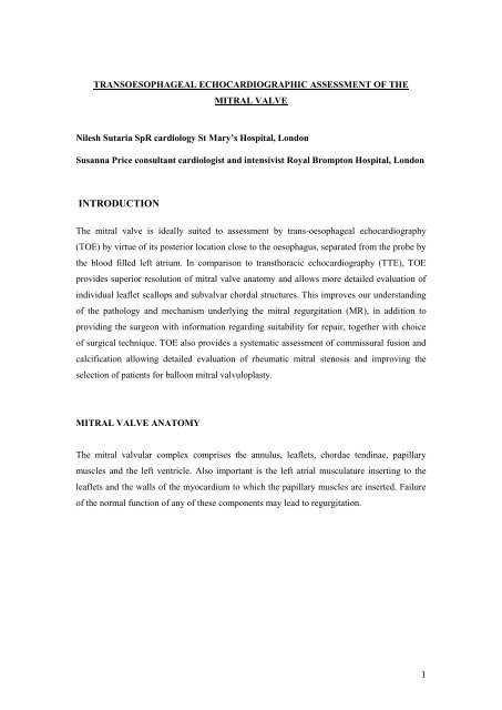
TRANSOESOPHAGEAL ECHOCARDIOGRAPHIC ASSESSMENT OF THE<br />
MITRAL VALVE<br />
Nilesh Sutaria SpR cardiology St Mary’s Hospital, London<br />
Susanna Price consultant cardiologist <strong>and</strong> intensivist Royal Brompton Hospital, London<br />
INTRODUCTION<br />
The <strong>mitral</strong> <strong>valve</strong> is ideally suited to <strong>assessment</strong> by trans-oesophageal echocardiography<br />
(<strong>TOE</strong>) by virtue of its posterior location close to the oesophagus, separated from the probe by<br />
the blood filled left atrium. In comparison to transthoracic echocardiography (TTE), <strong>TOE</strong><br />
provides superior resolution of <strong>mitral</strong> <strong>valve</strong> anatomy <strong>and</strong> allows more detailed evaluation of<br />
individual leaflet scallops <strong>and</strong> subvalvar chordal structures. This improves our underst<strong>and</strong>ing<br />
of the pathology <strong>and</strong> mechanism underlying the <strong>mitral</strong> regurgitation (MR), in addition to<br />
providing the surgeon with information regarding suitability for repair, together with choice<br />
of surgical technique. <strong>TOE</strong> also provides a systematic <strong>assessment</strong> of commissural fusion <strong>and</strong><br />
calcification allowing detailed evaluation of rheumatic <strong>mitral</strong> stenosis <strong>and</strong> improving the<br />
selection of patients for balloon <strong>mitral</strong> valvuloplasty.<br />
MITRAL VALVE ANATOMY<br />
The <strong>mitral</strong> valvular complex comprises the annulus, leaflets, chordae tendinae, papillary<br />
muscles <strong>and</strong> the left ventricle. Also important is the left atrial musculature inserting to the<br />
leaflets <strong>and</strong> the walls of the myocardium to which the papillary muscles are inserted. Failure<br />
of the normal function of any of these components may lead to regurgitation.<br />
1
The <strong>mitral</strong> annulus forms part of the fibrous skeleton of the heart <strong>and</strong> has a complex threedimensional<br />
saddle-shaped structure. (Figure 1).<br />
Figure 1 . A: Fibrous skeleton of the heart. B: Saddle shaped <strong>mitral</strong> annulus<br />
During <strong>TOE</strong>, annular dimensions should be assessed at end-systole in both the midoesophageal<br />
commissural view (low, longer axis) <strong>and</strong> mid-oesophageal long axis view (high,<br />
shorter axis) [1]<br />
2
The <strong>mitral</strong> <strong>valve</strong> is bi-leaflet. The Carpentier system of classification of the <strong>mitral</strong> <strong>valve</strong><br />
leaflet segments is based on an anatomical view of the <strong>valve</strong> <strong>and</strong> has been adopted by the<br />
ASE/SCA guidelines <strong>and</strong> many cardiac surgeons. (Figure 2)[2]<br />
Figure 2 Carpentier nomenclature for <strong>mitral</strong> <strong>valve</strong> segments<br />
PV = pulmonary <strong>valve</strong><br />
AV = aortic <strong>valve</strong><br />
TV = tricuspid <strong>valve</strong><br />
The anterior (aortic) leaflet is in fibrous continuity with the aortic <strong>valve</strong> <strong>and</strong> has a base which<br />
attaches to one third of the annular circumference. The posterior (mural) leaflet is long <strong>and</strong><br />
narrow, with a more extensive area of insertion to the annulus <strong>and</strong> is further divided by clefts<br />
into 3 scallops: anterolateral, middle <strong>and</strong> posteromedial or P1, P2 <strong>and</strong> P3. The middle or P2<br />
scallop of the posterior leaflet is most commonly involved in prolapse of a floppy <strong>mitral</strong><br />
<strong>valve</strong>. The opposing segments of the anterior leaflet are labelled A1, A2 <strong>and</strong> A3. The curved<br />
line of coaptation between the anterior <strong>and</strong> posterior <strong>mitral</strong> leaflets is generally regarded as<br />
having an anterolateral <strong>and</strong> posteromedial commissure at either end.<br />
3
Two papillary muscles (anterolateral <strong>and</strong> posteromedial) support the <strong>mitral</strong> <strong>valve</strong> <strong>and</strong> each<br />
attaches to both leaflets via thin fibrous chordae tendinae (Figure 3)<br />
Figure 3 Chordal relationships (ASE/SCA terminology as per Carpentier) [3]<br />
First order chordae attach to the fibrous b<strong>and</strong> running along the free edge of both leaflet tips<br />
ensuring coaptation of the contact surfaces (rough zone) of the leaflets. Second order chordae<br />
attach to the ventricular surface of both leaflets <strong>and</strong> contribute to ventricular function. Third<br />
order chordae arise from the ventricular wall <strong>and</strong> attach only to the posterior leaflet base as it<br />
inserts into the annulus. The anterolateral papillary muscle has dual blood supply from the<br />
LAD <strong>and</strong> circumflex coronary arteries <strong>and</strong> supports the anterolateral segments of each leaflet<br />
(A1, P1 <strong>and</strong> part of A2, P2). The posteromedial PM supports A3, P3 <strong>and</strong> part of A2, P2, it is<br />
usually supplied entirely by the right coronary artery <strong>and</strong> is therefore more susceptible to<br />
infarction <strong>and</strong> rupture with resultant severe MR.<br />
4
MITRAL REGURGITATION<br />
Carpentier’s functional classification of <strong>mitral</strong> regurgitation is widely used <strong>and</strong> describes the<br />
mechanism of MR according to whether leaflet motion is normal, excessive or restricted<br />
(Figure 4) [4].<br />
Figure 4. Carpentier’s functional classification of leaflet <strong>and</strong> chordal motion showing<br />
the direction of the MR jet [3]<br />
Type I<br />
MR associated with normal leaflet motion is caused by annular dilatation resulting in failure<br />
of coaptation, or leaflet perforation complicating endocarditis<br />
5
Type II<br />
Myxomatous degeneration is the commonest cause of MR. There is leaflet thickening <strong>and</strong><br />
redundancy, chordal elongation with possible rupture <strong>and</strong> annular dilatation. (Figure 5).<br />
Figure 5.<br />
Myxomatous <strong>mitral</strong> <strong>valve</strong> disease with a flail<br />
segment of the posterior leaflet<br />
This prevents normal apposition of the leaflet tips resulting in a spectrum of pathology<br />
including billowing, <strong>mitral</strong> <strong>valve</strong> prolapse (MVP) <strong>and</strong> flail <strong>mitral</strong> <strong>valve</strong> with variable degrees<br />
of <strong>mitral</strong> regurgitation. (Figure 6)<br />
Figure 6 Excessive leaflet motion (Carpentier type II) [5]<br />
Billowing occurs when part of the body of the <strong>mitral</strong> leaflet rises above the annulus in systole,<br />
but the coaptation point remains below the plane of the annulus.<br />
Prolapse occurs when the leaflet tip protrudes above the level of the <strong>mitral</strong> annulus in systole<br />
<strong>and</strong> is the leading cause of isolated <strong>mitral</strong> regurgitation <strong>and</strong> regurgitation requiring surgery.<br />
Early findings of a 3-15% prevalence of MVP have been overestimated because of nonuniform<br />
echo criteria. Due to the non-planar nature of the annulus, the 4-chamber view tends<br />
to over-diagnose the presence of prolapse. Analysis in the transthoracic long-axis parasternal<br />
or apical views are more specific <strong>and</strong> using this technique classic MVP, defined as superior<br />
displacement of the <strong>mitral</strong> leaflets >2mm during systole <strong>and</strong> maximal leaflet thickness of at<br />
least 5mm during diastasis, occurred in 1.3% of an unselected community based population<br />
[6].<br />
Flail leaflet is a condition in which the edge of the leaflet is free flowing in the left atrium<br />
throughout systole. The aetiology is usually ruptured chordae associated with myxomatous<br />
6
change or rarely papillary muscle rupture following myocardial infarction. The regurgitation<br />
generally develops more rapidly, <strong>and</strong> with increased severity compared with MVP.<br />
Where a single scallop is involved, both prolapse <strong>and</strong> flail leaflets give rise to an eccentric jet<br />
of <strong>mitral</strong> regurgitation directed away from the affected leaflet, often hugging the wall of the<br />
left atrium. Jet eccentricity may, however, be misleading where more than one leaflet of the<br />
<strong>mitral</strong> <strong>valve</strong> is involved.<br />
Type III<br />
Leaflet restriction during systole <strong>and</strong> diastole (IIIa) is seen with leaflet thickening <strong>and</strong><br />
calcification <strong>and</strong> subvalvular tethering from rheumatic <strong>valve</strong> disease. Leaflet restriction<br />
during systole (IIIb) occurs in cases of LV dysfunction where there is an altered geometric<br />
relationship between the papillary / chordal apparatus <strong>and</strong> <strong>mitral</strong> leaflets. With ventricular<br />
dilatation, the more globular shape of the LV results in lateral <strong>and</strong> apical displacement of the<br />
papillary muscles with resultant tethering or restriction of leaflet motion Figure 4D). The<br />
leaflets are morphologically normal but become “tented” with loss of the usual systolic leaflet<br />
overlap or with a visible coaptation defect (figure 7).<br />
Figure 7. Apical displacement (“tenting”) of the <strong>mitral</strong> <strong>valve</strong> leaflet tips<br />
In dilated cardiomyopathy the MR is usually central. In the case of MR associated with<br />
ischaemic heart disease, there may be localised dilatation of the myocardium causing<br />
asymmetrical leaflet tethering <strong>and</strong> papillary muscle dysfunction resulting in an eccentric<br />
regurgitant jet which is directed towards the side of the lesion. (Figure 4C)<br />
7
MITRAL VALVE REPAIR<br />
The advantages of surgical repair over replacement include anatomical restoration of the<br />
<strong>mitral</strong> subvalvar apparatus with preservation of left ventricular function, reduced<br />
thromboembolic risk avoiding the need for long-term warfarin <strong>and</strong> excellent durability.<br />
St<strong>and</strong>ard <strong>mitral</strong> <strong>valve</strong> repair involves firstly the correction of abnormalities of the leaflets <strong>and</strong><br />
chordal apparatus, <strong>and</strong> may also include stabilisation of the annulus by implantation of an<br />
annular ring. Pre-operative <strong>assessment</strong> should involve detailed <strong>and</strong> systematic analysis of<br />
each part of the <strong>valve</strong> <strong>and</strong> sub-valvar apparatus (as described below) to determine the<br />
mechanism(s) of regurgitation. In addition, the surgeon should be alerted to the presence of<br />
annular calcification. Following <strong>mitral</strong> <strong>valve</strong> repair, echocardiography should be directed<br />
towards (i) <strong>assessment</strong> of the anatomical surgical result <strong>and</strong> (ii) <strong>assessment</strong> of the degree of<br />
<strong>mitral</strong> regurgitation. Therefore some knowledge of the surgical techniques employed is<br />
essential for accurate interpretation.<br />
Isolated posterior leaflet prolapse is the most frequent cause of <strong>mitral</strong> regurgitation<br />
accounting for up to 60% of cases in most surgical series, <strong>and</strong> most commonly involving the<br />
middle scallop (P2). Repair of isolated P2 prolapse is highly successful <strong>and</strong> involves resecting<br />
a segment of leaflet with direct suturing of the defect. The traditional quadrangular resection<br />
has recently been replaced by triangular resection of the P2 scallop which allows better reapproximation<br />
<strong>and</strong> suturing of the resected margins. If the posterior leaflet is very wide <strong>and</strong><br />
redundant, systolic anterior motion (SAM) can result, causing left ventricular outflow<br />
obstruction <strong>and</strong> <strong>mitral</strong> regurgitation. On occasion this necessitates a sliding plasty which<br />
consists of detaching the posterior leaflet from the annulus on both sides of the quadrangular<br />
resection <strong>and</strong> resuturing it closer to the opposite segment; this not only decreases its surface<br />
area but also redistributes the stress in the suture lines <strong>and</strong> annulus. [7]<br />
Anterior leaflet prolapse is usually the result of elongation or rupture of the anterior leaflet<br />
chordae <strong>and</strong> repair is more challenging, involving the more dem<strong>and</strong>ing techniques of chordal<br />
shortening, chordal transfer, <strong>and</strong> implantation of artificial Gortex chordae. Repair can also be<br />
performed in bileaflet prolapse <strong>and</strong> rheumatic disease.<br />
In <strong>mitral</strong> regurgitation post-myocardial infarction, over-reduction of the <strong>mitral</strong> annulus to<br />
promote coaptation of the restricted leaflets may be achieved using an annuloplasty ring. This<br />
decreases the radius of this cavity <strong>and</strong> the distance between the papillary muscles,<br />
consequently decreasing also the tethering effect on the <strong>mitral</strong> leaflets [7].<br />
8
MITRAL STENOSIS<br />
Mitral stenosis is almost always the result of a previous attack of rheumatic fever, although<br />
50% of cases are subclinical. In symptomatic patients with severe <strong>mitral</strong> stenosis,<br />
percutaneous balloon <strong>mitral</strong> valvotomy (BMV) offers an alternative to surgical<br />
commissurotomy with equivalent results <strong>and</strong> has become the treatment of choice in young<br />
patients with pliant, non-calcified <strong>valve</strong>s with predominant commissural fusion. With the<br />
disappearance of rheumatic fever in the Western world, most cases of <strong>mitral</strong> stenosis<br />
encountered in centres such as ours are elderly patients with more severe degenerative <strong>valve</strong><br />
disease which is less suitable for BMV <strong>and</strong> echocardiography has a key role in the case<br />
selection. Occasionally, <strong>mitral</strong> stenosis is due to a congenital abnormality of the <strong>mitral</strong> <strong>valve</strong>.<br />
Here, it is important to exclude other cardiac abnormalities (especially stenotic lesions<br />
affecting the left heart <strong>and</strong> aorta).<br />
Pre-valvuloplasty <strong>assessment</strong>.<br />
The diagnosis <strong>and</strong> haemodynamic <strong>assessment</strong> of <strong>mitral</strong> stenosis can be made adequately from<br />
transthoracic echo. The leaflets are thickened initially at the tips with reduced mobility <strong>and</strong> a<br />
bowing motion in diastole. The subvalvar chordal structures are thickened, fibrosed <strong>and</strong><br />
shortened. The Wilkins Score is the most widely used transthoracic scoring system to guide<br />
case selection for BMV <strong>and</strong> examines four aspects of <strong>valve</strong> morphology: leaflet thickening,<br />
leaflet mobility, leaflet calcification <strong>and</strong> subvalvar disease. Each is graded 1-4, higher scores<br />
(>8) indicating more severe degenerative disease <strong>and</strong> predicting a worse outcome from<br />
valvuloplasty [8].<br />
The dominant mechanism of BMV is commissural splitting. Patients with extensive<br />
symmetrical commissural fusion gain an optimal result but calcified commissures resist<br />
splitting <strong>and</strong> result in leaflet tearing <strong>and</strong> <strong>mitral</strong> regurgitation. However, the Wilkins Score<br />
does not include commissural <strong>assessment</strong> <strong>and</strong> many have found this to be a weak predictor of<br />
outcome. Although both commissures can be visualised in the parasternal short axis view, in<br />
some patients resolution is poor, there is shielding from calcification <strong>and</strong> distinguishing<br />
between leaflet <strong>and</strong> commissural calcification is difficult. All these limitations are overcome<br />
by <strong>TOE</strong> which allows systematic scanning of each commissure for both the extent of fusion<br />
<strong>and</strong> localisation of calcification (figure 8). A Commissure Score based on this <strong>assessment</strong><br />
ranging from 0-4 is a more powerful predictor of outcome <strong>and</strong> is of additional predictive<br />
value in patients with a low Wilkins Scores (
surgical risk, BMV can offer a short term palliative option if there is any prospect of<br />
commissural splitting.<br />
The presence of moderate or severe MR is generally a contraindication to BMV <strong>and</strong> <strong>TOE</strong> is<br />
useful for <strong>assessment</strong> of MR severity. Furthermore, <strong>TOE</strong> is valuable to confirm a thin<br />
interatrial septum suitable for transeptal puncture <strong>and</strong> to exclude left atrial appendage<br />
thrombus (Figure 9) since these structures cannot be reliably assessed from the transthoracic<br />
approach.<br />
Figure 8<br />
Calcified anterolateral commissure<br />
Figure 9<br />
Mobile LA appendage thrombus<br />
10
SYSTEMATIC EXAMINATION OF MITRAL VALVE LEAFLETS USING <strong>TOE</strong><br />
Figure 10 Anatomical view of the <strong>mitral</strong> <strong>valve</strong> labelled according to the Carpentier<br />
system with planes of the views obtained during <strong>TOE</strong> [2][9]<br />
With the probe in the midoesophagus, the four chamber view (0) visualises the anterolateral<br />
part of the commissure <strong>and</strong> the coaptation line is scanned by advancing <strong>and</strong> withdrawing the<br />
probe. Conversely, the 2 chamber view (90) visualises the posteromedial end of the<br />
commissure <strong>and</strong> the coaptation line is scanned by rotating the probe clockwise <strong>and</strong><br />
anticlockwise.<br />
One method for systematic examination of the <strong>mitral</strong> <strong>valve</strong> scallops begins with the probe in<br />
the mid oesophagus at 0 <strong>and</strong> withdrawn or flexed to a five chamber view visualising the<br />
most anterolateral part of the commissure (nearest the LA appendage) between A1, P1.<br />
Advancing the probe from the annulus to the centre of the <strong>valve</strong> visualises coaptation between<br />
A2 <strong>and</strong> P2 with further advancement <strong>and</strong> retroflexion revealing A3, P3.<br />
At 60-70 in the mid oesophagus the scan plane cuts the curved <strong>mitral</strong> commissure twice<br />
(commissural or “seagull” view) <strong>and</strong> visualises from left to right P3, A2, P1 with P3/A3<br />
commissure on the left, A1/P1 on the right. (Figure 11) This view is useful for determining<br />
11
which part of the commissure is regurgitant <strong>and</strong> confirms the presence of P2 prolapse by<br />
visualising the prolapsing P2 segment in the centre of the annulus above A2. (Figure 12D).<br />
Where <strong>valve</strong> regurgitation is due to annular dilatation, multiple regurgitant jets may be seen.<br />
The mid oesophageal 2 chamber view is obtained at 90 with the LA appendage visible to the<br />
right. The scan plane cuts through P3 on the left <strong>and</strong> all 3 segments of the anterior leaflet on<br />
the right. This view shows coaptation between the posteromedial segments P3/A3 <strong>and</strong><br />
visualises the posteromedial papillary muscle. (Figure 13A)<br />
The midoesophageal long axis view (120-150) cuts the coaptation line perpendicularly<br />
through A2/P2 <strong>and</strong> the left ventricular outflow tract (LVOT) is in view. The papillary muscles<br />
are excluded. This is a useful view for evaluating SAM following <strong>valve</strong> repair <strong>and</strong><br />
measurement of the vena contracta. In this view, clockwise (right) rotation sweeps the plane<br />
towards the posteromedial commissure <strong>and</strong> anticlockwise (left) rotation sweeps towards the<br />
anterolateral commissure.<br />
The transgastric basal short axis view can allow visualisation of all <strong>mitral</strong> <strong>valve</strong> segments <strong>and</strong><br />
the entire line coaptation line (Figure 13B). Colour Doppler in this view is helpful in<br />
localising the origin of the regurgitant jet <strong>and</strong> prolapsing segments can often be seen. This is<br />
especially useful in confirming the localisation of regurgitant jets seen in the mid-oesophageal<br />
views. The transgastric 90 plane provides a long axis view of the LV <strong>and</strong> <strong>mitral</strong> <strong>valve</strong> <strong>and</strong> is<br />
useful for assessing the subvalvular chordal anatomy, particularly in the presence of leaflet<br />
calcification where the mid-oesophageal views may have been unhelpful.<br />
12
Figure 11 Inter-commissural view. Two jets of MR arising<br />
from P1-A2 <strong>and</strong> P3-A2 commissure<br />
Figure 12. Patient with severe MR due to prolapse P2 scallop<br />
A: 4 chamber view showing A2/P2 coaptation point<br />
B: 4 chamber view showing eccentric jet of MR directed away (anteriorly) from the lesion<br />
with large PISA<br />
C: Systolic flow reversal in the left upper pulmonary vein<br />
D: Inter-commissural view showing flail P2 scallop above A2<br />
E: Long-axis view showing A2/P2 coaptation point <strong>and</strong> LVOT<br />
13
Figure 13. Severe MR due to flail A3 segment<br />
A: 2 chamber view showing the posteromedial part of the <strong>valve</strong> with A3/P3 coaptation point<br />
<strong>and</strong> eccentric jet of MR directed away from the lesion (posteriorly)<br />
B: Transgastric view confirming the origin of the MR from the posteromedial part of the<br />
commissure<br />
14
In <strong>mitral</strong> stenosis, the anterolateral <strong>and</strong> posteromedial commissures should be scanned<br />
systematically in the 0 <strong>and</strong> 90mid oesophageal planes respectively from the annulus to the<br />
leaflet tips to determine the extent of fusion <strong>and</strong> presence of commissural calcification. Each<br />
commissure can be assigned a score to reflect the likelihood of splitting. A score of 0 is given<br />
if there is no fusion, or if commissural calcification, expected to resist splitting, is present.<br />
When non-calcified fusion is present, a score of 1 is given if the fusion is partial <strong>and</strong> a score<br />
of 2 given when such fusion is extensive. Each <strong>valve</strong> therefore has an overall ‘Commissure<br />
Score’ ranging from 0 to 4, a high score indicating extensively fused, non calcified<br />
commissures which are therefore more likely to split, whereas a low score reflects either<br />
minimal fusion or the presence of resistant commissural calcification [9].<br />
INTRA-OPERATIVE <strong>TOE</strong><br />
The effects of general anaesthesia (after-load <strong>and</strong> pre-load reduction) will<br />
significantly reduce the severity of MR <strong>and</strong> therefore grading the MR in theatre prebypass<br />
can be unreliable. It is more important to focus on the mechanism of MR <strong>and</strong><br />
suitability for repair. Echocardiographic predictors of an unsuccessful repair (or need<br />
for MVR) include annular diameter >5cm, <strong>mitral</strong> annular calcification, central MR jet<br />
<strong>and</strong> extensive bi-leaflet disease (3 or more scallops prolapse or flail)[11].<br />
Immediately after coming off bypass, interpretation of colour Doppler <strong>and</strong> a definitive<br />
<strong>assessment</strong> of residual MR should be delayed until the left ventricle is adequately filled <strong>and</strong><br />
appropriate vasoconstrictor therapy has been instituted to recover the awake haemodynamic<br />
state. Initial <strong>assessment</strong> should be directed towards systematic examination of the anatomy of<br />
the repair, with no residual prolapse, <strong>and</strong> a good area of coaptation between the leaflets. This<br />
should be made with knowledge of the details of the surgery undertaken. For example, where<br />
quadrangular resection has been performed, the posterior leaflet may appear short <strong>and</strong> less<br />
mobile. Second, it is important to assess the severity of any residual MR which may require a<br />
second bypass run. Significant MR could result from SAM, incomplete repair with residual<br />
prolapse, residual annular dilatation, suture dehiscence which may manifest as a leaflet<br />
perforation with the origin of the jet displaced from the coaptation line. Where the repair is<br />
borderline, the echocardiographer should assess other aspects of cardiac function that may<br />
affect the surgeon’s decision-making. (ie right <strong>and</strong> left ventricular function) Significant<br />
iatrogenic <strong>mitral</strong> stenosis is uncommon after repair but may be suggested by an elevated E<br />
wave velocity >2 m/s <strong>and</strong> shallow E wave deceleration slope <strong>and</strong> mean gradients of more than<br />
15
5 or 6 mmHg. It is important, however, to remember that the alternative (<strong>mitral</strong> <strong>valve</strong><br />
replacement) will have a significant residual gradient.<br />
Following <strong>mitral</strong> <strong>valve</strong> repair, SAM of the anterior leaflet can cause LVOT obstruction<br />
(Figure 14) with significant posteriorly directed MR.<br />
Figure 14. SAM of anterior leaflet into the LVOT [1].<br />
SAM is best visualised in the 5 chamber or long–axis planes <strong>and</strong> the transgastric view can<br />
allow alignment of CW Doppler across the LVOT to demonstrate high velocities (3-5 m/s).<br />
Factors increasing the risk of SAM include a small, non-dilated LV, use of a small<br />
annuloplasty ring or excessive posterior <strong>mitral</strong> leaflet tissue. Severe SAM may require<br />
surgical revision as described above. However, most cases respond to medical therapy with<br />
volume loading <strong>and</strong> discontinuing inotropes/vasodilators [1].<br />
New lateral or inferoposterior regional wall motion abnormality should alert the team to the<br />
possibility of circumflex coronary artery injury which is a rare but recognised complication of<br />
MV repair <strong>and</strong> replacement. The finding of severe aortic regurgitation post repair raises the<br />
possibility of aortic cusp injury during deep suture placement in the anterior annulus which<br />
my require AVR.<br />
16
Assessment of MR severity <strong>and</strong> deciding when to operate<br />
A number of ultrasound techniques are available for grading MR severity. Data from these<br />
should be considered collectively along with clinical information to decide whether the<br />
patient has significant MR to warrant intervention [12] (Table 1)<br />
Table 1. Echocardiographic parameters indicating severe MR<br />
Echo parameter<br />
Severe MR<br />
LA / LV Size<br />
Markedly increased (except in acute MR)<br />
Jet area (%LA) >50<br />
Spectral CW Doppler Dense triangular regurgitation signal with early peak <strong>and</strong><br />
late systolic reduction in velocity. E wave dominant with<br />
velocity >1.5 m/s.<br />
Vena contracta<br />
>7mm<br />
Pulmonary vein flow Systolic flow reversal<br />
Regurgitant volume (ml/beat) >60<br />
Regurgitant fraction >50%<br />
EROA (cm 2 ) >0.4<br />
PISA<br />
Large (radius >1 cm at aliasing velocity 40cm/s) but less<br />
accurate in eccentric jets<br />
PISA = Proximal Isovelocity Surface Area (radius r)<br />
EROA = Effective Regurgitant Orifice Area = 2 r 2 x Alias v<br />
MR Vmax<br />
Regurgitant volume = EROA x MR TVI<br />
In the setting of severe non-ischaemic MR, the ACC/AHA task force assign class I<br />
recommendations for surgery in patients with symptoms (NYHA II, III or IV), or in<br />
asymptomatic patients with evidence of LV disease (EF 45mm) [13].<br />
The guidelines stress the importance of using <strong>mitral</strong> <strong>valve</strong> repair when possible <strong>and</strong> in<br />
general suggest a lower threshold to intervene surgically when successful <strong>mitral</strong> <strong>valve</strong><br />
repair appears likely. A recent study from the Mayo clinic showed that in asymptomatic<br />
patients with organic <strong>mitral</strong> regurgitation treated medically, an EROA >0.4 cm 2 was a<br />
powerful predictor of adverse events <strong>and</strong> should prompt early referral for surgery if <strong>valve</strong><br />
repair is feasible. [14].<br />
17
Patients with poor LV function require careful consideration. Severe LV impairment will<br />
underestimate the severity of MR. Additionally, in the setting of severe MR, afterload is<br />
falsely low as the left ventricle ejects into the left atrium <strong>and</strong> following restoration of a<br />
competent <strong>mitral</strong> <strong>valve</strong> the LV may struggle severely to overcome the true afterload <strong>and</strong><br />
systemic vascular resistance.<br />
PROSTHETIC VALVES<br />
<strong>TOE</strong> has a key diagnostic role in patients with prosthetic <strong>valve</strong>s <strong>and</strong> a systematic examination<br />
should confirm normal leaflet or disk motion, proper seating in the native annulus, normal<br />
blood flow profile with absence of paravalvular or transvalvular regurgitation in addition to<br />
estimation of transvalvular pressure gradients. A basic knowledge of commonly used<br />
bioprosthetic <strong>and</strong> mechanical <strong>valve</strong>s, their flow dynamics <strong>and</strong> echocardiographic features is<br />
therefore essential. This is beyond the scope of our article <strong>and</strong> further reading is advised. The<br />
most commonly implanted mechanical <strong>valve</strong> is the bileaflet prosthesis (St Jude, Carbomedic).<br />
This is constructed of two semicircular leaflets suspended from four hinge points in a circular<br />
annulus surrounded by a sewing ring. Normal <strong>and</strong> abnormal function is shown in figures 15<br />
<strong>and</strong> 16.<br />
Figure 15 [15]<br />
Normal functioning bileaflet mechanical<br />
<strong>mitral</strong> <strong>valve</strong><br />
A&B: systolic frames showing normal small<br />
convergent jets of regurgitation (cleansing<br />
jets)<br />
C&D: diastolic frames showing the sewing<br />
ring <strong>and</strong> two parallel open leaflets. Colour<br />
Doppler shows acceleration of flow through<br />
two lateral orifices <strong>and</strong> a small central orifice<br />
18
Figure 16 [5] Bileaflet MVR<br />
Paravalvular regurgitation originating<br />
outside the sewing ring is always pathological<br />
19
REFERENCES (Essential reading highlighted in bold)<br />
1.Sidebotham, Merry <strong>and</strong> Legget. Practical perioperative transoesophageal echocardiography.<br />
Butterworth-Heineman 2004<br />
2. Shanewise JS, Cheung AT, Aronson S et al. ASE/SCA guidelines for performing a<br />
comprehensive intraoperative multiplane transesophageal echocardiography<br />
examination: recommendations of the American Society of Echocardiography Council<br />
for Intraoperative Echocardiography <strong>and</strong> the Society of Cardiovascular<br />
Anesthesiologists Task Force for Certification in Perioperative Transesophageal<br />
Echocardiography. Anesthesia <strong>and</strong> Analgesia 1999;89:870-884<br />
3. Savage R, Aronson S. Comprehensive textbook of intraoperative transesophageal<br />
echocardiography. Lippincott Williams & Wilkins 2005<br />
4.Carpentier A, Deloche A, Dauptain J et al. A new reconstructive operation for correction of<br />
<strong>mitral</strong> <strong>and</strong> tricuspid insufficiency. J Thorac Cardiovasc Surg 1971;61:1-13<br />
5. Perrino A, Reeves S. A practical approach to transesophageal echocardiography. Lippincott<br />
Williams & Wilkins. 2003<br />
6. Freed LA, Levy D, Levine RA, et al. Prevalence <strong>and</strong> clinical outcome of <strong>mitral</strong>-<strong>valve</strong><br />
prolapse. N Engl J Med 1999;341:1-7<br />
7. J M Ferrão de Oliveira <strong>and</strong> Manuel J Antunes. Mitral <strong>valve</strong> repair: better than replacement<br />
Heart 2006;92;275-281<br />
8. Abascal V, Wilkins G, Choong C, Thomas I. echocardiographic evaluation of <strong>mitral</strong> <strong>valve</strong><br />
structure <strong>and</strong> function in patients followed for at least six months after percutaneous <strong>mitral</strong><br />
valvuloplasty. J Am Coll Cardiol 1989;12:606-615<br />
9. D Pellerin, S Brecker, C Vetrat. Degenerative <strong>mitral</strong> <strong>valve</strong> disease with an emphysis on<br />
<strong>mitral</strong> <strong>valve</strong> prolapse. Heart 2002;88(Suppl IV):iv20-iv28<br />
20
10. N Sutaria, TRD Shaw, B Prendergast, D Northridge. Transoesophageal echocardiographic<br />
<strong>assessment</strong> of commissural fusion <strong>and</strong> calcification predicts outcome after balloon <strong>mitral</strong><br />
valvotomy. Heart 2006;92:52-57<br />
11. Omran A. Woo A, David T et al Intraoperative Transesophageal Echocardiography<br />
Accurately Predicts Mitral Valve Anatomy <strong>and</strong> Suitability for Repair. J Am Soc<br />
Echocardiogr 2002;15:950-7<br />
12. Zoghbi WA, Enriquez-Sarano M, Foster E et al. Recommendations for<br />
Evaluation of the Severity of Native Valvular Regurgitation with Twodimensional<br />
<strong>and</strong> Doppler Echocardiography A report from the American<br />
Society of Echocardiography’s Nomenclature <strong>and</strong> St<strong>and</strong>ards Committee <strong>and</strong><br />
The Task Force on Valvular Regurgitation. Am Soc Echocardiogr 2003;16:777-<br />
802.<br />
13. Robert O. Bonow, Blase Carabello, Antonio C. de Leon, Jr et al. Guidelines<br />
for the Management of Patients With Valvular Heart Disease. Executive<br />
Summary Report of the American College of Cardiology/American Heart<br />
Association Task Force on Practice Guidelines. (Committee on Management of<br />
Patients with Valvular Heart Disease) Circulation. 1998;98:1949 –1984.<br />
14. Enriquez-Sarano M, Avierinos JF, Messika-Zeitoun D et al. Quantitative<br />
Determinants of the Outcome of Asymptomatic Mitral Regurgitation N Engl J Med<br />
2005;352:875-83.<br />
15. Otto C. Textbook of Clinical Echocardiography. 3 rd Edition. Elsevier Saunders 2004.<br />
21