Thermostabilization of a Cytochrome P450 Peroxygenase
Thermostabilization of a Cytochrome P450 Peroxygenase
Thermostabilization of a Cytochrome P450 Peroxygenase

You also want an ePaper? Increase the reach of your titles
YUMPU automatically turns print PDFs into web optimized ePapers that Google loves.
<strong>Thermostabilization</strong> <strong>of</strong> a <strong>Cytochrome</strong><br />
<strong>P450</strong> <strong>Peroxygenase</strong><br />
Oriana Salazar, Patrick C. Cirino, and<br />
Frances H. Arnold* [a]<br />
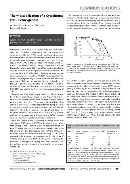
To characterize the thermostability <strong>of</strong> the peroxygenase<br />
variants identified during screening, we measured the fraction<br />
<strong>of</strong> folded heme domain remaining after heat-treatment, which<br />
we determined from the fraction <strong>of</strong> the ferrous heme-CO<br />
complex that retained the 450 nm absorbance peak characteristic<br />
<strong>of</strong> properly-folded <strong>P450</strong>. Figure 1 shows the percentage <strong>of</strong><br />
KEYWORDS:<br />
cytochrome <strong>P450</strong> ¥ directed evolution ¥ enzymes ¥ oxidation ¥<br />
peroxygenases ¥ thermostability<br />
<strong>Cytochrome</strong> <strong>P450</strong> BM-3 is a soluble fatty acid hydroxylase<br />
composed <strong>of</strong> a heme domain and a reductase domain on a<br />
single polypeptide chain. [1] We recently described a laboratoryevolved<br />
variant <strong>of</strong> the <strong>P450</strong> BM-3 heme domain which functions<br />
as an H 2 O 2 -driven hydroxylase (peroxygenase∫) and does not<br />
require NADPH, O 2 , or the reductase. [2] This variant, which we<br />
named 21B3, allows us to carry out cytochrome <strong>P450</strong>-catalyzed<br />
biotransformations under highly simplified reaction conditions:<br />
only the heme domain and hydrogen peroxide are needed for<br />
substrate (fatty acid) hydroxylation. Because its heme domain<br />
alone is competent for catalysis, <strong>P450</strong> BM-3 peroxygenase 21B3<br />
<strong>of</strong>fers a unique opportunity to create a thermostable, functional<br />
cytochrome <strong>P450</strong>. Here we report further directed evolution <strong>of</strong><br />
the 21B3 peroxygenase, resulting in an enzyme which is<br />
significantly more thermostable than wildtype cytochrome<br />
<strong>P450</strong> BM-3 and retains much <strong>of</strong> the peroxygenase activity <strong>of</strong><br />
21B3.<br />
Enzymes are <strong>of</strong>ten poorly stable under conditions encountered<br />
during production, storage, or use. Improving enzyme<br />
resistance to thermal denaturation has been a major focus <strong>of</strong><br />
protein engineering efforts. [3±5] Improved thermostability <strong>of</strong>ten<br />
correlates with longer shelf-life, longer life-time during use (even<br />
at low temperatures), and a higher temperature optimum for<br />
[6, 7]<br />
activity. There have been no reports <strong>of</strong> stabilizing the<br />
relatively unstable cytochrome <strong>P450</strong> enzymes by protein<br />
engineering, however, primarily because the <strong>P450</strong>s comprise<br />
multiple subunits and contain thermolabile c<strong>of</strong>actors.<br />
<strong>P450</strong> BM-3 heme domain containing the single amino acid<br />
substitution F87A (mutant HF87A) is significantly more active<br />
than wildtype heme domain (HWT) in reactions driven by<br />
H 2 O 2 . [8, 9] Variant 21B3 is much more active than HF87A in 10 mM<br />
H 2 O 2 , but is also less thermostable than HWT and HF87A. We<br />
therefore sought to improve the thermostability <strong>of</strong> 21B3 while<br />
maintaining its improved peroxygenase activity. Six cycles <strong>of</strong><br />
random mutagenesis or DNA shuffling and screening for<br />
retention <strong>of</strong> peroxygenase activity after heat treatment (see<br />
the Methods section) yielded the thermostable peroxygenase<br />
variant5H6.<br />
[a] Pr<strong>of</strong>. F. H. Arnold, Dr. O. Salazar, P. C. Cirino<br />
Division <strong>of</strong> Chemistry and Chemical Engineering<br />
California Institute <strong>of</strong> Technology<br />
Pasadena, CA 91125 (USA)<br />
Fax: (‡ 1) 626-568-8743<br />
E-mail: frances@cheme.caltech.edu<br />
Figure 1. Percentage <strong>of</strong> 450 nm CO-binding peak <strong>of</strong> cytochrome <strong>P450</strong> BM-3<br />
heme domain HWT (&), HF87A (*), and 5H6 (~) remaining after 10-minute<br />
incubations at the indicated temperatures. For the holoenzyme BWT (^), the<br />
percentage <strong>of</strong> initial NADPH-driven activity remaining after 10-minute<br />
incubations is shown.<br />
properly-folded heme domain protein remaining after 10-<br />
minute incubations at different, elevated temperatures. To allow<br />
comparison to the wildtype full-length enzyme (BWT), whose<br />
stability is limited by the stability <strong>of</strong> the reductase domain and<br />
therefore cannot be determined from the CO-binding measurement,<br />
we determined the residual (NADPH-driven) activity <strong>of</strong><br />
BWT following 10-minute incubations at the same temperatures.<br />
Data in Figure 1 were fit to a two-state model, and the resulting<br />
calculated temperatures corresponding to half denaturation for<br />
the 10-minute heat incubations (T 50 ) are listed in Table 1. These<br />
values are in good agreement with the midpoints <strong>of</strong> the melting<br />
curves <strong>of</strong> Figure 1. According to this measure <strong>of</strong> stability, variant<br />
5H6 (T 50 ˆ 618C) is much more thermostable than the natural<br />
catalytic system, BWT (T 50 ˆ 438C). Itis also significantly more<br />
thermostable than HF87A and 21B3.<br />
Table 1. Thermostability and activity parameters for evolved and parental<br />
<strong>P450</strong>s.<br />
Mutant [a]<br />
T 50 for 10-minute<br />
incubations [b] [ o C]<br />
t 1/2 at57.5 8C [c] [min]<br />
BWT 43 0.46 < 5<br />
HWT 57 n.d. < 5<br />
HF87A 54 2.3 23<br />
21B3 46 n.d. 430<br />
5H6 61 115 220<br />
<strong>Peroxygenase</strong><br />
Activity [d] [min 1 ]<br />
[a] BWT ˆ full-length, wildtype <strong>P450</strong> BM-3; HWT ˆ wildtype <strong>P450</strong> BM-3<br />
heme domain; HF87A ˆ <strong>P450</strong> BM-3 heme domain containing mutation<br />
F87A; 21B3, 5H6 ˆ evolved heme domain peroxygenase variants. [b] Calculated<br />
from the data in Figure 1, fit to a two-state denaturation equation.<br />
[c] Calculated from the data in Figure 2, fit to a first-order exponential decay<br />
equation. [d] Reported as initial rates at room temperature on 12-pNCA in<br />
10 mM H 2 O 2 and 6 % DMSO. n.d.: notdetermined.<br />
ChemBioChem 2003, 4, 891 ± 893 DOI: 10.1002/cbic.200300660 ¹ 2003 Wiley-VCH Verlag GmbH & Co. KGaA, Weinheim 891
<strong>Peroxygenase</strong> activities were measured at room temperature,<br />
using a colorimetric assay with 12-p-nitrophenoxycarboxylic acid<br />
(12-pNCA) as substrate (see ref. [10] and Methods Section). 5H6<br />
retains 50 % <strong>of</strong> the high activity <strong>of</strong> 21B3 and is almost ten times<br />
as active as HF87A (Table 1). Another useful measure <strong>of</strong> enzyme<br />
stability comes from the rate <strong>of</strong> inactivation at high temperature.<br />
Figure 2 shows the percentage <strong>of</strong> activity that remains for the<br />
Figure 2. Heat-inactivation <strong>of</strong> cytochrome <strong>P450</strong> BM-3 holoenzyme BWT (^)<br />
and peroxygenase mutants HF87A (*) and 5H6 (~), calculated as the<br />
percentage <strong>of</strong> activity remaining after incubation at 57.5 8C for the indicated<br />
periods <strong>of</strong> time. <strong>Peroxygenase</strong> activity was measured for HF87A and 5H6, while<br />
NADPH-driven activity was measured for BWT.<br />
different variants upon heating at 57.5 8C. The activities decay<br />
exponentially with time (first-order), and the half-life (t 1/2 ) <strong>of</strong> each<br />
catalytic system is listed in Table 1. HF87A (which is less<br />
thermostable than HWT) is significantly more resistant to<br />
inactivation at 57.5 8C compared to BWT. (The half-life <strong>of</strong> HF87A<br />
is also higher than that <strong>of</strong> BWT at room temperature (data not<br />
shown)). The half-life <strong>of</strong> 5H6 at57.5 8C is 50 times longer than<br />
that <strong>of</strong> HF87A and 250 times that <strong>of</strong> BWT. The fraction <strong>of</strong><br />
peroxygenase activity remaining after heat treatment correlated<br />
with the fraction <strong>of</strong> remaining CO-binding peak for HF87A and<br />
5H6. (Residual activity <strong>of</strong> HWT could not be correlated to the<br />
remaining CO-binding peak because HWT has essentially no<br />
peroxygenase activity.)<br />
Enzyme thermostabilization <strong>of</strong>ten leads to a shift in the<br />
activity-temperature pr<strong>of</strong>ile to higher temperatures, reflecting<br />
the higher stability <strong>of</strong> the folded protein. [6] Measurements <strong>of</strong><br />
peroxygenase activity at different temperatures, however,<br />
showed no significant increase in the optimum temperature<br />
for activity for 5H6 compared to HF87A (both were 25 ± 30 8C).<br />
Thermostable peroxygenase 5H6 contains eight new amino<br />
acid substitutions compared to 21B3 (see ref. [2]), and 15<br />
substitutions compared to HF87A. 5H6 also contains a deletion<br />
resulting in the removal <strong>of</strong> one His residue from the 6-His<br />
sequence included at the C terminus. The M145V substitution<br />
that had been found previously in mutant 21B3 reverted to Met<br />
upon back-crossing with HF87A (see Methods Section). Five <strong>of</strong><br />
the substitutions are conservative with regard to hydrophobicity<br />
and size: L52I, A184V, L324I, V340M and I366V. Ser 106 was<br />
converted to a positively charged Arg residue (S106R) and a<br />
negatively-charged Glu residue was converted to a positivelycharged<br />
Lys (E442K). It is difficult to rationalize how these<br />
mutations increase the enzyme's stability. According to the <strong>P450</strong><br />
BM-3 heme domain crystal structure, [11] substitutions S106R,<br />
L324I, V340M, I366V, and E442K are located on the protein<br />
surface; the others are buried. Four stabilizing mutations are<br />
close to positions where mutations that improved peroxygenase<br />
activity accumulated in earlier experiments: L52I (in b-sheet1 ±<br />
2) is adjacentto I58V (helix B) from 21B3, S106R (in a loop<br />
connecting helices C and D) lies next to mutation F107L from<br />
21B3, E442K (in b-sheet4 ± 2) lies adjacent to K434E (in b-<br />
sheet 4 ± 1) from 21B3, and the reversion to M145 (helix E) is<br />
adjacent to S274T (helix I) from 21B3. The new stabilizing<br />
mutations may serve to alleviate structural perturbations<br />
introduced by the original (activity) mutations.<br />
Two thermostable cytochrome <strong>P450</strong>s, CYP119 and CYP175A1,<br />
[12, 13]<br />
from thermophilic organisms have recently been described<br />
and their (heme domain) crystal structures determined. [14±16]<br />
CYP119 exhibits a melting temperature <strong>of</strong> 918C. Aromatic<br />
stacking, salt-link networks and shortened loops are believed to<br />
help stabilize these enzymes. Unfortunately, the functions <strong>of</strong><br />
these <strong>P450</strong>s are not known, and reported activities are extremely<br />
low (for example, 0.35 min 1 in the NADH-driven hydroxylation<br />
<strong>of</strong> lauric acid). [17]<br />
Directed evolution allows us to explore non-natural functions<br />
and properties <strong>of</strong> <strong>P450</strong>s. In this first example <strong>of</strong> engineering a<br />
<strong>P450</strong> to resist thermal denaturation, we have used directed<br />
evolution to improve the thermostability <strong>of</strong> BM-3 peroxygenase<br />
variant 21B3. These results reinforce the biotechnological<br />
relevance <strong>of</strong> cytochrome <strong>P450</strong> BM-3 and the catalytic potential<br />
<strong>of</strong> our biomimetic hydroxylase.<br />
Methods<br />
General Remarks: All chemical reagents were procured from Aldrich,<br />
Sigma, or Fluka. Restriction enzymes were purchased from New<br />
England Biolabs and Roche. Deep-well plates (96 wells, 1 mL volume<br />
per well) for growing mutant libraries were purchased from Becton<br />
Dickinson. Flat-bottom 96-well microplates (300 mL per well) for<br />
screening mutant library activities were purchased from Rainin.<br />
Enzyme Expression and Purification: <strong>P450</strong> BM-3 enzymes were<br />
expressed in catalase-deficient E. coli [18] using the b-D-thiogalactopyranoside<br />
(IPTG)-inducible pCWori ‡ vector. [19] The heme domain<br />
consisted <strong>of</strong> the first 463 amino acids <strong>of</strong> <strong>P450</strong> BM-3 followed by a<br />
6-His sequence at the C terminus, which had no significant influence<br />
on the activity. Cultures for protein production were grown and<br />
proteins were purified as described. [9] Purified enzyme samples were<br />
stored at 80 8C until use, at which time they were thawed at room<br />
temperature and then kept on ice. Concentrations <strong>of</strong> properly-folded<br />
<strong>P450</strong> enzyme were determined from the 450 nm CO-binding<br />
difference spectra <strong>of</strong> the reduced heme, as described. [20]<br />
Preparation <strong>of</strong> Mutant Libraries: Error-prone PCR libraries were<br />
prepared using standard protocols. [21] Starting with 21B3 as the<br />
parent, three rounds <strong>of</strong> error-prone PCR (using Taq DNA polymerase<br />
(Roche)) followed by screening were performed, and the most<br />
thermostable mutant which did not lose peroxygenase activity was<br />
chosen as the parent for the next generation. Two additional<br />
generations were prepared with the GeneMorph PCR Mutagenesis<br />
Kit (Stratagene). In the final generation leading to mutant 5H6, a<br />
recombinantlibrary was prepared by DNA shuffling [22] using Pfu Ultra<br />
892 ¹ 2003 Wiley-VCH Verlag GmbH & Co. KGaA, Weinheim www.chembiochem.org ChemBioChem 2003, 4, 891 ± 893
DNA Polymerase (Stratagene). Parents for the recombinant library<br />
included HF87A, mutants from the previous generation which were<br />
more stable but less active, and mutants with increased activity.<br />
Mutant Library Screening: Screening was performed essentially as<br />
described, [2] except cell lysates were additionally subjected to a heat<br />
inactivation step and screened for residual activity (see also ref. [23]).<br />
Briefly, cultures expressing mutants were grown in 96-well deep-well<br />
plates. After cell growth, the plates were centrifuged, cell pellets<br />
were frozen at 208C, and the cells were lysed in Tris-HCl buffer<br />
(100 mM, pH 8.2) containing lysozyme (0.5 ± 1 mg mL 1 ) and deoxyribonuclease<br />
I (1.5 ± 4 units mL 1 ). Clarified cell lysates were transferred<br />
to 96-well microplates for activity measurements at room<br />
temperature (described below). Lysates were also transferred to 96-<br />
well PCR plates (GeneMate) and heated to an appropriate temperature<br />
(48 ± 57.5 8C) in a PTC200 thermocycler (MJ Research) for 10 ± 15<br />
minutes, rapidly cooled to 48C, and then brought to room temperature.<br />
The residual activities <strong>of</strong> these heat-treated lysates were then<br />
measured in the same manner as the initial activities. Clones showing<br />
a higher fraction <strong>of</strong> activity remaining after heat treatment and high<br />
initial activity were characterized further.<br />
Activity Assay: Activity on 12-pNCA [10] was determined by monitoring<br />
the formation <strong>of</strong> p-nitrophenolate (pNP) (398 nm) at room<br />
temperature using a 96-well plate spectrophotometer (SPECTRAmax<br />
Plus, Molecular Devices), as described. [2] Reaction wells contained<br />
Tris-HCl buffer (140 mL <strong>of</strong> 100 mM, pH 8.2), a stock solution <strong>of</strong><br />
substrate (10 mL<strong>of</strong>4mM 12-pNCA) in DMSO, and purified enzyme or<br />
clarified lysate. Reactions were initiated by the addition <strong>of</strong> an H 2 O 2<br />
stock solution (10 mL). Data for accurate determination <strong>of</strong> 12-pNCA<br />
turnover rates with purified enzyme were collected using a BioSpec-<br />
1601 spectrophotometer (Shimadzu), where absorbance changes<br />
could be registered every 0.1 seconds. Typical final concentrations<br />
were 250 mM 12-pNCA, 6 % DMSO, 1 ± 10 mM H 2 O 2 , and 0.1 ± 1.0 mM<br />
<strong>P450</strong>. The extinction coefficient for pNP was determined from<br />
standard pNP solutions prepared under identical reaction conditions.<br />
NADPH-driven activity <strong>of</strong> BWT was determined spectrophotometrically<br />
from the initial rate <strong>of</strong> NADPH consumption (measured as the<br />
decrease in 340 nm absorbance) in the presence <strong>of</strong> myristic acid, as<br />
described. [24]<br />
Data for T 50 Determination: Purified enzyme samples ( 20 mM) in<br />
Tris-HCl buffer (100 mM, pH 8.2) were incubated for 10 minutes at<br />
different temperatures. Samples were then cooled on ice, and the<br />
concentration <strong>of</strong> properly-folded heme domain (diluted 8x) was<br />
estimated from the 450 nm CO-binding difference spectra and<br />
compared to the CO-binding peak prior to heat treatment. Residual<br />
NADPH-consumption activity was measured for BWT. Data in<br />
Figure 1 represent average values from at least two experiments.<br />
Data for t 1/2 Determination: Concentrated purified enzyme (70 mM)<br />
was added to pre-heated (57.5 8C) Tris-HCl buffer (100 mM, pH 8.2)<br />
and incubated at 57.5 8C. Samples were removed at time intervals<br />
(indicated in Figure 2), quenched by dilution into cold buffer,<br />
brought to room temperature, and assayed for residual activity. Data<br />
in Figure 2 represent average values from at least two experiments,<br />
with standard deviations less than 10 %.<br />
[1] A. W. Munro, D. G. Leys, K. J. McLean, K. R. Marshall, T. W. Ost, S. Daff, C. S.<br />
Miles, S. K. Chapman, D. A. Lysek, C. C. Moser, C. C. Page, P. L. Dutton,<br />
TrendsBiochem. Sci. 2002, 27, 250.<br />
[2] P. C. Cirino, F. H. Arnold, Angew. Chem., 2003, 115, 3421; Angew. Chem. Int.<br />
Edit., 2003, 42, 3299.<br />
[3] M. Lehmann, M. Wyss, Curr. Opin. Biotechnol. 2001, 12, 371.<br />
[4] M. Lehmann, L. Pasamontes, S. F. Lassen, M. Wyss, Biochim. Biophys. Acta<br />
2000, 1543, 408.<br />
[5] P. L. Wintrode, F. H. Arnold, in Evolutionary Protein Design, Vol. 55 (Ed.: F. H.<br />
Arnold), Academic Press, San Diego, 2001, pp. 161.<br />
[6] R. M. Daniel, M. J. Danson, R. Eisenthal, TrendsBiochem. Sci. 2001, 26, 223.<br />
[7] K. Martinek, V. V. Mozhaev, in Thermostability <strong>of</strong> Enzymes (Ed.: M. N.<br />
Gupta), Springer, Berlin, 1993, p. 76.<br />
[8] Q. S. Li, J. Ogawa, S. Shimizu, Biochem. Biophys. Res. Commun. 2001, 280,<br />
1258.<br />
[9] P. C. Cirino, F. H. Arnold, Adv. Synth. Catal. 2002, 344, 932.<br />
[10] U. Schwaneberg, C. Schmidt-Dannert, J. Schmitt, R. D. Schmid, Anal.<br />
Biochem. 1999, 269, 359.<br />
[11] K. G. Ravichandran, S. S. Boddupalli, C. A. Hasermann, J. A. Peterson, J.<br />
Deisenh<strong>of</strong>er, Science 1993, 261, 731.<br />
[12] Y. T. Chang, G. Loew, Biochemistry 2000, 39, 2484.<br />
[13] M. A. McLean, S. A. Maves, K. E. Weiss, S. Krepich, S. G. Sligar, Biochem.<br />
Biophys. Res. Commun. 1998, 252, 166.<br />
[14] J. K. Yano, L. S. Koo, D. J. Schuller, H. Li, P. R. Ortiz de Montellano, T. L.<br />
Poulos, J. Biol. Chem. 2000, 275, 31 086.<br />
[15] S. Y. Park, K. Yamane, S. Adachi, Y. Shiro, K. E. Weiss, S. A. Maves, S. G. Sligar,<br />
J. Inorg. Biochem. 2002, 91, 491.<br />
[16] S. Y. Park, K. Yamane, S. Adachi, Y. Shiro, K. E. Weiss, S. G. Sligar, Acta<br />
Crystallogr. Sect. D Biol. Crystallogr. 2000, 56 (Pt 9), 1173.<br />
[17] A. V. Puchkaev, L. S. Koo, P. R. Ortiz de Montellano, Arch. Biochem. Biophys.<br />
2003, 409, 52.<br />
[18] S. Nakagawa, S. Ishino, S. Teshiba, Biosci. Biotechnol. Biochem. 1996, 60,<br />
415.<br />
[19] H. J. Barnes, M. P. Arlotto, M. R. Waterman, Proc. Natl. Acad. Sci. USA 1991,<br />
88, 5597.<br />
[20] T. Omura, R. J. Sato, J. Biol. Chem. 1964, 239, 2370.<br />
[21] P. C. Cirino, K. M. Mayer, D. Umeno, in Directed Evolution Library Creation:<br />
Methodsand Protocols(Eds.: F. H. Arnold, G. Georgiou), Humana Press,<br />
Totowa, 2003, pp.3.<br />
[22] W. P. Stemmer, Nature 1994, 370, 389.<br />
[23] P. C. Cirino, R. Georgescu, in Directed Enzyme Evolution: Screening and<br />
Selection Methods (Eds.: F. H. Arnold, G. Georgiou), Humana Press, Totowa,<br />
2003, p. 117.<br />
[24] H. Yeom, S. G. Sligar, Arch. Biochem. Biophys. 1997, 337, 209.<br />
Received: May 16, 2003 [Z 660]<br />
The authors thank C. A. Olson for laboratory assistance. This work<br />
was supported by the Biotechnology Research and Development<br />
Corporation (Peoria, IL), an NSF Graduate Research Fellowship<br />
awarded to P.C.C., and a Visiting Scholar Grant from the Fulbright<br />
Commission awarded to O.S. for educational exchange between<br />
the USA and Chile.<br />
ChemBioChem 2003, 4, 891 ± 893 www.chembiochem.org ¹ 2003 Wiley-VCH Verlag GmbH & Co. KGaA, Weinheim 893