Wsparcie spoÅeczne u chorych z miażdżycÄ tÄtnic koÅczyn dolnych
Wsparcie spoÅeczne u chorych z miażdżycÄ tÄtnic koÅczyn dolnych
Wsparcie spoÅeczne u chorych z miażdżycÄ tÄtnic koÅczyn dolnych

Create successful ePaper yourself
Turn your PDF publications into a flip-book with our unique Google optimized e-Paper software.
28<br />
Wojciech Pawłowicz<br />
INTRODUCTION<br />
While general functions and physiology of<br />
epithelial tissue have been thoroughly recognized, less<br />
attention is focused on its motility, particularly the one<br />
related to movements of different substrates on<br />
epithelial surface. Various types of epithelium show<br />
different patterns of movement, which serves specific<br />
purposes, but the nature and exact mechanisms of its<br />
regulation are not fully known. To better understand<br />
the nature of these movements, a biological model<br />
could be helpful, aside from traditional methods which,<br />
like the Ussing apparatus, involve using tissue samples.<br />
In search for such a model, a set of similarities<br />
between snail pedal epithelium and other epithelia has<br />
been evaluated. As the animal moves forward series of<br />
repeated epithelial darkening areas called pedal waves<br />
are observed, which origin at the back of the foot and<br />
move forward towards the head as the animal moves<br />
forward [1]. Because snail movements are similar in<br />
physical nature to bowel movements, snails have the<br />
potential to be used as biological models. The wave<br />
progression and how it relates to overall mollusk<br />
adhesive locomotion in normal, unmodified conditions<br />
has already been studied [2,3,4]. The results showed<br />
that both pedal wave frequency and pedal wave length<br />
could play a key role in regulation of locomotion;<br />
however, these studies have so far proved inconclusive<br />
[5].<br />
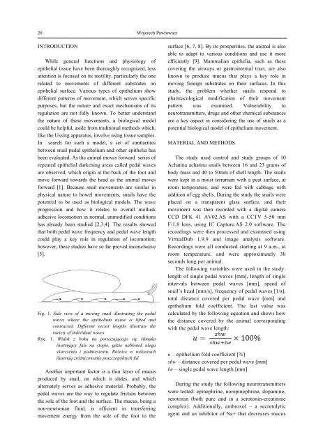
Fig. 1. Side view of a moving snail illustrating the pedal<br />
waves where the epithelium tissue is lifted and<br />
contracted. Different vector lengths illustrate the<br />
variety of individual waves<br />
Ryc. 1. Widok z boku na poruszającego się ślimaka<br />
ilustrujący fale na stopie, gdzie nabłonek ulega<br />
skurczeniu i podniesieniu. Różnice w wektorach<br />
ilustrują zróżnicowanie poszczególnych fal<br />
Another important factor is a thin layer of mucus<br />
produced by snail, on which it slides, and which<br />
alternately serves as adhesive material. Probably, the<br />
pedal waves are the way to regulate friction between<br />
the sole of the foot and the surface. The mucus, being a<br />
non-newtonian fluid, is efficient in transferring<br />
movement energy from the sole of the foot to the<br />
surface [6, 7, 8]. By its prosperities, the animal is also<br />
able to adapt to various conditions and use it more<br />
efficiently [9]. Mammalian epithelia, such as these<br />
covering the airways or gastrointernal tract, are also<br />
known to produce mucus that plays a key role in<br />
moving foreign substrates on their surfaces. In this<br />
study, the problem whether snails respond to<br />
pharmacological modification of their movement<br />
pattern was examined. Vulnerability to<br />
neurotransmitters, drugs and other chemical substances<br />
are a key aspect in considering the use of snails as a<br />
potential biological model of epithelium movement.<br />
MATERIAL AND METHODS<br />
The study used control and study groups of 10<br />
Achatina achatina snails between 16 and 23 grams of<br />
body mass and 40 to 50mm of shell length. The snails<br />
were kept in a moist terrarium with a peat surface, at<br />
room temperature, and were fed with cabbage with<br />
addition of egg shells. During the study the snails were<br />
placed on a transparent glass surface, and their<br />
movement was then recorded with a digital camera<br />
CCD DFK 41 AV02.AS with a CCTV 5-50 mm<br />
F/1.8 lens, using IC Capture.AS 2.0 software. The<br />
recordings were then processed and examined using<br />
VirtualDub 1.9.9 and image analysis software.<br />
Recordings were all conducted starting at 9 a.m., at<br />
room temperature, and were approximately 30<br />
seconds long per animal.<br />
The following variables were used in the study:<br />
length of single pedal waves [mm], length of single<br />
intervals between pedal waves [mm], speed of<br />
snail’s head [mm/s], frequency of pedal waves [1/s],<br />
total distance covered per pedal wave [mm] and<br />
epithelium fold coefficient. The last value was<br />
calculated by the following equation and shows how<br />
the distance covered by the animal corresponding<br />
with the pedal wave length:<br />
u – epithelium fold coefficient [%]<br />
shw – distance covered per pedal wave [mm]<br />
lw – single pedal wave length [mm]<br />
During the study the following neurotransmitters<br />
were tested: epinephrine, norepinephrine, dopamine,<br />
serotonin (both pure and in a serotonin-creatinine<br />
complex). Additionally, ambroxol – a secretolytic<br />
agent and an inhibitor of Na+ that decreases mucus